the biological functions of naa10. - biorxiv.org - the ... · nata complex, recently other ......
TRANSCRIPT

I
The biological functions of Naa10.
Max Doerfel1, Gholson J. Lyon
1,2,3*
1) Stanley Institute for Cognitive Genomics, Cold Spring Harbor Laboratory, NY, USA; 2)
Department of Psychiatry, Stony Brook University, Stony Brook, NY, USA; 3) Utah
Foundation for Biomedical Research, Salt Lake City, UT, USA.
Author emails:
Max Doerfel: [email protected]
*Gholson J. Lyon: [email protected]
*Corresponding author.
Key Words: amino-terminal acetylation; acetyltransferases; proteins; proteomics;
enzymology; Ogden Syndrome; NAA10; NAA15; NAA50.
.CC-BY 4.0 International licensenot peer-reviewed) is the author/funder. It is made available under aThe copyright holder for this preprint (which was. http://dx.doi.org/10.1101/014324doi: bioRxiv preprint first posted online Jan. 29, 2015;

II
A b s t r a c t : N-terminal acetylation (NTA) is one of the most abundant protein modifications known, and
the NAT machinery is conserved throughout all Eukarya. Over the past 50 years, the function
of NTA has begun to be slowly elucidated, and this includes the modulation of protein-protein
interaction, protein-stability, protein function, and as well as protein targeting to specific
cellular compartments. Many of these functions have been studied in the context of
Naa10/NatA; however, we are only starting to really understand the full complexity of this
picture. Roughly, about 40 % of all human proteins are substrates of Naa10 and the impact of
this modification has only been studied for a few of them. Besides acting as a NAT in the
NatA complex, recently other functions have been linked to Naa10, including post-
translational NTA, lysine acetylation and NAT/KAT-independent functions. Also, recent
publications have linked mutations in Naa10 to various diseases, emphasizing the importance
of Naa10 research in humans. The recent design and synthesis of the first 3 bisubstrate
inhibitors that potently and selectively inhibit the NatA/Naa10 complex, monomeric Naa10,
and hNaa50 further increases the toolset to analyze Naa10 function.
.CC-BY 4.0 International licensenot peer-reviewed) is the author/funder. It is made available under aThe copyright holder for this preprint (which was. http://dx.doi.org/10.1101/014324doi: bioRxiv preprint first posted online Jan. 29, 2015;

III
Table of content
I N T R O D U C T I O N 1
1.1 General Functions of Amino-terminal acetylation 3
1.2 The NatA complex 5
1.2.1 Mammalian Naa10 and isoforms 7
1.2.2 Mammalian Naa15 and isoforms 8
1.2.3 other NatA components 8
1.2.4 Localization of NatA 9
M A M M A L S 1 2
1.3 Naa10 function in mammals 12
1.3.1 Naa10 and cyclin D1 regulation 13
1.3.2 NF-kB/ DNA damage 16
1.3.3 Naa10 in cellular hypoxia 18
1.3.4 Naa10 in bone formation 22
1.3.5 Naa10 in neuronal development 23
1.3.6 Naa10 and disease 24
1.4 Function of Naa15/Naa16/Tubedown 27
1.5 Function of Naa50 27
N A T A I N O T H E R O R G A N I S M S 2 9
1.6 Naa10 in C. elegans 29
1.7 NatA in Yeast (S. cerevisiae unless indicated otherwise) 30
1.7.1 Mating type/silencing defect 30
1.7.2 NatA in ribosome function and protein targeting 33
1.7.3 NatA and proteasome function 35
1.7.4 NatA in prion propagation 38
1.8 NATs in prokaryotes (archaeabacteria, eubacteria) 39
1.8.1 In E. coli: RIMs 39
1.8.2 In Archaea 40
1.9 NATs in plants 41
1.10 Function of NTA in plants 42
C O N C L U S I O N 4 4
1.11 Co-translational NTA and protein quality control 44
1.12 Post-translational NTA of Naa10 45
1.13 NAT-independent functions of Naa10 and transcriptional regulation 46
R E F E R E N C E S 4 8
.CC-BY 4.0 International licensenot peer-reviewed) is the author/funder. It is made available under aThe copyright holder for this preprint (which was. http://dx.doi.org/10.1101/014324doi: bioRxiv preprint first posted online Jan. 29, 2015;

Date: 2015-01-28
1
1 I N T R O D U C T I O N
N-terminal acetylation (NTA) is one of the most abundant modifications of
eukaryotic proteins. Today it is believed that the majority of the proteome of higher
organisms is fully or partially acetylated. In fact, recent large-scale proteomics analyses have
identified peptides that were fully or partially acetylated at their designated N-terminus in the
following percentages: 13-19% in Halobacterium salinarum and Natronomonas pharaonis
(Falb et al., 2006; Aivaliotis et al., 2007), 29% in Haloferax volcanii (Kirkland et al., 2008),
16% in bacteria (45 different organisms (Bonissone et al., 2013), 60-70% in S. cerevisiae
(Arnesen et al., 2009b; Van Damme et al., 2011c; Bonissone et al., 2013; Van Damme et al.,
2014), 75% in Drosophila melanogaster (Goetze et al., 2009), 90% in Arabidopsis thaliana
(Bienvenut et al., 2012), at least 4% in C. elegans (Mawuenyega et al., 2003), 83% in mouse
(Lange and Overall, 2011), 90% in human erythrocytes (Lange et al., 2014) and 85% in HeLa
cells (Arnesen et al., 2009b; Van Damme et al., 2011c). However, it should be noted that
these values do not necessarily reflect the whole proteomes. A recent computational analysis
of large-scale proteome analyses was used to develop prediction software for NTA in archae
(P. furiosus, T. acidophilum, H. salinarum and N. pharaonis), animals (Homo sapiens,
Caenorhadbitis elegans, and Drosophila melanogaster), plants (A. thaliana and Oryza sativa)
and fungi (S. cerevisiae and N. crassa). The analysis revealed a bias for N-terminal acetylated
proteins in highly abundant cytosolic proteins (Martinez et al., 2008). This bias could indicate
that the reported percentage of acetylation is higher than the percentage in the actual
proteomes: archeae 1-6.5%, animal 58 %, fungi and plants 60% (Martinez et al., 2008).
Furthermore, in some studies only annotated N-termini were analyzed, others included N-
termini derived from alternative translation initiation sites.
In vitro data suggests that NTA occurs mainly co-translationally on the emerging
polypeptide chain at a length of approximately 25-80 residues (Strous et al., 1973; Filner and
Marcus, 1974; Strous et al., 1974; Driessen et al., 1985; Gautschi et al., 2003), either on the
initiating methionine (iMet) or on the second amino acid after methionine cleavage, also
known as N-terminal methionine excision (NME) (Kendall and Bradshaw, 1992; Xiao et al.,
2010; Bonissone et al., 2013). The removal of the iMet is the first occurring widespread
protein modification and involves peptide deformylases and methionine aminopeptidases
(MetAPs) (Giglione et al., 2014). In addition to co-translational acetylation, accumulating
evidence also supports the occurrence of post-translational N-acetylation. The ribosomal
protein L7/L12 in E. coli e.g. becomes acetylated post-translationally depending on the
availability of nutrients (Gordiyenko et al., 2008). Furthermore, proteomic analyses identified
NTA of internal peptides, further supporting the idea of post-translational acetylation (Helbig
et al., 2010; Helsens et al., 2011). This is especially interesting for many proteins that are
imported into organelles, after which the cleaved mature N-terminus of the protein (now
missing its target/transit peptide) is acetylated by dedicated NATs that reside in the respective
target organelle as shown for yeast mitochondrial localized proteins (Van Damme et al.,
2014) or chloroplast proteins in Chlamydomonas reinhardtii and Arabidopsis thaliana
(Zybailov et al., 2008; Bienvenut et al., 2011; Bienvenut et al., 2012).
Nα-terminal acetylation is catalyzed by distinct N
α-acetyltransferases (NATs) that
belong to the GCN5-related N-acetyltransferase (GNAT) family, a diverse family that
.CC-BY 4.0 International licensenot peer-reviewed) is the author/funder. It is made available under aThe copyright holder for this preprint (which was. http://dx.doi.org/10.1101/014324doi: bioRxiv preprint first posted online Jan. 29, 2015;

Date: 2015-01-28
2
catalyze the transfer of an acetyl group from acetyl-CoA to the primary amine of a wide
variety of substrates from small molecules to large proteins (Vetting et al., 2005). Besides the
NATs, this protein family also includes/contains lysine acetyltransferases (KATs) and histone
acetyltransferases (HATs) (Marmorstein and Zhou, 2014).
In 2009, a new nomenclature for the Nα-acetyltransferases was introduced (Polevoda
et al., 2009), in which the concept of multi-protein complexes for NATs was formalized. In
humans, six NATs, NatA-F, were defined that specifically co-translationally catalyze the
acetylation of the Nα-terminal amino group of a well-defined subset of proteins, although N
ε-
acetylation of internal lysines has also been reported (Kalvik and Arnesen, 2013). NatA
consists of the catalytic subunit Naa10 and the auxiliary subunit Naa15 and acetylates small
side chains such as Ser, Ala, Thr, Gly, Val after the initiator methionine has been cleaved by
methionine aminopeptidases (via NME) (see Figure 1). NatB and NatC are defined as
including the catalytic subunits Naa20 and Naa25 and the auxiliary subunits Naa30 and
Naa35/Naa38, respectively. They acetylate proteins with their methionine retained. The only
known substrates for NatD (Naa40) are histone H2A and H4. Naa50 is the catalytic subunit of
NatE, with a substrate specificity for N-termini starting with methionine followed by Leu,
Lys, Ala and Met. NatF is composed of Naa60 and has a substrate specificity that partially
overlaps with NatC and NatE. It is important to note that this might not be the complete
picture, as there are possibly other proteins binding and interacting with proteins in these
NATs as currently defined [for reviews see (Arnesen, 2011; Van Damme et al., 2011a;
Starheim et al., 2012)].
Figure 1: The co-translational N-terminal protein modification process. As soon as the nascent polypeptide chain
emerges from the ribosome exit tunnel, the initiator methionine is cleaved by methionine aminopeptideases
(MetAPs) if the following amino acid is small and uncharged. For the sake of simplicity, this process is illustrated
by one enzyme despite the fact that other enzymes including peptide deformylases are involved, depending on
organism and cellular cormpartment. Subsequently, the new N-termini can get acetylated by NatA, composed of
the catalytic Naa10 and the auxiliary subunit Naa15. The majority of cytosolic proteins fall into this category. If
the iMet is not processed, NTA can be accomplished by NatB (composed of Naa20 and Naa25), NatC (Naa30,
Naa35, Naa38), NatD (Naa40) NatE (Naa50 and possibly Naa15) and NatF (Naa60). Figure modified from
(Kalvik and Arnesen, 2013).
Modeling of the substrate binding pocket of the catalytic subunits of different NATs
revealed that 3 tyrosines in the catalytic center form a scaffold ready to interact with the
peptide backbone of the substrate, whereas the specificity of each NAT is tuned by
.CC-BY 4.0 International licensenot peer-reviewed) is the author/funder. It is made available under aThe copyright holder for this preprint (which was. http://dx.doi.org/10.1101/014324doi: bioRxiv preprint first posted online Jan. 29, 2015;

Date: 2015-01-28
3
surrounding amino acids that are not conserved between the different NATs (Grauffel et al.,
2012).
1.1 General Functions of Amino-terminal acetylation
Despite its discovery more than 50 years ago, very little is known about the biological
function of NTA. For many years, it has been generalized from a few examples that NTA
broadly protects many proteins from degradation. This was supported by the fact that
acetylation of globin and lysozyme prevents their degradation by the ubiquitin proteolytic
system from reticulocytes (Hershko et al., 1984). In line with this, the NTA of enkephalins
diminishes their proteolytic cleavage by aminopeptidase M. (Jayawardene and Dass, 1999),
improves the chemical stability of guinea pig myelin basic protein (de Haan et al., 2004) and
protects glucagon-like peptide (GLP-1) from DPP-IV- (dipeptidyl peptidase IV) mediated
degradation (John et al., 2008). Analysis of the half-life of β-galactosidase in a split-ubiquitin
system showed that proteins having N-terminal amino acids that are prone to acetylation
(Met, Ser, Ala, Thr, Val, or Gly) have a relative long half-life, whereas Arg, Lys, Phe, Leu, or
Asp at the amino-terminus have very short half-lives (Bachmair et al., 1986). One way that
NTA could contribute to protein stability is by blocking the access for N-terminal
ubiquitination as shown for p16 and p14/p19ARF
(Ben-Saadon et al., 2004; Ciechanover and
Ben-Saadon, 2004; Kuo et al., 2004). Also, p21Cip1
is acetylated (most likely by NatA) at its
N-terminus, whereas an N-terminally tagged variant that abolishes Nα-acetylation becomes
ubiquitylated (Chen et al., 2004). Another study suggests that NTA may play a role in the
structural stabilization of N-terminally flexible proteins, as bioinformatics analysis of the
yeast proteome showed that proteins with N-terminal-disordered regions are more likely to be
acetylated (Holmes et al., 2014). In line with that, many studies have shown directly that NTA
can stabilize an N-terminal -helix (Shoemaker et al., 1987; Fairman et al., 1989;
Chakrabartty et al., 1993; Doig et al., 1994; Greenfield et al., 1994; Jarvis et al., 1995; Fauvet
et al., 2012; Kang et al., 2012; Kang et al., 2013). However, other studies suggest that NTA
has no effects on protein stability (Greenfield et al., 1994; Yi et al., 2011). In addition, recent
studies showed that acetylation might regulate protein stability and degradation for some
proteins, depending on the cellular availability of interaction partners. The ubiquitin ligase
Doa10 recognizes Nt-acetylated Ala, Val, Ser, Thr, and Cys and earmarks acetylated
substrates for degradation (Hwang et al., 2010). NTA therefore creates protein degradation
signals (Ac
N-degrons) that are targeted by the Ac/N-end rule pathway resulting in
ubiquitylation and proteasome-mediated degradation by the Doa10 E3 N-recognin, in
conjunction with the Ubc6 and Ubc7 E2 enzymes (Varshavsky, 2011). Conversely, multiple
Doa10 substrates do not require Nα-acetylation for their degradation, and acetylation has only
mild effects on the stability of the tested substrates (Zattas et al., 2013). These discrepancies
can be explained with the findings that Nα-acetylation can have stabilizing effects when an
interaction partner is involved. In this case, the acetylated N-termini recruits the interaction
partners that then shield the Ac
N-degron, preventing ubiquitinylation and degradation to
regulate subunit stoichiometries (Shemorry et al., 2013). In agreement with this, it is widely
accepted that Nα-terminal acetylation can act as an avidity enhancer within protein complexes
(Deakin et al., 1980; Scott et al., 2011; Nazmi et al., 2012). Interestingly, NTA may also
.CC-BY 4.0 International licensenot peer-reviewed) is the author/funder. It is made available under aThe copyright holder for this preprint (which was. http://dx.doi.org/10.1101/014324doi: bioRxiv preprint first posted online Jan. 29, 2015;

Date: 2015-01-28
4
regulate NEDDylation, as Nα-terminal acetylation of the E2 enzyme, Ubc12, is absolutely
required for its interaction with E3, Dcn1, and promotes cullin neddylation (Scott et al.,
2011). This is also another good example where NTA acts as an avidity enhancer.
Another function of NTA has been implicated in protein sorting and secretory
processes. In yeast, the ARF-like GTPase Arl3p/ARP is acetylated by NatC and this
modification is required for its targeting to the Golgi apparatus, possibly through the
acetylation-dependent interaction with the integral membrane protein Sys1p (Setty et al.,
2004). Simultaneously, a different group confirmed that Sys1p is the receptor for Arl3p and
knockout of NatC or mutation of the NatC complex, that abrogated its acetyltransferase
activity, resulted in failure to target Arl3p to the Golgi (Behnia et al., 2004). Furthermore,
targeting of the human homologue of Arl3p, ARFRP1, is dependent on Sys1p and mutation of
the N-terminus of ARFRP1, that disagrees with acetylation by NatC, induced its mis-
localization in COS cells (Behnia et al., 2004). This and the fact that the N-terminus of Arl3p
is a potential NatC substrate in S. cerevisiae, D. melanogaster, C. elegans and plants
indicates that this system is well conserved. Interestingly, two other human lysosomal Arf-
like GTPases, Arl8a and Arl8b (also known as Arl10b/c and Gie1/2), and their single
homologue in Drosophila are potential substrates of NatC and mass spectrometric analyses
confirmed that human Arl8b is N-terminally acetylated (Hofmann and Munro, 2006). Later in
vitro acetylation assays showed that Arl8b is acetylated by NatC and knockdown of the
catalytic subunit of NatC (Starheim et al., 2009a) or replacement of the leucine in position 2
with alanine (Hofmann and Munro, 2006) resulted in a loss of its lysosomal localization. It
should be mentioned the protein was still found to be acetylated, presumably by NatA
following removal of the initiator methionine (Hofmann and Munro, 2006), indicating that
specifically the acetylated methionine rather than acetylation itself is important for lysosomal
targeting of Arl8b. Also, the inner nuclear membrane protein Trm1-II was found to be
mislocated to the nucleoplasm, when NatC was knocked out or when the penultimate amino
acid was mutated to inhibit NatC-dependent NTA (Murthi and Hopper, 2005). On the other
hand, systematic analysis of predicted N-terminal processing in yeast showed that
cytoplasmic proteins are typically acetylated, whereas those bound to ER are largely
unmodified (Forte et al., 2011). Mutation of the N-terminal amino acid of the secretory
protein carboxypeptidase Y, which allowed acetylation of this protein, inhibited targeting to
the ER (Forte et al., 2011). However, fluorescence microscopy analysis in yeast indicated
unaltered subcellular localization patterns for all 13 studied NatC substrates, after disruption
of NatC catalytic subunit (Aksnes et al., 2013). Furthermore, no disruption of the nuclear
membrane, endoplasmic reticulum, Golgi apparatus, mitochondria, or bud neck was observed,
suggesting the intactness of these organelles and subcellular structures as judged by the
unchanged shape, number, size and distribution in the cell (Aksnes et al., 2013). Taken
together, this indicates that NatC is not – at least not in general – a determinant for substrate
subcellular localization (Aksnes et al., 2013). Similarly, fluorescence microscopy analysis of
13 NatB substrates in wild type and NAA20 yeast cells revealed that acetylation by NatB is
not a general signal for protein localization (Caesar et al., 2006).
Other examples in which NTA can affect protein function and/or activity include
hemoglobin isoforms (Scheepens et al., 1995; Ashiuchi et al., 2005), phospholamban (PLB)
(Starling et al., 1996), N-TIMPs (N-terminal inhibitory domains of TIMPs /inhibitors of
.CC-BY 4.0 International licensenot peer-reviewed) is the author/funder. It is made available under aThe copyright holder for this preprint (which was. http://dx.doi.org/10.1101/014324doi: bioRxiv preprint first posted online Jan. 29, 2015;

Date: 2015-01-28
5
metalloproteinases (Van Doren et al., 2008), parvalbumin (Permyakov et al., 2012),
melanocyte-stimulating hormone (MSH) in the barfin flounder (Verasper moseri) (Kobayashi
et al., 2009), the contractile proteins actin and tropomyosin in fission and budding yeast
(Polevoda et al., 2003; Singer and Shaw, 2003; Coulton et al., 2010) as well as the stress-
induced carboxypeptidase Y inhibitor Tfs1p in yeast (Caesar and Blomberg, 2004).
In addition, NTA has been linked to various diseases, including apoptosis and cancer
(Kalvik and Arnesen, 2013), host parasite interaction in malaria (Chang et al., 2008), and has
been discussed to play a role in Parkinson's disease (see below). As pointed out in earlier
reviews: “Although…[NTA]…is essential for cell viability and survival, very little is known
about the physiological reasons associated with this crucial role” (Giglione et al., 2014) and
“there may be a variety of acetylation-dependent functions depending on the target protein,
rather than one general function [and] there is even the possibility that this modification
affects the function of only very few proteins” (Arnesen, 2011).
The best studied Nα-acetyltransferases NatA consists of the catalytic subunit Naa10
and the auxiliary subunit Naa15. In this review we mainly concentrate on Naa10 structure and
function and discuss the recent development of the field.
1.2 The NatA complex
As mentioned above, the NatA complex consists at least of the auxiliary and catalytic
subunit, Naa15 and Naa10, respectively and is evolutionarily conserved from yeast to
vertebrates (Mullen et al., 1989; Park and Szostak, 1992; Sugiura et al., 2003; Arnesen et al.,
2005a). We adopt here the nomenclature of inserting letters to indicate the species about
which we are discussing, so yNatA refers to NatA in yeast, where we are specifically referring
to S. cerevesiae, hNatA refers to NatA in humans, and mNatA refers to NatA in mice.
However, this nomenclature in 2009 did not address other species, and it might be worth
updating the nomenclature at some future international meeting focused on the NATs.
In any case, there is good in vitro and in vivo evidence that yNatA acetylates the N-
termini of small side chains like serine, alanine, glycine and threonine (Arnold et al., 1999;
Polevoda et al., 1999) and NatA from humans has identical or nearly identical specificities,
acetylating proteins starting with small side chains like serine, glycine, alanine, threonine and
cysteine (Arnesen et al., 2009b; Van Damme et al., 2011b; Van Damme et al., 2011c) after
the removal of the initiator methionine by methionine aminopeptidases. However, it is critical
to note that heterologous combinations of human and yeast subunits are not functional in
yeast, suggesting significant structural subunit differences between the species, presumably
due to lack of proper dimerization due to species-specific differences between the proteins
(Arnesen et al., 2009b). Interestingly, (Met-)Ala-N-termini are more prevalent in the human
proteome, whereas (Met)-Ser-N-termini are more abundant in the yeast proteome (Van
Damme et al., 2011c). Accordingly, hNatA displays a preference towards these Ala-N-termini
whereas yNatA seems to be the more efficient in acetylating Ser-starting N-termini, indicating
that NatA substrate specificity/efficiency of Nt-acetylation has co-evolved with the repertoire
of NatA type substrates expressed (Van Damme et al., 2014).
Size-exclusion chromatography and circular dichroism showed that purified human
Naa10 consists of a compact globular region comprising two thirds of the protein and a
flexible unstructured C-terminus (Sánchez-Puig and Fersht, 2006). The recent X-ray crystal
.CC-BY 4.0 International licensenot peer-reviewed) is the author/funder. It is made available under aThe copyright holder for this preprint (which was. http://dx.doi.org/10.1101/014324doi: bioRxiv preprint first posted online Jan. 29, 2015;

Date: 2015-01-28
6
structure of the 100 kD holo-NatA (Naa10/Naa15) complex from S. pombe revealed that the
auxiliary subunit Naa15 is composed of 37 -helices ranging from 8 to 32 residues in length,
among which 13 conserved helical bundle tetratricopeptide repeat (TPR) motifs can be
identified (Liszczak et al., 2013). These Naa15 helices form a ring-like structure that wraps
completely around the Naa10 catalytic subunit (Liszczak et al., 2013). TPR motifs mediate
protein-protein interactions, and it was speculated that TPR might be important for interaction
with other NatA-binding partners such as the ribosome, Naa50/NatE and the HYPK
chaperone (Liszczak et al., 2013). We discuss the possible interaction with NatE in more
detail below. Naa10 adopts a typical GNAT fold containing a N-terminal α1–loop–α2
segment that features one large hydrophibic interface and exhibits the most intimate
interactions with Naa15, a central acetyl CoA-binding region and C-terminal segments that
are similar to the corresponding regions in Naa50 (Liszczak et al., 2013). The X-ray crystal
structure of archaeal T. volcanium Naa10 has also been reported, revealing multiple distinct
modes of acetyl-Co binding involving the loops between β4 and α3 including the P-loop (Ma
et al., 2014). To our knowledge, there is not yet any published cryo-electron microscopy data
regarding larger complexes between the ribosome, nascent polypeptide chain and any NATs.
A very elegant cryo-EM structure of the ribosome, nascent polypeptide chain, and the signal
recognition particle was recently published (Voorhees et al., 2014).
Besides acting in a complex, it has been shown that a fraction of human Naa10 exists
independent of Naa15 in the cytoplasm and is able to acetylate acidic side chains like
aspartate and glutamate in γ- and β-actin (Van Damme et al., 2011b; Foyn et al., 2013a).
These Type I actins are natural NatB substrates (initiator methionine followed by amino acid
with acidic side chain) and are therefore initially acetylated by NatB at the methionine in
yeast and humans (Van Damme et al., 2012). However, further processing/cleavage by an Nα-
acetylaminopeptidase (ANAP), which specifically removes the N-terminal Ac-Met or Ac-Cys
from actin exposes the acidic N-terminal residue (Polevoda and Sherman, 2003b), which can
be subsequently acetylated by Naa10. This substrate switching of Naa10 from small side
chains towards acidic side chains could be explained by comparing the X-ray crystal
structures of complexed (Naa15-bound) and uncomplexed Naa10 of S. pombe. The
complexed form of Naa10 adopts a GNAT fold containing a central acetyl CoA–binding
region and flanking N- and C-terminal segments that allows the acetylation of conventional
substrates (Liszczak et al., 2013). In the uncomplexed form, Leu22 and Tyr26 shift out of the
active site of Naa10 and Glu24 is repositioned by ~5 Å resulting in a conformation that allows
for the acetylation of acidic N-termini (Liszczak et al., 2013). However, it should be noted
that some proteins starting with an N-terminal acidic amino acid are usually further modified
by arginyl-transferases and targeted by the Arg/N-end rule pathway for degradation
(Varshavsky, 2011). Therefore, further studies have to show if Type I actins are unique
substrates of non-complexed Naa10 and/or if more in vivo substrates with acidic N-termini
exist. Such studies also need to explore whether the NTA of actin does trigger any
downstream processing in the Arg/N-end rule pathway.
Apart from its function as an N-terminal acetyltransferase, NatA has been shown at
least in vitro to possess N-terminal propionyltransferase activity (Foyn et al., 2013b) and
lysine acetylation activity (Jeong et al., 2002; Lin et al., 2004; Lim et al., 2006; Yoo et al.,
2006; Lim et al., 2008; Lee et al., 2010b; Shin et al., 2014). Autoacetylation at an internal
.CC-BY 4.0 International licensenot peer-reviewed) is the author/funder. It is made available under aThe copyright holder for this preprint (which was. http://dx.doi.org/10.1101/014324doi: bioRxiv preprint first posted online Jan. 29, 2015;
Date: 2015-01-28
7
lysine K136 in hNaa10 e.g. has been shown to regulate its enzymatic activity (Seo et al.,
2010). However, LC/MS/MS analyses on human Naa10 expressed and purified from E. coli
did not show acetylation at any of the internal 16 lysines but identified autoacetylation on its
own N-terminal glycine (Murray-Rust et al., 2006). The physicochemical properties of a
lysine side chain are quite different from the known N-terminal substrates, thus it is not yet
known how much lysine is directly acetylated by monomeric Naa10. The degree to which
autoacetylation of Naa10 occurs in vivo is also currently not well characterized.
1.2.1 Mammalian Naa10 and isoforms
Naa10 (Nα-acetyltransferase 10; NatA catalytic subunit; ARD1, arrest-defective
protein 1 homolog; DXS707; TE2), the catalytic subunit of NatA, has an apparent molecular
weight of 26 kDa and contains a typical Gcn5-related N-acetyltransferases (GNAT) domain.
In mouse, NAA10 is located on chromosome X A7.3 and contains 9 exons. Two alternative
splicing products of mouse Naa10, mNaa10235
and mNaa10225
, were reported in NIH-3T3 and
JB6 cells that may have different activities and function in different subcellular compartments
(Chun et al., 2007). The human NAA10 is located on chromosome Xq28 and is encoded by 8
exons (Tribioli et al., 1994). According to RefSeq (NCBI) (Pruitt et al., 2007), three different
isoforms derived from alternate splicing (see Figure 2) exist. Additionally, a processed
NAA10 gene duplicate NAA11 (ARD2) has been identified that is expressed in several human
cell lines (Jurkat, HEK293, NPA) (Arnesen et al., 2006b). However, later studies have
revealed data arguing that Naa11 is not expressed in the human cell lines HeLa and HEK293
or in cancerous tissues, and NAA11 transcripts were only detected in testicular and placental
tissues (Pang et al., 2011). Naa11 has also been found in mouse, where it is mainly expressed
in the testis (Pang et al., 2009). NAA11 is located on chromosome 4q21.21 or 5 E3 for human
or mouse, respectively, and only contains two exons.
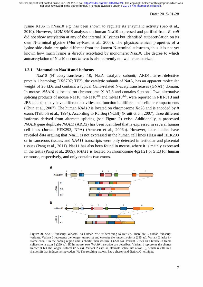
Figure 2: NAA10 transcript variants. A) Human NAA10 according to RefSeq. There are 3 human transcript
variants. Variant 1 represents the longest transcript and encodes the longest isoform (235 aa). Variant 2 lacks in-
frame exon 6 in the coding region and is shorter than isoform 1 (220 aa). Variant 3 uses an alternate in-frame
splice site in exon 3 (229 aa). B) In mouse, two NAA10 transcripts are described. Variant 1 represents the shorter
transcript but the longer isoform (235 aa). Variant 2 uses an alternate splice site (exon 8), which results in a
frameshift that induces a stop codon (*). The resulting isoform has a shorter and distinct C-terminus.
.CC-BY 4.0 International licensenot peer-reviewed) is the author/funder. It is made available under aThe copyright holder for this preprint (which was. http://dx.doi.org/10.1101/014324doi: bioRxiv preprint first posted online Jan. 29, 2015;
Date: 2015-01-28
8
1.2.2 Mammalian Naa15 and isoforms
Human Naa15 (Nα-acetyltransferase 15; NatA auxiliary subunit, NMDA Receptor-
Regulated Protein; NARG1; Tubedown-100; Tbdn100 ; tubedown-1, NATH) is considered to
be mainly the auxiliary subunit of NatA, although it certainly might have independent
functions. The human NAA15 gene is located on chromosome 4q31.1 and contains 23 exons.
Initially, 2 mRNA species were identified, 4.6 and 5.8 kb, both harboring the same open
reading frame encoding a putative protein of 866 amino acids (105 kDa) protein that can be
detected in most human adult tissues (Fluge et al., 2002). According to RefSeq/NCBI (Pruitt
et al., 2007), only one human transcript variant exists, although 2 more isoforms are
predicted. It should be noted that, in addition to full length Naa15, a N-terminally truncated
variant of Naa15 (named tubedown-1), Naa15273-865, has been described (Gendron et al.,
2000). However, northern blot analyses of poly(A) mRNA from mouse revealed that full
length Naa15 is widely expressed, whereas smaller transcripts were visualized merely in heart
and testis (Willis et al., 2002).
Similar to the situation with NAA10, a NAA15 gene variant has been identified,
NAA16, that originates from an early vertebrate duplication event (Arnesen et al., 2009a). The
encoded protein shares 70% sequence identity to hNaa15 and is expressed in a variety of
human cell lines, but is generally less abundant as compared to hNaa15 (Arnesen et al.,
2009a). Three isoforms of Naa16 are validated so far (NCBI RefSeq). Mouse NAA15 is
located on chromosome 2 D and contains 20 exons, whereas mouse NAA16 is located on
chromosome 14 D3 and consists of 21 exons.
It has been shown in principle that NatA could assemble from all the isoforms. Naa15
interacts with Naa11, in humans (Arnesen et al., 2006b) and mouse (Pang et al., 2009), and
Naa10 interacts with the Naa15 paralogue, Naa16, creating a more complex and flexible
system for Nα-terminal acetylation as compared to lower eukaryotes (Arnesen et al., 2009a).
Such a system might create the opportunity for functional redundancy or compensation in the
event of loss of Naa10 or Naa15, although we are not aware of any studies showing whether
NAA11 expression might be upregulated in tissues lacking or having reduced NAA10
expression.
1.2.3 other NatA components
As there is a known crystal structure of S. pombe Naa10 bound to NAA15, it is safe to
conclude that this is a very stable complex in that species. We await such crystal structures for
human Naa10 and Naa15. It is certainly possible that there are proteins interacting transiently
with NatA, but there could also be the possibility of a more stable trimeric or larger complex
involving Naa50 (Nα-acetyltransferase 50, NatE; NAT13; Mak3; Nat5, SAN separation
anxiety) or other proteins. Naa50 is the catalytic acetyltransferase subunit of NatE, is
expressed in several human cell lines, and has been shown to be associated with NatA in yeast
(Gautschi et al., 2003), fruit fly (Williams et al., 2003) and humans (Arnesen et al., 2006a).
Naa50 has a distinct substrate activity for Met followed by a hydrophobic amino acid in
human and yeast (Polevoda et al., 1999; Evjenth et al., 2009; Evjenth et al., 2012) and has
been claimed to possess -acetyltransferase activity towards K525 in -tubulin (Chu et al.,
2011) and histone 4 (Evjenth et al., 2009). Furthermore, hNaa50 has been shown to harbor
autoacetylation activity on internal lysines (K34, K37 and K140) in vitro, modulating Naa50
.CC-BY 4.0 International licensenot peer-reviewed) is the author/funder. It is made available under aThe copyright holder for this preprint (which was. http://dx.doi.org/10.1101/014324doi: bioRxiv preprint first posted online Jan. 29, 2015;

Date: 2015-01-28
9
substrate activity (Evjenth et al., 2009; Evjenth et al., 2012). However, in contradiction to
this, the X-ray crystal structure of human Naa50 revealed a GNAT fold with a specific
substrate binding groove that allows for acetylation of α-amino substrates but excludes lysine
side chains as a substrate (Liszczak et al., 2011). This seems to be strong evidence against a
role for Naa50 in direct acetylation of lysine side chains. Further studies have to sort out these
discrepancies.
Because Naa50 has a distinct/different substrate specificity and NAA50 cells did not
display the NatA phenotype in yeast (Gautschi et al., 2003), Naa50 was considered as an
independent NAT and was named NatE (Starheim et al., 2009b). Furthermore, in HeLa cells,
more than 80 % of endogenous Naa50 is not associated with the NatA complex (Hou et al.,
2007). Therefore, future experiments have to examine whether Naa50 has a distinct function
independent of NatA and/or if Naa50 works in a cooperative manner with NatA.
Recently, the chaperone like protein HYPK (Huntingtin Interacting Protein K) was
shown to interact with Naa10 and 15 and is required for NTA of the known in vivo NatA
substrate PCNP (Arnesen et al., 2010). However, it is an open question whether HYPK
generally is required for NatA-mediated acetylation of downstream substrates.
Further interaction partners of Naa10 have been identified including Myosin light-
chain kinase, MYLK (Shin et al., 2009), tuberous sclerosis 2, TSC2 (Kuo et al., 2010)
RelA/p65 (Xu et al., 2012) DNMT1 (DNA methyltransferase 1) (Lee et al., 2010a), androgen
receptor (Wang et al., 2012) and proteasome activator 28 (Min et al., 2013). In high
throughput screens CDC25A (cell division cycle 25 homolog) (Rual et al., 2005) and Rho
guanine nucleotide exchange factor 6 have been shown as interaction partners of NatA (Xiao
et al., 2007). Additionally, -Catenin (Lim et al., 2006; Lim et al., 2008), HIF-1 (Jeong et
al., 2002; Arnesen et al., 2005b) and methionine sulfoxide reductase A (Shin et al., 2014)
have been suggested to bind to Naa10. In a recent high-throughput study, multiple orthogonal
separation techniques were employed to resolve distinct protein complexes. Fractionation of
soluble cytoplasmic and nuclear extracts from HeLa S3 and HEK293 cells into 1,163 different
fractions identified several interaction partners for Naa10 (Naa15, Naa16, Mina, M89BB,
TCEA1 and PLC3) and Naa15 (RT21, ML12A, HYPK and Cap1), Naa16 (TCEA1, PLCB3,
Naa10 and Mina) (Havugimana et al., 2012). However, these interactions seem to be transient
in the cell and have not yet been shown to regulate or change NatA function; therefore, we do
not list them as part of any putative larger NatA complex. As we stated above, definitive
evidence of any sort of larger stable complex, other than the dimer between Naa10 and
Naa15, could come from structural studies, including possibly cryo-electron microscopy.
1.2.4 Localization of NatA
Mainly from yeast data, it is thought that the auxiliary subunits of NatA as well as
other NATs are associated with mono- and polysome fractions and co-translationally
acetylate the nascent polypeptide chain as it emerges from the ribosome (Gautschi et al.,
2003; Polevoda et al., 2008). In line with this, it has been shown that human Naa10 and
Naa15, HYPK (Arnesen et al., 2010), the human paralog of Naa15, Naa16 (Arnesen et al.,
2009a) as well as yeast Naa15 (Raue et al., 2007) and rat Naa15 (Yamada and Bradshaw,
1991) are associated with poly- or monosomes. In yeast, NatA binds via the ribosomal
.CC-BY 4.0 International licensenot peer-reviewed) is the author/funder. It is made available under aThe copyright holder for this preprint (which was. http://dx.doi.org/10.1101/014324doi: bioRxiv preprint first posted online Jan. 29, 2015;
Date: 2015-01-28
10
proteins, uL23 and uL29 (Polevoda et al., 2008). Further data indicates that NatA preferably
associates with translating ribosomes. Particularly, yNatA as well as other ribosome-
associated protein biogenesis factors (including the chaperones Ssb1/2 and ribosome-
associated complex, signal recognition particle and the aminopeptidases Map1 and Map2)
bind with increased apparent affinity to randomly translating ribosomes as compared with
non-translating ones (Raue et al., 2007). Interestingly, Hsp70 chaperones may be direct
targets of NatA and NTA by NatA contributes an unanticipated influence on protein
biogenesis, both through and independent of Hsp70 activity (Holmes et al., 2014), supporting
a role of NatA in protein biogegnesis. However, the NatA complex also exists in a ribosome-
free context. For instance it has been shown that the majority of hNatA is non-polysomal
(Arnesen et al., 2005a) and a minor fraction of cytosolic hNaa10 exists independent of the
NatA complex, carrying out post-translational acetylation as mentioned above (Van Damme
et al., 2011b). Mammalian Naa10, Naa11 and Naa15 and Naa50 (isoforms) have been
reported to be mainly localized in the cytoplasm and to a lesser extent to the nucleus (Fluge et
al., 2002; Sugiura et al., 2003; Bilton et al., 2005; Arnesen et al., 2006a; Arnesen et al.,
2006b; Chun et al., 2007; Xu et al., 2012; Park et al., 2014; Zeng et al., 2014). In mouse, an
isoform specific localization of Naa10 has been described. mNaa10235
was mainly nuclear in
NIH-3T3 and JB6 cells whereas another variant mNaa10225
, derived from alternative splicing
at a different 3’-splice site, was mainly localized in the cytoplasm (Chun et al., 2007). In
humans, Naa10225
is absent and Naa10235
was found to be evenly distributed in both
cytoplasm and nucleus of HeLa and HT1080 cells as seen by immunofluorescence, confocal
microscopy, and cell fractionation (Chun et al., 2007). Interestingly, Naa10 could be detected
in nuclear fractions of doxorubicin treated HEK293 cells whereas a deletion construct lacking
amino acids 1-35 could not be detected suggesting that a nuclear localization signal (NLS)
resides in the N-terminal part of Naa10 (Park et al., 2012). Sequence analysis had previously
identified a putative NLS more C-terminally in Naa10 between residues 78 and 83
(KRSHRR) (Arnesen et al., 2005a). In agreement with that, deletion of this NLS78-83 almost
completely abrogated nuclear localization of Naa10, whereas Naa10 wild type was imported
to the nuclei of proliferating HeLa and HEK293 cells, especially during S phase (Park et al.,
2014). Furthermore, the deletion of NLS78-83 altered the cell cycle and the expression levels of
cell cycle regulators and resulted in cell morphology changes and cellular growth impairment,
all of which was mostly rescued when the nuclear import of hARD1 was restored by
exogenous NLS (Park et al., 2014). It should be noted that Arnesen et al. reported that neither
leptomycin B nor actinomycin D significantly changed the localization patterns of Naa10 in
HeLa cells, indicating that Naa10 is not actively imported through importin β-dependent
mechanisms (Arnesen et al., 2005a).
Naa15 also harbors a putative NLS between residues 612-628
(KKNAEKEKQQRNQKKKK), however, only Naa10 was found to be localized in the nuclei
of HeLa, GaMg, HEK293, MCF-7 and NB4 cells, whereas Naa15 was predominantly
localized in the cytoplasm (Arnesen et al., 2005a). In contrast to this, a different study showed
that Naa15 localizes to the nucleus where it interacts with the osteocalcin promoter, as shown
by cellular fractionation and ChIP experiments in MC3T3E1 calvarial osteoblasts (Willis et
al., 2002). Further studies have to resolve these discrepancies and analyze possible cell-type
specific differences. Besides that, it has been shown that Naa10 associates with microtubules
.CC-BY 4.0 International licensenot peer-reviewed) is the author/funder. It is made available under aThe copyright holder for this preprint (which was. http://dx.doi.org/10.1101/014324doi: bioRxiv preprint first posted online Jan. 29, 2015;

Date: 2015-01-28
11
in dendrites in cultured neurons (Ohkawa et al., 2008) and Naa15 colocalizes with the actin-
binding protein cortactin and the F-actin cytoskeleton in the cytoplasm IEM mouse and
RF/6A rhesus endothelial cells (Paradis et al., 2008).
.CC-BY 4.0 International licensenot peer-reviewed) is the author/funder. It is made available under aThe copyright holder for this preprint (which was. http://dx.doi.org/10.1101/014324doi: bioRxiv preprint first posted online Jan. 29, 2015;

Date: 2015-01-28
12
2 M A M M A L S
2.1 Naa10 function in mammals
The N-acetyltransferase NatA is expressed widely in many tissues and NatA N-
termini are overrepresented in eukaryotic proteomes. As pointed out earlier, 80-90 % of
soluble human proteins are fully or partially acetylated and nearly 40-50 % of all proteins are
potential NatA substrates according to their sequence in S. cerevisiae, D. melanogaster and
humans (Van Damme et al., 2011c; Starheim et al., 2012). In the current model, Naa15 links
Naa10, Naa50 and possibly other factors like HYPK to the ribosome where NatA/Naa10
acetylates the Nα-amino group of canonical substrates and NatE/Naa50 acetylates methionine
followed by a hydrophobic amino acid in a co-translational manner (Figure 3). Some known
canonical NatA substrates include PCNP (Arnesen et al., 2010), androgen receptor (Wang et
al., 2012), caspase-2 (Yi et al., 2011), -tubulin (Ohkawa et al., 2008) and TSC2 (Kuo et al.,
2010), although a wealth of proteomic studies in recent years has suggested many more
(Arnesen et al., 2009b; Lange et al., 2014). Post-translational acetylation by non-ribosome-
associated NatA or monomeric Naa10 might occur as well.
Figure 3: Multiple functions of Naa10. Associated with the ribosome in the NatA complex, Naa10 co-
translationally acetylates the Nα-terminal amino group of the nascent polypeptide chains of classical substrates as
they emerge from the ribosome. Uncomplexed Naa10 post-translationally Nα-acetylates proteins starting with
acidic side chains and might also Nε-acetylate internal lysines. Furthermore, it has been suggested that Naa10
translocates into the nucleus where it acts in cooperation with transcription factors to modulate protein expression.
Furthermore, Naa15 as well as the signal recognition particle SRP and nascent polypeptide-associated complex
NAC might bind to similar regions on the ribosome near the exit tunnel (see below).
On the other hand, when Naa10 is not in a complex with Naa15, Naa10 adopts a
different catalytic activity towards N-termini with acidic side chains. Presumably, this
function occurs post-translationally in the cytosol, as Naa15 is necessary to link Naa10 to the
ribosome (Figure 3). Substrates with acidic N termini include γ- and β-actin (Van Damme et
al., 2011b). Proteins that have been reported to be substrates for -acetylation at lysines by
Naa10 include -catenin (Lim et al., 2006; Lim et al., 2008) or HIF-1 (Jeong et al., 2002;
Yoo et al., 2006; Lee et al., 2010b) although there is controversy in the field whether HIF-1
is an actual substrate of Naa10 or not (see below). Another ambiguous finding is that Naa10
.CC-BY 4.0 International licensenot peer-reviewed) is the author/funder. It is made available under aThe copyright holder for this preprint (which was. http://dx.doi.org/10.1101/014324doi: bioRxiv preprint first posted online Jan. 29, 2015;

Date: 2015-01-28
13
can translocate into the nucleus, regulating gene transcription, and this function might even be
independent of its catalytic activity (Figure 3). In H1299 lung cancer cells e.g., Naa10 binds
to nonmethylated DNA at the E-Cadherin promoter, directly interacts with DNMT1 (DNA
methyltransferase 1) thereby recruiting DNMT1 resulting in silencing of the E-Cahherin
promoterin a NAT-independent manner as analyzed by a Naa10-R82A mutant (Lee et al.,
2010a). Similarly, the enzymatic activity is not necessary for Naa10 to significantly suppress
migration, tumor growth, and metastasis in human cancer cells. Instead, Naa10 binds to the
GIT-binding domain of PIX (Rho guanine nucleotide exchange factor 7), thereby preventing
the formation of the GIT-PIX-Paxillin complex, resulting in reduced intrinsic Cdc42/Rac1
activity and decreased cell migration (Hua et al., 2011).
Due to its ubiquitous expression in almost all tissues and the broad substrate
specificity of NatA, it is perhaps not surprising that many pathways and cellular
functions/processes are regulated by NatA/Naa10 activity. This includes, as indicated above,
regulation of gene transcription and cell motility. In this regard, it has been shown that
overexpression or knock-down of Naa10 in HT1080 cells reduces migration and enhances
invasion (Shin et al., 2009). The authors also reported that Naa10 interacts with activated
(phosphorylated) MYLK (myosin light-chain kinase) and acetylates it at Lys608
, thereby
inactivating MYLK resulting in the dephosphorylation of MLC (Shin et al., 2009).
Additionally, many other functions of Naa10 have been discussed including cyclin D1
regulation, regulation of DNA-damage response pathways, cellular hypoxia, apoptosis and
cancer.
In S. cerevisiae, NatA function is not essential but disruption has other strong defects
(see below). However, the D. melanogaster homolog of Naa10 (variable nurse cells; vnc) was
found to be crucial for normal development and loss of Naa10 results in lethality. Particularly,
disruptions of this gene result in pleiotropic oogenesis defects including abnormal cyst
encapsulation, desynchronized cystocyte division, disrupted nurse cell chromosome
dispersion, and eventual lethality for the animal, with homozygotes for vncBDk (NAA10)
perishing during the second larval instar” (Wang et al., 2010). Additionally, Naa10 is
essential in controlling C. elegans life history (see below) and loss of the corresponding
homologs in T. brucei is lethal as well (Ingram et al., 2000; Chen et al., 2014). Surprisingly,
Naa10-knockout mice have very recently been reported to be viable, displaying a defect in
bone development (Yoon et al., 2014). However, a full characterization of the Naa10
knockout in mouse remains to be published. Below, we will summarize and discuss the recent
findings on Naa10 function. For a recent review on Naa10 function in cancer we refer the
reader to (Kalvik and Arnesen, 2013).
2.1.1 Naa10 and cyclin D1 regulation
The proto-oncogene cyclin D1 forms a complex with CDK4/6 (cyclin-dependent
kinase 4/6) and promotes G1/S cell cycle transition. The expression of cyclin D1 is tightly
regulated by many factors including growth factor-dependent activation of the canonical
MAPK (mitogen-activated protein kinase) pathway that stimulate the expression of AP-1
transcription factors (c-Fos and c-Jun), NF-B signaling, cytokines through the JAK-STAT
pathway and Wnt-signaling (Klein and Assoian, 2008).
.CC-BY 4.0 International licensenot peer-reviewed) is the author/funder. It is made available under aThe copyright holder for this preprint (which was. http://dx.doi.org/10.1101/014324doi: bioRxiv preprint first posted online Jan. 29, 2015;
Date: 2015-01-28
14
The Wnt signal transduction pathway is evolutionarily conserved and regulates many
cellular functions including cell migration, cell polarity and embryonic development. In the
canonical Wnt pathway, cytoplasmic β-catenin is marked for ubiquitination and proteasomal
degradation by the β-catenin destruction complex composed of axin, APC (adenomatous
polyposis coli), PP2A (protein phosphatase 2A), GSK3 (glycogen synthase kinase 3) and
CK1α (casein kinase 1α) in the absence of stimuli (Niehrs, 2012). Activation of Frizzled and
its co-receptor LRP5/6 by secreted glycoproteins called Wnt disrupts the destruction complex,
leading to a stabilization and accumulation of β-catenin that translocates to the nucleus where
it participates in the regulation of LEF/TCF (Lymphoid enhancer-binding factor 1/T-cell
factor) dependent genes like c-Myc, cyclin D1 or c-jun (Komiya and Habas, 2008). c-Myc
inhibits the transcription of the cyclin kinase inhibitor p21(WAF1/CIP1)
thereby activating cyclin-
dependent kinases and promoting cell cycle progression (Gartel et al., 2001).
The first evidence for an implication of Naa10 in the regulation of cyclin D1 came
from studies on the small lung cancer cell lines H1299 and A549. Knockdown of Naa10 by
siRNA in these cells attenuated -catenin acetylation, reduced the recruitment of -
catenin/TCF (T-cell factor) on cyclin D1 promoter in chromatin immunoprecipitations, and
diminished the transactivation activity of a TCF-reporter assay (TOP-FLASH) resulting in a
reduced cyclin D1 expression and proliferation inhibition (Lim et al., 2006). The direct
interaction of Naa10 and -catenin and the -acetylation of -catenin at lysine(s) could be
shown in pulldown and in vitro acetylation assays with ectopically expressed proteins,
respectively (Lim et al., 2006). A follow up study from the same group showed that this
regulation has implications in hypoxic stress management. Naa10 forms a stable complex
with -catenin and acetylates -catenin, which leads to the activation of TCF4 dependent
genes, promoting cell proliferation under normoxic conditions, whereas HIF-1 dissociates
the -catenin/Naa10 complex in hypoxia (Lim et al., 2008). Particularly, HIF-1 competes
via its oxygen-dependent degradation domain (ODDD) with -catenin for Naa10 binding,
thus leading to hypoacetylation and a repressed transcriptional activity of -catenin including
p21WAF1/CIP1-dependent growth arrest (Lim et al., 2008). In contrast to this, no changes in
-catenin expression or acetylation could be observed upon siRNA mediated depletion of
Naa10 in CAL62 and 8305C cells (Gromyko et al., 2010). Interestingly, CAL-62 and 8305C
cells, bearing p53 mutations, show little intrinsic -catenin/TCF activity (Adam et al., 2012),
whereas H1299 and A549 cells have activated -catenin (Lim et al., 2006; Lim et al., 2008).
This could indicate that activation of the -catenin/TCF pathway is an essential requirement
or prerequisite for Naa10 to acetylate -catenin. However, another mechanism by which HIF
could block -catenin signaling without affecting -catenin acetylation status has been
described: HIF-1 N-terminal domain directly competes with TCF4 for β-catenin complex
formation, thereby inhibiting -catenin/TCF4 transcriptional activity, resulting in a G1 arrest
that involves c-Myc and p21WAF1/CIP1 under hypoxic conditions (Kaidi et al., 2007).
Therefore, it has been speculated that the hypoxic repression of TCF4 is subject to double-
checking by (at least) two distinct mechanisms: competitive disruption of the Naa10/-catenin
complex and disruption of the -catenin/TCF4 transcriptional complex (Lim et al., 2008).
The enzymatic activity of Naa10 seems to be important for its ability to stimulate -
catenin/TCF transcriptional activity. Seo and colleagues showed that Naa10 undergoes
.CC-BY 4.0 International licensenot peer-reviewed) is the author/funder. It is made available under aThe copyright holder for this preprint (which was. http://dx.doi.org/10.1101/014324doi: bioRxiv preprint first posted online Jan. 29, 2015;
Date: 2015-01-28
15
autoacetylation at an internal lysine K136, which in turn increases its enzymatic activity
towards other substrates, including -catenin (Seo et al., 2010). Naa10 wild type augments
LiCl-induced recruitment of -catenin to the cyclin D1 promoter, amplifies TCF4 reporter in
HEK293 cells, enhances cell growth/increased cell proliferation in A549, HeLa, and
HEK293T cells, thereby leading to increased colony forming in an anchor-independent
colony formation assay and increased tumor growth in mouse xenografts with H460 human
lung cancer cells expressing wild type Naa10 (Seo et al., 2010). An autoacetylation-deficient
mutant K136R mutant abrogated these effects indication that the enzymatic activity of Naa10
and the resulting auto-acetylation is important for its signaling (Seo et al., 2010). In line with
this, knockdown of endogenous Naa10 decreased luciferase activity in cyclin D1 reporter
system and reduces cyclin D1 expression, which could be restored by Naa10 wild type but not
by the K136R mutant (Seo et al., 2010).
Taken together, Naa10 seems to activate and/or amplify the transcriptional activity of -
catenin/TCF transcriptional activity thereby stimulating cyclin D1 and c-Myc expression
leading to inhibition of p21WAF1/CIP1
and promoting the G1/S cell cycle transition. This
activation seems to be downstream of -catenin stabilization, as the knockdown of Naa10 did
not affect -catenin ubiquitination or degradation (Lim et al., 2006).
As mentioned above, Naa10-depletion has been shown to decrease cyclin D1 levels
and inhibit cell cycle promotion independently of -catenin in CAL62 and 8305C cells
(Gromyko et al., 2010). The authors speculate that Naa10 knockdown might induce the DNA-
damage response network and p53-dependent apoptosis independent of -catenin (Gromyko
et al., 2010). In this regard, it should be mentioned that Naa10 might regulate cyclin D1
expression also via the MAPK (mitogen-activated protein kinase)-pathway.
Phosphorylation/activation of the MAPK pathway activates Erk1/2 (extracellular signal-
regulated kinase 1/2) and stabilizes c-Fos by direct acetylation, which then associates with c-
Jun to form the transcriptionally active AP-1 (activator protein) complex that activates cyclin
D1 (Cargnello and Roux, 2011). Knockdown of Naa10 significantly decreases phorbol ester
TPA-induced phosphorylation of Erk1/2, attenuates c-Jun and c-Fos activation, decreases AP-
1-recruitment to the cyclin D1 promoter and results in a repression of the AP-1 target genes
including cyclin D1 (Seo et al., 2010). In conclusion, recent evidence indicates that Naa10-
dependent acetylation regulates cyclin D1 through various pathways, including Wnt/-catenin
and MAPK. It should be noted that Naa10 has been shown to interacts with STAT5a in in
vitro association and immunoprecipitation assays, thereby inhibiting NF-B-dependent IL1B
(interleukin-1) expression and reducing STAT5a-dependent ID1 (inhibitors of differentiation
1) expression (Zeng et al., 2014), which in turn could regulate cyclin D1 expression, as
cytokines, such as interleukin-3 and interleukin-6, stimulate cyclin D1 promoter activity via
STAT3 and STAT5 (Klein and Assoian, 2008). However, this has not been tested in the
above study (Zeng et al., 2014).
Aside from cyclin D1, Naa10 has been linked to other factors implicated in cell-cycle
and cell proliferation regulation. Naa10 has, for example, been identified in a yeast two-
hybrid screen as an interaction partner of CDC25A (cell division cycle 25 homolog) that is
required for G1/S cell cycle progression (Rual et al., 2005). Furthermore, Naa10 has been
shown to physically interact with TSC2 (tuberous sclerosis 1-2 complex), an inhibitor of
mTOR (mammalian target of rapamycin) (Kuo et al., 2010). Particularly, Naa10-dependent
.CC-BY 4.0 International licensenot peer-reviewed) is the author/funder. It is made available under aThe copyright holder for this preprint (which was. http://dx.doi.org/10.1101/014324doi: bioRxiv preprint first posted online Jan. 29, 2015;

Date: 2015-01-28
16
acetylation of TSC2 induced the stabilization of TSC2, repression of mTOR activity leading
to reduced cell proliferation and increased autophagy of MCF-10A and MDA-MB-435 cells
(Kuo et al., 2010). However, it should also be noted that other studies postulate that Naa10
does not regulate cell cycle progression, e.g. deficiency of Naa10/Naa15 had no effect on cell-
cycle progression in HeLa and U2OS cells (Yi et al., 2011).
2.1.2 NF-kB/ DNA damage
Data generated from yeast indicates that NatA is involved in chromosomal stability,
telomeric silencing and NHEJ repair (Aparicio et al., 1991; Ouspenski et al., 1999; Wilson,
2002). Indeed, a genome-wide RNA interference screen in D. melanogaster cells identified
Naa10 as a regulator of apoptosis as a response to doxorubicin-induced DNA damage (Yi et
al., 2007). Furthermore, Naa10 regulates cell death in response to doxorubicin in HeLa,
HT1080, and U2OS cells (Yi et al., 2011) and apoptosis in response to the DNA damage
inducer 5-fluorouracil in RKO and H1299 cells (Xu et al., 2012), indicating a function of
Naa10 in DNA damage control. The dimeric transcription factor NF-B (nuclear factor B) is
activated by DNA-damaging drugs such as doxorubicin, daunorubicin and mitoxantrone, that
intercalate into DNA and inhibit topoisomerase II as part of the cellular stress response (Karl
et al., 2009). To date, five members of the NF-B family are known: RelA (p56), RelB, c-
Rel, NF-B1 (p105/p50) and NF-B2 (p100/52). In unstimulated cells, the IκB (Inhibitor of
κB) proteins, mask the nuclear localization signals of NF-κB proteins, thereby keeping them
sequestered in an inactive complex in the cytoplasm (Huxford et al., 1998; Jacobs and
Harrison, 1998). Activation of the classic/canonical NF-B pathway by cytokines such as IL-1
(interleukin-1) or TNFα (tumor necrosis factor α) leads to the recruitment of several factors,
including RIP1 (receptor-interacting protein 1), TRAF2 (TNF receptor-associated factor 2)
and cIAPs (inhibitor of apoptosis proteins) that leads to the activation of IKK (IB kinase), a
complex of IKKα, IKKβ and NEMO (NFB essential modifier). This complex
phosphorylates and marks IB for ubiquitination and subsequent proteasomal degradation,
releases and activates NF-B that then translocates into the nucleus and induces the
transcription of downstream target genes (Hayden and Ghosh, 2012). In DNA damage, RIP1
forms a distinct complex with PIDD (P53 inducible death domain-containing protein) and
NEMO in the nucleus. NEMO is subsequently SUMOylated and phosphorylated, resulting in
the nuclear export of NEMO where it forms the IKK complex and activates NF-B (McCool
and Miyamoto, 2012).
Park and colleagues showed that siRNA-mediated knockdown attenuated, or
overexpression of Naa10 increased, respectively, doxorubicin-induced RIP1/PIDD/NEMO
complex formation, NEMO ubiquitination and NF-B activation in HEK293 cells (Park et al.,
2012). Specifically, Naa10 binds RIP1 via its acetyltransferase domain (aa45–130); however,
the N-terminus as well a functional active acetyltransferase domain of Naa10 are necessary to
induce NF-B activation (Park et al., 2012). TNF-induced NFB activation was not affected
by Naa10 knockdown (Yi et al., 2011; Park et al., 2012). In line with that, in vitro pull-down
and co-immunoprecipitation assays in three cancer cell lines (RKO, H1299 and A549)
showed that Naa10 directly interacts with NF-B (RelA/p56) and contributes in NF-B
transcriptional activation of MCL1 (induced myeloid leukemia cell differentiation protein 1),
.CC-BY 4.0 International licensenot peer-reviewed) is the author/funder. It is made available under aThe copyright holder for this preprint (which was. http://dx.doi.org/10.1101/014324doi: bioRxiv preprint first posted online Jan. 29, 2015;

Date: 2015-01-28
17
thereby protecting cells from apoptosis (Xu et al., 2012). In contrast to this, a very recent
report showed that overexpression or siRNA-mediated knockdown of Naa10 had no effects
on cisplatin-induced DNA damage in A549 and H1299 cells (Shin et al., 2014), suggesting
that Naa10 function in DNA damage depends on the stimulus as well as the cellular system.
On the other hand, Naa10 might also induce caspase-dependent cell death upon
treatment with DNA-damaging drugs. In this regard it has been shown that Naa10 is essential
for the activation of caspase-2/-3/-7 and -9 in HeLa cells after doxorubicin stimulation (Yi et
al., 2007). Later studies by the same group showed that Naa10 directly acetylates Caspase-2,
which induces its interaction with caspase-2 scaffolding complex RAIDD (RIP-associated
ICH-1/CED-3 homologous protein with a death domain), and activation of Caspase 2 (Yi et
al., 2011) thereby inducing apoptosis in response to DNA damage. Another study showed that
Naa10 associates with IB kinase (IKK) which phosphorylated Naa10 at Ser209, resulting
in destabilization and proteasome-mediated degradation of Naa10 (Kuo et al., 2009),
supporting the direct link between Naa10 and the NF-B pathway and providing a possible
feedback regulation of Naa10 during NF-B signaling. In line with that, a recent study found
that Runx2, a transcription factor in bone development, stabilizes Naa10 by inhibiting IKK-
dependent phosphorylation and degradation of Naa10 (Yoon et al., 2014). Interestingly,
Naa10 itself is degraded only at late stages of DNA-damage-induced apoptosis in HeLa and
HIH-3T3cells (Chun et al., 2007), which also might be a hint that it is important at early
stages of DNA damage regulation.
It should be noted that Naa10 might also directly induce DNA-damage response
pathways. hNatA-depletion in HCT116 cells, for instance, itself activates DNA damage
signaling through the γH2AX and Chk2 sensing system, leading to p53 dependent apoptosis
(Gromyko et al., 2010). In contrast to this, another study showed that Naa10 overexpression,
but not siRNA-mediated knockdown, increased γH2AX expression in A549 and H1299 cells
upon treatment with oxidative stress, but had no effect in untreated cells (Shin et al., 2014).
The reason for these discrepancies is not clear.
However, siRNA mediated knockdown of Naa10 in HeLa cells leads to apoptosis and
sensitizes cells for daunorubicin-induced apoptosis (Arnesen et al., 2006c) and knockdown of
Naa10 in the cancer cell line H1299 cells augmented the cellular response to doxorubicin (cell
death and caspase-3 activation) (Lim et al., 2008). This is in clear contrast to before, as
knockdown of Naa10 dramatically enhanced cell survival in the presence of doxorubicin in
HeLa and D. melanogaster Kc167 cells (Yi et al., 2007). These observed differences might be
due to the context dependent regulation of NF-B signaling and be dependent on the dose and
duration of the applied drug. Recent reports show that, while DNA damage dependent NF-B
activation mediates protection of normal and malignant cells from DNA damage-induced
apoptotic death, prolonged treatment or high dosing of cells with genotoxic
chemotherapeutics rather induces apoptosis (involving ubiquitination of RIP1 and assembly
of RIP1/NEMO/FADD/caspase-8) (McCool and Miyamoto, 2012).
However, it should be noted that Naa10 intrinsically is essential for cell survival in
higher eukaryotes. Loss-of-function of Naa10 in Drosophila affects cell survival/proliferation
and is lethal for the animal (Wang et al., 2010). Furthermore, knockdown of Naa10 impaired
proliferation, induces cycle arrest and/or induces cell death as shown for different thyroid
carcinoma and thyroid follicular epithelial cell lines (Gromyko et al., 2010), H1299 lung
.CC-BY 4.0 International licensenot peer-reviewed) is the author/funder. It is made available under aThe copyright holder for this preprint (which was. http://dx.doi.org/10.1101/014324doi: bioRxiv preprint first posted online Jan. 29, 2015;
Date: 2015-01-28
18
cancer cells (Lee et al., 2010a), LNCaP cells (Wang et al., 2012), HeLa (Arnesen et al., 2010)
and RKO and H1299 cells (Xu et al., 2012). For a recent review on Naa10 in tumor
progression see: (Kalvik and Arnesen, 2013)
2.1.3 Naa10 in cellular hypoxia
The hypoxia-inducible factor (HIF) mediates the cellular response to low oxygen
levels. HIF (hypoxia-inducible factors) is a heterodimeric transcription factor that regulates
many cellular processes including proliferation, migration, differentiation, glucose
metabolism and angiogenesis. HIF is composed of a constitutively expressed HIF1 subunit
and one of the three HIFα subunits (HIF1α, HIF2α, HIF3α). Under normoxic conditions,
HIFα is an exceptionally short-lived protein due to the O2-dependent hydroxylation of proline
(Pro) or asparagine (Asn) residues by PHD (prolyl hydroxylase domain) proteins (Benizri et
al., 2008). Particularly, hydroxylation of two conserved prolines (Pro402 and Pro564) in the
oxygen-dependent degradation domain (ODDD) of HIF-1α recruits the E3-ubiquitin ligase,
pVHL (von Hippel-Lindau), leading to ubiquitination and proteasomal degradation of HIF-
1α. Additionally, hydroxylation of HIF-1α at Asn903 within the C-terminal transactivation
domain (C-TAD) by another hydroxygenase termed FIH (factor inhibiting HIF) abrogates the
binding of the essential transcriptional co-activators p300/CBP, and hence the transcriptional
activity of the HIF-1α (Brahimi-Horn et al., 2007).
Under oxygen-limiting conditions, HIFα is stabilized, dimerizes with HIFβ and binds
to site-specific sequences termed hypoxia response elements (HRE), recruits the co-activator
p300/CBP and regulates the transcription of specific target genes like VEGF, glycolytic
enzymes and erythropoietin. Another regulation of HIF-1 that is independent of oxygen
seems to involve Lysine K532 in HIF-1 (Tanimoto et al., 2000; Chun et al., 2003) and
acetylation at this side chain accelerates HIF-1 proteasomal degradation (Demidenko et al.,
2005).
Early studies showed that mouse Naa10 expression is induced by hypoxia and
enhances the interaction of HIF-1 with VHL leading to HIF-1 ubiquitination and
proteasomal degradation (Jeong et al., 2002). HIF-1 could be detected in
immunoprecipitates with an acetyl-lysine antibody after induction of hypoxia or Naa10
transfection in HEK293 (Jeong et al., 2002), B16F10 and MKN74 (Lee et al., 2010b) and
MCF-7 (Yoo et al., 2006) cells, suggesting an in vivo acetylation of HIF-1 by Naa10,
although this result was alternatively interpreted as an acetylated protein like p300/CBP and
Hsp90 coprecipitating with HIF-1 (Fath et al., 2006). Nevertheless, it could be shown that
Naa10 directly binds to the oxygen degradation domain (ODD) of HIF-1 and -acetylates
HIF-1 at Lys532 in vitro (Jeong et al., 2002). Additionally, it has been shown that nickel (II)
or cobalt (II), ions that are known to induce hypoxia-like stress, down-regulated Naa10 and
FIH-1 mRNA in the human lung adenocarcinoma A549 cells (Ke et al., 2005). In contrast to
this, several studies showed that human Naa10 expression is not affected in hypoxia and/or
Naa10 does not acetylate and/or destabilize HIF-1 (Arnesen et al., 2005b; Bilton et al., 2005;
Fisher et al., 2005; Fath et al., 2006; Murray-Rust et al., 2006). Particularly, neither mRNA
nor protein levels of Naa10 are regulated by hypoxia in HeLa, HT1080, HEK293 or MCF-7
cells, and overexpression or silencing of Naa10 has no impact on HIF-1 stability (Bilton et
.CC-BY 4.0 International licensenot peer-reviewed) is the author/funder. It is made available under aThe copyright holder for this preprint (which was. http://dx.doi.org/10.1101/014324doi: bioRxiv preprint first posted online Jan. 29, 2015;
Date: 2015-01-28
19
al., 2005). Fisher and colleagues also found no effect of Naa10 knockdown in HIF-1 protein
level in HEK293T cells and HepG2 cells but detected that Naa10 is downregulated in a
number of cell lines in response to hypoxia or hypoxia mimicking compounds (Fisher et al.,
2005). Furthermore, knockdown of Naa10 decreases erythropoietin and VEGF protein
production under normoxic and hypoxic conditions and suppressed proliferation (Fisher et al.,
2005). In contrast, Arnesen and colleagues did not find a decreased Naa10 protein level in
hypoxia in HT1080, RCC4, HeLa, MCF-7 and HEK293 cells, but could confirm the
interaction of Naa10 with HIF-1 ODD suggesting a putative, still unclear, connection
between these proteins (Arnesen et al., 2005b).
More recently, a study suggested that different splicing forms from mouse and humans
differentially regulate the cellular response to hypoxia. As mentioned earlier, 2 mouse
(mNaa10225
and mNaa10235
) and 3 human (hNaa10235
, hNaa10220
and hNaa10229
) isoforms of
Naa10 that are generated by alternate splicing are listed in RefSeq (Pruitt et al., 2007). The
used isoforms implicated in hypoxia are summarized in Table 1.
Table 1: Transcript variants of human and murine Naa10 implicated in hypoxia. Summary of the
Naa10 isoforms that were used in hypoxia research including their accession number and
corresponding citation. Two other isoforms have been reported in the literature including hNaa10131
and mNaa10198
(Kim et al., 2006) and that there exists a predicted transcript variant X1 hNaa10184
(XM_005277911) according to RefSeq that are not included in this table.
name Gene bank accession no + isoform Citation
mARD1235 NM_019870 transcript variant 1 (Sugiura et al., 2003; Chun et al., 2007)
mARD1225 BC027219 transcript variant 2
(Jeong et al., 2002; Yoo et al., 2006; Chun et
al., 2007; Lee et al., 2010b)
hARD1235 NM_003491 transcript variant 1
(Arnesen et al., 2005b; Bilton et al., 2005;
Fisher et al., 2005; Chang et al., 2006; Murray-
Rust et al., 2006; Chun et al., 2007; Lim et al.,
2008)
hNaa10220 NM_001256119 transcript variant 2
hNaa10229 NM_001256120 transcript variant 3
The C-terminal domain (aa 158-225) of mouse Naa10225
completely differs from
mNaa10235
and hNaa10235
(Kim et al., 2006), whereas mNaa10235
and hNaa10235
share 96 %
sequence similarity. All isoforms share an N-terminal N-Acyltransferase superfamily domain.
Transfection of mNaa10225
into HeLa cells strongly decreased the HIF-1 protein and VEGF
mRNA levels under hypoxia whereas both other variants had only minor effects (Kim et al.,
2006). In immunoprecipitates isolated by an acetyl-lysine antibody from HeLa cells treated
with the proteasome inhibitor MG132, HIF-1 could be detected when the cells were
transfected with mNaa10225
but was nearly undetectable when the cells were transfected with
mNaa10235
or hNaa10235
(Kim et al., 2006). The authors conclude that mNaa10225
increased
the level of HIF-1 acetylation under normoxic conditions whereas the two other analyzed
variants had only weak effects. However, it still cannot be excluded that in these experiment
another, yet not identified, acetylated protein co-precipitates with HIF-1. Moreover, it is not
clear whether Naa10 directly acetylates HIF-1 or if another acetyl transferase is involved
that is stimulated by Naa10. Anyway, the expression pattern of Naa10 isoforms was analyzed
.CC-BY 4.0 International licensenot peer-reviewed) is the author/funder. It is made available under aThe copyright holder for this preprint (which was. http://dx.doi.org/10.1101/014324doi: bioRxiv preprint first posted online Jan. 29, 2015;

Date: 2015-01-28
20
by western blotting in the human cervical adenocarcinoma HeLa, fibrosarcoma HT1080, in
the lung adenocarcinoma H1299 cell line, as well as in the murine fibroblast cell line
NIH3T3. Interestingly, hNaa10235
was identified as the major form in the human cell lines,
whereas mNaa10235
and mNaa10225
were both detected in the murine cell line (Kim et al.,
2006), indicating, that – at least in human cells – the shorter Naa10 variant plays only a minor
role in hypoxia response.
Other studies suggest that the regulation of HIF-1 by Naa10 might require other
factors/regulators such as deacetylases and/or may include different signaling pathways. In
this context, it has been shown that the suppression of HIF-1α by Naa10 is dependent on the
expression level of MTA-1 (metastasis-associated protein 1) a component of the nucleosome
remodeling and histone deacetylase (HDAC; NuRD) complex. Upon CoCl2-induced hypoxia,
MTA-1 is expressed, binds to the ODD and C-terminal domain of HIF-1, stimulates
deacetylation of HIF-1 at Lys532, thereby stabilizing HIF-1 and enhancing its interaction
with HDAC1 leading to increased transcriptional activity (Yoo et al., 2006). Trichostation
(TSA), a potent specific inhibitor of HDAC, increases acetylation of HIF-1 and decreases
HIF-1 stability (Yoo et al., 2006). Therefore the authors speculate that MTA-1 may
counteract Naa10 in the regulation of HIF-1 stability by activating HDAC1. In contrast to
this, it has been shown that TSA does not induce acetylation or regulate stability of HIF-1
but induces hyperacetylation of p300 thereby reducing the interaction of p300 with HIF-1
repressing the transactivation potential of HIF-/p300 complex in HeLa cells (Fath et al.,
2006). Similarly, it could be shown that Naa10 transfection had no effect on hypoxia-induced
stabilization of HIF-1 in HeLa, HepG2 and MCF-7 cells that express high levels of MTA-1,
but suppresses stabilization in HEK293 cells (expressing low levels of MTA-1) (Yoo et al.,
2006). Under normoxic conditions, MTA-1 expression is repressed and overexpressed HIF-
1 is proteasomal degraded upon Naa10 transfection even in MCF-7 cells (Yoo et al., 2006).
These findings indicate that the expression level of MTA-1 as well as cellular oxygen status
might impact Naa10 function in regulating HIF-1 signaling. The pro-fibrotic connective
tissue growth factor (CTGF) can also regulate angiogenesis by interacting with VEGF.
Interestingly, transfection of the human lung adenocarcinoma cells CL1-5 and A549 with
CTGF leads to increased Naa10 expression levels, increased HIF-1 acetylation, enhanced its
interaction and ubiquitination with/by VHL and accelerated the proteasomal degradation of
HIF-1 (Chang et al., 2006). Additionally, knockdown of Naa10 with antisense decreased the
level of acetylated HIF-1 and restored VEGF-A expression in these cells (Chang et al.,
2006). Therefore the authors hypothesize that the Naa10-dependent regulation of HIF-1
might be dependent on CTGF and speculate that certain cellular proteins are induced by
CTGF that work coordinately with Naa10 to affect HIF-1α protein stability.
A different study showed that Naa10235
regulates the response to hypoxia through a
different pathway, suggesting crosstalk between HIF-1 and Wnt-signaling pathway. In cells
with activated Wnt-signaling, HIF-1 competitively dissociates Naa10 from -catenin
preventing its acetylation under hypoxic conditions (Lim et al., 2008). This in turn represses
-catenin/TCF4 transcriptional activity, resulting in c-Myc suppression and p21(cip1)
induction and proliferation inhibition (Lim et al., 2008). In this context, studies from APCMin/+
mice are very interesting. These mice harbor a Min (multiple intestinal neoplasia) mutation
.CC-BY 4.0 International licensenot peer-reviewed) is the author/funder. It is made available under aThe copyright holder for this preprint (which was. http://dx.doi.org/10.1101/014324doi: bioRxiv preprint first posted online Jan. 29, 2015;

Date: 2015-01-28
21
leading to a truncated version of APC (adenomatous polyposis coli), a component of the -
catenin-destruction complex. Whereas the APC knockout is embryonic lethal, heterozygous
mice expressing this variant are viable but spontaneously develop intestinal polyps.
ApcMin/+
/mNaa10225
mice (generated by crossbreeding ApcMin/+
and mice expressing
mNaa10225
from the actin promoter) are characterized by a decreased polyp size and number,
and the tumors contain a decreased VEGF-A level and microvessel density compared to
ApcMin/+
mice (Lee et al., 2010b). Furthermore, expression of mNaa10225
in B16F10 and
MKN74 reduced endogenous HIF-1 protein and VEGF-A mRNA and protein levels under
hypoxia and suppressed migration, whereas co-expression of HIF-1 K532 mutant abolished
these effects (Lee et al., 2010b).
Figure 4: Possible role of Naa10 in cellular hypoxia. Under normoxic conditions, HIF1 is hydroxylated by PHD
proteins on prolines, leading to the subsequent ubiquitination by pVHL and rapid proteasomal degradation of
HIF1. Furthermore, hydroxylation of Asn903 by FIH disrupts binding of HIF1 to its transcriptional co-activators
p300/CBP, and hence the transcriptional activity of the HIF-1α. Many reports have shown a direct interaction of
Naa10 with HIF-1α, suggesting a link in this pathway. Naa10 might either directly or indirectly stimulate HIF1
lysine acetylation, thereby destabilizing HIF1, possibly through promoting its interaction with pVHL and PHDs.
MTA1 counteracts the action of Naa10 by recruiting HDAC1 which leads to a deacetylation and subsequent
stabilization of HIF-1α.
Somewhat related to this is a study that analyzed Naa10 regulation in oxidative stress
response. Under normoxic conditions, reactive oxygen species (ROS) are inevitable generated
and enzymes have evolved to counteract these metabolites. One such enzyme is the
methionine sulfoxide reductase (MSR) that consists of MSRA (methionine sulfoxide
reductase A) and MSRB (methionine sulfoxide reductase B) that reduce S-sulfoxide and R-
sulfoxide, respectively. Naa10 interacts with MSRA and acetylates it on K49 thereby
inhibiting MSRA activity as shown in in vitro assays (Shin et al., 2014). Furthermore, Naa10
overexpression in A549 and H1299 cells or transgenic mouse (overexpressing Naa10 in
kidney and liver) reduced MSRA activity as measured by an increase of ROS, thereby
promoting cell death (Shin et al., 2014). The authors speculate that Naa10 regulation in
.CC-BY 4.0 International licensenot peer-reviewed) is the author/funder. It is made available under aThe copyright holder for this preprint (which was. http://dx.doi.org/10.1101/014324doi: bioRxiv preprint first posted online Jan. 29, 2015;

Date: 2015-01-28
22
hypoxia might be related to its role in oxidative stress, as intracellular ROS increases during
hypoxia; however, the mechanism remains unclear.
Although the direct interaction of Naa10 and HIF-1 is widely accepted and indicates
a link between Naa10 and hypoxia, the physiological significance of Naa10 in this context is
still unclear (Figure 4). It seems quite established that HIF-1 acetylation at K532 is a
destabilizing factor in normoxia. However, further studies have to address the following
questions:
Is HIF-1 K532 directly acetylated by Naa10? Many studies have questioned this idea
and the use of anti-lysine-antibodies is prone to misinterpretation as the position of
acetylation is not defined. p300 itself for example acetylates HIF-1 at K709 thereby
regulating its stability (Geng et al., 2012) and changes in the acetylation status on this
position would affect the above assay.
If Naa10 does not directly acetylate HIF-1 K532, does it promote its acetylation
indirectly? If so, what is the unknown enzyme and how does Naa10 regulate its target
recognition/catalytic activity?
What effect does the cellular status have on Naa10 function in normoxic/hypoxic
conditions? Several studies show that Naa10 function in hypoxia depends on other
factors including MTA-1, CTGF and -catenin. The activation of some of these
pathways in different model systems could explain some of the observed
discrepancies. For example, MTA-1 expression strongly correlates with hypoxia and
some cells only weakly express MTA-1 under normoxic conditions (Yoo et al., 2006).
Additionally, HIF-1 and -catenin are deregulated in many cancers (Zhong et al.,
1999; Talks et al., 2000; Clevers and Nusse, 2012) and therefore might be deregulated
in many cancer-derived cell lines. How does that influence the action of Naa10 in
hypoxia? Therefore, the choice of the cell system to study might strongly influence the
outcome of Naa10 knockdown or overexpression.
What are the isoform-specific Naa10 effects and what are the differences between
species? In mouse, mNaa10225
had a strong effect on HIF-1 acetylation and stability,
whereas mNaa10235
and hNaa10235
had only minor effects. However, all isoforms
interact with HIF-1 and possess an N-terminal aceyltransferase domain. Therefore
the differences in the regulation remain unclear. Furthermore, unlike in mouse, in
human cell lines, mainly Naa10235
is expressed, which seems to differ in its
functionality from mNaa10225
(Kim et al., 2006).
2.1.4 Naa10 in bone formation
In the early phase of bone development, runt-related transcription factor 2 (Runx2)
regulates osteoblast differentiation through transactivation of many genes including collagen
1, osteopontin, bone sialoprotein (BSP) and osteocalcin (OCN). A recent study showed that
Naa10 interacts with the RUNT domain of Runx2, acetylates it at Lys225, and disrupts
Runx2/CBF interaction, thereby inhibiting Runx2 transcriptional activity as shown in in
vitro association, acetylation assays, and reporter gene assays, respectively (Yoon et al.,
2014). Additionally, knockdown of Naa10 augmented the stimulatory effects of BMP-2 on
osteoblastogenesis in a rat calvarial critical-size defect model and enhanced the differentiation
.CC-BY 4.0 International licensenot peer-reviewed) is the author/funder. It is made available under aThe copyright holder for this preprint (which was. http://dx.doi.org/10.1101/014324doi: bioRxiv preprint first posted online Jan. 29, 2015;
Date: 2015-01-28
23
in primary mouse osteoblasts (Yoon et al., 2014). Furthermore, overexpression of Naa10 in
transgenic mice resulted in calvarial fontanels being less closed and bones being less dense, a
lower osteoblast surface and reduced mRNA of osteoblastic genes (delayed bone
development), whereas Naa10 knockout mice exhibit normal skeletal structure at day 3 after
birth and bones developed to a greater extent, supporting the idea that Naa10 functions in
Runx2-mediated osteogenesis in vivo (Yoon et al., 2014).
Naa15 also has been implicated in osteogenesis. Studies of the osteocalcin promoter
identified Naa15, Ku70 and Ku80 as possible regulators. These proteins bind the osteocalcin
promoter and activate osteocalcin expression synergistically with Runx2, suggesting a
functional interactions between Ku, Naa15, and Runx2 (Willis et al., 2002). In contrast to the
above, Yoon and colleagues showed that knockdown of Naa15 does not influence BMP-2
induced osteoblast differentiation and does not affect the Naa10-dependent acetylation of
Runx2 (Yoon et al., 2014).
2.1.5 Naa10 in neuronal development
An early study demonstrated that mNaa15 expression is temporally regulated in mouse
brains during development (Sugiura et al., 2001). In a follow up study, in situ hybridization
showed that mNaa10 and mNaa15 are highly expressed throughout mouse brain development
in areas of cell division and migration, but are down-regulated as neurons differentiate and
mitotic and migratory activities subside (Sugiura et al., 2003). Interestingly, during mouse
brain development, Naa10 and Naa15 levels stay high in the olfactory bulb, neocortex,
ventricular zone and hippocampus, regions where cell division and migration are still
prominent in the neonate, suggesting that NatA plays an important role in the generation and
differentiation of neurons (Sugiura et al., 2003). In line with that, it could be shown that the
expression of Naa10 and Naa15 increases during neuronal dendritic development of cerebellar
Purkinje cells, and knockdown of Naa10 significantly limited the dendritic extension of
cultured neurons (Ohkawa et al., 2008). Furthermore, both proteins colocalize with
microtubules (MTs) in dendrites, Naa10 rescues an arborization defect of HDAC6-(a major
deacetylases of -tubulin)-overexpressing cells, and an immunopurified NatA complex
acetylates purified -tubulin (Ohkawa et al., 2008). Tubulin acetylation is involved in neuron
polarization, neurite branching and promotes vesicular transport on MTs in differentiated
neurons (Perdiz et al., 2011). Therefore the authors speculate that Naa10 counteracts HDAC6
by acetylating -tubulin, thereby promoting MT stability for dendritic development.
Interestingly, a different study suggests that HDAC6 reverses Naa10-mediated acetylation of
Runx2, suggesting that HDAC6 might be a major counter player of Naa10 (Yoon et al.,
2014).
However, other studies have shown that Tat1 (-tubulin K40 acetyltransferase) is a
predominant/major -tubulin acetyltransferases in vivo in mammals and nematodes and fine-
tunes hippocampus development (Shida et al., 2010; Kim et al., 2013). As a side note, in D.
melanogaster, Naa50 has shown to acetylate -tubulin at lysine252 on soluble tubulin
heterodimers but not tubulins in MTs. thereby modulating MT polymerization (Chu et al.,
2011). More studies are needed to elucidate the mechanism for how Naa10 regulates neuronal
differentiation and what the involved substrates are.
.CC-BY 4.0 International licensenot peer-reviewed) is the author/funder. It is made available under aThe copyright holder for this preprint (which was. http://dx.doi.org/10.1101/014324doi: bioRxiv preprint first posted online Jan. 29, 2015;

Date: 2015-01-28
24
2.1.6 Naa10 and disease
Accumulating evidence links Naa10 to neurodegenerative diseases. It has been shown
that Naa10 binds via its 50 C-terminal amino acids to the cytoplasmic domain of the type I
membrane protein amyloid precursor protein (APP) APP (β-amyloid precursor protein) and
colocalizes with it in HEK293 cells (Asaumi et al., 2005). In Alzheimer’s disease, APP gets
processed into beta-amyloid (A) fragments that accumulate into plaques of abnormally
folded proteins in the brain. Interestingly, overexpression of the NatA complex (Naa10 wild
type/Naa15 wild type) in HEK293 cells decreases endocytosis of APP and suppresses Aβ40
secretion ,whereas the expression of an enzymatic-dead NatA (Naa10 R82A or G85A/Naa15
wild type) attenuated this suppression (Asaumi et al., 2005). However, the mechanism by
which NatA regulates Aβ production remains unclear. Neither a direct acetylation of APP or
any other of the involved proteins have been shown, and it is noteworthy that the N-terminus
of APP faces outside of the cell. Aside from that, it has been shown that the natural C-
terminal fragments of Tau (another Alzheimer’s disease-associated protein), and -synuclein
(associated with Parkinson’s disease) are NTA, and the sequence properties point to NatB as
the corresponding NAT. Indeed, NatB has been identified in a genetic screen for deletion
mutants with mislocalisation of -synuclein, and the knockout of NatB diminishes the
membrane localization of -synuclein in yeast (Zabrocki et al., 2008). Furthermore, NTA of
-synuclein promotes its soluble, intrinsically alpha-helical structure, thereby inhibiting
aggregation (Kang et al., 2012; Kang et al., 2013) and promoting membrane/vesicle binding
(Maltsev et al., 2012; Bartels et al., 2014) and interaction with calmodulin (Gruschus et al.,
2013). In contrast, NTA has been shown to be critical for -synuclein oligomer forming
(Trexler and Rhoades, 2012) and no differences in the acetylation state for -synuclein in
control or PD/DLB brain samples were found (Ohrfelt et al., 2011). In HeLa cells, N-
acetylation does not significantly influence -synuclein fibrillization, synaptosomal
membrane binding, or its subcellular localization in mammalian cells (Fauvet et al., 2012).
Future work has to resolve these discrepancies and elucidate whether NTA regulates
physiological dimer formation or pathological aggregation of -synuclein. For a recent review
on this topic see (Alderson and Markley, 2013). On a sidenote, NatA function has been shown
to regulate protein folding through the actions of chaperones, thereby promoting prion [PSI+]
propagation in yeast (see below), illustrating the important contributions of NTA to
amyloidogenesis and its biological consequences (Holmes et al., 2014).
Recently, two families with an lethal X-linked disorder of infancy were described,
with this new syndrome being named Ogden syndrome in honor of where the first family
lives (in Ogden, Utah). The disorder comprises a distinct combination of an aged appearance,
craniofacial anomalies, hypotonia, global developmental delays, cryptorchidism, and cardiac
arrhythmias (Rope et al., 2011). Using X chromosome exon sequencing and a recently
developed variant annotation, analysis, and selection tool (VAAST), a c.109T>C (p.Ser37Pro)
variant in NAA10 was identified (Rope et al., 2011). Functional analysis of this mutation
revealed a decreased enzymatic activity of the S37P mutant in in vitro acetylation assays
towards canonical (NatA) and new (likely monomeric Naa10) substrates (Rope et al., 2011),
and further characterization of the catalytic activity (kcat/Km) indicated that the S37P mutation
possibly alteres peptide substrate binding or release without affecting acetyl-CoA binding
.CC-BY 4.0 International licensenot peer-reviewed) is the author/funder. It is made available under aThe copyright holder for this preprint (which was. http://dx.doi.org/10.1101/014324doi: bioRxiv preprint first posted online Jan. 29, 2015;

Date: 2015-01-28
25
(Myklebust et al., 2014). Quantitative N-acetylome analyses (COFRADIC/MS) in a yeast
model of Ogden syndrome revealed a reduced degree of NTA for a majority of NatA
substrates in a yNatA knocked out strain (yNAA10/yNAA15) overexpressing the human
NatA (hNaa15/hNaa10-S37P) mutant complex compared to a strain overexpressing the
human NatA (hNaa15/hNaa10 wild type) complex (Van Damme et al., 2014). Similarly,
quantitative analysis of the proteome wide acetylation degree in B-cells and fibroblasts from
the Ogden families revealed a reduction of the in vivo acetylation status for a subset of N-
termini of classical NatA substrates and some NatE substrates (Myklebust et al., 2014).
Interestingly, co-immunoprecipitation experiments showed that the S37P Ogden mutation
also decreased the interaction of Naa10 with Naa50, the catalytic subunit of NatE, providing a
possible explanation for these findings (Myklebust et al., 2014). In yeast,
immunoprecipitation assays from these strains with an anti-Naa15 antibody showed a 2 fold
reduction in NatA complex formation and a reduced acetyltransferase activity of the mutated
Naa10 compared to wild type (Van Damme et al., 2014). In agreement with that,
immunoprecipitation experiments in HEK293 cells and patient fibroblasts showed a reduced
NatA complex formation, disruption of Naa10-Naa50 interaction as well as a impaired
enzymatic activity of Naa10-S37P (Myklebust et al., 2014). Interestingly, structural dynamic
simulations of both, wild-type human NatA complex and the S37P mutant, showed
perturbations of the hNaa10-hNaa15 interaction interface and changes in the flexibility of key
regions for substrate binding which would explain the impaired Nt-acetyltransferase activity
and complex stability of NatA-S37P (Myklebust et al., 2014). Taken together, this data
suggests a diminished functionality of hNatA S37P in vitro and in vivo.
The functional consequences of the S37P mutation were studied in various models. In
the recently developed Ogden syndrome heterologous yeast model, human NatA wild type
rescued the sensitivity of the yNatA knockout strain towards caffeine and cycloheximide
whereas the S37P mutant was qualitatively shown in yeast strain dotting to only partially
rescue the phenotypes (Van Damme et al., 2014), indicating an increased sensitivity of the
S37P cells towards stress. Fibroblasts from Ogden patients displayed altered morphology,
growth and migration characteristics, possibly linked to a perturbed retinoblastoma (Rb)
pathway, as Rb1 proteins levels were found to be increased; however, Naa10 protein levels or
subcellular localization of Naa10 were unaffected in B-cells and fibroblast from Ogden males
(Myklebust et al., 2014). These cellular studies indicate that the Ogden mutation induces
changes in general cellular key features, including proliferation, and sensitizes cells towards
stress. Interestingly, analysis of the X-chromosome inactivation pattern of the Ogden family
revealed skewing of the affected allele in fibroblasts from carrier women, indicating a
decreased fitness or production rate of these cells (Myklebust et al., 2014).
The mechanism for how reduced NTA acetylation of a subset of NatA substrates could
induce the severe phenotype observed in Ogden syndrome is still unknown. However, now
that some putative affected proteins are identified, further studies have to elucidate the
specific consequences of a decrease in acetylation on the function of these proteins. In a first
attempt, analysis of the stability of 7 affected proteins (reduced acetylation status) revealed no
change of the steady state protein levels except for THOC7 (THO complex subunit 7
homolog), and mutations of THOC7 (G2V or G2P) that partially or completely abrogated
NTA, respectively, strongly reduced steady-state protein level in A431cells, which could be
.CC-BY 4.0 International licensenot peer-reviewed) is the author/funder. It is made available under aThe copyright holder for this preprint (which was. http://dx.doi.org/10.1101/014324doi: bioRxiv preprint first posted online Jan. 29, 2015;

Date: 2015-01-28
26
rescued upon proteasomal inhibition (Myklebust et al., 2014). Similarly, siRNA-mediated
knockdown of NatA decreased the half-life of THOC7 in cycloheximide chase experiments,
indicating that NTA confers stability on THOC7 (Myklebust et al., 2014), which possibly
contributes to the Ogden phenotype. However, further studies are necessary to affirm this
assumption and analyze the effects of NTA-impairment on affected proteins.
Another group used exome sequencing to identify a R116W mutation in a boy and a
V107F mutation in a girl in two unrelated families with sporadic cases of non-syndromic
intellectual disabilities postnatal growth failure and skeletal anomalies (Rauch et al., 2012;
Popp et al., 2014). A relative mild phenotype in the boy compared to a strong defect in the
girl correlates with the remaining catalytic activity of Naa10 as measured in in vitro NTA
assays, suggesting that the “N-terminal acetyltransferase deficiency is clinically
heterogeneous with the overall catalytic activity determining the phenotypic severity” (Popp
et al., 2014). In a study from 2013 where 106 genes proposed to be involved in monogenic
forms of X-linked intellectual disability (XLID) were reassessed using data from the National
Heart, Lung, and Blood (NHLBI) Exome Sequencing Project, the mutation in Naa10 reported
in 2012 was marked as “awaiting replication” (Piton et al., 2013).
Another recent study implicated a mutation in NAA10 in a single family with Lenz
microphthalmia syndrome (LMS), a very rare, genetically heterogeneous X-linked recessive
disorder characterized by microphthalmia or anophthalmia, developmental delay, intellectual
disability, skeletal abnormalities and malformations of teeth, fingers and toes. Exome
sequencing in this family with three affected brothers of LMS identified a splice mutation in
the intron 7 splice donor site (c.471+2T→A) of NAA10 (Esmailpour et al., 2014). Patient
fibroblasts lacked expression of full length Naa10 protein and displayed cell proliferation
defects, although it was not clear from the study if the control (wild type) cell lines were taken
from unaffected family members or from completely unrelated individuals. STRA6, a retinol
binding protein receptor that mediates cellular uptake of retinol/vitamin A and plays a major
role in regulating the retinoic acid signaling pathway, was found to be dysregulated, as shown
by expression array studies, and retinol uptake was decreased in patient cells (Esmailpour et
al., 2014). Finding additional unrelated families with the exact same mutation, or at least other
mutations in NAA10 and also with this similar phenotype, will be very important, particularly
as there is very little overlap between Ogden Syndrome and this phenotype. The field of
human genetics has begun to coalesce around the notion that mutations should be found in
more than one family prior to making strong assertions that any reported mutation really
contributes to any particular phenotype being studied. In this regard, the two unrelated
families with Ogden Syndrome were both found to have the exact same mutation, with perfect
segregation in 5 affected boys in one family and in 3 affected boys in the other family,
alongside not being found in several unaffected males in at least one of the families.
However, even though this was reported in 2011, there have not yet been any reports of even
a third family with the exact same mutation, thus highlighting the major issues with finding
additional families, just as what appears to have occurred with these above singleton families.
However, accumulating evidence from S. cerevisiae suggests that phenotypic changes
observed with NatA mutations can be based on the reduced acetylation status of multiple
target proteins, as shown for the mating type switching (Geissenhöner et al., 2004; Wang et
al., 2004) and the [PSI+] prion phenotype (Holmes et al., 2014) (see below). Therefore, the
.CC-BY 4.0 International licensenot peer-reviewed) is the author/funder. It is made available under aThe copyright holder for this preprint (which was. http://dx.doi.org/10.1101/014324doi: bioRxiv preprint first posted online Jan. 29, 2015;
Date: 2015-01-28
27
observed discrepancies in the above studies might result from differences in the acetylation
status of proteins and/or protein complexes as well as acetylation-induced global changes,
including in protein biogenesis. This shows the inevitable need to study the effects of NTA on
individual proteins as well as on more general level.
2.2 Function of Naa15/Naa16/Tubedown
In contrast to Naa10, the auxiliary subunit of the NatA complex, Naa15, is less
intensively studied. Specific knockdown of hNAA16 in HEK293 cells induces cell death,
suggesting an essential role for hNaa16p in human cells (Arnesen et al., 2009a). Furthermore,
Naa15 is overexpressed in tumor cells in PTC (papillary thyroid carcinomas) and especially in
a Burkitt lymphoma cell line indicating a function in the progression of papillary thyroid
carcinomas, but heterologous expression of Naa15 did not alter the cellular proliferation rate
(Fluge et al., 2002).
Recent studies suggest that Naa15 may play a role in the maintenance of a healthy
retina. The expression of Naa15 during oxygen-induced retinal neovascularization in mice
and in a specimen of stage 3 human retinopathy of prematurity (IHC) is suppressed,
indicating that loss of retinal endothelial Naa15 expression is associated with retinal
fibrovascular growth and thickening (Gendron et al., 2006). In line with this, Naa15 loss
resulting from old age or conditional knockdown was associated with retinal lesions showing
significant extravascularly localized albumin and correlated with increased activity of
senescence (Gendron et al., 2010). Furthermore, in endothelial-specific-Naa15-knockdown
mice a significant leakage of albumin from retinal blood vessels compared with control age-
matched mice was observed contributing to the retinal pathology (Paradis et al., 2008). Naa15
interacts with the actin binding protein cortactin in cytoplasmic regions at the cortex of
cultured endothelial cells thereby regulation endothelial cell permeability (Paradis et al.,
2008).
Naa15 is also involved in tumor progression. Quantitative analysis of Naa15
expression in neuroblastic tumors (neuroblastomas, ganglioneuroblastomas, and
ganglioneuromas) and normal adrenal tissues revealed that Naa15 is highly expressed in
tumors with significant neuroblastic components, unfavorable histopathology, advanced stage,
high-risk group, and poor outcome. Additionally, retinoic acid-induced neuronal
differentiation responses of neuroblastoma cells were associated with a significant decrease in
Naa15 expression, whereas limited neuronal differentiation responses to retinoic acid were
associated with little or no decrease in Naa15 expression, indicating that Naa15 expression is
linked to the differentiation status and aggressiveness of neuroblastic tumors (Martin et al.,
2007).
2.3 Function of Naa50
In yeast (S. cerevesiae), deletion of the Nα-terminal acetyltransferase Naa50 does not
cause a detectable phenotype (Gautschi et al., 2003). However, early studies in Drosophila
showed that Naa50 is required for establishing sister chromatid cohesion. Mutation of
Drosophila melanogaster Naa50 (san/separation anxiety) disrupts centromeric sister
chromatid cohesion, activates the spindle checkpoint, and causes metaphase delay or arrest
.CC-BY 4.0 International licensenot peer-reviewed) is the author/funder. It is made available under aThe copyright holder for this preprint (which was. http://dx.doi.org/10.1101/014324doi: bioRxiv preprint first posted online Jan. 29, 2015;
Date: 2015-01-28
28
(Williams et al., 2003). This function seems to be conserved in metazoans as indicated in a
later study, where enzymatic active Naa50 was critical for sister chromatid cohesion in HeLa
cells, independently of NatA, as shown in rescue experiments with a catalytic-defective
Y124F mutant (Hou et al., 2007). However, the mechanism by which Naa50 regulates sister
chromatid cohesion has not been elucidated. One possible substrate that matches the preferred
N-terminal sequence for the NAT activity of hNaa50 is TIMELESS-interacting protein, a
protein that is reportedly involved in chromatid cohesion (Evjenth et al., 2009). But, we have
not found any published experimental data regarding this yet.
Recently, Naa50 was described to have -tubulin acetyltransferase activity. Dynamic
instability is a critical property of microtubules (MTs). Naa50 acetylates -tubulin at a novel
site (lysine 252) on soluble tubulin heterodimers but not on polymerized tubulins in MTs.
After cold-induced catastrophe, MT regrowth was accelerated in Naa50-siRNA knockdown
cells while the incorporation of acetylation-mimicking K252A/Q mutant tubulins was
severely impeded (Chu et al., 2011). Because K525 interacts with the -tubulin-bound GTP,
the authors propose that acetylation slows down tubulin incorporation into MTs by
neutralizing the positive charge on K252, which allows tubulin heterodimers to form a
conformation that disfavors tubulin incorporation.
.CC-BY 4.0 International licensenot peer-reviewed) is the author/funder. It is made available under aThe copyright holder for this preprint (which was. http://dx.doi.org/10.1101/014324doi: bioRxiv preprint first posted online Jan. 29, 2015;
Date: 2015-01-28
29
3 N A T A I N O T H E R O R G A N I S M S
Since its first identification in a yeast screen (Whiteway and Szostak, 1985)
homologues for Naa10 have been found in a variety of other organisms. In an early review,
Polevoda and Sherman used the general BLAST server from the National Center for
Biotechnology Information (NCBI) to identify orthologs of yeast NATs. They found
candidates for Naa10 in S. pombe, C. elegans, D. melanogaster, A. thaliana, T. brucei, D.
discoideum, M. musculus and H. sapiens, and orthologs for Naa50 were found in S. pombe, C.
elegans, D. melanogaster, A. thaliana and H. sapiens, showing the generality of the NTA
machinery in eukaryotes (Polevoda and Sherman, 2003b). Since then, many orthologs of
Naa10 have been identified in a variety of different organisms. Also, a proteomic analysis
comparison of acetylated proteins in S. cerevisiae and human HeLa cells revealed that the
NatA substrate specificity is almost the same in both organisms indicating that the acetylation
of proteins is evolutionary conserved (Arnesen et al., 2009b). Interestingly, in the archeae S.
solfataricus, only a single ssNaa10 has been identified to date with a more relaxed substrate
specificity (acetylating NatA, NatB and NatC-type substrates) (Mackay et al., 2007). A recent
structural comparison of ssNaa10 with S. pombe Naa10 showed that the active site of ssNaa10
represents a hybrid of Naa10 and Naa50 and, as a result, can facilitate acetylation of Met- and
non-Met-containing amino-terminal substrate peptides (Liszczak and Marmorstein, 2013).
Therefore, the authors suggest that this ssNaa10 is an ancestral NAT variant from which the
eukaryotic NAT machinery evolved (Liszczak and Marmorstein, 2013).
Naa10 homologues have also been identified in the protozoan parasites Trypanosoma
brucei and Plasmodium falciparum (Ingram et al., 2000; Chang et al., 2008). In T. brucei,
disruption of both alleles was lethal, indicating that Naa10 is essential.
3.1 Naa10 in C. elegans
The C. elegans ortholog to Naa10 (abnormal dauer formation-31, DAF-31, K07H8.3)
was first identified in a genetic screen to identify novel mutants affecting dauer and dauer-like
larvae (Chen et al., 2014). In favorable environments, the embryonic development in C.
elegans consists of four larval stages (L1-L4); however, when food is limited, the nematodes
arrest at the L1 dauer diapause, a long-lived stress resistant stage (Fielenbach and Antebi,
2008). The insulin/IGF-1 signaling pathway regulates, among others, dauer and stress
resistance in C. elegans. Activation of the insulin/IGF-1 transmembrane receptor (IGFR)
ortholog DAF-2 results in the activation of a signaling cascade that eventually inhibits the
DAF-16/FOXO (forkhead box O transcription factor) transcription factor, by promoting its
sequestration in the cytoplasm, thereby negatively regulating dauer formation, longevity, fat
metabolism and stress response (Murphy and Hu, 2013). The identified mutant animals
(bearing a 393 bp deletion in the promoter and N-terminal region of NAA10/daf-31) formed
dauer-like uncoordinated (Unc) larvae in the presence of dauer-inducing pheromones and
under starvation, but unlike wild type animals, were sensitive to SDS and did not resume
development when food was provided (Chen et al., 2014). Furthermore, overexpression of
Naa10 stimulated transcriptional activity of DAF-16 thereby enhancing the longevity
phenotype of DAF-2/insulin receptor mutants, indicating that Naa10 regulates C. elegans
.CC-BY 4.0 International licensenot peer-reviewed) is the author/funder. It is made available under aThe copyright holder for this preprint (which was. http://dx.doi.org/10.1101/014324doi: bioRxiv preprint first posted online Jan. 29, 2015;

Date: 2015-01-28
30
larval development dependent on the DAF-2/IGFR pathway (Chen et al., 2014). However, the
exact mechanism remains unclear and the sequence of DAF-16 (starting with MQLE…)
indicates that it is not a direct target for NTA by Naa10. Interestingly, in parallel with the
above, another report was published implicating NatC in the regulation of stress resistance
towards a broad-spectrum of physiologic stressors, including multiple metals, heat, and
oxidation. Particularly, Naa35 (NATC-1, NatC auxiliary subunit, T23B12.4) is
transcriptionally repressed by DAF-16 and mutations in NAA35 or NAA30 increased dauer
formation, indicating that NatC is an effector of the insulin/IGF-1 signaling pathway
downstream of DAF-16 (Warnhoff et al., 2014). Taken together, these data shows that NTA
may play an important role in C. elegans development at different levels, although the exact
mechanism for this action remains elusive. Interestingly, the ortholog of NAA15 in C. elegans,
hpo-29, was identified in an RNAi screen for genes that, when knocked down, result in
hypersensitivity towards pore-forming toxins (Kao et al., 2011). However, the mechanism of
this action was not analyzed.
3.2 NatA in Yeast (S. cerevisiae unless indicated otherwise)
The yeast NAA10 gene was first identified in a genetic screen searching for mutants
with an arrest-defective phenotype (mutants that exhibit a high proportion of budding cells
and are resistant to mating α-factor) and therefore named ARD1 (arrest-defective) (Whiteway
and Szostak, 1985). Later studies showed that NAA15 knockout cells exhibit the same
phenotype as a NAA10 knockout: temperature sensitivity, a growth defect, de-repression of
the silent mating type locus (HML) and failure to enter Go phase (Lee et al., 1989; Mullen et
al., 1989), highlighting that both Naa10 and Naa15 function together to regulate gene
silencing and mating in yeast. Interestingly, a later study found that disruption of NatA
elevated protein aggregation and modulated the expression as well as the recruitment of
molecular chaperones, including Hsp104 and Hsp70, to protein aggregates, potentially
contributing to the observed heat sensitivity (Holmes et al., 2014). In agreement with that,
overexpression of the Hsp70 protein Ssb1 partially suppressed the temperature sensitivity
and derepression of the silent mating type loci caused by NAA15 or NAA10 (Gautschi et
al., 2003). Furthermore, disruption of NatA (Naa10 or Naa15) affects general cell fitness as
assayed by the ability of cells to restart their cell cycle when nutrients are added after
starvation (Aragon et al., 2008; van Deventer et al., 2014).
3.2.1 Mating type/silencing defect
In yeast, sexual reproduction is regulated by nonhomologous alleles, the mating type.
The alleles of the mating type locus, MATa and MATα, encode for proteins specifically
regulating the expression of sex specific genes. Two silent loci, HML and HMR are located on
opposite sides of this locus and serve as donors during the recombinational process of mating
type switching. In most strains, each HML and HMR contain a cryptic copy of the mating type
sequences MATα and MATa, respectively (Haber, 2012). Silencing of the HML and HMR loci,
rDNA loci and telomeres is accomplished by trans-acting factors like Sir (silent information
regulator) proteins that bind to specific silencer sequences, called HML-E, HML-I, HMR-E
and HMR-I (Haber, 2012). Orc1p, the large subunit of the origin recognition complex (ORC),
.CC-BY 4.0 International licensenot peer-reviewed) is the author/funder. It is made available under aThe copyright holder for this preprint (which was. http://dx.doi.org/10.1101/014324doi: bioRxiv preprint first posted online Jan. 29, 2015;
Date: 2015-01-28
31
binds to the silencer and recruits Sir1p (Fox et al., 1997). The N-terminal domain of Orc1p is
related to Sir3p and is required for mating type silencing (Bell et al., 1995). Binding of these
proteins at the silencer region recruits the Sir2p/Sir3p/Sir4p complex. The histone deacetylase
Sir2p deacetylates the tails of histone H3 and H4, establishing binding sites for Sir3p/Sir4p
(they bind deacetylated tails of histones) (Steglich et al., 2013). This is thought to start a
sequential recruitment of additional Sir proteins, leading to the deacetylation of further
histones and spreading of Sir proteins along the silencer to repress the transcription in this
region (Rusche et al., 2003). Similarly, silencing adjacent to telomeres is regulated in yeast,
involving many of the components of the HML/HMR silencing machinery; however,
telomeric silencing is less robust (Haber, 2012).
As mentioned before, it is well established that mutations in yNAA10 prevent entry
into stationary phase G0 and induce de-repression of the normally silent α information at the
HML locus, leading to a mating defect in MATa cells (Whiteway and Szostak, 1985;
Whiteway et al., 1987; Aparicio et al., 1991). Interestingly, MATα ard1 cells respond
normally to a-factor (Whiteway and Szostak, 1985). Further studies showed that
overexpression of Sir1p suppresses the mating defect of yNAA10/yNAA15 cells (Stone et
al., 1991). Comparing wild type and NAA10 mutant strains and biochemical analyses of Sir
proteins revealed that NatA acetylates the Nα-terminal alanine residues of Sir3 and of Orc1
bromo-adjacent homology (BAH) domain but not Sir2 and this is important for the silencing
of HML (Wang et al., 2004). Interestingly, introducing either wild type Sir3 or Sir3-A2S, -
A2G or -A2T mutants (all theoretically susceptible to acetylation by the NatA complex)
restored the mating defect in sir3 MATa cells (Wang et al., 2004) whereas a sir3 sir3-A2Q
mutant that abrogates acetylation by NatA had severe defect for silencing at HML and
telomeres (Wang et al., 2004; Ruault et al., 2011). Interestingly, the introduction of Sir3-A2S
mutant could restore the mating of sir1/sir3 double knockout cells and only exhibited a
slight telomeric silencing effect, whereas the Sir3-A2G or -A2T mutants all failed to mate in
the absence of Sir1 (Stone et al., 2000; Wang et al., 2004; van Welsem et al., 2008). Taken
together, these data suggest that mutation of the N-terminal alanine of Sir3 causes different
phenotypes, ranging from a mild effect of the A2S mutation, an intermediate effect of the
A2T or A2G mutations, to the A2Q mutation, displaying the strongest effects. Similar effects
were shown for the MAT cells, although the characteristics of the mutations appears to be
not as severe as for the MATa cells (Wang et al., 2004). In this context, a later study found an
explanation for why NatA mutations preferentially de-repress the HML loci. When Sir3
becomes limiting, silencing at HMR is more stable than HML, indicating that the HMR locus
is better able to sustain silencing at very low levels of Sir3 (Motwani et al., 2012). In
agreement with that, weakening of HMR-E by using a synthetic variant that lacks much of the
functional redundancy of natural silencer (HMR SSI) leads to a strong mating defect upon
disruption of NAA15 (Geissenhöner et al., 2004).
An explanation for the impact of Sir3 acetylation on its function partly comes from
structural studies of acetylated and non-acetylated forms of Sir3. In vitro studies of purified
yeast chromatin fragments and Sir3 purified from wild type or NAA10 yeast showed a
reduced interaction of the unacetylated BAH domain (compared to the acetylated form) with
nucleosomes (Onishi et al., 2007; Sampath et al., 2009) and acetylated Sir3 formed Sir-
nucleosome filaments, whereas the non-acetylated Sir3 formed short filaments with a
.CC-BY 4.0 International licensenot peer-reviewed) is the author/funder. It is made available under aThe copyright holder for this preprint (which was. http://dx.doi.org/10.1101/014324doi: bioRxiv preprint first posted online Jan. 29, 2015;
Date: 2015-01-28
32
different morphology (Onishi et al., 2007). The crystal structure of non-acetylated Sir3
indicated that the N-terminal BAH domain is disordered (Armache et al., 2011). In contrast to
this, acetylated Sir3 exhibits a more structured N-terminus as demonstrated by the crystal
structure of the N-terminal BAH domain of Sir3 bound to the nucleosome core particle (NCP)
(Arnaudo et al., 2013). Particularly, the interaction of the N-terminal acetyl group and loop 3
of Sir3 appears to generate a rotation of the whole BAH domain toward the surface of the
NCP, positioning helix 8 closer to the core histones and thereby stabilizing the interaction of
Sir3 and the nucleosome (Arnaudo et al., 2013). Mutations within the N-terminal region that
either prevent N-terminal methionine cleavage or replace the Ala at position 2 would disrupt
this structure and reduce NPC binding as the residual initiator methionine would clash with
Trp142 or long side chains would clash with the short N-terminal helix (Arnaudo et al., 2013).
Methylation of histones is another important mechanisms in regulating Sir3
function/silencing. Methylation of H3 K79 by Dot1 e.g. abrogates binding of Sir3 to histone
H3 (Onishi et al., 2007; Wang et al., 2013). Furthermore, studies showed that Nα-terminal
acetylation of Sir3 enhances its specificity for nucleosomes that are unmethylated at H3K79
(van Welsem et al., 2008). The crystal structures of the unacetylated Sir3 suggests that
methylation of H3K79 decreases the potential of this lysine to form hydrogen bonds with E84
and E140 in Sir3 that would potentially result in a decreased affinity Sir3 for the nucleosome
(Armache et al., 2011). However, in the structure of the acetylated form a set of additional
interactions with the nucleosome arising from the acetylated N-terminus of Sir3 can be
observed and methylation of H3K79 would create repulsion with the residues in the
acetylated BAH loop 3 (Arnaudo et al., 2013), which could explain the increased affinity of
acetylated Sir3 towards nucleosomes and the increased sensitivity of acetylated Sir3 towards
nonmethylated nucleosomes. In contrast to this, the silencing defects of weak Sir3 mutants
such as sir3-A2G or sir3-A2T mutants could be suppressed by mutations in histones H3 and
H4, specifically, by H3 D77N and H4 H75Y mutations but not by knockout of Dot1,
indicating that methylation is not responsible for the silencing defect of these mutants
(Sampath et al., 2009).
As indicated above, another protein that has been studied, in regards to a silencing
defect in NatA mutants, is Orc1. The N-terminal BAH domain of Orc1 shares 50% identity
with Sir3 and the first eight residues of both proteins are completely identical (Geissenhöner
et al., 2004). Furthermore, similar to Sir3, Orc1 purified from wild type yeast is found to be
acetylated, but unacetylated in NAA10 or NAA15 cells (Geissenhöner et al., 2004; Wang et
al., 2004). Interestingly, tethering of Sir1 or Orc1 to the HMR silencer rescued the mating
defect of MATα yeast NAA15 cells, indicating that the silencing defect works upstream of
Sir1 and hence, through Orc1 (Geissenhöner et al., 2004). Mutations in the penultimate
residue of Orc1 (Orc1-A2P and Orc1-A2V) that inhibited its ability to be acetylated by NatA
caused a severe loss of telomeric silencing; however, silencing at HML or HMR was not
disrupted in these mutants (Geissenhöner et al., 2004). This is in line with the idea that
HML/HMR silencing is more robust than telomeric silencing (Haber, 2012) and also suggests
that the silencing phenotype of NAA10 and/or NAA15 cells is caused by an acetylation
deficiency of at least two proteins, Orc1 and Sir3.
.CC-BY 4.0 International licensenot peer-reviewed) is the author/funder. It is made available under aThe copyright holder for this preprint (which was. http://dx.doi.org/10.1101/014324doi: bioRxiv preprint first posted online Jan. 29, 2015;

Date: 2015-01-28
33
3.2.2 NatA in ribosome function and protein targeting
As pointed out earlier, the NatA complex is – at least partially - associated with the
ribosome. Besides that, it could also be proven that the ribosome itself is susceptible to
acetylation by NATs. Edman degradation of ribosomal proteins from S. cerevisiae identified
14 proteins whose electrophoretic mobility suggest they carry an additional charge,
presumably due to acetylation, in an NAA15 strain compared to the wild type (Takakura et
al., 1992). Two other studies used two-dimensional difference gel electrophoresis (2D-DIGE)
combined with mass spectrometric analysis to identify acetylated ribosomal proteins. In these
studies, 30/19 of the of 68/60 identified ribosomal proteins were Nα-terminally acetylated, and
24/17 of these were NatA substrates, only 4/2 were NatB substrates and two were unusual
NatD substrates (Arnold et al., 1999; Kamita et al., 2011). These data give arises the question
what the effect of this modification is on ribosome function.
A recent study suggests that NatA-dependent acetylation is necessary for ribosome
biogenesis in conjunction with Ebp2. The nuclear protein Ebp2 is required for ribosome
synthesis, especially for the maturation of 25S rRNA and assembly of 60S subunit. Mutation
of nuclear protein Ebp2 (ebp2-14 mutant) exhibited synergistic growth defects and defects in
the biogenesis of 60S ribosomal subunit with NAA10 or NAA15 mutants, which could be
repressed by overexpression of ribosomal protein eL36A (Wan et al., 2013). Indeed, eL36
was found to be N-terminally acetylated by NatA in one of the previous screens (Arnold et al.,
1999). However, mutation of the second amino acid to proline in eL36A or eL36B did not
exhibit the above mentioned effects, whereas mutation of Brx1 (brx1-S2P), a functional
partner of Ebp2, led to synthetic lethality with ebp2-14, suggesting that Brx1 and not eL36A
acetylation by NatA is important for ribosome biogenesis (Wan et al., 2013). It should be
noted that, according to their sequence, all analyzed proteins in this study are putative NatA
substrates. However, further studies are necessary to identify the exact acetylation targets that
impact ribosome biogenesis to elucidate the mechanism by which NTA affects this process.
To analyze the impact of N-terminal acetylation on translational activity, Kamita and
colleagues performed a polyU-dependent poly-(Phe) synthesis assay with purified ribosomes
from wild type and NAA15 strains (Kamita et al., 2011). The authors detected a decrease of
the ribosomal protein synthesis activity by 27% in the mutant compared to wild type
ribosomes and dilution spot assays revealed an increased sensitivity of the mutant cells
towards the antibiotic paromomycin and hygromycin, but not anisomycin and cycloheximide.
Anisomycin is a competitive inhibitor of A-site binding that sterically hinders positioning of
the acceptor end of A-site tRNA in the peptidyl transferase center (PTC) on the 60S subunit
of the ribosome, and cycloheximide is known to interfere with the translocation step in
protein synthesis by blocking translational elongation. Paromomycin is a translational error-
inducing antibiotic that binds to the decoding center on the ribosome's 40S subunit and
promotes conformational changes affecting formation of the codon–anticodon helix between
mRNA and tRNA at the A-site. Therefore, the authors speculate that Nα-acetylation of
ribosomal proteins by NatA has no specific effect on translocation or peptidyl transferase
activity but may be required to maintain proper translational fidelity (Kamita et al., 2011). In
a translational fidelity assay using a bicistronic reporter gene, elevated stop codon
readthrough was observed in the NAA15 mutant strain compared to wild type, and this effect
.CC-BY 4.0 International licensenot peer-reviewed) is the author/funder. It is made available under aThe copyright holder for this preprint (which was. http://dx.doi.org/10.1101/014324doi: bioRxiv preprint first posted online Jan. 29, 2015;
Date: 2015-01-28
34
was strongly influenced by the addition of paromomycin, indicating that Nα-acetylation by
NatA is indeed required for optimal translational termination (Kamita et al., 2011).
In addition to the above, NatA-dependent acetylation could influence ribosome
function through the action of ribosome-associated protein biogenesis factors (RPBs), which
act on newly synthesized proteins as soon as they emerge from the ribosome exit tunnel.
These RPBs regulate enzymatic processing, chaperone-assisted folding, and protein sorting.
RPBs include the NATs, methionine aminopeptidases, signal recognition particle (SRP), as
well as chaperones like the Ssb/Ssz/Zuotin triad and nascent polypeptide-associated complex
(NAC). For a review, see (Kramer et al., 2009). Recent findings suggest that NatA regulates
protein import into the endoplasmatic reticulum (ER). In eukaryotes, this translocation
process is mediated by the heterotrimeric Sec61 complex that consists of the yeast proteins
Sec61, Sbh1 and Sss1. During co-translational translocation, the nascent polypeptide chain is
recognized by the signal recognition particle SRP that, through its interaction with the SRP
receptor, targets the ribosome nascent chain complex (RNC) to the membrane of the ER. The
elongating polypeptide chain then is transferred to Sec61 and inserted into the channel. In
contrast to this, posttranslational translocation requires, besides the Sec61 complex, also the
Sec63 complex (Sec63, Sec62, Sec71 and Sec72) and the luminal chaperone BiP.
Additionally, in yeast, another complex, called Ssh1, can be formed by Ssh1, Sss1 and Sbh2
that is exclusively responsible for co-translational import into the ER. For recent reviews, the
reader is referred to (Robson and Collinson, 2006; Park and Rapoport, 2012; Akopian et al.,
2013).
Interestingly, NatA preferentially associates with translating ribosomes but seems to
discriminate ribosomes translating SRP substrates, as shown in cross-linking experiments of
ribosome nascent chain complexes in vitro translating Pgk1, pp-factor, and Dap2 (Raue et
al., 2007) (see Table 2). In line with this, systematic bioinformatic studies of predicted N-
terminal processing found that cytoplasmic proteins are typically biased in favor of
acetylation, whereas secretory proteins are not (Forte et al., 2011). Furthermore, mutation of
the penultimate amino acid of the post-transcriptionally targeted proteins CPY
(carboxypeptidase Y), Pdi1 and pp-factor that allowed acetylation by NatA inhibited their
translocation, whereas this effect was restored in NAA10 cells (Forte et al., 2011). It should
be noted that NAA10 cells itself showed no gross ER translocation defect for the pp-factor,
Sec62, or Pdi1 (Forte et al., 2011; Soromani et al., 2012). However, the artificial SRP-
substrates OPY and DHC-α translocated normally and were found to be unprocessed, even
after mutation of the N-terminus (Forte et al., 2011). Thus, the authors conclude that the Nα-
acetylation by Naa10 abolishes post-transcriptional, SRP-independent protein translocation in
vivo but does not regulate SRP-dependent translocation. The authors argue that SRP could
block the nascent polypeptide chain from N-terminal processing (Forte et al., 2011). In this
context it is noteworthy that both SRP and NatA bind to the ribosome via uL23/uL29 at the
tunnel exit (Pool et al., 2002; Halic et al., 2004; Polevoda et al., 2008), which supports the
above findings (competitive binding of SRP and NatA for ribosome). In contrast to this,
Soromani and colleagues found that two Sec proteins, Sec61 and Sec62, which are themselves
targeted to the ER membrane in an SRP-dependent fashion, are acetylated by NatA (Soromani
et al., 2012), indicating that NatA-dependent acetylation is still possible even if SRP is
binding to specific RNCs. In this context it is interesting that additional ribosome binding
.CC-BY 4.0 International licensenot peer-reviewed) is the author/funder. It is made available under aThe copyright holder for this preprint (which was. http://dx.doi.org/10.1101/014324doi: bioRxiv preprint first posted online Jan. 29, 2015;
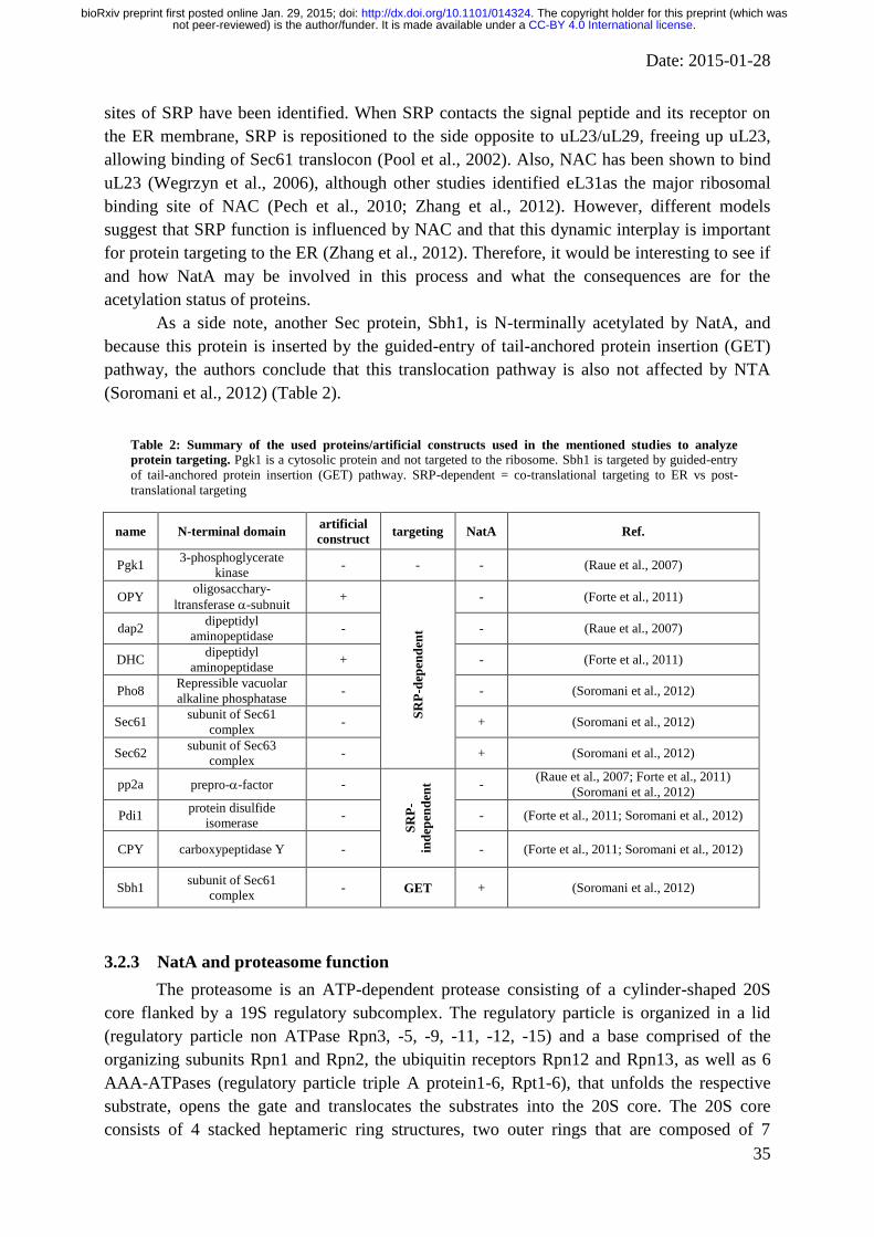
Date: 2015-01-28
35
sites of SRP have been identified. When SRP contacts the signal peptide and its receptor on
the ER membrane, SRP is repositioned to the side opposite to uL23/uL29, freeing up uL23,
allowing binding of Sec61 translocon (Pool et al., 2002). Also, NAC has been shown to bind
uL23 (Wegrzyn et al., 2006), although other studies identified eL31as the major ribosomal
binding site of NAC (Pech et al., 2010; Zhang et al., 2012). However, different models
suggest that SRP function is influenced by NAC and that this dynamic interplay is important
for protein targeting to the ER (Zhang et al., 2012). Therefore, it would be interesting to see if
and how NatA may be involved in this process and what the consequences are for the
acetylation status of proteins.
As a side note, another Sec protein, Sbh1, is N-terminally acetylated by NatA, and
because this protein is inserted by the guided-entry of tail-anchored protein insertion (GET)
pathway, the authors conclude that this translocation pathway is also not affected by NTA
(Soromani et al., 2012) (Table 2).
Table 2: Summary of the used proteins/artificial constructs used in the mentioned studies to analyze
protein targeting. Pgk1 is a cytosolic protein and not targeted to the ribosome. Sbh1 is targeted by guided-entry
of tail-anchored protein insertion (GET) pathway. SRP-dependent = co-translational targeting to ER vs post-
translational targeting
name N-terminal domain artificial
construct targeting NatA Ref.
Pgk1 3-phosphoglycerate
kinase - - - (Raue et al., 2007)
OPY oligosacchary-
ltransferase -subnuit +
SR
P-d
epen
den
t
- (Forte et al., 2011)
dap2 dipeptidyl
aminopeptidase - - (Raue et al., 2007)
DHC dipeptidyl
aminopeptidase + - (Forte et al., 2011)
Pho8 Repressible vacuolar
alkaline phosphatase - - (Soromani et al., 2012)
Sec61 subunit of Sec61
complex - + (Soromani et al., 2012)
Sec62 subunit of Sec63
complex - + (Soromani et al., 2012)
pp2a prepro--factor -
SR
P-
ind
epen
den
t - (Raue et al., 2007; Forte et al., 2011)
(Soromani et al., 2012)
Pdi1 protein disulfide
isomerase - - (Forte et al., 2011; Soromani et al., 2012)
CPY carboxypeptidase Y - - (Forte et al., 2011; Soromani et al., 2012)
Sbh1 subunit of Sec61
complex - GET + (Soromani et al., 2012)
3.2.3 NatA and proteasome function
The proteasome is an ATP-dependent protease consisting of a cylinder-shaped 20S
core flanked by a 19S regulatory subcomplex. The regulatory particle is organized in a lid
(regulatory particle non ATPase Rpn3, -5, -9, -11, -12, -15) and a base comprised of the
organizing subunits Rpn1 and Rpn2, the ubiquitin receptors Rpn12 and Rpn13, as well as 6
AAA-ATPases (regulatory particle triple A protein1-6, Rpt1-6), that unfolds the respective
substrate, opens the gate and translocates the substrates into the 20S core. The 20S core
consists of 4 stacked heptameric ring structures, two outer rings that are composed of 7
.CC-BY 4.0 International licensenot peer-reviewed) is the author/funder. It is made available under aThe copyright holder for this preprint (which was. http://dx.doi.org/10.1101/014324doi: bioRxiv preprint first posted online Jan. 29, 2015;

Date: 2015-01-28
36
structural α subunits and 7 catalytic β subunits that exhibit protease activity in the center of
the particle. For a recent review see (Bhattacharyya et al., 2014).
Some of the catalytic β-type subunits of the 20S core are synthesized as catalytically
inactive forms with an N-terminal propeptide, which is then cleaved during particle assembly.
Incomplete propeptide removal results in severe assembly defects of the proteasome (Groll et
al., 1997). Mutants in 5, 2 and 1, which lack the N-terminal propeptide, are defective for
specific peptidase activity, are more sensitive to environmental stresses, and have defects in
proteasome assembly (Arendt and Hochstrasser, 1999). Deletion of NAA10 or NAA15 restores
the phenotype, indicating that the propeptide protects the N-terminal threonine of the mature
form against acetylation (Arendt and Hochstrasser, 1999). A similar study showed that the
deletion of the N-terminal propeptides of the β1, β2 subunit precursors drastically inhibited
the enzymatic activity of the proteasome (Jäger et al., 1999). Further chemical analyses
revealed that acetylation of the N-terminal threonine in β1 is responsible for the measured
activity loss, supporting the idea that the propeptide protects the mature subunit moiety from
being acetylated prior to incorporation into the proteasome complex (Jäger et al., 1999). The
authors also propose a similar mechanism for β2 and β5; however, the N-termini have not
been analyzed for NTA (Jäger et al., 1999). Taken together, this indicates that the expression
of the β-chains together with the propeptide is a natural mechanism to circumvent acetylation
of an N-terminus that is important for catalytic function.
Furthermore, acetylation of other components of the proteasome has been identified in
vivo. Purified 20S proteasome subunits from wild type, NAA10, NAA20 and Naa30
mutant cells indicated that the 1, 2, 3, 4, 7, and 3 subunits were acetylated by NatA,
the 4 subunit was acetylated by NatB and the 5 and 6 subunits were acetylated by NatC
(Kimura et al., 2000). In a similar approach for the 26S regulatory subunit, the same group
found that 8 subunits, Rpt4, Rpt5, Rpt6, Rpn2, Rpn3, Rpn5, Rpn6, Rpn8, Rpn13 and Rpn15
were NatA substrates and that 2 subunits, Rpt3 and Rpn11, were NatB substrates, whereas
Rpt1 was unprocessed (Kimura et al., 2003; Kikuchi et al., 2010). Although these data
indicates that a multitude of proteasomal factors are acetylated in vivo, little is known about
the functional consequences of this modification and the so far analyzed effects seem to be
relative small. Analysis of the 20S proteasomes derived from either NAA15 mutant cells
revealed a very similar in vitro activity compared to wild type-derived proteasomes with
respect to chymotrypsin-like, trypsin-like, and peptidylglutamyl peptide hydrolyzing activities
(Kimura et al., 2000). In the absence of the chaotropic agent SDS, which is used in the in vitro
assay to induce activity of the 20S proteasome, the NAA15 proteasomes displayed a twofold-
higher chymotrypsin-like peptidase activity (Kimura et al., 2000). However, crude
proteasome extracts from the wild type strain and the NAA15 deletion mutant exposed a
similar accumulation level of the 26S proteasome in in vitro and exhibited normal
chymotrypsin-like activity in the presence of ATP (Kimura et al., 2003). On the other hand,
the accumulation level of 20S proteasome and the chymotrypsin-like activity were
appreciably higher in the extracts from the NAA15 deletion mutant (Kimura et al., 2003),
indicating that NTA might have a suppressing role in proteasome function.
In contrast to this, data generated with H. volcanii suggests that acetylation is required
for the assembly of the 20S core particle. In this archaea, the structural proteins α1 and α2 are
acetylated on their initiator methionine (Humbard et al., 2006). Interestingly, the acetylated
.CC-BY 4.0 International licensenot peer-reviewed) is the author/funder. It is made available under aThe copyright holder for this preprint (which was. http://dx.doi.org/10.1101/014324doi: bioRxiv preprint first posted online Jan. 29, 2015;
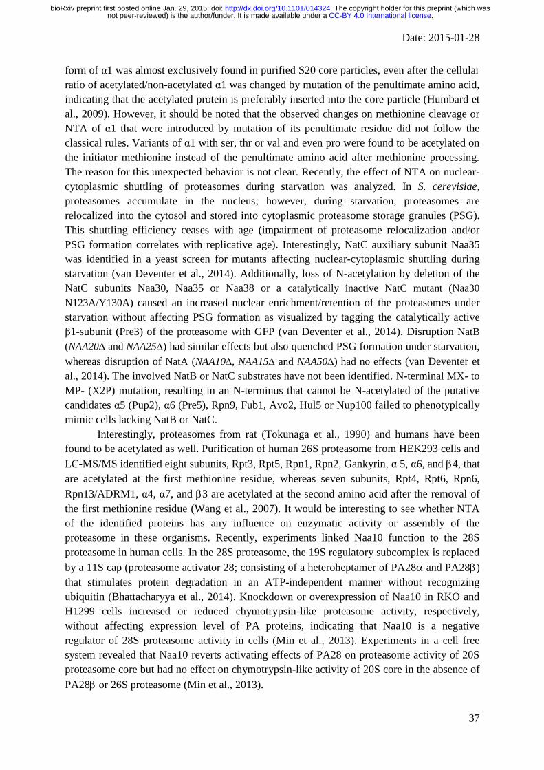
Date: 2015-01-28
37
form of α1 was almost exclusively found in purified S20 core particles, even after the cellular
ratio of acetylated/non-acetylated α1 was changed by mutation of the penultimate amino acid,
indicating that the acetylated protein is preferably inserted into the core particle (Humbard et
al., 2009). However, it should be noted that the observed changes on methionine cleavage or
NTA of α1 that were introduced by mutation of its penultimate residue did not follow the
classical rules. Variants of α1 with ser, thr or val and even pro were found to be acetylated on
the initiator methionine instead of the penultimate amino acid after methionine processing.
The reason for this unexpected behavior is not clear. Recently, the effect of NTA on nuclear-
cytoplasmic shuttling of proteasomes during starvation was analyzed. In S. cerevisiae,
proteasomes accumulate in the nucleus; however, during starvation, proteasomes are
relocalized into the cytosol and stored into cytoplasmic proteasome storage granules (PSG).
This shuttling efficiency ceases with age (impairment of proteasome relocalization and/or
PSG formation correlates with replicative age). Interestingly, NatC auxiliary subunit Naa35
was identified in a yeast screen for mutants affecting nuclear-cytoplasmic shuttling during
starvation (van Deventer et al., 2014). Additionally, loss of N-acetylation by deletion of the
NatC subunits Naa30, Naa35 or Naa38 or a catalytically inactive NatC mutant (Naa30
N123A/Y130A) caused an increased nuclear enrichment/retention of the proteasomes under
starvation without affecting PSG formation as visualized by tagging the catalytically active
β1-subunit (Pre3) of the proteasome with GFP (van Deventer et al., 2014). Disruption NatB
(NAA20 and NAA25) had similar effects but also quenched PSG formation under starvation,
whereas disruption of NatA (NAA10, NAA15 and NAA50) had no effects (van Deventer et
al., 2014). The involved NatB or NatC substrates have not been identified. N-terminal MX- to
MP- (X2P) mutation, resulting in an N-terminus that cannot be N-acetylated of the putative
candidates α5 (Pup2), α6 (Pre5), Rpn9, Fub1, Avo2, Hul5 or Nup100 failed to phenotypically
mimic cells lacking NatB or NatC.
Interestingly, proteasomes from rat (Tokunaga et al., 1990) and humans have been
found to be acetylated as well. Purification of human 26S proteasome from HEK293 cells and
LC-MS/MS identified eight subunits, Rpt3, Rpt5, Rpn1, Rpn2, Gankyrin, α 5, α6, and 4, that
are acetylated at the first methionine residue, whereas seven subunits, Rpt4, Rpt6, Rpn6,
Rpn13/ADRM1, α4, α7, and 3 are acetylated at the second amino acid after the removal of
the first methionine residue (Wang et al., 2007). It would be interesting to see whether NTA
of the identified proteins has any influence on enzymatic activity or assembly of the
proteasome in these organisms. Recently, experiments linked Naa10 function to the 28S
proteasome in human cells. In the 28S proteasome, the 19S regulatory subcomplex is replaced
by a 11S cap (proteasome activator 28; consisting of a heteroheptamer of PA28 and PA28)
that stimulates protein degradation in an ATP-independent manner without recognizing
ubiquitin (Bhattacharyya et al., 2014). Knockdown or overexpression of Naa10 in RKO and
H1299 cells increased or reduced chymotrypsin-like proteasome activity, respectively,
without affecting expression level of PA proteins, indicating that Naa10 is a negative
regulator of 28S proteasome activity in cells (Min et al., 2013). Experiments in a cell free
system revealed that Naa10 reverts activating effects of PA28 on proteasome activity of 20S
proteasome core but had no effect on chymotrypsin-like activity of 20S core in the absence of
PA28 or 26S proteasome (Min et al., 2013).
.CC-BY 4.0 International licensenot peer-reviewed) is the author/funder. It is made available under aThe copyright holder for this preprint (which was. http://dx.doi.org/10.1101/014324doi: bioRxiv preprint first posted online Jan. 29, 2015;

Date: 2015-01-28
38
3.2.4 NatA in prion propagation
Several studies have linked NatA to the Sup35/[PSI+] prion in S. cerevisiae. Prions are
protein-based units of inheritance that, by altering their conformational state and self-
assembly into amyloid fibers, serve as novel, self-replicating agents to determine phenotypic
traits (Sweeny and Shorter, 2008). The eukaryotic release factor 3, eRF3/Sup35 facilitates, in
its soluble nonprion [psi-] state, translation termination. In the [PSI
+] prion state, in the
alternative conformation that includes Sup35 aggregation, this leads to a termination defect
(to stop codon read-through) (Liebman and Chernoff, 2012). The N-terminal PrD (prion-
determining domain) is required for formation and propagation of the prion state.
Interestingly, disruption of NAA10 or NAA15 in [PSI+] yeast strains reversed the
[PSI+] phenotype measured by stop-codon readthrough assays (colony growth assays based
on expression of the ade1-14 (UGA) allele containing a premature stop codon that leads to
adenine auxotrophy in [psi-] but not in [PSI
+] cells and stop codon read-through in Pgk1-stop-
-galactosidase reporter) (Pezza et al., 2009). Furthermore, Sup35 aggregates from NatA
[PSI+] strain exhibit reduced thermodynamic stability, and are composed of smaller core
polymers compared to wild type [PSI+] but did not appear to alter the number of Sup35
aggregates or the solubility of Sup35 (Pezza et al., 2009). However, later studies by the same
group showed that the decrease in Sup35 aggregate size observed in a [PSI+]strong
NatA
strain leads to increased ribosome binding of the aggregates and a decreased stop-codon
readthrough, indicating an increase in aggregate-associated termination activity (Pezza et al.,
2014). Taken together, these data suggest that NatA function is required for [PSI+] strains to
display the prion phenotype. Interestingly, Sup35 was identified as a substrate of NatA in a
global qualitative and quantitative analysis of protein N-acetylation (Arnesen et al., 2009b).
Mutation of the penultimate amino acid to proline abrogates NTA of Sup35; indeed, S2P
mutation in [PSI+] induced reduced stability and core size of Sup35-S2P aggregates;
however, the effects were weaker compared to deletion of NatA, and the Sup35-S2P did not
revert stop-codon readthrough, indicating that Sup35 NTA alone is not sufficient and
additional factors contribute to the phenotype conversion of the NatA [PSI+] strain (Holmes
et al., 2014).
The propagation of the [PSI+] state involves the action of many chaperones. The
Hsp104 chaperone and the Hsp70 chaperone Ssa1/2 (constitutively expressed) and Ssa3/4
(stress inducible), promote fragmentation of prion fibers into smaller seeds, initiating new
rounds of prion propagation (Liebman and Chernoff, 2012). The Hsp40 protein Sis1 is an
important co-chaperone of HSP70 in prion propagation (Higurashi et al., 2008; Tipton et al.,
2008). In contrast to this, the Hsp70 chaperone Ssb1/2 seems to destabilize the prion state
[PSI+] (Liebman and Chernoff, 2012). In an early study on acetylated proteins in yeast,
Ssa1/2 and Ssb1/2 proteins but not Hsp104 have been shown to undergo NTA by NatA
(Polevoda et al., 1999). Notably, Ssb was also found to be acetylated by ssNaa10 in S.
solfataricus, despite the fact that the N-terminal sequence (MEEK…) of Ssb points towards
NatB as the corresponding NAT (Mackay et al., 2007; Polevoda et al., 2009). This again
indicates that the sequence specificity of the NAT has co-evolved with its substrates
sequence. However, the sequence of Ssa3/4 (starting MS…) also indicates that these proteins
are targets of NatA in yeast, which has been confirmed biochemically (Van Damme et al.,
2014). Interestingly, loss of NatA strongly increased heat-shock response (HSR) as measured
.CC-BY 4.0 International licensenot peer-reviewed) is the author/funder. It is made available under aThe copyright holder for this preprint (which was. http://dx.doi.org/10.1101/014324doi: bioRxiv preprint first posted online Jan. 29, 2015;

Date: 2015-01-28
39
by Hsf1 reporter gene expression, increased Hsp104, Ssa1/2, Ssb1/2 and Sis1 protein levels
and induced a change in the recruitment of the chaperones Ssb1/2 Ssa1/2 to Sup35 as seen in
immunoprecipitation experiments in [PSI+] cells (Holmes et al., 2014), but did not affect
Hsp104 or Ssa1/2 expression levels in [psi-] cells (Pezza et al., 2009). Additionally, elevation
of HSR activity by using a constitutively active Hsf1 mutant (Hsf1ON), reduces Sup35
aggregate size in [PSI+] cells (mimics the effects of NatA disruption), whereas
downregulation of HSR by expressing a dominant-negative mutant of Hsf1 (Hsf1DN)
resulted in the loss of the smallest Sup35 core polymers (Holmes et al., 2014). Because
disruption of NatA in [PSI+] strain led to the accumulation of protein aggregates, as assayed
by a centrifugation assay, sensitivity to translational inhibitors and Hsf1-mediated gene
expression, the authors conclude that loss of NTA promotes general protein misfolding, a
redeployment of chaperones to these substrates, and a corresponding stress response that
subsequently reduces the size of prion aggregates and reverse their phenotypic consequences
(Holmes et al., 2014). However, single mutation of Ssa1, Ssa2, Ssb1, Ssb2 or Sup35 only
partially mimicked the effects of NatA (Pezza et al., 2009; Holmes et al., 2014), whereas
mutation of all of these proteins [ssa1(S2P)ssa2, ssb1(S2P)ssb2, sup35(S2P)] did mimic
the effects of disruption of NatA in [PSI+] strain, including induction of smaller sized Sup35
aggregates and reversion of stop-codon readthrough in colony growth ade1-14 (UGA) assays
(Holmes et al., 2014). This indicates that the increased chaperones and loss of Sup35 NTA
combinatorially contribute to the effect of NatA disruption on prion propagation (Holmes et
al., 2014).
3.3 NATs in prokaryotes (archaeabacteria, eubacteria)
3.3.1 In E. coli: RIMs
In contrast to eukaryotes, in Escherichia coli and Salmonella typhimurium, only a few
proteins seem to be N-terminally acetylated (Bernal-Perez et al., 2012; Ansong et al., 2013)
and ectopically expressed human proteins in E. coli usually lack Nα-acetylation as shown for
human ββ alcohol dehydrogenase (Höög et al., 1987), rat chaperonin 10 (Ball et al., 1994),
Annexin II tetramer (Kang et al., 1997) or Spartin and Tropomyosin (Johnson et al., 2010).
However, some proteins are found to be fully or partially acetylated when expressed in E.
coli, including stathmin-like subdomains (Charbaut et al., 2002) prothymosin α (Wu et al.,
2006) interferon- (Honda et al., 1989), interferon- (Takao et al., 1987) or the serine
proteinase inhibitor eglin c (Grütter et al., 1985). However, a recent analysis of 112 million
mass spectrometric data from 57 bacterial species revealed that NTA might be markedly more
common than accepted, as about 16% of bacterial N-termini were found to be N-acetylated
(Bonissone et al., 2013).
To date only three Nα-acetyltransferases have been identified in prokaryotes which
specifically modifies only one protein each, as opposed to eukaryotes, in which these
enzymes are much less specific (Nesterchuk et al., 2011). These enzymes, RimI, RimJ and
RimL (ribosomal modification) specifically acetylate the ribosomal proteins S18 (Isono and
Isono, 1980; Yoshikawa et al., 1987), S5 (Cumberlidge and Isono, 1979; Yoshikawa et al.,
1987) and L7/L12 (Isono and Isono, 1981; Tanaka et al., 1989), respectively. According to
their low sequence identity with the NATs it is most likely that the RIM proteins do not have
.CC-BY 4.0 International licensenot peer-reviewed) is the author/funder. It is made available under aThe copyright holder for this preprint (which was. http://dx.doi.org/10.1101/014324doi: bioRxiv preprint first posted online Jan. 29, 2015;
Date: 2015-01-28
40
a common ancestor and evolved independently (Polevoda and Sherman, 2003a; Vetting et al.,
2008).
Mutation of each of these Rims leads to temperature sensitivity in the E. coli strain
K12 (Isono and Isono, 1978), however, this phenotype is independent of the acetylation of
S18, S5 and S12 (Cumberlidge and Isono, 1979; Isono and Isono, 1980; Isono and Isono,
1981; Yoshikawa et al., 1987). Nevertheless, there is evidence, that the RimI and RimJ-
mediated acetylation occurs post translationally and is required for the assembly of the
ribosomal subunits or translation initiation (or more generally for ribosome function) (Poot et
al., 1997; Recht and Williamson, 2001; Roy-Chaudhuri et al., 2008).
RimJ has evolved dual functionality; it functions in ribosomal-protein acetylation and
as a ribosome assembly factor, where the enzymatic activity is not needed in E. coli (Roy-
Chaudhuri et al., 2008). Furthermore it has been shown to regulate Pyelonephritis-associated
pilus (Pap) transcription in response to multiple environmental cues (White-Ziegler et al.,
2002).
RimL seems to have a regulatory function during the growth cycle. A shift in
acetylation occurs during the growth cycle: L7 is the acetylated form of L12 (Terhorst et al.,
1973). L12/L7 is not acetylated during early log phase but becomes acetylated at the
stationary phase (Ramagopal and Subramanian, 1974). Later mass spectrometric data from
intact ribosomes showed that NTA of L12 during the stationary phase correlates with an
increased ribosomal stability mediates the adaption of the cells to nutrient deprivation
(Gordiyenko et al., 2008).
Taken together, it seem like the main function of NTA is regulation/initiation of
translation/transcription. If acetylation can also act as a signal for protein degradation as has
recently been proposed for eukaryotic NTA (Hwang et al., 2010) has to be shown. However,
that was questioned as the enzyme that recognizes acetylated N-termini in eukaryotes and tags
them for destruction (Doa10 ubiquitin ligase) does not have orthologues in prokaryotes such
as E. coli (Jones and O'Connor, 2011).
3.3.2 In Archaea
In contrast to bacteria, NTA is quite common in archaea. Large-scale proteomics
studies in Halobacterium salinarum, Natronomonas pharaonis (Falb et al., 2006) and
Haloferax volcanii (Kirkland et al., 2008) showed that up to 14%, 19% and 29% of the
identified proteins are acetylated, respectively. In the former study, acetylation was limited to
cleaved N-termini starting with serine or alanine residues, suggesting NatA activity (Falb et
al., 2006; Aivaliotis et al., 2007), whereas the latter study also found NatB like substrates
(Kirkland et al., 2008). However, many of the encountered proteins were only acetylated
partially.
So far, only one NAT in Sulfolobus solfataricus has been studied in more detail.
However, homologous proteins have also been identified in the archaea Pyrobaculum
aerophilum, Natronomonas pharaonis and Halobacterium salinarium (Mackay et al., 2007),
Aeropyrum pernix (Polevoda and Sherman, 2003b) as well as Thermoplasma acidophilum
(Han et al., 2006) and Thermoplasma volcanium (Ma et al., 2014). The S. solfataricus
ssNaa10 represents an ancestral variant of the eukaryotic NATs and it adopts a more relaxed
substrate specificity as it acetylates NatA, B, C and E substrates (Mackay et al., 2007). In line
.CC-BY 4.0 International licensenot peer-reviewed) is the author/funder. It is made available under aThe copyright holder for this preprint (which was. http://dx.doi.org/10.1101/014324doi: bioRxiv preprint first posted online Jan. 29, 2015;
Date: 2015-01-28
41
with that, in vitro acetylation assay showed that although ssNaa10 has higher sequence
similarity to hNaa10 over hNaa50 (34% vs. 24% sequence identity, respectively) and
therefore has a preference for Ser- over Met-amino-terminal substrates, it still is able to
accommodate NatA and NatE substrates (Liszczak and Marmorstein, 2013). Furthermore,
studies on the X-ray crystal structure of ssNaa10 combined with mutagenesis and kinetic
analyses showed that the active site of ssNaa10 represents a hybrid of the NatA and NatE
active sites (Liszczak and Marmorstein, 2013), explaining the above findings. The X-ray
crystal structure of archaeal T. volcanium Naa10 has also been reported, revealing multiple
distinct modes of acetyl-Co binding involving the loops between β4 and α3 including the P-
loop (Ma et al., 2014). It should be noted that the acetylation activity of ssNaa10 has been
confirmed in vitro for a variety of substrates including the S. solfataricus proteins Holliday
junction resolving enzymes (Hjc and Hje), single-stranded DNA-binding protein (SSB) as
well as Alba1 (Mackay et al., 2007; Ma et al., 2014).
Until now, no auxiliary subunit of any of the NATs has been described in archaea, and
the function of NTA in archaea is unknown.
3.4 NATs in plants
In 2003, Polevoda and Sherman identified the first candidates for NATs (Naa10,
Naa20 and Naa30) in A. thaliana by homology search with the amino acid sequences of yeast
NATs (Polevoda and Sherman, 2003b). More recent studies identified orthologs and their
gene duplicates for 6 catalytic subunits and 5 auxiliary subunits corresponding to NatA-F
from humans in Populus nigra (Liu et al., 2013) and Populus trichocarpa, Chlamydomonas
reinhardtii, Medicago truncatula, and Vitis vinifera (Bienvenut et al., 2012), implying that all
known NATs are conserved in woody plants. Additionally, six distinct methionine
aminopeptidases have been identified and characterized in A. thaliana (Giglione et al., 2000).
To date, 200/1054 proteins in A. thaliana (Baerenfaller et al., 2008; Bienvenut et al.,
2012) and 58 proteins in Populus nigra (Liu et al., 2013) proteins have been found to be N-
terminally acetylated. A large-scale N-terminomic study in the model diatom Thalassiosira
pseudonana (phytoplankton) revealed that about 70% of cytosolic proteins were completely
or partially acetylated (Huesgen et al., 2013). Furthermore, similar acetylation patterns
between animal and plant kingdoms have been observed, suggesting a strong convergence of
the characterized modification (Liu et al., 2013). Also, the observed patterns for methionine
removal follow the same rules as those of other eukaryotes (Huesgen et al., 2013).
In addition to cytosolic proteins, posttranslational acetylation of proteins imported into
organelles has been observed. A large scale analysis of chloroplast preparations from
Arabidopsis thaliana identified 47 N-terminal acetylated nuclear encoded proteins (Zybailov
et al., 2008) and proteomic studies detected NTA on 30% of nuclear encoded stromal
chloroplast proteins in the green algae Chlamydomonas reinhardti (Bienvenut et al., 2011)
and on 50 % of nuclear encoded plastid proteins in Thalassiosira pseudonana (Huesgen et al.,
2013). Similarly, a comparative large scale characterization of plant and mammalian
proteomes found that 25 % of the identified acetylated Arabidopsis thaliana proteins become
Nα-acetylated at a position downstream of the annotated position 1 or 2 (compared to 8 % in
humans) and the majority of these proteins (>80 %) reside in the chloroplasts and become
.CC-BY 4.0 International licensenot peer-reviewed) is the author/funder. It is made available under aThe copyright holder for this preprint (which was. http://dx.doi.org/10.1101/014324doi: bioRxiv preprint first posted online Jan. 29, 2015;
Date: 2015-01-28
42
acetylated at the new N-terminus after removal of the chloroplast transit peptide (Bienvenut et
al., 2012). Furthermore, the authors indicate that the sequence of the acetylated chloroplast
appears to match to NatA substrates, suggesting a dedicated NatA might exist in this
organelle (Bienvenut et al., 2012). Interestingly, in Thalassiosira pseudonana, a nuclear
encoded plastid-targeted putative N-acetyltransferase was identified that could be responsible
for the observed NTA of imported and plastid-encoded proteins (Huesgen et al., 2013).
All these data indicate that NTA is very common in plants. Also, it appears that most
NAT components have been identified in the analyzed species so far, and the fact that the
same rules for N-terminal processing apply, indicates that N-terminal processing is very well
conserved in plants. Plants also possess chloroplasts and many studies support post-
translational NTA in these plastids. It would be very interesting to identify and characterize
the dedicated acetyltransferase in these plastids to understand the evolution of the NATs. On a
side note, recently two mitochondrial proteins have been identified to be (partially) acetylated in a
NatA-dependent manner, and it has speculated that this NTA is accomplished either post-
translationally in mitochondria upon import of Naa10 or alternatively post-translationally upon
mitochondrial export of the substrates (Van Damme et al., 2014).
3.5 Function of NTA in plants
Compared to mammals or yeast, the function of NTA in plant is even less studied or
understood. However, some data point out that NTA in plants may have similar effects as
described earlier. Analysis of the acetylation status of stromal proteins and in combination
with pulse-chase experiments e.g. revealed that among all identified N-terminally acetylated
stromal proteins with a short half-life the NTA was underrepresented indicating that NTA
may have a stabilizing role in the plastids of plants (Bienvenut et al., 2011) in contrast to the
proposed destabilizing Ac/N-end rule pathway in the mammalian cytosol (Varshavsky, 2011).
The authors speculate that, because a) the majority of the plastid proteins have an unusual N-
terminus due to the cleavage of their transit peptides and b) the chloroplast does not have a
proteasome which is indispensable for the Ac
N-degron degradation process, but contains a
number of bacterial-type chambered proteases, including ClpP (which appears to display
specificity for such unusual N-termini), an -acetylated N-terminus could protect stromal
proteins from rapid degradation (Bienvenut et al., 2011). However, further studies have to
analyze the effect of NTA on the stability of plastid and cytosolic plant proteins.
Interestingly, the -subunit of chloroplast ATP synthase has been found to be partially
acetylated in Citrullus lanatus L. (wild watermelon) and during drought, the non-acetylated
form gets degraded by metalloaminopeptidases (Hoshiyasu et al., 2013) indicating that the
effect of NTA on stability might be more complex and might even depend on the respective
substrate. It should be mentioned that NTA might be generally important for photosynthesis.
An Arabidopsis thaliana Naa30 (AtMak3) mutant exhibit a decreased effective quantum yield
of photosystem II and a growth defect (Pesaresi et al., 2003). However, in the latter case, it is
not clear how a cytoplasmatic N-acetylation defect would influence chloroplast function. The
authors speculate that NTA may be relevant for “stability and/or import of organellar
precursor proteins and thus, indirectly, for organellar function” (Pesaresi et al., 2003). This
could indicate that, as discussed for yeast, targeting of proteins is regulated by NTA. In this
context it is interesting to mention that components of the photosystem II also have been
.CC-BY 4.0 International licensenot peer-reviewed) is the author/funder. It is made available under aThe copyright holder for this preprint (which was. http://dx.doi.org/10.1101/014324doi: bioRxiv preprint first posted online Jan. 29, 2015;
Date: 2015-01-28
43
found to be acetylated in Spinacia oleracea (spinach) (Michel et al., 1988). Thus, direct
acetylation of photosystem compounds by an organelle-localized NAT could also influence
photosystem function.
As for mammalian NATs, NTA has also been implicated in plant development. A loss
of function mutation of Naa20 (NatB catalytic subunit, nbc-1) or Naa25 (NatB auxiliary
subunit, transcurvata2, tcu2) in Arabidopsis thaliana caused a strong phenotype that includes
leaf reticulation, early flowering, unfertilized or aborted ovules in siliques, indicating that the
NatB auxiliary subunit is important for vegetative and reproductive development (Ferrández-
Ayela et al., 2013). Furthermore, genetic interaction of NAA25 and AGO10 (Argonaute10)
suggest a link between NatB-mediated Nα-terminal acetylation and the microRNA pathway
(Ferrández-Ayela et al., 2013). The mechanism for this genetic interaction is not clear and
future studies have to explore whether other NATs are also involved in plant development.
Interestingly, systematic investigation of publicly available microarray data showed that the
expression levels of NatA-F are relatively low during development; these NATs are expressed
at low levels but share distinct tissue-specific expression patterns (Zhu et al., 2014).
.CC-BY 4.0 International licensenot peer-reviewed) is the author/funder. It is made available under aThe copyright holder for this preprint (which was. http://dx.doi.org/10.1101/014324doi: bioRxiv preprint first posted online Jan. 29, 2015;
Date: 2015-01-28
44
4 C O N C L U S I O N
NTA is one of the most abundant protein modifications known, and the NAT
machinery is conserved throughout all Eukarya. Over the past 50 years, the function of NTA
has begun to be slowly elucidated, and this includes the modulation of protein-protein
interaction, protein-stability, protein function, and as well as protein targeting to specific
cellular compartments. Many of these functions have been studied in the context of
Naa10/NatA; however, we are only starting to really understand the full complexity of this
picture. Roughly, about 40 % of all human proteins are substrates of Naa10 and the impact of
this modification has only been studied for a few of them. Besides acting as a NAT in the
NatA complex, recently other functions have been linked to Naa10, including post-
translational NTA, lysine acetylation and NAT/KAT-independent functions. Also, recent
publications have linked mutations in Naa10 to various diseases, emphasizing the importance
of Naa10 research in humans. The recent design and synthesis of the first 3 bisubstrate
inhibitors that potently and selectively inhibit the NatA/Naa10 complex, monomeric Naa10,
and hNaa50 further increases the toolset to analyze Naa10 function (Foyn et al., 2013a).
4.1 Co-translational NTA and protein quality control
The highly complicated process of protein translation requires the interplay of multiple
factors, including non-ribosomal proteins that permanently or transiently associate with the
ribosome and welcome the emerging protein as soon as a nascent polypeptide reaches the exit
from the ribosomal tunnel. The “welcoming committee” consists of ribosome-associated
protein biogenesis factors (RPBs) that co-translationally regulate various processes, including
protein folding, targeting protein sorting, protein quality control and protein modifications
(Giglione et al., 2014). RPBs include the NATs, MetAPs (methionine aminopeptidases), SRP
(signal recognition particle) and chaperones like Hsp70/Hsp40 and NAC (nascent
polypeptide-associated complex) (Kramer et al., 2009). The region around the exit tunnel of
the proteasome, which is comprised of the ribosomal proteins uL22, uL23, uL24, uL29 as
well as eL19, eL31 and eL39 in archaea and eukaryotes, constitutes a general docking
platform for RPBs, and recent research indicates that uL23 might play an eminent role as it
interacts with almost all RPBs analyzed (Kramer et al., 2009).
It is widely accepted that NATs and NatA, in particular, are linked to the ribosome and
co-translationally acetylate nascent chain as they emerge from the ribosomal exit tunnel
(Strous et al., 1973; Filner and Marcus, 1974; Strous et al., 1974; Driessen et al., 1985;
Yamada and Bradshaw, 1991; Gautschi et al., 2003; Arnesen et al., 2005a; Raue et al., 2007;
Polevoda et al., 2008; Arnesen et al., 2009a; Arnesen et al., 2010). Recent findings suggest
that NatA negatively regulates protein sorting/targeting to the ER through SRP-dependent
and/or SRP-independent mechanisms, and the bias observed in secretory proteins towards not
being NTA supports this idea, although some ER-targeted proteins, including Sec61 and
Sec62, have been shown to be NTA by NatA (Raue et al., 2007; Forte et al., 2011; Soromani
et al., 2012). Interestingly, both, SRP and NatA bind to the ribosome via uL23/uL29 at the
tunnel exit; however, SRP seems to reposition to the opposite side of the tunnel during signal
peptide recognition to allowing binding of the Sec61 translocon to uL23 (Pool et al., 2002;
.CC-BY 4.0 International licensenot peer-reviewed) is the author/funder. It is made available under aThe copyright holder for this preprint (which was. http://dx.doi.org/10.1101/014324doi: bioRxiv preprint first posted online Jan. 29, 2015;

Date: 2015-01-28
45
Halic et al., 2004; Polevoda et al., 2008). In addition to SRP and NatA, NAC has been shown
to interact with uL23; however, eL31 seems to be the major ribosomal binding site of NAC
(Wegrzyn et al., 2006; Pech et al., 2010; Zhang et al., 2012). This data on the one hand
indicates that RPBs compete for nascent chains at the ribosome as suggested for NATs and N-
myristoyltransferases (Utsumi et al., 2001) and on the other hand suggests that NATs might
functionally regulate adjacent RPBs. We are only beginning to understand the complicated
interplay of RPBs on the ribosome, but recent studies suggest that NAC might modulate the
function of SRP possibly through the Hsp70/40 chaperone system in an isoform-specific
fashion, and this dynamic interplay is important for proper protein targeting and folding
(Koplin et al., 2010; del Alamo et al., 2011; Sedwick, 2011; Zhang et al., 2012; Pechmann et
al., 2013; Holmes et al., 2014). The role of Naa10/NatA in this context is obscure but the data
summarized here strongly implicates a connection of NATs to the protein sorting and protein
folding machinery, which could explain, at least in part, the connection of Naa10 and other
NATs to neurodegenerative diseases such as Alzheimer’s or Parkinson’s disease, where
misfolding of certain protein species leads to the accumulation of toxic amyloid aggregates
(Asaumi et al., 2005; Zabrocki et al., 2008; Pezza et al., 2009; Kang et al., 2012; Kang et al.,
2013; Holmes et al., 2014; Pezza et al., 2014). However, it should be noted that the ribosome
itself is susceptible to acetylation by NatA and other NATs (Takakura et al., 1992; Arnold et
al., 1999; Kamita et al., 2011), and NTA of ribosomal proteins seem to affect various
processes, including ribosome biogenesis, ribosomal fidelity/translational activity and
translational termination (Kamita et al., 2011; Wan et al., 2013)
4.2 Post-translational NTA of Naa10
Besides acting co-translationally, accumulating reports have reported post-
translational NTA by Naa10. Indeed, despite being reported to be mainly associated with
ribosomes in yeast, a major pool of hNaa10 exists in a ribosome-free context, and a fraction
of cytosolic hNaa10 even exists independent of the NatA complex (Arnesen et al., 2005a; Van
Damme et al., 2011b). In this context it should be mentioned that NatB has also been shown
to not always co-express and co-localize with its catalytic counterpart Naa20 in differentiated
neurons in mouse, implying that the auxiliary subunit may function either with an unidentified
NAT protein partner(s) or possibly in a NAT-independent manner (Ohyama et al., 2012).
However, large-scale N-terminomic analyses are shedding light on post-translational NTA by
identifying N-terminally acetylated proteins starting beyond position 1 or 2, such as
mitochondrial or chloroplast proteins in plants, that become post-translationally NTA after
cleavage of their transit peptide (Zybailov et al., 2008; Helbig et al., 2010; Bienvenut et al.,
2011; Helsens et al., 2011; Van Damme et al., 2011b; Bienvenut et al., 2012; Huesgen et al.,
2013; Van Damme et al., 2014). Until now, it is not clear if a dedicated NAT enzyme resides
in the organelles or if, for example, Naa10 is imported into the respective organelle
(Bienvenut et al., 2012; Van Damme et al., 2014). In this regard it is interesting to note that
monomeric Naa10 seems to have a different substrate specificity, preferentially acetylating
acidic N-termini, e.g. the non-cannonical substrates γ- and β-actin (Van Damme et al., 2011b;
Van Damme et al., 2014). The reason for this change in substrate specificity are structural
rearrangements of the substrate binding pocket in Naa10 induced by Naa15 binding,
(Liszczak et al., 2013). However, the occurrence of classical NatA-substrates in organelles
.CC-BY 4.0 International licensenot peer-reviewed) is the author/funder. It is made available under aThe copyright holder for this preprint (which was. http://dx.doi.org/10.1101/014324doi: bioRxiv preprint first posted online Jan. 29, 2015;
Date: 2015-01-28
46
would suggest that somehow the complete NatA complex is present in organelles, although
future studies must address this question in more detail.
4.3 NAT-independent functions of Naa10 and transcriptional
regulation
Many studies have addressed the intercellular localization of Naa10 in a variety of cell
systems, showing that Naa10 mainly localized to the cytoplasm and to a lesser extent in the
nucleus, but also isoform specific localization patterns of Naa10 have been suggested (Fluge
et al., 2002; Sugiura et al., 2003; Bilton et al., 2005; Arnesen et al., 2006a; Arnesen et al.,
2006b; Chun et al., 2007; Xu et al., 2012; Park et al., 2014; Zeng et al., 2014). In agreement
with this, a functional nuclear localization signal (KRSHRR) has been identified and
characterized (Arnesen et al., 2005a; Park et al., 2012; Park et al., 2014). Interestingly, Naa15
also harbors a putative NLS; however, analysis of the nuclear localization of Naa15 revealed
discrepant results (Willis et al., 2002; Arnesen et al., 2005a).
The function for a possible nuclear redistribution of Naa10 is not yet clear, but
accumulating studies showed that Naa10 directly or indirectly regulates transcriptional
activity in different pathways, including Wnt/-catenin, MAPK, JAK/STAT5, AP-1, mTOR,
NF-kB and BMP signaling (Lim et al., 2006; Kaidi et al., 2007; Lim et al., 2008; Kuo et al.,
2009; Kuo et al., 2010; Seo et al., 2010; Park et al., 2012; Xu et al., 2012; Yoon et al., 2014;
Zeng et al., 2014). However, the exact mechanism of its action is not known. Despite
speculations that Naa10 might directly act as a transcription factor, to our knowledge, a direct
interaction of Naa10 and promoter regions has only been described for the E-cadherin
promoter (Lee et al., 2010a) and the c-Myc promoter (Lim et al., 2008). The goal of future
studies will be to exactly analyze whether Naa10 indeed acts as a transcription factor and/or
modulates transcription indirectly through acetylation of substrates or via other NAT-
independent functions.
Despite the fact that the NAT-dependent function of Naa10 has been addressed in
numerous studies, a possible NAT-independent function of Naa10 is widely understudied and
the mechanism of such action is still not clear. The use of an enzymatic-dead hNaa10 mutant
helped to differentiate between NAT-dependent and -independent functions. This R82A
mutation is located in a R82
-x-x-G85
-x-A87
consensus sequence, critical for the binding of
acetyl-CoA, and has been shown to exhibit low acetyltransferase activity and inhibited
autoacetylation of Naa10 (Asaumi et al., 2005; Seo et al., 2010; Seo et al., 2014). According
to the widely accepted idea that the acetyltransferase activity of Naa10 is crucial for its
function, it has been shown that disruption of the catalytic activity abrogated Naa10 function
to decrease endocytosis of APP and suppress Aβ40 secretion, to decrease ubiquitination and
increase TSC2 stability thereby repressing mTOR signaling, and for doxorubicin-induced NF-
B activation as well as promotion of cyclin D1 expression through acetylation-dependent
activation of β-catenin and AP-1(Asaumi et al., 2005; Kuo et al., 2010; Seo et al., 2010; Park
et al., 2012). However, in some cases, eliminating of NAT activity did not affect Naa10
function. For example, reintroduction of Naa10 R82A restored colony forming in Naa10-
depleted cells, although not to the same degree as wild type hNaa10, and overexpression of
the mutant increased DNMT activity similarly to wild type Naa10 (Lee et al., 2010a). The
.CC-BY 4.0 International licensenot peer-reviewed) is the author/funder. It is made available under aThe copyright holder for this preprint (which was. http://dx.doi.org/10.1101/014324doi: bioRxiv preprint first posted online Jan. 29, 2015;

Date: 2015-01-28
47
authors conclude that Naa10 faciliates DNMT1-mediated gene silencing in an
acetyltransferase (AT)-independent manner by recruiting DNMT1 to the E-cadherin
promoter, where DNMT1 can silence the transcription of the tumor suppressor E-cadherin
through methylation (Lee et al., 2010a). Similarly, Naa10 has been shown to competitively
bind to PIX proteins in an AT-independent manner, thereby disrupting the GIT-PIX-Paxillin
complex, resulting in reduced activation of the cell migration machinery (Hua et al., 2011).
However, the authors did not completely rule out acetylation-dependent mechanisms in
regulating cell mobility, as the Naa10-R82A mutant may still have some remaining function
to acetylate some other specific proteins that participate in cell mobility control (Hua et al.,
2011). A different study showed that Naa10 regulates Janus kinase 2-STAT5a transcriptional
activity independent of its acetyltransferase activity (Zeng et al., 2014). Particularly,
overexpression of either wild type Naa10 or Naa10-R82A in MCF-7 and MDA-MB-231
breast cancer cells induced a similar decrease in STAT5a binding to ID1 promoter, similar
inhibition of ID1 protein levels, and similarly reduced migration of these cells (Zeng et al.,
2014). Furthermore, because STAT5a was not found to be acetylated at lysine residue(s) and
its protein expression was not affected by the exogenous wild-type or mutant Naa10, the
authors speculate that the acetyltransferase activity of Naa10 could be dispensable for
inhibiting STAT5a-dependent ID1 expression and suppressing invasiveness of breast cancer
cells (Zeng et al., 2014). It should be noted that although STAT5a itself is a potential
substrate for NTA by Naa10 according to its sequence (starting: MAG…), the authors did not
check the acetylation status of STAT5a. However, in another study, AT-independent activity
Naa10 has been implicated in the activation of the 28S proteasome; however, the mechanism
for this action is not clear (Min et al., 2013). The authors speculate that Naa10, through
binding to components of the 28S activation complex PA28, could sterically hinder peptide
entrance into the proteasome, although this would need further investigation (Min et al.,
2013).
Acknowledgements
The laboratory of G.J.L. is supported by funds from the Stanley Institute for Cognitive
Genomics at Cold Spring Harbor Laboratory (CSHL).The authors thank Jason O’Rawe for
critical reading of the manuscript.
Competing Interests
G.J.L serves on the medical advisory board of GenePeeks, Inc. and the scientific advisory
board of Omicia, Inc. The study did not involve these companies and did not use products
from these companies.
.CC-BY 4.0 International licensenot peer-reviewed) is the author/funder. It is made available under aThe copyright holder for this preprint (which was. http://dx.doi.org/10.1101/014324doi: bioRxiv preprint first posted online Jan. 29, 2015;

Date: 2015-01-28
48
5 R E F E R E N C E S
Adam, V., Ekblad, M., Sweeney, K., Müller, H., Busch, K.H., Johnsen, C.T., Kang, N.R.,
Lemoine, N.R. and Halldén, G., 2012. Synergistic and Selective Cancer Cell Killing
Mediated by the Oncolytic Adenoviral Mutant AdΔΔ and Dietary Phytochemicals in
Prostate Cancer Models. Hum Gene Ther 23, 1003-15.
Aivaliotis, M., Gevaert, K., Falb, M., Tebbe, A., Konstantinidis, K., Bisle, B., Klein, C.,
Martens, L., Staes, A., Timmerman, E., Van Damme, J., Siedler, F., Pfeiffer, F.,
Vandekerckhove, J. and Oesterhelt, D., 2007. Large-scale identification of N-terminal
peptides in the halophilic archaea Halobacterium salinarum and Natronomonas
pharaonis. J Proteome Res 6, 2195-204.
Akopian, D., Shen, K., Zhang, X. and Shan, S.O., 2013. Signal recognition particle: an
essential protein-targeting machine. Annu Rev Biochem 82, 693-721.
Aksnes, H., Osberg, C. and Arnesen, T., 2013. N-Terminal Acetylation by NatC Is Not a
General Determinant for Substrate Subcellular Localization in Saccharomyces
cerevisiae. PLoS One 8, e61012.
Alderson, T.R. and Markley, J.L., 2013. Biophysical characterization of α-synuclein and its
controversial structure. Intrinsically Disord Proteins 1, 18-39.
Ansong, C., Wu, S., Meng, D., Liu, X., Brewer, H.M., Deatherage Kaiser, B.L., Nakayasu,
E.S., Cort, J.R., Pevzner, P., Smith, R.D., Heffron, F., Adkins, J.N. and Pasa-Tolic, L.,
2013. Top-down proteomics reveals a unique protein S-thiolation switch in
Salmonella Typhimurium in response to infection-like conditions. Proc Natl Acad Sci
U S A 110, 10153-8.
Aparicio, O.M., Billington, B.L. and Gottschling, D.E., 1991. Modifiers of position effect are
shared between telomeric and silent mating-type loci in S. cerevisiae. Cell 66, 1279-
87.
Aragon, A.D., Rodriguez, A.L., Meirelles, O., Roy, S., Davidson, G.S., Tapia, P.H., Allen, C.,
Joe, R., Benn, D. and Werner-Washburne, M., 2008. Characterization of differentiated
quiescent and nonquiescent cells in yeast stationary-phase cultures. Mol Biol Cell 19,
1271-80.
Arendt, C.S. and Hochstrasser, M., 1999. Eukaryotic 20S proteasome catalytic subunit
propeptides prevent active site inactivation by N-terminal acetylation and promote
particle assembly. EMBO J 18, 3575-85.
Armache, K.J., Garlick, J.D., Canzio, D., Narlikar, G.J. and Kingston, R.E., 2011. Structural
basis of silencing: Sir3 BAH domain in complex with a nucleosome at 3.0 Å
resolution. Science 334, 977-82.
Arnaudo, N., Fernández, I.S., McLaughlin, S.H., Peak-Chew, S.Y., Rhodes, D. and Martino,
F., 2013. The N-terminal acetylation of Sir3 stabilizes its binding to the nucleosome
core particle. Nat Struct Mol Biol.
Arnesen, T., 2011. Towards a functional understanding of protein N-terminal acetylation.
PLoS Biol 9, e1001074.
Arnesen, T., Anderson, D., Baldersheim, C., Lanotte, M., Varhaug, J.E. and Lillehaug, J.R.,
2005a. Identification and characterization of the human ARD1-NATH protein
acetyltransferase complex. Biochem J 386, 433-43.
Arnesen, T., Anderson, D., Torsvik, J., Halseth, H.B., Varhaug, J.E. and Lillehaug, J.R.,
2006a. Cloning and characterization of hNAT5/hSAN: an evolutionarily conserved
component of the NatA protein N-alpha-acetyltransferase complex. Gene 371, 291-5.
Arnesen, T., Betts, M.J., Pendino, F., Liberles, D.A., Anderson, D., Caro, J., Kong, X.,
Varhaug, J.E. and Lillehaug, J.R., 2006b. Characterization of hARD2, a processed
.CC-BY 4.0 International licensenot peer-reviewed) is the author/funder. It is made available under aThe copyright holder for this preprint (which was. http://dx.doi.org/10.1101/014324doi: bioRxiv preprint first posted online Jan. 29, 2015;

Date: 2015-01-28
49
hARD1 gene duplicate, encoding a human protein N-alpha-acetyltransferase. BMC
Biochem 7, 13.
Arnesen, T., Gromyko, D., Kagabo, D., Betts, M.J., Starheim, K.K., Varhaug, J.E., Anderson,
D. and Lillehaug, J.R., 2009a. A novel human NatA Nalpha-terminal acetyltransferase
complex: hNaa16p-hNaa10p (hNat2-hArd1). BMC Biochem 10, 15.
Arnesen, T., Gromyko, D., Pendino, F., Ryningen, A., Varhaug, J.E. and Lillehaug, J.R.,
2006c. Induction of apoptosis in human cells by RNAi-mediated knockdown of
hARD1 and NATH, components of the protein N-alpha-acetyltransferase complex.
Oncogene 25, 4350-60.
Arnesen, T., Kong, X., Evjenth, R., Gromyko, D., Varhaug, J.E., Lin, Z., Sang, N., Caro, J.
and Lillehaug, J.R., 2005b. Interaction between HIF-1 alpha (ODD) and hARD1 does
not induce acetylation and destabilization of HIF-1 alpha. FEBS Lett 579, 6428-32.
Arnesen, T., Starheim, K.K., Van Damme, P., Evjenth, R., Dinh, H., Betts, M.J., Ryningen,
A., Vandekerckhove, J., Gevaert, K. and Anderson, D., 2010. The chaperone-like
protein HYPK acts together with NatA in cotranslational N-terminal acetylation and
prevention of Huntingtin aggregation. Mol Cell Biol 30, 1898-909.
Arnesen, T., Van Damme, P., Polevoda, B., Helsens, K., Evjenth, R., Colaert, N., Varhaug,
J.E., Vandekerckhove, J., Lillehaug, J.R., Sherman, F. and Gevaert, K., 2009b.
Proteomics analyses reveal the evolutionary conservation and divergence of N-
terminal acetyltransferases from yeast and humans. Proc Natl Acad Sci U S A 106,
8157-62.
Arnold, R.J., Polevoda, B., Reilly, J.P. and Sherman, F., 1999. The action of N-terminal
acetyltransferases on yeast ribosomal proteins. J Biol Chem 274, 37035-40.
Asaumi, M., Iijima, K., Sumioka, A., Iijima-Ando, K., Kirino, Y., Nakaya, T. and Suzuki, T.,
2005. Interaction of N-terminal acetyltransferase with the cytoplasmic domain of beta-
amyloid precursor protein and its effect on A beta secretion. J Biochem 137, 147-55.
Ashiuchi, M., Yagami, T., Willey, R.J., Padovan, J.C., Chait, B.T., Popowicz, A., Manning,
L.R. and Manning, J.M., 2005. N-terminal acetylation and protonation of individual
hemoglobin subunits: position-dependent effects on tetramer strength and
cooperativity. Protein Sci 14, 1458-71.
Bachmair, A., Finley, D. and Varshavsky, A., 1986. In vivo half-life of a protein is a function
of its amino-terminal residue. Science 234, 179-86.
Baerenfaller, K., Grossmann, J., Grobei, M.A., Hull, R., Hirsch-Hoffmann, M., Yalovsky, S.,
Zimmermann, P., Grossniklaus, U., Gruissem, W. and Baginsky, S., 2008. Genome-
scale proteomics reveals Arabidopsis thaliana gene models and proteome dynamics.
Science 320, 938-41.
Ball, H.L., Giuliani, P., Lucietto, P., Fossati, G. and Mascagni, P., 1994. Chemical synthesis
and characterisation of rat chaperonin 10: effect of chain length, ions, heat and N-
terminal acetylation on unchaperoned folding into its heptameric form. Biomed Pept
Proteins Nucleic Acids 1, 39-44.
Bartels, T., Kim, N.C., Luth, E.S. and Selkoe, D.J., 2014. N-alpha-acetylation of α-synuclein
increases its helical folding propensity, GM1 binding specificity and resistance to
aggregation. PLoS One 9, e103727.
Behnia, R., Panic, B., Whyte, J.R. and Munro, S., 2004. Targeting of the Arf-like GTPase
Arl3p to the Golgi requires N-terminal acetylation and the membrane protein Sys1p.
Nat Cell Biol 6, 405-13.
Bell, S.P., Mitchell, J., Leber, J., Kobayashi, R. and Stillman, B., 1995. The multidomain
structure of Orc1p reveals similarity to regulators of DNA replication and
transcriptional silencing. Cell 83, 563-8.
Ben-Saadon, R., Fajerman, I., Ziv, T., Hellman, U., Schwartz, A.L. and Ciechanover, A.,
2004. The tumor suppressor protein p16(INK4a) and the human papillomavirus
.CC-BY 4.0 International licensenot peer-reviewed) is the author/funder. It is made available under aThe copyright holder for this preprint (which was. http://dx.doi.org/10.1101/014324doi: bioRxiv preprint first posted online Jan. 29, 2015;

Date: 2015-01-28
50
oncoprotein-58 E7 are naturally occurring lysine-less proteins that are degraded by the
ubiquitin system. Direct evidence for ubiquitination at the N-terminal residue. J Biol
Chem 279, 41414-21.
Benizri, E., Ginouvès, A. and Berra, E., 2008. The magic of the hypoxia-signaling cascade.
Cell Mol Life Sci 65, 1133-49.
Bernal-Perez, L.F., Prokai, L. and Ryu, Y., 2012. Selective N-terminal fluorescent labeling of
proteins using 4-chloro-7-nitrobenzofurazan: a method to distinguish protein N-
terminal acetylation. Anal Biochem 428, 13-5.
Bhattacharyya, S., Yu, H., Mim, C. and Matouschek, A., 2014. Regulated protein turnover:
snapshots of the proteasome in action. Nat Rev Mol Cell Biol 15, 122-33.
Bienvenut, W.V., Espagne, C., Martinez, A., Majeran, W., Valot, B., Zivy, M., Vallon, O.,
Adam, Z., Meinnel, T. and Giglione, C., 2011. Dynamics of post-translational
modifications and protein stability in the stroma of Chlamydomonas reinhardtii
chloroplasts. Proteomics 11, 1734-50.
Bienvenut, W.V., Sumpton, D., Martinez, A., Lilla, S., Espagne, C., Meinnel, T. and
Giglione, C., 2012. Comparative large scale characterization of plant versus mammal
proteins reveals similar and idiosyncratic N-α-acetylation features. Mol Cell
Proteomics 11, M111.015131.
Bilton, R., Mazure, N., Trottier, E., Hattab, M., Déry, M.A., Richard, D.E., Pouysségur, J. and
Brahimi-Horn, M.C., 2005. Arrest-defective-1 protein, an acetyltransferase, does not
alter stability of hypoxia-inducible factor (HIF)-1alpha and is not induced by hypoxia
or HIF. J Biol Chem 280, 31132-40.
Bonissone, S., Gupta, N., Romine, M., Bradshaw, R.A. and Pevzner, P.A., 2013. N-terminal
protein processing: a comparative proteogenomic analysis. Mol Cell Proteomics 12,
14-28.
Brahimi-Horn, M.C., Chiche, J. and Pouysségur, J., 2007. Hypoxia signalling controls
metabolic demand. Curr Opin Cell Biol 19, 223-9.
Caesar, R. and Blomberg, A., 2004. The stress-induced Tfs1p requires NatB-mediated
acetylation to inhibit carboxypeptidase Y and to regulate the protein kinase A
pathway. J Biol Chem 279, 38532-43.
Caesar, R., Warringer, J. and Blomberg, A., 2006. Physiological importance and identification
of novel targets for the N-terminal acetyltransferase NatB. Eukaryot Cell 5, 368-78.
Cargnello, M. and Roux, P.P., 2011. Activation and function of the MAPKs and their
substrates, the MAPK-activated protein kinases. Microbiol Mol Biol Rev 75, 50-83.
Chakrabartty, A., Doig, A.J. and Baldwin, R.L., 1993. Helix capping propensities in peptides
parallel those in proteins. Proc Natl Acad Sci U S A 90, 11332-6.
Chang, C.C., Lin, M.T., Lin, B.R., Jeng, Y.M., Chen, S.T., Chu, C.Y., Chen, R.J., Chang,
K.J., Yang, P.C. and Kuo, M.L., 2006. Effect of connective tissue growth factor on
hypoxia-inducible factor 1alpha degradation and tumor angiogenesis. J Natl Cancer
Inst 98, 984-95.
Chang, H.H., Falick, A.M., Carlton, P.M., Sedat, J.W., DeRisi, J.L. and Marletta, M.A., 2008.
N-terminal processing of proteins exported by malaria parasites. Mol Biochem
Parasitol 160, 107-15.
Charbaut, E., Redeker, V., Rossier, J. and Sobel, A., 2002. N-terminal acetylation of ectopic
recombinant proteins in Escherichia coli. FEBS Lett 529, 341-5.
Chen, D., Zhang, J., Minnerly, J., Kaul, T., Riddle, D.L. and Jia, K., 2014. daf-31 Encodes the
Catalytic Subunit of N Alpha-Acetyltransferase that Regulates Caenorhabditis elegans
Development, Metabolism and Adult Lifespan. PLoS Genet 10, e1004699.
Chen, X., Chi, Y., Bloecher, A., Aebersold, R., Clurman, B.E. and Roberts, J.M., 2004. N-
acetylation and ubiquitin-independent proteasomal degradation of p21(Cip1). Mol
Cell 16, 839-47.
.CC-BY 4.0 International licensenot peer-reviewed) is the author/funder. It is made available under aThe copyright holder for this preprint (which was. http://dx.doi.org/10.1101/014324doi: bioRxiv preprint first posted online Jan. 29, 2015;

Date: 2015-01-28
51
Chu, C.W., Hou, F., Zhang, J., Phu, L., Loktev, A.V., Kirkpatrick, D.S., Jackson, P.K., Zhao,
Y. and Zou, H., 2011. A novel acetylation of β-tubulin by San modulates microtubule
polymerization via down-regulating tubulin incorporation. Mol Biol Cell 22, 448-56.
Chun, K.H., Cho, S.J., Choi, J.S., Kim, S.H., Kim, K.W. and Lee, S.K., 2007. Differential
regulation of splicing, localization and stability of mammalian ARD1235 and
ARD1225 isoforms. Biochem Biophys Res Commun 353, 18-25.
Chun, Y.S., Lee, K.H., Choi, E., Bae, S.Y., Yeo, E.J., Huang, L.E., Kim, M.S. and Park, J.W.,
2003. Phorbol ester stimulates the nonhypoxic induction of a novel hypoxia-inducible
factor 1alpha isoform: implications for tumor promotion. Cancer Res 63, 8700-7.
Ciechanover, A. and Ben-Saadon, R., 2004. N-terminal ubiquitination: more protein
substrates join in. Trends Cell Biol 14, 103-6.
Clevers, H. and Nusse, R., 2012. Wnt/β-catenin signaling and disease. Cell 149, 1192-205.
Coulton, A.T., East, D.A., Galinska-Rakoczy, A., Lehman, W. and Mulvihill, D.P., 2010. The
recruitment of acetylated and unacetylated tropomyosin to distinct actin polymers
permits the discrete regulation of specific myosins in fission yeast. J Cell Sci 123,
3235-43.
Cumberlidge, A.G. and Isono, K., 1979. Ribosomal protein modification in Escherichia coli.
I. A mutant lacking the N-terminal acetylation of protein S5 exhibits
thermosensitivity. J Mol Biol 131, 169-89.
de Haan, E.C., Wauben, M.H., Wagenaar-Hilbers, J.P., Grosfeld-Stulemeyer, M.C., Rijkers,
D.T., Moret, E.E. and Liskamp, R.M., 2004. Stabilization of peptide guinea pig
myelin basic protein 72-85 by N-terminal acetylation-implications for immunological
studies. Mol Immunol 40, 943-8.
Deakin, J.F., Doströvsky, J.O. and Smyth, D.G., 1980. Influence of N-terminal acetylation
and C-terminal proteolysis on the analgesic activity of beta-endorphin. Biochem J 189,
501-6.
del Alamo, M., Hogan, D.J., Pechmann, S., Albanese, V., Brown, P.O. and Frydman, J., 2011.
Defining the specificity of cotranslationally acting chaperones by systematic analysis
of mRNAs associated with ribosome-nascent chain complexes. PLoS Biol 9,
e1001100.
Demidenko, Z.N., Rapisarda, A., Garayoa, M., Giannakakou, P., Melillo, G. and
Blagosklonny, M.V., 2005. Accumulation of hypoxia-inducible factor-1alpha is
limited by transcription-dependent depletion. Oncogene 24, 4829-38.
Doig, A.J., Chakrabartty, A., Klingler, T.M. and Baldwin, R.L., 1994. Determination of free
energies of N-capping in alpha-helices by modification of the Lifson-Roig helix-coil
therapy to include N- and C-capping. Biochemistry 33, 3396-403.
Driessen, H.P., de Jong, W.W., Tesser, G.I. and Bloemendal, H., 1985. The mechanism of N-
terminal acetylation of proteins. CRC Crit Rev Biochem 18, 281-325.
Esmailpour, T., Riazifar, H., Liu, L., Donkervoort, S., Huang, V.H., Madaan, S., Shoucri,
B.M., Busch, A., Wu, J., Towbin, A., Chadwick, R.B., Sequeira, A., Vawter, M.P.,
Sun, G., Johnston, J.J., Biesecker, L.G., Kawaguchi, R., Sun, H., Kimonis, V. and
Huang, T., 2014. A splice donor mutation in NAA10 results in the dysregulation of the
retinoic acid signalling pathway and causes Lenz microphthalmia syndrome. J Med
Genet 51, 185-96.
Evjenth, R., Hole, K., Karlsen, O.A., Ziegler, M., Arnesen, T. and Lillehaug, J.R., 2009.
Human Naa50p (Nat5/San) displays both protein N alpha- and N epsilon-
acetyltransferase activity. J Biol Chem 284, 31122-9.
Evjenth, R.H., Brenner, A.K., Thompson, P.R., Arnesen, T., Frøystein, N. and Lillehaug, J.R.,
2012. Human protein N-terminal acetyltransferase hNaa50p (hNAT5/hSAN) follows
ordered sequential catalytic mechanism: combined kinetic and NMR study. J Biol
Chem 287, 10081-8.
.CC-BY 4.0 International licensenot peer-reviewed) is the author/funder. It is made available under aThe copyright holder for this preprint (which was. http://dx.doi.org/10.1101/014324doi: bioRxiv preprint first posted online Jan. 29, 2015;
Date: 2015-01-28
52
Fairman, R., Shoemaker, K.R., York, E.J., Stewart, J.M. and Baldwin, R.L., 1989. Further
studies of the helix dipole model: effects of a free alpha-NH3+ or alpha-COO- group
on helix stability. Proteins 5, 1-7.
Falb, M., Aivaliotis, M., Garcia-Rizo, C., Bisle, B., Tebbe, A., Klein, C., Konstantinidis, K.,
Siedler, F., Pfeiffer, F. and Oesterhelt, D., 2006. Archaeal N-terminal protein
maturation commonly involves N-terminal acetylation: a large-scale proteomics
survey. J Mol Biol 362, 915-24.
Fath, D.M., Kong, X., Liang, D., Lin, Z., Chou, A., Jiang, Y., Fang, J., Caro, J. and Sang, N.,
2006. Histone deacetylase inhibitors repress the transactivation potential of hypoxia-
inducible factors independently of direct acetylation of HIF-alpha. J Biol Chem 281,
13612-9.
Fauvet, B., Fares, M.B., Samuel, F., Dikiy, I., Tandon, A., Eliezer, D. and Lashuel, H.A.,
2012. Characterization of Semisynthetic and Naturally Nα-Acetylated α-Synuclein in
Vitro and in Intact Cells: IMPLICATIONS FOR AGGREGATION AND
CELLULAR PROPERTIES OF α-SYNUCLEIN. J Biol Chem 287, 28243-62.
Ferrández-Ayela, A., Micol-Ponce, R., Sánchez-García, A.B., Alonso-Peral, M.M., Micol,
J.L. and Ponce, M.R., 2013. Mutation of an Arabidopsis NatB N-alpha-terminal
acetylation complex component causes pleiotropic developmental defects. PLoS One
8, e80697.
Fielenbach, N. and Antebi, A., 2008. C. elegans dauer formation and the molecular basis of
plasticity. Genes Dev 22, 2149-65.
Filner, B. and Marcus, A., 1974. TMV coat protein synthesis in vivo: analysis of the N-
terminal acetylation. Virology 61, 537-46.
Fisher, T.S., Etages, S.D., Hayes, L., Crimin, K. and Li, B., 2005. Analysis of ARD1 function
in hypoxia response using retroviral RNA interference. J Biol Chem 280, 17749-57.
Fluge, Ø., Bruland, O., Akslen, L.A., Varhaug, J.E. and Lillehaug, J.R., 2002. NATH, a novel
gene overexpressed in papillary thyroid carcinomas. Oncogene 21, 5056-68.
Forte, G.M., Pool, M.R. and Stirling, C.J., 2011. N-terminal acetylation inhibits protein
targeting to the endoplasmic reticulum. PLoS Biol 9, e1001073.
Fox, C.A., Ehrenhofer-Murray, A.E., Loo, S. and Rine, J., 1997. The origin recognition
complex, SIR1, and the S phase requirement for silencing. Science 276, 1547-51.
Foyn, H., Jones, J.E., Lewallen, D., Narawane, R., Varhaug, J.E., Thompson, P.R. and
Arnesen, T., 2013a. Design, Synthesis, and Kinetic Characterization of Protein N-
Terminal Acetyltransferase Inhibitors. ACS Chem Biol.
Foyn, H., Van Damme, P., Støve, S.I., Glomnes, N., Evjenth, R., Gevaert, K. and Arnesen, T.,
2013b. Protein N-terminal acetyltransferases act as N-terminal propionyltransferases
in vitro and in vivo. Mol Cell Proteomics 12, 42-54.
Gartel, A.L., Ye, X., Goufman, E., Shianov, P., Hay, N., Najmabadi, F. and Tyner, A.L.,
2001. Myc represses the p21(WAF1/CIP1) promoter and interacts with Sp1/Sp3. Proc
Natl Acad Sci U S A 98, 4510-5.
Gautschi, M., Just, S., Mun, A., Ross, S., Rücknagel, P., Dubaquié, Y., Ehrenhofer-Murray,
A. and Rospert, S., 2003. The yeast N(alpha)-acetyltransferase NatA is quantitatively
anchored to the ribosome and interacts with nascent polypeptides. Mol Cell Biol 23,
7403-14.
Geissenhöner, A., Weise, C. and Ehrenhofer-Murray, A.E., 2004. Dependence of ORC
silencing function on NatA-mediated Nalpha acetylation in Saccharomyces cerevisiae.
Mol Cell Biol 24, 10300-12.
Gendron, R.L., Adams, L.C. and Paradis, H., 2000. Tubedown-1, a novel acetyltransferase
associated with blood vessel development. Dev Dyn 218, 300-15.
.CC-BY 4.0 International licensenot peer-reviewed) is the author/funder. It is made available under aThe copyright holder for this preprint (which was. http://dx.doi.org/10.1101/014324doi: bioRxiv preprint first posted online Jan. 29, 2015;

Date: 2015-01-28
53
Gendron, R.L., Good, W.V., Miskiewicz, E., Tucker, S., Phelps, D.L. and Paradis, H., 2006.
Tubedown-1 (Tbdn-1) suppression in oxygen-induced retinopathy and in retinopathy
of prematurity. Mol Vis 12, 108-16.
Gendron, R.L., Laver, N.V., Good, W.V., Grossniklaus, H.E., Miskiewicz, E., Whelan, M.A.,
Walker, J. and Paradis, H., 2010. Loss of tubedown expression as a contributing factor
in the development of age-related retinopathy. Invest Ophthalmol Vis Sci 51, 5267-77.
Geng, H., Liu, Q., Xue, C., David, L.L., Beer, T.M., Thomas, G.V., Dai, M.S. and Qian, D.Z.,
2012. HIF1α protein stability is increased by acetylation at lysine 709. J Biol Chem
287, 35496-505.
Giglione, C., Fieulaine, S. and Meinnel, T., 2014. N-terminal protein modifications: Bringing
back into play the ribosome. Biochimie.
Giglione, C., Serero, A., Pierre, M., Boisson, B. and Meinnel, T., 2000. Identification of
eukaryotic peptide deformylases reveals universality of N-terminal protein processing
mechanisms. EMBO J 19, 5916-29.
Goetze, S., Qeli, E., Mosimann, C., Staes, A., Gerrits, B., Roschitzki, B., Mohanty, S.,
Niederer, E.M., Laczko, E., Timmerman, E., Lange, V., Hafen, E., Aebersold, R.,
Vandekerckhove, J., Basler, K., Ahrens, C.H., Gevaert, K. and Brunner, E., 2009.
Identification and functional characterization of N-terminally acetylated proteins in
Drosophila melanogaster. PLoS Biol 7, e1000236.
Gordiyenko, Y., Deroo, S., Zhou, M., Videler, H. and Robinson, C.V., 2008. Acetylation of
L12 increases interactions in the Escherichia coli ribosomal stalk complex. J Mol Biol
380, 404-14.
Grauffel, C., Abboud, A., Liszczak, G., Marmorstein, R., Arnesen, T. and Reuter, N., 2012.
Specificity and versatility of substrate binding sites in four catalytic domains of human
N-terminal acetyltransferases. PLoS One 7, e52642.
Greenfield, N.J., Stafford, W.F. and Hitchcock-DeGregori, S.E., 1994. The effect of N-
terminal acetylation on the structure of an N-terminal tropomyosin peptide and alpha
alpha-tropomyosin. Protein Sci 3, 402-10.
Groll, M., Ditzel, L., Löwe, J., Stock, D., Bochtler, M., Bartunik, H.D. and Huber, R., 1997.
Structure of 20S proteasome from yeast at 2.4 A resolution. Nature 386, 463-71.
Gromyko, D., Arnesen, T., Ryningen, A., Varhaug, J.E. and Lillehaug, J.R., 2010. Depletion
of the human Nα-terminal acetyltransferase A induces p53-dependent apoptosis and
p53-independent growth inhibition. Int J Cancer 127, 2777-89.
Gruschus, J.M., Yap, T.L., Pistolesi, S., Maltsev, A.S. and Lee, J.C., 2013. NMR Structure of
Calmodulin Complexed to an N-Terminally Acetylated α-Synuclein Peptide.
Biochemistry.
Grütter, M.G., Märki, W. and Walliser, H.P., 1985. Crystals of the complex between
recombinant N-acetyleglin c and subtilisin. A preliminary characterization. J Biol
Chem 260, 11436-7.
Haber, J.E., 2012. Mating-type genes and MAT switching in Saccharomyces cerevisiae.
Genetics 191, 33-64.
Halic, M., Becker, T., Pool, M.R., Spahn, C.M., Grassucci, R.A., Frank, J. and Beckmann, R.,
2004. Structure of the signal recognition particle interacting with the elongation-
arrested ribosome. Nature 427, 808-14.
Han, S.H., Ha, J.Y., Kim, K.H., Oh, S.J., Kim, d.J., Kang, J.Y., Yoon, H.J., Kim, S.H., Seo,
J.H., Kim, K.W. and Suh, S.W., 2006. Expression, crystallization and preliminary X-
ray crystallographic analyses of two N-terminal acetyltransferase-related proteins from
Thermoplasma acidophilum. Acta Crystallogr Sect F Struct Biol Cryst Commun 62,
1127-30.
Havugimana, P.C., Hart, G.T., Nepusz, T., Yang, H., Turinsky, A.L., Li, Z., Wang, P.I.,
Boutz, D.R., Fong, V., Phanse, S., Babu, M., Craig, S.A., Hu, P., Wan, C., Vlasblom,
.CC-BY 4.0 International licensenot peer-reviewed) is the author/funder. It is made available under aThe copyright holder for this preprint (which was. http://dx.doi.org/10.1101/014324doi: bioRxiv preprint first posted online Jan. 29, 2015;

Date: 2015-01-28
54
J., Dar, V.U., Bezginov, A., Clark, G.W., Wu, G.C., Wodak, S.J., Tillier, E.R.,
Paccanaro, A., Marcotte, E.M. and Emili, A., 2012. A census of human soluble protein
complexes. Cell 150, 1068-81.
Hayden, M.S. and Ghosh, S., 2012. NF-κB, the first quarter-century: remarkable progress and
outstanding questions. Genes Dev 26, 203-34.
Helbig, A.O., Rosati, S., Pijnappel, P.W., van Breukelen, B., Timmers, M.H., Mohammed, S.,
Slijper, M. and Heck, A.J., 2010. Perturbation of the yeast N-acetyltransferase NatB
induces elevation of protein phosphorylation levels. BMC Genomics 11, 685.
Helsens, K., Van Damme, P., Degroeve, S., Martens, L., Arnesen, T., Vandekerckhove, J. and
Gevaert, K., 2011. Bioinformatics analysis of a Saccharomyces cerevisiae N-terminal
proteome provides evidence of alternative translation initiation and post-translational
N-terminal acetylation. J Proteome Res 10, 3578-89.
Hershko, A., Heller, H., Eytan, E., Kaklij, G. and Rose, I.A., 1984. Role of the alpha-amino
group of protein in ubiquitin-mediated protein breakdown. Proc Natl Acad Sci U S A
81, 7021-5.
Higurashi, T., Hines, J.K., Sahi, C., Aron, R. and Craig, E.A., 2008. Specificity of the J-
protein Sis1 in the propagation of 3 yeast prions. Proc Natl Acad Sci U S A 105,
16596-601.
Hofmann, I. and Munro, S., 2006. An N-terminally acetylated Arf-like GTPase is localised to
lysosomes and affects their motility. J Cell Sci 119, 1494-503.
Holmes, W.M., Mannakee, B.K., Gutenkunst, R.N. and Serio, T.R., 2014. Loss of amino-
terminal acetylation suppresses a prion phenotype by modulating global protein
folding. Nat Commun 5, 4383.
Honda, S., Asano, T., Kajio, T. and Nishimura, O., 1989. Escherichia coli-derived human
interferon-gamma with Cys-Tyr-Cys at the N-terminus is partially N alpha-acylated.
Arch Biochem Biophys 269, 612-22.
Höög, J.O., Weis, M., Zeppezauer, M., Jörnvall, H. and von Bahr-Lindström, H., 1987.
Expression in Escherichia coli of active human alcohol dehydrogenase lacking N-
terminal acetylation. Biosci Rep 7, 969-74.
Hoshiyasu, S., Kohzuma, K., Yoshida, K., Fujiwara, M., Fukao, Y., Yokota, A. and Akashi,
K., 2013. Potential involvement of N-terminal acetylation in the quantitative
regulation of the ε subunit of chloroplast ATP synthase under drought stress. Biosci
Biotechnol Biochem 77, 998-1007.
Hou, F., Chu, C.W., Kong, X., Yokomori, K. and Zou, H., 2007. The acetyltransferase
activity of San stabilizes the mitotic cohesin at the centromeres in a shugoshin-
independent manner. J Cell Biol 177, 587-97.
Hua, K.T., Tan, C.T., Johansson, G., Lee, J.M., Yang, P.W., Lu, H.Y., Chen, C.K., Su, J.L.,
Chen, P.B., Wu, Y.L., Chi, C.C., Kao, H.J., Shih, H.J., Chen, M.W., Chien, M.H.,
Chen, P.S., Lee, W.J., Cheng, T.Y., Rosenberger, G., Chai, C.Y., Yang, C.J., Huang,
M.S., Lai, T.C., Chou, T.Y., Hsiao, M. and Kuo, M.L., 2011. N-α-acetyltransferase 10
protein suppresses cancer cell metastasis by binding PIX proteins and inhibiting
Cdc42/Rac1 activity. Cancer Cell 19, 218-31.
Huesgen, P.F., Alami, M., Lange, P.F., Foster, L.J., Schröder, W.P., Overall, C.M. and Green,
B.R., 2013. Proteomic amino-termini profiling reveals targeting information for
protein import into complex plastids. PLoS One 8, e74483.
Humbard, M.A., Stevens, S.M. and Maupin-Furlow, J.A., 2006. Posttranslational
modification of the 20S proteasomal proteins of the archaeon Haloferax volcanii. J
Bacteriol 188, 7521-30.
Humbard, M.A., Zhou, G. and Maupin-Furlow, J.A., 2009. The N-terminal penultimate
residue of 20S proteasome alpha1 influences its N(alpha) acetylation and protein
.CC-BY 4.0 International licensenot peer-reviewed) is the author/funder. It is made available under aThe copyright holder for this preprint (which was. http://dx.doi.org/10.1101/014324doi: bioRxiv preprint first posted online Jan. 29, 2015;

Date: 2015-01-28
55
levels as well as growth rate and stress responses of Haloferax volcanii. J Bacteriol
191, 3794-803.
Huxford, T., Huang, D.B., Malek, S. and Ghosh, G., 1998. The crystal structure of the
IkappaBalpha/NF-kappaB complex reveals mechanisms of NF-kappaB inactivation.
Cell 95, 759-70.
Hwang, C.S., Shemorry, A. and Varshavsky, A., 2010. N-terminal acetylation of cellular
proteins creates specific degradation signals. Science 327, 973-7.
Ingram, A.K., Cross, G.A. and Horn, D., 2000. Genetic manipulation indicates that ARD1 is
an essential N(alpha)-acetyltransferase in Trypanosoma brucei. Mol Biochem
Parasitol 111, 309-17.
Isono, K. and Isono, S., 1980. Ribosomal protein modification in Escherichia coli. II. Studies
of a mutant lacking the N-terminal acetylation of protein S18. Mol Gen Genet 177,
645-51.
Isono, S. and Isono, K., 1978. Mutations affecting the structural genes and the genes coding
for modifying enzymes for ribosomal proteins in Escherichia coli. Mol Gen Genet
165, 15-20.
Isono, S. and Isono, K., 1981. Ribosomal protein modification in Escherichia coli. III. Studies
of mutants lacking an acetylase activity specific for protein L12. Mol Gen Genet 183,
473-7.
Jacobs, M.D. and Harrison, S.C., 1998. Structure of an IkappaBalpha/NF-kappaB complex.
Cell 95, 749-58.
Jäger, S., Groll, M., Huber, R., Wolf, D.H. and Heinemeyer, W., 1999. Proteasome beta-type
subunits: unequal roles of propeptides in core particle maturation and a hierarchy of
active site function. J Mol Biol 291, 997-1013.
Jarvis, J.A., Ryan, M.T., Hoogenraad, N.J., Craik, D.J. and Høj, P.B., 1995. Solution structure
of the acetylated and noncleavable mitochondrial targeting signal of rat chaperonin 10.
J Biol Chem 270, 1323-31.
Jayawardene, D.S. and Dass, C., 1999. The effect of N-terminal acetylation and the inhibition
activity of acetylated enkephalins on the aminopeptidase M-catalyzed hydrolysis of
enkephalins. Peptides 20, 963-70.
Jeong, J.W., Bae, M.K., Ahn, M.Y., Kim, S.H., Sohn, T.K., Bae, M.H., Yoo, M.A., Song,
E.J., Lee, K.J. and Kim, K.W., 2002. Regulation and destabilization of HIF-1alpha by
ARD1-mediated acetylation. Cell 111, 709-20.
John, H., Maronde, E., Forssmann, W.G., Meyer, M. and Adermann, K., 2008. N-terminal
acetylation protects glucagon-like peptide GLP-1-(7-34)-amide from DPP-IV-
mediated degradation retaining cAMP- and insulin-releasing capacity. Eur J Med Res
13, 73-8.
Johnson, M., Coulton, A.T., Geeves, M.A. and Mulvihill, D.P., 2010. Targeted amino-
terminal acetylation of recombinant proteins in E. coli. PLoS One 5, e15801.
Jones, J.D. and O'Connor, C.D., 2011. Protein acetylation in prokaryotes. Proteomics 11,
3012-22.
Kaidi, A., Williams, A.C. and Paraskeva, C., 2007. Interaction between beta-catenin and HIF-
1 promotes cellular adaptation to hypoxia. Nat Cell Biol 9, 210-7.
Kalvik, T.V. and Arnesen, T., 2013. Protein N-terminal acetyltransferases in cancer.
Oncogene 32, 269-76.
Kamita, M., Kimura, Y., Ino, Y., Kamp, R.M., Polevoda, B., Sherman, F. and Hirano, H.,
2011. N(α)-Acetylation of yeast ribosomal proteins and its effect on protein synthesis.
J Proteomics 74, 431-41.
Kang, H.M., Kassam, G., Jarvis, S.E., Fitzpatrick, S.L. and Waisman, D.M., 1997.
Characterization of human recombinant annexin II tetramer purified from bacteria:
role of N-terminal acetylation. Biochemistry 36, 2041-50.
.CC-BY 4.0 International licensenot peer-reviewed) is the author/funder. It is made available under aThe copyright holder for this preprint (which was. http://dx.doi.org/10.1101/014324doi: bioRxiv preprint first posted online Jan. 29, 2015;
Date: 2015-01-28
56
Kang, L., Janowska, M.K., Moriarty, G.M. and Baum, J., 2013. Mechanistic Insight into the
Relationship between N-Terminal Acetylation of α-Synuclein and Fibril Formation
Rates by NMR and Fluorescence. PLoS One 8, e75018.
Kang, L., Moriarty, G.M., Woods, L.A., Ashcroft, A.E., Radford, S.E. and Baum, J., 2012. N-
terminal acetylation of α-synuclein induces increased transient helical propensity and
decreased aggregation rates in the intrinsically disordered monomer. Protein Sci 21,
911-7.
Kao, C.Y., Los, F.C., Huffman, D.L., Wachi, S., Kloft, N., Husmann, M., Karabrahimi, V.,
Schwartz, J.L., Bellier, A., Ha, C., Sagong, Y., Fan, H., Ghosh, P., Hsieh, M., Hsu,
C.S., Chen, L. and Aroian, R.V., 2011. Global functional analyses of cellular
responses to pore-forming toxins. PLoS Pathog 7, e1001314.
Karl, S., Pritschow, Y., Volcic, M., Häcker, S., Baumann, B., Wiesmüller, L., Debatin, K.M.
and Fulda, S., 2009. Identification of a novel pro-apopotic function of NF-kappaB in
the DNA damage response. J Cell Mol Med 13, 4239-56.
Ke, Q., Kluz, T. and Costa, M., 2005. Down-regulation of the expression of the FIH-1 and
ARD-1 genes at the transcriptional level by nickel and cobalt in the human lung
adenocarcinoma A549 cell line. Int J Environ Res Public Health 2, 10-3.
Kendall, R.L. and Bradshaw, R.A., 1992. Isolation and characterization of the methionine
aminopeptidase from porcine liver responsible for the co-translational processing of
proteins. J Biol Chem 267, 20667-73.
Kikuchi, J., Iwafune, Y., Akiyama, T., Okayama, A., Nakamura, H., Arakawa, N., Kimura, Y.
and Hirano, H., 2010. Co- and post-translational modifications of the 26S proteasome
in yeast. Proteomics 10, 2769-79.
Kim, G.W., Li, L., Gorbani, M., You, L. and Yang, X.J., 2013. Mice Lacking α-Tubulin
Acetyltransferase 1 Are Viable but Display α-Tubulin Acetylation Deficiency and
Dentate Gyrus Distortion. J Biol Chem 288, 20334-50.
Kim, S.H., Park, J.A., Kim, J.H., Lee, J.W., Seo, J.H., Jung, B.K., Chun, K.H., Jeong, J.W.,
Bae, M.K. and Kim, K.W., 2006. Characterization of ARD1 variants in mammalian
cells. Biochem Biophys Res Commun 340, 422-7.
Kimura, Y., Saeki, Y., Yokosawa, H., Polevoda, B., Sherman, F. and Hirano, H., 2003. N-
Terminal modifications of the 19S regulatory particle subunits of the yeast
proteasome. Arch Biochem Biophys 409, 341-8.
Kimura, Y., Takaoka, M., Tanaka, S., Sassa, H., Tanaka, K., Polevoda, B., Sherman, F. and
Hirano, H., 2000. N(alpha)-acetylation and proteolytic activity of the yeast 20 S
proteasome. J Biol Chem 275, 4635-9.
Kirkland, P.A., Humbard, M.A., Daniels, C.J. and Maupin-Furlow, J.A., 2008. Shotgun
proteomics of the haloarchaeon Haloferax volcanii. J Proteome Res 7, 5033-9.
Klein, E.A. and Assoian, R.K., 2008. Transcriptional regulation of the cyclin D1 gene at a
glance. J Cell Sci 121, 3853-7.
Kobayashi, Y., Mizusawa, K., Yamanome, T., Chiba, H. and Takahashi, A., 2009. Possible
paracrine function of alpha-melanocyte-stimulating hormone and inhibition of its
melanin-dispersing activity by N-terminal acetylation in the skin of the barfin
flounder, Verasper moseri. Gen Comp Endocrinol 161, 419-24.
Komiya, Y. and Habas, R., 2008. Wnt signal transduction pathways. Organogenesis 4, 68-75.
Koplin, A., Preissler, S., Ilina, Y., Koch, M., Scior, A., Erhardt, M. and Deuerling, E., 2010.
A dual function for chaperones SSB-RAC and the NAC nascent polypeptide-
associated complex on ribosomes. J Cell Biol 189, 57-68.
Kramer, G., Boehringer, D., Ban, N. and Bukau, B., 2009. The ribosome as a platform for co-
translational processing, folding and targeting of newly synthesized proteins. Nat
Struct Mol Biol 16, 589-97.
.CC-BY 4.0 International licensenot peer-reviewed) is the author/funder. It is made available under aThe copyright holder for this preprint (which was. http://dx.doi.org/10.1101/014324doi: bioRxiv preprint first posted online Jan. 29, 2015;

Date: 2015-01-28
57
Kuo, H.P., Lee, D.F., Chen, C.T., Liu, M., Chou, C.K., Lee, H.J., Du, Y., Xie, X., Wei, Y.,
Xia, W., Weihua, Z., Yang, J.Y., Yen, C.J., Huang, T.H., Tan, M., Xing, G., Zhao, Y.,
Lin, C.H., Tsai, S.F., Fidler, I.J. and Hung, M.C., 2010. ARD1 stabilization of TSC2
suppresses tumorigenesis through the mTOR signaling pathway. Sci Signal 3, ra9.
Kuo, H.P., Lee, D.F., Xia, W., Lai, C.C., Li, L.Y. and Hung, M.C., 2009. Phosphorylation of
ARD1 by IKKbeta contributes to its destabilization and degradation. Biochem
Biophys Res Commun 389, 156-61.
Kuo, M.L., den Besten, W. and Sherr, C.J., 2004. N-Terminal polyubiquitination of the ARF
tumor suppressor, a natural lysine-less protein. Cell Cycle 3, 1367-9.
Lange, P.F., Huesgen, P.F., Nguyen, K. and Overall, C.M., 2014. Annotating N termini for
the human proteome project: N termini and Nα-acetylation status differentiate stable
cleaved protein species from degradation remnants in the human erythrocyte
proteome. J Proteome Res 13, 2028-44.
Lange, P.F. and Overall, C.M., 2011. TopFIND, a knowledgebase linking protein termini with
function. Nat Methods 8, 703-4.
Lee, C.F., Ou, D.S., Lee, S.B., Chang, L.H., Lin, R.K., Li, Y.S., Upadhyay, A.K., Cheng, X.,
Wang, Y.C., Hsu, H.S., Hsiao, M., Wu, C.W. and Juan, L.J., 2010a. hNaa10p
contributes to tumorigenesis by facilitating DNMT1-mediated tumor suppressor gene
silencing. J Clin Invest 120, 2920-30.
Lee, F.J., Lin, L.W. and Smith, J.A., 1989. N alpha acetylation is required for normal growth
and mating of Saccharomyces cerevisiae. J Bacteriol 171, 5795-802.
Lee, M.N., Lee, S.N., Kim, S.H., Kim, B., Jung, B.K., Seo, J.H., Park, J.H., Choi, J.H., Yim,
S.H., Lee, M.R., Park, J.G., Yoo, J.Y., Kim, J.H., Lee, S.T., Kim, H.M., Ryeom, S.,
Kim, K.W. and Oh, G.T., 2010b. Roles of arrest-defective protein 1(225) and hypoxia-
inducible factor 1alpha in tumor growth and metastasis. J Natl Cancer Inst 102, 426-
42.
Liebman, S.W. and Chernoff, Y.O., 2012. Prions in yeast. Genetics 191, 1041-72.
Lim, J.H., Chun, Y.S. and Park, J.W., 2008. Hypoxia-inducible factor-1alpha obstructs a Wnt
signaling pathway by inhibiting the hARD1-mediated activation of beta-catenin.
Cancer Res 68, 5177-84.
Lim, J.H., Park, J.W. and Chun, Y.S., 2006. Human arrest defective 1 acetylates and activates
beta-catenin, promoting lung cancer cell proliferation. Cancer Res 66, 10677-82.
Lin, S., Tsai, S.C., Lee, C.C., Wang, B.W., Liou, J.Y. and Shyu, K.G., 2004. Berberine
inhibits HIF-1alpha expression via enhanced proteolysis. Mol Pharmacol 66, 612-9.
Liszczak, G., Arnesen, T. and Marmorstein, R., 2011. Structure of a ternary Naa50p
(NAT5/SAN) N-terminal acetyltransferase complex reveals the molecular basis for
substrate-specific acetylation. J Biol Chem 286, 37002-10.
Liszczak, G., Goldberg, J.M., Foyn, H., Petersson, E.J., Arnesen, T. and Marmorstein, R.,
2013. Molecular basis for N-terminal acetylation by the heterodimeric NatA complex.
Nat Struct Mol Biol.
Liszczak, G. and Marmorstein, R., 2013. Implications for the evolution of eukaryotic amino-
terminal acetyltransferase (NAT) enzymes from the structure of an archaeal ortholog.
Proc Natl Acad Sci U S A.
Liu, C.C., Zhu, H.Y., Dong, X.M., Ning, D.L., Wang, H.X., Li, W.H., Yang, C.P. and Wang,
B.C., 2013. Identification and analysis of the acetylated status of poplar proteins
reveals analogous N-terminal protein processing mechanisms with other eukaryotes.
PLoS One 8, e58681.
Ma, C., Pathak, C., Jang, S., Lee, S.J., Nam, M., Kim, S.J., Im, H. and Lee, B.J., 2014.
Structure of Thermoplasma volcanium Ard1 belongs to N-acetyltransferase family
member suggesting multiple ligand binding modes with acetyl coenzyme A and
coenzyme A. Biochim Biophys Acta 1844, 1790-7.
.CC-BY 4.0 International licensenot peer-reviewed) is the author/funder. It is made available under aThe copyright holder for this preprint (which was. http://dx.doi.org/10.1101/014324doi: bioRxiv preprint first posted online Jan. 29, 2015;

Date: 2015-01-28
58
Mackay, D.T., Botting, C.H., Taylor, G.L. and White, M.F., 2007. An acetylase with relaxed
specificity catalyses protein N-terminal acetylation in Sulfolobus solfataricus. Mol
Microbiol 64, 1540-8.
Maltsev, A.S., Ying, J. and Bax, A., 2012. Impact of N-terminal acetylation of α-synuclein on
its random coil and lipid binding properties. Biochemistry 51, 5004-13.
Marmorstein, R. and Zhou, M.M., 2014. Writers and readers of histone acetylation: structure,
mechanism, and inhibition. Cold Spring Harb Perspect Biol 6, a018762.
Martin, D.T., Gendron, R.L., Jarzembowski, J.A., Perry, A., Collins, M.H., Pushpanathan, C.,
Miskiewicz, E., Castle, V.P. and Paradis, H., 2007. Tubedown expression correlates
with the differentiation status and aggressiveness of neuroblastic tumors. Clin Cancer
Res 13, 1480-7.
Martinez, A., Traverso, J.A., Valot, B., Ferro, M., Espagne, C., Ephritikhine, G., Zivy, M.,
Giglione, C. and Meinnel, T., 2008. Extent of N-terminal modifications in cytosolic
proteins from eukaryotes. Proteomics 8, 2809-31.
Mawuenyega, K.G., Kaji, H., Yamuchi, Y., Shinkawa, T., Saito, H., Taoka, M., Takahashi, N.
and Isobe, T., 2003. Large-scale identification of Caenorhabditis elegans proteins by
multidimensional liquid chromatography-tandem mass spectrometry. J Proteome Res
2, 23-35.
McCool, K.W. and Miyamoto, S., 2012. DNA damage-dependent NF-κB activation: NEMO
turns nuclear signaling inside out. Immunol Rev 246, 311-26.
Michel, H., Hunt, D.F., Shabanowitz, J. and Bennett, J., 1988. Tandem mass spectrometry
reveals that three photosystem II proteins of spinach chloroplasts contain N-acetyl-O-
phosphothreonine at their NH2 termini. J Biol Chem 263, 1123-30.
Min, L., Xu, H., Wang, J., Qu, L., Jiang, B., Zeng, Y., Meng, L., Jin, H. and Shou, C., 2013.
N-α-acetyltransferase 10 protein is a negative regulator of 28S proteasome through
interaction with PA28β. FEBS Lett.
Motwani, T., Poddar, M. and Holmes, S.G., 2012. Sir3 and epigenetic inheritance of silent
chromatin in Saccharomyces cerevisiae. Mol Cell Biol 32, 2784-93.
Mullen, J.R., Kayne, P.S., Moerschell, R.P., Tsunasawa, S., Gribskov, M., Colavito-
Shepanski, M., Grunstein, M., Sherman, F. and Sternglanz, R., 1989. Identification
and characterization of genes and mutants for an N-terminal acetyltransferase from
yeast. EMBO J 8, 2067-75.
Murphy, C.T. and Hu, P.J., 2013. Insulin/insulin-like growth factor signaling in C. elegans.
WormBook, 1-43.
Murray-Rust, T.A., Oldham, N.J., Hewitson, K.S. and Schofield, C.J., 2006. Purified
recombinant hARD1 does not catalyse acetylation of Lys532 of HIF-1alpha fragments
in vitro. FEBS Lett 580, 1911-8.
Murthi, A. and Hopper, A.K., 2005. Genome-wide screen for inner nuclear membrane protein
targeting in Saccharomyces cerevisiae: roles for N-acetylation and an integral
membrane protein. Genetics 170, 1553-60.
Myklebust, L.M., Van Damme, P., Stove, S.I., Dorfel, M.J., Abboud, A., Kalvik, T.V.,
Grauffel, C., Jonckheere, V., Wu, Y., Swensen, J., Kaasa, H., Liszczak, G.,
Marmorstein, R., Reuter, N., Lyon, G.J., Gevaert, K. and Arnesen, T., 2014.
Biochemical and cellular analysis of Ogden syndrome reveals downstream Nt-
acetylation defects. Hum Mol Genet.
Nazmi, A.R., Ozorowski, G., Pejic, M., Whitelegge, J.P., Gerke, V. and Luecke, H., 2012. N-
terminal acetylation of annexin A2 is required for S100A10 binding. Biol Chem 393,
1141-50.
Nesterchuk, M.V., Sergiev, P.V. and Dontsova, O.A., 2011. Posttranslational Modifications
of Ribosomal Proteins in Escherichia coli. Acta Naturae 3, 22-33.
.CC-BY 4.0 International licensenot peer-reviewed) is the author/funder. It is made available under aThe copyright holder for this preprint (which was. http://dx.doi.org/10.1101/014324doi: bioRxiv preprint first posted online Jan. 29, 2015;
Date: 2015-01-28
59
Niehrs, C., 2012. The complex world of WNT receptor signalling. Nat Rev Mol Cell Biol 13,
767-79.
Ohkawa, N., Sugisaki, S., Tokunaga, E., Fujitani, K., Hayasaka, T., Setou, M. and Inokuchi,
K., 2008. N-acetyltransferase ARD1-NAT1 regulates neuronal dendritic development.
Genes Cells 13, 1171-83.
Ohrfelt, A., Zetterberg, H., Andersson, K., Persson, R., Secic, D., Brinkmalm, G., Wallin, A.,
Mulugeta, E., Francis, P.T., Vanmechelen, E., Aarsland, D., Ballard, C., Blennow, K.
and Westman-Brinkmalm, A., 2011. Identification of novel α-synuclein isoforms in
human brain tissue by using an online nanoLC-ESI-FTICR-MS method. Neurochem
Res 36, 2029-42.
Ohyama, K., Yasuda, K., Onga, K., Kakizuka, A. and Mori, N., 2012. Spatio-temporal
expression pattern of the NatB complex, Nat5/Mdm20 in the developing mouse brain:
implications for co-operative versus non-co-operative actions of Mdm20 and Nat5.
Gene Expr Patterns 12, 36-45.
Onishi, M., Liou, G.G., Buchberger, J.R., Walz, T. and Moazed, D., 2007. Role of the
conserved Sir3-BAH domain in nucleosome binding and silent chromatin assembly.
Mol Cell 28, 1015-28.
Ouspenski, I.I., Elledge, S.J. and Brinkley, B.R., 1999. New yeast genes important for
chromosome integrity and segregation identified by dosage effects on genome
stability. Nucleic Acids Res 27, 3001-8.
Pang, A.L., Clark, J., Chan, W.Y. and Rennert, O.M., 2011. Expression of human NAA11
(ARD1B) gene is tissue-specific and is regulated by DNA methylation. Epigenetics 6,
1391-9.
Pang, A.L., Peacock, S., Johnson, W., Bear, D.H., Rennert, O.M. and Chan, W.Y., 2009.
Cloning, characterization, and expression analysis of the novel acetyltransferase
retrogene Ard1b in the mouse. Biol Reprod 81, 302-9.
Paradis, H., Islam, T., Tucker, S., Tao, L., Koubi, S. and Gendron, R.L., 2008. Tubedown
associates with cortactin and controls permeability of retinal endothelial cells to
albumin. J Cell Sci 121, 1965-72.
Park, E. and Rapoport, T.A., 2012. Mechanisms of Sec61/SecY-mediated protein
translocation across membranes. Annu Rev Biophys 41, 21-40.
Park, E.C. and Szostak, J.W., 1992. ARD1 and NAT1 proteins form a complex that has N-
terminal acetyltransferase activity. EMBO J 11, 2087-93.
Park, J., Kanayama, A., Yamamoto, K. and Miyamoto, Y., 2012. ARD1 binding to RIP1
mediates doxorubicin-induced NF-κB activation. Biochem Biophys Res Commun 422,
291-7.
Park, J.H., Seo, J.H., Wee, H.J., Vo, T.T., Lee, E.J., Choi, H., Cha, J.H., Ahn, B.J., Shin,
M.W., Bae, S.J. and Kim, K.W., 2014. Nuclear translocation of hARD1 contributes to
proper cell cycle progression. PLoS One 9, e105185.
Pech, M., Spreter, T., Beckmann, R. and Beatrix, B., 2010. Dual binding mode of the nascent
polypeptide-associated complex reveals a novel universal adapter site on the
ribosome. J Biol Chem 285, 19679-87.
Pechmann, S., Willmund, F. and Frydman, J., 2013. The ribosome as a hub for protein quality
control. Mol Cell 49, 411-21.
Perdiz, D., Mackeh, R., Poüs, C. and Baillet, A., 2011. The ins and outs of tubulin acetylation:
more than just a post-translational modification? Cell Signal 23, 763-71.
Permyakov, S.E., Vologzhannikova, A.A., Emelyanenko, V.I., Knyazeva, E.L., Kazakov,
A.S., Lapteva, Y.S., Permyakova, M.E., Zhadan, A.P. and Permyakov, E.A., 2012.
The impact of alpha-N-acetylation on structural and functional status of parvalbumin.
Cell Calcium 52, 366-76.
.CC-BY 4.0 International licensenot peer-reviewed) is the author/funder. It is made available under aThe copyright holder for this preprint (which was. http://dx.doi.org/10.1101/014324doi: bioRxiv preprint first posted online Jan. 29, 2015;
Date: 2015-01-28
60
Pesaresi, P., Gardner, N.A., Masiero, S., Dietzmann, A., Eichacker, L., Wickner, R., Salamini,
F. and Leister, D., 2003. Cytoplasmic N-terminal protein acetylation is required for
efficient photosynthesis in Arabidopsis. Plant Cell 15, 1817-32.
Pezza, J.A., Langseth, S.X., Raupp Yamamoto, R., Doris, S.M., Ulin, S.P., Salomon, A.R.
and Serio, T.R., 2009. The NatA acetyltransferase couples Sup35 prion complexes to
the [PSI+] phenotype. Mol Biol Cell 20, 1068-80.
Pezza, J.A., Villali, J., Sindi, S.S. and Serio, T.R., 2014. Amyloid-associated activity
contributes to the severity and toxicity of a prion phenotype. Nat Commun 5, 4384.
Piton, A., Redin, C. and Mandel, J.L., 2013. XLID-Causing Mutations and Associated Genes
Challenged in Light of Data From Large-Scale Human Exome Sequencing. Am J
Hum Genet.
Polevoda, B., Arnesen, T. and Sherman, F., 2009. A synopsis of eukaryotic Nalpha-terminal
acetyltransferases: nomenclature, subunits and substrates. BMC Proc 3 Suppl 6, S2.
Polevoda, B., Brown, S., Cardillo, T.S., Rigby, S. and Sherman, F., 2008. Yeast N(alpha)-
terminal acetyltransferases are associated with ribosomes. J Cell Biochem 103, 492-
508.
Polevoda, B., Cardillo, T.S., Doyle, T.C., Bedi, G.S. and Sherman, F., 2003. Nat3p and
Mdm20p are required for function of yeast NatB Nalpha-terminal acetyltransferase
and of actin and tropomyosin. J Biol Chem 278, 30686-97.
Polevoda, B., Norbeck, J., Takakura, H., Blomberg, A. and Sherman, F., 1999. Identification
and specificities of N-terminal acetyltransferases from Saccharomyces cerevisiae.
EMBO J 18, 6155-68.
Polevoda, B. and Sherman, F., 2003a. Composition and function of the eukaryotic N-terminal
acetyltransferase subunits. Biochem Biophys Res Commun 308, 1-11.
Polevoda, B. and Sherman, F., 2003b. N-terminal acetyltransferases and sequence
requirements for N-terminal acetylation of eukaryotic proteins. J Mol Biol 325, 595-
622.
Pool, M.R., Stumm, J., Fulga, T.A., Sinning, I. and Dobberstein, B., 2002. Distinct modes of
signal recognition particle interaction with the ribosome. Science 297, 1345-8.
Poot, R.A., Jeeninga, R.E., Pleij, C.W. and van Duin, J., 1997. Acetylation of ribosomal
protein S5 affected by defects in the central pseudoknot in 16S ribosomal RNA?
FEBS Lett 401, 175-9.
Popp, B., Støve, S.I., Endele, S., Myklebust, L.M., Hoyer, J., Sticht, H., Azzarello-Burri, S.,
Rauch, A., Arnesen, T. and Reis, A., 2014. De novo missense mutations in the
NAA10 gene cause severe non-syndromic developmental delay in males and females.
Eur J Hum Genet.
Pruitt, K.D., Tatusova, T. and Maglott, D.R., 2007. NCBI reference sequences (RefSeq): a
curated non-redundant sequence database of genomes, transcripts and proteins.
Nucleic Acids Res 35, D61-5.
Ramagopal, S. and Subramanian, A.R., 1974. Alteration in the acetylation level of ribosomal
protein L12 during growth cycle of Escherichia coli. Proc Natl Acad Sci U S A 71,
2136-40.
Rauch, A., Wieczorek, D., Graf, E., Wieland, T., Endele, S., Schwarzmayr, T., Albrecht, B.,
Bartholdi, D., Beygo, J., Di Donato, N., Dufke, A., Cremer, K., Hempel, M., Horn, D.,
Hoyer, J., Joset, P., Röpke, A., Moog, U., Riess, A., Thiel, C.T., Tzschach, A.,
Wiesener, A., Wohlleber, E., Zweier, C., Ekici, A.B., Zink, A.M., Rump, A.,
Meisinger, C., Grallert, H., Sticht, H., Schenck, A., Engels, H., Rappold, G., Schröck,
E., Wieacker, P., Riess, O., Meitinger, T., Reis, A. and Strom, T.M., 2012. Range of
genetic mutations associated with severe non-syndromic sporadic intellectual
disability: an exome sequencing study. Lancet 380, 1674-82.
.CC-BY 4.0 International licensenot peer-reviewed) is the author/funder. It is made available under aThe copyright holder for this preprint (which was. http://dx.doi.org/10.1101/014324doi: bioRxiv preprint first posted online Jan. 29, 2015;
Date: 2015-01-28
61
Raue, U., Oellerer, S. and Rospert, S., 2007. Association of protein biogenesis factors at the
yeast ribosomal tunnel exit is affected by the translational status and nascent
polypeptide sequence. J Biol Chem 282, 7809-16.
Recht, M.I. and Williamson, J.R., 2001. Central domain assembly: thermodynamics and
kinetics of S6 and S18 binding to an S15-RNA complex. J Mol Biol 313, 35-48.
Robson, A. and Collinson, I., 2006. The structure of the Sec complex and the problem of
protein translocation. EMBO Rep 7, 1099-103.
Rope, A.F., Wang, K., Evjenth, R., Xing, J., Johnston, J.J., Swensen, J.J., Johnson, W.E.,
Moore, B., Huff, C.D., Bird, L.M., Carey, J.C., Opitz, J.M., Stevens, C.A., Jiang, T.,
Schank, C., Fain, H.D., Robison, R., Dalley, B., Chin, S., South, S.T., Pysher, T.J.,
Jorde, L.B., Hakonarson, H., Lillehaug, J.R., Biesecker, L.G., Yandell, M., Arnesen,
T. and Lyon, G.J., 2011. Using VAAST to identify an X-linked disorder resulting in
lethality in male infants due to N-terminal acetyltransferase deficiency. Am J Hum
Genet 89, 28-43.
Roy-Chaudhuri, B., Kirthi, N., Kelley, T. and Culver, G.M., 2008. Suppression of a cold-
sensitive mutation in ribosomal protein S5 reveals a role for RimJ in ribosome
biogenesis. Mol Microbiol 68, 1547-59.
Rual, J.F., Venkatesan, K., Hao, T., Hirozane-Kishikawa, T., Dricot, A., Li, N., Berriz, G.F.,
Gibbons, F.D., Dreze, M., Ayivi-Guedehoussou, N., Klitgord, N., Simon, C., Boxem,
M., Milstein, S., Rosenberg, J., Goldberg, D.S., Zhang, L.V., Wong, S.L., Franklin,
G., Li, S., Albala, J.S., Lim, J., Fraughton, C., Llamosas, E., Cevik, S., Bex, C.,
Lamesch, P., Sikorski, R.S., Vandenhaute, J., Zoghbi, H.Y., Smolyar, A., Bosak, S.,
Sequerra, R., Doucette-Stamm, L., Cusick, M.E., Hill, D.E., Roth, F.P. and Vidal, M.,
2005. Towards a proteome-scale map of the human protein-protein interaction
network. Nature 437, 1173-8.
Ruault, M., De Meyer, A., Loïodice, I. and Taddei, A., 2011. Clustering heterochromatin:
Sir3 promotes telomere clustering independently of silencing in yeast. J Cell Biol 192,
417-31.
Rusche, L.N., Kirchmaier, A.L. and Rine, J., 2003. The establishment, inheritance, and
function of silenced chromatin in Saccharomyces cerevisiae. Annu Rev Biochem 72,
481-516.
Sampath, V., Yuan, P., Wang, I.X., Prugar, E., van Leeuwen, F. and Sternglanz, R., 2009.
Mutational analysis of the Sir3 BAH domain reveals multiple points of interaction
with nucleosomes. Mol Cell Biol 29, 2532-45.
Sánchez-Puig, N. and Fersht, A.R., 2006. Characterization of the native and fibrillar
conformation of the human Nalpha-acetyltransferase ARD1. Protein Sci 15, 1968-76.
Scheepens, A., Mould, R., Hofmann, O. and Brittain, T., 1995. Some effects of post-
translational N-terminal acetylation of the human embryonic zeta globin protein.
Biochem J 310 ( Pt 2), 597-600.
Scott, D.C., Monda, J.K., Bennett, E.J., Harper, J.W. and Schulman, B.A., 2011. N-terminal
acetylation acts as an avidity enhancer within an interconnected multiprotein complex.
Science 334, 674-8.
Sedwick, C., 2011. SRP and NAC: guiding a nascent protein's first steps. PLoS Biol 9,
e1001103.
Seo, J.H., Cha, J.H., Park, J.H., Jeong, C.H., Park, Z.Y., Lee, H.S., Oh, S.H., Kang, J.H., Suh,
S.W., Kim, K.H., Ha, J.Y., Han, S.H., Kim, S.H., Lee, J.W., Park, J.A., Jeong, J.W.,
Lee, K.J., Oh, G.T., Lee, M.N., Kwon, S.W., Lee, S.K., Chun, K.H., Lee, S.J. and
Kim, K.W., 2010. Arrest defective 1 autoacetylation is a critical step in its ability to
stimulate cancer cell proliferation. Cancer Res 70, 4422-32.
.CC-BY 4.0 International licensenot peer-reviewed) is the author/funder. It is made available under aThe copyright holder for this preprint (which was. http://dx.doi.org/10.1101/014324doi: bioRxiv preprint first posted online Jan. 29, 2015;
Date: 2015-01-28
62
Seo, J.H., Park, J.H., Lee, E.J., Vo, T.T., Choi, H., Jang, J.K., Wee, H.J., Ahn, B.J., Cha, J.H.,
Shin, M.W. and Kim, K.W., 2014. Autoacetylation regulates differentially the roles of
ARD1 variants in tumorigenesis. Int J Oncol.
Setty, S.R., Strochlic, T.I., Tong, A.H., Boone, C. and Burd, C.G., 2004. Golgi targeting of
ARF-like GTPase Arl3p requires its Nalpha-acetylation and the integral membrane
protein Sys1p. Nat Cell Biol 6, 414-9.
Shemorry, A., Hwang, C.S. and Varshavsky, A., 2013. Control of protein quality and
stoichiometries by N-terminal acetylation and the N-end rule pathway. Mol Cell 50,
540-51.
Shida, T., Cueva, J.G., Xu, Z., Goodman, M.B. and Nachury, M.V., 2010. The major alpha-
tubulin K40 acetyltransferase alphaTAT1 promotes rapid ciliogenesis and efficient
mechanosensation. Proc Natl Acad Sci U S A 107, 21517-22.
Shin, D.H., Chun, Y.S., Lee, K.H., Shin, H.W. and Park, J.W., 2009. Arrest defective-1
controls tumor cell behavior by acetylating myosin light chain kinase. PLoS One 4,
e7451.
Shin, S.H., Yoon, H., Chun, Y.S., Shin, H.W., Lee, M.N., Oh, G.T. and Park, J.W., 2014.
Arrest defective 1 regulates the oxidative stress response in human cells and mice by
acetylating methionine sulfoxide reductase A. Cell Death Dis 5, e1490.
Shoemaker, K.R., Kim, P.S., York, E.J., Stewart, J.M. and Baldwin, R.L., 1987. Tests of the
helix dipole model for stabilization of alpha-helices. Nature 326, 563-7.
Singer, J.M. and Shaw, J.M., 2003. Mdm20 protein functions with Nat3 protein to acetylate
Tpm1 protein and regulate tropomyosin-actin interactions in budding yeast. Proc Natl
Acad Sci U S A 100, 7644-9.
Soromani, C., Zeng, N., Hollemeyer, K., Heinzle, E., Klein, M.C., Tretter, T., Seaman, M.N.
and Römisch, K., 2012. N-acetylation and phosphorylation of Sec complex subunits in
the ER membrane. BMC Cell Biol 13, 34.
Starheim, K.K., Gevaert, K. and Arnesen, T., 2012. Protein N-terminal acetyltransferases:
when the start matters. Trends Biochem Sci 37, 152-61.
Starheim, K.K., Gromyko, D., Evjenth, R., Ryningen, A., Varhaug, J.E., Lillehaug, J.R. and
Arnesen, T., 2009a. Knockdown of human N alpha-terminal acetyltransferase
complex C leads to p53-dependent apoptosis and aberrant human Arl8b localization.
Mol Cell Biol 29, 3569-81.
Starheim, K.K., Gromyko, D., Velde, R., Varhaug, J.E. and Arnesen, T., 2009b. Composition
and biological significance of the human Nalpha-terminal acetyltransferases. BMC
Proc 3 Suppl 6, S3.
Starling, A.P., Sharma, R.P., East, J.M. and Lee, A.G., 1996. The effect of N-terminal
acetylation on Ca(2+)-ATPase inhibition by phospholamban. Biochem Biophys Res
Commun 226, 352-5.
Steglich, B., Sazer, S. and Ekwall, K., 2013. Transcriptional regulation at the yeast nuclear
envelope. Nucleus 4, 379-389.
Stone, E.M., Reifsnyder, C., McVey, M., Gazo, B. and Pillus, L., 2000. Two classes of sir3
mutants enhance the sir1 mutant mating defect and abolish telomeric silencing in
Saccharomyces cerevisiae. Genetics 155, 509-22.
Stone, E.M., Swanson, M.J., Romeo, A.M., Hicks, J.B. and Sternglanz, R., 1991. The SIR1
gene of Saccharomyces cerevisiae and its role as an extragenic suppressor of several
mating-defective mutants. Mol Cell Biol 11, 2253-62.
Strous, G.J., Berns, A.J. and Bloemendal, H., 1974. N-terminal acetylation of the nascent
chains of alpha-crystallin. Biochem Biophys Res Commun 58, 876-84.
Strous, G.J., van Westreenen, H. and Bloemendal, H., 1973. Synthesis of lens protein in vitro.
N-terminal acetylation of alpha-crystallin. Eur J Biochem 38, 79-85.
.CC-BY 4.0 International licensenot peer-reviewed) is the author/funder. It is made available under aThe copyright holder for this preprint (which was. http://dx.doi.org/10.1101/014324doi: bioRxiv preprint first posted online Jan. 29, 2015;

Date: 2015-01-28
63
Sugiura, N., Adams, S.M. and Corriveau, R.A., 2003. An evolutionarily conserved N-terminal
acetyltransferase complex associated with neuronal development. J Biol Chem 278,
40113-20.
Sugiura, N., Patel, R.G. and Corriveau, R.A., 2001. N-methyl-D-aspartate receptors regulate a
group of transiently expressed genes in the developing brain. J Biol Chem 276, 14257-
63.
Sweeny, E.A. and Shorter, J., 2008. Prion proteostasis: Hsp104 meets its supporting cast.
Prion 2, 135-40.
Takakura, H., Tsunasawa, S., Miyagi, M. and Warner, J.R., 1992. NH2-terminal acetylation
of ribosomal proteins of Saccharomyces cerevisiae. J Biol Chem 267, 5442-5.
Takao, T., Kobayashi, M., Nishimura, O. and Shimonishi, Y., 1987. Chemical
characterization of recombinant human leukocyte interferon A using fast atom
bombardment mass spectrometry. J Biol Chem 262, 3541-7.
Talks, K.L., Turley, H., Gatter, K.C., Maxwell, P.H., Pugh, C.W., Ratcliffe, P.J. and Harris,
A.L., 2000. The expression and distribution of the hypoxia-inducible factors HIF-
1alpha and HIF-2alpha in normal human tissues, cancers, and tumor-associated
macrophages. Am J Pathol 157, 411-21.
Tanaka, S., Matsushita, Y., Yoshikawa, A. and Isono, K., 1989. Cloning and molecular
characterization of the gene rimL which encodes an enzyme acetylating ribosomal
protein L12 of Escherichia coli K12. Mol Gen Genet 217, 289-93.
Tanimoto, K., Makino, Y., Pereira, T. and Poellinger, L., 2000. Mechanism of regulation of
the hypoxia-inducible factor-1 alpha by the von Hippel-Lindau tumor suppressor
protein. EMBO J 19, 4298-309.
Terhorst, C., Möller, W., Laursen, R. and Wittmann-Liebold, B., 1973. The primary structure
of an acidic protein from 50-S ribosomes of Escherichia coli which is involved in GTP
hydrolysis dependent on elongation factors G and T. Eur J Biochem 34, 138-52.
Tipton, K.A., Verges, K.J. and Weissman, J.S., 2008. In vivo monitoring of the prion
replication cycle reveals a critical role for Sis1 in delivering substrates to Hsp104. Mol
Cell 32, 584-91.
Tokunaga, F., Aruga, R., Iwanaga, S., Tanaka, K., Ichihara, A., Takao, T. and Shimonishi, Y.,
1990. The NH2-terminal residues of rat liver proteasome (multicatalytic proteinase
complex) subunits, C2, C3 and C8, are N alpha-acetylated. FEBS Lett 263, 373-5.
Trexler, A.J. and Rhoades, E., 2012. N-Terminal acetylation is critical for forming α-helical
oligomer of α-synuclein. Protein Sci 21, 601-5.
Tribioli, C., Mancini, M., Plassart, E., Bione, S., Rivella, S., Sala, C., Torri, G. and Toniolo,
D., 1994. Isolation of new genes in distal Xq28: transcriptional map and identification
of a human homologue of the ARD1 N-acetyl transferase of Saccharomyces
cerevisiae. Hum Mol Genet 3, 1061-7.
Utsumi, T., Sato, M., Nakano, K., Takemura, D., Iwata, H. and Ishisaka, R., 2001. Amino
acid residue penultimate to the amino-terminal gly residue strongly affects two
cotranslational protein modifications, N-myristoylation and N-acetylation. J Biol
Chem 276, 10505-13.
Van Damme, P., Arnesen, T. and Gevaert, K., 2011a. Protein alpha-N-acetylation studied by
N-terminomics. FEBS J 278, 3822-34.
Van Damme, P., Evjenth, R., Foyn, H., Demeyer, K., De Bock, P.J., Lillehaug, J.R.,
Vandekerckhove, J., Arnesen, T. and Gevaert, K., 2011b. Proteome-derived peptide
libraries allow detailed analysis of the substrate specificities of N(alpha)-
acetyltransferases and point to hNaa10p as the post-translational actin N(alpha)-
acetyltransferase. Mol Cell Proteomics 10, M110.004580.
Van Damme, P., Hole, K., Pimenta-Marques, A., Helsens, K., Vandekerckhove, J., Martinho,
R.G., Gevaert, K. and Arnesen, T., 2011c. NatF contributes to an evolutionary shift in
.CC-BY 4.0 International licensenot peer-reviewed) is the author/funder. It is made available under aThe copyright holder for this preprint (which was. http://dx.doi.org/10.1101/014324doi: bioRxiv preprint first posted online Jan. 29, 2015;
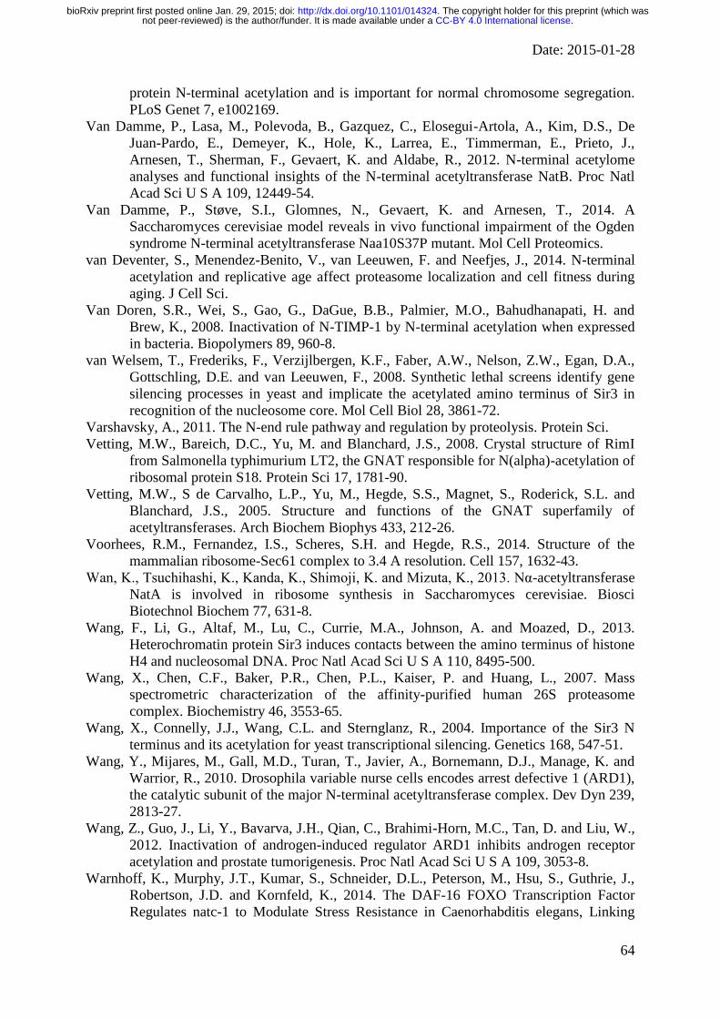
Date: 2015-01-28
64
protein N-terminal acetylation and is important for normal chromosome segregation.
PLoS Genet 7, e1002169.
Van Damme, P., Lasa, M., Polevoda, B., Gazquez, C., Elosegui-Artola, A., Kim, D.S., De
Juan-Pardo, E., Demeyer, K., Hole, K., Larrea, E., Timmerman, E., Prieto, J.,
Arnesen, T., Sherman, F., Gevaert, K. and Aldabe, R., 2012. N-terminal acetylome
analyses and functional insights of the N-terminal acetyltransferase NatB. Proc Natl
Acad Sci U S A 109, 12449-54.
Van Damme, P., Støve, S.I., Glomnes, N., Gevaert, K. and Arnesen, T., 2014. A
Saccharomyces cerevisiae model reveals in vivo functional impairment of the Ogden
syndrome N-terminal acetyltransferase Naa10S37P mutant. Mol Cell Proteomics.
van Deventer, S., Menendez-Benito, V., van Leeuwen, F. and Neefjes, J., 2014. N-terminal
acetylation and replicative age affect proteasome localization and cell fitness during
aging. J Cell Sci.
Van Doren, S.R., Wei, S., Gao, G., DaGue, B.B., Palmier, M.O., Bahudhanapati, H. and
Brew, K., 2008. Inactivation of N-TIMP-1 by N-terminal acetylation when expressed
in bacteria. Biopolymers 89, 960-8.
van Welsem, T., Frederiks, F., Verzijlbergen, K.F., Faber, A.W., Nelson, Z.W., Egan, D.A.,
Gottschling, D.E. and van Leeuwen, F., 2008. Synthetic lethal screens identify gene
silencing processes in yeast and implicate the acetylated amino terminus of Sir3 in
recognition of the nucleosome core. Mol Cell Biol 28, 3861-72.
Varshavsky, A., 2011. The N-end rule pathway and regulation by proteolysis. Protein Sci.
Vetting, M.W., Bareich, D.C., Yu, M. and Blanchard, J.S., 2008. Crystal structure of RimI
from Salmonella typhimurium LT2, the GNAT responsible for N(alpha)-acetylation of
ribosomal protein S18. Protein Sci 17, 1781-90.
Vetting, M.W., S de Carvalho, L.P., Yu, M., Hegde, S.S., Magnet, S., Roderick, S.L. and
Blanchard, J.S., 2005. Structure and functions of the GNAT superfamily of
acetyltransferases. Arch Biochem Biophys 433, 212-26.
Voorhees, R.M., Fernandez, I.S., Scheres, S.H. and Hegde, R.S., 2014. Structure of the
mammalian ribosome-Sec61 complex to 3.4 A resolution. Cell 157, 1632-43.
Wan, K., Tsuchihashi, K., Kanda, K., Shimoji, K. and Mizuta, K., 2013. Nα-acetyltransferase
NatA is involved in ribosome synthesis in Saccharomyces cerevisiae. Biosci
Biotechnol Biochem 77, 631-8.
Wang, F., Li, G., Altaf, M., Lu, C., Currie, M.A., Johnson, A. and Moazed, D., 2013.
Heterochromatin protein Sir3 induces contacts between the amino terminus of histone
H4 and nucleosomal DNA. Proc Natl Acad Sci U S A 110, 8495-500.
Wang, X., Chen, C.F., Baker, P.R., Chen, P.L., Kaiser, P. and Huang, L., 2007. Mass
spectrometric characterization of the affinity-purified human 26S proteasome
complex. Biochemistry 46, 3553-65.
Wang, X., Connelly, J.J., Wang, C.L. and Sternglanz, R., 2004. Importance of the Sir3 N
terminus and its acetylation for yeast transcriptional silencing. Genetics 168, 547-51.
Wang, Y., Mijares, M., Gall, M.D., Turan, T., Javier, A., Bornemann, D.J., Manage, K. and
Warrior, R., 2010. Drosophila variable nurse cells encodes arrest defective 1 (ARD1),
the catalytic subunit of the major N-terminal acetyltransferase complex. Dev Dyn 239,
2813-27.
Wang, Z., Guo, J., Li, Y., Bavarva, J.H., Qian, C., Brahimi-Horn, M.C., Tan, D. and Liu, W.,
2012. Inactivation of androgen-induced regulator ARD1 inhibits androgen receptor
acetylation and prostate tumorigenesis. Proc Natl Acad Sci U S A 109, 3053-8.
Warnhoff, K., Murphy, J.T., Kumar, S., Schneider, D.L., Peterson, M., Hsu, S., Guthrie, J.,
Robertson, J.D. and Kornfeld, K., 2014. The DAF-16 FOXO Transcription Factor
Regulates natc-1 to Modulate Stress Resistance in Caenorhabditis elegans, Linking
.CC-BY 4.0 International licensenot peer-reviewed) is the author/funder. It is made available under aThe copyright holder for this preprint (which was. http://dx.doi.org/10.1101/014324doi: bioRxiv preprint first posted online Jan. 29, 2015;

Date: 2015-01-28
65
Insulin/IGF-1 Signaling to Protein N-Terminal Acetylation. PLoS Genet 10,
e1004703.
Wegrzyn, R.D., Hofmann, D., Merz, F., Nikolay, R., Rauch, T., Graf, C. and Deuerling, E.,
2006. A conserved motif is prerequisite for the interaction of NAC with ribosomal
protein L23 and nascent chains. J Biol Chem 281, 2847-57.
White-Ziegler, C.A., Black, A.M., Eliades, S.H., Young, S. and Porter, K., 2002. The N-
acetyltransferase RimJ responds to environmental stimuli to repress pap fimbrial
transcription in Escherichia coli. J Bacteriol 184, 4334-42.
Whiteway, M., Freedman, R., Van Arsdell, S., Szostak, J.W. and Thorner, J., 1987. The yeast
ARD1 gene product is required for repression of cryptic mating-type information at
the HML locus. Mol Cell Biol 7, 3713-22.
Whiteway, M. and Szostak, J.W., 1985. The ARD1 gene of yeast functions in the switch
between the mitotic cell cycle and alternative developmental pathways. Cell 43, 483-
92.
Williams, B.C., Garrett-Engele, C.M., Li, Z., Williams, E.V., Rosenman, E.D. and Goldberg,
M.L., 2003. Two putative acetyltransferases, san and deco, are required for
establishing sister chromatid cohesion in Drosophila. Curr Biol 13, 2025-36.
Willis, D.M., Loewy, A.P., Charlton-Kachigian, N., Shao, J.S., Ornitz, D.M. and Towler,
D.A., 2002. Regulation of osteocalcin gene expression by a novel Ku antigen
transcription factor complex. J Biol Chem 277, 37280-91.
Wilson, T.E., 2002. A genomics-based screen for yeast mutants with an altered
recombination/end-joining repair ratio. Genetics 162, 677-88.
Wu, J., Chang, S., Gong, X., Liu, D. and Ma, Q., 2006. Identification of N-terminal
acetylation of recombinant human prothymosin alpha in Escherichia coli. Biochim
Biophys Acta 1760, 1241-7.
Xiao, K., McClatchy, D.B., Shukla, A.K., Zhao, Y., Chen, M., Shenoy, S.K., Yates, J.R. and
Lefkowitz, R.J., 2007. Functional specialization of beta-arrestin interactions revealed
by proteomic analysis. Proc Natl Acad Sci U S A 104, 12011-6.
Xiao, Q., Zhang, F., Nacev, B.A., Liu, J.O. and Pei, D., 2010. Protein N-terminal processing:
substrate specificity of Escherichia coli and human methionine aminopeptidases.
Biochemistry 49, 5588-99.
Xu, H., Jiang, B., Meng, L., Ren, T., Zeng, Y., Wu, J., Qu, L. and Shou, C., 2012. N-α-
acetyltransferase 10 protein inhibits apoptosis through RelA/p65-regulated MCL1
expression. Carcinogenesis 33, 1193-202.
Yamada, R. and Bradshaw, R.A., 1991. Rat liver polysome N alpha-acetyltransferase:
isolation and characterization. Biochemistry 30, 1010-6.
Yi, C.H., Pan, H., Seebacher, J., Jang, I.H., Hyberts, S.G., Heffron, G.J., Vander Heiden,
M.G., Yang, R., Li, F., Locasale, J.W., Sharfi, H., Zhai, B., Rodriguez-Mias, R.,
Luithardt, H., Cantley, L.C., Daley, G.Q., Asara, J.M., Gygi, S.P., Wagner, G., Liu,
C.F. and Yuan, J., 2011. Metabolic regulation of protein N-alpha-acetylation by Bcl-
xL promotes cell survival. Cell 146, 607-20.
Yi, C.H., Sogah, D.K., Boyce, M., Degterev, A., Christofferson, D.E. and Yuan, J., 2007. A
genome-wide RNAi screen reveals multiple regulators of caspase activation. J Cell
Biol 179, 619-26.
Yoo, Y.G., Kong, G. and Lee, M.O., 2006. Metastasis-associated protein 1 enhances stability
of hypoxia-inducible factor-1alpha protein by recruiting histone deacetylase 1. EMBO
J 25, 1231-41.
Yoon, H., Kim, H.L., Chun, Y.S., Shin, D.H., Lee, K.H., Shin, C.S., Lee, D.Y., Kim, H.H.,
Lee, Z.H., Ryoo, H.M., Lee, M.N., Taeg Oh, G. and Park, J.W., 2014. NAA10
controls osteoblast differentiation and bone formation as a feedback regulator of
Runx2. Nat Commun 5, 5176.
.CC-BY 4.0 International licensenot peer-reviewed) is the author/funder. It is made available under aThe copyright holder for this preprint (which was. http://dx.doi.org/10.1101/014324doi: bioRxiv preprint first posted online Jan. 29, 2015;

Date: 2015-01-28
66
Yoshikawa, A., Isono, S., Sheback, A. and Isono, K., 1987. Cloning and nucleotide
sequencing of the genes rimI and rimJ which encode enzymes acetylating ribosomal
proteins S18 and S5 of Escherichia coli K12. Mol Gen Genet 209, 481-8.
Zabrocki, P., Bastiaens, I., Delay, C., Bammens, T., Ghillebert, R., Pellens, K., De Virgilio,
C., Van Leuven, F. and Winderickx, J., 2008. Phosphorylation, lipid raft interaction
and traffic of alpha-synuclein in a yeast model for Parkinson. Biochim Biophys Acta
1783, 1767-80.
Zattas, D., Adle, D.J., Rubenstein, E.M. and Hochstrasser, M., 2013. N-terminal acetylation
of the yeast Derlin Der1 is essential for Hrd1 ubiquitin-ligase activity toward luminal
ER substrates. Mol Biol Cell 24, 890-900.
Zeng, Y., Min, L., Han, Y., Meng, L., Liu, C., Xie, Y., Dong, B., Wang, L., Jiang, B., Xu, H.,
Zhuang, Q., Zhao, C., Qu, L. and Shou, C., 2014. Inhibition of STAT5a by Naa10p
contributes to decreased breast cancer metastasis. Carcinogenesis 35, 2244-53.
Zhang, Y., Berndt, U., Gölz, H., Tais, A., Oellerer, S., Wölfle, T., Fitzke, E. and Rospert, S.,
2012. NAC functions as a modulator of SRP during the early steps of protein targeting
to the endoplasmic reticulum. Mol Biol Cell 23, 3027-40.
Zhong, H., De Marzo, A.M., Laughner, E., Lim, M., Hilton, D.A., Zagzag, D., Buechler, P.,
Isaacs, W.B., Semenza, G.L. and Simons, J.W., 1999. Overexpression of hypoxia-
inducible factor 1alpha in common human cancers and their metastases. Cancer Res
59, 5830-5.
Zhu, H.Y., Li, C.M., Wang, L.F., Bai, H., Li, Y.P., Yu, W.X., Xia, D.A. and Liu, C.C., 2014.
In Silico Identification and Characterization of N-Terminal Acetyltransferase Genes of
Poplar (Populus trichocarpa). Int J Mol Sci 15, 1852-64.
Zybailov, B., Rutschow, H., Friso, G., Rudella, A., Emanuelsson, O., Sun, Q. and van Wijk,
K.J., 2008. Sorting signals, N-terminal modifications and abundance of the chloroplast
proteome. PLoS One 3, e1994.
.CC-BY 4.0 International licensenot peer-reviewed) is the author/funder. It is made available under aThe copyright holder for this preprint (which was. http://dx.doi.org/10.1101/014324doi: bioRxiv preprint first posted online Jan. 29, 2015;