the biology and functional morphology of macoma biota (bivalvia
TRANSCRIPT
ZOOLOGIA 28 (3): 321–333, June, 2011doi: 10.1590/S1984-46702011000300006
© 2011 Sociedade Brasileira de Zoologia | www.sbzoologia.org.br | All rights reserved.
Tellinoidea is one of the largest groups of Bivalvia, andspecies of this family are conspicuous by their high diversityin tropical and subtropical waters. Tellinoidea are ecologicallyimportant in many places, and they frequently constitute foodand economic resources for many human coastal populations.All examined species of this family also possess a cruciformmuscle near the base of the inhalant siphon, and most spe-cies also have a long, unfused siphon (YONGE 1949). Since thestudy of YONGE (1949), knowledge of the mode of life oftellinoideans has increased, revealing that this family is com-posed of both deposit and suspension feeding species (e.g.,BRAFIELD & NEWELL 1961, POHLO 1969, 1982, NARCHI 1972, 1978,GILBERT 1977, DOMANESCHI 1995, ARRUDA et al. 2003, PASSOS &DOMANESCHI 2004). The deposit feeders have long and mobileinhalant siphons, which are characteristic of many Tellinidae,whereas the suspension feeders are more phenotypically di-verse and culminate in the short and passive siphons presentin the Donacidae.
Both deposit and suspension feeding individuals can befound in Tellinidae, even within the same species (BRAFIELD &NEWELL 1961, GILBERT 1977). This is the largest tellinoidean groupand is composed of two subfamilies, Tellininae and Macominae,distinguished by the presence of lateral hinge teeth in Tellininaeand their absence in Macominae. In some places, the diversityof tellinids (and tellinoideans in general) is probably underes-
timated because many species have very similar shells that canonly be distinguished by accurate morphological examinations,including examination of the soft parts. The Brazilian coast isa location that provides an example of this issue. As a result ofa substantial effort by the “Marine benthic biodiversity of SãoPaulo State” program (BIOTA/FAPESP) to describe the marinediversity in the state of São Paulo, 30 tellinoideans were re-corded, including one new species, Macoma biota by Arruda &Domaneschi, 2005. This species is a large bivalve that can beeasily confused with the well-known Macoma constricta(Bruguière, 1792) (ARRUDA & DOMANESCHI 2005).
Because of the high diversity of Tellinidae, the biologyof relatively few species is known. Among the 43 reported Bra-zilian species (RIOS 1994, 2009), only three were studied andexamined for anatomical details. The Macominae have receivedthe most attention, with the examination of Temnoconchabrasiliana Dall, 1921 (BOSS & KENK 1964), M. constricta (NARCHI
2003) and M. biota (ARRUDA & DOMANESCHI 2005). NARCHI (2003)studied the relationship between the siphonal organ and thelabial palps of the species, and recently, ARRUDA & DOMANESCHI
(2005) investigated the gross morphology of the soft parts ofthis species in a comparative study with M. biota. The shells ofspecies of Tellina Linnaeus, 1758, from the western coast of theAtlantic were compared (BOSS 1966, TENÓRIO 1984), but no ana-tomical data were furnished.
The biology and functional morphology of Macoma biota(Bivalvia: Tellinidae: Macominae)
Pedro Ribeiro Piffer1; Eliane Pintor de Arruda2 & Flávio Dias Passos3
1 Programa de Pós-Graduação em Ecologia, Departamento de Biologia Animal, Instituto de Biologia, Universidade Estadualde Campinas. 13083-970 Campinas, SP, Brazil. Corresponding author. Email: [email protected] Universidade Federal de São Carlos, Campus Sorocaba. Rodovia João Leme dos Santos, km 110, Itinga,18052-780 Sorocaba, SP, Brazil.3 Departamento de Biologia Animal, Instituto de Biologia, Universidade Estadual de Campinas. Caixa Postal 6109,13083-970 Campinas, SP, Brasil.
ABSTRACT. Macoma biota Arruda & Domaneschi, 2005, is a recently described species known only from the intertidal
zone of Praia da Cidade, Caraguatatuba Bay, in the state of São Paulo, southeastern Brazil. The main purpose of the
present paper is to describe the biology of M. biota, beginning with a detailed analysis of its anatomy and functional
morphology and how these attributes are correlated with its habitat and life history. The morphology of the organs in
the pallial cavity and their sorting devices indicate that this species has efficient mechanisms to process large amounts
of particles that enter this cavity via the inhalant current. M. biota can rapidly select the material suitable for ingestion
and direct the undesired excess to the rejection mantle tracts. These characteristics along with the siphon’s behavior
and the digestive tract configuration reveal that this species can be classified primarily as a deposit feeder, like other
species of the genus; however, it can also behave as a suspension feeder, depending on the environmental conditions.
KEY WORDS. Anatomy; behavior; deposit-feeder; mode of life; Mollusca.
322 P. R. Piffer et al.
ZOOLOGIA 28 (3): 321–333, June, 2011
Macominae, which is probably a polyphyletic branch,originated in the Eocene and includes nine recent genera (COAN
et al. 2000). In addition to T. brasiliana and Cymatoica orientalis(Dall, 1890), ten species of Macoma have been recorded fromBrazilian shores (TENÓRIO et al. 1986, RIOS 1994, 2009, ARRUDA &DOMANESCHI 2005). Data on the life histories of these species arevery scarce once M. constricta is the only species that has beenobserved alive (ARRUDA et al. 2003). A few other Macominaespecies have already been investigated, such as Macoma balthica(Linnaeus, 1758), which was analyzed by BRAFIELD & NEWELL
(1961) and GILBERT (1977), and eight other species, which wereinvestigated by REID & REID (1969). In the latter study, depositfeeding was recorded among some species, while others werecharacterized as suspension feeders. Another species ofMacomona, Blainville, 1814, from Thailand was analyzed bySIMONE & WILKISON (2008).
In the present study, a detailed anatomical investigationof M. biota was undertaken to contribute to the knowledge ofM. biota biology. These data will help to further distinguishthis species from other Macominae species, providing basicinformation for future studies on the taxonomy and phylog-eny of this diverse group. Special attention has been given tothe structure and functioning of the pallial organs, as they aredirectly involved in food capture and therefore define the rolethat this species plays in the community structure of Brazilianmuddy beaches.
MATERIAL AND METHODS
During low tide, living specimens of M. biota were col-lected manually using a common shovel in the intertidal zoneof Praia da Cidade, which is a sheltered beach near downtownCaraguatatuba in the state of São Paulo, Brazil (23º37’31.08”S,45º23’57.38”W). This is the type locality of this species and theonly area in which it is presently known. The physical character-istics of the beach were described by ARRUDA & DOMANESCHI (2005).The sediment is composed mainly of fine and very fine sand andretains a large amount of silt-clay (mean sediment size: 2.82Ø).The sediment includes 6.64% organic matter and 8.88% calciumcarbonate, and the interstitial seawater salinity is 34% S.
The individuals of M. biota were found in the muddy-sand substratum near beds of the mytilid Mytella charruana(d’Orbigny, 1842). In the laboratory, living animals were keptin aquaria containing seawater and clean sand at 21ºC withconstant aeration. The specimens were fed powdered fish foodthat was poured in the surrounding seawater three times a week.Under these conditions, specimens survived for 60 consecu-tive days. The burrowing and feeding behavior and the posi-tion adopted by M. biota within the sediment were observedusing small aquaria containing sand immersed in seawater.Anatomical and behavioral studies were conducted with healthyspecimens during the first few weeks of confinement in aquaria.After the first month, the specimens began to weaken in re-
sponse to the lack of a proper food supply. This affected theirbehavior and could have interfered with the experiments; there-fore, when specimens began to show signs of weakness, theywere anesthetized and fixed for further use.
For the morphological study of the organs of the mantlecavity and digestive tract, specimens were anesthetized using a2.5% solution of magnesium chloride or menthol crystals, fixedwith a 4% formaldehyde solution and preserved in 70% alco-hol. Living specimens were dissected and observed under a dis-secting microscope, and ciliary currents and sorting mechanismswere elucidated using powdered carmine and fine organic andmineral particles. The organs and structures were compared inboth preserved and living conditions and drawn using a cameralucida attached to a Zeiss STEMI SV-6 stereomicroscope.
For histological examination, specimens fixed withBouin’s fixative had their shells removed and were then dehy-drated in a crescent alcoholic series (70, 80, 95, and 100%),embedded in paraffin and serially microtomized in 7-10 ¼mthick sections. The sections were stained with hematoxylin andcounterstained with either eosin or Mallory’s triple stain.
Voucher specimens of this study were deposited in themolluscan collection of the “Museu de Zoologia da UnicampProf. Dr. Adão José Cardoso” (lot ZUEC-BIV 2192).
RESULTS
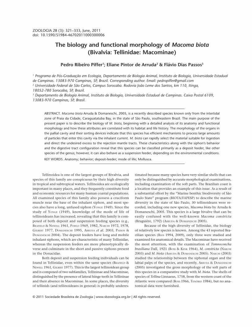
Macoma biota lives burrowed deep in the sediment, ap-proximately 20 to 40 cm below the surface (Fig. 1). When re-moved from the sediment and left lying on the right or left valve,the animal extends its foot antero-ventrally and bends it towardthe substratum in an attempt to anchor itself. Once the foot isanchored in the substratum, the anterior and posterior pedalretractor muscles contract, lifting the shell upward to a verticalposition. From this position, the animal begins to burrow, as-sisted by an abrupt shutting of the valves, which expels the wa-ter contained in the pallial cavity and displaces the sedimentthus opening space for the shell to penetrate. During this pro-cess, the siphons remain partially protracted and are not affectedby the harsh shutting of the valves because of a small openingin the posterior region of the shell. Specimens measuring 4 cmlong buried themselves in approximately 15 sec.
Once fully buried, M. biota assumes a horizontal position,lying on the left shell valve (Fig. 1); at this point, the siphons areprotracted beyond the surface of the sediment and re-initiatethe intake of food and water. The fact that this species lies onthe left valve is corroborated by a small flexure to the right inthe posterior region of the shell, exactly where the animal pro-tracts its siphons. In laboratory conditions, certain specimensemerged from the substrate and remained lying on the surface.This tended to occur after a few weeks of confinement in theaquarium, probably resulting from the fact that the animalsbegan to become unhealthy when submitted to conditions thatdiffered from those of their natural environment.

323The biology and functional morphology of Macoma biota
ZOOLOGIA 28 (3): 321–333, June, 2011
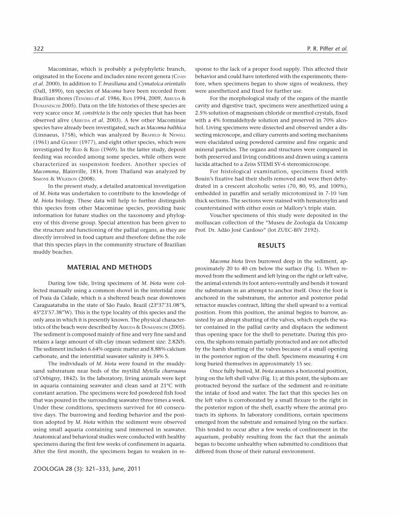
The adductor muscles were unequal in cross section; theanterior muscle was dorso-ventrally elongated, and the poste-rior muscle was ovate. In addition to the two typical adductormuscles, M. biota possesses an accessory adductor muscle unit-ing the valves in the ventral-posterior region and serving thesame function as the adductors (Fig. 2). This muscle originatesfrom the hypertrophy of the pallial musculature and is locatedbetween the anterior and posterior insertions of the cruciformmuscle. It leaves an ovate scar in both valves that is unitedwith the pallial line scar at its posterior extremity. At the baseof this accessory adductor muscle is the cruciform muscle,which is composed of two muscular bundles, each with its an-terior extremity fixed in one valve and its posterior extremityfixed in the opposite valve (Fig. 2). Halfway between the fixa-tion points in the valves, the fibers of each bundle intercalate,crossing each other and then reconstituting the original bundle.The posterior extremity of each bundle penetrates between themantle lobe tissues and bifurcates into two sub-equal bundlesimmediately before being affixed to the valve, which leavestwo unequal scars. Anteriorly to the bifurcation, each muscu-
lar bundle is separated completely by a gap, which communi-cates with a sensorial organ that is typical of Tellinoidea.
The mantle lobes are thin and translucent, except at theirthickened, muscular, three-folded ventral margins. The mantlelobes are completely unattached antero-ventrally, forming anextensive pedal opening. At the posterior end, the opposinginner folds fuse and are involved in siphon formation. At thebase of the inhalant siphon, the pallial musculature hypertro-phies, originating the accessory adductor muscle and the cru-ciform muscle. The inner mantle folds are short, translucent
Figure 1. Macoma biota: specimen fully buried in its natural habi-tat, lying on the left shell valve. The arrows indicate the directionsof the inhalant and exhalant currents.
13 cm
30 cm
4 cm
Figure 2. Macoma biota: ventral view representing 3/4 of a speci-men with the valves half-open showing the position of the siphonalorgans and the accessory adductor and cruciform muscles. (acm)Accessory adductor muscle, (cm) cruciform muscle, (ex) exhalantsiphon, (f) foot, (in) inhalant siphon, (isa) inhalant siphon proximalaperture, (lamf) left additional mantle fold, (ld) left inner demibranch,(lso) left siphonal organ, (ramf) right additional mantle fold, (rd)right inner demibranch, (rso) right siphonal organ. Scale bar: 5 mm.
in
ramf
rso
isa
ex
cm
acm
lso
lamf
ldf
rd

324 P. R. Piffer et al.
ZOOLOGIA 28 (3): 321–333, June, 2011
and barely visible, even in live specimens dissected under amicroscope. The middle sensorial fold bears a single row ofregularly spaced, short and digitiform tentacles. The outer foldis less developed than the others, and it is very difficult to visu-alize.
Both mantle lobes present a long and smooth additionalfold located dorsally to the inner fold, isolating a waste chan-nel for pseudofeces (Fig. 2). Both additional mantle folds ex-tend posteriorly from the mid-point of the pedal opening upto the vicinity of the cruciform muscle. At this point, the leftadditional fold ends, and the right fold bends abruptly dor-sally and forward, extending anteriorly and increasing in height,with the free edge becoming lobulated and often intensivelyramified.
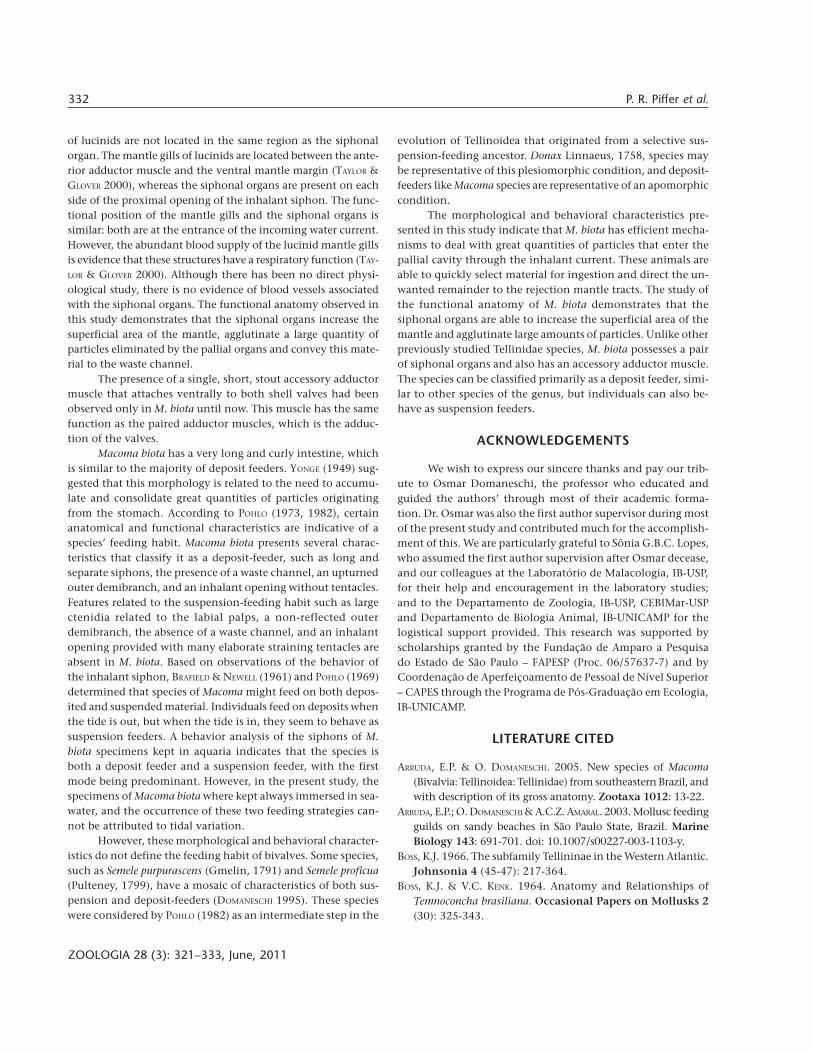
The material that comes in contact with the internalmantle epithelium is quickly conduced ventrally and posteri-orly by two very active ciliary currents. The first current col-lects the material that precipitates close to the anterior adductormuscle and transfers it first to the dorsal surface of the addi-tional mantle fold and then to the base of the inhalant siphon.The second current is dorso-ventrally directed throughout theumbo line; the material precipitated in the anterior and poste-rior regions of the mantle epithelium converge at this current.The material dragged dorso-ventrally to the siphonal organs isequally concentrated in the proximity of the inhalant siphonbase (Fig. 3).
All material removed from the surface of the left mantlelobe is concentrated in the posterior extremity of the left addi-tional mantle fold. In the opposite lobe, the material is con-centrated in the interior of the hollow formed by the flexionof the right additional mantle fold (Fig. 3).
A pair of asymmetric siphonal organs is located close tothe proximal aperture of the inhalant siphon in both mantlelobes (Fig. 2). The left siphonal organ is more developed thanthe right, but both are slightly cushion-like at the base andbordered by a thin, plicate sheath at the free apical edge. Theleft and right siphonal organs of M. biota present similar andvery active ciliary currents in both surfaces. On the dorsal sur-face, the cilia drag the material mostly to the anterior marginof the siphonal organ, transferring it to the ventral surface. Onthe ventral surface, the ciliary currents transfer the particles tothe base of the siphonal organ and then posteriorly to a regionnear the proximal aperture of the inhalant siphon (Fig. 4).
All the material that originated from the rejection cur-rents of the ctenidia, labial palps, visceral mass and mantleepithelium is concentrated close to the proximal aperture ofthe inhalant siphon and is eliminated as pseudofeces throughthe inhalant siphon by an abrupt adduction of the valves.
The siphons are formed exclusively by the fusion andhypertrophy of the inner folds of the mantle. These siphonsare present in the posterior region of the bivalve and are classi-fied as type A based on the definitions of YONGE (1948, 1982).The siphons are long, cylindrical, separated and very active,
moving freely and diminishing gradually in diameter in thedirection of the tip. The inhalant siphon is longer than theexhalant and, when fully extended, reaches four to five timesthe shell length. The inhalant aperture is fringed with six simplelobular projections, forming small tentacles that are visible onlywhen it is fully distended. The tentacles are inconspicuous andbarely detectable when the animal is fixed or when it contractsthe siphons (Fig. 5). The exhalant aperture is more elaborate asa result of the occurrence of six small digitiform tentacles thatare intercalated by digitiform papillae (Fig. 5). The siphons arewhitish, smooth and thin organs with a segmented appear-ance caused by the presence of circular and regularly spacedconstrictions. The siphon walls present six longitudinal whitelines that are regularly spaced and visible due to transparency.These lines correspond to the nerves that end in the interior ofthe six projections in both apertures. The siphon walls andapertures are sensitive to mechanical stimulation and harshmovements of the water in their proximity, and they respondwith local contractions and/or full retraction to the interior ofthe sediment or shell.
The siphons can be fully retracted to an isolated spaceexternal to the pallial cavity, called the siphonal space. Whenfully retracted, the proximal portions of the siphons remainnear the cruciform muscle line. This is in contrast to othertellinids in which the siphons retract further into the anteriorregion and occupy a considerable part of the pallial cavity. Theretraction process is facilitated by the fan-shaped siphon re-tractor musculature, which is well developed in the Tellinoidea.
In the absence of perturbations, M. biota can protract itssiphons slowly, exposing the apertures above the sediment sur-face and remaining like this for long periods of time. The pro-traction of the siphons is relatively slow and continuous and isa result of the combined actions of the intrinsic musculatureand the blood.
The specimens maintained in the aquaria behaved as ei-ther deposit feeders or suspension feeders. When they fed onmaterial in suspension, the inhalant siphon remained outsideof the substratum, either passively lying on the surface or ex-tended into the water column. When they fed on material de-posited in the substratum, the specimens protracted theinhalant siphon well above the surface and bent it in the shapeof a hook, putting the aperture in contact with the surface andsucking the material precipitated in the sediment. As a result,large quantities of sediment entered the mantle cavity.
When the exhalant siphon was protracted above the sur-face of the substratum, it remained passive most of the time.Sporadically, the exhalant siphon performed circular move-ments to launch feces far from the inhalant aperture. In manyinstances, the exhalant siphon remained retracted in the inte-rior of the substratum with its aperture located below the sur-face of the sediment.
The foot is a huge, axe-shaped and very active muscularorgan that expands far beyond the antero-ventral margin of

325The biology and functional morphology of Macoma biota
ZOOLOGIA 28 (3): 321–333, June, 2011
the shell. The proximal portion of the foot remains protectedinside the shell and holds the visceral mass.
The intrinsic pedal musculature is composed of two typesof muscular bundles: the first is arranged transversely, unitingthe epithelium of both sides of the foot, and the second is ar-
ranged obliquely in the lateral walls of the foot. The extrinsicpedal musculature is composed of three pairs of bundles thatare attached to the shell valves and extend to different regionsof the foot. These include one pair of protractor muscles andtwo pairs of retractor muscles (anterior and posterior) (Fig. 6).
isaisa
Inhalant Exhalant
3
4 5
Figures 3-5. Macoma biota. (3) Ciliary currents on the inner surface of the right mantle lobe. Observe the accumulation of pseudofecesin the space formed by the additional mantle fold. (4) Ciliary currents on the surface of the left siphonal organ; (A) ventral surface, (B):dorsal surface. (5) distal extremity of the siphons, showing the inhalant aperture with six lobular small tentacles and the exhalantaperture with six small digitiform tentacles, intercalated by digitiform papillae. (dp) Digitiform papillae, (dt) digitiform tentacle, (isa)Inhalant siphon proximal aperture, (st) small tentacle. Scale bars: 3 = 1.5 cm, 4 = 5.0 mm, 5 = 2.0 mm.
st dt dp
A B

326 P. R. Piffer et al.
ZOOLOGIA 28 (3): 321–333, June, 2011
The insertion of the anterior pedal retractor muscles (arm)is elongated and lies dorsally to the anterior adductor muscle.These muscles form the innermost muscular layers of the footand extend nearly ventrally. The muscular bundles of the an-terior retractor muscles intercross in the middle portion of thesagittal plane of the bivalve. The right bundles cross with thoseoriginating from the left and vice versa, forming a thicker re-gion that then separates again and extends ventrally.
The insertion of the posterior pedal retractor muscle (prm)is triangular and lies dorsally to the posterior adductor muscle.These muscles form the middle muscular layer of the foot be-tween the anterior retractor and protractor muscles. The rightand left posterior pedal retractor muscles proceed anteriorly,converging in the sagittal plane and fusing just beneath thekidney, thus forming a thick median bundle. At this point, thebundles of fibers intersect and split; the fibers from the rightside pass deeply into the left side of the foot and vice versa,irradiating to the whole lateral area of the foot.
The pedal protractor muscles (ppm) on each side of thefoot penetrate the anterior adductor muscle transversely, at-taching to their respective valves with the anterior adductormuscle fibers. Splitting of the muscular bundles occurs a shortdistance before the pedal protractors penetrate the anterioradductor to form a thicker ventral portion that concentratesmost of the muscular fibers and a thinner dorsal portion that
has few muscular fibers. From their insertion on the shell valve,the pedal protractors follow posteriorly and twist abruptly asthey enter the foot and then spread in a fan pattern toward allproximal regions of the foot that hold the visceral mass. Thesemuscles form the outermost muscular layer of the foot, andthey are external to the posterior retractor.
The foot epithelium presents ciliary activity only on theportion that holds the visceral mass. This activity occurs onthe area in contact with the inner demibranchs and labial palps.This ciliary activity is relatively weak, and the material thatenters into contact with the epithelium of the visceral mass inthis region can be captured by the cilia on the smooth face ofthe internal labial palp or the demibranchs. When this doesnot occur, the material is carried ventrally and posteriorly andaccumulated in the posterior region of the visceral mass, whereit is covered by the inner demibranch (Fig. 7). The material isthen agglutinated by mucus, which causes precipitation to themantle. At the mantle, the material is transferred to the proxi-mal aperture of the inhalant siphon and is eliminated aspseudofeces.
The general configuration of the organs and structuresof the pallial complex can be viewed in figure 8. The ctenidiaare complete, eulamellibranch and totally smooth (homorha-bidic). The interfilamentar and interlamellar junctions occurregularly in the ctenidia. The inner demibranch is complete,
Figure 6. Macoma biota: the arrangement of the musculature. (aam) anterior adductor muscle, (acm) accessory adductor muscle, (arm)anterior pedal retractor muscle, (cm) cruciform muscle, (f) foot, (pam) posterior adductor muscle, (ppm) pedal protractor muscle,(prm) posterior pedal retractor muscle. Scale bar: 1.5 cm.
f
aam
arm
ppm
acm
cm
pam
prm

327The biology and functional morphology of Macoma biota
ZOOLOGIA 28 (3): 321–333, June, 2011
Figure 7. Macoma biota: ciliary currents on the proximal region of the foot’s epithelium that holds the visceral mass. The left ctenidiumand labial palps were removed and the dashed lines indicates their original position in the live animal. Scale bar: 1.5 cm.
Figure 8. Macoma biota: organs of the pallial cavity viewed from the left side after the removal of the left shell valve and mantle lobe.(aam) anterior adductor muscle, (acm) accessory adductor muscle, (cm) cruciform muscle, (ex) exhalant siphon, (f) foot, (id) innerdemibranch, (ilp) inner labial palp, (in) inhalant siphon, (lso) left siphonal organ, (mm) mantle margin, (od) outer demibranch, (olp)outer labial palp, (pam) posterior adductor muscle, (ramf) right additional mantle fold, (rso) right siphonal organ. Scale bar: 1.5 cm.
pam
ex
od
idolp
aam
in
lso
rso
cmacmramf
f
ilp
mm

328 P. R. Piffer et al.
ZOOLOGIA 28 (3): 321–333, June, 2011
with both ascending and descending lamellae. The outerdemibranch is reduced to the single descending upturned lamel-lae, leaving the inner demibranch uncovered. Both demi-branchs have the same length and height (Fig. 9). The innerdemibranch has a shallow marginal food groove along its freemargin. The ctenidia axis is located diagonally in relation tothe antero-posterior axis of the bivalve body, and it extendsfrom the umbonal concavity to the base of the siphons.
The filaments of both demibranchs have frontal, latero-frontal and lateral cilia. The frontal face of the filaments pre-sents a longitudinal band of equally sized frontal cilia, whichare followed laterally by the latero-frontal cilia. The latero-fron-tal cilia are long and rigid, and as their extremities touch andintercalate with the cilia from the adjacent filament, they forma barrier to larger particles brought by the inhalant current,obstructing the entrance of these particles into the interior ofdemibranch. Bands of lateral cilia occur on the sides of thefilaments; their vibration promotes the water flow trough themantle cavity. The filaments are internally reinforced withchitin developed on the lateral walls. No pro-latero-frontal ciliawere detected in M. biota.
The ciliary currents on the ctenidia are represented infigure 9. The material introduced to the pallial cavity by theinhalant current that then reaches the outer demibranch isconduced to the ctenidia axis, where an oral current of accep-tance transfers the smaller particles in a dorso-anterior direc-tion to the distal oral groove, which is formed between theouter labial palp and the demibranchs. Excess and rejectedparticles are transferred to the descendent lamellae of the in-ner demibranch. In both lamellae of the inner demibranch,ciliary currents conduct material to the free margin, and thesmaller particles are agglutinated in mucus, transferred to themarginal food grove and conducted orally for digestion or fornew selection in the labial palps. Larger particles, agglomer-ates of particles and surplus materials that are in contact withthe inner demibranch are discharged from the free margin tothe mantle epithelium.
The labial palps are trigonal, large, wide and approxi-mately two-thirds of the shell length. The labial palps andctenidia have similar lengths, although the labial palps aresometimes slightly longer. The inner labial palps are locatedbetween the visceral mass and the ascending lamellae of theinner demibranch, and the outer labial palps are located be-tween the mantle and the descending lamellae of the samedemibranch. The association between the ctenidia and the la-bial palps of M. biota is classified as type III based on the defini-tions of STASEK (1963) (Fig. 10).
The external surfaces of the labial palps are smooth, andthe internal surfaces are highly and conspicuously folded. Thefolded face has two smooth borders: one is large and dorsal inrelation to the folds, and the other is thin and ventral, with afree margin that can be affected by muscular contractions form-ing simple or highly truncate lobular formations, assuming a
fringed aspect (Fig. 10). Histological sections showed that thelabial palp folds have a smooth oral face and an aboral facewith longitudinal thickenings that give them a wavy aspect(Fig. 11).
Particles that come into contact with the external, smoothface of the labial palps are removed transversely in a dorsaldirection and transferred to the broad, dorsal, smooth borderon the internal face (Fig. 10). On this border, longitudinal con-vergent currents concentrate the particles to a specific pointthat is set back from the distal extremities of the palps, and theparticles are then transferred to the folds for selection. Isolatedparticles from the distal oral groove, the frontal face of thefilaments of the anterior region of the inner demibranch, themarginal food groove, and the inner demibranch are alsobrought to the folded area. Particles agglutinated in a mucusstrand that reaches the palps through the distal portion of theoral groove and the marginal food groove go directly to themouth.
Different ciliary currents on the folded faces of the labialpalps (Figs 10 and 11) rigorously select the material to be in-gested. A very active current (a) carries isolated particles andagglomerated particles perpendicularly across the crests of thefolds, being transferred from fold to fold. This current operatesobliquely in an oral direction and markedly in a ventral direc-tion, and it can act either as an acceptance or a rejection cur-rent, depending upon the size or total volume of the particles.The fold inclination determines whether the material will betransferred to the proximal oral groove between the palps andthen directed to the mouth or transferred to a strong longitu-dinal rejection current (b) along the ventral, smooth border ofthe palps. The material on the rejection current is carried tothe distal free extremity of the palp and then discharged to themantle epithelium.
Other ciliary currents occur along the grooves formedbetween the folds and on the aboral face of these folds, andthese currents act as rejection or acceptance mechanisms, re-spectively. The longitudinal currents (d) on the aboral face ofthe folds impede the immediate discharge of material, whereasfine particles that precipitate on the floor of the grooves be-tween the folds are carried longitudinally (current “c”) to therejection tract “b” on the free ventral margin of the palp. Thelongitudinal current “d” along the aboral face of each fold actsto re-sort particles, capturing them from current “a” and keep-ing them on the palp for re-selection. The joint action betweencurrents “a” and “d” retains the particles for a longer time inthe folded face to allow for gradual selection of particles foringestion or rejection. This interaction is responsible for thezigzag trajectory that carries the material to the base of thepalps, preventing the particles to reach the rejection tract “b”on the smooth ventral border.
Large, isolated particles or particle agglomerates precipi-tated in large quantities on the folds are captured by current“a” and are conduced to rejection tract “b” on the free ventral

329The biology and functional morphology of Macoma biota
ZOOLOGIA 28 (3): 321–333, June, 2011
border of the palps. This activity is enhanced when muscularaction approaches the fold crests or curls or wraps the organ.By approaching the folds, the bivalve creates a barrier thatblocks the penetration of particles in the space between thefolds, thus making current “a” act as a rejection current. The
curling and wrapping of the labial palps also results in a barrierand facilitates the capture of material by rejection current “b”.
The labial palps can contract severely with consequentwrinkling but without wrapping. This contraction forms fourto five folds (“secondary folds”) transverse to the longitudinalaxis of the organ. These folds are temporary functional forma-tions that speed up the rejection process of large quantities ofmaterial by favoring current “a”, which carries the materialquickly to rejection tract “b”.
Beyond the mouth is the flattened and relatively shortesophagus, which extends posteriorly and opens into the stom-ach dorso-anteriorly (Fig. 12). The stomach is globular, withthin, fragile and semi-translucent walls. From the posterior re-gion of the stomach roof, slightly bending toward the rightside, projects the short and digitiform appendix, which is ho-mologous to the posterior caecum. Additionally, the dorsal hoodprojects from the stomach roof and bends posteriorly. Belowthe dorsal hood, there is a broad left pouch that opens into theanterior region of the stomach in line with the esophagus open-ing. The left pouch has two branches: a thinner branch thatextends anteriorly and a wider branch that extends posteri-
Figures 9-10. Macoma biota. (9) Diagrammatic vertical section through the ctenidium showing its functional organization: arrowsventralward and bullets oralward ciliary currents. (10) Relationship between the left labial palps and inner demibranch, and respectiveciliary currents. (b) Rejection current along the ventral smooth border, (ca) ctenidium axis, (dog) distal oral groove, (id) inner demibranch,(ilp) inner labial palp, (m) mouth, (mfg) marginal food groove, (od) outer demibranch, (olp) outer labial palp, (pog) proximal oralgroove. Scale bar: 5 mm.
9
od
10
id
dogolp
ilp
b
m
pogca
id
od
mfg
Figure 11. Macoma biota: diagram of the ciliary mechanisms onthe folded surface of the labial palp, to show the various ciliarytracts. For lettering (a), (c), (d), see text.
oral
a
aboral
d
c

330 P. R. Piffer et al.
ZOOLOGIA 28 (3): 321–333, June, 2011
orly. The left pouch receives some ducts from the digestive di-verticula in the distal extremity of the anterior branch and fromall along the free margin of the posterior branch.
The remainder of the ducts that comes from the densemass of digestive diverticula, which covers the entire front,left and right sides of the stomach and style-sac, opens intothe stomach via the right and left caeca. The right caecum isshort and highly reduced, and it is located on the right side ofthe stomach and extends anteriorly. The right caecum receivessome ducts from the digestive diverticula mostly on its distalextremity, but some branches also arrive close to the begin-ning of the caecum. The left caecum opens into the anteriorregion of the stomach, and it is cylindrical and far more devel-oped than the right caecum. The left caecum receives severalducts from the digestive diverticula and extends ventrally un-til approximately the middle portion of the style-sac. At thatpoint, the left caecum divides into two portions: one spreadsto the left side of the style-sac and the other to the right.
The style-sac and the intestine open together in thepostero-ventral region of the stomach floor, separated by theminor and major typhlosoles. The style-sac is conical and ex-tends postero-ventrally. The crystalline style has the shape of abaseball bat, is quite rigid and is always present in both live
and fixed specimens. The distal extremity of the crystallinestyle projects into the stomach, occupying a large amount ofinternal space in this organ.
The intestine continues from the distal region of the style-sac and is located in a very compact cavity that is well separatedfrom other organs. The intestinal loops and gonads form a verycompact mass. The diameter or caliber of the intestine is rela-tively constant during its entire length, and its walls are extremelyelastic and have great powers of distention. The intestine canacquire a diameter almost five times larger than its normal di-ameter, depending on the quantity of fecal pellets accumulatedin its interior. The intestine describes a large number of loops,divided into two sets. In the first set, the intestine describes threecircular loops in the horizontal plan, and after that, it assumes awinding aspect, describing three to four more irregular loops inthe same plan. In the first set, the loops are very compact andentangled. From the middle portion of its trajectory, the intes-tine describes a second set of loops, constituted by seven to eightcircular loops in a more vertical plan. After that, the intestinereaches the pericardium and goes through the ventricle. Aftercrossing the pericardium, the intestine passes over the dorsalsurface of the posterior adductor muscle and ends with the anus,which has two small papillae in its opening.
Figure 12. Macoma biota: left side view of a dissected specimen showing the alimentary canal. (ap) Anal papillae, (ax) appendix, (dh)dorsal hood, (eso) esophagus, (i) intestine, (lc) left caecum, (lp) lef pouch, (m) mouth, (pe) pericardium, (ss) style-sac, (sto) stomach.Scale bar: 1.5 cm.
i
pelpax
dh
sto
eso
m
lc ss ap

331The biology and functional morphology of Macoma biota
ZOOLOGIA 28 (3): 321–333, June, 2011
DISCUSSION
Macoma biota demonstrates a series of adaptations thatallow it to colonize a habitat with large amounts of suspendedsediment. The limited horizontal mobility of individuals thatremain burrowed deeply in the substratum for long periods inthe same place is related to the mode of life on calm beacheswith little surf, which is the only locale where the species hasbeen recorded. The inhalant siphons of M. biota do not possessany barriers that could block the entrance of large amounts ofsediment in the pallial cavity. However, when the particles reachthe pallial cavity, they are quickly selected by the intense cili-ary activity of the pallial organs.
According to YONGE (1949), the ctenidia shape of M. biota,with the complete inner demibranch and incomplete outerdemibranch, is an adaptation to deal with large amounts ofmaterial that enter the pallial cavity. The outer demibranch,which is reduced to a single descending, upturned lamellae,and the inner demibranch form a continuous surface that pro-pitiates a greater efficiency of transport and selection for thesediment that comes in contact with the ctenidia and also con-tributes to reduce the risk of damaging the organ. The strongcurrents observed on the marginal food groove and on thectenidia axis ensure the efficiency of particle transport in theoral direction. The labial palps, which are more developed thanthe ctenidia, play a major role in the selection of the particlesthat are directed to the mouth. The presence of labial palpsthat are more developed than the ctenidia is also observed inspecies that live in muddy habitats where the material thatenters the pallial cavity is very fine (YONGE 1949).
The large ciliary activity of the labial palps and the for-mation of a “cone of rejection” that occurs when these curl orwrap up also indicates that these animals deal with a large in-flow of deposited material that requires efficient selectionmechanisms to sort between the particles to be ingested andthe ones to be rejected. Moreover, the large ciliary activity ofthe labial palps of M. biota is related to the re-selection of ma-terial brought by the inhalant current.
The presence of an additional fold in each mantle lobe,forming a ventral waste channel that was named by KELLOGG
(1915), is characteristic of many deposit feeding tellinoids(YONGE 1949) and also some suspension feeding veneroids(NARCHI & DI DARIO 2002). The waste channel isolates unwantedsediment from the strong inhalant current and can drag thesematerials back to the pallial cavity organs. The ventral channelwas described for Abra alba (Wood, 1801) and Scrobicularia plana(da Costa, 1778) by YONGE (1949), T. brasiliana by BOSS & KENK
(1964) and M. constricta by NARCHI (2003). REID & REID (1969)also described the presence of a waste channel in Macoma brotaDall, 1916, M. calcarea (Gmelin, 1791), M. elimata Dunnil &Coan, 1968, M. incongrua (Martens, 1865), M. inquinata(Deshayes, 1855), M. secta (Conrad, 1837), M. lipara Dall, 1916,and M. nasuta (Conrad, 1837). In M. biota, the additional mantle
fold forms a waste channel slightly different from those de-scribed by KELLOGG (1915). In M. biota, the rejected material iscarried dorsally in relation to the folds, not ventrally, and itaccumulates close to the proximal aperture of the inhalant si-phon in the interior of the space formed by the flexion of theright additional mantle fold. At this location, it is also isolatedfrom the inhalant current as a result of the development of theright additional mantle fold.
Macoma biota possesses a pair of siphonal organs, de-scribed for the first time by BOSS & KENK (1964) in T. brasilianaand by NARCHI (2003) in M. constricta. In contrast with T.brasiliana and M. constricta, which have unique left siphonalorgans, M. biota has a paired structure, and the left organ ismore developed than the right. According to NARCHI (2003), M.constricta has a strongly wavy sheath in the distal extremity ofthe outer left labial palp that maintains an intimate functionalrelationship with the unilateral left siphonal organ. The par-ticles rejected by the left labial palp are first passed to the pli-cate wall of the free margin of the siphonal organ and laterpassed to the aperture of the inhalant siphon. With the inflowof water from the inhalant current, this material can be con-veyed to the ctenidia to be processed again. NARCHI (2003) con-cluded that the presence of ciliary currents conveying particlesto the aperture of the inhalant opening shows that the bivalvecan re-sort material rejected by the labial palps. The presenceof an always-moving plicate wall around the cushion-like baseof the unilateral left siphonal organ also suggests that this or-gan is sensory (NARCHI 2003).
In M. biota, this sheath is absent; however, the ciliary cur-rents on the paired siphonal organ convey particles from thelabial palps and mantle cavity to the aperture of the inhalantopening, similar to M. constricta. However, we did not observethe inflow of water from the inhalant current conveying par-ticles back to the ctenidia. The particles caught by the pairedsiphonal organs are conveyed to the posterior extremity of thewaste channel and eliminated as pseudofeces. The pairedsiphonal organs agglutinate large quantities of particles that areeliminated by labial palps and ctenidia on the mantle cavity.The organs probably handle with larger amounts of material thanthe unique left siphonal organ of M. constricta. The sensory func-tion of the siphonal organ could not be confirmed in this study.
The siphonal organ may be present in other species. REID
& REID (1969) studied the feeding processes of Macoma speciesand named a structure present on the right mantle lobe in asimilar position to the siphonal organ the “mantle fold”. SIMONE
& WILKINSON (2008) described an organ in the left mantle lobecalled the “pseudogill” in Moerella cf. nitens (Deshayes, 1854),Pinguitellina cf. pinguis (Hanley, 1844), Elpidollina sp., Tellinidestimorensis (Lamarck, 1818) and Macomona species. According theauthors, the pseudogill is located in an equivalent place to thesiphonal organ, and “the functional name is based on similarstructures present in that region in other bivalves, e.g., lucinids”(SIMONE & WILKINSON 2008). The structures known as mantle gills
332 P. R. Piffer et al.
ZOOLOGIA 28 (3): 321–333, June, 2011
of lucinids are not located in the same region as the siphonalorgan. The mantle gills of lucinids are located between the ante-rior adductor muscle and the ventral mantle margin (TAYLOR &GLOVER 2000), whereas the siphonal organs are present on eachside of the proximal opening of the inhalant siphon. The func-tional position of the mantle gills and the siphonal organs issimilar: both are at the entrance of the incoming water current.However, the abundant blood supply of the lucinid mantle gillsis evidence that these structures have a respiratory function (TAY-LOR & GLOVER 2000). Although there has been no direct physi-ological study, there is no evidence of blood vessels associatedwith the siphonal organs. The functional anatomy observed inthis study demonstrates that the siphonal organs increase thesuperficial area of the mantle, agglutinate a large quantity ofparticles eliminated by the pallial organs and convey this mate-rial to the waste channel.
The presence of a single, short, stout accessory adductormuscle that attaches ventrally to both shell valves had beenobserved only in M. biota until now. This muscle has the samefunction as the paired adductor muscles, which is the adduc-tion of the valves.
Macoma biota has a very long and curly intestine, whichis similar to the majority of deposit feeders. YONGE (1949) sug-gested that this morphology is related to the need to accumu-late and consolidate great quantities of particles originatingfrom the stomach. According to POHLO (1973, 1982), certainanatomical and functional characteristics are indicative of aspecies’ feeding habit. Macoma biota presents several charac-teristics that classify it as a deposit-feeder, such as long andseparate siphons, the presence of a waste channel, an upturnedouter demibranch, and an inhalant opening without tentacles.Features related to the suspension-feeding habit such as largectenidia related to the labial palps, a non-reflected outerdemibranch, the absence of a waste channel, and an inhalantopening provided with many elaborate straining tentacles areabsent in M. biota. Based on observations of the behavior ofthe inhalant siphon, BRAFIELD & NEWELL (1961) and POHLO (1969)determined that species of Macoma might feed on both depos-ited and suspended material. Individuals feed on deposits whenthe tide is out, but when the tide is in, they seem to behave assuspension feeders. A behavior analysis of the siphons of M.biota specimens kept in aquaria indicates that the species isboth a deposit feeder and a suspension feeder, with the firstmode being predominant. However, in the present study, thespecimens of Macoma biota where kept always immersed in sea-water, and the occurrence of these two feeding strategies can-not be attributed to tidal variation.
However, these morphological and behavioral character-istics do not define the feeding habit of bivalves. Some species,such as Semele purpurascens (Gmelin, 1791) and Semele proficua(Pulteney, 1799), have a mosaic of characteristics of both sus-pension and deposit-feeders (DOMANESCHI 1995). These specieswere considered by POHLO (1982) as an intermediate step in the
evolution of Tellinoidea that originated from a selective sus-pension-feeding ancestor. Donax Linnaeus, 1758, species maybe representative of this plesiomorphic condition, and deposit-feeders like Macoma species are representative of an apomorphiccondition.
The morphological and behavioral characteristics pre-sented in this study indicate that M. biota has efficient mecha-nisms to deal with great quantities of particles that enter thepallial cavity through the inhalant current. These animals areable to quickly select material for ingestion and direct the un-wanted remainder to the rejection mantle tracts. The study ofthe functional anatomy of M. biota demonstrates that thesiphonal organs are able to increase the superficial area of themantle and agglutinate large amounts of particles. Unlike otherpreviously studied Tellinidae species, M. biota possesses a pairof siphonal organs and also has an accessory adductor muscle.The species can be classified primarily as a deposit feeder, simi-lar to other species of the genus, but individuals can also be-have as suspension feeders.
ACKNOWLEDGEMENTS
We wish to express our sincere thanks and pay our trib-ute to Osmar Domaneschi, the professor who educated andguided the authors’ through most of their academic forma-tion. Dr. Osmar was also the first author supervisor during mostof the present study and contributed much for the accomplish-ment of this. We are particularly grateful to Sônia G.B.C. Lopes,who assumed the first author supervision after Osmar decease,and our colleagues at the Laboratório de Malacologia, IB-USP,for their help and encouragement in the laboratory studies;and to the Departamento de Zoologia, IB-USP, CEBIMar-USPand Departamento de Biologia Animal, IB-UNICAMP for thelogistical support provided. This research was supported byscholarships granted by the Fundação de Amparo a Pesquisado Estado de São Paulo – FAPESP (Proc. 06/57637-7) and byCoordenação de Aperfeiçoamento de Pessoal de Nível Superior– CAPES through the Programa de Pós-Graduação em Ecologia,IB-UNICAMP.
LITERATURE CITED
ARRUDA, E.P. & O. DOMANESCHI. 2005. New species of Macoma(Bivalvia: Tellinoidea: Tellinidae) from southeastern Brazil, andwith description of its gross anatomy. Zootaxa 1012: 13-22.
ARRUDA, E.P.; O. DOMANESCHI & A.C.Z. AMARAL. 2003. Mollusc feedingguilds on sandy beaches in São Paulo State, Brazil. MarineBiology 143: 691-701. doi: 10.1007/s00227-003-1103-y.
BOSS, K.J. 1966. The subfamily Tellininae in the Western Atlantic.Johnsonia 4 (45-47): 217-364.
BOSS, K.J. & V.C. KENK. 1964. Anatomy and Relationships ofTemnoconcha brasiliana. Occasional Papers on Mollusks 2(30): 325-343.

333The biology and functional morphology of Macoma biota
ZOOLOGIA 28 (3): 321–333, June, 2011
BRAFIELD, A.E. & G.E. NEWELL. 1961. The behavior of Macomabalthica (L.). Journal of Marine Biological Association ofthe United Kingdom 41: 81-87.
COAN, E.V.; P.V. SCOTT & F.R. BERNARD. 2000. Bivalvia Seashellsof Western North America. Marine Bivalve Molluscksfrom Artic Alaska to Baja California. Santa BarbaraMuseum of Natural History, 764p.
DOMANESCHI, O. 1995. A comparative study of the functionalmorphology of Semele purpuracens (Gmelin, 1791) and Semeleproficua (Pulteney, 1799) (Bivalvia: Semelidae). The Veliger38 (4): 323-342.
GILBERT, M.A. 1977. The behavior and functional morphologyof deposit feeding in Macoma balthica (Linné, 1758) in NewEngland. Journal of Molluscan Studies 43: 18-27.
KELLOGG, J.L. 1915. Cilliary mechanisms of Lamellibranchs.Journal of Morphology 26: 625-701.
NARCHI, W. 1972. On the biology of Iphigenia brasiliensisLamarck, 1818 (Bivalvia, Donacidae). Proccedings of theMalacological Society of London 40 (2): 79-91.
NARCHI, W. 1978. Functional anatomy of Donax hanleyanusPhilippi 1874 (donacidae-Bivalvia). Boletim de Zoologiada Universidade de São Paulo 3: 121-142.
NARCHI, W. 2003. The relationship between the unilateral siphonalorgan and labial palps of Macoma constricta (Bruguière, 1792)(BIvalvia: Tellinidae). Journal of Molluscan Studies 69: 359-363.
NARCHI, W. & F. DI DARIO. 2002. The anatomy and functionalmorphology of Tivela ventricosa (Gray, 1838) (Bivalvia: Vene-ridae). The Nautilus 116 (1): 13-24.
PASSOS, F.D. & O. DOMANESCHI. 2004. Biologia e anatomia funci-onal de Donax gemmula Morrison (Bivalvia, Donacidae) dolitoral de Sao Paulo. Brasil. Revista Brasileira de Zoologia21 (4): 1017-1032.
POHLO, R.H. 1969. Confusion concerning deposit feeding in theTellinacea. Proceedings of the Malacological Society ofLondon 38: 361-364.
POHLO, R.H. 1973. Feeding and associated functional morphologyin Tagelus californianus and Florimetis obesa (Bivalvia: Tellinacea).Malacologia 12 (1): 1-11.
POHLO, R.H. 1982. Evolution of the Tellinacea (Bivalvia). Journalof Molluscan Studies 48 (3): 245-256.
REID, R.G.B. & A. REID. 1969. Feeding of members of the genusMacoma (Mollusca: Bivalvia). Canadian Journal of Zoology47 (4): 649-657.
RIOS, E.C. 1994. Seashells of Brasil. Rio Grande, Museu Ocea-nográfico Prof. E.C. Rios, Fundação Universidade do RioGrande, 2nd ed., 368p.
RIOS, E.C. 2009. Compendium of Brazilian Sea Shells. RioGrande, Editora Evangraf, 668p.
SIMONE, L.R. L. & S. WILKINSON. 2008. Comparative morphologicalstudy of some Tellinidae from Thailand (Bivalvia:Tellinoidea). The Raffles Bulletin of Zoology, Suplement18: 151-190.
STASEK, C.R. 1963. Synopsis and discussion of the association ofctenidia and labial palps in the bivalved Mollusca. TheVeliger 6 (2): 91-97.
TAYLOR, J.D. & E.A. GLOVER. 2000. Functional anatomy,chemosymbiosis and evolution of the Lucinidae, p. 207-225. In: E.M. HARPER; J.D. TAYLOR & J.A. CRAMER (Eds). TheEvolutionary Biology of the Bivalvia. London, GeologicalSociety London, Special Publication 177.
TENÓRIO, D.O. 1984. O gênero Tellina Linnaeus, 1758 (Mollusca,Bivalvia) na plataforma continental brasileira. Trabalhos Oce-anográficos, Universidade Federal de Pernambuco 8: 7-138.
TENÓRIO, D.O.; R.L.S. MELLO & O.C. SILVA. 1986. O gênero MacomaLeach, 1819 (Bivalvia Tellinidae) na plataforma continentalbrasileira. Caderno Ômega da Universidade Federal Ru-ral de Pernambuco, Série Ciências Aquáticas, 2: 7-39.
YONGE, C.M. 1948. Formation of siphons in Lamellibranchia.Nature 161: 198-199.
YONGE, C.M. 1949. On the structure and adaptation of theTellinacea, deposit-feeding Eulamellibranchia. PhilosophicalTransactions of the Royal Society of London, Series B,Biological Sciences 234 (609): 29-76.
YONGE, C.M. 1982. Mantle margins with a revision of thesiphonal types in the Bivalvia. Journal of Molluscan Studies48 (1): 102-103.
Submitted: 01.IX.2010; Accepted: 14.V.2011.Editorial responsibility: Rosana M. da Rocha