the biotechnology education company ® r e v i s e d ... · purifi cation of the restriction...
TRANSCRIPT

Updated
Revised
and
The Biotechnology Education Company ®
EDVOTEK, Inc. • 1-800-EDVOTEK • www.edvotek.com
302.100817
EDVO-Kit #
302Purifi cation of theRestriction Enzyme Eco RI
Storage: See Page 3 for specifi c storage instructions
EXPERIMENT OBJECTIVE:
In this experiment, students will purify a restriction endonuclease, test its enzyme activity, and visu-
alize the test results by agarose gel electrophoresis.
This experiment is designed for DNA staining with InstaStain® Ethidium Bromide.
SAMPLE LITERATURE
Please
refer
to in
cluded
weblin
k for c
orrect
versi
on.
Purifi cation of the Restriction Enzyme Eco RI
302.100817
2
xxx302EDVO-Kit #
EDVOTEK - The Biotechnology Education Company® • 1-800-EDVOTEK • www.edvotek.com
EDVOTEK, The Biotechnology Education Company, and InstaStain are registered trademarks of EDVOTEK, Inc.. Ready-to-Load and UltraSpec-Agarose are trademarks of EDVOTEK, Inc.
Page
Experiment Components 3Experiment Requirements 3Background Information 5
Experiment Procedures Experiment Overview and General Instructions 7 Partial Purifi cation of Eco RI 8 Analysis of Eco RI Activity (First Assay) 10 Quantifi cation of Eco RI Activity (Second Assay) 14 Activity Determination in Units 16 Study Questions 17 Instructor's Guidelines Notes to the Instructor 20 Pre-Lab Preparations 23 Experiment Results and Analysis 25 Study Questions and Answers 26
Appendices
A. 0.8% Agarose Gel Preparation Reference Tables for DNA Staining with InstaStain® Ethidium Bromide 28
B 0.8% Agarose Gel Electrophoresis Reference Tables Quantity Gel Preparation 29 C Staining and Visualization of DNA with InstaStain® Ethidium Bromide Cards 30
Safety Data Sheets are found on our website: www.edvotek.com/safety-data-sheets
Table of Contents

Purifi cation of the Restriction Enzyme Eco RI
302.100817
3
EDVOTEK - The Biotechnology Education Company® 1.800.EDVOTEK • www.edvotek.com
FAX: 202.370.1501 • email: [email protected]
302EDVO-Kit #
Experiment Components
All components are intended for educational research only. They are not to be used for diagnostic or drug purposes, nor admin-istered to or consumed by humans or animals.
THIS EXPERIMENT DOES NOT CONTAIN HUMAN DNA. None of the experi-ment components are de-rived from human sources.
This experiment is designed for 5 laboratory groups.
Components Storage
A E. coli RY (Eco RI) Extract (lyophilized) FreezerB DEAE-Cellulose Room temperatureC 10x Equilibration Buffer FreezerD 50% Glycerol FreezerE KCl Room temperatureF Eco RI Reaction Buffer FreezerG Qualifi ed Water FreezerH Lambda DNA FreezerI Lambda/Eco RI Marker FreezerJ Eco RI Dilution Buffer Freezer
Reagents & Supplies
• UltraSpec-Agarose™ powder • Concentrated electrophoresis buffer • 10x Gel Loading Solution • Chromatography columns • InstaStain® Ethidium Bromide
This experiment is designed for DNA staining with InstaStain® Ethidium Bromide.
Components & Requirements
STORAGE OF PERISHABLES
This experiment includes perishable components which were sent on wet ice. Store these components at -20°C (-4°F). Please note what type of freezer you have and store components accordingly.
Frost-free Freezer
Most refrigerator/freezers in homes are frost free. This means the freezer goes through warming cycles to eliminate frost (defrost cycle). If using this type of freezer, keep the enzymes in the foam chest (with the ice brick) in which they were sent. This will help maintain the enzymes at -20°C when the freezer goes through the defrost cycle.
Non Frost-free Freezer
These older model freezers, which are still sold but are harder to fi nd, do not go through warming cycles. Therefore, ice will build up on freezer walls over time. If using this type of freezer, check to make sure that it maintains temperature at -20°C.
Purifi cation of the Restriction Enzyme Eco RI
302.100817
4
xxx302EDVO-Kit #
EDVOTEK - The Biotechnology Education Company® • 1-800-EDVOTEK • www.edvotek.com
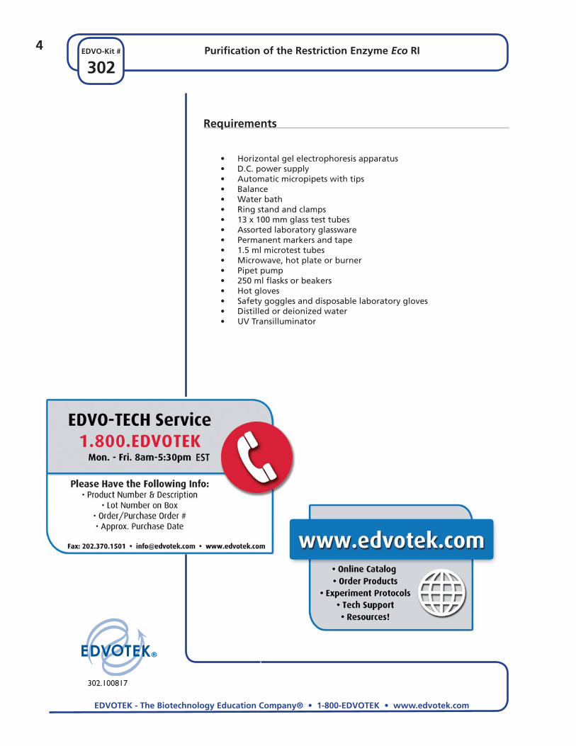
Requirements
• Horizontal gel electrophoresis apparatus • D.C. power supply • Automatic micropipets with tips • Balance • Water bath • Ring stand and clamps • 13 x 100 mm glass test tubes • Assorted laboratory glassware • Permanent markers and tape • 1.5 ml microtest tubes • Microwave, hot plate or burner • Pipet pump • 250 ml fl asks or beakers • Hot gloves • Safety goggles and disposable laboratory gloves • Distilled or deionized water • UV Transilluminator

5Purifi cation of the Restriction Enzyme Eco RI
302EDVO-Kit #
The Biotechnology Education Company® • 1-800-EDVOTEK • www.edvotek.com
Duplication of this document, in conjunction with use of accompanying reagents, is permitted for classroom/labora-tory use only. This document, or any part, may not be reproduced or distributed for any other purpose without the written consent of EDVOTEK, Inc. Copyright © 1996,1998, 1999, 2001, 2002, 2006, 2007, 2010 EDVOTEK, Inc., all rights reserved 302.100817
Backg
rou
nd
Info
rmatio
n
Purifi cation of the Restriction Enzyme Eco RI
Sequence-specifi c, or Type II, endonucleases are commonly known as restric-tion enzymes. In contrast with nonspecifi c endonucleases, these enzymes generate reproducible fragments from specifi c DNAs. They cleave double-stranded DNA by hydrolyzing two phosphodiester bonds (one per strand) within defi ned nucleotide sequences. Over 3,000 enzymes have been discov-ered since the fi rst report by H.O. Smith and collaborators. These enzymes are extracted from a variety of bacterial strains.
The name of a restriction enzyme is derived from the genus and species of bacterium from which it is isolated. The fi rst letter of the genus name and fi rst two letters of the species are combined to form the enzyme name. This is followed by a strain designation if applicable. In many instances, a bacterial strain contains more than one restric-tion endonuclease. When this occurs, each enzyme is assigned a Roman numeral. For example, Bam HI was the fi rst enzyme activity reported from Bacillus amyloliquefaciens strain H.
Most restriction enzymes are composed of two polypoeptides of equal subunits with molecular weights of 20,000-25,000 or single polypeptides with molecular weights of 30,000-35,000. Enzyme activities can be differentiated from each other by their characteristic digestion patterns of small viral DNAs. The DNA from bacteriophage lambda is the most widely used substrate for screening restriction enzymes. Because it is often diffi cult to determine a characteristic pattern from a lambda digest, smaller DNAs, such as the replicative form of bac-
teriophage ØX174 and SV40 DNA are also used as substrates. The resulting DNA restriction enzyme digests are displayed on agarose gels and visualized by staining with ethidium bromide.
A given recognition sequence in DNA can often be cleaved by more than one restriction enzyme. The term "isoschizomers" describes a group of restric-tion enzymes that recognize the same sequence in DNA. The sequences recognized by these enzymes are for the most part centrosymmetric "palin-dromic" sequences that are usually hexamers, pentamers, or tetramers. Sev-eral Type II restriction enzymes recognize DNA at a specifi c site and hydro-lyze phosphodiester bonds at a defi ned distance from that site. An example of this group of enzymes is Bgl I, which recognizes a sequence containing two groups of specifi ed residues separated by completely unspecifi ed resi-dues - GCCNNNNNGGC; it therefore generates DNA fragments with variable end groups.
Eco RIBacillus globigii
Restriction Enzyme Recognition Site
5’-GCCNNNNNGGC-3’3’-CGGNNNNNCCG-5’
5’-AAGCTT-3’3’-TTCGAA-5’
5’-GGATCC-3’3’-CCTAGG-5’
5’-GGCC-3’3’-CCGG-5’
5’-GAATTC-3’3’-CTTAAG-5’
Bgl IEscherichia coli RY13
Bam HIBacillus amyloliquefaciens H
Hae IIIHaemophilus aegyptius
Hind IIIHaemophilus influenzae R4
Figure 1: Examples of Restriction Enzymes and their recognition sites

Duplication of this document, in conjunction with use of accompanying reagents, is permitted for classroom/labora-tory use only. This document, or any part, may not be reproduced or distributed for any other purpose without the written consent of EDVOTEK, Inc. Copyright © 1996,1998, 1999, 2001, 2002, 2006, 2007, 2010 EDVOTEK, Inc., all rights reserved 302.100817
6
302Purifi cation of the Restriction Enzyme Eco RI
The Biotechnology Education Company® • 1-800-EDVOTEK • www.edvotek.com
EDVO-Kit #B
ackg
rou
nd
Info
rmat
ion
There is considerable diversity in the fragment termini produced in cleavage by Type II endonucleases that recognize and cleave within the same se-quence. In some cases, the 5' extension may be as short as two nucleotides or as long as fi ve. Points of cleavage on each strand may be opposite each other; this results in blunt (square ends). Several restriction endonucleases produce 3' extensions of two to four nucleotides. However, all Type II endo-nucleases produce fragments with a 5'-terminal phosphate and a 3'-terminal hydroxyl residue (Figure 1).
Enzymes in the Type II restriction enzyme family are amenable to purifi cation by chromatographic procedures. Ion exchangers at nearly neutral pH are used as separation matrices after extracts have been freed of cellular nucleic acids. At this stage of purifi cation, short-term assays often make it possible to visualize enzyme fractions that contain restriction enzymes. A variety of enzymes have been fractionated with affi nity chromatography. This method takes advantage of biospecifi c interactions not offered by conventional frac-tionation methods. The advantages of affi nity chromatography are speed of purifi cation and often protection against denaturation during fractionation.
Effects of Reaction Conditions on Restriction Enzymes
Several reports have described apparent changes in specifi city of restriction endonucleases in association with altered reaction environments. Conditions that alter specifi city have included changes in ionic concentration, pH of the reaction buffer, and the amounts of glycerol in the storage and the reaction mixture. For example, when lambda DNA is incubated with Eco RI or Bam HI in the presence of glycerol at various concentrations, a progressive change in the DNA digestion pattern is observed.
A change in recognition specifi city of enzymes include Bam HI and Eco RI ac-tivity. The second activity is designated as “.1” (as Bam HI.1). A similar activ-ity is displayed by Eco RI. Increasing the pH of the reaction from 7.0 to 9.0 in the absence of monovalent cations stimulates alternate activities. Decreases in the ionic strength have a similar effect.
Purifi cation of the Restriction Enzyme Eco RI

7Purifi cation of the Restriction Enzyme Eco RI
302EDVO-Kit #
The Biotechnology Education Company® • 1-800-EDVOTEK • www.edvotek.com
Duplication of this document, in conjunction with use of accompanying reagents, is permitted for classroom/labora-tory use only. This document, or any part, may not be reproduced or distributed for any other purpose without the written consent of EDVOTEK, Inc. Copyright © 1996,1998, 1999, 2001, 2002, 2006, 2007, 2010 EDVOTEK, Inc., all rights reserved 302.100817
The Exp
erimen
t
EXPERIMENT OBJECTIVE:
In this experiment, students will purify a restriction endonuclease, test its enzyme activity, and visualize the test results by agarose gel electrophoresis.
LABORATORY SAFETY
1. Gloves and goggles should be worn routinely as good laboratory prac-tice.
2. Exercise extreme caution when working with equipment that is used in conjunction with the heating and/or melting of reagents.
3. DO NOT MOUTH PIPET REAGENTS - USE PIPET PUMPS.
4. Exercise caution when using any electrical equipment in the laboratory.
5. Always wash hands thoroughly with soap and water after handling reagents or biological materials in the laboratory.
6. This experiment utilizes InstaStain® Ethidium Bromide for staining and visualization of DNA after gel electrophoresis. Always wear gloves when handling InstaStain® cards. Alhough there is only a very small amount of Ethidium bromide on InstaStain® EtBr cards, it is a listed mutagen. Wear UV-re-sistant safety goggles when working with ultraviolet light since it can cause irreparable damage to the eyes. Exposure to skin should also be avoided.
Experiment Overview and General Instructions

Duplication of this document, in conjunction with use of accompanying reagents, is permitted for classroom/labora-tory use only. This document, or any part, may not be reproduced or distributed for any other purpose without the written consent of EDVOTEK, Inc. Copyright © 1996,1998, 1999, 2001, 2002, 2006, 2007, 2010 EDVOTEK, Inc., all rights reserved 302.100817
8
302Purifi cation of the Restriction Enzyme Eco RI
The Biotechnology Education Company® • 1-800-EDVOTEK • www.edvotek.com
EDVO-Kit #Th
e Ex
per
imen
t
Partial Purifi cation of Eco RI
PACKING AND EQUILIBRATING THE COLUMN
1. Vertically mount the column on a ring stand. Make sure it is straight.
2. Slide the cap onto the spout at the bottom of the column.
3. Mix the DEAE-Cellulose (ion-exchanger matrix) thoroughly by swirling or gently stirring.
4. Carefully pipet the mixed DEAE-Cellulose into the column by letting it stream down the inside walls of the column.
If the fl ow is stopped by an air pocket, stop adding the DEAE-Cellulose and fi rmly tap the column until the air is removed and the exchanger fl ows down. Continue adding the exchanger.
5. Place an empty beaker under the column to collect wash material.
6. Remove the cap from the bottom of the column and allow the matrix to pack into the column.
7. Wash the packed column with 25 ml of Eq (1x equilibration buffer).
Do not allow the column to dry.
Note
Loading the column and subsequent elution will be done at room tem-perature. The elution buffers and the frac-tions collected should be stored on ice as they elute from the column.
9Purifi cation of the Restriction Enzyme Eco RI
302EDVO-Kit #
The Biotechnology Education Company® • 1-800-EDVOTEK • www.edvotek.com
Duplication of this document, in conjunction with use of accompanying reagents, is permitted for classroom/labora-tory use only. This document, or any part, may not be reproduced or distributed for any other purpose without the written consent of EDVOTEK, Inc. Copyright © 1996,1998, 1999, 2001, 2002, 2006, 2007, 2010 EDVOTEK, Inc., all rights reserved 302.100817
The Exp
erimen
t
Partial Purifi cation of Eco RI
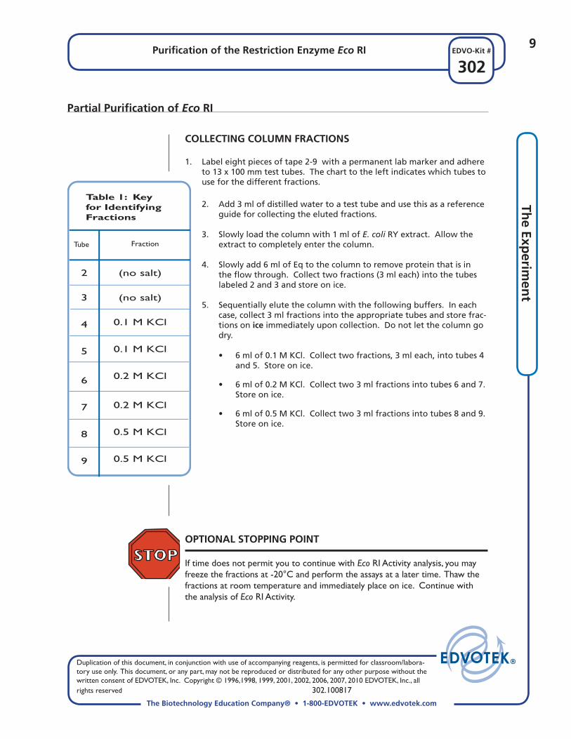
OPTIONAL STOPPING POINT
If time does not permit you to continue with Eco RI Activity analysis, you may freeze the fractions at -20°C and perform the assays at a later time. Thaw the fractions at room temperature and immediately place on ice. Continue with the analysis of Eco RI Activity.
COLLECTING COLUMN FRACTIONS
1. Label eight pieces of tape 2-9 with a permanent lab marker and adhere to 13 x 100 mm test tubes. The chart to the left indicates which tubes to use for the different fractions.
Tube Fraction
Table 1: Key for Identifying Fractions
2 3
4
5
6
7
8
9
(no salt)
(no salt)
0.1 M KCl
0.1 M KCl
0.2 M KCl
0.2 M KCl
0.5 M KCl
0.5 M KCl
2. Add 3 ml of distilled water to a test tube and use this as a reference guide for collecting the eluted fractions.
3. Slowly load the column with 1 ml of E. coli RY extract. Allow the extract to completely enter the column.
4. Slowly add 6 ml of Eq to the column to remove protein that is in the fl ow through. Collect two fractions (3 ml each) into the tubes labeled 2 and 3 and store on ice.
5. Sequentially elute the column with the following buffers. In each case, collect 3 ml fractions into the appropriate tubes and store frac-tions on ice immediately upon collection. Do not let the column go dry.
• 6 ml of 0.1 M KCl. Collect two fractions, 3 ml each, into tubes 4 and 5. Store on ice.
• 6 ml of 0.2 M KCl. Collect two 3 ml fractions into tubes 6 and 7. Store on ice.
• 6 ml of 0.5 M KCl. Collect two 3 ml fractions into tubes 8 and 9. Store on ice.

Duplication of this document, in conjunction with use of accompanying reagents, is permitted for classroom/labora-tory use only. This document, or any part, may not be reproduced or distributed for any other purpose without the written consent of EDVOTEK, Inc. Copyright © 1996,1998, 1999, 2001, 2002, 2006, 2007, 2010 EDVOTEK, Inc., all rights reserved 302.100817
10
302Purifi cation of the Restriction Enzyme Eco RI
The Biotechnology Education Company® • 1-800-EDVOTEK • www.edvotek.com
EDVO-Kit #Th
e Ex
per
imen
t
6. Cap the tubes tightly and tap on the lab bench to collect samples at the bottom of the tubes or quick spin balanced tubes in a microcen-trifuge.
7. Mix the samples and incubate in a 37°C waterbath for 15 minutes.
8. Make sure your set of reagent tubes are labeled with your initials or group number and store in the refrigerator for later use in the second assay.
9. After the 15 minute incubation is complete, add 5 µl of 10x gel load-ing solution to each tube to stop the reactions.
This prepares the Eco RI digestion products for separation by agarose gel electrophoresis.
Analysis of Eco RI Activity (First Assay)
Lambda DNA will be incubated with the fractions collected and the samples will be electrophoresed in an agarose gel to determine the peak activity of Eco RI endonuclease. Lambda DNA cut with Eco RI yields a characteristic and recognizable fragmentation pattern.
Table 2: Reagents for the Incubation of Eco RI with Lambda DNA
1 ml
100 µl
100 µl
100 µl
45 µl
Water (G) Eco RI Rxn Buffer (F) Lambda DNA (H)
10x Gel Load
Marker
on ice
on ice
on ice
Important Note
Reagents listed in Table 2 will be used for two assays. Label your set of reagent tubes with your initials or group number and store re-agents in the refrigera-tor between assays.
1. With a permanent marker, label 9 microtest tubes 1-9. Put your initials and group number on each tube.
2. Each group will assay Eco RI using 2 µl, 4 µl, 6 µl, 8 µl, or 10 µl as as-signed by your instructor. In Table 3 on the next page, the “x” equals your assigned volume for analysis.
3. Use an automatic micropipet to add (40-x µl) of Qualifi ed water to each
of the 9 tubes
4. Use an automatic micropipet to dispense 5 µl of the Eco RI Rxn Buffer and 5 µl of Lambda DNA to each of the 9 tubes.
5. Use a clean pipet tip for each fraction and add 2 µl, 4 µl, 6 µl, 8 µl, or 10 µl as assigned, from each fraction to the appropriate tube.

11Purifi cation of the Restriction Enzyme Eco RI
302EDVO-Kit #
The Biotechnology Education Company® • 1-800-EDVOTEK • www.edvotek.com
Duplication of this document, in conjunction with use of accompanying reagents, is permitted for classroom/labora-tory use only. This document, or any part, may not be reproduced or distributed for any other purpose without the written consent of EDVOTEK, Inc. Copyright © 1996,1998, 1999, 2001, 2002, 2006, 2007, 2010 EDVOTEK, Inc., all rights reserved 302.100817
The Exp
erimen
t
* Volumes of Eco RI in fractions should be varied among different groups within the range of 2 to 5 µl, with 1 µl increments. Water in the assay should be adjusted accordingly.
** To be added after incubation at 37°C.
OPTIONAL STOPPING POINT If time does not permit you to continue with agarose gel electrophoresis at this time, you may freeze the fractions at -20°C and perform the electrophoresis at a later date. Thaw the fractions at room temperature and heat the samples at 65°C before loading the gel.
Eco RIReaction
Buffer (µl)
RxnTube
Lambda DNA(µl)
QualifiedWater
(µl)Fraction
ReactionVolume
(µl)
37°CIncubation(minutes)
Sequence for Restriction Enzyme Reactions
1 2 3
4
5
6
7
8
9
40
(40 - x)
(40 - x)
(40 - x)
(40 - x)
(40 - x)
(40 - x)
(40 - x)
(40 - x)
5 5 5
5
5
5
5
5
5
5 5 5
5
5
5
5
5
5
None
x µl tube 2(no salt)
x µl tube 3(no salt)
x µl tube 4(0.1 M KCl)
x µl tube 5(0.1 M KCl)
x µl tube 6(0.2 M KCl)
x µl tube 7(0.2 M KCl)
x µl tube 8(0.5 M KCl)
x µl tube 9(0.5 M KCl)
15
15
15
15
15
15
15
15
15
10xGel Load
(µl)
5 5 5
5
5
5
5
5
5
50
50
50
50
50
50
50
50
50
Analysis of Eco RI Activity (First Assay)

Duplication of this document, in conjunction with use of accompanying reagents, is permitted for classroom/labora-tory use only. This document, or any part, may not be reproduced or distributed for any other purpose without the written consent of EDVOTEK, Inc. Copyright © 1996,1998, 1999, 2001, 2002, 2006, 2007, 2010 EDVOTEK, Inc., all rights reserved 302.100817
12
302Purifi cation of the Restriction Enzyme Eco RI
The Biotechnology Education Company® • 1-800-EDVOTEK • www.edvotek.com
EDVO-Kit #Th
e Ex
per
imen
t
AGAROSE GEL REQUIREMENTS FOR THE FIRST ASSAY
• Recommended gel size: 7 x 14 cm
• Number of sample wells required: 10
• Agarose gel concentration: 0.8%
PREPARING THE AGAROSE GEL
1. Close off the open ends of a clean and dry gel bed (casting tray) by us-ing rubber dams or tape.
2. Place a well-former template (comb) in the fi rst set of notches at the end of the bed. Make sure the comb sits fi rmly and evenly across the bed.
3. To a 250 ml fl ask or beaker, add agarose powder and buffer as indicated in the Reference Tables (Appendix A) provided by your instructor. Swirl the mixture to disperse clumps of agarose powder.
4. With a marking pen, indicate the level of the solution volume on the outside of the fl ask.
5. Heat the mixture using a microwave oven or burner to dissolve the aga-rose powder.
6. Cool the agarose solution to 60°C with careful swirling to promote even dissipation of heat. If detectable evaporation has occurred, add distilled water to bring the solution up to the original volume marked in step 4.
After the gel is cooled to 60°C:
7. Place the bed on a level surface and pour the cooled agarose solution into the bed.
8. Allow the gel to completely solidify. It will become fi rm and cool to the touch after approximately 20 minutes.
9. After the gel is solidifi ed, be careful not to damage or tear the wells while removing the rubber dams or tape and comb(s) from the gel bed.
10. Place the gel (on its bed) into the electrophoresis chamber, properly oriented, centered and level on the platform.
11. Fill the electrophoresis apparatus chamber with the appropriate amount of diluted (1x) electrophoresis buffer (refer to Table B on the instruction sheet from the Appendix provided by your instructor).
Analysis of Eco RI Activity (First Assay)
If you are unfamiliar with agarose gel preparation and electrophoresis, detailed instructions and helpful resources are available at www.edvotek.com
Important Note
Continue heating until the fi nal solution appears clear (like water) without any un-dissolved particles. Check the solution carefully. If you see "crystal" particles, the agarose is not completely dissolved.
13Purifi cation of the Restriction Enzyme Eco RI
302EDVO-Kit #
The Biotechnology Education Company® • 1-800-EDVOTEK • www.edvotek.com
Duplication of this document, in conjunction with use of accompanying reagents, is permitted for classroom/labora-tory use only. This document, or any part, may not be reproduced or distributed for any other purpose without the written consent of EDVOTEK, Inc. Copyright © 1996,1998, 1999, 2001, 2002, 2006, 2007, 2010 EDVOTEK, Inc., all rights reserved 302.100817
The Exp
erimen
t
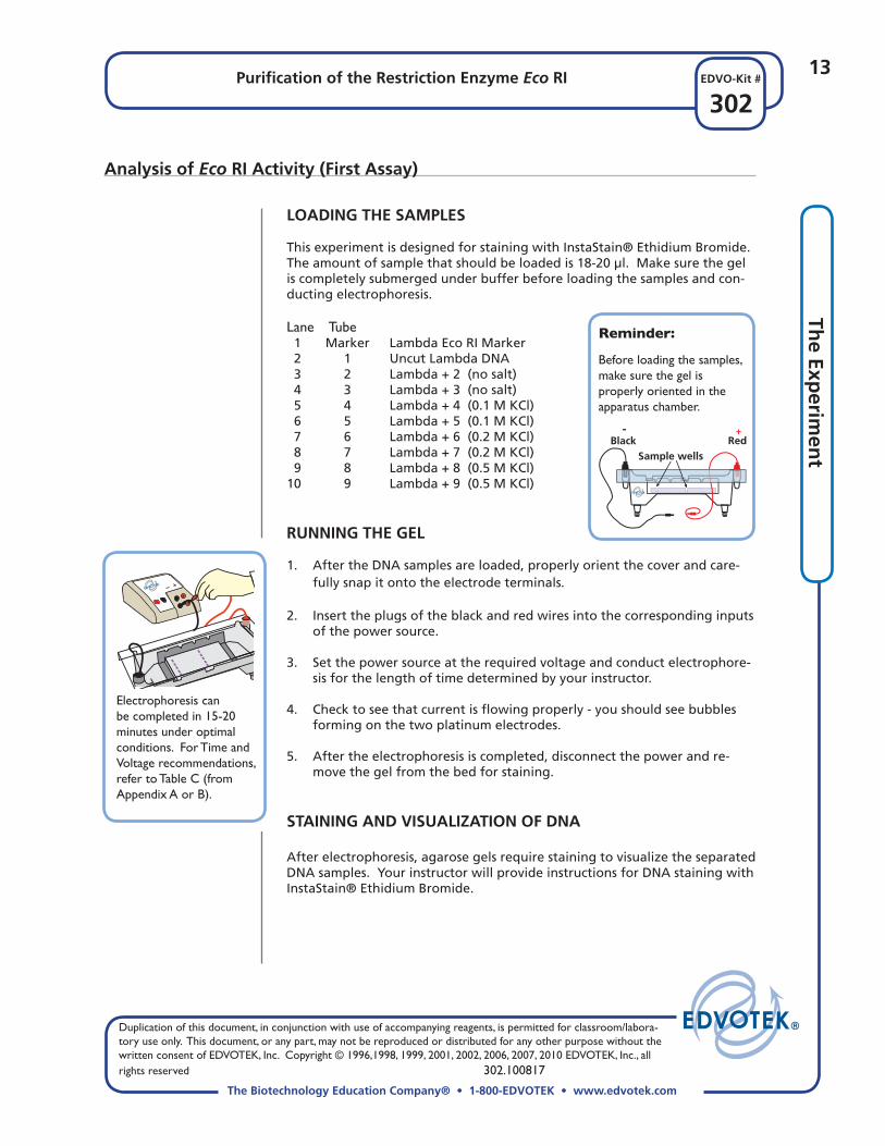
LOADING THE SAMPLES
This experiment is designed for staining with InstaStain® Ethidium Bromide. The amount of sample that should be loaded is 18-20 µl. Make sure the gel is completely submerged under buffer before loading the samples and con-ducting electrophoresis.
Lane Tube 1 Marker Lambda Eco RI Marker 2 1 Uncut Lambda DNA 3 2 Lambda + 2 (no salt) 4 3 Lambda + 3 (no salt) 5 4 Lambda + 4 (0.1 M KCl) 6 5 Lambda + 5 (0.1 M KCl) 7 6 Lambda + 6 (0.2 M KCl) 8 7 Lambda + 7 (0.2 M KCl) 9 8 Lambda + 8 (0.5 M KCl)10 9 Lambda + 9 (0.5 M KCl)
RUNNING THE GEL
1. After the DNA samples are loaded, properly orient the cover and care-fully snap it onto the electrode terminals.
2. Insert the plugs of the black and red wires into the corresponding inputs of the power source.
3. Set the power source at the required voltage and conduct electrophore-sis for the length of time determined by your instructor.
4. Check to see that current is fl owing properly - you should see bubbles forming on the two platinum electrodes.
5. After the electrophoresis is completed, disconnect the power and re-move the gel from the bed for staining.
STAINING AND VISUALIZATION OF DNA After electrophoresis, agarose gels require staining to visualize the separated DNA samples. Your instructor will provide instructions for DNA staining with InstaStain® Ethidium Bromide.
Analysis of Eco RI Activity (First Assay)
Reminder:
Before loading the samples, make sure the gel is properly oriented in the apparatus chamber.
Electrophoresis can be completed in 15-20 minutes under optimal conditions. For Time and Voltage recommendations, refer to Table C (from Appendix A or B).
+-Black Red
Sample wells

Duplication of this document, in conjunction with use of accompanying reagents, is permitted for classroom/labora-tory use only. This document, or any part, may not be reproduced or distributed for any other purpose without the written consent of EDVOTEK, Inc. Copyright © 1996,1998, 1999, 2001, 2002, 2006, 2007, 2010 EDVOTEK, Inc., all rights reserved 302.100817
14
302Purifi cation of the Restriction Enzyme Eco RI
The Biotechnology Education Company® • 1-800-EDVOTEK • www.edvotek.com
EDVO-Kit #Th
e Ex
per
imen
t
Quantifi cation of Eco RI Activity (Second Assay)
Units of enzyme activity are defi ned by convention. A restriction enzyme unit is defi ned as the amount of enzyme activity that will digest 1 µg of lambda DNA at 37°C within one hour under the defi ned assay conditions. To determine the total units of Eco RI purifi ed in this experiment, re-assay pooled enzyme fractions at various enzyme dilutions to determine the minimum amount of enzyme that yields complete digestion of 1 µg of lambda DNA.
1. Pool the enzyme fractions that have Eco RI activity as judged by the fi rst assay. If a fraction has only a trace of activity, do not pool it since it will dilute the enzyme which may result in activity loss.
2. Measure and record the volume of pooled Eco RI fractions.
3. Gently mix the pooled fraction to get a representative sample for assaying.
4. Dilute the pooled Eco RI enzyme fraction with Eco RI Dilu-tion Buffer, using the dilution factors indicated in Table 4.
5. Prepare each Eco RI dilution (from Table 4) for incubation as outlined in Table 5. Use the remaining reagents from the fi rst assay that were stored in the refrigerator.
6. After completing the incubations as outlined in Table 5, add 10x Gel Loading Solution to each tube to stop the reactions.
7. Separate the Eco RI digestion products by agarose gel elec-trophoresis.Store all fractions
on ice.
Label fractions ac-cording to dilution factors. 10 μl of each dilution will be used as shown in Table 5.
Table 4: Dilution of Pooled Eco RI
10
10
30
40
100
100
0
1:2
1:3
1:4
1:10
1:20
TotalVolume
(µl)
PooledEnzyme
(µl)
DilutionBuffer(µl)
DilutionFactor
10
5
10
10
10
5
0
5
20
30
90
95
Eco RIReaction
Buffer (µl)
RxnTube
Lambda DNA(µl)
QualifiedWater
(µl)
Eco RIDilution
(from Table 4)
ReactionVolume
(µl)
37°CIncubation(minutes)
Table 5: Assay to Determine Total Units of Eco RI
1
2
3
4
5
6
7
40
30
30
30
30
30
30
5
5
5
5
5
5
5
5
5
5
5
5
5
5
None
10 µl of 0
10 µl of 1:2
10 µl of 1:3
10 µl of 1:4
10 µl of 1:10
10 µl of 1:20
30
30
30
30
30
30
30
10xGel Load*
(µl)
5
5
5
5
5
5
5
50
50
50
50
50
50
50
*To be added after 37°C incubation

15Purifi cation of the Restriction Enzyme Eco RI
302EDVO-Kit #
The Biotechnology Education Company® • 1-800-EDVOTEK • www.edvotek.com
Duplication of this document, in conjunction with use of accompanying reagents, is permitted for classroom/labora-tory use only. This document, or any part, may not be reproduced or distributed for any other purpose without the written consent of EDVOTEK, Inc. Copyright © 1996,1998, 1999, 2001, 2002, 2006, 2007, 2010 EDVOTEK, Inc., all rights reserved 302.100817
The Exp
erimen
t
AGAROSE GEL REQUIREMENTS FOR THE SECOND ASSAY
• Recommended gel size: 7 x 14 cm
• Number of sample wells required: 8
• Agarose gel concentration: 0.8%
1. Prepare a 0.8% agarose gel for the second assay according to instruc-tions previously described.
2. Load 20 µl of each DNA sample in the following manner:
Lane Tube 1 Marker Lambda Eco RI Marker 2 1 Uncut Lambda DNA 3 2 Lambda + Undiluted Eco RI 4 3 Lambda + 1:2 Dilution 5 4 Lambda + 1:3 Dilution 6 5 Lambda + 1:4 Dilution 7 6 Lambda + 1:10 Dilution 8 7 Lambda + 1:20 Dilution 3. After the samples are loaded, conduct electrophoresis and stain the gel
with InstaStain® Ethidium Bromide for visualization. 3. Examine the gel or take a photograph to determine which lane gives
complete digestion determined as follows:
• No undigested or partially digested lambda DNA is visible. • All the DNA digestion products (5 bands) are visible.
Quantifi cation of Eco RI Activity (Second Assay)

Duplication of this document, in conjunction with use of accompanying reagents, is permitted for classroom/labora-tory use only. This document, or any part, may not be reproduced or distributed for any other purpose without the written consent of EDVOTEK, Inc. Copyright © 1996,1998, 1999, 2001, 2002, 2006, 2007, 2010 EDVOTEK, Inc., all rights reserved 302.100817
16
302Purifi cation of the Restriction Enzyme Eco RI
The Biotechnology Education Company® • 1-800-EDVOTEK • www.edvotek.com
EDVO-Kit #Th
e Ex
per
imen
t
Example for Determining Specifi c Activity
Specifi c Activity 1.8 mg
7200 units 4,000 units/mg
= =
Conversion for a 1 hour digestion assay:
Total Activity units = 3600 units X 2 = 7200
9000 μl10 μl
X 4 = 3600 units
Example for Determining Total units.
Pooled volume is 9 ml = 9000 μlEco RI volume for assay = 10 μlDilution factor = 4
Specifi c activity is defi ned as the number of enzyme units per mg of total protein in the enzyme fraction. The less total protein the Eco RI fraction contains, the higher is its spe-cifi c activity.
• For this experiment we have equated 1.0 absorbance unit at A280. In 9 ml, the amount of protein is 0.2 mg/ml X 9 ml = 1.8 ml.
Activity Determination in Units
SPECIFIC ACTIVITY DETERMINATION (OPTIONAL)
Restriction enzyme unit = amount of enzyme activity that will digest 1 µg of lambda DNA at 37°C within one hour·
for a 30 minute digestion
= Specifi c Activity mg of protein
Total units
Total units: 7200 units for the total volume of 9 mlTotal mg. of protein - 1.8 mg
DETERMINATION OF TOTAL ACTIVITY
Total units (units) is the amount of enzyme activity recov-ered from the preparation. It does not indicate the level of enzyme purity.
= XTotal
Activity (units) Volume used for
assay (µl)
Pooled volume (µl)Dilution factor

17Purifi cation of the Restriction Enzyme Eco RI
302EDVO-Kit #
The Biotechnology Education Company® • 1-800-EDVOTEK • www.edvotek.com
Duplication of this document, in conjunction with use of accompanying reagents, is permitted for classroom/labora-tory use only. This document, or any part, may not be reproduced or distributed for any other purpose without the written consent of EDVOTEK, Inc. Copyright © 1996,1998, 1999, 2001, 2002, 2006, 2007, 2010 EDVOTEK, Inc., all rights reserved 302.100817
The Exp
erimen
t
Study Questions
Answer the following study questions in your laboratory notebook or on a separate worksheet.
1. What is the recognition site for Eco RI?
2. How is E. coli host DNA protected against action of the Eco RI endonu-clease?
3. How many Eco RI sites are there in lambda DNA?
4. What is the difference between total activity versus specifi c activity?

Purifi cation of the Restriction Enzyme Eco RI
302.100817
18
xxx302EDVO-Kit #
EDVOTEK - The Biotechnology Education Company® • 1-800-EDVOTEK • www.edvotek.com
Purifi cation of the Restriction Enzyme Eco RI
302.100817
19
EDVOTEK - The Biotechnology Education Company® 1.800.EDVOTEK • www.edvotek.com
FAX: 202.370.1501 • email: [email protected]
302EDVO-Kit #
Instructor’s Guide
Class size, length of laboratory sessions, and availability of equipment are factors which must be considered in the planning and the implementation of this experiment with your students. These guidelines can be adapted to fi t your specifi c set of circumstances. If you do not fi nd the answers to your questions in this section, a variety of resources are continuously being added to the EDVOTEK web site. In addition, Technical Service is available from 9:00 am to 6:00 pm, Eastern time zone. Call for help from our knowledge-able technical staff at 1-800-EDVOTEK (1-800-338-6835).
EDUCATIONAL RESOURCES
Electrophoresis Hints, Help and Frequently Asked Questions
EDVOTEK Experiments are designed for maximum success in the classroom setting. However, even the most experienced students and teachers occa-sionally encounter experimental problems or diffi culties. The EDVOTEK web site provides several suggestions and reminders for conducting electrophore-sis, as well as answers to frequently asked electrophoresis questions.
Duplication of this document, in conjunction with use of accompanying reagents, is permitted for classroom/labora-tory use only. This document, or any part, may not be reproduced or distributed for any other purpose without the written consent of EDVOTEK, Inc. Copyright © 1996,1998, 1999, 2001, 2002, 2006, 2007, 2010 EDVOTEK, Inc., all rights reserved 302.100817
20
302Purifi cation of the Restriction Enzyme Eco RI
The Biotechnology Education Company® • 1-800-EDVOTEK • www.edvotek.com
EDVO-Kit #Th
e Ex
per
imen
t
Time and VoltageRecommendations
Minimum / Maximum
Volts
150
125
70
50
15 / 20 min
20 / 30 min
35 / 45 min
50 / 80 min
Table
CEDVOTEK Electrophoresis Model
M6+ M12 & M36Minimum / Maximum
25 / 35 min
35 / 45 min
60 / 90 min
95 / 130 min
Notes to the Instructor:
MICROPIPETTING BASICS AND PRACTICE GEL LOADING
Accurate pipeting is critical for maximizing successful experiment results. EDVOTEK Series 300 experiments are designed for students who have had previous experience with agarose gel electrophoresis and micropipeting techniques. If your students are unfamiliar with using micropipets, EDVOTEK highly recommends that students perform Experiment # S-44, Micropipetting Basics, or other Series 100 or 200 electrophoresis experiment prior to con-ducting this advanced level experiment.
APPROXIMATE TIME REQUIREMENTS
• Pre-lab preparations Pre-lab preparations and dispensing of biologicals and reagents take ap-
proximately 1-2 hours.
• Restriction Enzyme Digestion The approximate time required for students to perform the restric-
tion enzyme digestion and prepare samples for electrophoresis is 50-75 minutes. Extending the restriction enzyme digest incubation time to 60 minutes will help ensure complete cleavage of DNA.
• Agarose Gel preparation Whether you choose to prepare the gel(s) in advance or have the stu-
dents prepare their own, allow approximately 30-40 minutes for this procedure. Generally, 20 minutes of this time is required for gel solidifi -cation. See section “Options for Preparing Agarose Gels” below.
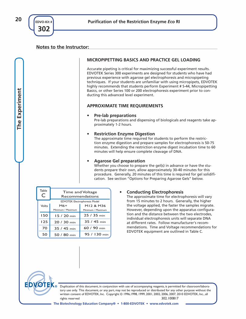
• Conducting Electrophoresis The approximate time for electrophoresis will vary
from 15 minutes to 2 hours. Generally, the higher the voltage applied, the faster the samples migrate. However, depending upon the apparatus confi gura-tion and the distance between the two electrodes, individual electrophoresis units will separate DNA at different rates. Follow manufacturer's recom-mendations. Time and Voltage recommendations for EDVOTEK equipment are outlined in Table C.

21Purifi cation of the Restriction Enzyme Eco RI
302EDVO-Kit #
The Biotechnology Education Company® • 1-800-EDVOTEK • www.edvotek.com
Duplication of this document, in conjunction with use of accompanying reagents, is permitted for classroom/labora-tory use only. This document, or any part, may not be reproduced or distributed for any other purpose without the written consent of EDVOTEK, Inc. Copyright © 1996,1998, 1999, 2001, 2002, 2006, 2007, 2010 EDVOTEK, Inc., all rights reserved 302.100817
Instru
ctor’s G
uid
e
Notes to the Instructor:
OPTIONS FOR PREPARING AGAROSE GELS
This experiment is designed for DNA staining after electrophoresis with In-staStain® Ethidium Bromide. There are several options for preparing agarose gels for the experiment.
1. Individual Gel Casting: Each student lab group can be responsible for casting their own indi-
vidual gel prior to conducting the experiment.
2. Preparing Gels in Advance: Gels may be prepared ahead and stored for later use. Solidifi ed gels can
be stored under buffer in the refrigerator for up to 2 weeks.
Do not store gels at -20°C. Freezing will destroy the gels.
Gels that have been removed from their trays for storage, should be "anchored" back to the tray with a few drops of hot, molten agarose before placing the gels into the apparatus for electrophoresis. This will prevent the gels from sliding around in the trays and the chambers.
3. Batch Gel Preparation: A batch of agarose gel can be prepared for sharing by the class. To save
time, a larger quantity of UltraSpec-Agarose can be prepared for sharing by the class. See instructions for "Batch Gel Preparation".
GEL CONCENTRATION AND VOLUME
The gel concentration required is 0.8%. Prepare 7 x 14 cm gels according to Table A.1 or A.2 in Appendix A.

Duplication of this document, in conjunction with use of accompanying reagents, is permitted for classroom/labora-tory use only. This document, or any part, may not be reproduced or distributed for any other purpose without the written consent of EDVOTEK, Inc. Copyright © 1996,1998, 1999, 2001, 2002, 2006, 2007, 2010 EDVOTEK, Inc., all rights reserved 302.100817
22
302Purifi cation of the Restriction Enzyme Eco RI
The Biotechnology Education Company® • 1-800-EDVOTEK • www.edvotek.com
EDVO-Kit #In
stru
cto
r’s
Gu
ide
Notes to the Instructor:
GEL STAINING AND DESTAINING AFTER ELECTROPHORESIS This experiment features InstaStain® Ethidium Bromide for gel staining after electrophoresis. It is a proprietary staining method which saves time and reduces liquid waste. DNA staining with InstaStain® Methylene Blue is not recommended because it will not yield optimal results.
Instastain® Ethidium Bromide
• InstaStain® Ethidium Bromide Appendix C
Optimal visualization of DNA fragments on gels is obtained by staining with InstaStain® Ethidium Bromide (InstaStain® EtBr) cards.
Caution: Ethidium Bromide is a listed mutagen. Disposal of the InstaStain® EtBr cards, which contain only a few micrograms of ethidium bromide, is minimal compared to the large volume of liquid waste generated by tradi-tional ethidium bromide staining procedures. Disposal of InstaStain® cards and gels should follow institutional guidelines for chemical waste.
PHOTODOCUMENTATION OF DNA (OPTIONAL)
There are many different photodocumentation systems available, including digital systems that are interfaced directly with computers. Specifi c instruc-tions will vary depending upon the type of photodocumentation system you are using.

23Purifi cation of the Restriction Enzyme Eco RI
302EDVO-Kit #
The Biotechnology Education Company® • 1-800-EDVOTEK • www.edvotek.com
Duplication of this document, in conjunction with use of accompanying reagents, is permitted for classroom/labora-tory use only. This document, or any part, may not be reproduced or distributed for any other purpose without the written consent of EDVOTEK, Inc. Copyright © 1996,1998, 1999, 2001, 2002, 2006, 2007, 2010 EDVOTEK, Inc., all rights reserved 302.100817
Instru
ctor’s G
uid
e
Each group requires:
• DEAE-Cellulose• Eq Buffer• 0.1 M KCl• 0.2 M KCl• 0.5 M KCl• E. coli RY extract• 1 chromatography column• 1 ring stand with clamp• 9 test tubes (13 x 100 mm)• 10 microtest tubes• Automatic micropipet & tips• 5 ml pipets and pipet pumps
Pre-Lab Preparations
PARTIAL PURIFICATION OF ECO RI (Packing the Column and Collecting Fractions)
DEAE-Cellulose Matrix
1. Hydrate the ion-exchanger, DEAE-Cellulose (B) in 35 ml of 10x equilibra-tion buffer (C).
5. To make Eq + KCl Buffers, mix the following:
Eq Buffer (1x) KCl
0.1 M KCl 100 ml 0.75 g 0.2 M KCl 100 ml 1.5 g 0.5 M KCl 100 ml 3.75 g
E. coli Cell Extract Containing Eco RI Restriction Enzyme
6. Re-hydrate the sample by adding 0.5 ml of distilled or deionized water to tube component A and let sit for 5 minutes.
7. Mix vigorously by vortexing and transfer the entire contents to a 50 ml conical tube. Rinse tube A six times - each time with 1 ml of 1x Equilibration buffer (diluted component C) and add the rinse material to the 50 ml conical tube. Mix the tube well.
8. Label 5 tubes "E. coli RY extract". Aliquot 1 ml of the re-hydrat-ed extract for each of the student groups. Store extracts on ice.
2. Stir occasionally for a minimum of 30 min-utes.
3. Aliquot 6 ml for each of the fi ve groups.
Buffers
4. Prepare 500 ml of 1x equilibration buffer (Eq) in a 600 ml fl ask or beaker. To prepare, add the following and stir thoroughly:
350 ml Distilled water 50 ml 10x Equilibration buffer (C) 100 ml 50% glycerol (D)
Use this prepared Eq buffer to make buffers in step 5.
Summary of Reagent Preparations
6 ml
35 ml
6 ml
6 ml
6 ml
1 ml
DEAE-Cellulose (Matrix, B)
Eq Buffer (1x equil buffer, diluted C)
0.1 M KCl
0.2 M KCl
0.5 M KCl
E. coli RY extract (A)
Quick Reference
on ice
on ice
on ice
on ice
on ice
on ice
The 10x equilibration buffer used to hydrate the DEAE-Cellulose con-tains potassium phos-phate, pH 7.4, EDTA, and β-mercaptoethanol.

Duplication of this document, in conjunction with use of accompanying reagents, is permitted for classroom/labora-tory use only. This document, or any part, may not be reproduced or distributed for any other purpose without the written consent of EDVOTEK, Inc. Copyright © 1996,1998, 1999, 2001, 2002, 2006, 2007, 2010 EDVOTEK, Inc., all rights reserved 302.100817
24
302Purifi cation of the Restriction Enzyme Eco RI
The Biotechnology Education Company® • 1-800-EDVOTEK • www.edvotek.com
EDVO-Kit #In
stru
cto
r’s
Gu
ide
Pre-Lab Preparations
ANALYSIS AND QUANTIFICATION OF ECO RI ACTIVITY (First and Second Assays)
Incubation of Fractions with Lambda DNA
Important: Students should be reminded that the reagents they receive are for two assays.
1. Label 5 tubes "water" and dispense 1 ml Qualifi ed Water (G) into the tubes. Store on ice.
2. Label 5 tubes "Eco RI Rxn Buffer" and dispense 100 µl of Eco RI Reaction Buffer (F) into the tubes. Store on ice.
3. Label 5 tubes "Lambda DNA" and dis-pense 100 µl of Lambda DNA (H) into the tubes. Store on ice.
4. Label 5 tubes "10x Gel Load" and dis-pense 100 µl 10x Gel Loading Solution into the tubes.
5. Label 5 tubes "Marker" and dispense 45 µl Lambda/Eco RI Marker (I) into the tubes.
6. Label 5 tubes "Eco RI Diln Buffer" and dispense 250 µl of Eco RI Dilution Buffer (J) into the tubes. Store on ice.
7. Have a 37°C waterbath ready for Eco RI activity analysis.
Reagents for First & Second Assays
1 ml
100 µl
100 µl
100 µl
45 µl
Water (G)
Eco RI Rxn Buffer (F)
Lambda DNA (H)
10x Gel Load
Marker
on ice
on ice
on ice
Summary of Reagent Preparations
Additional Reagent for Second Assay
Lambda DNA (H) 250 µl on ice
Quick Reference

25Purifi cation of the Restriction Enzyme Eco RI
302EDVO-Kit #
The Biotechnology Education Company® • 1-800-EDVOTEK • www.edvotek.com
Duplication of this document, in conjunction with use of accompanying reagents, is permitted for classroom/labora-tory use only. This document, or any part, may not be reproduced or distributed for any other purpose without the written consent of EDVOTEK, Inc. Copyright © 1996,1998, 1999, 2001, 2002, 2006, 2007, 2010 EDVOTEK, Inc., all rights reserved 302.100817
Instru
ctor’s G
uid
e
Experiment Results and Analysis
In the idealized schematic, the relative positions of DNA fragments are shown but are not depict-ed to scale. The schematic depicts an idealized gel result for identifying column fractions with Eco RI activity.
Lane Tube
1 Marker Lambda Eco RI Markers 2 1 Uncut Lambda DNA 3 2 Lambda + 2 (no salt) 4 3 Lambda + 3 (no salt) 5 4 Lambda + 4 (0.1 M KCl) 6 5 Lambda + 5 (0.1 M KCl) 7 6 Lambda + 6 (0.2 M KCl) 8 7 Lambda + 7 (0.2 M KCl) 9 8 Lambda + 8 (0.5 M KCl) 10 9 Lambda + 9 (0.5 M KCl)
* Results may vary between different groups and from the schematic depicted to the left. Some bands may be faint and thus diffi cult to see. You may also see extra bands due to partial digestion of the DNA. The amount of activity in the fl ow through may also vary.
Results of the second assay will show varying results depending upon the amount of purifi ed enzyme activity.
PARTIAL PURIFICATION OF ECO RI (FIRST ASSAY)
( + )
( - )
1 2 3 4 5 6
7 8 9 10

Please refer to the kit insert for the Answers to
Study Questions

Purifi cation of the Restriction Enzyme Eco RI
302.100817
27
EDVOTEK - The Biotechnology Education Company® 1.800.EDVOTEK • www.edvotek.com
FAX: 202.370.1501 • email: [email protected]
302EDVO-Kit #
A 0.8% Agarose Gel Preparation For DNA Staining with InstaStain® Ethidium Bromide
B 0.8% Quantity Preparations for Agarose Gel Electrophoresis C Staining and Visualization of DNA with InstaStain® Ethidium Bromide Cards
Safety Data Sheets are found on our website: www.edvotek.com/safety-data-sheets
Appendices

Duplication of this document, in conjunction with use of accompanying reagents, is permitted for classroom/labora-tory use only. This document, or any part, may not be reproduced or distributed for any other purpose without the written consent of EDVOTEK, Inc. Copyright © 1996,1998, 1999, 2001, 2002, 2006, 2007, 2010 EDVOTEK, Inc., all rights reserved 302.100817
28
The Biotechnology Education Company® • 1-800-EDVOTEK • www.edvotek.com
302EDVO-Kit #
For DNA analysis, the recom-mended electrophoresis buffer is Tris-acetate-EDTA, pH 7.8. The formula for diluting EDVOTEK (50x) concentrated buffer is one volume of buffer concentrate to every 49 volumes of distilled or deionized water. Prepare buffer as required for your electropho-resis unit.
If preparing the gel with concentrated (50x) buffer, use Table A.1.
0.8% Agarose Gel Preparation Reference Tables forDNA Staining with InstaStain® Ethidium Bromide
If preparing the gel with diluted (1x) buffer, use Table A.2.
Time and Voltage recommendations for EDVOTEK equipment are outlined in Table C. The approxi-mate time for electrophoresis will vary from ap-proximately 15 minutes to 2 hours depending upon various factors. Conduct electrophoresis for the length of time determined by your instructor.
Amt ofAgarose
(g)
ConcentratedBuffer (50X)
(ml)
Size of Gel(cm)
7 x 7
7 x 14
0.2
0.4
0.5
1.0
+
Table
A.1 Individual 0.8%* UltraSpec-Agarose™ GelDNA Staining with InstaStain® EtBr
DistilledWater(ml)
TotalVolume
(ml)
24.5
49.0
25
50
=+
Amt ofAgarose
(g)
DilutedBuffer (1x)
(ml)
Size of Gel(cm)
7 x 7
7 x 14
0.2
0.4
25
50
+
Table
A.2Individual 0.8%*
UltraSpec-Agarose™ GelDNA Staining with
InstaStain® Ethidium Bromide
50x Conc.Buffer (ml)
DistilledWater (ml)
6
8
10
20
294
392
490
980
+EDVOTEKModel #
Total Volume Required (ml)
Electrophoresis (Chamber) Buffer
M6+
M12
M36 (blue)
M36 (clear)
300
400
500
1000
Dilution
Table
B
Time and VoltageRecommendations
Minimum / Maximum
Volts
150
125
70
50
15 / 20 min
20 / 30 min
35 / 45 min
50 / 80 min
Table
CEDVOTEK Electrophoresis Model
M6+ M12 & M36Minimum / Maximum
25 / 35 min
35 / 45 min
60 / 90 min
95 / 130 min
Appendix A
* 0.77 UltraSpec-Agarose™ gel percentage rounded up to 0.8%

The Biotechnology Education Company® • 1-800-EDVOTEK • www.edvotek.com
Duplication of this document, in conjunction with use of accompanying reagents, is permitted for classroom/labora-tory use only. This document, or any part, may not be reproduced or distributed for any other purpose without the written consent of EDVOTEK, Inc. Copyright © 1996,1998, 1999, 2001, 2002, 2006, 2007, 2010 EDVOTEK, Inc., all rights reserved 302.100817
29
302EDVO-Kit #
To save time, electrophoresis buffer and agarose gel solution can be prepared in larger quantities for sharing by the class. Unused diluted buffer can be used at a later time and solidifi ed agarose gel can be remelted.
0.8% Agarose Gel Electrophoresis Reference TablesQuantity Preparations
BULK ELECTROPHORESIS BUFFER
Quantity (bulk) preparation for 3 liters of 1x electro-phoresis buffer is outlined in Table D.
BATCH AGAROSE GELS (0.8%)
For quantity (batch) preparation of 0.8% agarose gels, see Table E.
1. Use a 500 ml fl ask to prepare the diluted gel buf-fer
2. Pour 3.0 grams of UltraSpec-Agarose™ into the prepared buffer. Swirl to disperse clumps.
3. With a marking pen, indicate the level of solution volume on the outside of the fl ask.
4. Heat the agarose solution as outlined previously for individual gel preparation. The heating time will require adjustment due to the larger total volume of gel buffer solution.
5. Cool the agarose solution to 60°C with swirling to promote even dissipation of heat. If evaporation has occurred, add distilled water to bring the solution up to the original volume as marked on the fl ask in step 3.
6. Dispense the required volume of cooled agarose solution for casting each gel. The volume re-quired is dependent upon the size of the gel bed.
7. Allow the gel to completely solidify. It will become fi rm and cool to the touch after approxi-mately 20 minutes. Then proceed with preparing the gel for electrophoresis.
60˚C
Table
D
ConcentratedBuffer (50x)
(ml)
DistilledWater(ml)
TotalVolume
(ml)
60 2,940 3000 (3 L)
=+
Bulk Preparation of Electrophoresis Buffer
Note: The UltraSpec-Agarose™ kit component is of-ten labeled with the amount it contains. In many cases, the entire contents of the bottle is 3.0 grams. Please read the label carefully. If the amount of agarose is not specifi ed or if the bottle's plastic seal has been broken, weigh the agarose to ensure you are using the correct amount.
Table
E
Amt ofAgarose
(g)
ConcentratedBuffer (50x)
(ml)
DistilledWater(ml)
TotalVolume
(ml)
3.0 7.5 382.5 390
+ =+
Batch Preparation of 0.8%* UltraSpec-Agarose™
*0.77% UltraSpec-Agarose™ gel percentage rounded up to 0.8%
Appendix B

Duplication of this document, in conjunction with use of accompanying reagents, is permitted for classroom/labora-tory use only. This document, or any part, may not be reproduced or distributed for any other purpose without the written consent of EDVOTEK, Inc. Copyright © 1996,1998, 1999, 2001, 2002, 2006, 2007, 2010 EDVOTEK, Inc., all rights reserved 302.100817
30
The Biotechnology Education Company® • 1-800-EDVOTEK • www.edvotek.com
302EDVO-Kit #
DNA InstaStain™
Patents Pending DNA InstaStain™
Patents Pending
DNA InstaStain™
Patents Pending
DNA InstaStain™
Patents Pending
- - - - -
DNA InstaStain™
Patents Pending DNA InstaStain™
Patents Pending
- - - - -
1
2
3
4
5
Press firmly.
Moisten the gel.
Place the InstaStain® card on the gel.
Place a small weight to ensure good contact.
View on U.V. (300 nm) transilluminator
Wear gloves and safety goggles
Do not stain gel(s) in the electrophoresis apparatus.
1. After electrophoresis, place the gel on a piece of plastic wrap on a fl at surface. Moisten the gel with a few drops of electrophoresis buf-fer.
2. Wearing gloves, remove the clear plastic pro-tective sheet, and place the unprinted side of the InstaStain® EtBr card on the gel.
3. Firmly run your fi ngers over the entire surface of the InstaStain® EtBr. Do this several times.
Visit our web site for an animated demonstration of InstaStain® EtBr.
Disposal of InstaStain
Disposal of InstaStain® cards and gels should follow institutional guidelines for chemical waste.
Additional Notes About Staining
• If bands appear faint, or if you are not using EDVOTEK UltraSpec-Agarose™, gels may take longer to stain with InstaStain® EtBr. Repeat staining and increase
Caution: Ethidium Bromide is a listed mutagen.
Staining and Visualization of DNA
INSTASTAIN® ETHIDIUM BROMIDE CARDS
the staining time an additional 10-15 minutes.
• Gels stained alternatively with InstaStain Methylene Blue or liquid methylene blue may fade with time. Re-stain the gel to visualize the DNA bands.
• DNA 200 bp markers should be visible after staining even if the amplifi ed DNA samples are faint or absent. If markers are not visible, troubleshoot for problems with the electrophoretic separation.
4. Place the gel casting tray and a small empty beaker on top to ensure that the InstaStain® card maintains direct contact with the gel surface.
Allow the InstaStain® EtBr card to stain the gel for 10-15 minutes.
5. After 10-15 minutes, remove the InstaStain® EtBr card. Transfer the gel to a ultraviolet (300 nm) transilluminator for viewing. Be sure to wear UV protec-tive goggles.
Appendix C