the body works? - online academic community · pdf fileinto how the body works and what ... a...
TRANSCRIPT
The Body Works?Part 2
Part of the UVic Retirees Association (UVRA)
Elder Academy Program
Presenters: David Docherty, Ph.D., withChris Pengilly, M.D., Mike Bassett, M.D.and Dr. Helen Martendale. Ph.D., O.D.
Overall approach:
Purpose: To provide some insight into how the body works and what can go wrong so you are able to understand what goes on in your body and communicate more effectively with medical professionals.
Presentations: two parts
1.The anatomy and function of four new selected systems
2.Things that can go wrong and the medical interventions commonly available
4 New Systems
• The Brain-Dr. Mike Bassett
• The Endocrine System-Dr. Chris Pengilly
• The Respiratory System-Dr. Chris Pengilly
• The Special Senses (Vision)-Dr Helen Martendale
Presentation 1: The Brain(and associated parts!)
However, before we start.....
Differences between men’s brains and women’s brains with apologies to Mark Gungor (marriage expert)

Compared the two brains!
Complex network Boxes
Woman’s brain Man’s brain
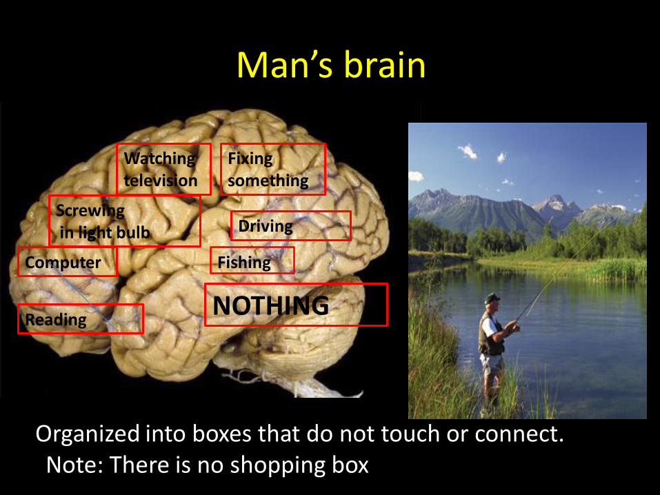
Man’s brain
Organized into boxes that do not touch or connect.
Reading
Computer
Screwingin light bulb
Watching television
Fixing something
NOTHING
Driving
Note: There is no shopping box
Fishing

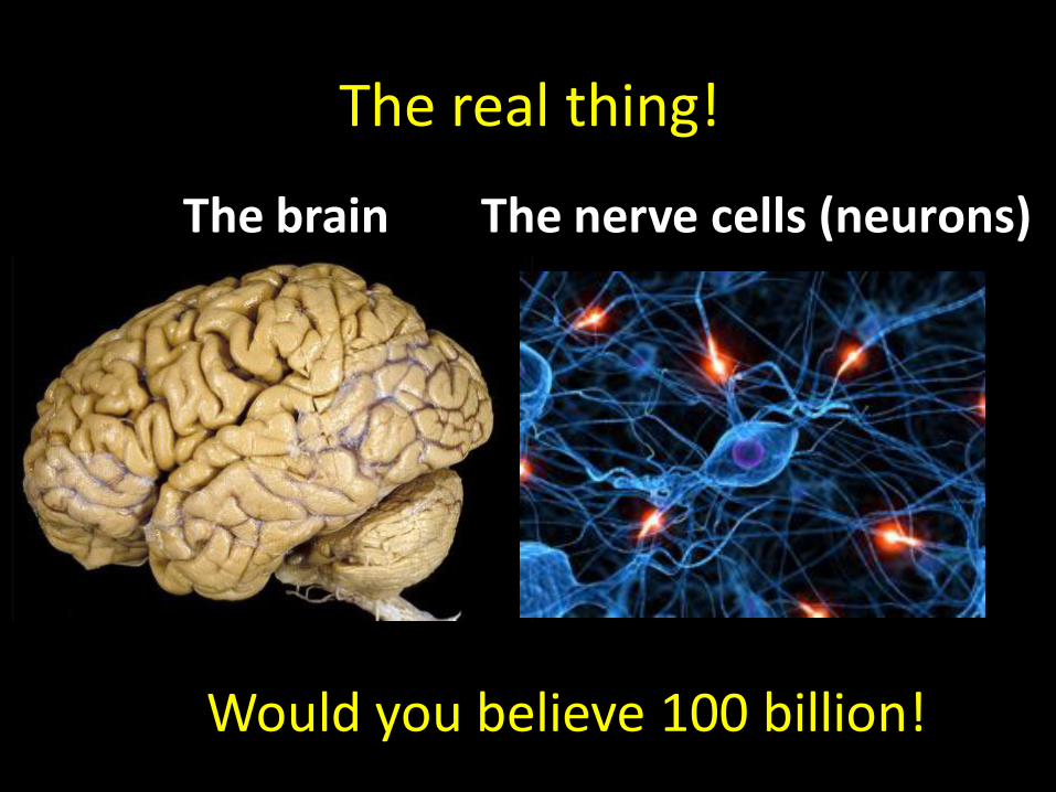
The real thing!
The brain The nerve cells (neurons)
Would you believe 100 billion!

The Nervous System
• The nervous system includes all of the neural tissue in the body.

Outline of presentation (first part)
• Neurons and how they communicate
• Organization of the brain and nervous system
• How messages get to their targets and how information is relayed back.
• Brief mention of the Autonomic Nervous System
• How the brain is protected.
• Circulation of blood and CSF in the brain.

Functional divisions of nervous system
• Afferent– Sensory information from receptors to CNS
• Efferent – Motor commands to muscles and glands
• Somatic division
–Voluntary control over skeletal muscle
• Autonomic division
– Involuntary regulation of smooth and cardiac muscle, glands
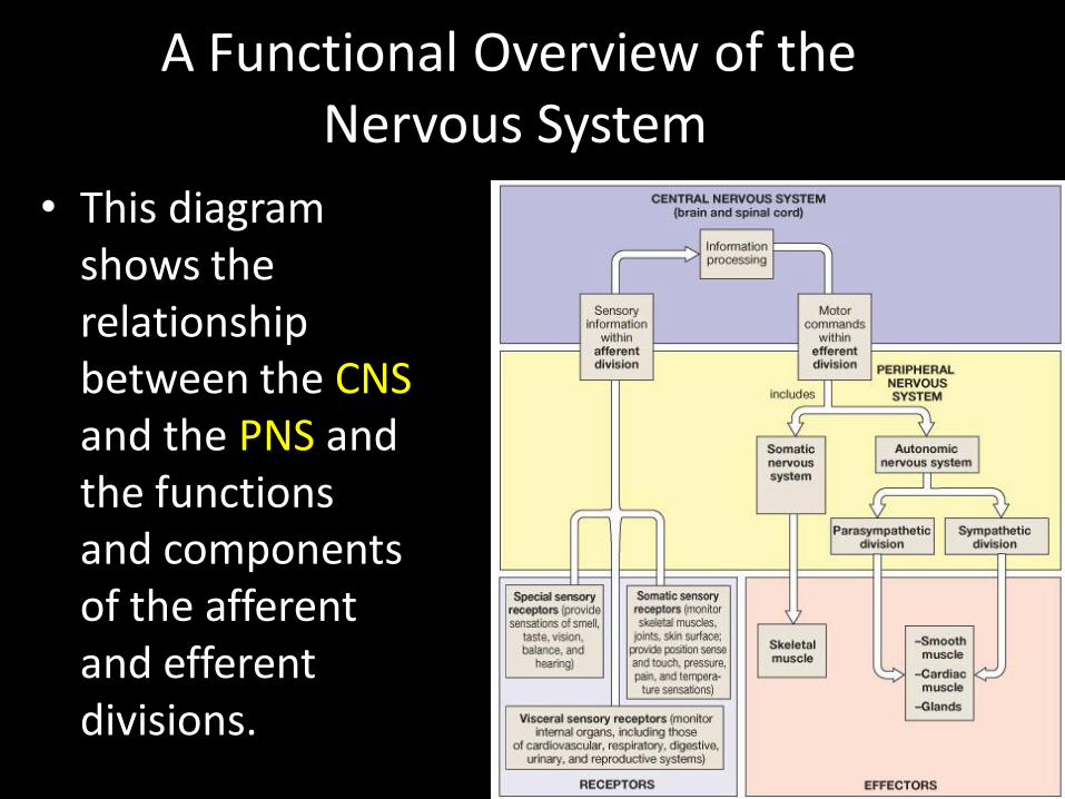
A Functional Overview of theNervous System
• This diagram shows the relationship between the CNSand the PNS and the functions and components of the afferent and efferent divisions.
Nerve Cells• Neuroglial cells (supporting structure,
phagocytic role, and isolates the neurons from surrounding tissue)
–Neuroglial cells come in different shapes and sizes and perform a variety of roles
• Neurons (responsible for transferring and processing information)
–Neurons come in many different shapes and perform several different functions
• Four types of neuroglial cells in the CNS
–Astrocytes
–Oligodendrocytes
–Microglia
–Ependymal cells
Neuroglia cells

The categories and functions of the various neuroglial cell types.
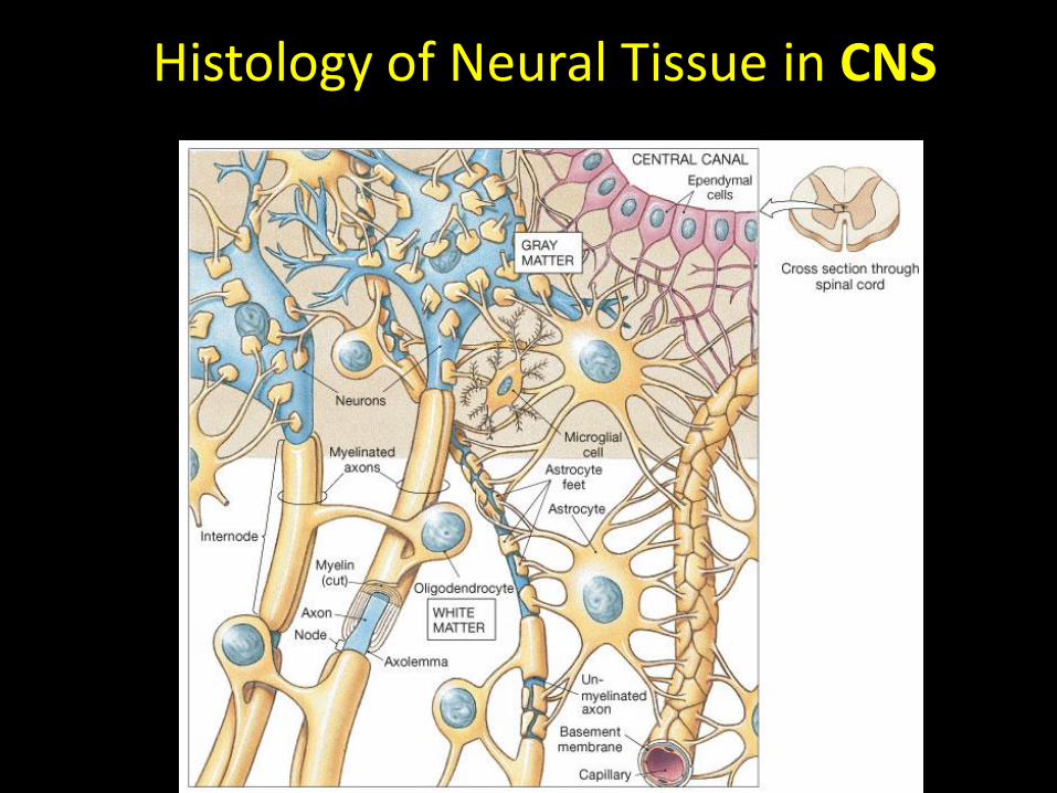
Histology of Neural Tissue in CNS

Neuron Structure
• The relationship of the four parts of a neuron (dendrites, cell body, axon, synaptic terminals).
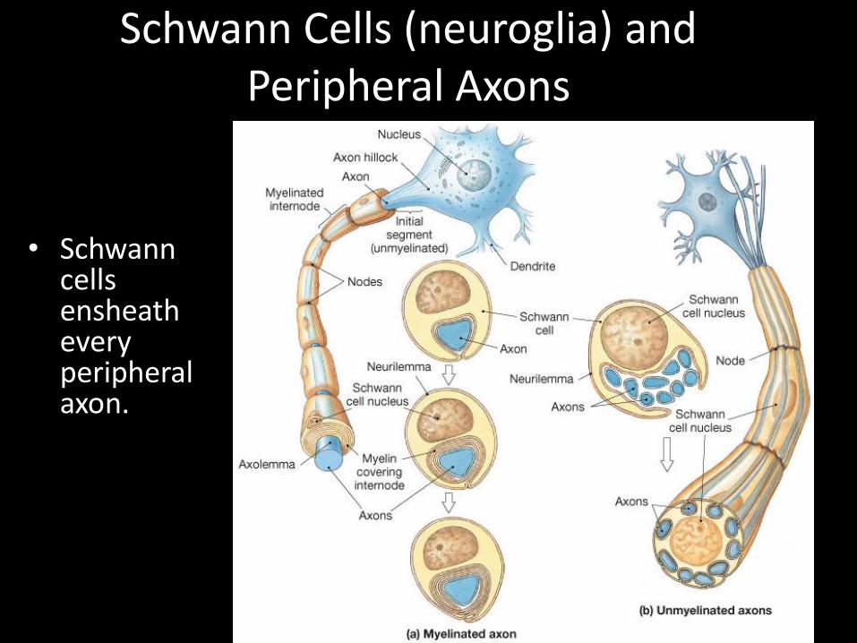
Schwann Cells (neuroglia) and Peripheral Axons
• Schwann cells ensheath every peripheral axon.
• Afferent division of PNS• Deliver sensory information from
sensory receptors to CNS– Exteroceptors– Proprioceptors– Interoceptors
Sensory Neurons

• Efferent pathways
• Stimulate peripheral structures
– Somatic motor neurons
Innervate skeletal muscle
– Visceral motor neurons
Innervate all other peripheral effectors
Preganglionic and postganglionic neurons
Motor Neurons
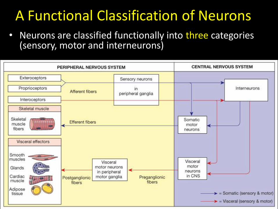
A Functional Classification of Neurons• Neurons are classified functionally into three categories
(sensory, motor and interneurons)
The Nerve Impulse
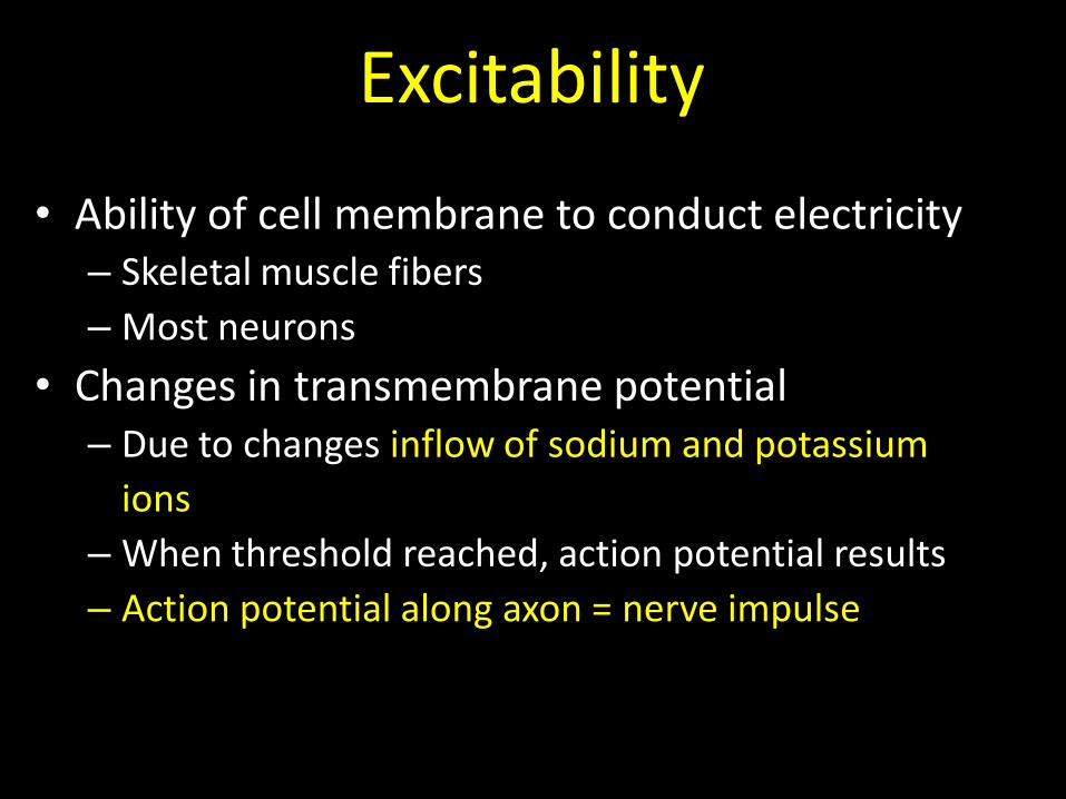
• Ability of cell membrane to conduct electricity– Skeletal muscle fibers
– Most neurons
• Changes in transmembrane potential– Due to changes inflow of sodium and potassium
ions
– When threshold reached, action potential results
– Action potential along axon = nerve impulse
Excitability

TRIGGERING AND PROPOGATING THE ACTION POTENTIAL
hwenger04
Depolarized potential = +30 mV (due to influx of Na++)
Resting potential = -70mV
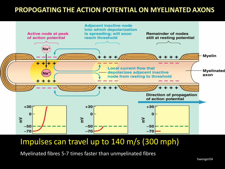
PROPOGATING THE ACTION POTENTIAL ON MYELINATED AXONS
hwenger04
Impulses can travel up to 140 m/s (300 mph)Myelinated fibres 5-7 times faster than unmyelinated fibres
Synaptic Communication
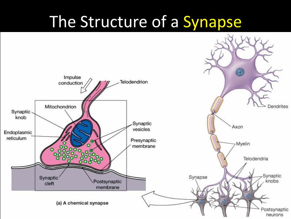
The Structure of a Synapse
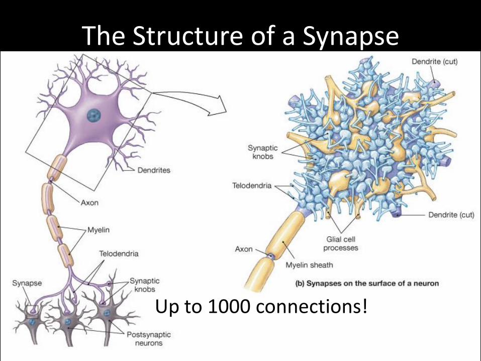
The Structure of a Synapse
Up to 1000 connections!

How neurons communicate
• https://www.youtube.com/watch?v=o9p2ou1IyC0

Anatomy of a Representative Neuron
• A neuron has a cell body, some branching dendrites and a single axon.
telodendria
(Clusters of ribosomes)
(Cytoplasm)

Neurons can connect with:
Other neurons
Skeletal or smooth muscle
Glands

• Action potential at presynaptic neuron synaptic knob
• Release of neurotransmitters (40-50 types)
• Neurotransmitter binds to receptors on postsynaptic neuron
• Change in permeability of postsynaptic neuron– Excitatory or inhibitory effects
• Degree of excitation may initiate action potential
• Effects of neurotransmitter fades rapidly– Enzymes break down neurotransmitters quickly
Steps at chemical synapse
Neural Regeneration

Nerve Regeneration in PNS after Injury
• Limited ability in PNS
• Severed peripheral nerve successfully regenerates a fraction of the axons
– Function is permanently impaired
– Schwann cells participate
• Wallerian degeneration
– Loss of axon distal to damage
Regeneration in PNS (cont)

• More complicated than PNS regeneration
• Far more limited
• More axons involved
• Astrocytes produce scar tissue preventing axonal regrowth
• Astrocytes release chemicals blocking regrowth
Regeneration in CNS

Human Anatomy, 3rd edition
Prentice Hall, © 2001
Figure 13-02:Functional overview of the Nervous System


The Limbic system
A collection of different structures with a similar function.
Function:Processing of memories, creation of emotional states, drives, and associated behaviours
The main parts of the Brain and what they do
• https://www.youtube.com/watch?v=kMKc8nfPATI
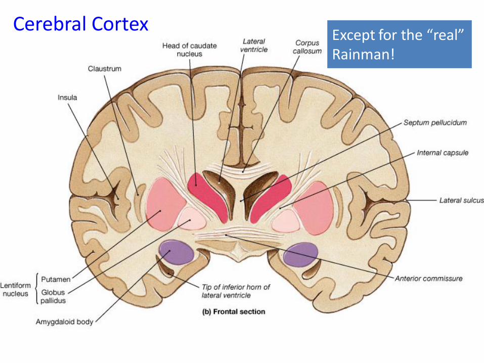
Cerebral CortexExcept for the “real”Rainman!
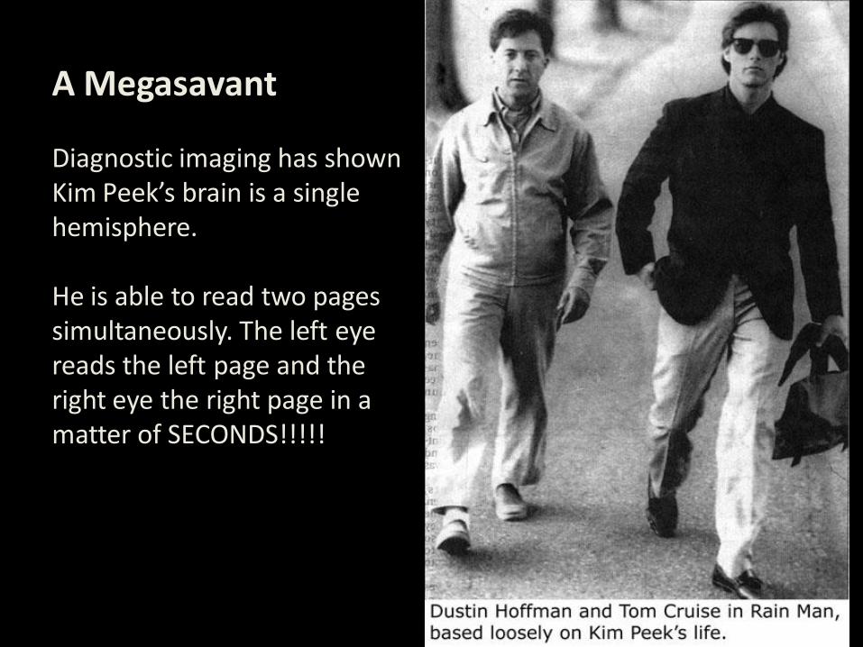
A Megasavant
Diagnostic imaging has shown Kim Peek’s brain is a single hemisphere.
He is able to read two pagessimultaneously. The left eye reads the left page and theright eye the right page in a matter of SECONDS!!!!!

Fig 15.10: Central White Matter-Communication Tracts
Tracts:•Commisural•Projection•Association

anatomical and functional landmarks

Cerebral hemispheres (lateral view)
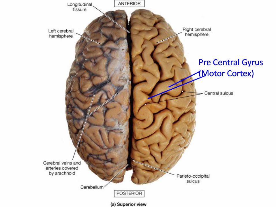
Figure 15-08a: Cerebral hemispheres (superior view)
Pre Central Gyrus (Motor Cortex)Pre Central Gyrus (Motor Cortex)

Levels of somatic motor control

Frontal section of basal nuclei/ganglia
Two distinct types of neurons: one that is excitatory (ACh) and one that is inhibitory (GABA). Excitatory usually inactive (dopamine from substantia nigra)

• The basal ganglia are responsible for voluntary motor control, procedural learning, and eye movement, as well as cognitive and emotional functions.
•
Source: Boundless. “The Role of the Basal Ganglia in Movement.” Boundless Anatomy and Physiology. Boundless, 12 Oct. 2016. Retrieved 09 Nov. 2016 from https://www.boundless.com/physiology/textbooks/boundless-anatomy-and-physiology-textbook/peripheral-nervous-system-13/motor-pathways-135/the-role-of-the-basal-ganglia-in-movement-724-8216//
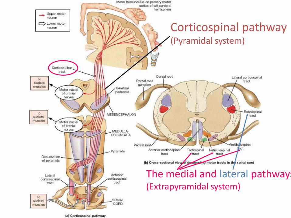
Corticospinal pathway(Pyramidal system)
The medial and lateral pathways(Extrapyramidal system)
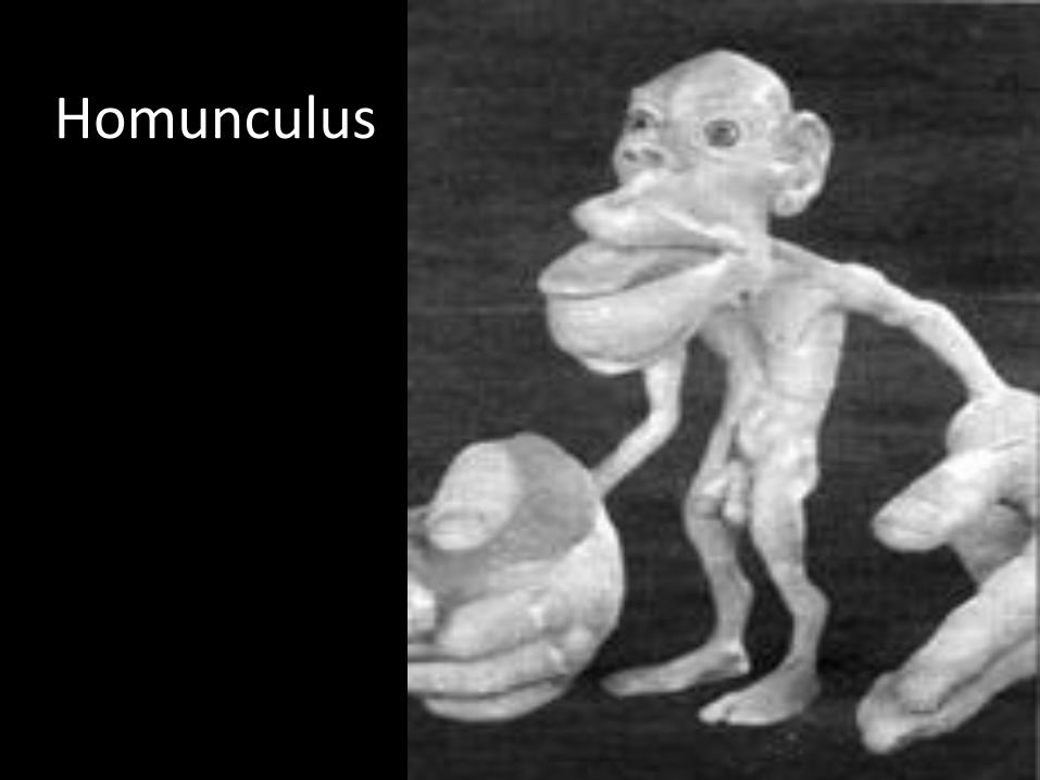
Homunculus
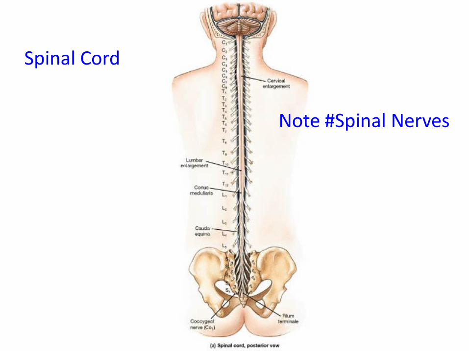
Spinal Cord
Note #Spinal Nerves
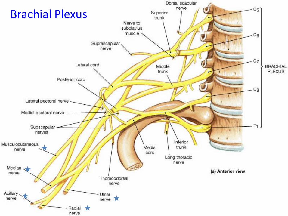
Brachial Plexus
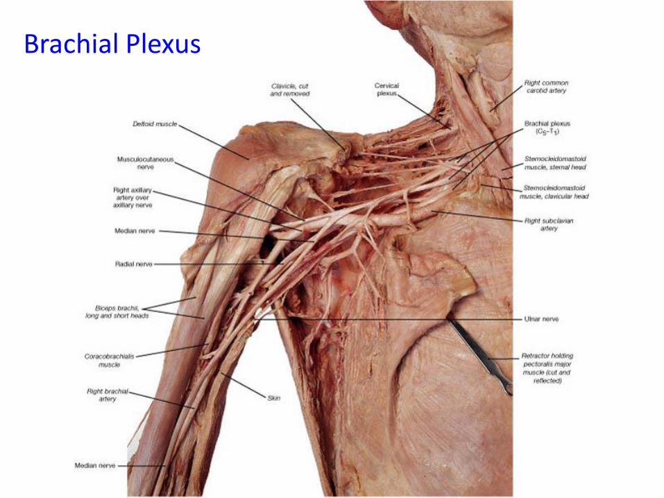
Brachial Plexus
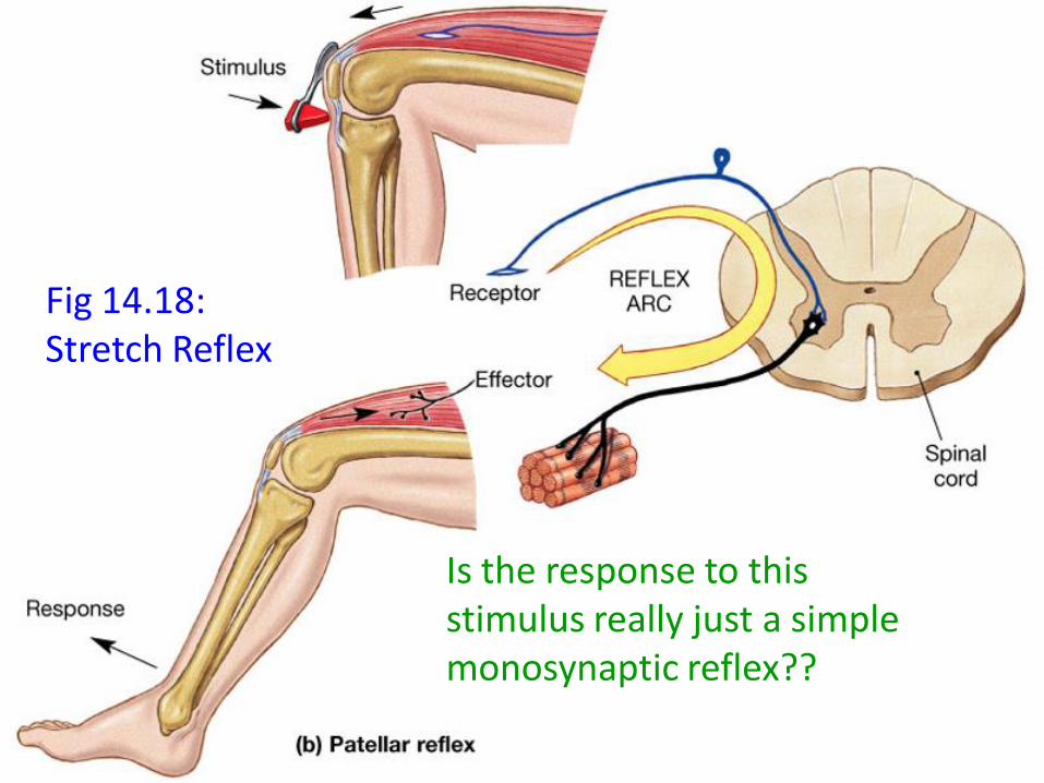
Fig 14.18: Stretch Reflex
Is the response to this stimulus really just a simple monosynaptic reflex??

Fig 16.2: Anterior Spinothalamic Tracts
Note decussation at spinal cord level
NB: Lateral spinothalamic tract:Pain and temperaturesensations
Human Anatomy, 3rd editionPrentice Hall, © 2001
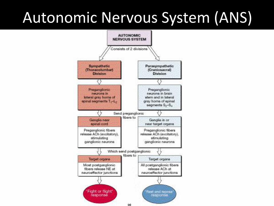
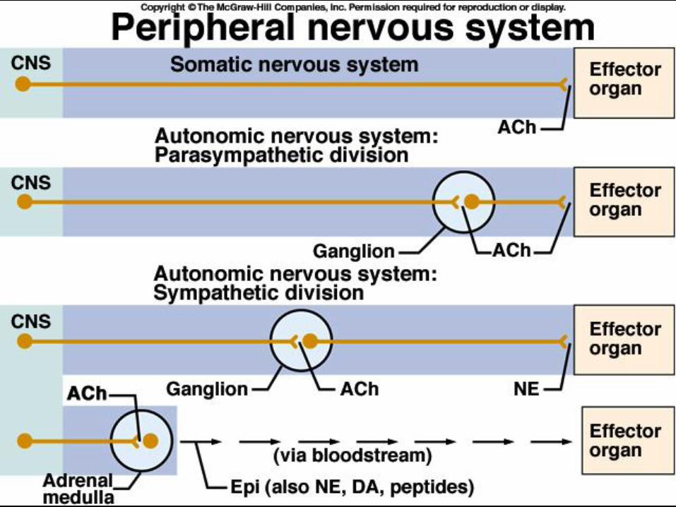
Autonomic Nervous System (ANS)

Human Anatomy, 3rd editionPrentice Hall, © 2001
Distribution of sympathetic postganglionic fibers (sympathetic chain)

Human Anatomy, 3rd editionPrentice Hall, © 2001
Sympathetic division of ANS
Acetylcholine T (ACh)
Catecholamines T (E & NE)
E
NE
Human Anatomy, 3rd editionPrentice Hall, © 2001
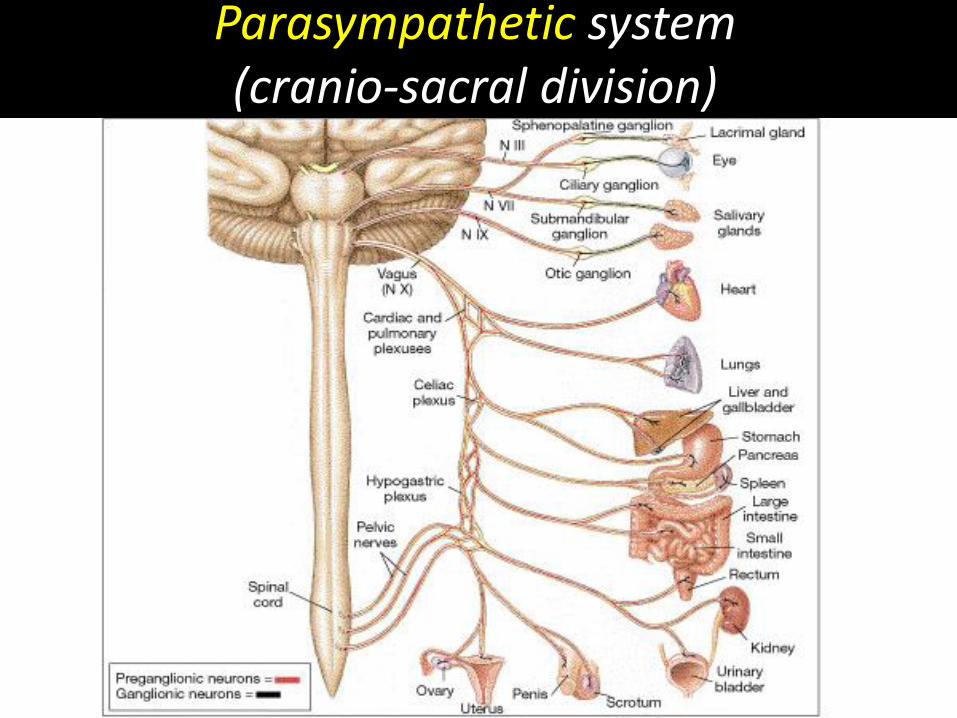
Parasympathetic system (cranio-sacral division)

Human Anatomy, 3rd editionPrentice Hall, © 2001
Organization of parasympatheticnervous system
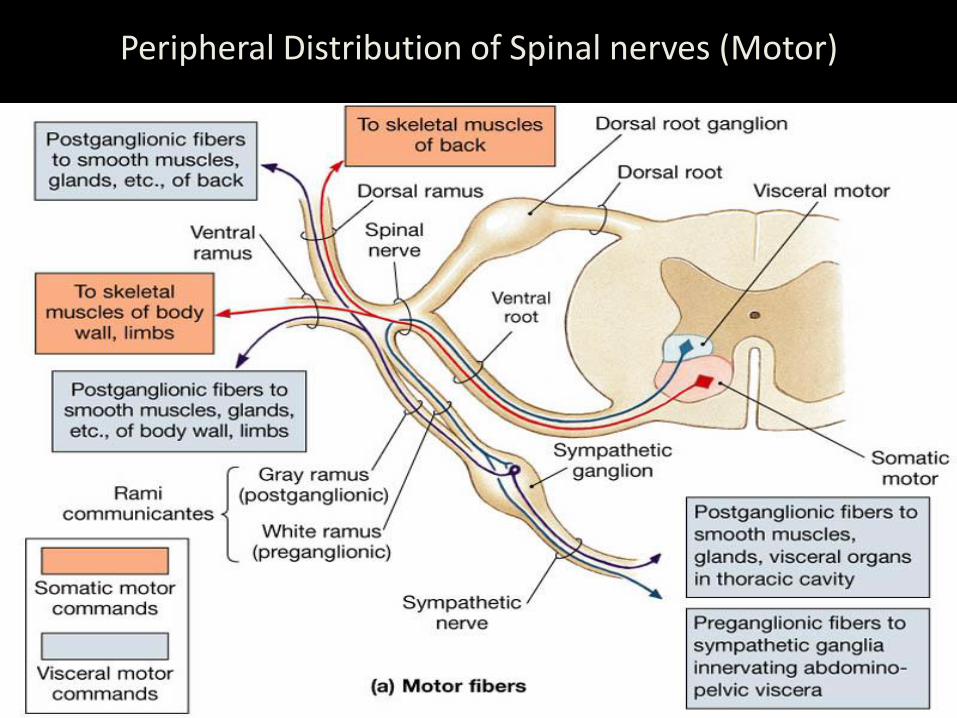
Peripheral Distribution of Spinal nerves (Motor)
Human Anatomy, 3rd editionPrentice Hall, © 2001

Protection of the brain
Bone (Skull)
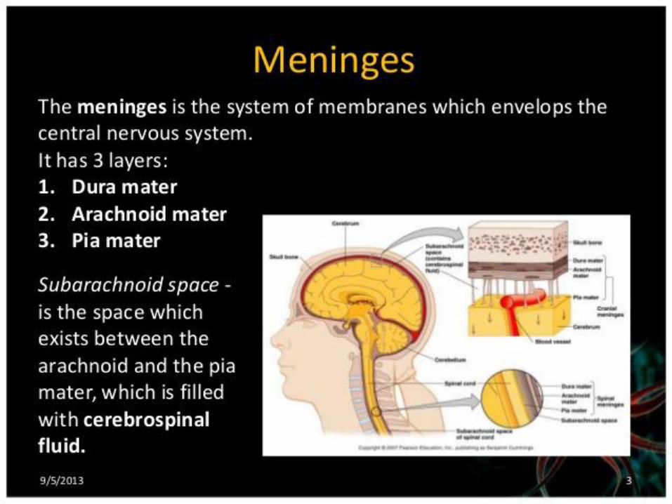
Connective tissue (meninges)
Fluid (CSF)

Figure 15-04a: Cadaver - cranial meninges

Superior view of cranial fossa
Tentorium cerebelli

Brain, cranium & meninges

Choroid plexus & brain barrier
Ependymal Cells
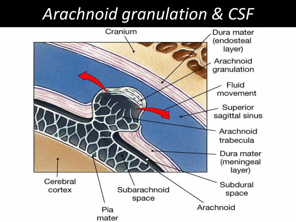
Arachnoid granulation & CSF

Anterior view - Ventricles

Ventricles of the brain
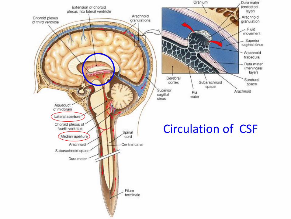
Figure 15-06: Circulation of CSF
Circulation of CSF
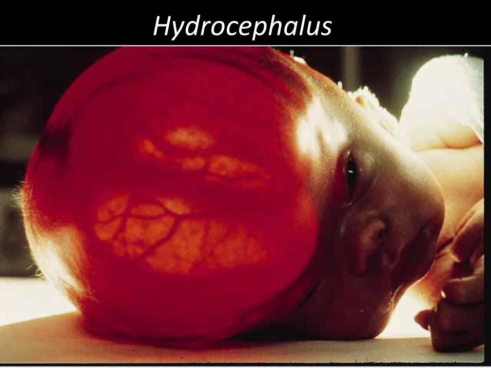
Hydrocephalus

Fig 14.4 Lumbar Puncture (Spinal tap)
The twelve cranial nerves
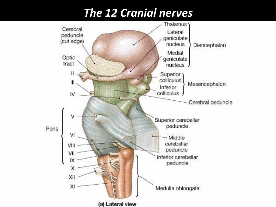
The 12 Cranial nerves
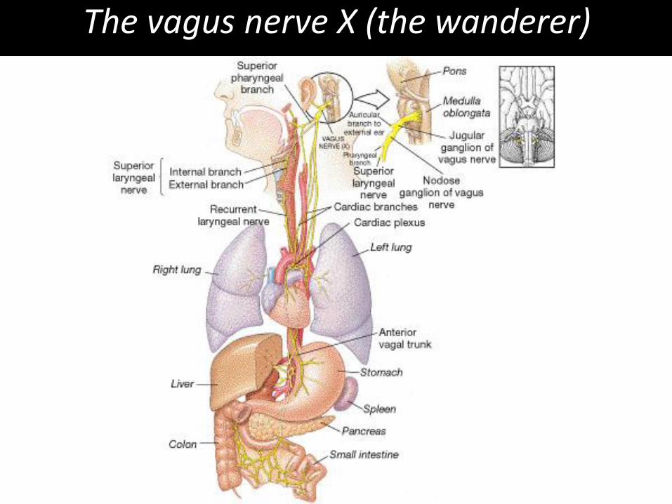
The vagus nerve X (the wanderer)
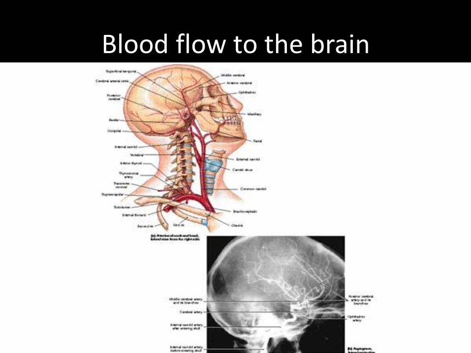
Blood flow to the brain
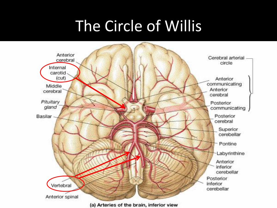
The Circle of Willis

Circle of Willis (up close)
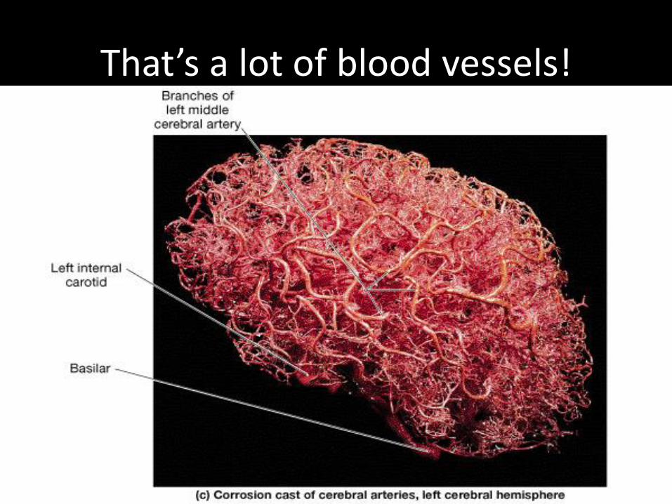
That’s a lot of blood vessels!
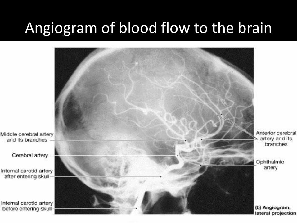
Angiogram of blood flow to the brain

Next!
Some medical conditions associated with aging!But first a short break

Neurodegenerative DiseasesMedicineNet.com
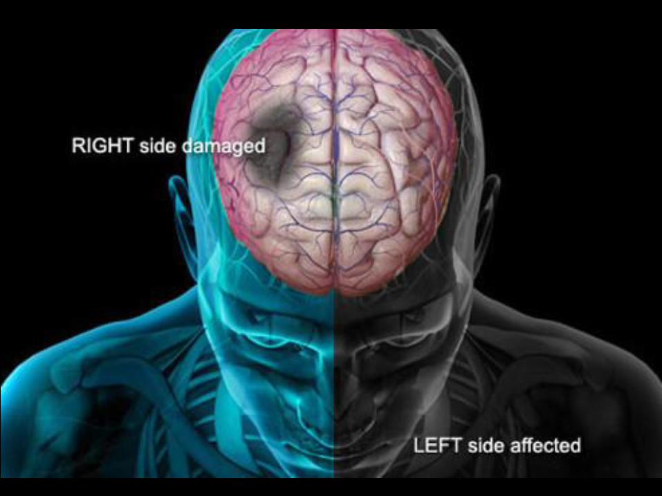
• Stroke
• Parkinson’s Disease
• Huntington’s Disease
• Dementia (Alzheimer’s)
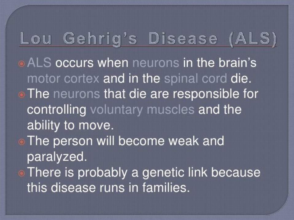
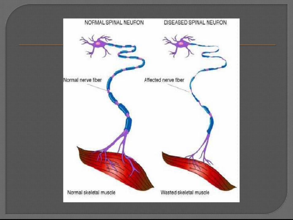
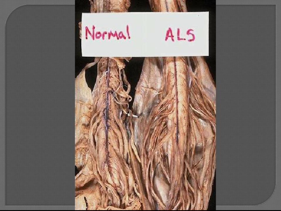
• Motor neuron diseases (amyotrophic lateral sclerosis [ALS], multiple sclerosis [MS])
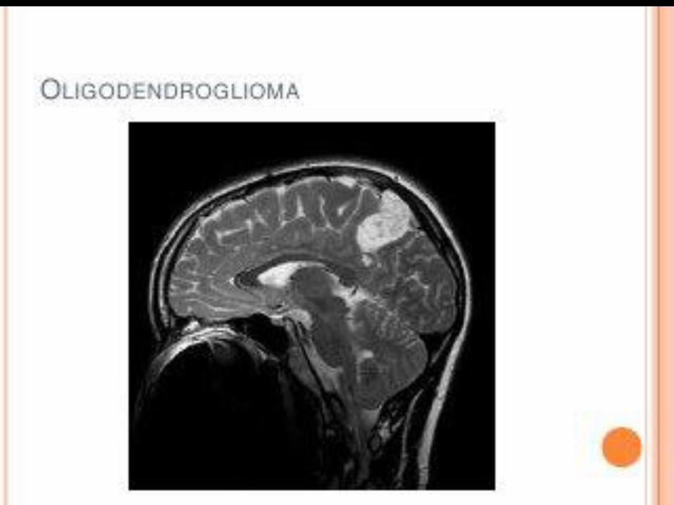
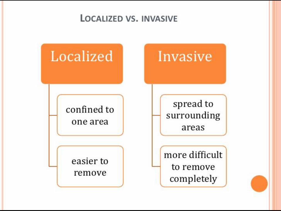
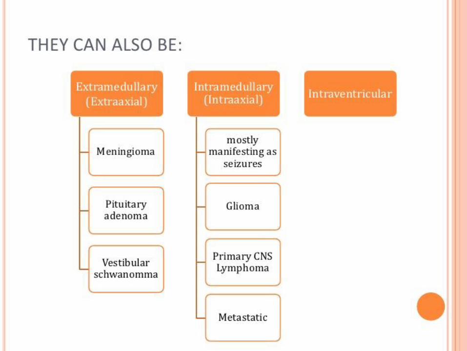
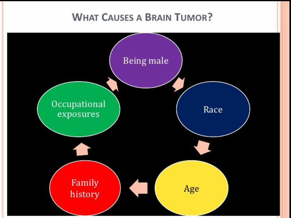
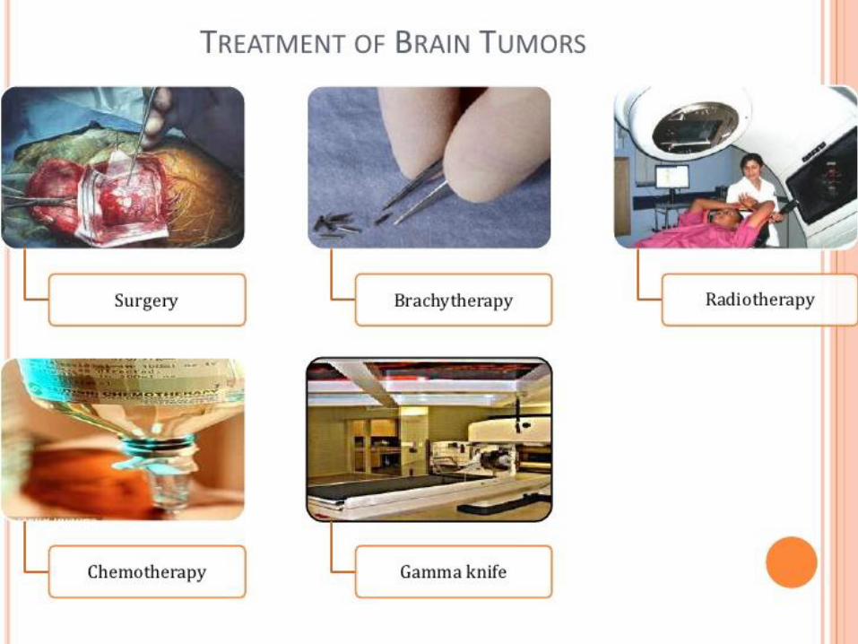
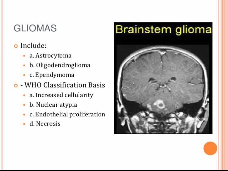
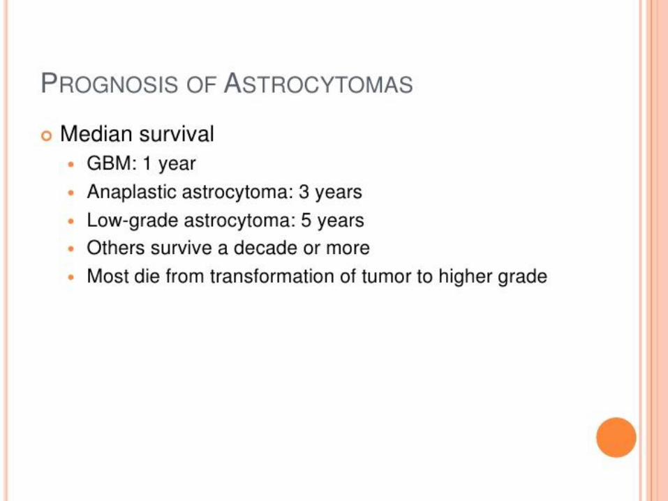
• Cancer (tumour)
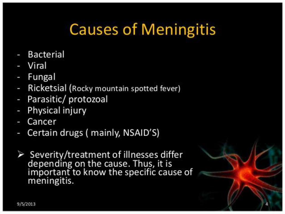
• Meningitis
• Other?

Causes of Stroke!

Ischemic Stroke

Hemorrhagic Stroke

Mini strokes (Transient Ischemic Attacks: TIA’s)
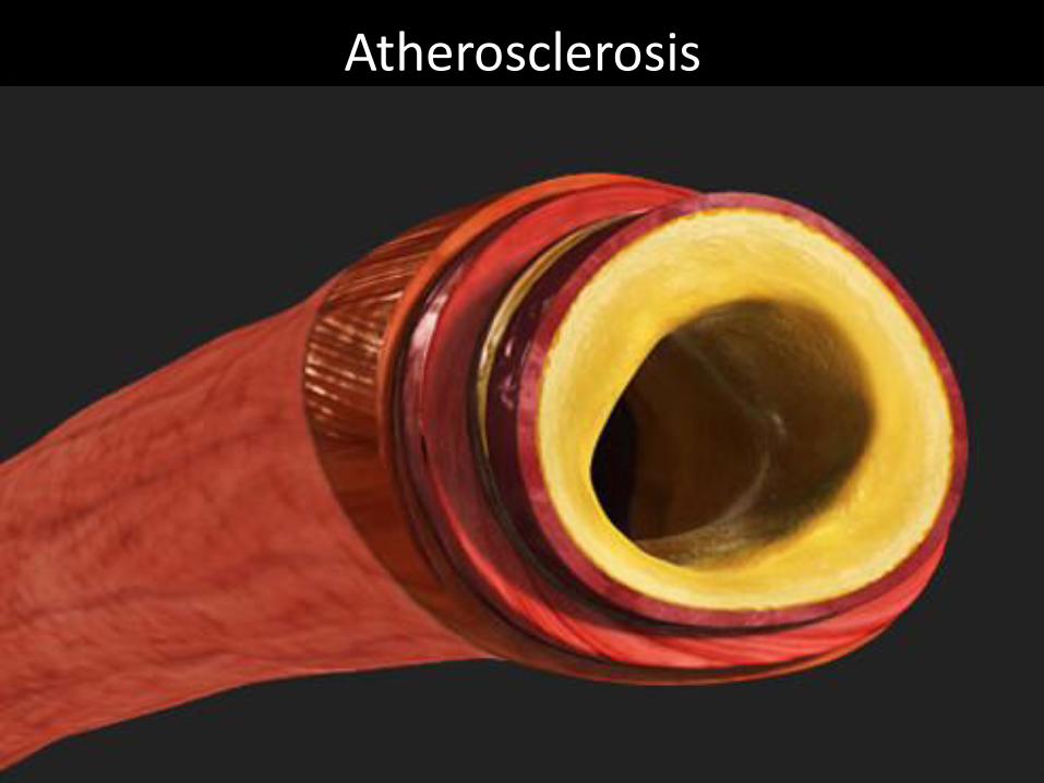
Atherosclerosis

Treatments

Medications
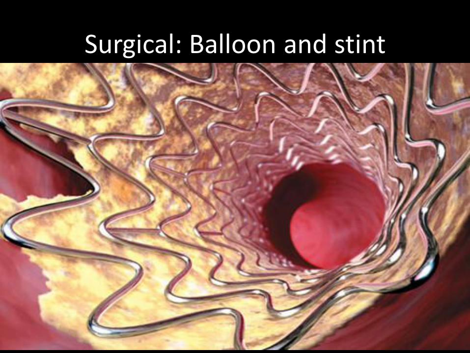
Surgical: Balloon and stint
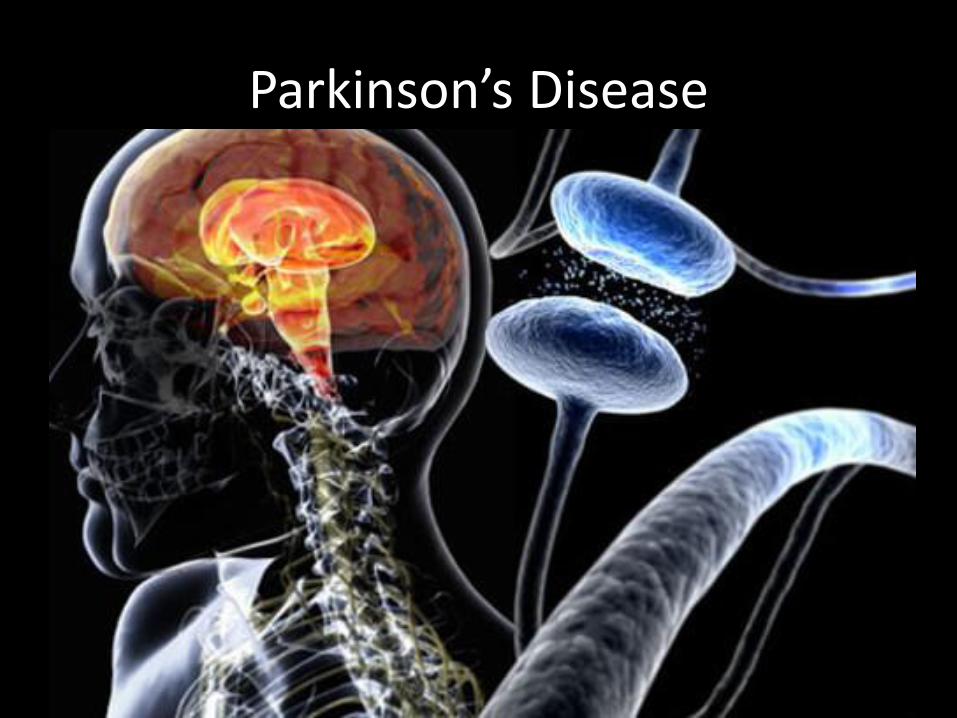
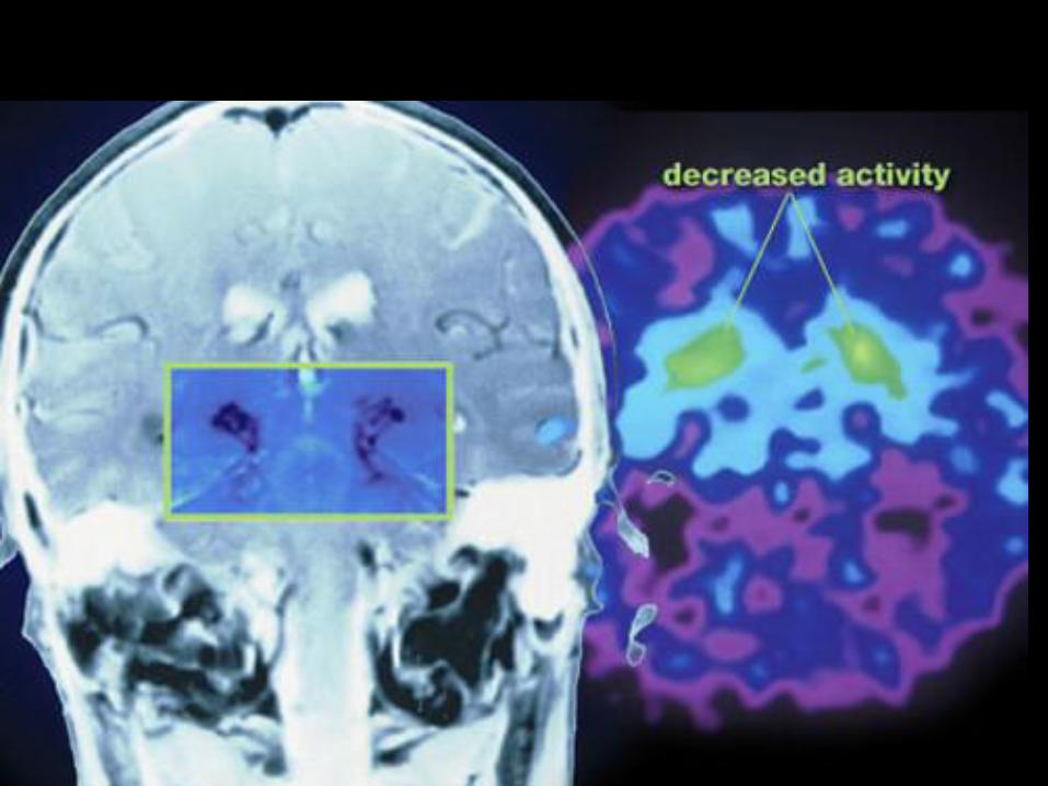
Parkinson’s Disease

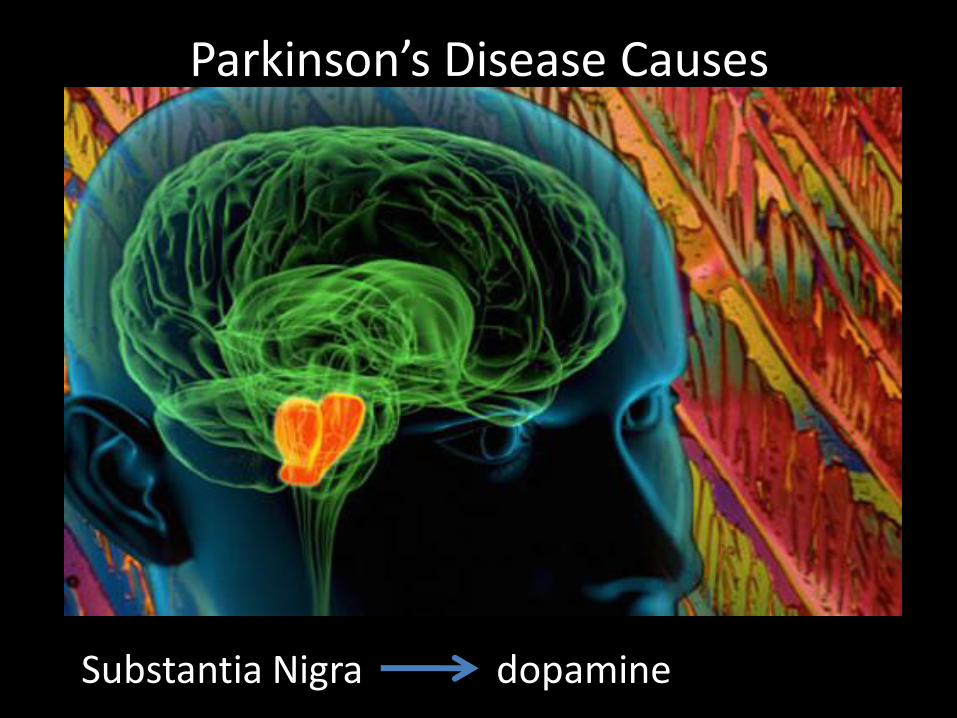
Parkinson’s Disease Causes
Substantia Nigra dopamine

TreatmentParkinson’s Treatment: LevodopaLevodopa, in the form of carbidopa and levodopa combined in a single tablet, has been the most effective medication to reduce or temporarily stop Parkinson's disease symptoms. The brain tissue converts this drug to dopamine. However, over time (about 6 years) the symptomatic reduction caused by the drug starts to fade and higher doses and other medications may be added. In addition, side effects of levodopa may develop (nausea, vomiting, mental changes, and involuntary movements), especially with use over years. These side effects can be reduced by slowly increasing the medication dose over time.
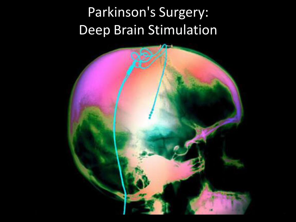
Parkinson's Surgery: Deep Brain Stimulation
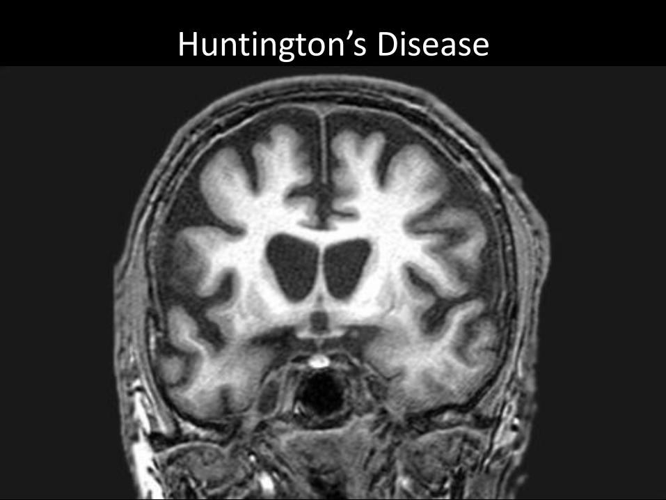
Huntington’s Disease
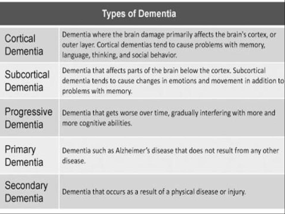
Dementia
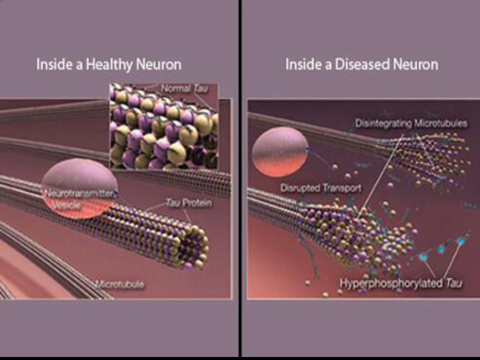
Alzeimer disease
https://en.wikipedia.org/wiki/Alzheimer%27s_disease

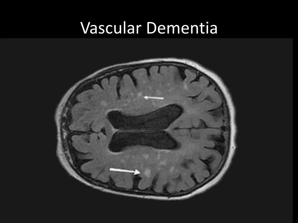
Vascular Dementia
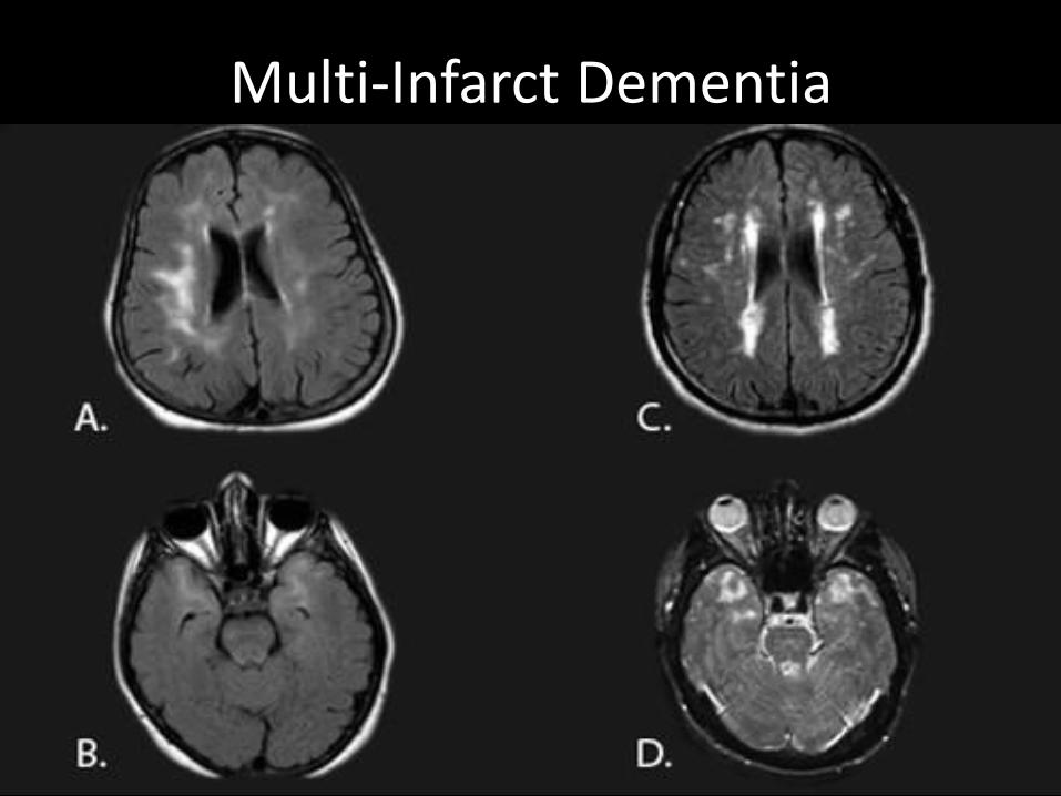
Multi-Infarct Dementia
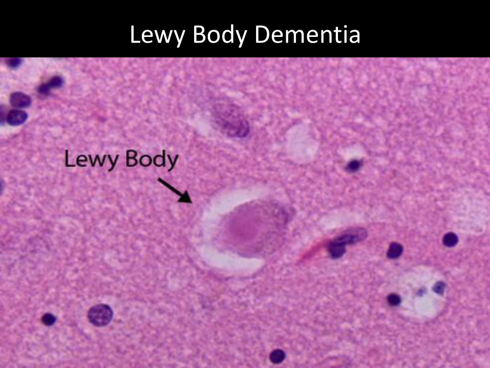
Lewy Body Dementia

Frontotemporal dementia (FTD)Pick’s Disease

Dementia Pugilistica

Corticobasal Degeneration (CBD)
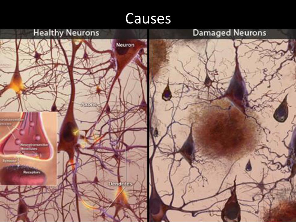
Causes

Treatments
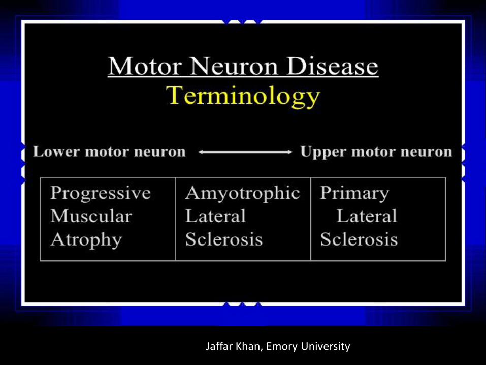
Jaffar Khan, Emory University


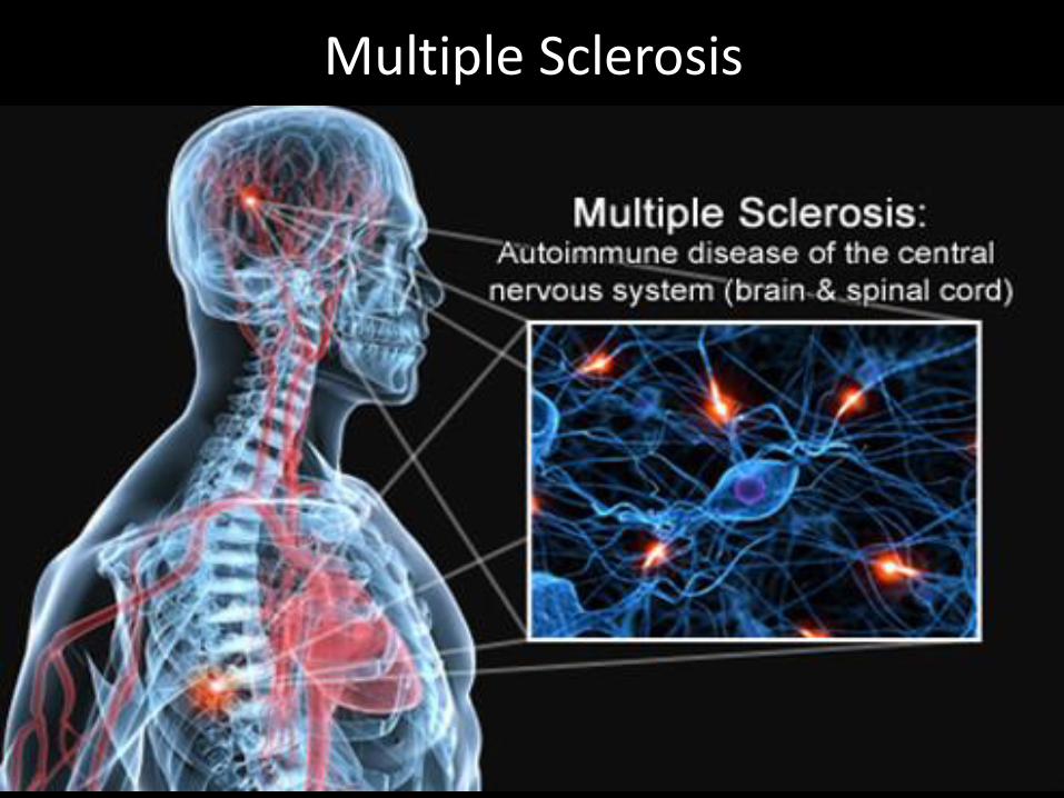
Multiple Sclerosis

What is MS?

Symptoms of MS
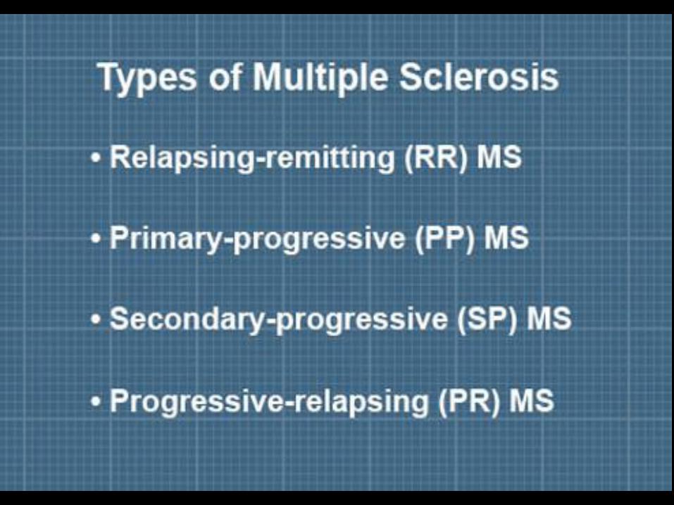

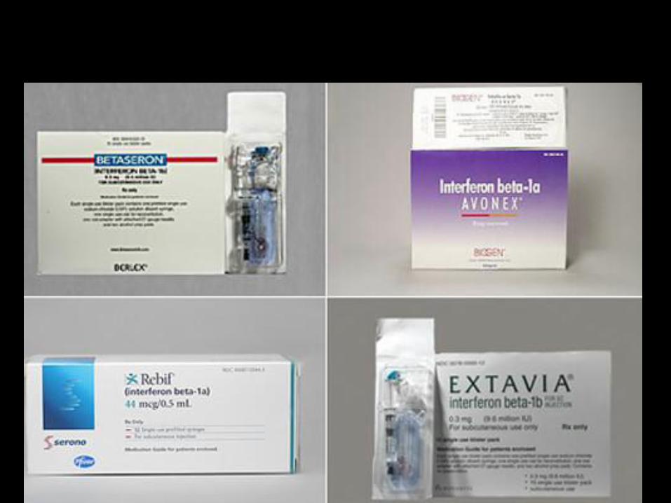
Symptoms and treatment


(From “slide share.net”)


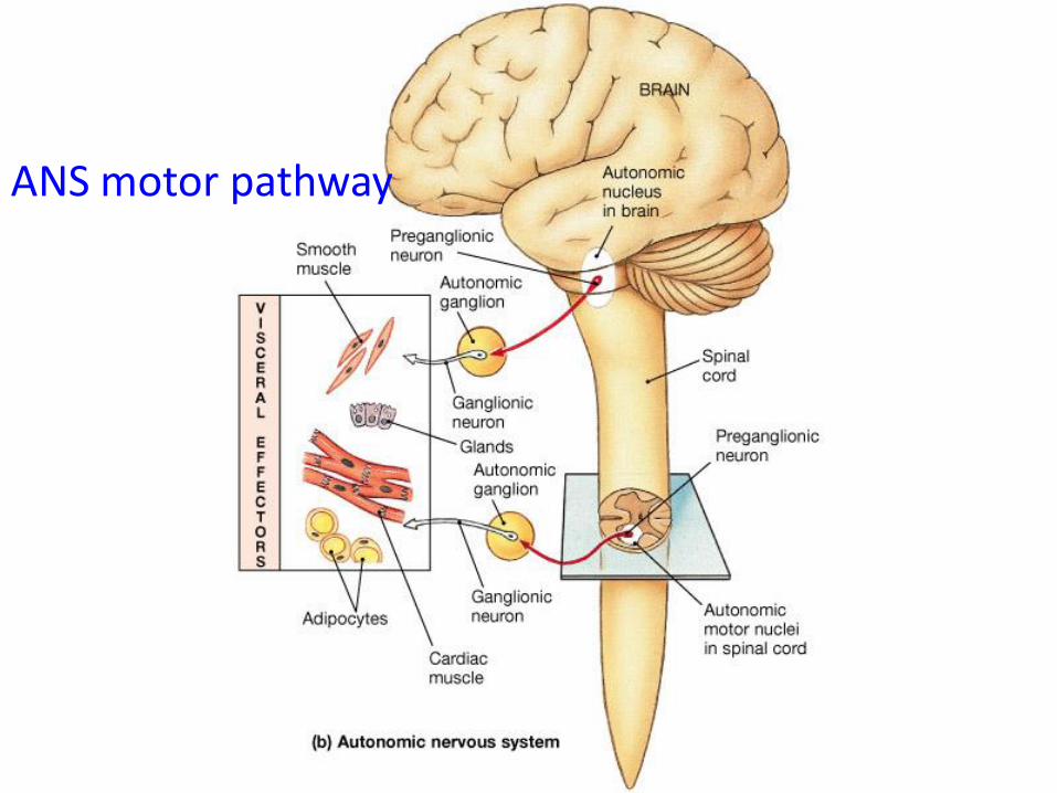
ANS motor pathway
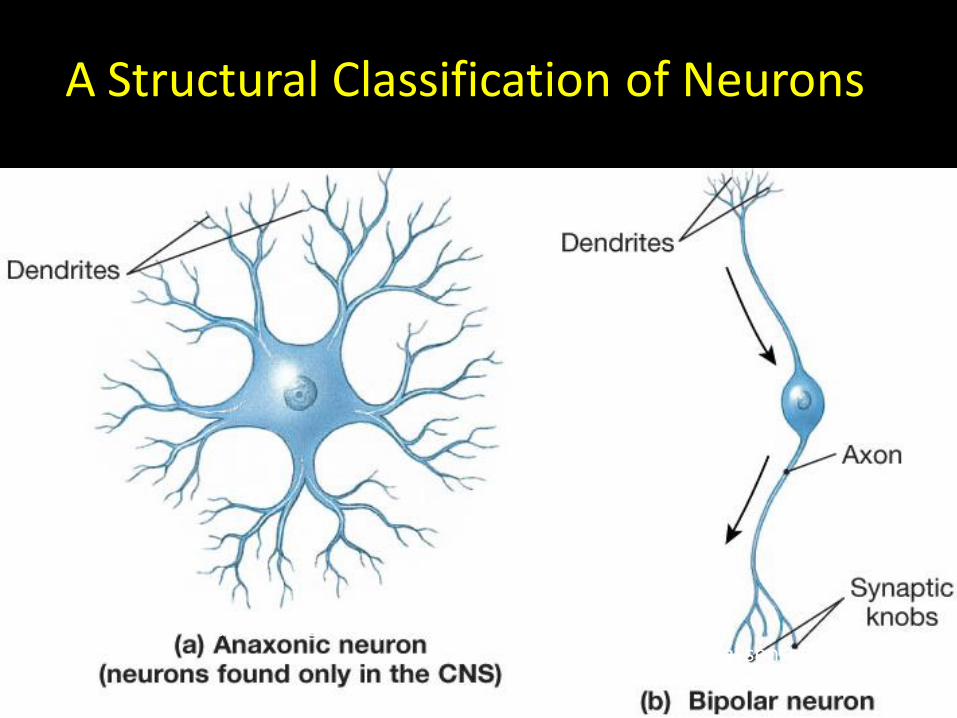
A Structural Classification of Neurons
• This classification is based on the placement of the cell body and the number of associated processes.
A Structural Classification of Neurons
(function not clear)(e.g. special senses)

A Structural Classification of Neurons
Sensory receptors Motor neurons
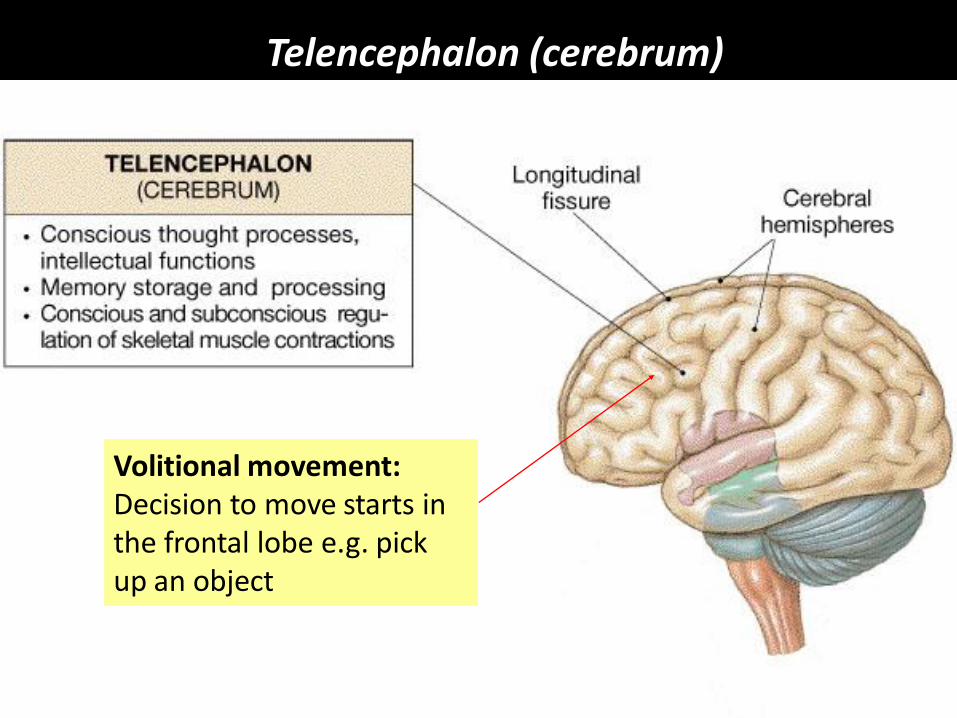
Telencephalon (cerebrum)
Volitional movement:Decision to move starts in the frontal lobe e.g. pickup an object
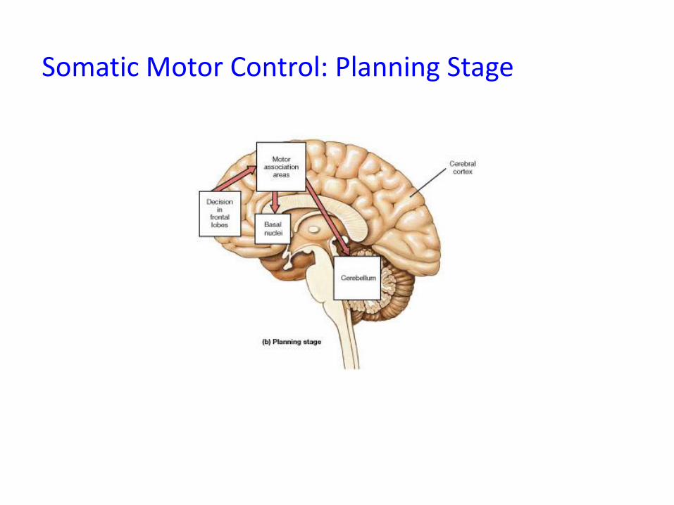
Somatic Motor Control: Planning Stage
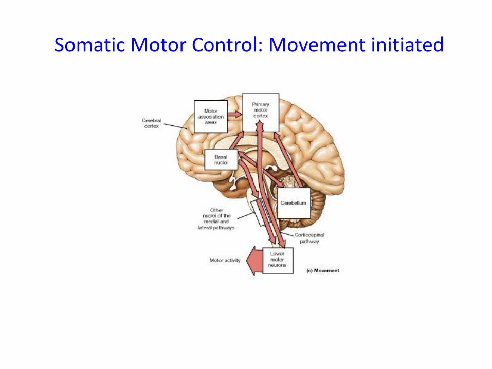
Somatic Motor Control: Movement initiated
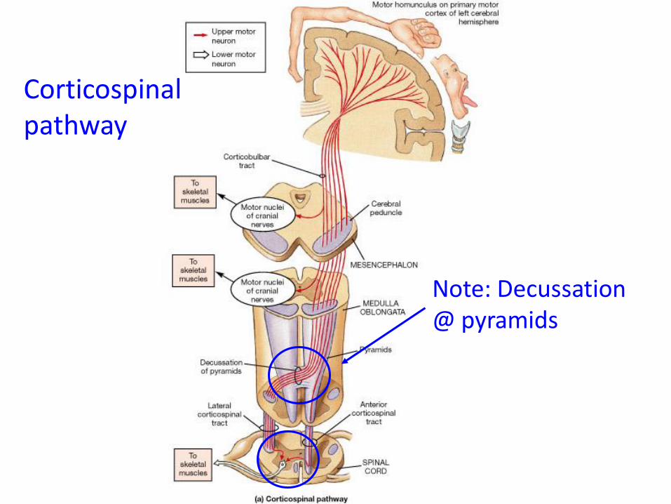
Corticospinalpathway
Note: Decussation @ pyramids
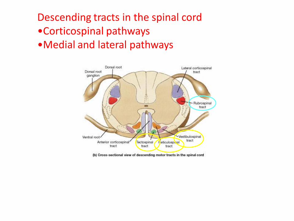
Descending tracts in the spinal cord•Corticospinal pathways•Medial and lateral pathways

Nuclei of the medial and lateral pathways
Previously calledthe Extrapyramidalpathway or system
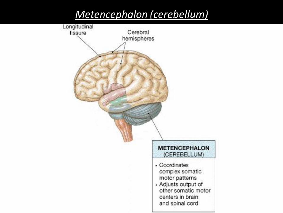
Metencephalon (cerebellum)

Medulla Oblongata
Connects S.Cord to Brain Stem
Cranial Nerves (VIII-XII)

Somatic
nervous system
Upper motor neuron
Lower motor neuron
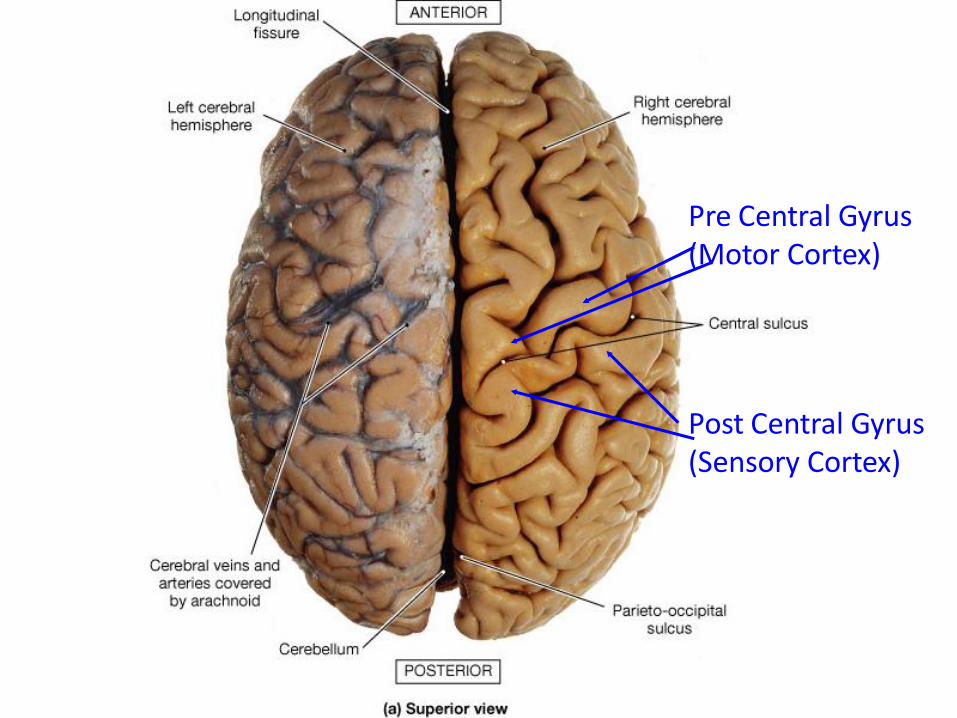
Figure 15-08a: Cerebral hemispheres (superior view)
Pre Central Gyrus (Motor Cortex)
Post Central Gyrus (Sensory Cortex)

Human Anatomy, 3rd editionPrentice Hall, © 2001
Neurotransmitters and receptors
Human Anatomy, 3rd editionPrentice Hall, © 2001
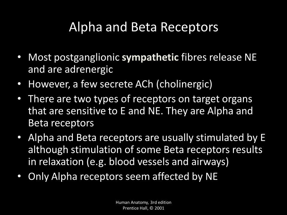
Alpha and Beta Receptors
• Most postganglionic sympathetic fibres release NE and are adrenergic
• However, a few secrete ACh (cholinergic)
• There are two types of receptors on target organs that are sensitive to E and NE. They are Alpha and Beta receptors
• Alpha and Beta receptors are usually stimulated by E although stimulation of some Beta receptors results in relaxation (e.g. blood vessels and airways)
• Only Alpha receptors seem affected by NE

Human Anatomy, 3rd editionPrentice Hall, © 2001
Nicotinic and Muscarinic Receptors
• Nicotinic receptors are found on ganglion cells of both sympathetic and parasympathetic nervous systems as well as at neuromuscular junctions. They are always stimulated by the release of Ach
• Muscarinic receptors are found in the neuroeffector junctions in parasympathetic nervous system as well as at the few cholinergic junctions in the sympathetic system. The effects may be excitatory or inhibitory depending on the specific enzymes in the target organ.
• In the parasympathetic system the effects of Ach may be excitatory or inhibitory depending on receptor