the c f bond as a conformational tool in organic and ... · the c–f bond as a conformational tool...
TRANSCRIPT
Page 1 of(page number not for citation purposes)
14
The C–F bond as a conformational tool in organicand biological chemistry
Luke Hunter
Review Open Access
Address:School of Chemistry, The University of Sydney, NSW 2006, Australia
Email:Luke Hunter - [email protected]
Keywords:conformation; functional molecules; organofluorine chemistry;stereochemistry; stereoelectronic effects
Beilstein Journal of Organic Chemistry 2010, 6, No. 38.doi:10.3762/bjoc.6.38
Received: 12 January 2010Accepted: 15 March 2010Published: 20 April 2010
Guest Editor: D. O’Hagan
© 2010 Hunter; licensee Beilstein-Institut.License and terms: see end of document.
AbstractOrganofluorine compounds are widely used in many different applications, ranging from pharmaceuticals and agrochemicals to
advanced materials and polymers. It has been recognised for many years that fluorine substitution can confer useful molecular prop-
erties such as enhanced stability and hydrophobicity. Another impact of fluorine substitution is to influence the conformations of
organic molecules. The stereoselective introduction of fluorine atoms can therefore be exploited as a conformational tool for the
synthesis of shape-controlled functional molecules. This review will begin by describing some general aspects of the C–F bond and
the various conformational effects associated with C–F bonds (i.e. dipole–dipole interactions, charge–dipole interactions and hyper-
conjugation). Examples of functional molecules that exploit these conformational effects will then be presented, drawing from a
diverse range of molecules including pharmaceuticals, organocatalysts, liquid crystals and peptides.
Page 1 of(page number not for citation purposes)
14
ReviewGeneral aspects of the C–F bondFluorine is a small atom, with an atomic radius intermediate
between that of hydrogen and oxygen (Table 1). The small size
of fluorine means that it can be incorporated into an organic
molecule as a replacement for hydrogen without dramatically
affecting the overall molecular size. However, fluorine is the
most electronegative element in the periodic table, consequently
the C–F bond is highly polarised and in this sense it is a
dramatic change from a C–H bond [1,2]. In the highly polar-
ised C–F bond, the fluorine atom bears a partial negative charge
and the carbon atom bears a partial positive charge, and these
charges attract each other. Hence, the C–F bond has significant
ionic character; it is a very short and strong bond. The fluorine
atom has three lone pairs, but because of fluorine’s high elec-
tronegativity these lone pairs are tightly held by the nucleus and
are therefore quite unreactive (fluorine is only a very weak
H-bond acceptor, for example). Another consequence of the
highly polarised nature of the C–F bond is a low-energy σ*
antibonding orbital, which is located behind the carbon atom in
the plane of the C–F bond. This vacant orbital can accept elec-
tron density from nearby electron-donating groups such as lone
Beilstein Journal of Organic Chemistry 2010, 6, No. 38.
Page 2 of(page number not for citation purposes)
14
pairs or σ-bonds and the importance of this will be discussed in
the next section. Overall, the C–F bond can be thought of as
short, strong, polarised and unreactive.
Table 1: Properties of some common elements and of their bonds tocarbon [2,3].
H F O N C Cl Br
Van der Waalsradius (Å)
1.20 1.47 1.52 1.55 1.70 1.75 1.85
Paulingelectronegativity
2.1 4.0 3.5 3.0 2.5 3.2 2.8
Length of singlebond to carbon(Å)
1.09 1.40 1.43 1.47 1.54 1.77 1.97
Strength of bondto carbon(kcal/mol)
98 105 84 70 83 77 66
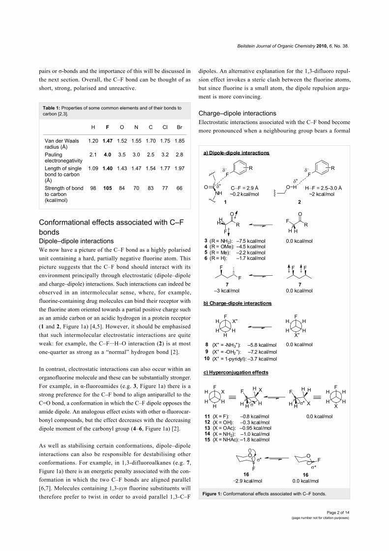
Conformational effects associated with C–FbondsDipole–dipole interactionsWe now have a picture of the C–F bond as a highly polarised
unit containing a hard, partially negative fluorine atom. This
picture suggests that the C–F bond should interact with its
environment principally through electrostatic (dipole–dipole
and charge–dipole) interactions. Such interactions can indeed be
observed in an intermolecular sense, where, for example,
fluorine-containing drug molecules can bind their receptor with
the fluorine atom oriented towards a partial positive charge such
as an amide carbon or an acidic hydrogen in a protein receptor
(1 and 2, Figure 1a) [4,5]. However, it should be emphasised
that such intermolecular electrostatic interactions are quite
weak: for example, the C–F···H–O interaction (2) is at most
one-quarter as strong as a “normal” hydrogen bond [2].
In contrast, electrostatic interactions can also occur within an
organofluorine molecule and these can be substantially stronger.
For example, in α-fluoroamides (e.g. 3, Figure 1a) there is a
strong preference for the C–F bond to align antiparallel to the
C=O bond, a conformation in which the C–F dipole opposes the
amide dipole. An analogous effect exists with other α-fluorocar-
bonyl compounds, but the effect decreases with the decreasing
dipole moment of the carbonyl group (4–6, Figure 1a) [2].
As well as stabilising certain conformations, dipole–dipole
interactions can also be responsible for destabilising other
conformations. For example, in 1,3-difluoroalkanes (e.g. 7,
Figure 1a) there is an energetic penalty associated with the con-
formation in which the two C–F bonds are aligned parallel
[6,7]. Molecules containing 1,3-syn fluorine substituents will
therefore prefer to twist in order to avoid parallel 1,3-C–FFigure 1: Conformational effects associated with C–F bonds.
dipoles. An alternative explanation for the 1,3-difluoro repul-
sion effect invokes a steric clash between the fluorine atoms,
but since fluorine is a small atom, the dipole repulsion argu-
ment is more convincing.
Charge–dipole interactionsElectrostatic interactions associated with the C–F bond become
more pronounced when a neighbouring group bears a formal
Beilstein Journal of Organic Chemistry 2010, 6, No. 38.
Page 3 of(page number not for citation purposes)
14
charge [8]. For example, in the 2-fluoroethylammonium ion (8)
and protonated 2-fluoroethanol (9) (Figure 1b), the gauche
conformers are strongly preferred because these bring the
(partially negative) fluorine atoms close to the formally posi-
tively-charged oxygen or nitrogen [9]. It is possible to envisage
an intramolecular hydrogen bond helping to stabilise the gauche
conformers of 8 and 9, but the gauche preference is also main-
tained in systems such as 10 (Figure 1b) which cannot accom-
modate a hydrogen bond [10], confirming that the
charge–dipole interaction is more important than any weak
H-bonding in these systems.
Hyperconjugation effectsConsider the well-studied molecule 1,2-difluoroethane (11,
Figure 1c). There are two possible staggered conformers, with
the fluorine atoms either gauche or anti. NMR and molecular
modelling studies have shown that the gauche conformer is
lower in energy, which is perhaps a surprising result since the
fluorine atoms might reasonably be expected to repel each
other. What effect overrides the difluoro repulsion and stabil-
ises the gauche conformer?
There is a vacant low-energy σ* antibonding orbital associated
with each C–F bond (Figure 1c). In the gauche conformer of 11,
both σ*CF orbitals are aligned with adjacent C–H bonds, which
can donate electron density into the σ*CF orbitals in a process
known as hyperconjugation [1,2]. Feeding electron density into
an antibonding orbital in this way is equivalent to partially
breaking the bond, so when hyperconjugation occurs the C–F
bonds of 11 become longer and less covalent in character.
However the bonds are still strong because the fluorine atoms
have now become even more negative, so they are more
strongly attracted to the partially positive carbon atoms.
Overall, hyperconjugation is a stabilising effect and thus will
lower the energy of the gauche conformer of 11. In contrast, in
the anti conformer of 11 each σ*CF orbital is now aligned with
an adjacent C–F bond, which is highly polarised and less elec-
tron releasing than a C–H bond and hence hyperconjugation
does not occur.
The gauche effect is only a subtle conformational influence
compared with the dipole–dipole and charge–dipole interac-
tions described earlier. Nevertheless, the gauche effect is very
general and applies in many other systems in addition to 1,2-
difluoroalkanes. For example, compounds containing F–C–C–O
and F–C–C–N also experience this effect (12–15, Figure 1c)
[9,11-13]. In general, more electronegative substituents give
rise to stronger gauche effects. It should be noted that there are
other explanations for the gauche effect in addition to the
hyperconjugation argument presented above. For example, the
“bent bond” theory [11] is an alternative explanation for the
gauche preference of compounds 11–15 (Figure 1c). However,
the hyperconjugation argument is more widely cited today [2]
and will be exclusively quoted in this review.
The examples of hyperconjugation presented thus far (11–15,
Figure 1c) all feature σ-bonds as the electron-donating groups.
However, hyperconjugation can also occur with other electron
donors such as lone pairs [1,14] or π-systems [15]. In each case,
conformations which align the electron-donating group with the
σ*CF orbital will be favoured (e.g. 16, Figure 1c).
In summary, fluorine atoms influence the conformation of
organic molecules through dipole–dipole interactions,
charge–dipole interactions and hyperconjugation effects. All of
these influences can be rationalised by considering that the C–F
bond is short, strong and highly polarised. The remainder of this
review will focus on examples of shape-controlled functional
molecules that exploit the C–F bond as a conformational tool.
Bioactive small moleculesDespite being the most abundant halogen in the Earth’s crust,
fluorine is almost completely absent from natural products
chemistry [16]. However, in contrast to the paucity of fluorin-
ated molecules in nature, there are many synthetic (non-natural)
organofluorine compounds with valuable biological activity. Of
these, an interesting subset exploit the C–F bond specifically as
a conformational tool and some examples of such molecules are
examined below.
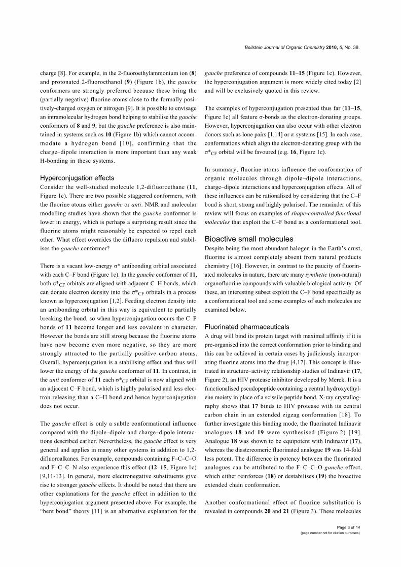
Fluorinated pharmaceuticalsA drug will bind its protein target with maximal affinity if it is
pre-organised into the correct conformation prior to binding and
this can be achieved in certain cases by judiciously incorpor-
ating fluorine atoms into the drug [4,17]. This concept is illus-
trated in structure–activity relationship studies of Indinavir (17,
Figure 2), an HIV protease inhibitor developed by Merck. It is a
functionalised pseudopeptide containing a central hydroxyethyl-
ene moiety in place of a scissile peptide bond. X-ray crystallog-
raphy shows that 17 binds to HIV protease with its central
carbon chain in an extended zigzag conformation [18]. To
further investigate this binding mode, the fluorinated Indinavir
analogues 18 and 19 were synthesised (Figure 2) [19].
Analogue 18 was shown to be equipotent with Indinavir (17),
whereas the diastereomeric fluorinated analogue 19 was 14-fold
less potent. The difference in potency between the fluorinated
analogues can be attributed to the F–C–C–O gauche effect,
which either reinforces (18) or destabilises (19) the bioactive
extended chain conformation.
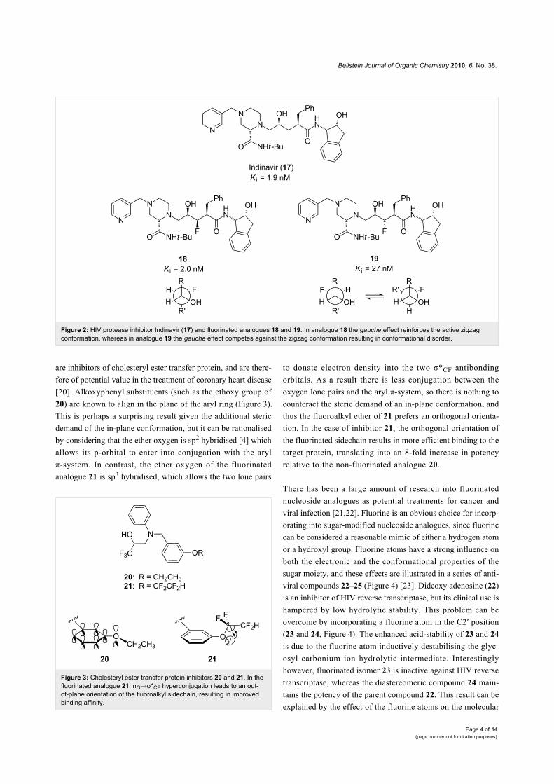
Another conformational effect of fluorine substitution is
revealed in compounds 20 and 21 (Figure 3). These molecules
Beilstein Journal of Organic Chemistry 2010, 6, No. 38.
Page 4 of(page number not for citation purposes)
14
Figure 2: HIV protease inhibitor Indinavir (17) and fluorinated analogues 18 and 19. In analogue 18 the gauche effect reinforces the active zigzagconformation, whereas in analogue 19 the gauche effect competes against the zigzag conformation resulting in conformational disorder.
Figure 3: Cholesteryl ester transfer protein inhibitors 20 and 21. In thefluorinated analogue 21, nO→σ*CF hyperconjugation leads to an out-of-plane orientation of the fluoroalkyl sidechain, resulting in improvedbinding affinity.
are inhibitors of cholesteryl ester transfer protein, and are there-
fore of potential value in the treatment of coronary heart disease
[20]. Alkoxyphenyl substituents (such as the ethoxy group of
20) are known to align in the plane of the aryl ring (Figure 3).
This is perhaps a surprising result given the additional steric
demand of the in-plane conformation, but it can be rationalised
by considering that the ether oxygen is sp2 hybridised [4] which
allows its p-orbital to enter into conjugation with the aryl
π-system. In contrast, the ether oxygen of the fluorinated
analogue 21 is sp3 hybridised, which allows the two lone pairs
to donate electron density into the two σ*CF antibonding
orbitals. As a result there is less conjugation between the
oxygen lone pairs and the aryl π-system, so there is nothing to
counteract the steric demand of an in-plane conformation, and
thus the fluoroalkyl ether of 21 prefers an orthogonal orienta-
tion. In the case of inhibitor 21, the orthogonal orientation of
the fluorinated sidechain results in more efficient binding to the
target protein, translating into an 8-fold increase in potency
relative to the non-fluorinated analogue 20.
There has been a large amount of research into fluorinated
nucleoside analogues as potential treatments for cancer and
viral infection [21,22]. Fluorine is an obvious choice for incorp-
orating into sugar-modified nucleoside analogues, since fluorine
can be considered a reasonable mimic of either a hydrogen atom
or a hydroxyl group. Fluorine atoms have a strong influence on
both the electronic and the conformational properties of the
sugar moiety, and these effects are illustrated in a series of anti-
viral compounds 22–25 (Figure 4) [23]. Dideoxy adenosine (22)
is an inhibitor of HIV reverse transcriptase, but its clinical use is
hampered by low hydrolytic stability. This problem can be
overcome by incorporating a fluorine atom in the C2′ position
(23 and 24, Figure 4). The enhanced acid-stability of 23 and 24
is due to the fluorine atom inductively destabilising the glyc-
osyl carbonium ion hydrolytic intermediate. Interestingly
however, fluorinated isomer 23 is inactive against HIV reverse
transcriptase, whereas the diastereomeric compound 24 main-
tains the potency of the parent compound 22. This result can be
explained by the effect of the fluorine atoms on the molecular
Beilstein Journal of Organic Chemistry 2010, 6, No. 38.
Page 5 of(page number not for citation purposes)
14
conformations of 23 and 24 [24]. In isomer 23, the fluorine
atom aligns gauche to the ring oxygen, resulting in a C3′-endo
ring pucker which is not recognised by HIV reverse tran-
scriptase [24,25]. By contrast, in isomer 24 the fluorine once
again aligns gauche to the ring oxygen, but this leads to a C3′-
exo ring pucker which is known to be optimal for biological
activity. This effect can be explored further by incorporating a
second fluorine atom at the C3′ position (25, Figure 4). If the
C3′ stereochemistry is appropriate, the C3′-exo ring pucker can
be further reinforced, with both fluorines aligned gauche to the
ring oxygen (note that a potential difluoro gauche effect is over-
ridden in this case) [24,26].
Figure 4: HIV reverse transcriptase inhibitor 22 and acid-stable fluorin-ated analogues 23–25. The F–C–C–O gauche effect influences thering conformations of 23–25.
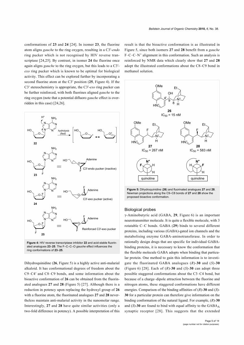
Dihydroquinidine (26, Figure 5) is a highly active anti-malarial
alkaloid. It has conformational degrees of freedom about the
C9–C4′ and C8–C9 bonds, and some information about the
bioactive conformation of 26 can be obtained from the fluorin-
ated analogues 27 and 28 (Figure 5) [27]. Although there is a
reduction in potency upon replacing the hydroxyl group of 26
with a fluorine atom, the fluorinated analogues 27 and 28 never-
theless maintain anti-malarial activity in the nanomolar range.
Interestingly, 27 and 28 have quite similar activities (only a
two-fold difference in potency). A possible interpretation of this
result is that the bioactive conformation is as illustrated in
Figure 5, since both isomers 27 and 28 benefit from a gauche
F–C–C–N+ alignment in this conformation. Such an analysis is
reinforced by NMR data which clearly show that 27 and 28
adopt the illustrated conformations about the C8–C9 bond in
methanol solution.
Figure 5: Dihydroquinidine (26) and fluorinated analogues 27 and 28.Newman projections along the C9–C8 bonds of 27 and 28 show theproposed bioactive conformation.
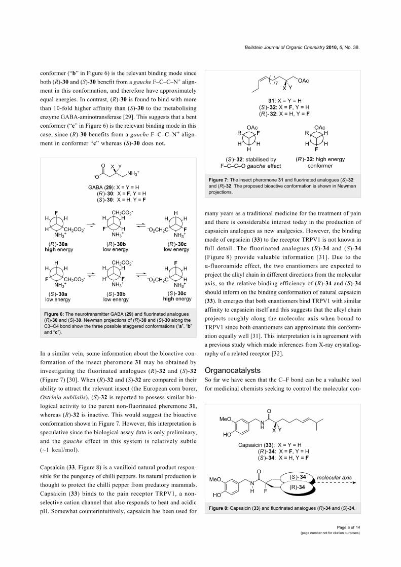
Biological probesγ-Aminobutyric acid (GABA, 29, Figure 6) is an important
neurotransmitter molecule. It is quite a flexible molecule, with 3
rotatable C–C bonds. GABA (29) binds to several different
proteins, including various (GABA)-gated ion channels and the
metabolising enzyme GABA-aminotransferase. In order to
rationally design drugs that are specific for individual GABA-
binding proteins, it is necessary to know the conformation that
the flexible molecule GABA adopts when binding that particu-
lar protein. One method to gain this information is to investi-
gate the fluorinated GABA analogues (R)-30 and (S)-30
(Figure 6) [28]. Each of (R)-30 and (S)-30 can adopt three
possible staggered conformations about the C3–C4 bond, but
because of a charge–dipole attraction between the fluorine and
nitrogen atoms, these staggered conformations have different
energies. Comparison of the binding affinities of (R)-30 and (S)-
30 for a particular protein can therefore give information on the
binding conformation of the natural ligand. For example, (R)-30
and (S)-30 are found to bind with equal affinity to the GABAA
synaptic receptor [28]. This suggests that the extended
Beilstein Journal of Organic Chemistry 2010, 6, No. 38.
Page 6 of(page number not for citation purposes)
14
conformer (“b” in Figure 6) is the relevant binding mode since
both (R)-30 and (S)-30 benefit from a gauche F–C–C–N+ align-
ment in this conformation, and therefore have approximately
equal energies. In contrast, (R)-30 is found to bind with more
than 10-fold higher affinity than (S)-30 to the metabolising
enzyme GABA-aminotransferase [29]. This suggests that a bent
conformer (“c” in Figure 6) is the relevant binding mode in this
case, since (R)-30 benefits from a gauche F–C–C–N+ align-
ment in conformer “c” whereas (S)-30 does not.
Figure 6: The neurotransmitter GABA (29) and fluorinated analogues(R)-30 and (S)-30. Newman projections of (R)-30 and (S)-30 along theC3–C4 bond show the three possible staggered conformations (“a”, “b”and “c”).
In a similar vein, some information about the bioactive con-
formation of the insect pheromone 31 may be obtained by
investigating the fluorinated analogues (R)-32 and (S)-32
(Figure 7) [30]. When (R)-32 and (S)-32 are compared in their
ability to attract the relevant insect (the European corn borer,
Ostrinia nubilalis), (S)-32 is reported to possess similar bio-
logical activity to the parent non-fluorinated pheremone 31,
whereas (R)-32 is inactive. This would suggest the bioactive
conformation shown in Figure 7. However, this interpretation is
speculative since the biological assay data is only preliminary,
and the gauche effect in this system is relatively subtle
(~1 kcal/mol).
Capsaicin (33, Figure 8) is a vanilloid natural product respon-
sible for the pungency of chilli peppers. Its natural production is
thought to protect the chilli pepper from predatory mammals.
Capsaicin (33) binds to the pain receptor TRPV1, a non-
selective cation channel that also responds to heat and acidic
pH. Somewhat counterintuitively, capsaicin has been used for
Figure 7: The insect pheromone 31 and fluorinated analogues (S)-32and (R)-32. The proposed bioactive conformation is shown in Newmanprojections.
Figure 8: Capsaicin (33) and fluorinated analogues (R)-34 and (S)-34.
many years as a traditional medicine for the treatment of pain
and there is considerable interest today in the production of
capsaicin analogues as new analgesics. However, the binding
mode of capsaicin (33) to the receptor TRPV1 is not known in
full detail. The fluorinated analogues (R)-34 and (S)-34
(Figure 8) provide valuable information [31]. Due to the
α-fluoroamide effect, the two enantiomers are expected to
project the alkyl chain in different directions from the molecular
axis, so the relative binding efficiency of (R)-34 and (S)-34
should inform on the binding conformation of natural capsaicin
(33). It emerges that both enantiomers bind TRPV1 with similar
affinity to capsaicin itself and this suggests that the alkyl chain
projects roughly along the molecular axis when bound to
TRPV1 since both enantiomers can approximate this conform-
ation equally well [31]. This interpretation is in agreement with
a previous study which made inferences from X-ray crystallog-
raphy of a related receptor [32].
OrganocatalystsSo far we have seen that the C–F bond can be a valuable tool
for medicinal chemists seeking to control the molecular con-
Beilstein Journal of Organic Chemistry 2010, 6, No. 38.
Page 7 of(page number not for citation purposes)
14
formation of drugs and bioprobes. This section will show that
the C–F bond is also emerging as a useful tool in the field of
catalysis. Recent reports have shown that organocatalysts can
be conformationally “fine-tuned” by fluorine substitution for
improved activity and selectivity.
Pyrrolidine 35 (Figure 9) is a highly selective catalyst for the
epoxidation of α,β-unsaturated aldehydes (e.g. 36) [33]. In the
first step of the reaction, aldehyde 36 and pyrrolidine 35 react
together to form the iminium ion 37. This has a LUMO-
lowering effect (analogous to Lewis-acid activation of 36)
which makes 37 more reactive towards nucleophiles [34]. In
intermediate 37, the fluorine atom aligns gauche to the positive-
ly-charged nitrogen atom (Figure 9, inset), resulting in a phenyl
group shielding the top (re) face of the alkene. Hydrogen
peroxide consequently attacks from the bottom (si) face, leading
to epoxide 38 with high enantioselectivity. In a control experi-
ment, the related organocatalyst 2-(diphenylmethyl)pyrrolidine
(containing a hydrogen atom instead of the fluorine atom of 35)
also catalyses the same reaction but with lower enantioselectiv-
ity suggesting that the fluorine atom of 35 helps to rigidify the
activated intermediate and thereby enhances selectivity.
Figure 9: Asymmetric epoxidation reaction catalysed by pyrrolidine 35.Inset: the geometry of the activated iminium ion intermediate 37 isstabilised by a gauche F–C–C–N+ alignment.
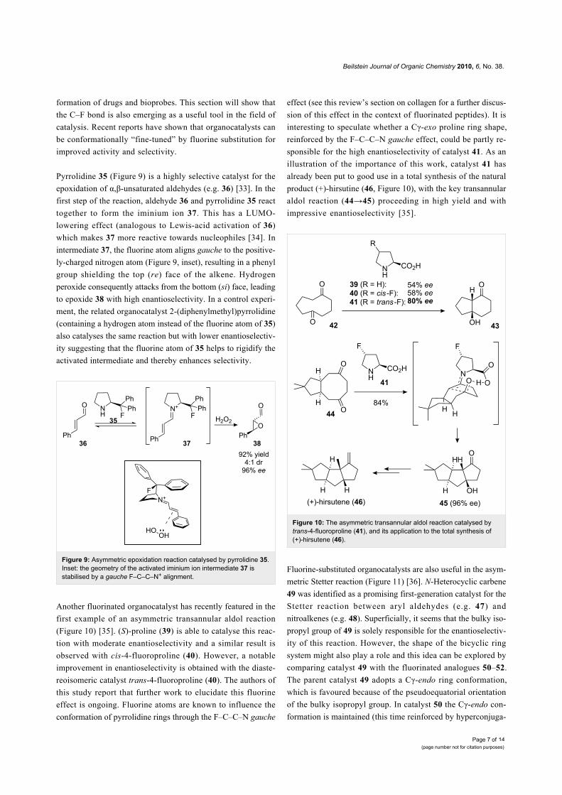
Another fluorinated organocatalyst has recently featured in the
first example of an asymmetric transannular aldol reaction
(Figure 10) [35]. (S)-proline (39) is able to catalyse this reac-
tion with moderate enantioselectivity and a similar result is
observed with cis-4-fluoroproline (40). However, a notable
improvement in enantioselectivity is obtained with the diaste-
reoisomeric catalyst trans-4-fluoroproline (40). The authors of
this study report that further work to elucidate this fluorine
effect is ongoing. Fluorine atoms are known to influence the
conformation of pyrrolidine rings through the F–C–C–N gauche
effect (see this review’s section on collagen for a further discus-
sion of this effect in the context of fluorinated peptides). It is
interesting to speculate whether a Cγ-exo proline ring shape,
reinforced by the F–C–C–N gauche effect, could be partly re-
sponsible for the high enantioselectivity of catalyst 41. As an
illustration of the importance of this work, catalyst 41 has
already been put to good use in a total synthesis of the natural
product (+)-hirsutine (46, Figure 10), with the key transannular
aldol reaction (44→45) proceeding in high yield and with
impressive enantioselectivity [35].
Figure 10: The asymmetric transannular aldol reaction catalysed bytrans-4-fluoroproline (41), and its application to the total synthesis of(+)-hirsutene (46).
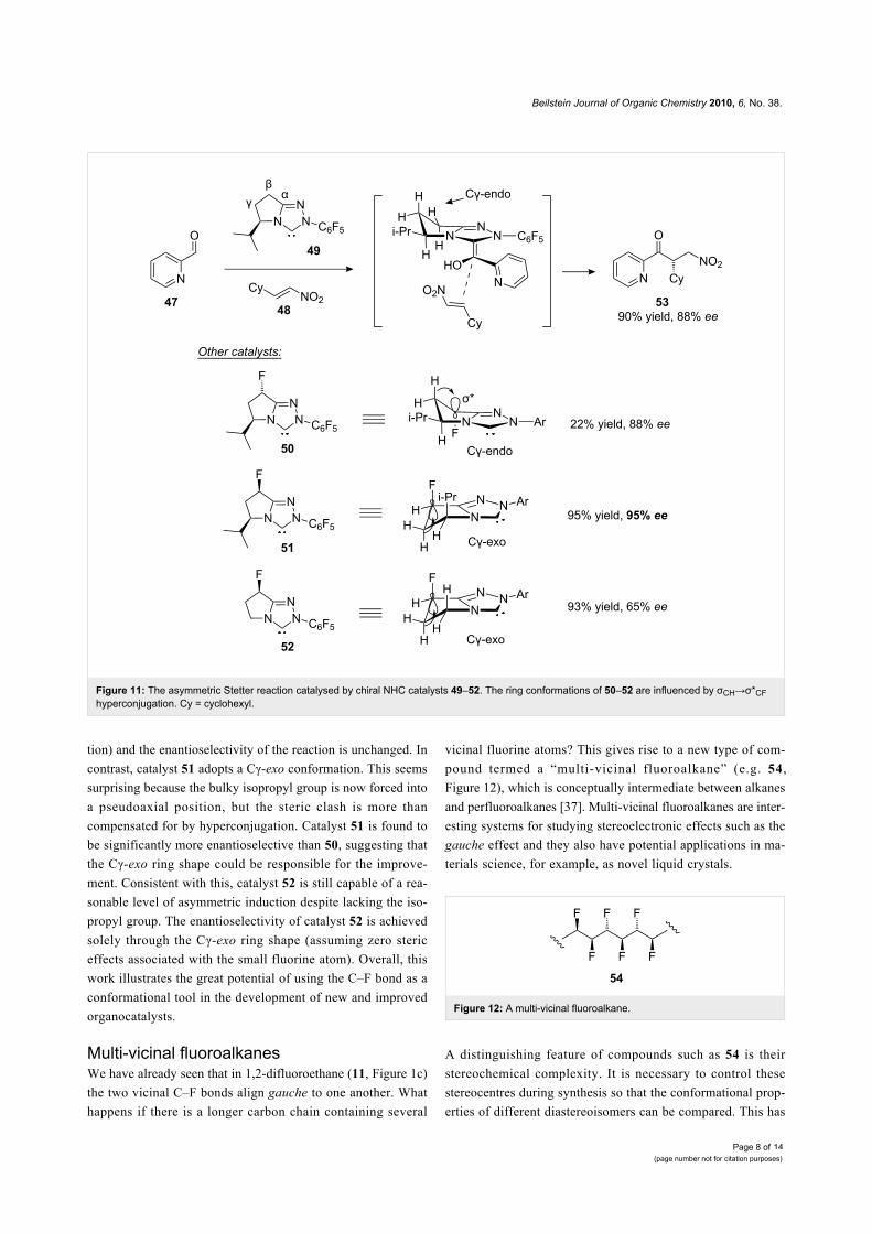
Fluorine-substituted organocatalysts are also useful in the asym-
metric Stetter reaction (Figure 11) [36]. N-Heterocyclic carbene
49 was identified as a promising first-generation catalyst for the
Stetter reaction between aryl aldehydes (e.g. 47) and
nitroalkenes (e.g. 48). Superficially, it seems that the bulky iso-
propyl group of 49 is solely responsible for the enantioselectiv-
ity of this reaction. However, the shape of the bicyclic ring
system might also play a role and this idea can be explored by
comparing catalyst 49 with the fluorinated analogues 50–52.
The parent catalyst 49 adopts a Cγ-endo ring conformation,
which is favoured because of the pseudoequatorial orientation
of the bulky isopropyl group. In catalyst 50 the Cγ-endo con-
formation is maintained (this time reinforced by hyperconjuga-
Beilstein Journal of Organic Chemistry 2010, 6, No. 38.
Page 8 of(page number not for citation purposes)
14
Figure 11: The asymmetric Stetter reaction catalysed by chiral NHC catalysts 49–52. The ring conformations of 50–52 are influenced by σCH→σ*CFhyperconjugation. Cy = cyclohexyl.
tion) and the enantioselectivity of the reaction is unchanged. In
contrast, catalyst 51 adopts a Cγ-exo conformation. This seems
surprising because the bulky isopropyl group is now forced into
a pseudoaxial position, but the steric clash is more than
compensated for by hyperconjugation. Catalyst 51 is found to
be significantly more enantioselective than 50, suggesting that
the Cγ-exo ring shape could be responsible for the improve-
ment. Consistent with this, catalyst 52 is still capable of a rea-
sonable level of asymmetric induction despite lacking the iso-
propyl group. The enantioselectivity of catalyst 52 is achieved
solely through the Cγ-exo ring shape (assuming zero steric
effects associated with the small fluorine atom). Overall, this
work illustrates the great potential of using the C–F bond as a
conformational tool in the development of new and improved
organocatalysts.
Multi-vicinal fluoroalkanesWe have already seen that in 1,2-difluoroethane (11, Figure 1c)
the two vicinal C–F bonds align gauche to one another. What
happens if there is a longer carbon chain containing several
vicinal fluorine atoms? This gives rise to a new type of com-
pound termed a “multi-vicinal fluoroalkane” (e.g. 54,
Figure 12), which is conceptually intermediate between alkanes
and perfluoroalkanes [37]. Multi-vicinal fluoroalkanes are inter-
esting systems for studying stereoelectronic effects such as the
gauche effect and they also have potential applications in ma-
terials science, for example, as novel liquid crystals.
Figure 12: A multi-vicinal fluoroalkane.
A distinguishing feature of compounds such as 54 is their
stereochemical complexity. It is necessary to control these
stereocentres during synthesis so that the conformational prop-
erties of different diastereoisomers can be compared. This has
Beilstein Journal of Organic Chemistry 2010, 6, No. 38.
Page 9 of(page number not for citation purposes)
14
been explored with compounds containing up to six vicinal
fluorines [37-39] and it emerges that the conformations of these
compounds are governed by two main considerations: parallel
1,3-C–F bonds are avoided, and gauche 1,2-C–F bonds are
favoured. For example, consider the all-syn hexafluoroalkane
55 (Figure 13) [39]. This molecule cannot adopt a zigzag con-
formation because this would incur multiple 1,3-difluoro repul-
sions. Instead, 55 adopts a helical shape in which each pair of
vicinal fluorines is aligned gauche but no 1,3-difluoro repul-
sion is present. In contrast, the diastereoisomeric compound 56
does adopt the zigzag conformation (Figure 13). This affords
three out of a possible five 1,2-difluoro gauche alignments,
while the different stereochemistry of the molecule prevents
1,3-difluoro repulsion from occurring.
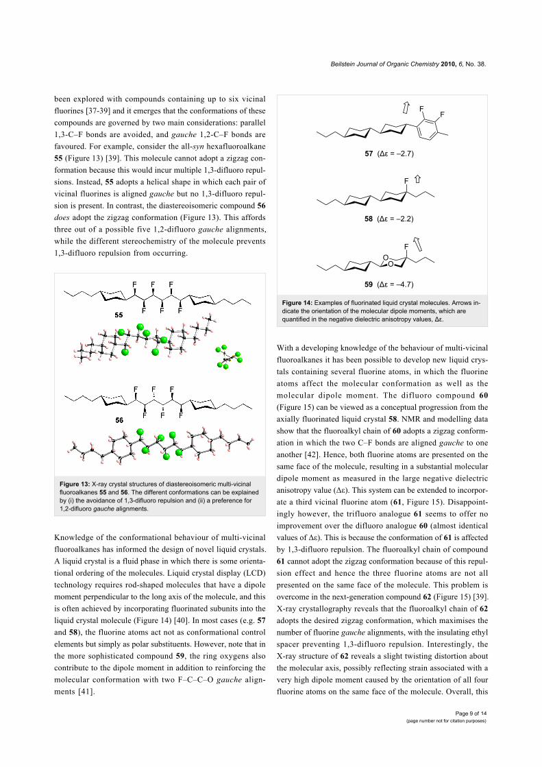
Figure 13: X-ray crystal structures of diastereoisomeric multi-vicinalfluoroalkanes 55 and 56. The different conformations can be explainedby (i) the avoidance of 1,3-difluoro repulsion and (ii) a preference for1,2-difluoro gauche alignments.
Knowledge of the conformational behaviour of multi-vicinal
fluoroalkanes has informed the design of novel liquid crystals.
A liquid crystal is a fluid phase in which there is some orienta-
tional ordering of the molecules. Liquid crystal display (LCD)
technology requires rod-shaped molecules that have a dipole
moment perpendicular to the long axis of the molecule, and this
is often achieved by incorporating fluorinated subunits into the
liquid crystal molecule (Figure 14) [40]. In most cases (e.g. 57
and 58), the fluorine atoms act not as conformational control
elements but simply as polar substituents. However, note that in
the more sophisticated compound 59, the ring oxygens also
contribute to the dipole moment in addition to reinforcing the
molecular conformation with two F–C–C–O gauche align-
ments [41].
Figure 14: Examples of fluorinated liquid crystal molecules. Arrows in-dicate the orientation of the molecular dipole moments, which arequantified in the negative dielectric anisotropy values, Δε.
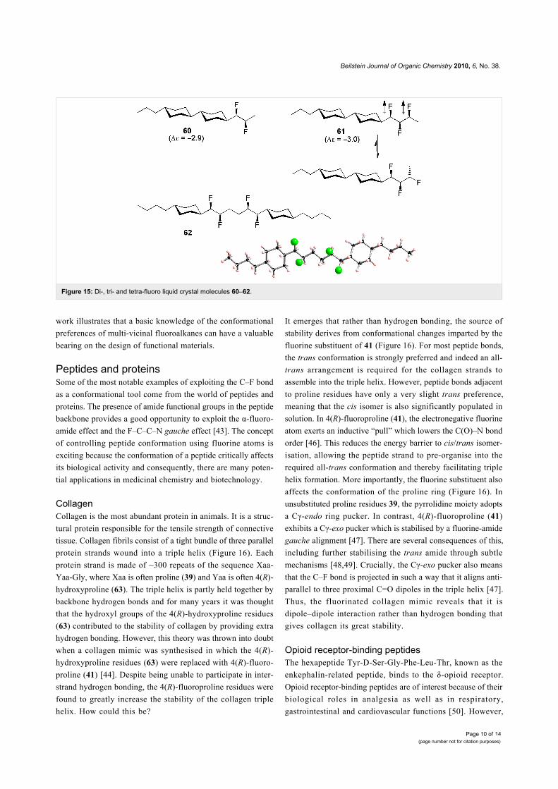
With a developing knowledge of the behaviour of multi-vicinal
fluoroalkanes it has been possible to develop new liquid crys-
tals containing several fluorine atoms, in which the fluorine
atoms affect the molecular conformation as well as the
molecular dipole moment. The difluoro compound 60
(Figure 15) can be viewed as a conceptual progression from the
axially fluorinated liquid crystal 58. NMR and modelling data
show that the fluoroalkyl chain of 60 adopts a zigzag conform-
ation in which the two C–F bonds are aligned gauche to one
another [42]. Hence, both fluorine atoms are presented on the
same face of the molecule, resulting in a substantial molecular
dipole moment as measured in the large negative dielectric
anisotropy value (Δε). This system can be extended to incorpor-
ate a third vicinal fluorine atom (61, Figure 15). Disappoint-
ingly however, the trifluoro analogue 61 seems to offer no
improvement over the difluoro analogue 60 (almost identical
values of Δε). This is because the conformation of 61 is affected
by 1,3-difluoro repulsion. The fluoroalkyl chain of compound
61 cannot adopt the zigzag conformation because of this repul-
sion effect and hence the three fluorine atoms are not all
presented on the same face of the molecule. This problem is
overcome in the next-generation compound 62 (Figure 15) [39].
X-ray crystallography reveals that the fluoroalkyl chain of 62
adopts the desired zigzag conformation, which maximises the
number of fluorine gauche alignments, with the insulating ethyl
spacer preventing 1,3-difluoro repulsion. Interestingly, the
X-ray structure of 62 reveals a slight twisting distortion about
the molecular axis, possibly reflecting strain associated with a
very high dipole moment caused by the orientation of all four
fluorine atoms on the same face of the molecule. Overall, this
Beilstein Journal of Organic Chemistry 2010, 6, No. 38.
Page 10 of(page number not for citation purposes)
14
Figure 15: Di-, tri- and tetra-fluoro liquid crystal molecules 60–62.
work illustrates that a basic knowledge of the conformational
preferences of multi-vicinal fluoroalkanes can have a valuable
bearing on the design of functional materials.
Peptides and proteinsSome of the most notable examples of exploiting the C–F bond
as a conformational tool come from the world of peptides and
proteins. The presence of amide functional groups in the peptide
backbone provides a good opportunity to exploit the α-fluoro-
amide effect and the F–C–C–N gauche effect [43]. The concept
of controlling peptide conformation using fluorine atoms is
exciting because the conformation of a peptide critically affects
its biological activity and consequently, there are many poten-
tial applications in medicinal chemistry and biotechnology.
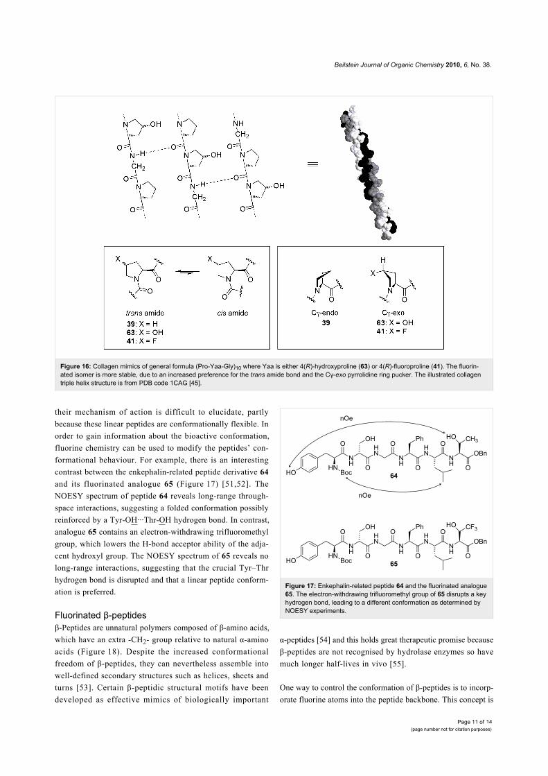
CollagenCollagen is the most abundant protein in animals. It is a struc-
tural protein responsible for the tensile strength of connective
tissue. Collagen fibrils consist of a tight bundle of three parallel
protein strands wound into a triple helix (Figure 16). Each
protein strand is made of ~300 repeats of the sequence Xaa-
Yaa-Gly, where Xaa is often proline (39) and Yaa is often 4(R)-
hydroxyproline (63). The triple helix is partly held together by
backbone hydrogen bonds and for many years it was thought
that the hydroxyl groups of the 4(R)-hydroxyproline residues
(63) contributed to the stability of collagen by providing extra
hydrogen bonding. However, this theory was thrown into doubt
when a collagen mimic was synthesised in which the 4(R)-
hydroxyproline residues (63) were replaced with 4(R)-fluoro-
proline (41) [44]. Despite being unable to participate in inter-
strand hydrogen bonding, the 4(R)-fluoroproline residues were
found to greatly increase the stability of the collagen triple
helix. How could this be?
It emerges that rather than hydrogen bonding, the source of
stability derives from conformational changes imparted by the
fluorine substituent of 41 (Figure 16). For most peptide bonds,
the trans conformation is strongly preferred and indeed an all-
trans arrangement is required for the collagen strands to
assemble into the triple helix. However, peptide bonds adjacent
to proline residues have only a very slight trans preference,
meaning that the cis isomer is also significantly populated in
solution. In 4(R)-fluoroproline (41), the electronegative fluorine
atom exerts an inductive “pull” which lowers the C(O)–N bond
order [46]. This reduces the energy barrier to cis/trans isomer-
isation, allowing the peptide strand to pre-organise into the
required all-trans conformation and thereby facilitating triple
helix formation. More importantly, the fluorine substituent also
affects the conformation of the proline ring (Figure 16). In
unsubstituted proline residues 39, the pyrrolidine moiety adopts
a Cγ-endo ring pucker. In contrast, 4(R)-fluoroproline (41)
exhibits a Cγ-exo pucker which is stabilised by a fluorine-amide
gauche alignment [47]. There are several consequences of this,
including further stabilising the trans amide through subtle
mechanisms [48,49]. Crucially, the Cγ-exo pucker also means
that the C–F bond is projected in such a way that it aligns anti-
parallel to three proximal C=O dipoles in the triple helix [47].
Thus, the fluorinated collagen mimic reveals that it is
dipole–dipole interaction rather than hydrogen bonding that
gives collagen its great stability.
Opioid receptor-binding peptidesThe hexapeptide Tyr-D-Ser-Gly-Phe-Leu-Thr, known as the
enkephalin-related peptide, binds to the δ-opioid receptor.
Opioid receptor-binding peptides are of interest because of their
biological roles in analgesia as well as in respiratory,
gastrointestinal and cardiovascular functions [50]. However,
Beilstein Journal of Organic Chemistry 2010, 6, No. 38.
Page 11 of(page number not for citation purposes)
14
Figure 16: Collagen mimics of general formula (Pro-Yaa-Gly)10 where Yaa is either 4(R)-hydroxyproline (63) or 4(R)-fluoroproline (41). The fluorin-ated isomer is more stable, due to an increased preference for the trans amide bond and the Cγ-exo pyrrolidine ring pucker. The illustrated collagentriple helix structure is from PDB code 1CAG [45].
their mechanism of action is difficult to elucidate, partly
because these linear peptides are conformationally flexible. In
order to gain information about the bioactive conformation,
fluorine chemistry can be used to modify the peptides’ con-
formational behaviour. For example, there is an interesting
contrast between the enkephalin-related peptide derivative 64
and its fluorinated analogue 65 (Figure 17) [51,52]. The
NOESY spectrum of peptide 64 reveals long-range through-
space interactions, suggesting a folded conformation possibly
reinforced by a Tyr-OH···Thr-OH hydrogen bond. In contrast,
analogue 65 contains an electron-withdrawing trifluoromethyl
group, which lowers the H-bond acceptor ability of the adja-
cent hydroxyl group. The NOESY spectrum of 65 reveals no
long-range interactions, suggesting that the crucial Tyr–Thr
hydrogen bond is disrupted and that a linear peptide conform-
ation is preferred.
Fluorinated β-peptidesβ-Peptides are unnatural polymers composed of β-amino acids,
which have an extra -CH2- group relative to natural α-amino
acids (Figure 18). Despite the increased conformational
freedom of β-peptides, they can nevertheless assemble into
well-defined secondary structures such as helices, sheets and
turns [53]. Certain β-peptidic structural motifs have been
developed as effective mimics of biologically important
Figure 17: Enkephalin-related peptide 64 and the fluorinated analogue65. The electron-withdrawing trifluoromethyl group of 65 disrupts a keyhydrogen bond, leading to a different conformation as determined byNOESY experiments.
α-peptides [54] and this holds great therapeutic promise because
β-peptides are not recognised by hydrolase enzymes so have
much longer half-lives in vivo [55].
One way to control the conformation of β-peptides is to incorp-
orate fluorine atoms into the peptide backbone. This concept is
Beilstein Journal of Organic Chemistry 2010, 6, No. 38.
Page 12 of(page number not for citation purposes)
14
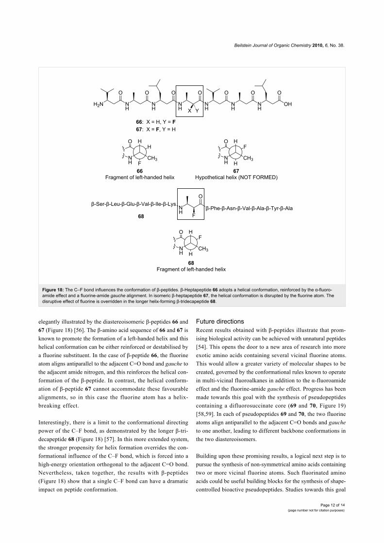
Figure 18: The C–F bond influences the conformation of β-peptides. β-Heptapeptide 66 adopts a helical conformation, reinforced by the α-fluoro-amide effect and a fluorine-amide gauche alignment. In isomeric β-heptapeptide 67, the helical conformation is disrupted by the fluorine atom. Thedisruptive effect of fluorine is overridden in the longer helix-forming β-tridecapeptide 68.
elegantly illustrated by the diastereoisomeric β-peptides 66 and
67 (Figure 18) [56]. The β-amino acid sequence of 66 and 67 is
known to promote the formation of a left-handed helix and this
helical conformation can be either reinforced or destabilised by
a fluorine substituent. In the case of β-peptide 66, the fluorine
atom aligns antiparallel to the adjacent C=O bond and gauche to
the adjacent amide nitrogen, and this reinforces the helical con-
formation of the β-peptide. In contrast, the helical conform-
ation of β-peptide 67 cannot accommodate these favourable
alignments, so in this case the fluorine atom has a helix-
breaking effect.
Interestingly, there is a limit to the conformational directing
power of the C–F bond, as demonstrated by the longer β-tri-
decapeptide 68 (Figure 18) [57]. In this more extended system,
the stronger propensity for helix formation overrides the con-
formational influence of the C–F bond, which is forced into a
high-energy orientation orthogonal to the adjacent C=O bond.
Nevertheless, taken together, the results with β-peptides
(Figure 18) show that a single C–F bond can have a dramatic
impact on peptide conformation.
Future directionsRecent results obtained with β-peptides illustrate that prom-
ising biological activity can be achieved with unnatural peptides
[54]. This opens the door to a new area of research into more
exotic amino acids containing several vicinal fluorine atoms.
This would allow a greater variety of molecular shapes to be
created, governed by the conformational rules known to operate
in multi-vicinal fluoroalkanes in addition to the α-fluoroamide
effect and the fluorine-amide gauche effect. Progress has been
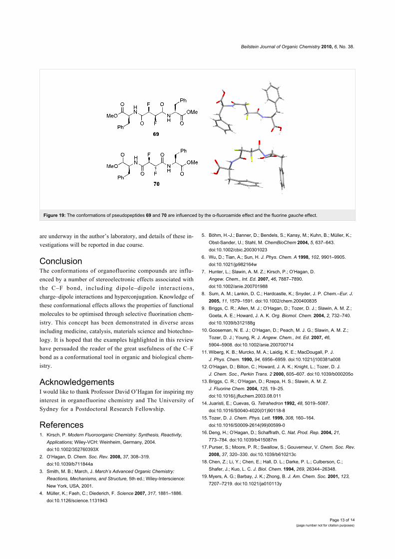
made towards this goal with the synthesis of pseudopeptides
containing a difluorosuccinate core (69 and 70, Figure 19)
[58,59]. In each of pseudopeptides 69 and 70, the two fluorine
atoms align antiparallel to the adjacent C=O bonds and gauche
to one another, leading to different backbone conformations in
the two diastereoisomers.
Building upon these promising results, a logical next step is to
pursue the synthesis of non-symmetrical amino acids containing
two or more vicinal fluorine atoms. Such fluorinated amino
acids could be useful building blocks for the synthesis of shape-
controlled bioactive pseudopeptides. Studies towards this goal
Beilstein Journal of Organic Chemistry 2010, 6, No. 38.
Page 13 of(page number not for citation purposes)
14
Figure 19: The conformations of pseudopeptides 69 and 70 are influenced by the α-fluoroamide effect and the fluorine gauche effect.
are underway in the author’s laboratory, and details of these in-
vestigations will be reported in due course.
ConclusionThe conformations of organofluorine compounds are influ-
enced by a number of stereoelectronic effects associated with
the C–F bond, including dipole–dipole interactions,
charge–dipole interactions and hyperconjugation. Knowledge of
these conformational effects allows the properties of functional
molecules to be optimised through selective fluorination chem-
istry. This concept has been demonstrated in diverse areas
including medicine, catalysis, materials science and biotechno-
logy. It is hoped that the examples highlighted in this review
have persuaded the reader of the great usefulness of the C–F
bond as a conformational tool in organic and biological chem-
istry.
AcknowledgementsI would like to thank Professor David O’Hagan for inspiring my
interest in organofluorine chemistry and The University of
Sydney for a Postdoctoral Research Fellowship.
References1. Kirsch, P. Modern Fluoroorganic Chemistry: Synthesis, Reactivity,
Applications; Wiley-VCH: Weinheim, Germany, 2004.doi:10.1002/352760393X
2. O’Hagan, D. Chem. Soc. Rev. 2008, 37, 308–319.doi:10.1039/b711844a
3. Smith, M. B.; March, J. March’s Advanced Organic Chemistry:Reactions, Mechanisms, and Structure, 5th ed.; Wiley-Interscience:New York, USA, 2001.
4. Müller, K.; Faeh, C.; Diederich, F. Science 2007, 317, 1881–1886.doi:10.1126/science.1131943
5. Böhm, H.-J.; Banner, D.; Bendels, S.; Kansy, M.; Kuhn, B.; Müller, K.;Obst-Sander, U.; Stahl, M. ChemBioChem 2004, 5, 637–643.doi:10.1002/cbic.200301023
6. Wu, D.; Tian, A.; Sun, H. J. Phys. Chem. A 1998, 102, 9901–9905.doi:10.1021/jp982164w
7. Hunter, L.; Slawin, A. M. Z.; Kirsch, P.; O’Hagan, D.Angew. Chem., Int. Ed. 2007, 46, 7887–7890.doi:10.1002/anie.200701988
8. Sum, A. M.; Lankin, D. C.; Hardcastle, K.; Snyder, J. P. Chem.–Eur. J.2005, 11, 1579–1591. doi:10.1002/chem.200400835
9. Briggs, C. R.; Allen, M. J.; O’Hagan, D.; Tozer, D. J.; Slawin, A. M. Z.;Goeta, A. E.; Howard, J. A. K. Org. Biomol. Chem. 2004, 2, 732–740.doi:10.1039/b312188g
10. Gooseman, N. E. J.; O’Hagan, D.; Peach, M. J. G.; Slawin, A. M. Z.;Tozer, D. J.; Young, R. J. Angew. Chem., Int. Ed. 2007, 46,5904–5908. doi:10.1002/anie.200700714
11. Wiberg, K. B.; Murcko, M. A.; Laidig, K. E.; MacDougall, P. J.J. Phys. Chem. 1990, 94, 6956–6959. doi:10.1021/j100381a008
12. O’Hagan, D.; Bilton, C.; Howard, J. A. K.; Knight, L.; Tozer, D. J.J. Chem. Soc., Perkin Trans. 2 2000, 605–607. doi:10.1039/b000205o
13. Briggs, C. R.; O’Hagan, D.; Rzepa, H. S.; Slawin, A. M. Z.J. Fluorine Chem. 2004, 125, 19–25.doi:10.1016/j.jfluchem.2003.08.011
14. Juaristi, E.; Cuevas, G. Tetrahedron 1992, 48, 5019–5087.doi:10.1016/S0040-4020(01)90118-8
15. Tozer, D. J. Chem. Phys. Lett. 1999, 308, 160–164.doi:10.1016/S0009-2614(99)00599-0
16. Deng, H.; O’Hagan, D.; Schaffrath, C. Nat. Prod. Rep. 2004, 21,773–784. doi:10.1039/b415087m
17. Purser, S.; Moore, P. R.; Swallow, S.; Gouverneur, V. Chem. Soc. Rev.2008, 37, 320–330. doi:10.1039/b610213c
18. Chen, Z.; Li, Y.; Chen, E.; Hall, D. L.; Darke, P. L.; Culberson, C.;Shafer, J.; Kuo, L. C. J. Biol. Chem. 1994, 269, 26344–26348.
19. Myers, A. G.; Barbay, J. K.; Zhong, B. J. Am. Chem. Soc. 2001, 123,7207–7219. doi:10.1021/ja010113y
Beilstein Journal of Organic Chemistry 2010, 6, No. 38.
Page 14 of(page number not for citation purposes)
14
20. Massa, M. A.; Spangler, D. P.; Durley, R. C.; Hickory, B. S.;Connolly, D. T.; Witherbee, B. J.; Smith, M. E.; Sikorski, J. A.Bioorg. Med. Chem. Lett. 2001, 11, 1625–1628.doi:10.1016/S0960-894X(01)00244-X
21. Pankiewicz, K. W. Carbohydr. Res. 2000, 327, 87–105.doi:10.1016/S0008-6215(00)00089-6
22. Meng, W.-D.; Qing, F.-L. Curr. Top. Med. Chem. 2006, 6, 1499–1528.doi:10.2174/156802606777951082
23. Marquez, V. E.; Tseng, C. K.-H.; Mitsuya, H.; Aoki, S.; Kelley, J. A.;Ford, H.; Roth, J. S.; Broder, S.; Johns, D. G.; Driscoll, J. S.J. Med. Chem. 1990, 33, 978–985. doi:10.1021/jm00165a015
24. Barchi, J. J.; Karki, R. G.; Nicklaus, M. C.; Siddiqui, M. A.; George, C.;Mikhailopulo, I. A.; Marquez, V. E. J. Am. Chem. Soc. 2008, 130,9048–9057. doi:10.1021/ja800964g
25. Van Roey, P.; Salerno, J. M.; Chu, C. K.; Schinazi, R. F.Proc. Natl. Acad. Sci. U. S. A. 1989, 86, 3929–3933.doi:10.1073/pnas.86.11.3929
26. Mikhailopulo, I. A.; Pricota, T. I.; Sivets, G. G.; Altona, C. J. Org. Chem.2003, 68, 5897–5908. doi:10.1021/jo0340859
27. Bucher, C.; Sparr, C.; Schweizer, W. B.; Gilmour, R. Chem.–Eur. J.2009, 15, 7637–7647. doi:10.1002/chem.200900505
28. Deniau, G.; Slawin, A. M. Z.; Lebl, T.; Chorki, F.; Issberner, J. P.;Van Mourik, T.; Heygate, J. M.; Lambert, J. J.; Etherington, L.-A.;Sillar, K. T.; O’Hagan, D. ChemBioChem 2007, 8, 2265–2274.doi:10.1002/cbic.200700371
29. Clift, M. D.; Ji, H.; Deniau, G.; O’Hagan, D.; Silverman, R. B.Biochemistry 2007, 46, 13819–13828. doi:10.1021/bi701249q
30. Khrimian, A. P.; Oliver, J. E.; Waters, R. M.; Panicker, S.;Nicholson, J. M.; Klun, J. A. Tetrahedron: Asymmetry 1996, 7, 37–40.doi:10.1016/0957-4166(95)00415-7
31. Winkler, M.; Moraux, T.; Khairy, H. A.; Scott, R. H.; Slawin, A. M. Z.;O’Hagan, D. ChemBioChem 2009, 10, 823–828.doi:10.1002/cbic.200800709
32. Jordt, S.-E.; Julius, D. Cell 2002, 108, 421–430.doi:10.1016/S0092-8674(02)00637-2
33. Sparr, C.; Schweizer, W. B.; Senn, H. M.; Gilmour, R.Angew. Chem., Int. Ed. 2009, 48, 3065–3068.doi:10.1002/anie.200900405
34. MacMillan, D. W. C. Nature 2008, 455, 304–308.doi:10.1038/nature07367
35. Chandler, C. L.; List, B. J. Am. Chem. Soc. 2008, 130, 6737–6739.doi:10.1021/ja8024164
36. DiRocco, D. A.; Oberg, K. M.; Dalton, D. M.; Rovis, T.J. Am. Chem. Soc. 2009, 131, 10872–10874. doi:10.1021/ja904375q
37. Hunter, L.; O’Hagan, D. Org. Biomol. Chem. 2008, 6, 2843–2848.doi:10.1039/b809432b
38. Farran, D.; Slawin, A. M. Z.; Kirsch, P.; O’Hagan, D. J. Org. Chem.2009, 74, 7168–7171. doi:10.1021/jo901360e
39. Hunter, L.; Kirsch, P.; Slawin, A. M. Z.; O’Hagan, D.Angew. Chem., Int. Ed. 2009, 48, 5457–5460.doi:10.1002/anie.200901956
40. Hird, M. Chem. Soc. Rev. 2007, 36, 2070–2095. doi:10.1039/b610738a41. Kirsch, P.; Hahn, A.; Fröhlich, R.; Haufe, G. Eur. J. Org. Chem. 2006,
4819–4824. doi:10.1002/ejoc.20060052942. Nicoletti, M.; Bremer, M.; Kirsch, P.; O’Hagan, D. Chem. Commun.
2007, 5075–5077. doi:10.1039/b711839b43. Briggs, C. R. S.; O’Hagan, D.; Howard, J. A. K.; Yufit, D. S.
J. Fluorine Chem. 2003, 119, 9–13.doi:10.1016/S0022-1139(02)00243-9
44. Holmgren, S. K.; Taylor, K. M.; Bretscher, L. E.; Raines, R. T. Nature1998, 392, 666–667. doi:10.1038/33573
45. Bella, J.; Eaton, M.; Brodsky, B.; Berman, H. M. Science 1994, 266,75–81. doi:10.1126/science.7695699
46. Eberhardt, E. S.; Panasik, N.; Raines, R. T. J. Am. Chem. Soc. 1996,118, 12261–12266. doi:10.1021/ja9623119
47. Holmgren, S. K.; Bretscher, L. E.; Taylor, K. M.; Raines, R. T.Chem. Biol. 1999, 6, 63–70. doi:10.1016/S1074-5521(99)80003-9
48. Bretscher, L. E.; Jenkins, C. L.; Taylor, K. M.; DeRider, M. L.;Raines, R. T. J. Am. Chem. Soc. 2001, 123, 777–778.doi:10.1021/ja005542v
49. Panasik, N.; Eberhardt, E. S.; Edison, A. S.; Powell, D. R.;Raines, R. T. Int. J. Pept. Protein Res. 1994, 44, 262–269.doi:10.1111/j.1399-3011.1994.tb00169.x
50. Marcotte, I.; Separovic, F.; Auger, M.; Gagné, S. M. Biophys. J. 2004,86, 1587–1600. doi:10.1016/S0006-3495(04)74226-5
51. Kitamoto, T.; Marubayashi, S.; Yamazaki, T. Chem. Lett. 2006, 35,1264–1265. doi:10.1246/cl.2006.1264
52. Kitamoto, T.; Marubayashi, S.; Yamazaki, T. Tetrahedron 2008, 64,1888–1894. doi:10.1016/j.tet.2007.11.085
53. Seebach, D.; Hook, D. F.; Glättli, A. Pept. Sci. 2006, 84, 23–37.doi:10.1002/bip.20391
54. Seebach, D.; Gardiner, J. Acc. Chem. Res. 2008, 41, 1366–1375.doi:10.1021/ar700263g
55. Hook, D. F.; Gessier, F.; Noti, C.; Kast, P.; Seebach, D. ChemBioChem2004, 5, 691–706. doi:10.1002/cbic.200300827
56. Mathad, R. I.; Gessier, F.; Seebach, D.; Jaun, B. Helv. Chim. Acta2005, 88, 266–280. doi:10.1002/hlca.200590008
57. Mathad, R. I.; Jaun, B.; Flögel, O.; Gardiner, J.; Löweneck, M.;Codée, J. D. C.; Seeberger, P. H.; Seebach, D. Helv. Chim. Acta 2007,90, 2251–2273. doi:10.1002/hlca.200790235
58. Schüler, M.; O’Hagan, D.; Slawin, A. M. Z. Chem. Commun. 2005,4324–4326. doi:10.1039/b506010a
59. O’Hagan, D.; Rzepa, H. S.; Schüler, M.; Slawin, A. M. Z.Beilstein J. Org. Chem. 2006, 2, No. 19. doi:10.1186/1860-5397-2-19
License and TermsThis is an Open Access article under the terms of the
Creative Commons Attribution License
(http://creativecommons.org/licenses/by/2.0), which
permits unrestricted use, distribution, and reproduction in
any medium, provided the original work is properly cited.
The license is subject to the Beilstein Journal of Organic
Chemistry terms and conditions:
(http://www.beilstein-journals.org/bjoc)
The definitive version of this article is the electronic one
which can be found at:
doi:10.3762/bjoc.6.38