the carbohydrate-binding site in galectin-3 is preorganized to recognize a sugarlike framework of...
TRANSCRIPT

The Carbohydrate-Binding Site in Galectin-3 Is Preorganized ToRecognize a Sugarlike Framework of Oxygens: Ultra-High-ResolutionStructures and Water DynamicsKadhirvel Saraboji,† Maria Hakansson,‡ Samuel Genheden,§ Carl Diehl,∥ Johan Qvist,∥ Ulrich Weininger,∥
Ulf J. Nilsson,⊥ Hakon Leffler,@ Ulf Ryde,§ Mikael Akke,*,∥ and Derek T. Logan*,†,‡
†Department of Biochemistry and Structural Biology, Center for Molecular Protein Science, Box 124, Lund University, SE-221 00Lund, Sweden‡SARomics Biostructures AB, Scheelevagen 22, SE-220 07 Lund, Sweden§Department of Theoretical Chemistry, Lund University, SE-221 00 Lund, Sweden∥Department of Biophysical Chemistry, Center for Molecular Protein Science, Box 124, Lund University, SE-221 00 Lund, Sweden⊥Department of Organic Chemistry, Lund University, SE-221 00 Lund, Sweden@Section MIG, Department of Laboratory Medicine, Solvegatan 23, Lund University, SE-223 62 Lund, Sweden
*S Supporting Information
ABSTRACT: The recognition of carbohydrates by proteins isa fundamental aspect of communication within and betweenliving cells. Understanding the molecular basis of carbohy-drate−protein interactions is a prerequisite for the rationaldesign of synthetic ligands. Here we report the high- to ultra-high-resolution crystal structures of the carbohydrate recog-nition domain of galectin-3 (Gal3C) in the ligand-free state(1.08 Å at 100 K, 1.25 Å at 298 K) and in complex with lactose(0.86 Å) or glycerol (0.9 Å). These structures reveal striking similarities in the positions of water and carbohydrate oxygen atomsin all three states, indicating that the binding site of Gal3C is preorganized to coordinate oxygen atoms in an arrangement that isnearly optimal for the recognition of β-galactosides. Deuterium nuclear magnetic resonance (NMR) relaxation dispersionexperiments and molecular dynamics simulations demonstrate that all water molecules in the lactose-binding site exchange withbulk water on a time scale of nanoseconds or shorter. Nevertheless, molecular dynamics simulations identify transient waterbinding at sites that agree well with those observed by crystallography, indicating that the energy landscape of the binding site ismaintained in solution. All heavy atoms of glycerol are positioned like the corresponding atoms of lactose in the Gal3Ccomplexes. However, binding of glycerol to Gal3C is insignificant in solution at room temperature, as monitored by NMRspectroscopy or isothermal titration calorimetry under conditions where lactose binding is readily detected. These observationsmake a case for protein cryo-crystallography as a valuable screening method in fragment-based drug discovery and further suggestthat identification of water sites might inform inhibitor design.
Interactions between carbohydrates and proteins mediatenumerous important biological functions, such as signal
transduction, cell adhesion, host−pathogen recognition, andthe immune response.1−4 Carbohydrate-recognizing proteinsare involved in a number of human disease states, includinginflammation and cancer. These key functional properties havestimulated significant efforts in drug design targetingcarbohydrate-binding proteins.5−8 Carbohydrate−protein inter-actions are typically relatively weak, with dissociation constantsof ∼1 mM. Binding affinity is typically driven by a favorableenthalpic component9−12 that is partly offset by negativeentropy. The low affinity is related to the inherent properties ofsaccharides, such as their lack of charges and lack of extendedhydrophobic surfaces, which both reduce the likelihood offorming strong interactions with proteins. Instead, theformation of carbohydrate−protein complexes involves rela-
tively weak van der Waals interactions and hydrogen bonds tothe carbohydrate hydroxyl groups, acetamides, and ring andglycosidic oxygens. As a result of these specific properties ofcarbohydrate−protein interactions, the design of high-affinityinhibitors has proven to be challenging,6−8 and the relativeimportance of the various types of interactions in drivingcarbohydrate recognition is a matter of intense research.13−18
During the ligand binding process, significant solventreorganization takes place across the contact surface.19−22
Thus, considerable attention has been paid to the structure anddynamics of water molecules in carbohydrate-binding sites andto the role these play in mediating carbohydrate recognition by
Received: September 19, 2011Revised: November 19, 2011Published: November 23, 2011
Article
pubs.acs.org/biochemistry
© 2011 American Chemical Society 296 dx.doi.org/10.1021/bi201459p | Biochemistry 2012, 51, 296−306
proteins.20,23−26 Nonetheless, our current understanding of thethermodynamics and kinetics of the solvent reorganizationprocess at the microscopic level remains incomplete.27 A fullunderstanding of carbohydrate recognition benefits stronglyfrom atomic-resolution descriptions of both the liganded andunliganded states.Galectins are small soluble proteins that constitute a family of
mammalian lectins, defined by a carbohydrate recognitiondomain (CRD) with a conserved sequence motif that confersaffinity for β-galactoside-containing glycans.28 Galectins haveseveral important functions in both carbohydrate-dependentextracellular and carbohydrate-independent intracellular activ-ities.28−33 Even though galectins are synthesized in andprimarily remain in the cytosol, they reach the extracellularspace or lumen of vesicles9,34 by a nonclassical secretorypathway35,36 and can then take part in regulation of cellulartrafficking of glycoproteins, signaling, and cell adhesion.31,37 Agrowing body of evidence links galectins to important roles incell growth, cell differentiation, cell cycle regulation, andapoptosis, making them potential pharmaceutical targets ininflammation, immunity, and cancer.29,38−41 Thus, it is criticalto understand the molecular driving forces for the ligandbinding specificity of galectins.Structures of several galectins in complex with natural and
designed ligands are known, and it has been seen that galectinsbind oligosaccharides in a conserved recognition modeinvolving a network of hydrogen bonds and several boundwaters, which form bridges between hydrogen-bondingpartners in the protein−ligand complex.16,25,42,43 As is typicalfor lectins, galectins bind the monosaccharide galactose withdissociation constants in the millimolar range, in theirconserved binding site. However, sugars adjacent to thegalactose may interact with neighboring sites to providestepwise boosts in affinity.28,44 Thus, for galectin-3 additionof glucose, as in lactose, enhances affinity 50-fold (Kd ∼ 0.2mM), and additional saccharides at position 3 of the galactosecan give affinities in the low micromolar range. Moreover, non-natural derivatives at this position may enhance the affinity tothe nanomolar range, e.g., by exploiting cation−π interactionswith the surface residue Arg144.16,45 Furthermore, recent workshows that triazole derivatization at C3 of galactose results in ahigh affinity, similar to that observed for aromatic amidocompounds.46,47 Several studies have highlighted the signifi-cance of high-affinity and selective galectin inhibitors that actintracellularly, with potential use in modulating inflammatoryprocesses and cancer growth.30,48−51 To develop effectiveapproaches for the structure-based design of potent galectin-3inhibitors, it is important to understand the detailed molecularbasis for carbohydrate recognition, based on the three-dimensional structure and physicochemical properties of theconserved binding motif. High-resolution structural informa-tion greatly aids in this respect. Here we report a study thatcombines X-ray crystallography, NMR spectroscopy, moleculardynamics (MD) simulations, and isothermal titration calorim-etry (ITC) to probe the role of water molecules in the bindingsite and the details of the hydrogen-bonding networks at theprotein−ligand interface of Gal3C. Our results highlight therole of an oxygen coordination framework in the extendedcarbohydrate-binding site of galectin-3 that potentially can befurther exploited in future drug design efforts.
■ MATERIALS AND METHODS
Protein Expression and Purification. The galectin-3carbohydrate recognition domain (Gal3C; amino acid residues113−250) was expressed and purified either as a thioredoxinfusion construct52 or as isolated Gal3C.43 In the first case, DNAencoding amino acids 113−250 was amplified via polymerasechain reaction from galectin-3 in pET3C53 and cloned into thepET-32 Ek/LIC vector (Novagen, Madison, WI) according tothe manufacturer’s instructions, and as described for galectin-8previously.44 In the second case, DNA encoding amino acids113−250 was inserted into pET 3C without additional tags.The expression protocol for the two constructs is identical andhas been reported previously.52 The purification protocol forthe isolated Gal3C domain is highly similar to that reportedpreviously,52,53 except that the final separation step aftercleavage of the expressed product using a lactosyl-Sepharosecolumn is not needed. Typical yields were 150 mg/L of cultureof isolated Gal3C. The absence of lactose from the apo-Gal3Csamples (used to determine the crystal structures of apo-Gal3Cand glycerol-bound Gal3C) was verified using NMR spectros-copy.
Crystallization and Structure Determination. AllGal3C crystals were obtained using the hanging drop vapordiffusion method in NeXtal plates (Qiagen). The Gal3C−lactose complex was obtained by mixing 9.5 μL of proteinsolution [20 mg/mL Gal3C, 10 mM β-mercaptoethanol, 100mM lactose, 10 mM sodium phosphate buffer (pH 7.5), 100mM NaCl, and 0.02% NaN3] with 0.5 μL of 100 mM 3′-benzamido-N-acetyllactosamine on ice. After being incubatedfor 1 h, the solution was centrifuged at 7000 rpm for 10 min at4 °C, and hanging drops were set up using 4 μL dropscontaining equal volumes of a protein solution and a reservoirsolution [30% (w/v) PEG 4000, 0.1 M Tris-HCl (pH 7.5), 0.1M MgCl2, 0.4 M NaSCN, and 8 mM β-mercaptoethanol].Immediately after setup, 0.3 μL of a seed bead solution(Hampton Research protocol) was added to the drop. The seedbead solution was made from lactose-containing Gal3C crystalscrushed in stabilizing solution [1 mM lactose, 29% PEG 4000,0.1 M Tris-HCl (pH 7.5), 0.1 M MgCl2, 0.4 M NaSCN, and 8mM β-mercaptoethanol]. The largest crystals grew todimensions of 0.4 mm × 0.4 mm × 0.5 mm within 1 month.This was an attempt to replace lactose with 3′-benzamido-N-acetyllactosamine, which failed because even if the affinity oflactose is lower (Kd = 231 μM) than that of 3′-benzamido-N-acetyllactosamine (Kd = 18.2 μM) for Gal3C,45 the lactoseconcentration was 19-fold higher (95 mM vs 5 mM). Thecomplex of 3′-benzamido-N-acetyllactosamine and Gal3C hasbeen determined using the same crystallization conditions butwithout lactose present.45
Apo-Gal3C crystals were obtained from 4 μL dropscontaining equal volumes of a protein solution [19 mg/mLGal3C, 10 mM sodium phosphate buffer (pH 7.5), 100 mMNaCl, 10 mM β-mercaptoethanol, and 0.02% NaN3] and areservoir solution [30% (w/v) PEG 4000, 0.1 M Tris-HCl (pH7.5 or 8.0), 0.1 M MgCl2, 0.4 M NaSCN, and 8 mM β-mercaptoethanol]. Apo crystals appeared overnight and grewwithin a few days to dimensions of 0.1 mm × 0.1 mm × 0.2mm.The Gal3C−lactose crystal was flash-cooled to 100 K in the
cold N2 gas stream of a Cryojet (Oxford Diffraction) using acryo solution consisting of 15% (v/v) glycerol, 25.5% (w/v)PEG 4000, 0.25 M NaSCN, 85 mM Tris-HCl (pH 7.5), 85 mM
Biochemistry Article
dx.doi.org/10.1021/bi201459p | Biochemistry 2012, 51, 296−306297

MgCl2, 7 mM β-mercaptoethanol, and 4 mM 3′-benzamido-N-acetyllactosamine. The same cryoprotectant but without 3′-benzamido-N-acetyllactosamine was used to cryoprotect theGal3C−glycerol complex crystals, whereas 15% (v/v) PEG 400[instead of 15% (v/v) glycerol] was used for apo-Gal3C. Fordata collection at room temperature, the crystal was mountedusing the MicroRT kit (MiTeGen). The X-ray diffraction datafor all the crystals were collected on a 165 mm marResearchCCD detector on beamline I911-5 of the MAX-II synchrotronin Lund, Sweden. Diffraction data were integrated and scaledusing XDS and XSCALE.54 Data statistics are listed in Table 1.The structure of the lactose−Gal3C complex was determined
by rigid-body refinement using the structure of anotherGal3C−ligand complex as the initial model45 and Refmac5,55
as implemented in the CCP4 suite.56 The apo and glycerol−Gal3C structures were determined in a similar manner, with thelactose−Gal3C structure minus the ligand serving as a startingmodel. In the initial stages, all refinement was conducted to aresolution of 1.4 Å using Refmac5, with 5% of the totalreflections randomly set aside for cross validation. Subse-quently, the resolution was gradually extended to the fullresolution range, and refinement was conducted usingSHELXL-97.57 Manual model building was conducted usingCoot.58 The structure of lactose and the accompanyingrefinement restraints were generated using the CCP4 programMonomer Library Sketcher. In the final stages of refinement,many of the hydrogen atoms were visible in difference electrondensity maps and were added at calculated positions using theSHELXL riding model. Alternating steps of anisotropicrefinement and minor structure adjustment were performed
until convergence. Refinement statistics are listed in Table 1.Molecular images were generated using PyMOL.59
High-Resolution Protein NMR Experiments. Lactose-bound Gal3C and apo-Gal3C samples were prepared asreported previously,52 using proteins expressed in [15N,1-13C1]-glucose-containing minimal medium.60,61 1H−15N HSQCspectra were acquired with increasing concentrations of glycerolon a sample of Gal3C that was initially in the apo state at 0.46mM. Nine additions of 10 μL of 25 mM glycerol in NMRbuffer were made, yielding a final glycerol concentration of 5.9mM, corresponding to 16 equiv with respect to Gal3C. The 1Hand 15N spectral widths were 8000 and 1825 Hz, respectively.The static magnetic field strength was 14.1 T, and thetemperature was 301 K.The tautomeric state of histidines was monitored by 1H−15N
HMQC and 1H−13C HSQC spectra, centered at 200 and 128ppm in the 15N and 13C dimensions, respectively, and coveringspectral widths of 180 and 28 ppm, respectively. Themagnetization transfer delay for 1H−15N HMQC was set to22.2 ms, to refocus magnetization arising from 1JNH couplings.The static magnetic field strength was 11.7 T, and thetemperature was 298 K.
Low-field 2H Relaxation Dispersion Experiments.Lactose-bound Gal3C and apo-Gal3C samples were preparedas reported previously,52 except that the solvent contained 50%2H2O and the lactose concentration in the former sample was20 mM. The longitudinal relaxation rate R1 of the water 2Hmagnetization was measured with an accuracy of ∼0.5−1.0% atseven different frequencies, using conventional cryomagnets forthe higher frequencies and an iron core magnet (Drusch EAR-
Table 1. Crystallographic Data Collection and Refinement Statisticsa
lactose glycerol apo (100 K) apo (room temperature)
Data Collectionwavelength (Å) 0.9078 0.9078 0.9078 0.9080resolution (Å) 30.0−0.86 (0.88−0.86) 30.0−0.90 (0.92−0.90) 30.0−1.08 (1.11−1.08) 30.0−1.25 (1.28−1.25)space group P212121 P212121 P212121 P212121unit cell parameters (a, b, c) (Å) 37.8, 58.3, 63.1 35.8, 58.2, 62.5 35.7, 58.3, 62.8 36.5, 58.2, 63.7no. of measured reflections 680438 549501 319133 294647no. of unique reflections 111079 93342 56940 37798completeness (%) 98.9 (95.4) 95.8 (87.3) 99.9 (99.9) 98.6 (97.7)multiplicity 6.1 (4.0) 5.9 (4.6) 5.6 (4.7) 7.8 (5.7)Rmerge (%)
b 5.0 (85.3) 3.8 (79.2) 7.0 (68.2) 5.3 (85.2)⟨I/σ(I)⟩ 19.3 (1.8) 20.4 (2.1) 11.6 (2.4) 18.7 (2.5)
Refinementresolution limits (Å) 20.0−0.86 20.0−0.90 20.0−1.08 20.0−1.25Rmodel (%)
c 12.7 13.2 14.9 11.9Rfree (%)
d 14.2 15.0 18.2 16.6no. of waters 275 262 320 168rmsd from ideal values
bond lengths (Å)e 0.017 0.015 0.13 0.015angle distances (Å)f 0.042 0.032 0.032 0.033
average B factor (Å2) (protein/solvent/ligand)
11.5/25.8/18.0 12.7/24.8/15.1 15.3/31.9/na 16.6/42.8/na
average anisotropy for all atoms (protein/solvent/ligand)
0.35/0.30/0.32 0.36/0.34/0.35 0.40/0.33/na 0.38/0.41/na
data/parameter ratio 7.4 6.6 3.6 2.9Ramachandran plotg
residues in most favored regions 98.5% (134/136) 98.5% (134/136) 97.1% (132/136) 97.8% (133/136)aValues in parentheses correspond to the highest-resolution shell. The resolution limit was taken to be the point at which the ⟨I/σ(I)⟩ ratio wasapproximately equal to 2. na means not available bRmerge = ∑hkl∑i|I(hkl)i − I(hkl)|/∑hkl∑iI(hkl)i.
cRmodel = ∑hkl|Fo(hkl) − Fc(hkl)|/∑hkl|Fo(hkl)|,where Fo and Fc are the observed and calculated structure factors, respectively.
dA 5% random test set. eCalculated from DFIX restraints in SHELXL.fCalculated from DANG restraints in SHELXL. gCalculated using Molprobity.79
Biochemistry Article
dx.doi.org/10.1021/bi201459p | Biochemistry 2012, 51, 296−306298

35N) interfaced with a Tecmag Discovery console for the lowerfrequencies. The sample temperature was regulated to 299.8 ±0.1 K. The relaxation rate was measured at each frequency forboth apo-Gal3C and lactose-bound Gal3C, as well as for twomatching reference solutions that did not contain Gal3C.The relaxation dispersion is given by
ω = + ω + ωR R J J( ) 0.2 ( ) 0.8 (2 )1 0 bulk 0 0
where the spectral density is modeled as
ω = α + β τ+ ωτ
J( )1 ( )2
in which α describes a frequency-independent contributionfrom water molecules that have rotational correlation timessignificantly longer than those in bulk water, but shorter than 1ns, due to interactions with the external protein surface. βdescribes the frequency-dependent contribution from watermolecules with longer correlation times due to interactions withinternal sites in the protein and is given by
β =ω
β βNN SQ
2
T
2
where ωQ is the 2H quadrupole coupling frequency, NT is theknown ratio of water and protein molecules in the sample, Nβ isthe number of water molecules with a τ of >1 ns, and Sβ is theorientational order parameter of bound waters.Isothermal Titration Calorimetry. Experiments were
performed as described previously,45 in two series of 30injections of 10 μL of either 58 or 117 mM glycerol (firstinjection of 5 μL) in 5 mM HEPES buffer (pH 7.4). Theconcentration of Gal3C was 0.1 mM.Molecular Dynamics Simulations and Analysis. MD
simulations were performed as described previously,62 startingfrom the 100 K apo-Gal3C crystal structure reported here. Tenindependent simulations were used, each 5 ns long. Snapshotswere saved every picosecond for analysis.Water occupancy was investigated by the clustering approach
of Friesner and co-workers.63,64 In each snapshot, water oxygenatoms within the active site were stored for clustering. Theactive site was defined by fitting each MD snapshot to thelactose−Gal3C structure by superimposing the backbone atomsof all residues within 10 Å of lactose. The active site was thendefined as the maximal extent of the lactose molecule plus 1 Åin each direction. Subsequently, the water oxygen atoms withinthe active site were clustered by an iterative approach. In eachiteration, the number of water oxygen atoms in all snapshotswithin 1 Å of a particular water oxygen atom in a givensnapshot was counted and the water molecule with the largestnumber was marked as a water site. That water molecule and allof its neighbors within 1 Å were removed from the list, and theprocess was repeated until the number of neighbors of a watersite was lower than in bulk water.The clustering procedure was applied to the 10 independent
simulations, and the water sites identified in each individualsimulation were clustered using a single-linkage hierarchicalapproach.65 Constraints were imposed such that two water sitesidentified from the same simulation could not be in the samecluster. When the minimal distance between two clusters waslarger than 1 Å, the clustering was stopped. Euclidian distancesbetween the water sites were used to determine their closeness.For each cluster of water sites, the population number and themaximal spatial extent were calculated. The occupancy is the
fraction of the 10 independent simulations in which the watersite was found. The maximal spatial extent is the maximaldistance between two water molecules in the same cluster andindicates the precision of the position of the water site. Thegeometric centers of the water site clusters are reported.
■ RESULTS AND DISCUSSIONWe have determined the crystal structures of the lactose-bound[Protein Data Bank (PDB) entry 3ZSJ], glycerol-bound (PDBentry 3ZSK), and ligand-free (apo; PDB entry 3ZSL at 100 Kand PDB entry 3ZSM at room temperature) states of Gal3C atatomic or near-atomic resolution (Table 1). Overall, thesestructures are highly isomorphous to each other (Figure 1A andTable S1 of the Supporting Information) and served as a basisfor further investigations of oxygen recognition within thebinding site of Gal3C. To this end, we performed MDsimulations of apo-Gal3C to determine the population densityand residence times of water molecules in the binding site.Furthermore, we conducted nuclear magnetic relaxationdispersion measurements to experimentally verify the residencetimes of water molecules bound to Gal3C. Finally, ITC andchemical shift mapping by NMR spectroscopy were used tostudy whether glycerol binds to Gal3C in solution underambient conditions.Crystal structures similar to those reported here have been
published previously,66 but these were determined at lowerresolution (1.35−2.45 Å) from samples with mixed populationsof the glycerol-bound, lactose-bound, and apo states. Both ofthese factors may compromise the interpretation of theobserved electron density. Our data mitigate such complica-tions, because the diffraction experiments were conducted withcrystals of well-defined samples with only a single state present.Thus, these structures form a solid basis for investigations ofwater and ligand coordination within the carbohydrate-bindingsite and for detailed comparisons with results from MDsimulations and NMR spectroscopy.
The Structure of Lactose-Bound Gal3C Reveals WaterMolecules and Hydrogen Atoms. The 0.86 Å resolutionstructure of Gal3C in complex with lactose yields a highlydetailed description of the lactose-binding site (Figure 1B,C),including the positions of many hydrogen atoms (see furtherbelow). The conformation of lactose is identical to that in thepreviously reported crystal structures of lactose- and N-acetyllactosamine (LacNAc)-bound Gal3C at lower resolu-tion.16,43,66 As observed previously,66 the electron densityclearly reveals the presence of both α and β anomers of lactosewith equal occupancy (Figure S1A of the SupportingInformation). However, the quality and the detail of theelectron density map were markedly improved at this resolutioncompared to those of previous structures. In particular, we canidentify a significantly increased number of water molecules,289 (of which 14 have partial occupancy), compared to the 197seen in the previously reported structure at 1.35 Å.66 In ourstructures, we have maintained a consistent numbering systemfor the water molecules, such that wherever possible anexperimentally observed water molecule at a given position hasthe same number in all structures. Details are provided in TableS2 of the Supporting Information.The binding site includes 11 water molecules. Five of these
(W1−W5) are conserved in all available lactose- and LacNAc-bound Gal3C structures.16,43,66 W2−W4 make importantbridging hydrogen bonds between lactose and Gal3C residuesArg144, Asn160, Glu165, and Glu184 (Figure 1B). In addition,
Biochemistry Article
dx.doi.org/10.1021/bi201459p | Biochemistry 2012, 51, 296−306299

six water molecules (W6−W11) that have not previously beenseen were identified. These coordinate lactose throughhydrogen bonds involving O1−O3 of the galactose moietyand O1′, O2′, and O6′ of the glucose moiety (Figure 1B) butare loosely bound and do not contact any protein atoms.The ultra-high-resolution structure clearly reveals the
positions of many hydrogen atoms (Figure 1C and FigureS1B of the Supporting Information). A total of 497 positivepeaks above 2σ were experimentally observed at ideal hydrogenpositions using the Fo − Fc difference omit map, in which allhydrogen atoms were removed from the model. These peaksrepresent 45% of the theoretical number and are mostly locatedclose to the backbone N and Cα atoms (corresponding to 70and 85% of all HN and Hα atoms, respectively). One hundredtwenty intramolecular N−H···O hydrogen bonds werepredicted (excluding those to water molecules) based on thepositions of riding hydrogen atoms, using HBPLUS with amaximal D−H···A distance of 3.5 Å and a minimal D−H···Aangle of 90° as criteria.67 The H atom omit map experimentallyconfirms the presence of 80 hydrogen bonds, comprising 63 of80 possible main chain−main chain, nine of 18 possible sidechain−main chain, and eight of 18 side chain−side chaininteractions. Within the binding site, 49% of the possible Hatoms were experimentally observed. These primarily belong tothe main chain (80% visible), whereas the side chain hydrogensare less well determined (40% visible).Because the ligand binding environment is a crucial factor to
consider in the design of synthetic ligands, a detailed analysis ofthe hydrogen bonding pattern in the binding site is of particularinterest. Direct identification of the hydrogen atoms improvesthe description of the hydrogen bonding patterns for keyresidues. Hydrogens are visible on several conserved amino acidresidues of the binding site (Figure 1C). In addition, severalhydrogens of lactose are observed, which allow the directassignment of individual hydroxyl groups as either hydrogenbond donors or acceptors. For example, the electron densityclearly defines the tautomeric state of His158, showing that Nδ1
carries the hydrogen (Figure 1C). Further, Nε2 is seen to accepta hydrogen bond from the galactose O4 hydroxyl group, whoseH atom is observed. The tautomer assignment of His158 isverified by the 13C and 15N chemical shifts and cross-peakpatterns in the 1H−13C and 1H−15N correlation spectra of theimidazole ring (Figure S2A of the Supporting Information),which provide unequivocal evidence that Nδ1 is protonated.68
Two Hη atoms of Arg162 are observed to coordinate O3′ of theglucose moiety, which is the only glucose atom involved in adirect interaction with the protein (Figure 1C).Hydrogens were also identified on the galactose hydroxyl
groups O2 and O3, which interact with surface water molecules(W1, W4, and W7−W9). Aliphatic H atoms in four of thelactose CH groups (C1, C3, C5, and C4′) show good electrondensity (Figure 1C), whereas those on C3 and C5 of galactoseinteract with the π-electron system of Trp181.
Figure 1. Atomic-resolution structures of Gal3C, in the presence oflactose (green) or glycerol (pink) and in the apo form (blue). (A)Gal3C displays an identical conformation in all complexes, and thebinding sites are highly comparable. Lactose and glycerol moleculesare shown as green and pink sticks, respectively. Key side chainscoordinating the ligands are indicated. (B) The carbohydrate-bindingsite of the lactose−Gal3C complex at 0.86 Å resolution reveals watermolecules W1−W5 (dark green spheres) that mediate hydrogenbonding between lactose and Gal3C. Additional water molecules thatcoordinate lactose, but not the protein, are shown as light green
Figure 1. continued
spheres. Hydrogen bonds are represented as dotted lines. (C) Electrondensity in the lactose-binding site, identifying important hydrogenatoms. The 2Fo − Fc electron density map contoured at 1.5σ (graymesh) and the Fo − Fc map contoured at 2.0σ (pink) were obtainedafter removal of H atoms followed by 10 refinement cycles to removebias. The Fo − Fc map was also contoured at a lower level of 1.6σ tovisualize the H density around C6 and O6 of lactose.
Biochemistry Article
dx.doi.org/10.1021/bi201459p | Biochemistry 2012, 51, 296−306300

Structure of Glycerol-Bound Gal3C. It is of interest tocompare the binding modes of lactose and glycerol in acomplex with Gal3C, because glycerol can be viewed as afragment of lactose. As a result of a quick soak of apo-Gal3Ccrystals with glycerol, the carbohydrate-binding site of Gal3Creveals electron density at 0.9 Å resolution that can be fitted
with two glycerol molecules. In one of these, the oxygen andcarbon atoms of glycerol are positioned identically to galactoseatoms O4−O6 and C4−C6. The second glycerol molecule,modeled with 50% occupancy, was found in the same positionas the O1, O3′, O5′, and C3′−C5′ atoms of glucose. Thissecond glycerol molecule was not seen at 1.35 Å.66 In addition
Figure 2. Carbohydrate-binding site of Gal3C. The 2Fo − Fc electron density maps, contoured at 1.0σ, are shown for the binding sites of glycerol-bound Gal3C and apo-Gal3C, superimposed on the structure of lactose (stick model) in lactose-bound Gal3C. (A) Electron density for glycerol-bound Gal3C. Glycerol and water molecules in the glycerol compex are shown as magenta sticks and spheres, respectively. To illustrate themolecular mimicry of lactose by glycerol, lactose is shown as thin sticks. The lactose and glycerol atom annotations are colored green and magenta,respectively. (B) Electron density for water molecules in apo-Gal3C. Water molecules are shown as blue spheres. Labels a and b are used to denotealternate positions of water molecules W8 and W9. As in panel A, lactose is shown as thin sticks as an aid to interpretation. (C) Superposition of thelactose-bound Gal3C, glycerol-bound Gal3C, and apo-Gal3C structures, demonstrating the common oxygen recognition motif of the binding site.The oxygens of lactose (green) and glycerol (magenta) occupy the same positions as water molecules found in apo (blue spheres) and glycerol-bound (magenta spheres) Gal3C.
Figure 3.Water sites identified in the MD simulations. The sites are labeled S1−S12 from left to right. The lactose molecule (green) is shown in theactive site together with the water sites. The color scheme ranges from high occupancy (red, present in all 10 simulations) to low occupancy (yellow,present in only two of 10 simulations). The size of a sphere is related to the maximal extent (see the text); i.e., water sites defined with high precisionare depticted as smaller spheres and vice versa. For reference, the water molecules in the crystal structures of lactose-bound Gal3C, glycerol-boundGal3C, and apo-Gal3C are shown as green, magenta, and blue spheres, respectively.
Biochemistry Article
dx.doi.org/10.1021/bi201459p | Biochemistry 2012, 51, 296−306301

to glycerol, the electron density can be fitted with six watermolecules (W12−W17), five of which have partial occupancy(Figure 2A). W1−W5, present in the lactose- and LacNAc-bound structures, are also present in glycerol-bound Gal3C.The water- and glycerol-binding sites partially overlap,indicating that the electron density reports on the ensembleaverage of different glycerol binding modes throughout thecrystal. A third partially occupied glycerol molecule might beinvoked (not shown) to explain the electron density (partlyassigned to W12) that appears near the sites where thegalactose C1 and C2 atoms are located in the lactose-boundstate. Together, the oxygen atoms of glycerol in theseoverlapping binding modes perfectly map out the binding siteobserved for lactose, except for the most peripheral part of theglucose moiety (Figure 2A,C). To this extent, glycerol and
lactose constitute a naturally occurring illustration of theprinciple of fragment-based drug discovery in that the atoms ofthe initial fragments (glycerol) maintain their originalorientations in the elaborated, higher-affinity ligand (lactose).
Binding of Glycerol to Gal3C Is Insignificant at RoomTemperature. We used ITC and NMR to monitor thebinding of glycerol to Gal3C at room temperature and underconditions (protein and ligand concentrations) similar to thoseused to study lactose binding. ITC experiments show nosignificant heat evolution upon addition of up to 130 equiv ofglycerol to apo-Gal3C, beyond the heat of dilution (Figure S3Aof the Supporting Information). Similarly, 1H−15N HSQCspectra of Gal3C acquired with increasing concentrations ofglycerol show that the protein chemical shifts are unperturbedby the addition of 16 equiv of glycerol (Figure S3B of theSupporting Information). Thus, both ITC and NMR show thatbinding of glycerol to Gal3C is very weak (Kd > 100 mM) atroom temperature. By contrast, lactose binds to Gal3C with adissociation constant (Kd) of 230 μM, governed by a favorableenthalpy, but an unfavorable entropy,45,69 as is typical forinteractions of galectin with oligosaccharides.70 The lack ofglycerol binding at ambient temperature is consistent with thethermodynamics of binding of a carbohydrate to Gal3C.Evidently, the enthalpy of formation of the glycerol−Gal3Ccomplex cannot overcome the entropic penalty at roomtemperature. This observation is explained by the smaller sizeof glycerol, which is essentially one-quarter of a lactosemolecule and therefore engages in fewer oxygen-binding siteson Gal3C at a time, relative to lactose. In contrast, at the lowtemperature (100 K) of the cryocooled X-ray diffractionexperiments, the thermodynamic balance is likely reversed sothat the entropic penalty is now overcome by the favorableenthalpy of binding. In addition, the glycerol concentration inthe cryoprotectant solution is very high (2 M), which furtherserves to drive glycerol into the binding site of Gal3C in thecrystal. The fact that binding of glycerol to Gal3C could not bedetected in solution at room temperature yet occurs perfectly asa fragment lead that can be elaborated into lactose emphasizesthe important role of protein crystallography in fragment-baseddrug discovery.71,72
Structure of Apo-Gal3C. The crystal structure of apo-Gal3C was determined at 1.08 Å using PEG 400 as a
Table 2. Details of the Water Site Clusters within 1.6 Å of Lactose Obtained from the MD Simulations of Apo-Gal3C
water site occupancya maximal extent (Å)b r(Lac) (Å)c r(Gal3C) (Å)d r(H2O) (Å)e
S1 1.0 0.6 1.0 (O3) 3.3 (H158 CE1) 1.6 (W1/W14)S2 1.0 0.8 1.2 (O4) 3.0 (H158 NE2/R144 NE1) 0.4 (W4)S3 1.0 0.6 0.5 (O4) 2.5 (H158 NE2) 0.6 (W10)S4 1.0 1.7 0.8 (C5) 3.4 (W181 CE2) 0.6 (W9)S5 0.4 0.6 0.4 (C6) 3.5 (H158 CD2) 0.5 (W8a)S6 1.0 1.3 0.2 (O6) 3.1 (N174 ND2) 0.2 (W8b)S7 0.7 1.1 1.0 (O6) 3.0 (N174 ND2) 1.4 (W8b)S8 0.7 2.5 1.0 (O1) 3.0 (R162 NH2) 0.4 (W11)S9 1.0 1.7 0.9 (O3′) 2.1 (R162 NH2) 1.1 (W15)S10 0.2 0.5 0.3 (O3′) 2.2 (R162 NH1) 0.2 (W15)S11 0.7 1.0 1.0 (O3′) 2.6 (R162 NH1) 1.0 (W15)S12 0.9 0.8 1.6 (O2′) 3.1 (E184 OE2) 1.0 (W16)S13 0.8 1.1 1.6 (O6′) 4.1 (W181 CB) 1.3 (W17)
aOccupancy is the fraction of the 10 independent simulations in which a given water site was identified. bMaximal extent is the maximal distancebetween two members in a water site. cr(Lac) is the shortest distance to a lactose atom, identified within parentheses. dr(Gal3C) is the shortestdistance to a protein atom, identified within parentheses. er(H2O) is the shortest distance to a water oxygen in the apo crystal structure, identifiedwithin parentheses.
Figure 4. Nuclear magnetic relaxation dispersion of apo-Gal3C (○)and lactose-bound Gal3C (●). Empty and filled squares represent thecorresponding data from the reference samples without Gal3C. Therelaxation rates for the lactose samples are downshifted by a small(0.05 s−1) and frequency-independent difference between thereference samples to facilitate comparison. The errors are of thesame size as the symbols.
Biochemistry Article
dx.doi.org/10.1021/bi201459p | Biochemistry 2012, 51, 296−306302

cryoprotectant, to avoid glycerol binding. There are nosignificant differences in the conformation of the binding sitefrom that of either the lactose- or glycerol-bound forms (Figure2). Despite the slightly lower resolution of the apo structure,the 2Fo − Fc electron density map contoured at 2σ showselectron density at Nδ1 of His158. As for lactose-bound Gal3C,the tautomeric state is corroborated by the 1H−13C and1H−15N correlation spectra, which are virtually identical tothose of the lactose-bound state (Figure S2B of the SupportingInformation). Thus, the tautomeric state is the same in bothforms, and the hydrogen bonding interaction remains in theapo state in solution.The electron density at the binding site reveals six water
molecules (W8−W11, W14, and W15) that align very well withoxygen positions of lactose (O6, O5, O4, O1, O3, and O3′,respectively) (see Figure 2B). The water molecules located atthe O5 (W9a) and O6 (W8a) atoms were modeled withalternate conformations (W9b and W8b) that coincide with theC5 and C6 atoms of lactose, and they were assignedoccupancies of 0.4 and 0.3, respectively. Two other watermolecules with half-occupancy were found at positionsequivalent to those of O2′ (W16) and O6′ (W17) in lactose.For comparison, the previously determined apo-Gal3Cstructure included only three water molecules in the bindingsite (corresponding to O3, O4′, and O6′).66
To eliminate the possibility that the observed watermolecules could be explained by PEG 400 or fragmentsthereof, the structure of apo-Gal3C was also determined atroom temperature (298 K), without the addition of eitherglycerol or PEG 400. The resulting structure at 1.25 Åresolution confirms the position of the water moleculesobserved in the data at 100 K, except for the two partiallyoccupied water molecules at the glucose O2′ and O6′ positions(W16 and W17 in Figure 2B), which have the fewest contactswith the protein. Conversely, the room-temperature dataactually reveal another fully occupied water molecule at theposition equivalent to O5′ of glucose. The close agreementbetween the two apo structures in the number and positions ofbound waters indicates that the low temperature of thecryocooled experiment does not alter the water bindingproperties of Gal3C in any major way.The Binding Site Is Preorganized To Recognize a
Sugarlike Framework of Oxygens. The crystal structurespresented here demonstrate that Gal3C coordinates water orhydroxyl oxygens (e.g., of lactose or glycerol) at identicalpositions in its binding site, as summarized in Figure 2C. Thestructures also emphasize the significance of conserved water-mediated hydrogen bonding in stabilizing the Gal3C ligands.Further, side chain conformations within the binding site andthe loop regions on either side of the binding groove areindistinguishable among the apo, glycerol-bound, and lactose-bound forms; i.e., the site is fully preorganized in thecrystallized apo form to accommodate a sugarlike framework.This view contrasts with the solution structure of apo-Gal3C, inwhich the loops surrounding the binding site have a differentconformation in the absence of ligand.73 Also, the NMR orderparameter for the Trp181 side chain of apo-Gal3C indicatesthat it is highly flexible in solution.45 These differences inconformation between the solution and crystalline states reflectcrystal contacts near the binding site as well as differentflexibilities of the protein in solution and in the crystal. Takentogether, the NMR and crystal structures suggest that thecrystal traps a conformation of the protein that is not the
dominant one in solution but is optimized for ligand binding.Consistent with this conclusion, the previous observation thatlactose diffuses very slowly out of Gal3C crystals66 suggests thatthe crystalline environment restricts the protein flexibilityrequired for ligand recognition and release.
Enhanced Occupancy of Water Molecules at SpecificSites in Apo-Gal3C in Solution. Molecular dynamicssimulations of apo-Gal3C in water were conducted to studythe water structure in the binding site (Figure 3 and Table 2),as a further probe of whether the observations made for thecrystal structures also hold in solution. We identified 13 sites(S1−S13) with enhanced water density within the binding site.In general, the centers of the identified water sites are localizedat or close to the water molecules observed in the apo-Gal3Cstructure. Sites S3, S6, S7, and S9−S12 are very close to thewater sites identified in a previous MD simulation initiatedfrom the crystal structure of LacNAc-bound Gal3C.74 However,that study was limited to identifying water sites based on radialand angular distribution functions around expected H-bindingatoms in the protein, while our identification was unbiased byprior expectations and identified all sites with high waterdensity regardless of their position relative to any hydrogenbonding partners in the protein.Water sites S2−S6, S8, and S10 are located very close (≤0.6
Å) to the water molecules observed in apo-Gal3C at the sites oflactose atoms O4, C6, O6, O1, and O3′, respectively[corresponding to W4, W10, W9, W8a, W8b, W11, and W15in the apo structures, respectively (see Figure 2B)]. Moreover,S1, S7, S9, and S11−S13 are only slightly offset (≤1.6 Å) fromthe observed positions of water or lactose oxygens. Notably,S12 matches well with the position of the acetyl group ofLacNAc in the LacNAc−Gal3C complex.43 S9 and S11 can beseen as alternative positions of the low-occupancy S10 site,0.9−1.0 Å from O3′. S5 has a low occupancy of 0.4, inagreement with the 30% occupancy estimated for the watermolecule (W8b) close to C6 in the apo-Gal3C structure.Similarly, site S12 is close to W16, which is only half-occupied.Thus, essentially all water molecules in the apo crystalstructures are close to water sites in the MD simulation ofthe apo state, which further supports our conclusion that theapo structure actually is devoid of ligands, such as traces oflactose or PEG, and that the water sites persist in solution.Comparing the MD-derived water sites in apo-Gal3C with
the electron densities of the glycerol-bound Gal3C structurefurther aids in the interpretation of the latter. As already statedabove, we find water sites on and close to the glycerolmolecules. Most of the glycerol molecule that overlaps with theO4−O6 atoms of lactose maps to water sites (S3−S6) in theMD simulation of apo-Gal3C, while the other, partiallyoccupied glycerol molecule overlaps with only two water sites(S10 and S13). Notably, the relative populations of the twoglycerol molecules match the relative number and occupancy ofwater sites. However, two water molecules in the glycerol-bound Gal3C structure [W12 and W13, located near the C2and O2 sites of lactose, respectively (Figure 2A)] do notcorrespond to any identified water site in the MD simulations,nor do they correspond to any waters in the apo-Gal3Cstructure. This observation further supports the hypothesis thatthe electron density of these tentative waters might in fact beexplained by a third glycerol molecule with low occupancy.
The Bound Water Molecules in the Lactose-BindingSite Exchange Rapidly with Bulk Water. To investigate theresidence times of water molecules bound to Gal3C, we used
Biochemistry Article
dx.doi.org/10.1021/bi201459p | Biochemistry 2012, 51, 296−306303
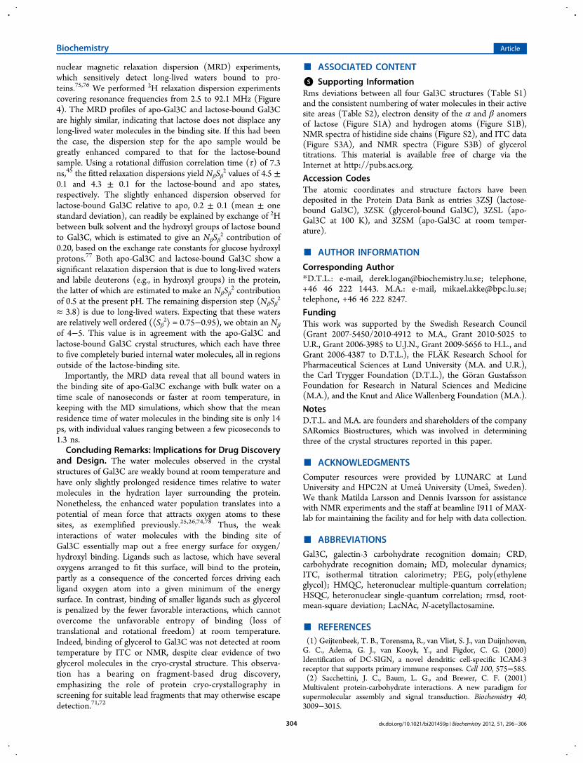
nuclear magnetic relaxation dispersion (MRD) experiments,which sensitively detect long-lived waters bound to pro-teins.75,76 We performed 2H relaxation dispersion experimentscovering resonance frequencies from 2.5 to 92.1 MHz (Figure4). The MRD profiles of apo-Gal3C and lactose-bound Gal3Care highly similar, indicating that lactose does not displace anylong-lived water molecules in the binding site. If this had beenthe case, the dispersion step for the apo sample would begreatly enhanced compared to that for the lactose-boundsample. Using a rotational diffusion correlation time (τ) of 7.3ns,45 the fitted relaxation dispersions yield NβSβ
2 values of 4.5 ±0.1 and 4.3 ± 0.1 for the lactose-bound and apo states,respectively. The slightly enhanced dispersion observed forlactose-bound Gal3C relative to apo, 0.2 ± 0.1 (mean ± onestandard deviation), can readily be explained by exchange of 2Hbetween bulk solvent and the hydroxyl groups of lactose boundto Gal3C, which is estimated to give an NβSβ
2 contribution of0.20, based on the exchange rate constants for glucose hydroxylprotons.77 Both apo-Gal3C and lactose-bound Gal3C show asignificant relaxation dispersion that is due to long-lived watersand labile deuterons (e.g., in hydroxyl groups) in the protein,the latter of which are estimated to make an NβSβ
2 contributionof 0.5 at the present pH. The remaining dispersion step (NβSβ
2
≈ 3.8) is due to long-lived waters. Expecting that these watersare relatively well ordered (⟨Sβ
2⟩ = 0.75−0.95), we obtain an Nβ
of 4−5. This value is in agreement with the apo-Gal3C andlactose-bound Gal3C crystal structures, which each have threeto five completely buried internal water molecules, all in regionsoutside of the lactose-binding site.Importantly, the MRD data reveal that all bound waters in
the binding site of apo-Gal3C exchange with bulk water on atime scale of nanoseconds or faster at room temperature, inkeeping with the MD simulations, which show that the meanresidence time of water molecules in the binding site is only 14ps, with individual values ranging between a few picoseconds to1.3 ns.Concluding Remarks: Implications for Drug Discovery
and Design. The water molecules observed in the crystalstructures of Gal3C are weakly bound at room temperature andhave only slightly prolonged residence times relative to watermolecules in the hydration layer surrounding the protein.Nonetheless, the enhanced water population translates into apotential of mean force that attracts oxygen atoms to thesesites, as exemplified previously.25,26,74,78 Thus, the weakinteractions of water molecules with the binding site ofGal3C essentially map out a free energy surface for oxygen/hydroxyl binding. Ligands such as lactose, which have severaloxygens arranged to fit this surface, will bind to the protein,partly as a consequence of the concerted forces driving eachligand oxygen atom into a given minimum of the energysurface. In contrast, binding of smaller ligands such as glycerolis penalized by the fewer favorable interactions, which cannotovercome the unfavorable entropy of binding (loss oftranslational and rotational freedom) at room temperature.Indeed, binding of glycerol to Gal3C was not detected at roomtemperature by ITC or NMR, despite clear evidence of twoglycerol molecules in the cryo-crystal structure. This observa-tion has a bearing on fragment-based drug discovery,emphasizing the role of protein cryo-crystallography inscreening for suitable lead fragments that may otherwise escapedetection.71,72
■ ASSOCIATED CONTENT
*S Supporting InformationRms deviations between all four Gal3C structures (Table S1)and the consistent numbering of water molecules in their activesite areas (Table S2), electron density of the α and β anomersof lactose (Figure S1A) and hydrogen atoms (Figure S1B),NMR spectra of histidine side chains (Figure S2), and ITC data(Figure S3A), and NMR spectra (Figure S3B) of glyceroltitrations. This material is available free of charge via theInternet at http://pubs.acs.org.
Accession CodesThe atomic coordinates and structure factors have beendeposited in the Protein Data Bank as entries 3ZSJ (lactose-bound Gal3C), 3ZSK (glycerol-bound Gal3C), 3ZSL (apo-Gal3C at 100 K), and 3ZSM (apo-Gal3C at room temper-ature).
■ AUTHOR INFORMATION
Corresponding Author*D.T.L.: e-mail, [email protected]; telephone,+46 46 222 1443. M.A.: e-mail, [email protected];telephone, +46 46 222 8247.
FundingThis work was supported by the Swedish Research Council(Grant 2007-5450/2010-4912 to M.A., Grant 2010-5025 toU.R., Grant 2006-3985 to U.J.N., Grant 2009-5656 to H.L., andGrant 2006-4387 to D.T.L.), the FLAK Research School forPharmaceutical Sciences at Lund University (M.A. and U.R.),the Carl Trygger Foundation (D.T.L.), the Goran GustafssonFoundation for Research in Natural Sciences and Medicine(M.A.), and the Knut and Alice Wallenberg Foundation (M.A.).
NotesD.T.L. and M.A. are founders and shareholders of the companySARomics Biostructures, which was involved in determiningthree of the crystal structures reported in this paper.
■ ACKNOWLEDGMENTS
Computer resources were provided by LUNARC at LundUniversity and HPC2N at Umea University (Umea, Sweden).We thank Matilda Larsson and Dennis Ivarsson for assistancewith NMR experiments and the staff at beamline I911 of MAX-lab for maintaining the facility and for help with data collection.
■ ABBREVIATIONS
Gal3C, galectin-3 carbohydrate recognition domain; CRD,carbohydrate recognition domain; MD, molecular dynamics;ITC, isothermal titration calorimetry; PEG, poly(ethyleneglycol); HMQC, heteronuclear multiple-quantum correlation;HSQC, heteronuclear single-quantum correlation; rmsd, root-mean-square deviation; LacNAc, N-acetyllactosamine.
■ REFERENCES(1) Geijtenbeek, T. B., Torensma, R., van Vliet, S. J., van Duijnhoven,G. C., Adema, G. J., van Kooyk, Y., and Figdor, C. G. (2000)Identification of DC-SIGN, a novel dendritic cell-specific ICAM-3receptor that supports primary immune responses. Cell 100, 575−585.(2) Sacchettini, J. C., Baum, L. G., and Brewer, C. F. (2001)Multivalent protein-carbohydrate interactions. A new paradigm forsupermolecular assembly and signal transduction. Biochemistry 40,3009−3015.
Biochemistry Article
dx.doi.org/10.1021/bi201459p | Biochemistry 2012, 51, 296−306304

(3) Karlsson, A., Feuk-Lagerstedt, E., Almkvist, J., Leffler, H., andDahlgren, C. (1999) Priming is required for galectin-3-inducedneutrophil NADPH-oxidase activity. J. Leukocyte Biol.,, 25.(4) Henderson, N. C., and Sethi, T. (2009) The regulation ofinflammation by galectin-3. Immunol. Rev. 230, 160−171.(5) von Itzstein, M. (2007) The war against influenza: Discovery anddevelopment of sialidase inhibitors. Nat. Rev. Drug Discovery 6, 967−974.(6) Bernardi, A., and Cheshev, P. (2008) Interfering with the SugarCode: Design and Synthesis of Oligosaccharide Mimics. Chem.Eur.J. 14, 7434−7441.(7) Ernst, B., and Magnani, J. L. (2009) From carbohydrate leads toglycomimetic drugs. Nat. Rev. Drug Discovery 8, 661−677.(8) Lepenies, B., Yin, J., and Seeberger, P. H. (2010) Applications ofsynthetic carbohydrates to chemical biology. Curr. Opin. Chem. Biol.,,1−8.(9) Dam, T. K., and Brewer, C. F. (2002) Thermodynamic studies oflectin-carbohydrate interactions by isothermal titration calorimetry.Chem. Rev. 102, 387−429.(10) Toone, E. J. (1994) Structure and Energetics of ProteinCarbohydrate Complexes. Curr. Opin. Struct. Biol. 4, 719−728.(11) Garcia Hernandez, E., Zubillaga, R. A., Rojo Dominguez, A.,Rodriguez Romero, A., and Hernandez Arana, A. (1997) New insightsinto the molecular basis of lectin-carbohydrate interactions: Acalorimetric and structural study of the association of hevein tooligomers of N-acetylglucosamine. Proteins: Struct., Funct., Genet. 29,467−477.(12) Garcia-Hernandez, E., and Hernandez-Arana, A. (1999)Structural bases of lectin-carbohydrate affinities: Comparison withprotein-folding energetics. Protein Sci. 8, 1075−1086.(13) Lemieux, R. U. (1999) How water provides the impetus formolecular recognition in aqueous solution (vol 29, pg 373, 1996). Acc.Chem. Res. 32, 631.(14) Lis, H., and Sharon, N. (1998) Lectins: Carbohydrate-specificproteins that mediate cellular recognition. Chem. Rev. 98, 637−674.(15) Davis, A. P., and Wareham, R. S. (1999) Carbohydraterecognition through noncovalent interactions: A challenge forbiomimetic and supramolecular chemistry. Angew. Chem., Int. Ed. 38,2978−2996.(16) Sorme, P., Arnoux, P., Kahl-Knutsson, B., Leffler, H., Rini, J. M.,and Nilsson, U. J. (2005) Structural and thermodynamic studies oncation-π interactions in lectin-ligand complexes: High-affinity galectin-3 inhibitors through fine-tuning of an arginine-arene interaction. J. Am.Chem. Soc. 127, 1737−1743.(17) del Carmen Fernandez-Alonso, M., Canada, F. J., Jimenez-Barbero, J., and Cuevas, G. (2005) Molecular recognition ofsaccharides by proteins. Insights on the origin of the carbohydrate-aromatic interactions. J. Am. Chem. Soc. 127, 7379−7386.(18) Laughrey, Z. R., Kiehna, S. E., Riemen, A. J., and Waters, M. L.(2008) Carbohydrate-π interactions: What are they worth? J. Am.Chem. Soc. 130, 14625−14633.(19) Ladbury, J. E. (1996) Just add water! The effect of water on thespecificity of protein-ligand binding sites and its potential applicationto drug design. Chem. Biol. 3, 973−980.(20) Clarke, C., Woods, R. J., Gluska, J., Cooper, A., Nutley, M. A.,and Boons, G. J. (2001) Involvement of water in carbohydrate-proteinbinding. J. Am. Chem. Soc. 123, 12238−12247.(21) Chervenak, M. C., and Toone, E. J. (1995) Calorimetric analysisof the binding of lectins with overlapping carbohydrate-binding ligandspecificities. Biochemistry 34, 5685−5695.(22) Dunitz, J. D. (1994) The entropic cost of bound water incrystals and biomolecules. Science 264, 670.(23) Kadirvelraj, R., Foley, B. L., Dyekjaer, J. D., and Woods, R. J.(2008) Involvement of Water in Carbohydrate-Protein Binding:Concanavalin A Revisited. J. Am. Chem. Soc. 130, 16933−16942.(24) Lemieux, R. U. (1996) How water provides the impetus formolecular recognition in aqueous solution. Acc. Chem. Res. 29, 373−380.
(25) Di Lella, S., Ma, L., Ricci, J. C., Rabinovich, G. A., Asher, S. A.,and Alvarez, R. M. (2009) Critical role of the solvent environment ingalectin-1 binding to the disaccharide lactose. Biochemistry 48, 786−791.(26) Li, Z., and Lazaridis, T. (2005) The effect of water displacementon binding thermodynamics: concanavalin A. J. Phys. Chem. B 109,662−670.(27) Asensio, J. L., Siebert, H. C., von der Lieth, C. W., Laynez, J.,Bruix, M., Soedjanaamadja, U. M., Beintema, J. J., Canada, F. J.,Gabius, H. J., and Jimenez-Barbero, J. (2000) NMR investigations ofprotein-carbohydrate interactions: Studies on the relevance of Trp/Tyr variations in lectin binding sites as deduced from titrationmicrocalorimetry and NMR studies on hevein domains. Determinationof the NMR structure of the complex between pseudohevein andN,N′,N″-triacetylchitotriose. Proteins: Struct., Funct., Genet. 40, 218−236.(28) Leffler, H., Carlsson, S., Hedlund, M., Qian, Y., and Poirier, F.(2004) Introduction to galectins. Glycoconjugate J. 19, 433−440.(29) Rabinovich, G. A., Liu, F. T., Hirashima, M., and Anderson, A.(2007) An emerging role for galectins in tuning the immune response:Lessons from experimental models of inflammatory disease,autoimmunity and cancer. Scand. J. Immunol. 66, 143−158.(30) MacKinnon, A. C., Farnworth, S. L., Hodkinson, P. S.,Henderson, N. C., Atkinson, K. M., Leffler, H., Nilsson, U. J.,Haslett, C., Forbes, S. J., and Sethi, T. (2008) Regulation of alternativemacrophage activation by galectin-3. J. Immunol. 180, 2650−2658.(31) Delacour, D., Koch, A., and Jacob, R. (2009) The role ofgalectins in protein trafficking. Traffic 10, 1405−1413.(32) Liu, F. T., and Rabinovich, G. A. (2010) Galectins: Regulators ofacute and chronic inflammation. Ann. N.Y. Acad. Sci. 1183, 158−182.(33) Grigorian, A., and Demetriou, M. (2010) Manipulating cellsurface glycoproteins by targeting N-glycan-galectin interactions.Methods Enzymol. 480, 245−266.(34) Nieminen, J., Kuno, A., Hirabayashi, J., and Sato, S. (2007)Visualization of galectin-3 oligomerization on the surface ofneutrophils and endothelial cells using fluorescence resonanceenergy transfer. J. Biol. Chem. 282, 1374−1383.(35) Seelenmeyer, C., Wegehingel, S., Tews, I., Kunzler, M., Aebi, M.,and Nickel, W. (2005) Cell surface counter receptors are essentialcomponents of the unconventional export machinery of galectin-1. J.Cell Biol. 171, 373−381.(36) Baptiste, T. A., James, A., Saria, M., and Ochieng, J. (2007)Mechano-transduction mediated secretion and uptake of galectin-3 inbreast carcinoma cells: Implications in the extracellular functions of thelectin. Exp. Cell Res. 313, 652−664.(37) Dennis, J. W., Lau, K. S., Demetriou, M., and Nabi, I. R. (2009)Adaptive regulation at the cell surface by N-glycosylation. Traffic 10,1569−1578.(38) Almkvist, J., and Karlsson, A. (2004) Galectins as inflammatorymediators. Glycoconjugate J. 19, 575−581.(39) Dumic, J., Dabelic, S., and Flogel, M. (2006) Galectin-3: Anopen-ended story. Biochim. Biophys. Acta 1760, 616−635.(40) Liu, F. T. (2005) Regulatory roles of galectins in the immuneresponse. Int. Arch. Allergy Immunol. 136, 385−400.(41) Liu, F. T., and Rabinovich, G. A. (2005) Galectins as modulatorsof tumour progression. Nat. Rev. Cancer 5, 29−41.(42) Walti, M. A., Walser, P. J., Thore, S., Grunler, A., Bednar, M.,Kunzler, M., and Aebi, M. (2008) Structural basis for chitotetraosecoordination by CGL3, a novel galectin-related protein fromCoprinopsis cinerea. J. Mol. Biol. 379, 146−159.(43) Seetharaman, J., Kanigsberg, A., Slaaby, R., Leffler, H., Barondes,S. H., and Rini, J. M. (1998) X-ray crystal structure of the humangalectin-3 carbohydrate recognition domain at 2.1-Å resolution. J. Biol.Chem. 273, 13047−13052.(44) Carlsson, S., Oberg, C. T., Carlsson, M. C., Sundin, A., Niisson,U. J., Smith, D., Cummings, R. D., Almkvist, J., Karlsson, A., andLeffler, H. (2007) Affinity of galectin-8 and its carbohydraterecognition domains for ligands in solution and at the cell surface.Glycobiology 17, 663−676.
Biochemistry Article
dx.doi.org/10.1021/bi201459p | Biochemistry 2012, 51, 296−306305

(45) Diehl, C., Genheden, S., Engstrom, O., Delaine, T., Hakansson,M., Leffler, H., Nilsson, U. J., Ryde, U., and Akke, M. (2010) Proteinflexibility and conformational entropy in ligand design targeting thecarbohydrate recognition domain of galectin-3. J. Am. Chem. Soc. 132,14577−14589.(46) Salameh, B. A., Leffler, H., and Nilsson, U. J. (2005) 3-(1,2,3-Triazol-1-yl)-1-thio-galactosides as small, efficient, and hydrolyticallystable inhibitors of galectin-3. Bioorg. Med. Chem. Lett. 15, 3344−3346.(47) Salameh, B. A., Cumpstey, I., Sundin, A., Leffler, H., andNilsson, U. J. (2010) 1H-1,2,3-Triazol-1-yl thiodigalactosidederivatives as high affinity galectin-3 inhibitors. Bioorg. Med. Chem.18, 5367−5378.(48) Yang, R. Y., Rabinovich, G. A., and Liu, F. T. (2008) Galectins:Structure, function and therapeutic potential. Expert Rev. Mol. Med. 10,e17.(49) Wu, M. H., Hong, T. M., Cheng, H. W., Pan, S. H., Liang, Y. R.,Hong, H. C., Chiang, W. F., Wong, T. Y., Shieh, D. B., Shiau, A. L., Jin,Y. T., and Chen, Y. L. (2009) Galectin-1-mediated tumor invasion andmetastasis, up-regulated matrix metalloproteinase expression, andreorganized actin cytoskeletons. Mol. Cancer Res. 7, 311−318.(50) Delaine, T., Cumpstey, I., Ingrassia, L., Le Mercier, M.,Okechukwu, P., Leffler, H., Kiss, R., and Nilsson, U. J. (2008)Galectin-inhibitory thiodigalactoside ester derivatives haveantimigratory effects in cultured lung and prostate cancer cells. J.Med. Chem. 51, 8109−8114.(51) Lin, C. I., Whang, E. E., Donner, D. B., Jiang, X., Price, B. D.,Carothers, A. M., Delaine, T., Leffler, H., Nilsson, U. J., Nose, V.,Moore, F. D., and Ruan, D. T. (2009) Galectin-3 targeted therapy witha small molecule inhibitor activates apoptosis and enhances bothchemosensitivity and radiosensitivity in papillary thyroid cancer. Mol.Cancer Res. 7, 1655−1662.(52) Diehl, C., Genheden, S., Modig, K., Ryde, U., and Akke, M.(2009) Conformational entropy changes upon lactose binding to thecarbohydrate recognition domain of galectin-3. J. Biomol. NMR 45,157−169.(53) Massa, S. M., Cooper, D. N., Leffler, H., and Barondes, S. H.(1993) L-29, an endogenous lectin, binds to glycoconjugate ligandswith positive cooperativity. Biochemistry 32, 260−267.(54) Kabsch, W. (2010) Integration, scaling, space-group assignmentand post-refinement. Acta Crystallogr. D66, 133−144.(55) Murshudov, G. N. (1997) Refinement of macromolecularstructures by the maximum-likelihood method. Acta Crystallogr. D53,240−255.(56) Potterton, E., Briggs, P., Turkenburg, M., and Dodson, E.(2003) A graphical user interface to the CCP4 program suite. ActaCrystallogr. D59, 1131−1137.(57) Sheldrick, G. M. (2008) A short history of SHELX. ActaCrystallogr. A64, 112−122.(58) Emsley, P., Lohkamp, B., Scott, W. G., and Cowtan, K. (2010)Features and development of Coot. Acta Crystallogr. D66, 486−501.(59) DeLano, W. L. (2002) The PyMOL Molecular Graphics System,DeLano Scientific, Palo Alto, CA.(60) Teilum, K., Brath, U., Lundstrom, P., and Akke, M. (2006)Biosynthetic 13C labeling of aromatic side chains in proteins for NMRrelaxation measurements. J. Am. Chem. Soc. 128, 2506−2507.(61) Lundstrom, P., Teilum, K., Carstensen, T., Bezsonova, I.,Wiesner, S., Hansen, D. F., Religa, T. L., Akke, M., and Kay, L. E.(2007) Fractional 13C enrichment of isolated carbons using [1-13C]- or[2- 13C]-glucose facilitates the accurate measurement of dynamics atbackbone Cα and side-chain methyl positions in proteins. J. Biomol.NMR 38, 199−212.(62) Genheden, S., Luchko, T., Gusarov, S., Kovalenko, A., and Ryde,U. (2010) An MM/3D-RISM approach for ligand binding affinities. J.Phys. Chem. B 114, 8505−8516.(63) Young, T., Abel, R., Kim, B., Berne, B. J., and Friesner, R. A.(2007) Motifs for molecular recognition exploiting hydrophobicenclosure in protein-ligand binding. Proc. Natl. Acad. Sci. U.S.A. 104,808−813.
(64) Abel, R., Young, T., Farid, R., Berne, B. J., and Friesner, R. A.(2008) Role of the active-site solvent in the thermodynamics of factorXa ligand binding. J. Am. Chem. Soc. 130, 2817−2831.(65) Shao, J. Y., Tanner, S. W., Thompson, N., and Cheatham, T. E.(2007) Clustering molecular dynamics trajectories: 1. Characterizingthe performance of different clustering algorithms. J. Chem. TheoryComput. 3, 2312−2334.(66) Collins, P. M., Hidari, K. I., and Blanchard, H. (2007) Slowdiffusion of lactose out of galectin-3 crystals monitored by X-raycrystallography: Possible implications for ligand-exchange protocols.Acta Crystallogr. D63, 415−419.(67) McDonald, I. K., and Thornton, J. M. (1994) Satisfyinghydrogen bonding potential in proteins. J. Mol. Biol. 238, 777−793.(68) Pelton, J. G., Torchia, D. A., Meadow, N. D., and Roseman, S.(1993) Tautomeric states of the active-site histidines ofphosphorylated and unphosphorylated III(Glc), a signal-transducingprotein from Escherichia coli, using two-dimensional heteronuclearNMR techniques. Protein Sci. 2, 543−558.(69) Bachhawat, K., Thomas, C. J., Amutha, B., Krishnasastry, M. V.,Khan, M. I., and Surolia, A. (2001) On the stringent requirement ofmannosyl substitution in mannooligosaccharides for the recognition bygarlic (Allium sativum) lectin. A surface plasmon resonance study. J.Biol. Chem. 276, 5541−5546.(70) Ahmad, N., Gabius, H. J., Sabesan, S., Oscarson, S., and Brewer,C. F. (2004) Thermodynamic binding studies of bivalentoligosaccharides to galectin-1, galectin-3, and the carbohydraterecognition domain of galectin-3. Glycobiology 14, 817−825.(71) Murray, C. W., and Blundell, T. L. (2010) Structural biology infragment-based drug design. Curr. Opin. Struct. Biol. 20, 497−507.(72) Burley, S. K., Hirst, G., Sprengeler, P., and Reich, S. (2010)Fragment-based structure-guided drug discovery: Strategy, process,and lessons from human protein kinases. In Drug Design: Structure- andligand-based approaches (Merz, K. M., Ringe, D., and Reynolds, C. H.,Eds.) pp 30−40, Cambridge University Press, New York.(73) Umemoto, K., Leffler, H., Venot, A., Valafar, H., and Prestegard,J. H. (2003) Conformational differences in liganded and unligandedstates of galectin-3. Biochemistry 42, 3688−3695.(74) Gauto, D. F., Di Lella, S., Guardia, C. M., Estrin, D. A., andMarti, M. A. (2009) Carbohydrate-binding proteins: Dissecting ligandstructures through solvent environment occupancy. J. Phys. Chem. B113, 8717−8724.(75) Halle, B., and Denisov, V. P. (2001) Magnetic relaxationdispersion studies of biomolecular solutions. Methods Enzymol. 338,178−201.(76) Halle, B. (2004) Protein hydration dynamics in solution: Acritical survey. Philos. Trans. R. Soc. London, Ser. B 359, 1207−1223.(77) Hills, B. P. (1991) Multinuclear NMR-studies of water insolutions of simple carbohydrates. 1. Proton and deuterium relaxation.Mol. Phys. 72, 1099−1121.(78) Di Lella, S., Marti, M. A., Alvarez, R. M. S., Estrin, D. A., andRicci, J. C. D. (2007) Characterization of the galectin-1 carbohydraterecognition domain in terms of solvent occupancy. J. Phys. Chem. B111, 7360−7366.(79) Davis, I. W., Leaver-Fay, A., Chen, V. B., Block, J. N., Kapral, G.J., Wang, X., Murray, L. W., Arendall, W. B. III, Snoeyink, J.,Richardson, J. S., and Richardson, D. C. (2007) MolProbity: All-atomcontacts and structure validation for proteins and nucleic acids. NucleicAcids Res. 35, W375−W383.
Biochemistry Article
dx.doi.org/10.1021/bi201459p | Biochemistry 2012, 51, 296−306306