the cardiovascular system - brazosport.edu · –some cardiac muscle cells are self-excitable ......
TRANSCRIPT
© Annie Leibovitz/Contact Press Images
Chapter 18 Part A
The
Cardiovascular
System
MDufilho 1/19/16 1
2
Similarities of Cardiac and Skeletal Muscle
• RMP
• Ion concentration
• Deploarization
• Action Potential
• Repolarization
• Restoring resting membrane potential
• Types of Cardiac muscle fibers
1/19/16 MDufilho
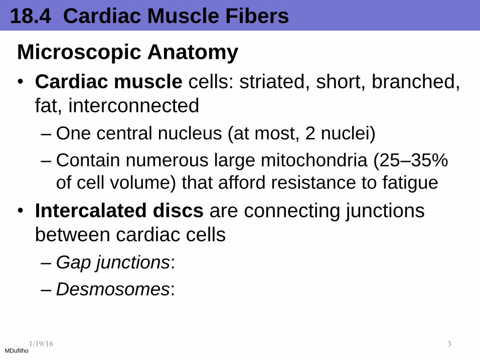
18.4 Cardiac Muscle Fibers
Microscopic Anatomy
• Cardiac muscle cells: striated, short, branched,
fat, interconnected
– One central nucleus (at most, 2 nuclei)
– Contain numerous large mitochondria (25–35%
of cell volume) that afford resistance to fatigue
• Intercalated discs are connecting junctions
between cardiac cells
– Gap junctions:
– Desmosomes:
MDufilho 1/19/16 3

Figure 18.11a Microscopic
anatomy of cardiac muscle.
MDufilho
Desmosomes (keep
myocytes from pulling apart)
Gap junctions (electrically
connect myocytes) Nucleus
Intercalated
discs
Cardiac
muscle cell
1/19/16 4
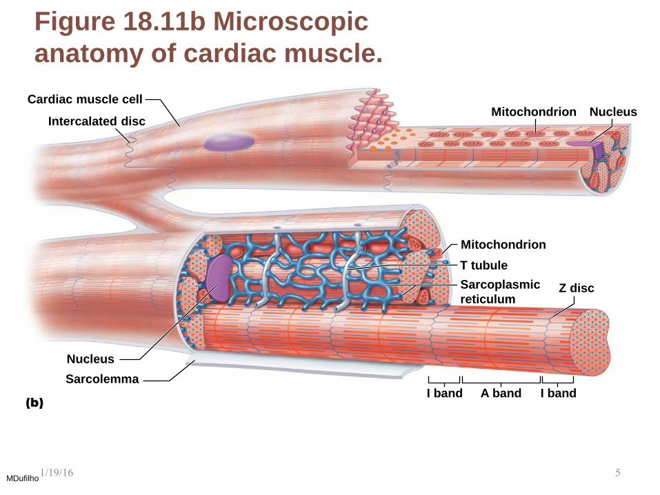
Figure 18.11b Microscopic
anatomy of cardiac muscle.
MDufilho
Nucleus
Nucleus
I band
Cardiac muscle cell
A band Sarcolemma
Z disc
Mitochondrion
Mitochondrion
T tubule
Sarcoplasmic
reticulum
I band
Intercalated disc
1/19/16 5
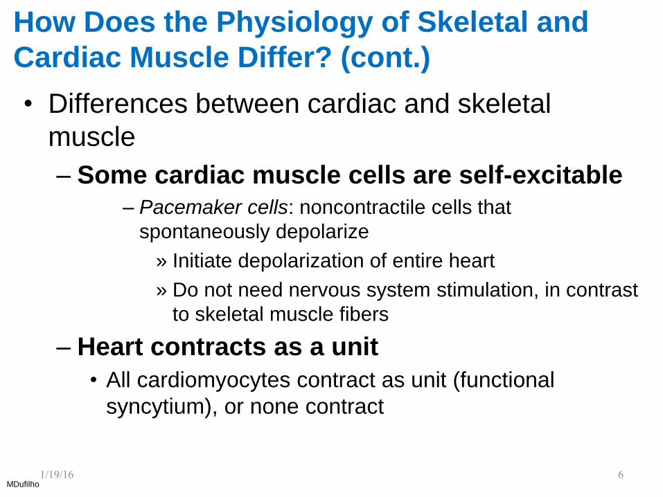
How Does the Physiology of Skeletal and
Cardiac Muscle Differ? (cont.)
• Differences between cardiac and skeletal
muscle
– Some cardiac muscle cells are self-excitable
– Pacemaker cells: noncontractile cells that
spontaneously depolarize
» Initiate depolarization of entire heart
» Do not need nervous system stimulation, in contrast
to skeletal muscle fibers
– Heart contracts as a unit
• All cardiomyocytes contract as unit (functional
syncytium), or none contract
MDufilho 1/19/16 6
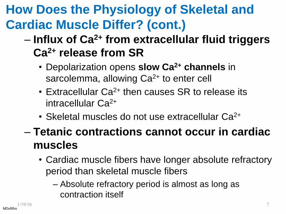
How Does the Physiology of Skeletal and
Cardiac Muscle Differ? (cont.) – Influx of Ca2+ from extracellular fluid triggers
Ca2+ release from SR
• Depolarization opens slow Ca2+ channels in
sarcolemma, allowing Ca2+ to enter cell
• Extracellular Ca2+ then causes SR to release its
intracellular Ca2+
• Skeletal muscles do not use extracellular Ca2+
– Tetanic contractions cannot occur in cardiac
muscles
• Cardiac muscle fibers have longer absolute refractory
period than skeletal muscle fibers
– Absolute refractory period is almost as long as
contraction itself
MDufilho 1/19/16 7

How Does the Physiology of Skeletal and
Cardiac Muscle Differ? (cont.)
– The heart relies almost exclusively on
aerobic respiration
• Cardiac muscle has more mitochondria than skeletal
muscle so has greater dependence on oxygen
– Cannot function without oxygen
• Skeletal muscle can go through fermentation when
oxygen not present
• Both types of tissues can use other fuel sources
– Cardiac is more adaptable to other fuels, including
lactic acid, but must have oxygen
MDufilho 1/19/16 8
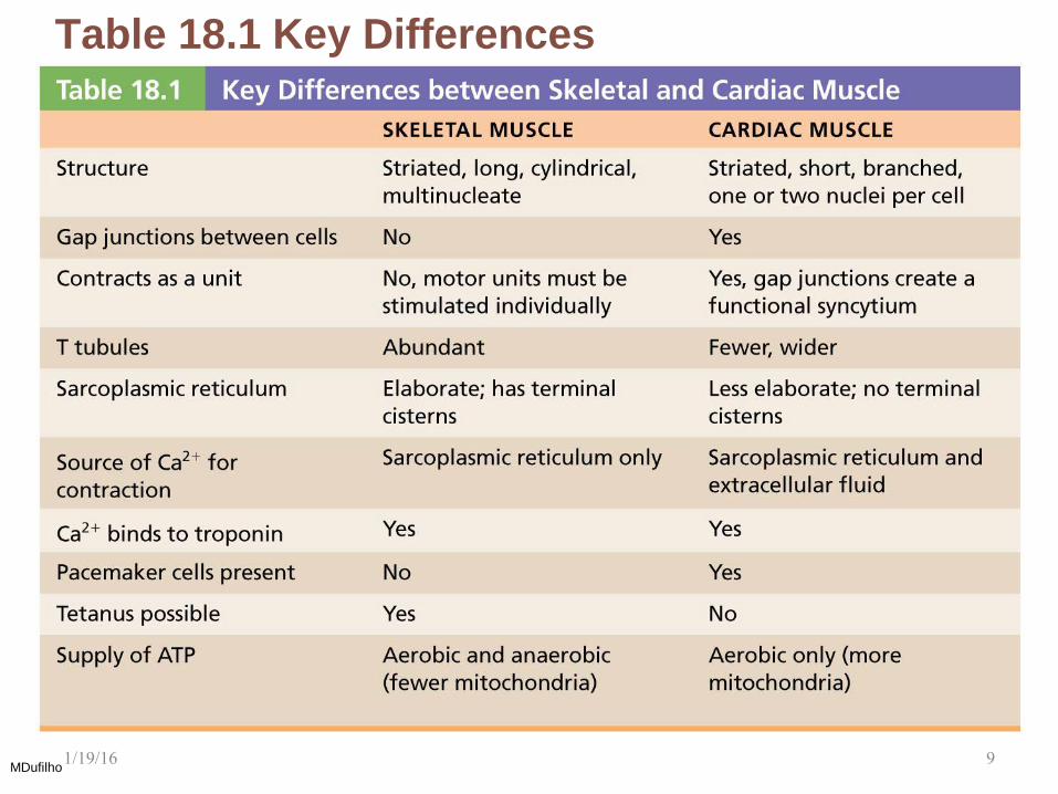
Table 18.1 Key Differences
between Skeletal and Cardiac
Muscle
MDufilho 1/19/16 9
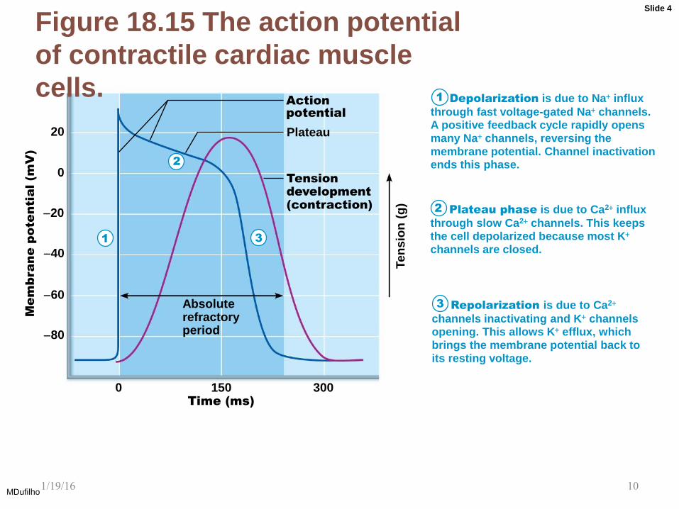
Figure 18.15 The action potential
of contractile cardiac muscle
cells.
MDufilho
80
60
40
20
0
20 Plateau
0 150 300
Plateau phase is due to Ca2+ influx
through slow Ca2+ channels. This keeps the cell depolarized because most K+
channels are closed.
Time (ms)
Absolute refractory period
Tension
development
(contraction)
Action
potential
Ten
sio
n (
g)
Me
mb
ra
ne
p
ote
ntia
l (m
V)
Repolarization is due to Ca2+
channels inactivating and K+ channels opening. This allows K+ efflux, which brings the membrane potential back to its resting voltage.
Depolarization is due to Na+ influx
through fast voltage-gated Na+ channels. A positive feedback cycle rapidly opens many Na+ channels, reversing the membrane potential. Channel inactivation ends this phase.
1
2
3
3
2
1
Slide 4
1/19/16 10
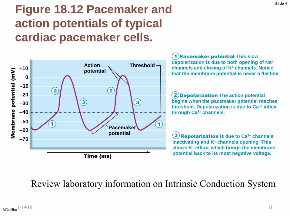
Figure 18.12 Pacemaker and
action potentials of typical
cardiac pacemaker cells.
MDufilho
Time (ms)
70
Action potential
Threshold
Pacemaker potential
60
40
30
20
10
0
+10
50
Mem
brane potential (m
V)
Pacemaker potential This slow
depolarization is due to both opening of Na+
channels and closing of K+ channels. Notice that the membrane potential is never a flat line.
Depolarization The action potential
begins when the pacemaker potential reaches threshold. Depolarization is due to Ca2+ influx through Ca2+ channels.
Repolarization is due to Ca2+ channels
inactivating and K+ channels opening. This allows K+ efflux, which brings the membrane potential back to its most negative voltage.
1
2
3
3
2
3
1 1
2
Slide 4
Review laboratory information on Intrinsic Conduction System
1/19/16 11
Figure 18.13 Intrinsic cardiac
conduction system and action
potential succession during one
heartbeat.
MDufilho
Pacemaker potential
Plateau
Internodal pathway
Superior vena cava Right atrium
Left atrium
Subendocardial conducting network (Purkinje fibers)
Inter- ventricular septum
Pacemaker potential
Ventricular
muscle
AV node
Atrial muscle
SA node
0 200 400 600
Milliseconds
Comparison of action potential shape
at various locations
Anatomy of the intrinsic conduction system showing the sequence
of electrical excitation
The sinoatrial
(SA) node (pacemaker) generates impulses.
The impulses pause (0.1 s) at the atrioventricular
(AV) node.
The atrioventricular
(AV) bundle
connects the atria to the ventricles.
The bundle branches conduct the impulses through the interventricular septum.
The subendocardial
conducting network
depolarizes the contractile cells of both ventricles.
1
2
3
4
5
Slide 6
1/19/16 12
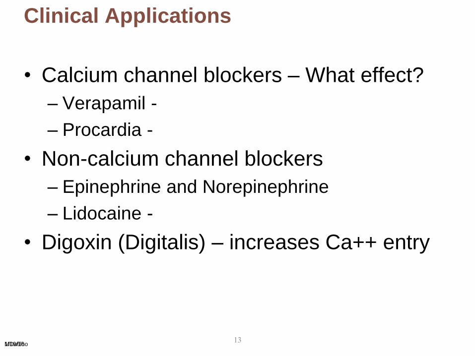
Clinical Applications
• Calcium channel blockers – What effect?
– Verapamil -
– Procardia -
• Non-calcium channel blockers
– Epinephrine and Norepinephrine
– Lidocaine -
• Digoxin (Digitalis) – increases Ca++ entry
MDufilho 1/19/16 13

18.6 Mechanical Events of Heart
• Systole: period of heart contraction
• Diastole: period of heart relaxation
• Cardiac cycle: blood flow through heart during
one complete heartbeat
– Atrial systole and diastole are followed by
ventricular systole and diastole
– Cycle represents series of pressure and blood
volume changes
– Mechanical events follow electrical events seen
on ECG
• Three phases of the cardiac cycle (following left
side, starting with total relaxation) MDufilho
1/19/16 14
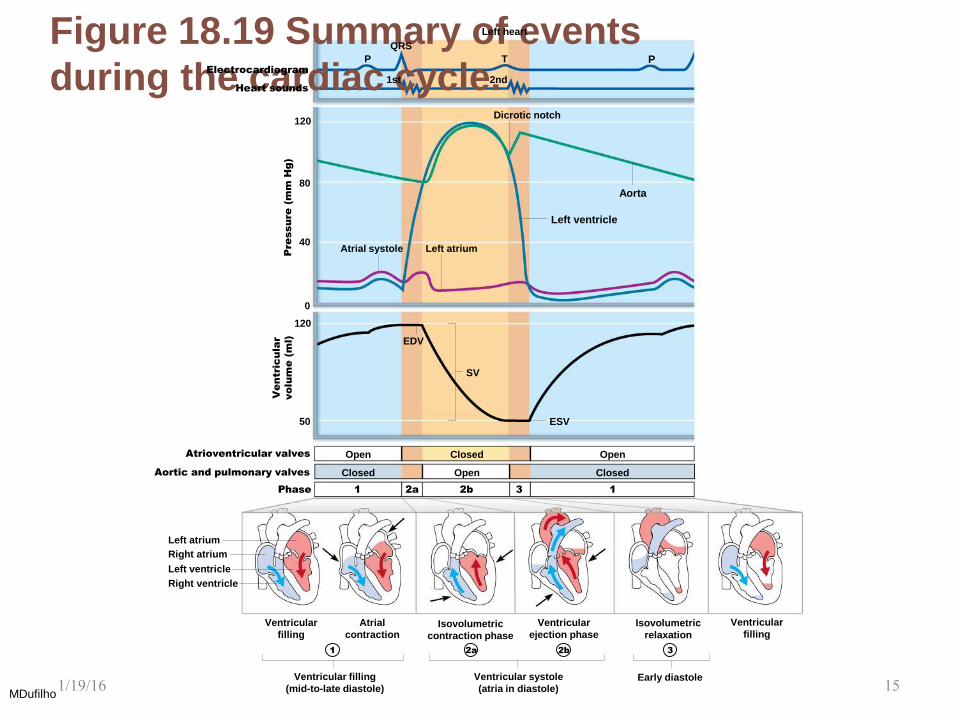
Figure 18.19 Summary of events
during the cardiac cycle.
MDufilho
120
80
40
0
Left heart
P
1st 2nd
QRS
P
120
50
Atrial systole
Dicrotic notch
Left ventricle
Left atrium
EDV
SV
Aorta
Open Open Closed
Closed Closed Open
ESV
Left atrium
Right atrium
Left ventricle
Right ventricle
Ventricular
filling
Atrial
contraction
Ventricular filling
(mid-to-late diastole)
Ventricular systole
(atria in diastole)
Isovolumetric
contraction phase
Ventricular
ejection phase
Early diastole
Isovolumetric
relaxation
Ventricular
filling
T
1 2a 2b 3
Atrioventricular valves
Aortic and pulmonary valves
Phase
Ve
ntric
ula
r
vo
lu
me
(m
l)
Pre
ssu
re
(m
m H
g)
Heart sounds
Electrocardiogram
1 2a 2b 3 1
1/19/16 15
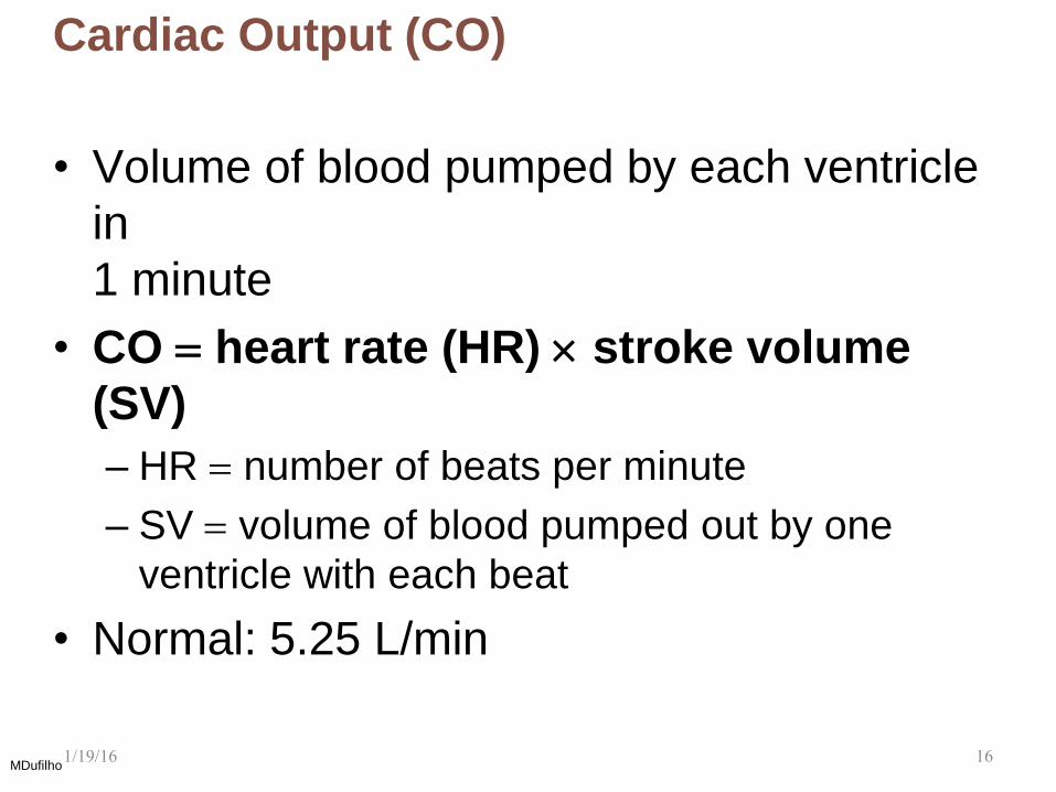
Cardiac Output (CO)
• Volume of blood pumped by each ventricle
in
1 minute
• CO = heart rate (HR) stroke volume
(SV)
– HR = number of beats per minute
– SV = volume of blood pumped out by one
ventricle with each beat
• Normal: 5.25 L/min
MDufilho 1/19/16 16

18.7 Regulation of Pumping
• Cardiac output: amount of blood pumped out
by each ventricle in 1 minute
– Equals heart rate (HR) times stroke volume (SV)
• Stroke volume: volume of blood pumped out by one
ventricle with each beat
– Correlates with force of contraction
• At rest:
CO (ml/min) = HR (75 beats/min) SV (70 ml/beat)
= 5.25 L/min
MDufilho 1/19/16 17
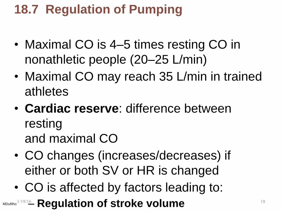
18.7 Regulation of Pumping
• Maximal CO is 4–5 times resting CO in
nonathletic people (20–25 L/min)
• Maximal CO may reach 35 L/min in trained
athletes
• Cardiac reserve: difference between
resting
and maximal CO
• CO changes (increases/decreases) if
either or both SV or HR is changed
• CO is affected by factors leading to:
– Regulation of stroke volume
– Regulation of heart rates
MDufilho 1/19/16 18

Regulation of Stroke Volume
• Mathematically: SV = EDV ESV
– EDV is affected by length of ventricular
diastole and venous pressure (120 ml/beat)
– ESV is affected by arterial BP and force of
ventricular contraction (50 ml/beat)
– Normal SV = 120 ml 50 ml = 70 ml/beat
• Three main factors that affect SV:
– Preload
– Contractility
– Afterload
MDufilho 1/19/16 19
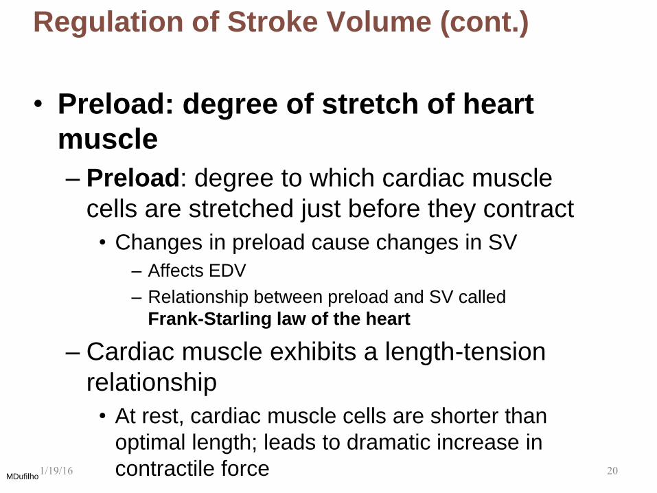
Regulation of Stroke Volume (cont.)
• Preload: degree of stretch of heart
muscle
– Preload: degree to which cardiac muscle
cells are stretched just before they contract
• Changes in preload cause changes in SV
– Affects EDV
– Relationship between preload and SV called
Frank-Starling law of the heart
– Cardiac muscle exhibits a length-tension
relationship
• At rest, cardiac muscle cells are shorter than
optimal length; leads to dramatic increase in
contractile force MDufilho 1/19/16 20
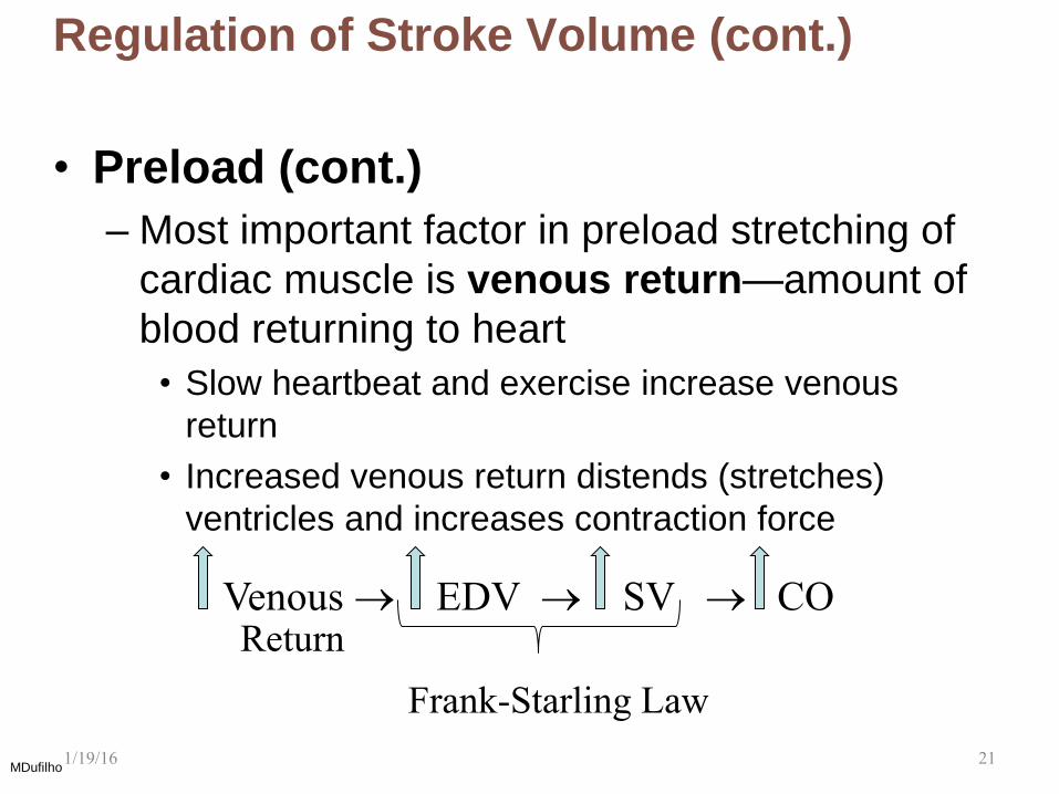
Regulation of Stroke Volume (cont.)
• Preload (cont.)
– Most important factor in preload stretching of
cardiac muscle is venous return—amount of
blood returning to heart
• Slow heartbeat and exercise increase venous
return
• Increased venous return distends (stretches)
ventricles and increases contraction force
MDufilho
Frank-Starling Law
Return Venous EDV SV CO
1/19/16 21
Regulation of Stroke Volume (cont.)
• Contractility
– Contractile strength at given muscle length
• Independent of muscle stretch and EDV
– Increased contractility lowers ESV; caused by:
• Sympathetic epinephrine release stimulates
increased Ca2+ influx, leading to more cross bridge
formations
• Positive inotropic agents increase contractility
– Thyroxine, glucagon, epinephrine, digitalis, high
extracellular Ca2+
– Decreased by negative inotropic agents
• Acidosis (excess H+), increased extracellular K+,
calcium channel blockers MDufilho 1/19/16 22
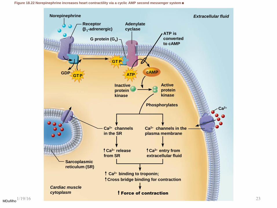
Figure 18.22 Norepinephrine increases heart contractility via a cyclic AMP second messenger system.
MDufilho
Norepinephrine
Adenylate
cyclase
ATP cAM P
GT P
G protein (Gs)
Active
protein
kinase
Phosphorylates
Sarcoplasmic
reticulum (SR)
Inactive
protein
kinase
Cardiac muscle
cytoplasm
Extracellular fluid
ATP is
converted
to cAMP
GT P GD P
Receptor
(1-adrenergic)
Ca2+ channels in the
plasma membrane
Ca2+ channels
in the SR
Ca2+
Ca2+ entry from
extracellular fluid
Ca2+ release
from SR
Ca2+ binding to troponin;
Cross bridge binding for contraction
Force of contraction
1/19/16 23

Regulation of Stroke Volume (cont.)
• Afterload: back pressure exerted by
arterial blood
– Afterload is pressure that ventricles must
overcome to eject blood
• Back pressure from arterial blood pushing on SL
valves is major pressure
– Aortic pressure is around 80 mm Hg
– Pulmonary trunk pressure is around 10 mm Hg
– Hypertension increases afterload, resulting in
increased ESV and reduced SV
MDufilho 1/19/16 24

Regulation of Heart Rate
• If SV decreases as a result of decreased
blood volume or weakened heart, CO can
be maintained by increasing HR and
contractility
– Positive chronotropic factors increase heart
rate
– Negative chronotropic factors decrease heart
rate
• Heart rate can be regulated by:
– Autonomic nervous system
– Chemicals
– Other factors MDufilho
1/19/16 25
Regulation of Heart Rate (cont.)
• Autonomic nervous system regulation
of heart rate
– Sympathetic nervous system can be activated
by emotional or physical stressors
– Norepinephrine is released and binds to
1-adrenergic receptors on heart, causing:
• Pacemaker to fire more rapidly, increasing HR
– EDV decreased because of decreased fill time
• Increased contractility
– ESV decreased because of increased volume of ejected
blood
MDufilho 1/19/16 26
Regulation of Heart Rate (cont.)
• Autonomic nervous system regulation
of heart rate (cont.)
– Because both EDV and ESV decrease, SV
can remain unchanged
– Parasympathetic nervous system opposes
sympathetic effects
• Acetylcholine hyperpolarizes pacemaker cells by
opening K+ channels, which slows HR
• Has little to no effect on contractility
MDufilho 1/19/16 27
Regulation of Heart Rate (cont.)
• Autonomic nervous system regulation
of heart rate (cont.)
– Heart at rest exhibits vagal tone
• Parasympathetic is dominant influence on heart
rate
• Decreases rate about 25 beats/min
• Cutting vagal nerve leads to HR of 100
MDufilho 1/19/16 28
Regulation of Heart Rate (cont.)
• Autonomic nervous system regulation
of heart rate (cont.)
– When sympathetic is activated,
parasympathetic is inhibited, and vice-versa
– Atrial (Bainbridge) reflex: sympathetic reflex
initiated by increased venous return, hence
increased atrial filling
• Atrial walls are stretched with increased volume
• Stimulates SA node, which increases HR
• Also stimulates atrial stretch receptors that activate
sympathetic reflexes
MDufilho 1/19/16 29
Regulation of Heart Rate (cont.)
• Chemical regulation of heart rate
– Hormones
• Epinephrine from adrenal medulla increases heart
rate and contractility
• Thyroxine increases heart rate; enhances effects
of norepinephrine and epinephrine
– Ions
• Intra- and extracellular ion concentrations (e.g.,
Ca2+ and K+) must be maintained for normal heart
function
– Imbalances are very dangerous to heart
MDufilho 1/19/16 30
Clinical – Homeostatic Imbalance 18.7
• Hypocalcemia: depresses heart
• Hypercalcemia: increases HR and
contractility
• Hyperkalemia: alters electrical activity,
which can lead to heart block and cardiac
arrest
• Hypokalemia: results in feeble heartbeat;
arrhythmias
MDufilho 1/19/16 31
Regulation of Heart Rate (cont.)
• Other factors that influence heart rate
– Age
• Fetus has fastest HR; declines with age
– Gender
• Females have faster HR than males
– Exercise
• Increases HR
• Trained athletes can have slow HR
– Body temperature
• HR increases with increased body temperature
MDufilho 1/19/16 32
Homeostatic Imbalance of Cardiac Output
• Congestive heart failure (CHF)
– Progressive condition; CO is so low that blood
circulation is inadequate to meet tissue needs
– Reflects weakened myocardium caused by:
• Coronary atherosclerosis: clogged arteries
caused by fat buildup; impairs oxygen delivery to
cardiac cells
– Heart becomes hypoxic, contracts inefficiently
MDufilho 1/19/16 33
Homeostatic Imbalance of Cardiac Output
(cont.)
• Congestive heart failure (CHF) (cont.) • Persistent high blood pressure: aortic pressure
90 mmHg causes myocardium to exert more
force
– Chronic increased ESV causes myocardium hypertrophy
and weakness
• Multiple myocardial infarcts: heart becomes
weak as contractile cells are replaced with scar
tissue
• Dilated cardiomyopathy (DCM): ventricles stretch
and become flabby, and myocardium deteriorates
– Drug toxicity or chronic inflammation may play a role
MDufilho 1/19/16 34
Homeostatic Imbalance of Cardiac Output
(cont.)
• Congestive heart failure (CHF) (cont.)
– Either side of heart can be affected:
• Left-sided failure results in pulmonary congestion
– Blood backs up in lungs
• Right-sided failure results in peripheral congestion
– Blood pools in body organs, causing edema
– Failure of either side ultimately weakens other
side
• Leads to decompensated, seriously weakened heart
• Treatment: removal of fluid, drugs to reduce afterload
and increase contractility
MDufilho 1/19/16 35