the cardiovascular system dr. mona soliman, mbbs, msc, phd dr. mona soliman, mbbs, msc, phd...
TRANSCRIPT

The Cardiovascular System
Dr. Mona Soliman, MBBS, MSc, PhD
Department of PhysiologyCollege of Medicine
KSU
November 2012
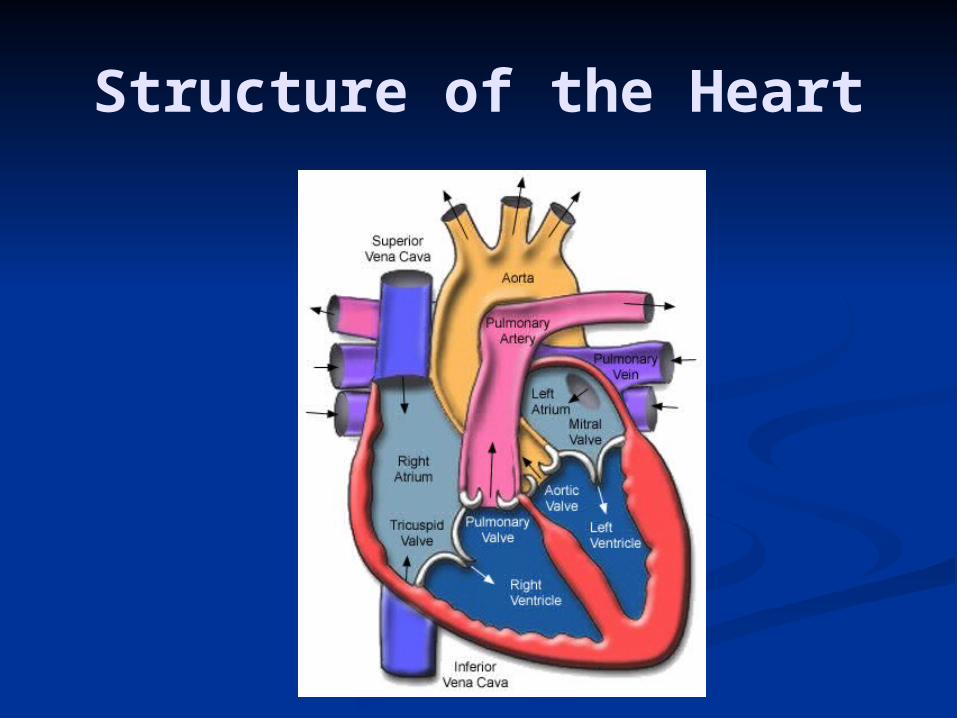
Structure of the Heart
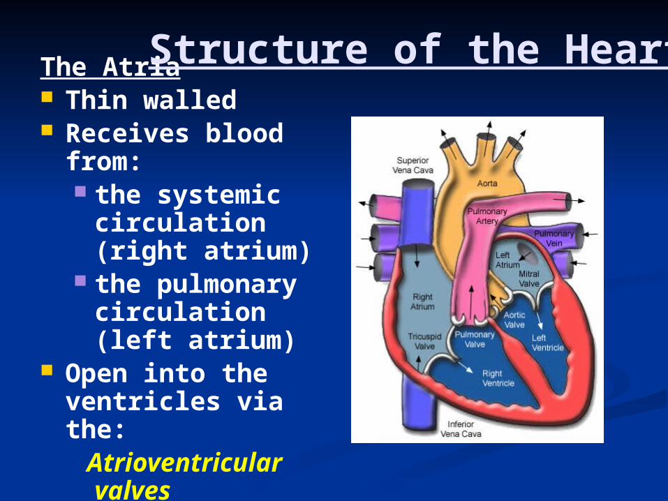
The Atria Thin walled Receives blood
from: the systemic
circulation (right atrium)
the pulmonary circulation (left atrium)
Open into the ventricles via the: Atrioventricular
valves (AV valves)
Structure of the Heart
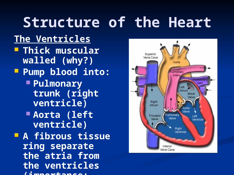
The Ventricles Thick muscular
walled (why?) Pump blood into:
Pulmonary trunk (right ventricle)
Aorta (left ventricle)
A fibrous tissue ring separate the atria from the ventricles (importance: electrical activity, AV valve)
Structure of the Heart

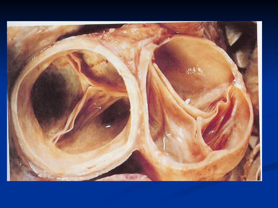
The Valves of the HeartThe Atrioventricular Valves
1. The Tricuspid Valve… between the right atrium and the right ventricle, 3 cusps
2. The Mitral Valve (bicuspid valve) … between the left atrium and the left ventricle, 2 cusps

Prevent back flow of blood from the ventricles to the atria
Held by chordae tendineae to papillary muscle
Contraction of papillary muscle…
The Valves of the HeartThe Atrioventricular
Valves

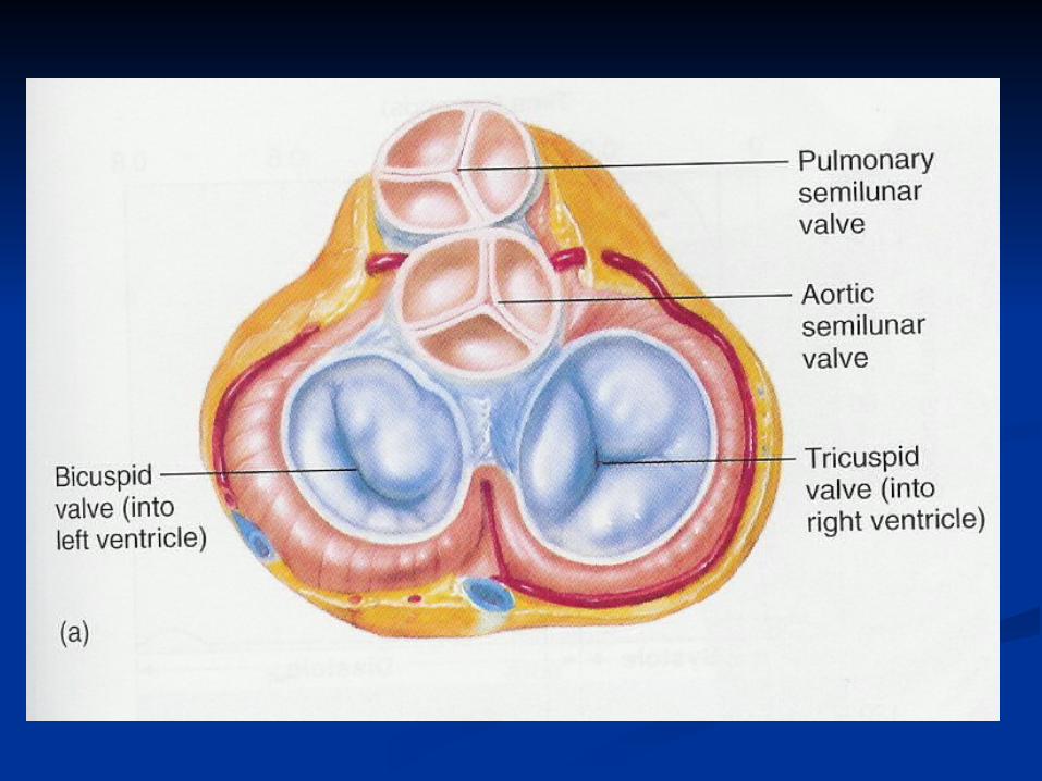

The Valves of the Heart The Semilunar Valves
Located at the origin of the pulmonary artery and aorta
Open during ventricular contraction…why?
Close during ventricular relaxation…why?
1. The Aortic Valve2. The Pulmonary
Valve

Cardiac muscle cell
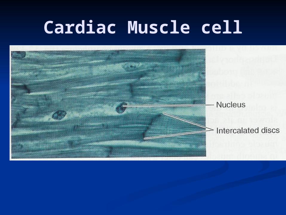
Cardiac Muscle cell
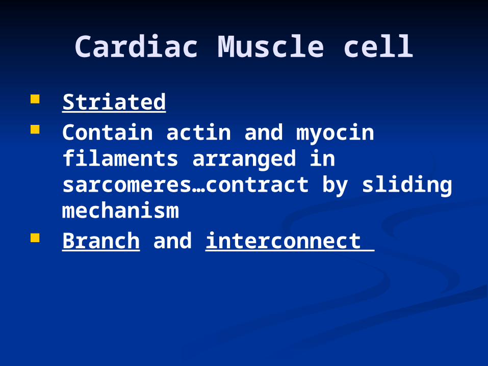
Striated Contain actin and myocin
filaments arranged in sarcomeres…contract by sliding mechanism
Branch and interconnect
Cardiac Muscle cell

Gap junctions Trans-membrane channel proteins,
connecting the cytoplasm of the cells
Allow spreading of the action potential from one fiber to another
Allow cardiac muscle to function as a syncytium “all or none law”: stimulation of a single muscle fiber results in contraction of all the muscle fibers
Intercalated discs
Cardiac Muscle cell

Cardiac Muscle cell

Electrical Activity of the
Heart

Electrical Activity of the Heart
Automaticity: capable of originating action potential

Resting membrane potential in myocardial cells -90 mVStimulation of myocardial cell
Myocardial action potential
Myocardial action potential

Myocardial action potential
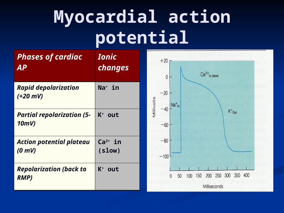
Phases of cardiac AP
Ionic changes
Rapid depolarization (+20 mV)
Na+ in
Partial repolarization (5-10mV)
K+ out
Action potential plateau (0 mV)
Ca2+ in (slow)
Repolarization (back to RMP)
K+ out
Myocardial action potential
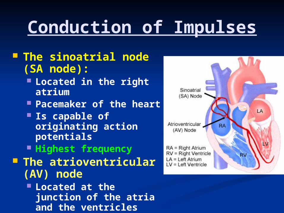
Conduction of Impulses The sinoatrial node
(SA node): Located in the right
atrium Pacemaker of the heart Is capable of
originating action potentials
Highest frequency The atrioventricular
(AV) node Located at the junction
of the atria and the ventricles
Delay in the conduction of impulses…why?
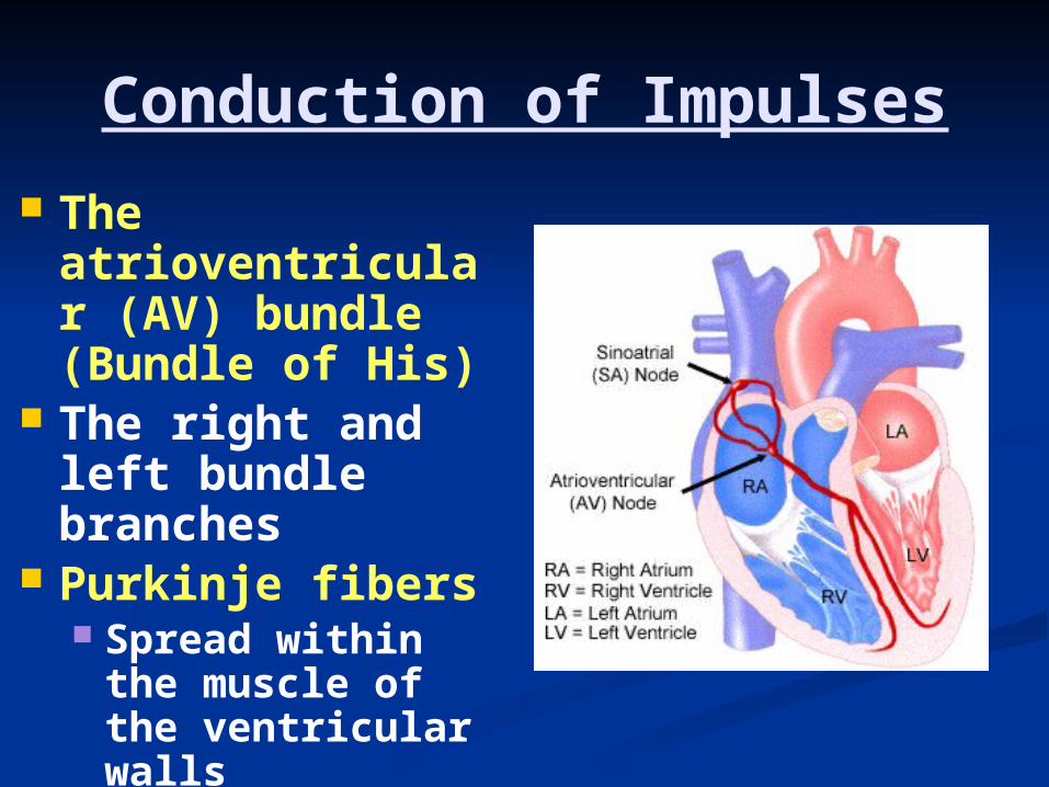
Conduction of Impulses
The atrioventricular (AV) bundle (Bundle of His)
The right and left bundle branches
Purkinje fibers Spread within
the muscle of the ventricular walls
Highest speed of conduction
Contractility
Contractility is the ability of cardiac muscle to convert chemical energy into mechanical work
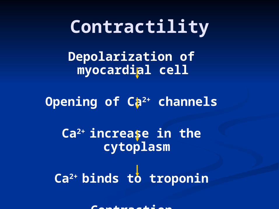
Depolarization of myocardial cell
Opening of Ca2+ channels
Ca2+ increase in the cytoplasm
Ca2+ binds to troponin
Contraction
Contractility
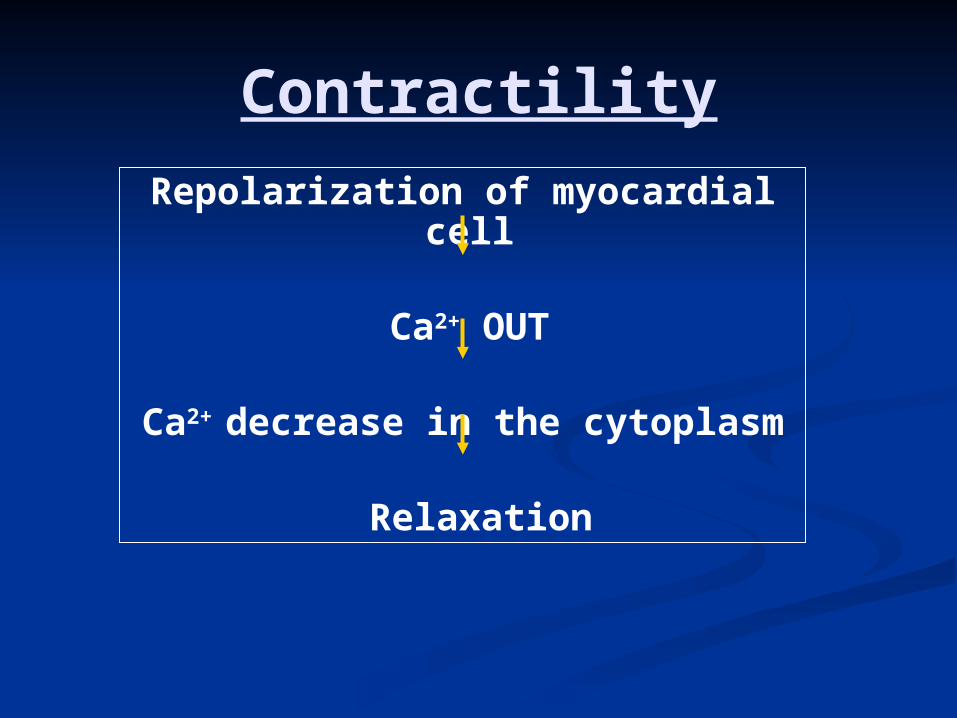
Repolarization of myocardial cell
Ca2+ OUT
Ca2+ decrease in the cytoplasm
Relaxation
Contractility
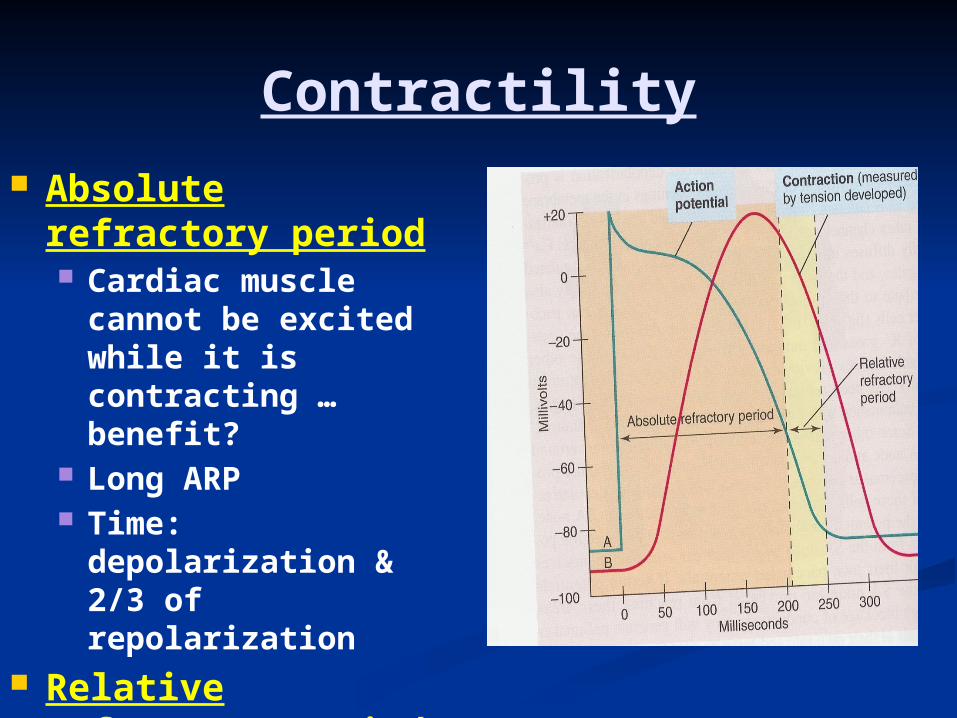
Contractility Absolute refractory
period Cardiac muscle
cannot be excited while it is contracting … benefit?
Long ARP Time: depolarization
& 2/3 of repolarization
Relative refractory period Time: last 1/3
repolarization Strong stimulus can
give rise to contraction
The Cardiac Cycle

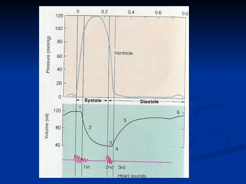
The Cardiac Cycle
The repeating pattern of contraction (systole) and relaxation (diastole) of the heart
Duration of cardiac cycle = 0.8 seconds
Diastole longer than systole Ventricular contraction follows
atrial contraction (0.1 to 0.2 second later)…why?

The end diastolic volume: the total volume of blood in the ventricles at the end of diastole (120 ml)
Stroke volume is the volume of blood pumped by each ventricle per beat (70 ml)
Residual volume: amount of blood left in each ventricle at the end of systole (50 ml)
The Cardiac Cycle
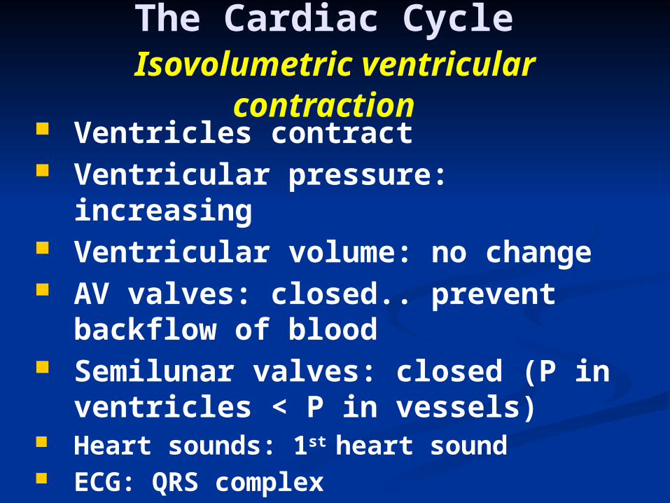
Ventricles contract Ventricular pressure: increasing Ventricular volume: no change AV valves: closed.. prevent
backflow of blood Semilunar valves: closed (P in
ventricles < P in vessels) Heart sounds: 1st heart sound ECG: QRS complex
The Cardiac Cycle Isovolumetric ventricular
contraction

Ventricular pressure: increasing > the pressure in the aortic and pulmonary vessels
Left ventricular pressure up to 120 mmHg
Right ventricular pressure up to 25 mmHg
Ventricular volume: decreasing Semilunar valves: open AV valves: closed.. prevent
backflow of blood
The Cardiac CycleEjection phase


Ventricles relax Ventricular pressure:
decreasing Ventricular volume: no
change AV valves: closed Semilunar valves: closed Heart sounds: 2nd heart sound ECG: T wave
The Cardiac CycleIsovolumetric relaxation


Ventricular pressure: below atrial pressure ( slightly above zero)
Ventricular volume: increasing
AV valves: open when pressure in the atria> the pressure in the ventricles
Semilunar valves: closed Passive ventricular filling via
AV valves (80%)
The Cardiac Cycle Rapid filling of the ventricles
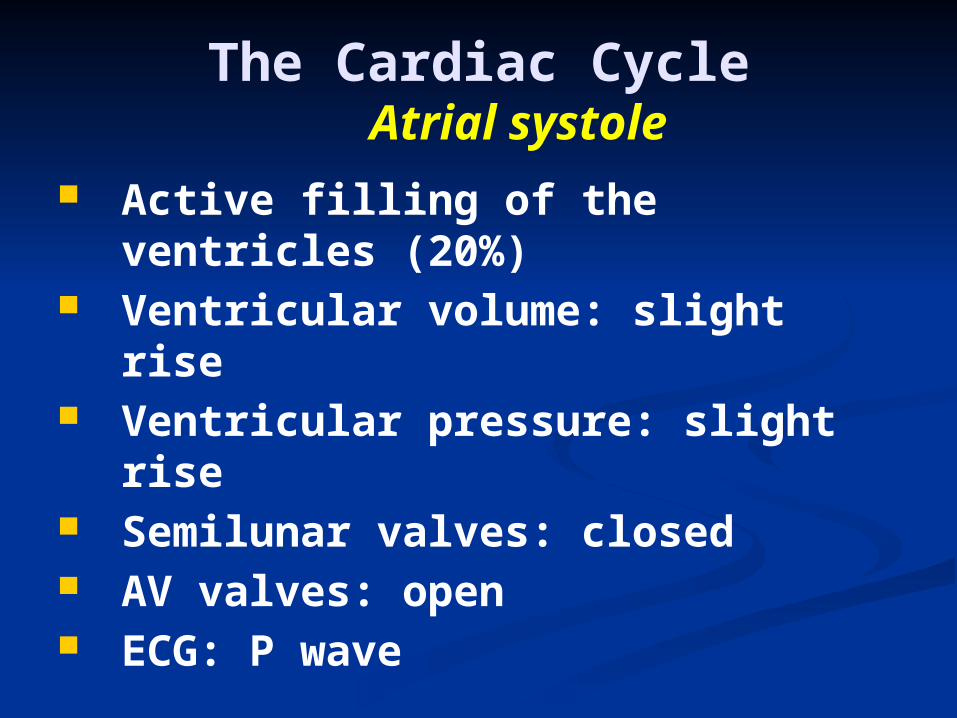
Active filling of the ventricles (20%)
Ventricular volume: slight rise Ventricular pressure: slight rise Semilunar valves: closed AV valves: open ECG: P wave
The Cardiac CycleAtrial systole

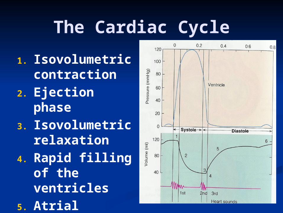
The Cardiac Cycle
1. Isovolumetric contraction
2. Ejection phase
3. Isovolumetric relaxation
4. Rapid filling of the ventricles
5. Atrial systole
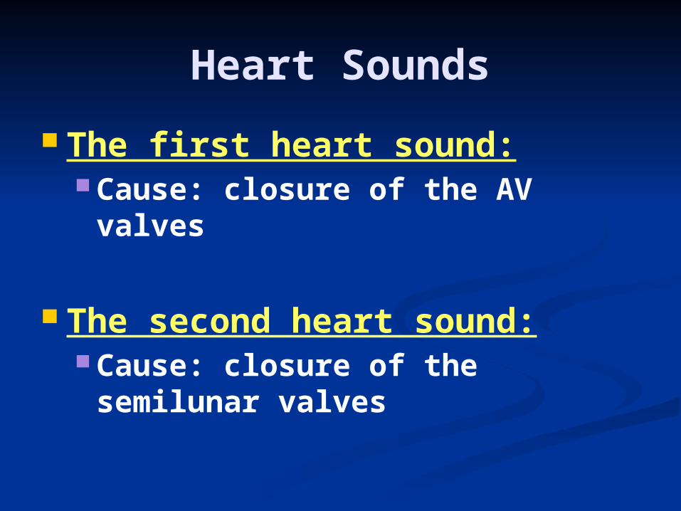
Heart Sounds
The first heart sound: Cause: closure of the AV valves
The second heart sound: Cause: closure of the semilunar
valves

Cardiac Output
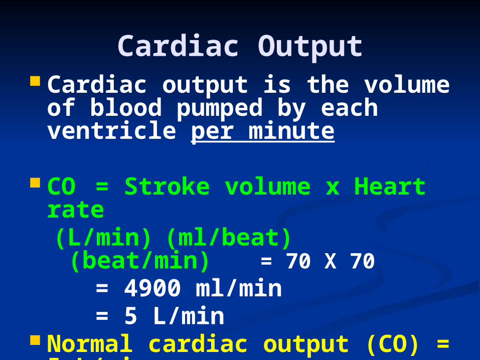
Cardiac Output Cardiac output is the volume
of blood pumped by each ventricle per minute
CO = Stroke volume x Heart rate(L/min) (ml/beat) (beat/min) = 70 X 70
= 4900 ml/min= 5 L/min
Normal cardiac output (CO) = 5 L/min
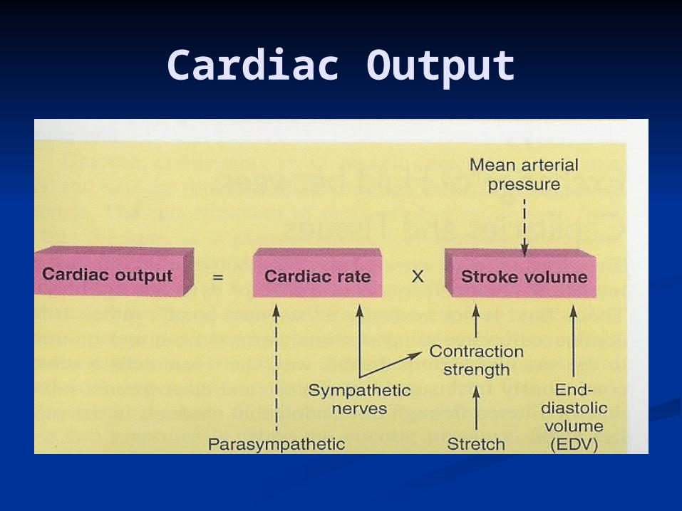
Cardiac Output

Sympathetic stimulation HR (positive chronotropic
effect) CO
Parasympathetic stimulation HR CO
Cardiac centers in the medulla oblangata
Cardiac OutputRegulation of Heart Rate

End Diastolic Volume (EDV) Frank- Starling Law of the
Heart venous return EDV length
of cardiac muscle (stretch) force of contraction stroke volume cardiac output
Cardiac OutputRegulation of Stroke
Volume

Positive ionotropic effect strength of contraction
Sympathetic stimulation Adrenaline
Negative ionotropic effect strength of contraction
Parasympathetic stimulation Acetylcholine Vagal stimulation
Cardiac Output Regulation of Stroke
Volume

Blood Pressure

Blood pressure
The blood pressure is the pressure the blood exerts against the inner walls of the blood vessels
Arterial blood pressure (BP) =cardiac output (CO) x peripheral
resistance
Heart Stroke Rate volume Vasoconstriction
Normal BP = 120/80 mmHg

Sympathetic stimulation vasoconstriction Peripheral resistance BP
Parasympathetic stimulation less important b/c of limited vasodilatation in the GIT, external genitalia and salivary glands
Arterial blood pressure Peripheral resistance

Sympathetic stimulation HR (positive chronotropic
effect) CO BP
Parasympathetic stimulation HR CO BP
Arterial blood pressure Heart rate
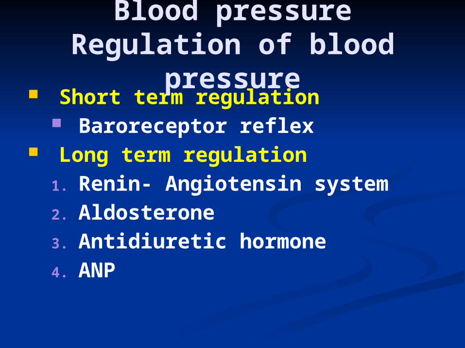
Short term regulation Baroreceptor reflex
Long term regulation1. Renin- Angiotensin system2. Aldosterone3. Antidiuretic hormone4. ANP
Blood pressureRegulation of blood
pressure

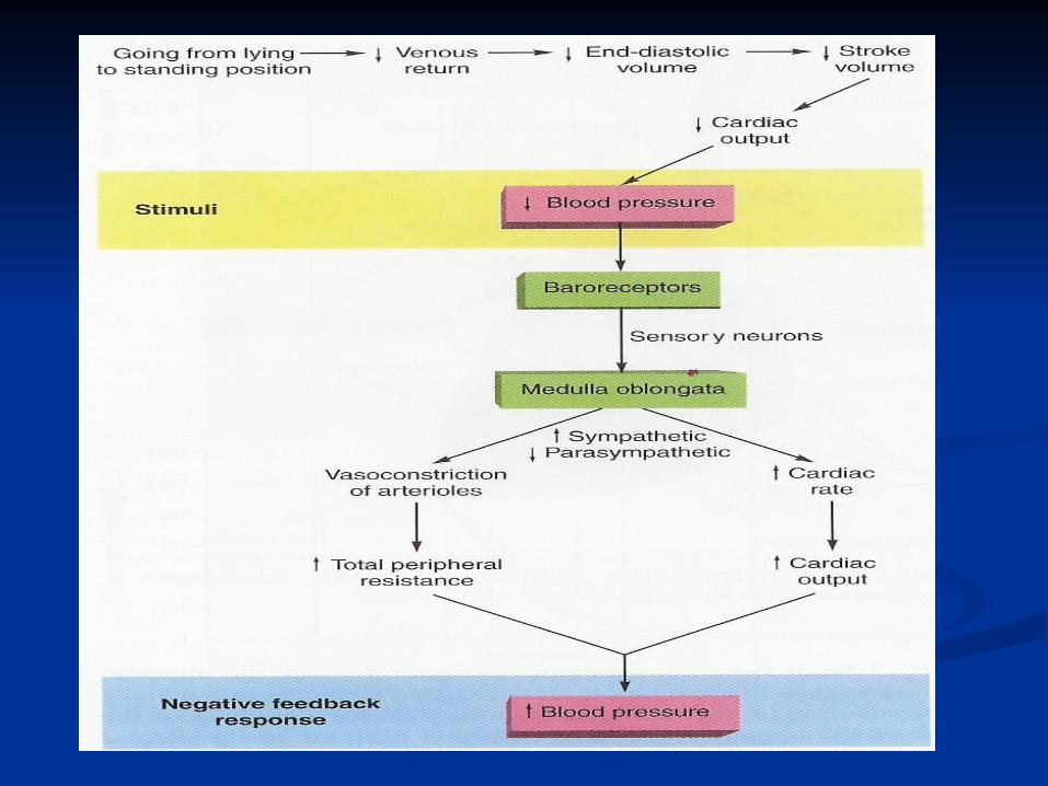
Arterial blood pressureBaroreceptor reflex
Stretch receptors Located in:1. The aortic arch2. The carotid sinus (at the
bifurcation of the common carotid artery)
Sensory nerve activity via the vagus and glossopharyngeal nerves
Cardiac centers in the medulla oblangata
The baroreceptor reflex is activated by changes in the BP

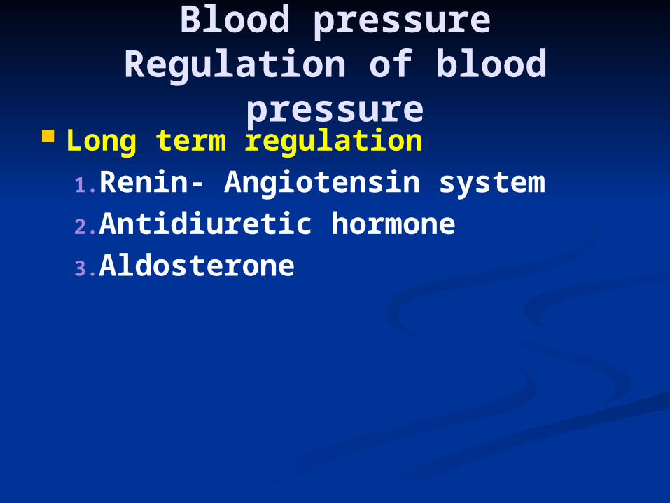
Blood pressureRegulation of blood
pressure Long term regulation
1.Renin- Angiotensin system2.Antidiuretic hormone 3.Aldosterone
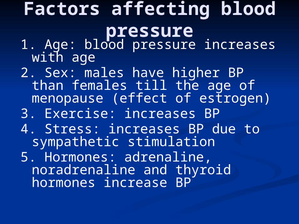
Factors affecting blood pressure
1. Age: blood pressure increases with age
2. Sex: males have higher BP than females till the age of menopause (effect of estrogen)
3. Exercise: increases BP4. Stress: increases BP due to
sympathetic stimulation5. Hormones: adrenaline,
noradrenaline and thyroid hormones increase BP
