the cargo-binding domain regulates structure and activity of myosin 5
TRANSCRIPT
© 2006 Nature Publishing Group
The cargo-binding domain regulates structure andactivity of myosin 5Kavitha Thirumurugan1, Takeshi Sakamoto2, John A. Hammer III3, James R. Sellers2 & Peter J. Knight1
Myosin 5 is a two-headed motor protein that moves cargoes alongactin filaments1,2. Its tail ends in paired globular tail domains(GTDs) thought to bind cargo3. At nanomolar calcium levels,actin-activated ATPase is low and the molecule is folded. Micro-molar calcium concentrations activate ATPase and the moleculeunfolds3–6. Here we describe the structure of folded myosin andthe GTD’s role in regulating activity. Electron microscopy showsthat the two heads lie either side of the tail, contacting the GTDsat a lobe of the motor domain (,Pro 117–Pro 137) that containsconserved acidic side chains, suggesting ionic interactionsbetween motor domain and GTD. Myosin 5 heavy meromyosin,a constitutively active fragment lacking the GTDs, is inhibited andfolded by a dimeric GST–GTD fusion protein. Motility assaysreveal that at nanomolar calcium levels heavy meromyosin movesrobustly on actin filaments whereas few myosins bind or move.These results combine to show that with no cargo, the GTDs bindin an intramolecular manner to the motor domains, producing aninhibited and compact structure that binds weakly to actin andallows the molecule to recycle towards new cargoes.Myosin 5 has two heavy chains, each contributing an amino-
terminal motor domain, an a-helical lever stabilized by six sequen-tially bound calmodulin light chains, a coiled-coil region thatdimerizes with the other heavy chain to form the tail and finallythe GTD3 (Fig. 1o). The unregulated high activity of myosin 5 heavymeromyosin (HMM) implicates the GTD in downregulation ofactivity at nanomolar calcium. Our initial images of the inhibitedstate showed a compact conformation, but identification of sub-structure was ambiguous4. The structures seen by other groups wereless compact5,6. To understand the mechanism of regulation of cargotransport by myosins requires an understanding of the structure andproperties of the inhibited state. Therefore we have studied thestructure of folded myosin 5, and the GTD’s mechanism ofinhibition.Single-particle image processing7 of folded myosin 5 molecules
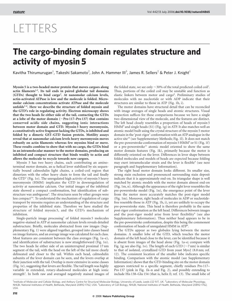
negative-stained in ATP at nanomolar calcium levels reveals detailedsubstructure. Briefly, molecules abstracted from raw images (Sup-plementary Fig. 1) were aligned together, grouped into classes basedon image features, and an average image was calculated for each class.Most features are consistent between the class averages (Fig. 1a–e),and identification of substructure is now straightforward (Fig. 1o).The two heads lie either side of an uninterrupted proximal 17 nmsegment of the tail, with the head on the left of the tail more clearlydelineated and closer to the tail. Within each head, calmodulinsubunits of the lever domain can be seen, and the levers overlap attheir junction with the tail. Overlap is more extensive in some classes(Fig. 1e), suggesting that levers are flexible. The tail length was highlyvariable in extended, rotary-shadowed molecules at high ionicstrength3. In both raw and averaged negatively stained images of
the folded state, we see only,30% of the total predicted coiled coil3.Thus, portions of the coiled coil may be unstable and function aselastic linkers between motor and cargo8. Preliminary studies ofmolecules with no nucleotide or with ADP indicate that theirstructures are similar to those in ATP (Fig. 1h, i).The motor domains have structural detail that can be reconciled
with image averages of single heads and atomic structures. Visualinspection suffices for these comparisons because we have a singletwo-dimensional view of the molecule, and the features are distinct.The left head closely resembles a proportion of heads of myosin 5HMM9 and single heads (S1) (Fig. 1g) in ATP. It also matches well anatomic model built using the crystal structure of the myosin 5 motordomain in the ‘post-rigor’ conformationwith an ATPanalogue in theactive site10 (see Supplementary Methods; Fig. 1l). It does not matchthe pre-powerstroke conformation ofmyosin 5HMM9 or S1 (Fig. 1f)or a pre-powerstroke11 atomic model oriented to show the samemotor domain features (Fig. 1k), primarily because the motor isdifferently oriented on the lever. Differences in lever shape betweenfolded molecules and models of heads are expected because foldingmay exert intramolecular strain and the lever is flexible9 (see nextparagraph and Supplementary Information).The right head motor domain looks different. Its smaller size,
strong stain exclusion and pronounced surrounding stain depositindicate that it is approximately end-on. Its triangular shape is wellmatched by atomic models with the motor domain almost end-on(Fig. 1m, n). Although the appearance of the right lever resembles thepre-powerstroke model (Fig. 1n), the emergence point of the leverfrom the motor more accurately matches the post-rigor model(Fig. 1m). Moreover, right heads of molecules in ADP or nucleotide-free resemble those in ATP (Fig. 1h, i), yet are unlikely to occupy thepre-powerstroke state. This head is therefore probably in the samepost-rigor conformation as the left head. Differences between imagesand the post-rigor model arise from lever flexibility9 (see alsoSupplementary Information). Thus neither head appears to be inthe pre-powerstroke conformation, despite this being the commonestconformation of heads of unregulated HMM in ATP9.The GTDs appear as two globules lying between the motor
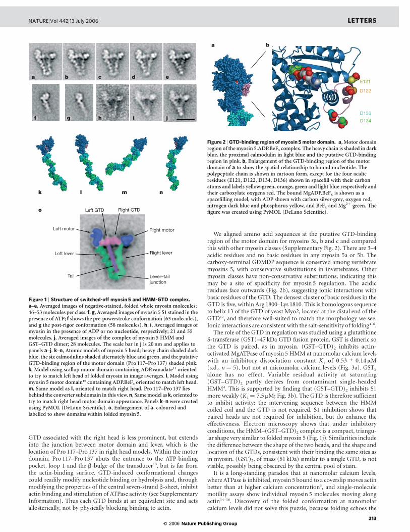
domains. A smaller lobe of the GTD, which touches the motordomain of the left head close to the lever, can be identified because itis absent from images of the head alone (Fig. 1a–e; compare withFig. 1g; see also Fig. 1o). The length of each GTD (,7 nm) is similarto that of isolated, crystallized GTD from yeast Myo2 (8.8 nm; ref.12). The consistent location of the smaller lobe indicates specificbinding. Comparison with the atomic model (see SupplementaryInformation) shows that the GTD-binding site on the motor domainappears restricted to a specific segment, chiefly residues Pro 117–Pro 137 (pink in Fig. 1k–n and Fig. 2), and possibly extending toinclude His 138–Glu 154 (that is, helix E; ref. 13). The small lobe of
LETTERS
1Institute of Molecular and Cellular Biology, and Astbury Centre for Structural Molecular Biology, University of Leeds, Leeds LS2 9JT, UK. 2Laboratory of Molecular Physiology,NHLBI, National Institutes of Health, Bethesda, Maryland 20892-1762, USA. 3Laboratory of Cell Biology, NHLBI, National Institutes of Health, Bethesda, Maryland 20892-1762,USA.
Vol 442|13 July 2006|doi:10.1038/nature04865
212
© 2006 Nature Publishing Group
GTD associated with the right head is less prominent, but extendsinto the junction between motor domain and lever, which is thelocation of Pro 117–Pro 137 in right head models. Within the motordomain, Pro 117–Pro 137 abuts the entrance to the ATP-bindingpocket, loop 1 and the b-bulge of the transducer10, but is far fromthe actin-binding surface. GTD-induced conformational changescould readily modify nucleotide binding or hydrolysis and, throughmodifying the properties of the central seven-strand b-sheet, inhibitactin binding and stimulation of ATPase activity (see SupplementaryInformation). Thus each GTD binds at an equivalent site and actsallosterically, not by physically blocking binding to actin.
We aligned amino acid sequences at the putative GTD-bindingregion of the motor domain for myosins 5a, b and c and comparedthis with other myosin classes (Supplementary Fig. 2). There are 3–4acidic residues and no basic residues in any myosin 5a or 5b. Thecarboxy-terminal GDMDP sequence is conserved among vertebratemyosins 5, with conservative substitutions in invertebrates. Othermyosin classes have non-conservative substitutions, indicating thismay be a site of specificity for myosin 5 regulation. The acidicresidues face outwards (Fig. 2b), suggesting ionic interactions withbasic residues of the GTD. The densest cluster of basic residues in theGTD is five, within Arg 1800–Lys 1810. This is homologous sequenceto helix 13 of the GTD of yeast Myo2, located at the distal end of theGTD12, and therefore well-suited to match the morphology we see.Ionic interactions are consistent with the salt-sensitivity of folding4–6.The role of the GTD in regulation was studied using a glutathione
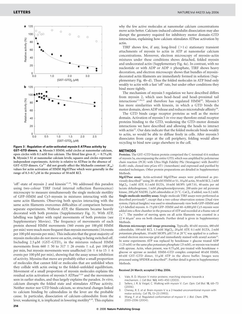
S-transferase (GST)–47 kDa GTD fusion protein. GST is dimeric sothe GTD is paired, as in myosin. (GST–GTD)2 inhibits actin-activated MgATPase of myosin 5 HMM at nanomolar calcium levelswith an inhibitory dissociation constant K i of 0.53 ^ 0.14 mM(s.d., n ¼ 5), but not at micromolar calcium levels (Fig. 3a). GST2
alone has no effect. Variable residual activity at saturating(GST–GTD)2 partly derives from contaminant single-headedHMM4. This is supported by finding that (GST–GTD)2 inhibits S1more weakly (K i ¼ 7.5 mM; Fig. 3b). The GTD is therefore sufficientto inhibit activity: the intervening sequence between the HMMcoiled coil and the GTD is not required. S1 inhibition shows thatpaired heads are not required for inhibition, but do enhance theeffectiveness. Electron microscopy shows that under inhibitoryconditions, the HMM–(GST–GTD)2 complex is a compact, triangu-lar shape very similar to foldedmyosin 5 (Fig. 1j). Similarities includethe difference between the shape of the two heads, and the shape andlocation of the GTDs, consistent with their binding the same sites asin myosin. (GST)2, of mass (51 kDa) similar to a single GTD, is notvisible, possibly being obscured by the central pool of stain.It is a long-standing paradox that at nanomolar calcium levels,
where ATPase is inhibited, myosin 5 bound to a coverslip moves actinbetter than at higher calcium concentration3, and single-moleculemotility assays show individual myosin 5 molecules moving alongactin14–16. Discovery of the folded conformation at nanomolarcalcium levels did not solve this puzzle, because folding echoes the
Figure 1 | Structure of switched-off myosin 5 and HMM-GTD complex.a–e, Averaged images of negative-stained, folded whole myosin molecules;46–53 molecules per class. f, g, Averaged images of myosin 5 S1 stained in thepresence of ATP; f shows the pre-powerstroke conformation (63 molecules),and g the post-rigor conformation (58 molecules). h, i, Averaged images ofmyosin in the presence of ADP or no nucleotide, respectively; 21 and 55molecules. j, Averaged images of the complex of myosin 5 HMM andGST–GTD dimer; 28 molecules. The scale bar in j is 20 nm and applies topanels a–j. k–n, Atomic models of myosin 5 head; heavy chain shaded darkblue, the six calmodulins shaded alternately blue and green, and the putativeGTD-binding region of the motor domain (Pro 117–Pro 137) shaded pink.k, Model using scallop motor domain containing ADP.vanadate11 orientedto try to match left head of folded myosin in image averages. l, Model usingmyosin 5 motor domain10 containing ADP.BeFx oriented to match left head.m, Same model as l, oriented to match right head. Pro 117–Pro 137 liesbehind the converter subdomain in this view. n, Same model as k, oriented totry to match right head motor domain appearance. Panels k–n were createdusing PyMOL (DeLano Scientific). o, Enlargement of a, coloured andlabelled to show domains within folded myosin 5.
Figure 2 |GTD-binding region ofmyosin 5motor domain. a, Motor domainregion of the myosin 5.ADP.BeFx complex. The heavy chain is shaded in darkblue, the proximal calmodulin in light blue and the putative GTD-bindingregion in pink. b, Enlargement of the GTD-binding region of the motordomain of a to show the spatial relationship to bound nucleotide. Thepolypeptide chain is shown in cartoon form, except for the four acidicresidues (E121, D122, D134, D136) shown in spacefill with their carbonatoms and labels yellow-green, orange, green and light blue respectively andtheir carboxylate oxygens red. The bound MgADP.BeFx is shown as aspacefilling model, with ADP shown with carbon silver-grey, oxygen red,nitrogen dark blue and phosphorus yellow, and BeFx and Mg2þ green. Thefigure was created using PyMOL (DeLano Scientific).
NATURE|Vol 442|13 July 2006 LETTERS
213
© 2006 Nature Publishing Group
‘off ’-state of myosin 2 and kinesin17,18. We addressed this paradoxusing two-colour TIRF (total internal reflection fluorescence)microscopy to measure simultaneously the single molecule motilityof GFP–HMM and Cy3-myosin in mixtures interacting with thesame actin filaments. Observing both species interacting with thesame actin filaments overcomes difficulties of comparison betweenseparate experiments. Without ATP, the filaments became heavilydecorated with both proteins (Supplementary Fig. 3). With ATP,labelling was lighter with rapid movements of both proteins (seeSupplementary Movies). The frequency of movements of eachprotein showed HMM movements (560 events per 100 pM HMMpermin)weremuchmore frequent thanmyosinmovements (14 eventsper 100 pMmyosin permin). This indicates that the greatmajority ofmyosinmolecules do notmove on actin, owing to being switched off.Including 2.5 mM (GST–GTD)2 in the mixtures reduced HMMmovements from 660 ^ 50 to 317 ^ 26 events ^ s.d. per 100 pMper min, but myosin movements were unaffected (16 ^ 6 to 15 ^ 6events per 100 pM per min), showing that the assay senses inhibitionof activity. Myosins that move are probably either a small proportionof molecules that cannot fold or molecules that are unfolded whenthey collide with actin owing to the folded–unfolded equilibrium.Movement of a small proportion of myosin molecules explains theresidual actin activation of myosin 5 ATPase3,4,6 and the movementsseen in earlier studies, and thus largely explains the paradox. In vitro,calcium disrupts the folded state and stimulates ATPase activity.Neither motor nor GTD binds calcium, so structural changes linkedto calcium binding by calmodulins in the lever are the probablecause. In particular, dissociation of calcium-calmodulin from thelever, weakening it, is implicated in lowering motility6,19. This explains
why the few active molecules at nanomolar calcium concentrationsmove actin better. Calcium-induced calmodulin dissociation may alsodisrupt the geometry required for inhibitory motor domain–GTDinteractions, explaining how calcium stimulates ATPase activation byactin.TIRF shows few, if any, long-lived (.1 s) stationary transient
attachments of myosin to actin in ATP at nanomolar calciumconcentrations. Moreover, electron microscopy of myosin–actinmixtures under these conditions shows detached, folded myosinand undecorated actin (Supplementary Fig. 4a). In contrast, with nonucleotide or with ADP or ADP þ phosphate, TIRF shows heavydecoration, and electron microscopy shows that bundles of myosin-decorated actin filaments are immediately formed in solution (Sup-plementary Fig. 4b–d). Thus the folded molecules in ATP bind onlyweakly to actin with a fast ‘off ’ rate, but under other conditions theybind more tightly.The mechanism of myosin 5 regulation we have described differs
from myosin 2, which uses head–head and head–proximal tailinteractions17,20,21 and therefore has regulated HMM20. Myosin 5has more similarities with kinesin, in which a GTD binds themotor domain, slows ADP release and reducesmicrotubule affinity18.The GTD binds cargo receptor proteins as well as the motor
domain. Activation of myosin 5 in vivomay therefore entail receptorproteins binding to the GTD, weakening the GTD–motor domaininteractions we have described and allowing the heads to interactwith actin22. Our data indicate that the folded molecule binds weaklyto actin, so would be able to diffuse freely in cells. After myosin 5dissociates from cargo at the cell periphery, folding would allowrecycling to bind new cargo elsewhere in the cell.
METHODSProteins. The GST–GTD fusion protein comprised the C-terminal 414 residuesof myosin 5a, encompassing the entire GTD, whichwas amplified by polymerasechain reaction (PCR) with Ultra High Fidelity Pfu (Stratagene) with BamH1/EcoR1 ends, cloned into pGex-4T1 (Amersham) and expressed and purified bystandard techniques. Other protein preparations are detailed in SupplementaryMethods.MgATPase assay. Actin-activated MgATPase assays were performed as pre-viously described23 using 20–40 nMHMMor S1, 10 mMactin, 50mMKCl, 2mMMgCl2, 1mM ATP, 0.1mM EGTA, 10mM MOPS (pH7.0), 40 units per mllactate dehydrogenase, 1mM phosphoenolpyruvate, 200 units per ml pyruvatekinase, 200mMNADH, 2mMcalmodulin at 25 8C. CaCl2, if added, was 0.2mM.Single-molecule motility assay. Single-molecule TIRF assays were performed asdescribed previously23, except that a two-colour observation system (Dual viewsystem, Optical Insights) was used to simultaneously view both GFP–HMMandCy3-labelled myosin. 6–75 pM GFP–HMM and 60–4,000 pM Cy3-myosin wereadded into a flow chamber in the presence of ATP and recorded at a frame rate of2 s21. The number of moving spots on all actin filaments was counted in a22 £ 44 mm2 area on both channels. Further detail is given in SupplementaryMethods.Electron microscopy and image processing. Typically, 40 nM myosin, 480 nMcalmodulin, 100mM KCl, 1.5mM MgCl2, 20 mM ATP, 0.1mM EGTA, 2mMpotassium phosphate, 10mM MOPS, pH7.0 at 20 8C was applied to a carbon-coated electron microscope grid and immediately stained with uranyl acetate24.In some experiments ATP was replaced by hexokinase þ glucose treated ADP(1.25mM) or the same plus potassium phosphate (25mM), ormyosinwas treatedwith apyrase. Actin, when present, was 0.75mM, pre-treated with hexokinase þglucose or apyrase as needed. HMM–GTD complex comprised 40nM HMM,60 nM GST–GTD dimer, 55 mM ATP in the above buffer. Images wereprocessed using SPIDER as described24. Further detail is given in SupplementaryMethods.
Received 24 March; accepted 3 May 2006.
1. Vale, R. D. Myosin V motor proteins: marching stepwise towards amechanism. J. Cell Biol. 163, 445–-450 (2003).
2. Sellers, J. R. & Veigel, C. Walking with myosin V. Curr. Opin. Cell Biol. 18, 68–-73(2006).
3. Cheney, R. E. et al. Brain myosin-V is a 2-headed unconventional myosin withmotor-activity. Cell 75, 13–-23 (1993).
4. Wang, F. et al. Regulated conformation of myosin V. J. Biol. Chem. 279,2333–-2336 (2004).
Figure 3 | Regulation of actin-activated myosin 5 ATPase activity byGST–GTD dimers. a, Myosin 5 HMM; solid circles at nanomolar calcium,open circles with 0.1 mM free calcium. The fitted line gives K i ¼ 0.75mM.b, Myosin 5 S1 at nanomolar calcium levels; squares and circles representindependent experiments. Activity is relative to ATPase in the absence ofGST–GTD dimers. Ca2þ did not greatly affect the Michaelis constant (Km)values for actin activation of HMM MgATPase which were generally in therange of 0.3–0.7mM in the presence of 50 mM KCl.
LETTERS NATURE|Vol 442|13 July 2006
214
© 2006 Nature Publishing Group
5. Li, X. D., Mabuchi, K., Ikebe, R. & Ikebe, M. Ca2þ-induced activation of ATPaseactivity of myosin Va is accompanied with a large conformational change.Biochem. Biophys. Res. Commun. 315, 538–-545 (2004).
6. Krementsov, D. N., Krementsova, E. B. & Trybus, K. M. Myosin V: regulation bycalcium, calmodulin, and the tail domain. J. Cell Biol. 164, 877–-886 (2004).
7. Frank, J. et al. SPIDER and WEB: Processing and visualization of images in 3Delectron microscopy and related fields. J. Struct. Biol. 116, 190–-199 (1996).
8. Schilstra, M. J. & Martin, S. R. An elastically tethered viscous load imposes aregular gait on the motion of myosin-V. Simulation of the effect of transientforce relaxation on a stochastic process. J. R. Soc. Interf. 3, 153–-165 (2006).
9. Burgess, S. et al. The prepower stroke conformation of myosin V. J. Cell Biol.159, 983–-991 (2002).
10. Coureux, P. D., Sweeney, H. L. & Houdusse, A. Three myosin V structuresdelineate essential features of chemo-mechanical transduction. EMBO J. 23,4527–-4537 (2004).
11. Houdusse, A., Szent-Gyorgyi, A. G. & Cohen, C. Three conformational states ofscallop myosin S1. Proc. Natl Acad. Sci. USA 97, 11238–-11243 (2000).
12. Pashkova, N., Jin, Y., Ramaswamy, S. & Weisman, L. S. Structural basis for myosinV discrimination between distinct cargoes. EMBO J. 25, 693–-700 (2006).
13. Cope, M., Whisstock, J., Rayment, I. & Kendrick Jones, J. Conservation withinthe myosin motor domain: Implications for structure and function. Structure 4,969–-987 (1996).
14. Sakamoto, T., Amitani, I., Yokota, E. & Ando, T. Direct observation ofprocessive movement by individual myosin V molecules. Biochem. Biophys. Res.Commun. 272, 586–-590 (2000).
15. Yildiz, A. et al. Myosin V walks hand-over-hand: Single fluorophore imagingwith 1.5-nm localization. Science 300, 2061–-2065 (2003).
16. Forkey, J. N., Quinlan, M. E., Shaw, M. A., Corrie, J. E. T. & Goldman, Y. E.Three-dimensional structural dynamics of myosin V by single-moleculefluorescence polarization. Nature 422, 399–-404 (2003).
17. Wendt, T., Taylor, D., Trybus, K. M. & Taylor, K. Three-dimensional imagereconstruction of dephosphorylated smooth muscle heavy meromyosin revealsasymmetry in the interaction between myosin heads and placement ofsubfragment 2. Proc. Natl Acad. Sci. USA 98, 4361–-4366 (2001).
18. Hackney, D. D. & Stock, M. F. Kinesin’s IAK tail domain inhibits initial
microtubule-stimulated ADP release. Nature Cell Biol. 2, 257–-260 (2000).
19. Nguyen, H. & Higuchi, H. Motility of myosin V regulated by the dissociation of
single calmodulin. Nature Struct. Mol. Biol. 12, 127–-132 (2005).
20. Sellers, J. R. Myosins (ed. Sheterline, P.) (Oxford Univ. Press, Oxford, 1999).
21. Ankrett, R. J., Rowe, A. J., Cross, R. A., Kendrick-Jones, J. & Bagshaw, C. R. A
folded (10 S) conformer of myosin from a striated muscle and its implications
for regulation of ATPase activity. J. Mol. Biol. 217, 323–-335 (1991).
22. Li, X. D., Ikebe, R. & Ikebe, M. Activation of myosin Va function by
melanophilin, a specific docking partner of myosin Va. J. Biol. Chem. 280,
17815–-17822 (2005).
23. Sakamoto, T. et al. Neck length and processivity of myosin V. J. Biol. Chem.
278, 29201–-29207 (2003).
24. Burgess, S. A., Walker, M. L., Thirumurugan, K., Trinick, J. & Knight, P. J. Use of
negative stain and single-particle image processing to explore dynamic
properties of flexible macromolecules. J. Struct. Biol. 147, 247–-258 (2004).
Supplementary Information is linked to the online version of the paper atwww.nature.com/nature.
Acknowledgements We thank S. A. Burgess for expert advice and assistancewith image processing, F. Zhang for technical assistance and The WellcomeTrust for support. T.S. was supported by a fellowship from the Japanese Societyfor the Promotion of Science.
Author contributions Electron microscopy and image processing wereperformed by K.T., TIRF microscopy by T.S., ATPase assays by J.R.S., andprotein preparations by T.S. GST–GTD was created by J.A.H., molecular modelsby P.J.K., sequence alignments by J.R.S. and P.J.K., and the paper by all authors.J.R.S. and P.J.K. contributed equally to this work.
Author Information Reprints and permissions information is available atnpg.nature.com/reprintsandpermissions. The authors declare no competingfinancial interests. Correspondence and requests for materials should beaddressed to P.J.K. ([email protected]).
NATURE|Vol 442|13 July 2006 LETTERS
215