the central dogma of biology - mr. aitken's biology class...
TRANSCRIPT

10-2
The Central Dogma of Biology

10-3
10.2 DNA, not protein,
is the genetic material Hershey and Chase Experiment
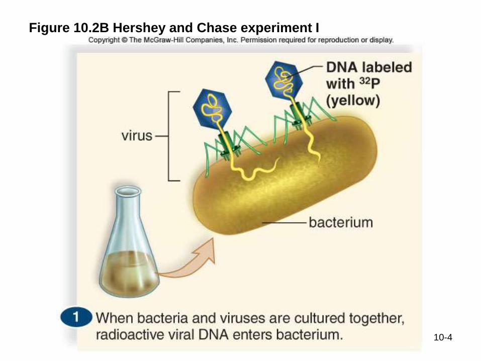
In their experiment, Hershey and Chase relied on a chemical
difference between DNA and protein to solve whether DNA or
protein was the genetic material
Figure 10.2A Structure of
the virus (T2 bacteriophage)
used by Hershey and Chase
10-4
Figure 10.2B Hershey and Chase experiment I
10-5
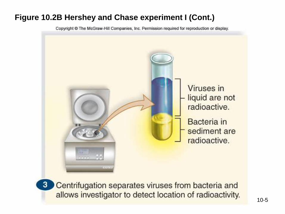
Figure 10.2B Hershey and Chase experiment I (Cont.)
10-6
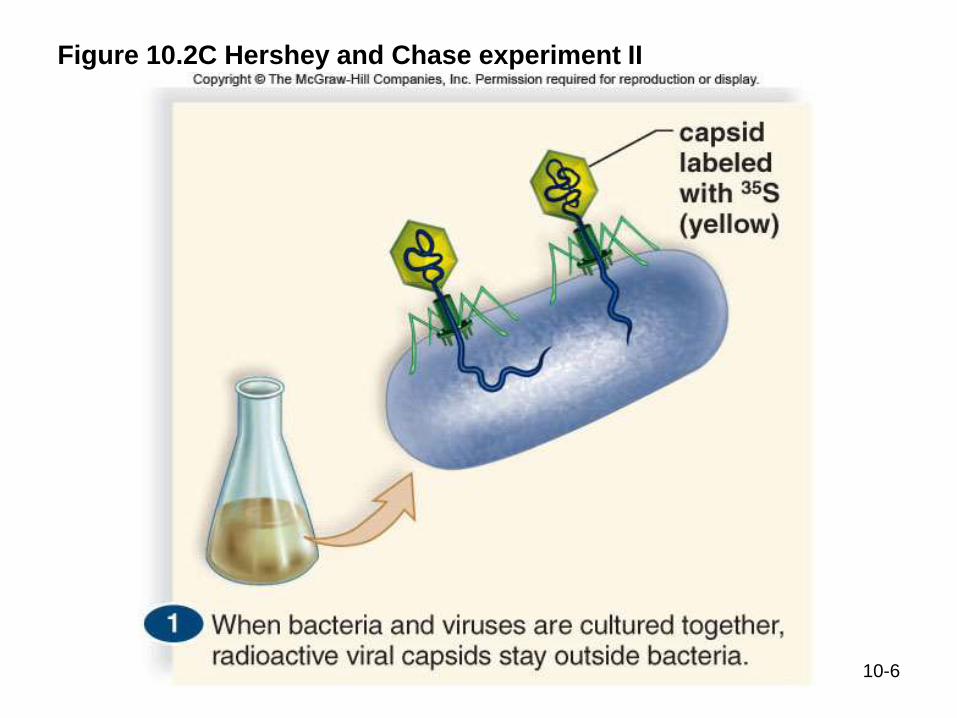
Figure 10.2C Hershey and Chase experiment II
10-7
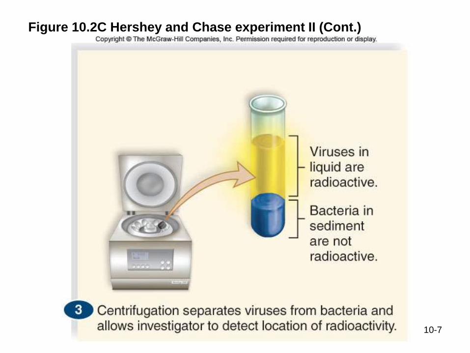
Figure 10.2C Hershey and Chase experiment II (Cont.)
Hershey and Chase take-home
The results from Hershey and Chase
experiments suggested that the DNA of the virus
entered the hosts (and not the protein), where
viral reproduction takes place.
Therefore, DNA is the genetic material and not
proteins.
10-8

10-9
10.3 DNA and RNA are
polymers of nucleotides
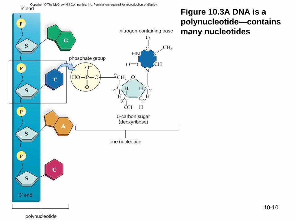
Nucleic acids contain only nucleotides, molecules that are composed of a nitrogen-containing base, a phosphate, and a pentose
(5-carbon sugar)
DNA (deoxyribonucleic acid) contains the
5-carbon sugar deoxyribose
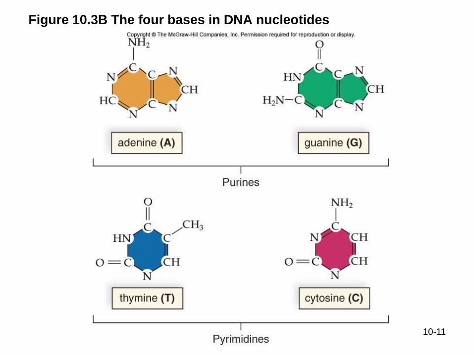
DNA contains four nucleotides with different bases Adenine, Guanine, Thymine, and Cytosine
10-10
Figure 10.3A DNA is a
polynucleotide—contains
many nucleotides
10-11
Figure 10.3B The four bases in DNA nucleotides

10-12
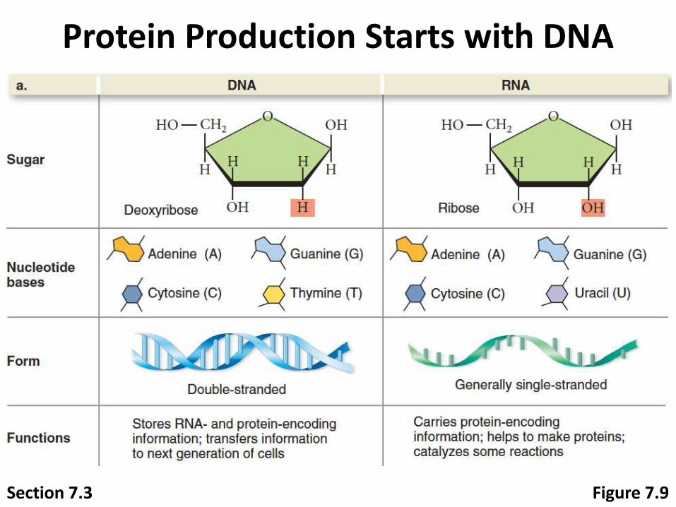
RNA RNA (ribonucleic acid) another polymer of nucleotides
RNA differs from DNA
Has ribose as a sugar, not deoxyribose
Has uracil in place of thymine
Figure 10.3C The
uracil nucleotide
in RNA replaces
thymine in DNA
10-13
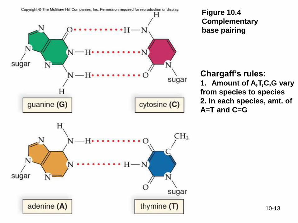
Figure 10.4
Complementary
base pairing
Chargaff’s rules:1. Amount of A,T,C,G vary
from species to species
2. In each species, amt. of
A=T and C=G

10-14
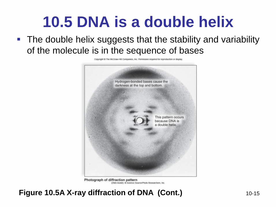
10.5 DNA is a double helix The double helix suggests that the stability and variability
of the molecule is in the sequence of bases
Figure 10.5A X-ray diffraction of DNA
10-15
10.5 DNA is a double helix The double helix suggests that the stability and variability
of the molecule is in the sequence of bases
Figure 10.5A X-ray diffraction of DNA (Cont.)
10-16
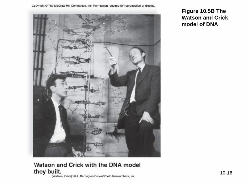
Figure 10.5B The
Watson and Crick
model of DNA
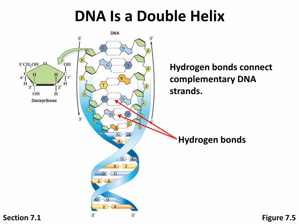
Section 7.1 Figure 7.5
Hydrogen bonds connect complementary DNA strands.
Hydrogen bonds
DNA Is a Double Helix
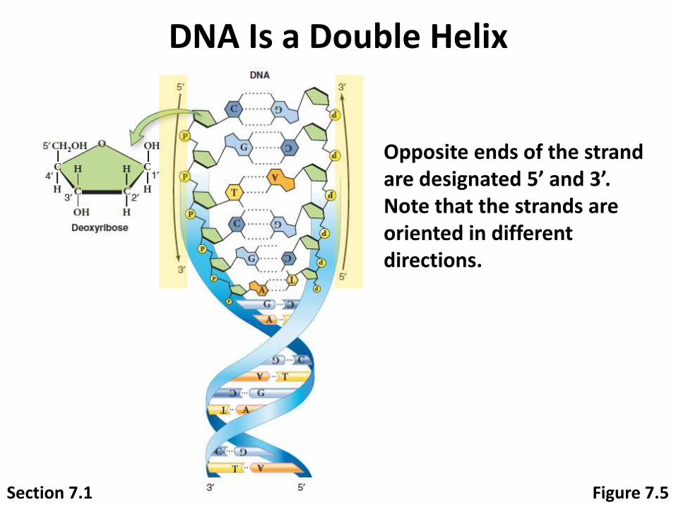
Section 7.1 Figure 7.5
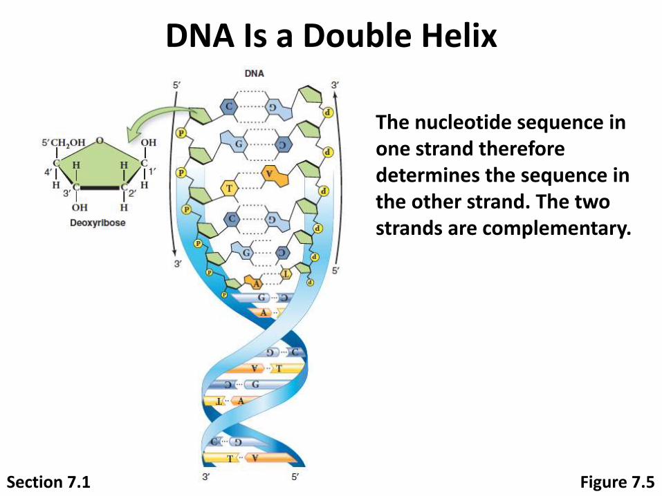
Opposite ends of the strand are designated 5’ and 3’. Note that the strands are oriented in different directions.
DNA Is a Double Helix

10-19
Figure 10.5B The
Watson and Crick
model of DNA
(Cont.)
Section 7.1 Figure 7.5
The nucleotide sequence in one strand therefore determines the sequence in the other strand. The two strands are complementary.
DNA Is a Double Helix

What forces make DNA a double
helix?
Attractiveness of bases
Repulsion forces of oxygen in phosphate
backbone
Hydrophobic interactions of bases with water
10-21
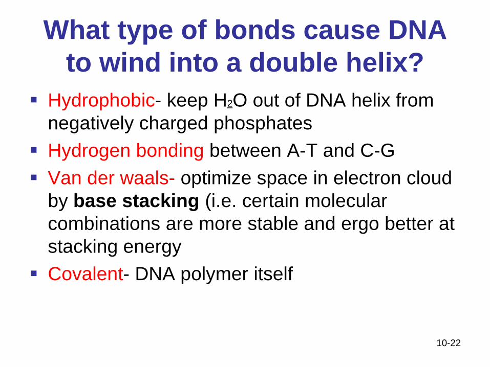
What type of bonds cause DNA
to wind into a double helix?
Hydrophobic- keep H2O out of DNA helix from
negatively charged phosphates
Hydrogen bonding between A-T and C-G
Van der waals- optimize space in electron cloud
by base stacking (i.e. certain molecular
combinations are more stable and ergo better at
stacking energy
Covalent- DNA polymer itself
10-22

10-24

10-25
DNA Can Be Duplicated

Objectives
Know the order, steps, location and enzymes
responsible for the replication of DNA
** Will be an essay question on your exam**
10-26

•Humans share 50% of their DNA with bananas.
•Cells can contain 6-9 feet of DNA. If all the DNA in your
body was put end to end, it would reach to the sun and back
over 600 times.
•DNA in all humans is 99.9 percent identical. It is about one
tenth of one percent that makes us all unique, or about 3
million nucleotides difference.
Amazing DNA facts

Amazing facts cont.
• DNA can store 25 gigabytes of information per inch and
is the most efficient storage system known to human.
So, humans are better than computers!!
• In an average meal, you eat approximately 55,000,000
cells or between 63,000 to 93,000 miles of DNA.
• It would take a person typing 60 words per minute, eight
hours a day, around 50 years to type the human
genome.
10-28

10-30
Central Dogma of Biology

10-31
10.8 Genes are linked to proteins
Figure 10.8 Chemical basis of
sickle-cell disease in humans

10-32
10.8 Genes are linked to proteinsFigure 10.8 Chemical basis of
sickle-cell disease in humans
(Cont.)
Protein Production Starts with DNA
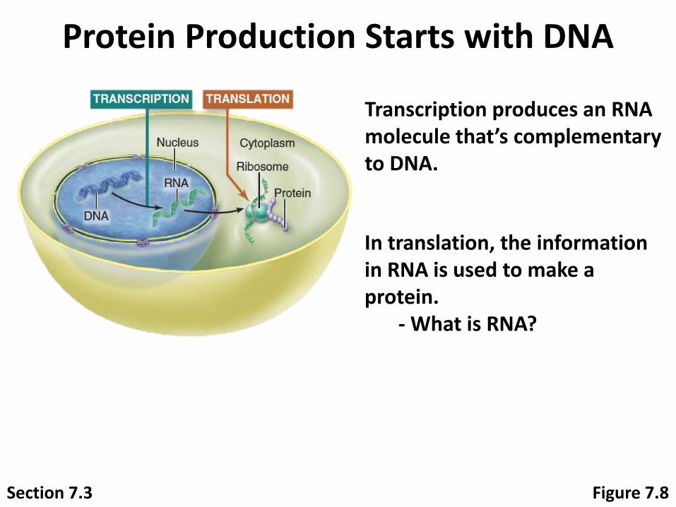
Section 7.3 Figure 7.8
Transcription produces an RNA molecule that’s complementary to DNA.
In translation, the information in RNA is used to make a protein.
- What is RNA?
Protein Production Starts with DNA
Section 7.3 Figure 7.9

Protein Production Starts with DNA
Section 7.3 Figure 7.8
RNA plays an important role in protein production.

Protein Production Starts with DNA
Section 7.3 Figure 7.8
Three types of RNA interact to produce proteins:
- Messenger RNA (mRNA)- Ribosomal RNA (rRNA)- Transfer RNA (tRNA)
Clicker Question #1
What is the main function of DNA?
A. encode proteins B. produce ATP C. speed up cell reactionsD. provide structural support to the cellE. All of the choices are correct.
Flower: © Doug Sherman/Geofile/RF
Clicker Question #1
What is the main function of DNA?
A. encode proteins B. produce ATP C. speed up cell reactionsD. provide structural support to the cellE. All of the choices are correct.
Flower: © Doug Sherman/Geofile/RF
10-39
The making of a protein requires
transcription and translation
Gene - segment of DNA that specifies the amino acid sequence of a protein
During transcription DNA serves as a template for RNA formation
DNA is transcribed, monomer by monomer, into RNA
During translation an RNA transcript directs the sequence of amino acids in a polypeptide
10-40
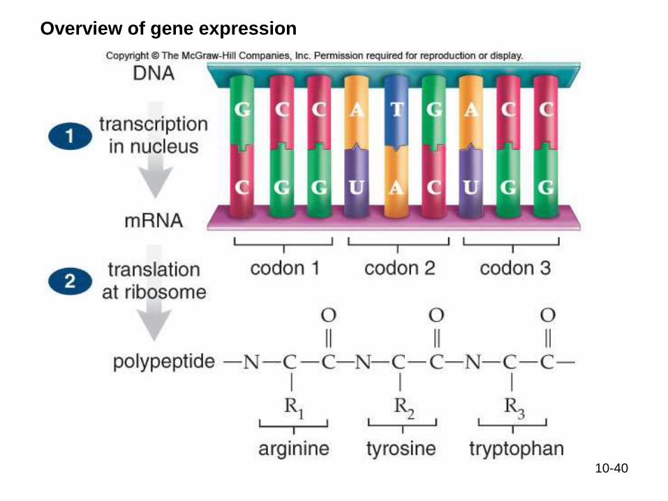
Overview of gene expression
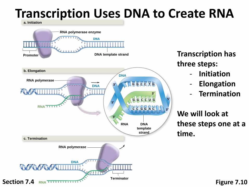
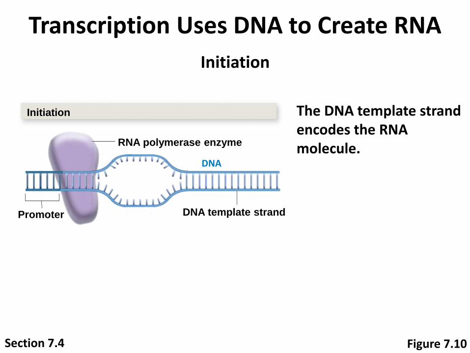
Transcription Uses DNA to Create RNA
Transcription has three steps:
- Initiation- Elongation- Termination
We will look at these steps one at a time.
DNA
a. Initiation
RNA polymerase enzyme
Promoter DNA template strand
b. Elongation
RNA polymerase
RNA
DNA
c. Termination
RNA polymerase
DNA
TerminatorRNA
G T
U
C
A
C
C C
C CC
G
G G
G G
G G
DNA
DNA
template
strand
RNA
3’
5’
5’5’
3’
3’
Figure 7.10Section 7.4
10-42
During transcription, a gene passes its coded
information to an mRNA
messenger RNA (mRNA) - takes instructions
from DNA in the nucleus to the ribosomes in the
cytoplasm
RNA polymerase joins the nucleotides together
Promoter defines the start of a gene, the direction of
transcription, and the strand to be transcribed
Stop sequence causes RNA polymerase to stop
transcribing the DNA and to release the mRNA
molecule, called an mRNA transcript
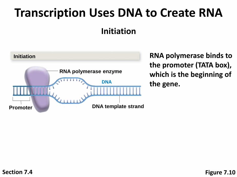
Transcription Uses DNA to Create RNA
RNA polymerase binds to the promoter (TATA box), which is the beginning of the gene.DNA
Initiation
RNA polymerase enzyme
Promoter DNA template strand
Initiation
Figure 7.10Section 7.4
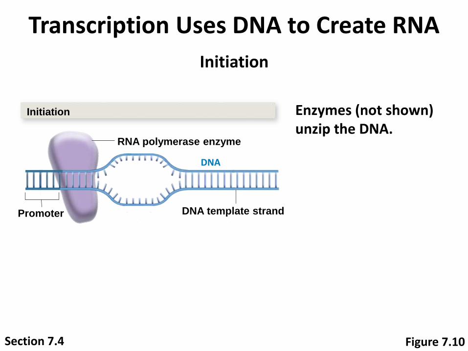
Transcription Uses DNA to Create RNA
Enzymes (not shown) unzip the DNA.
DNA
Initiation
RNA polymerase enzyme
Promoter DNA template strand
Initiation
Figure 7.10Section 7.4
Transcription Uses DNA to Create RNA
The DNA template strand encodes the RNA molecule.
DNA
Initiation
RNA polymerase enzyme
Promoter DNA template strand
Initiation
Figure 7.10Section 7.4

Transcription Uses DNA to Create RNA
Figure 7.10
RNA polymerase moves along the template strand, making an RNA copy.
Elongation
RNA polymerase
RNA
DNA
Elongation
3’
5’
5’
5’3’
3’
Section 7.4

Transcription Uses DNA to Create RNA
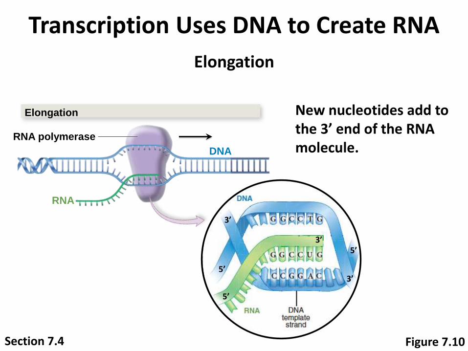
Notice that the RNA molecule is complementary to the DNA template strand.
Elongation
RNA polymerase
RNA
DNA
Elongation
Section 7.4
3’
5’
5’
5’3’
3’
Figure 7.10

Transcription Uses DNA to Create RNA
Also note that the strands are antiparallel: the 3’ end of RNA matches the 5’ end of DNA.
Elongation
RNA polymerase
RNA
DNA
Elongation
Section 7.4
3’
5’
5’
5’3’
3’
Figure 7.10
Transcription Uses DNA to Create RNA
New nucleotides add to the 3’ end of the RNA molecule.
Elongation
RNA polymerase
RNA
DNA
Elongation
Section 7.4
3’
5’
5’
5’3’
3’
Figure 7.10
Transcription Uses DNA to Create RNA
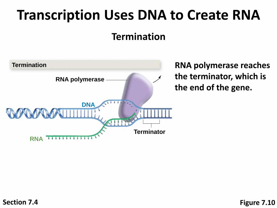
RNA polymerase reaches the terminator, which is the end of the gene.
Termination
RNA polymerase
DNA
TerminatorRNA
Termination
Figure 7.10Section 7.4
Transcription Uses DNA to Create RNA
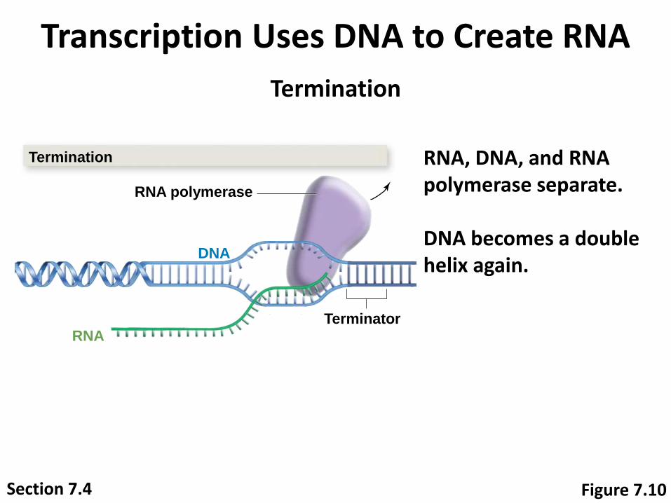
RNA, DNA, and RNA polymerase separate.
DNA becomes a double helix again.
Termination
RNA polymerase
DNA
TerminatorRNA
Termination
Figure 7.10Section 7.4

Transcription Uses DNA to Create RNA
The cell produced an RNA copy of a gene!
Termination
RNA polymerase
DNA
TerminatorRNA
Termination
Figure 7.10Section 7.4
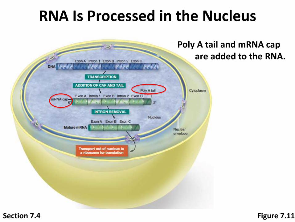
RNA Is Processed in the Nucleus
Figure 7.11
Poly A tail and mRNA cap are added to the RNA.
Section 7.4
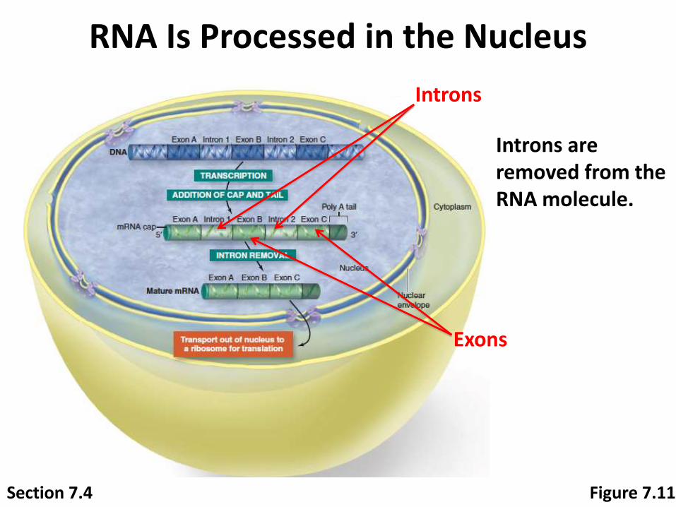
RNA Is Processed in the Nucleus
Introns are removed from the RNA molecule.
Introns
Exons
Figure 7.11Section 7.4
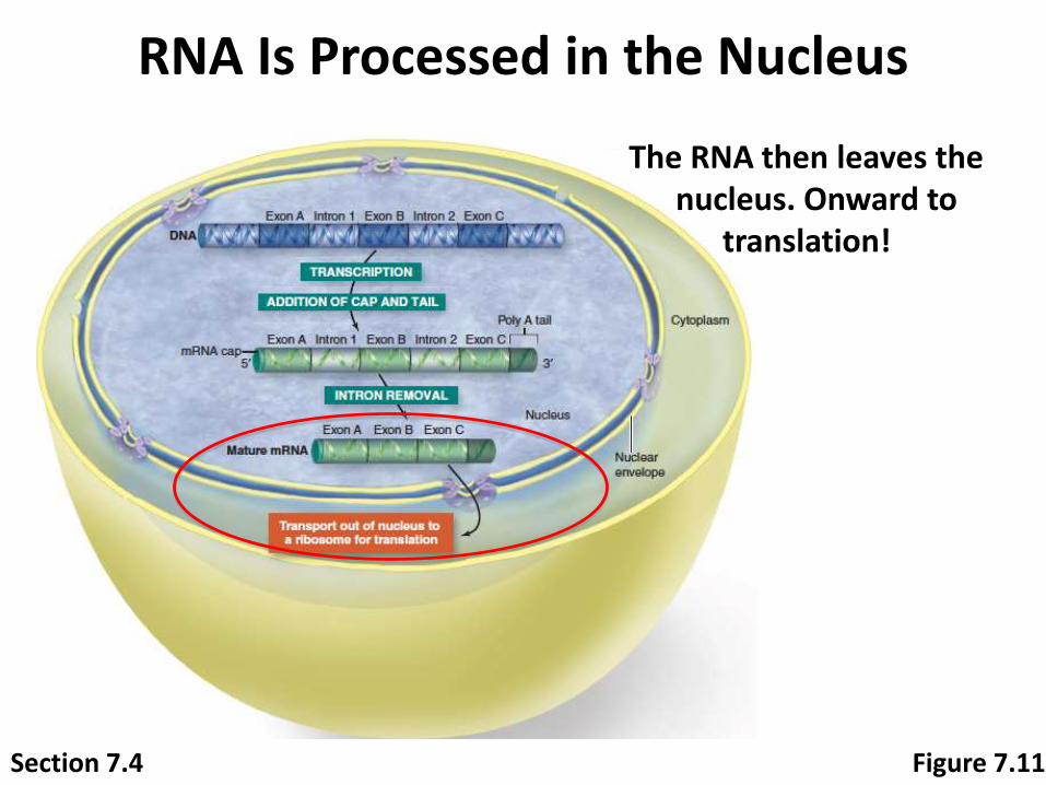
RNA Is Processed in the Nucleus
The RNA then leaves the nucleus. Onward to
translation!
Figure 7.11Section 7.4

10-56
In eukaryotes, an mRNA
is processed before
leaving the nucleus
Primary mRNA is composed of exons and introns The exons of mRNA will be expressed, but the
introns will not
Function of Introns Might allow exons to be put together in different
sequences so that various mRNAs and proteins can result from a single gene
Some introns might regulate gene expression by feeding back to determine which coding genes are to be expressed and how they should be spliced

10-57
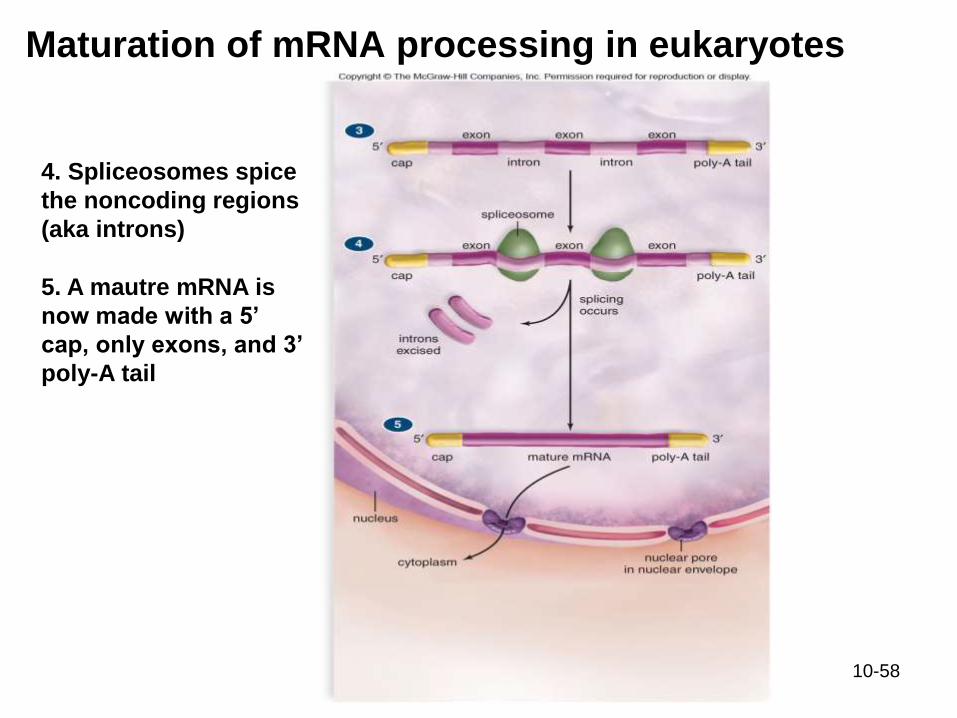
Maturation of mRNA processing in eukaryotes
1. RNA Polymerase
makes a transcript
of DNA using RNA
bases
2. A pre-mRNA from
DNA is made that has
both introns and
exons
3. A 5’ cap and 3’ poly-
A tail are put onto pre-
mRNA
10-58
Maturation of mRNA processing in eukaryotes
4. Spliceosomes spice
the noncoding regions
(aka introns)
5. A mautre mRNA is
now made with a 5’
cap, only exons, and 3’
poly-A tail

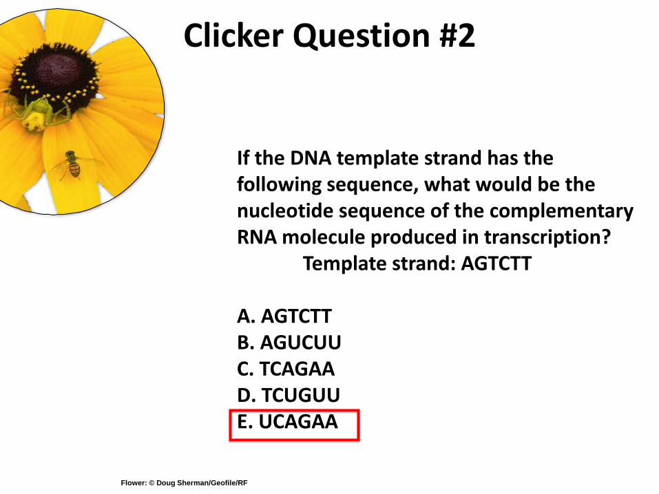
Clicker Question #2
If the DNA template strand has the following sequence, what would be the nucleotide sequence of the complementary RNA molecule produced in transcription?
Template strand: AGTCTT
A. AGTCTTB. AGUCUUC. TCAGAAD. TCUGUUE. UCAGAA
Flower: © Doug Sherman/Geofile/RF
Clicker Question #2
If the DNA template strand has the following sequence, what would be the nucleotide sequence of the complementary RNA molecule produced in transcription?
Template strand: AGTCTT
A. AGTCTTB. AGUCUUC. TCAGAAD. TCUGUUE. UCAGAA
Flower: © Doug Sherman/Geofile/RF

Translation Builds the Protein
Section 7.5 Figure 7.8
Now let’s look at how a ribosome uses RNA to produce a protein.

Translation Builds the Protein
Figure 7.12
A
A A
A AG
G G
G UU C
CTT T C
C
DNA template strandDNA
TRANSCRIPTION
mRNA
TRANSLATION
Protein
CodonCodonCodon
Lysine ValineSerine
Polypeptide (amino acid sequence)
A codon is a three-nucleotide sequence that encodes one amino acid.
Section 7.5
10-63
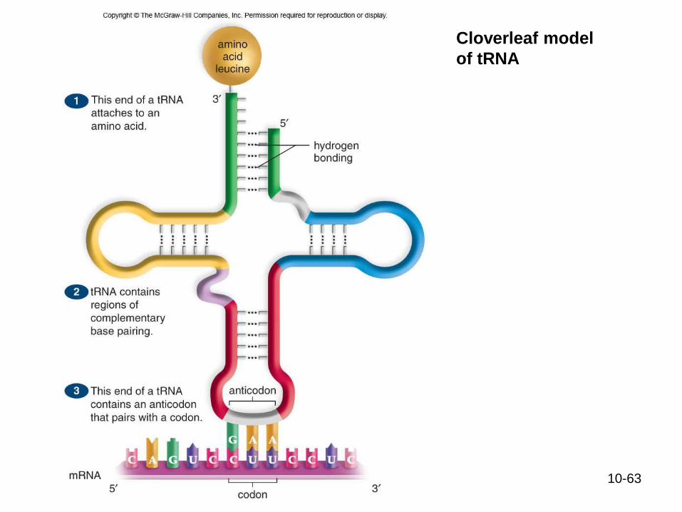
Cloverleaf model
of tRNA

Translation Builds the Protein
U
C
A
G
U C A G
Firs
t le
tte
r o
f co
do
n
U
C
A
G
U
C
A
G
U
C
A
G
U
C
A
G
Third
lette
r of co
do
nA A AG G UU CC
mRNA
TRANSLATION
Protein
CodonCodonCodon
Lysine ValineSerine
Polypeptide (amino acid sequence)
The Genetic Code
Second letter of codon
UUU
UUC
UUA
UUG
CUU
CUC
CUA
CUG
AUU
AUC
AUA
AUG
GUU
GUC
GUA
GUG
UAU
UAC
CCA
CCG
UAA
UCU
UCC
UCA
UCG
CCU
CCC
UAG
UGU
UGC
UGA
UGG
CAU
CAC
CAA
CAG
CGU
CGC
CGA
CGG
ACU
ACC
ACA
ACG
AAU
AAC
AAA
AAG
AGU
AGC
AGA
AGG
GCU
GCC
GCA
GCG
GAU
GAC
GAA
GAG
GGU
GGC
GGA
GGG
Leucine (Leu; L)
Phenylalanine (Phe; F)
Leucine (Leu; L)
Isoleucine (Ile; I)
Start Methionine (Met; M)
Valine (Val; V)
Serine (Ser; S)
Proline (Pro; P)
Proline (Pro; P)
Proline (Pro; P)
Tyrosine (Tyr; Y)
Histidine (His; H)
Glutamine (Gln; Q)
Asparagine (Asn; N)
Lysine (Lys; K)
Aspartic acid (Asp; D)
Glutamic acid (Glu; E)
Cysteine (Cys; C)
Tryptophan (Trp; W)
Stop
Arginine (Arg; R)
Serine (Ser; S)
Arginine (Arg; R)
Glysine (Gly; G)
Stop
Stop
The genetic code shows which mRNA codons correspond to which amino acids.
Section 7.5 Figure 7.12
10-65
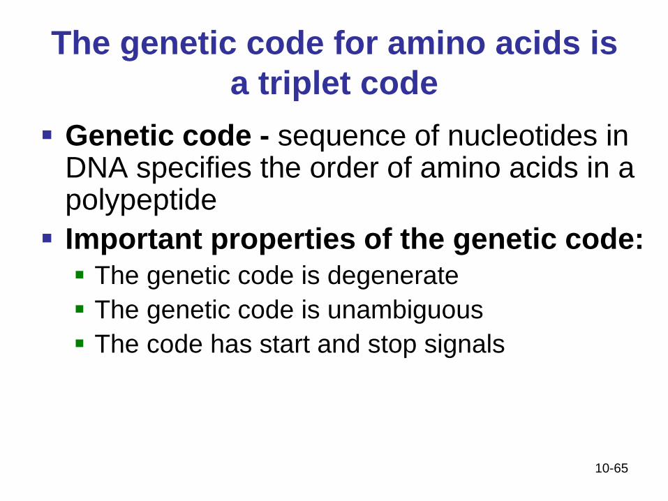
The genetic code for amino acids is
a triplet code
Genetic code - sequence of nucleotides in DNA specifies the order of amino acids in a polypeptide
Important properties of the genetic code:
The genetic code is degenerate
The genetic code is unambiguous
The code has start and stop signals

10-66
RNA codons

10-67
Ribosome structure and function
10-68
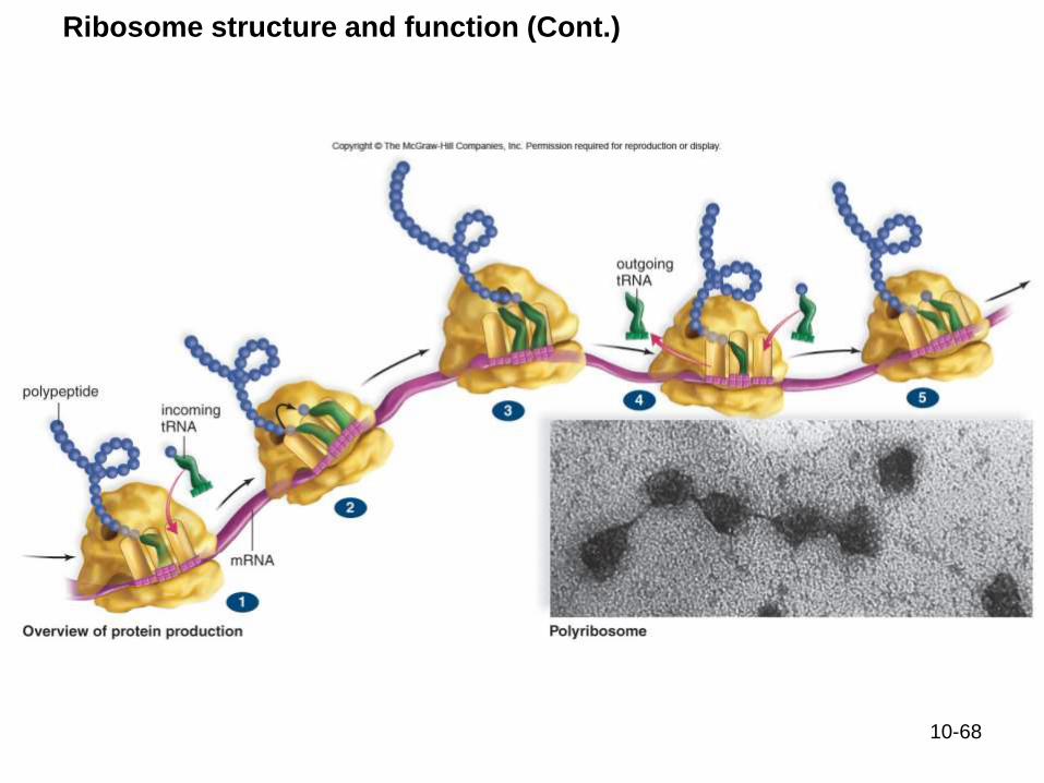
Ribosome structure and function (Cont.)
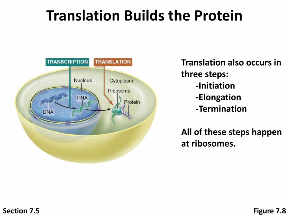
Translation Builds the Protein
Figure 7.8
Translation also occurs in three steps:
-Initiation-Elongation-Termination
All of these steps happen at ribosomes.
Section 7.5

10-70
Initiation begins the process of
polypeptide production
Initiation - the step that brings all the translation
components together
Initiation
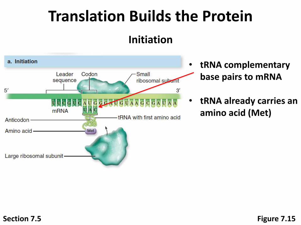
Translation Builds the Protein
• tRNA complementary base pairs to mRNA
• tRNA already carries an amino acid (Met)
Initiation
Section 7.5 Figure 7.15
10-72
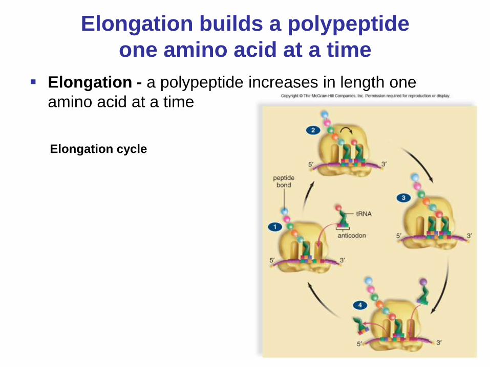
Elongation builds a polypeptide
one amino acid at a time
Elongation - a polypeptide increases in length one
amino acid at a time
Elongation cycle
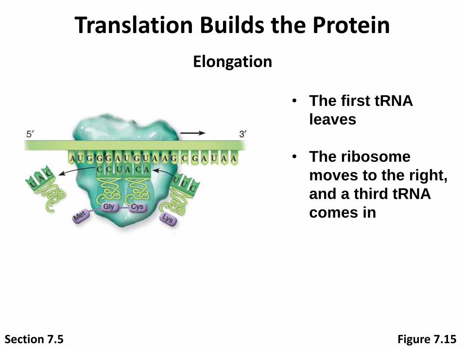
Translation Builds the Protein
Elongation
• The first tRNA
leaves
• The ribosome
moves to the right,
and a third tRNA
comes in
Section 7.5 Figure 7.15
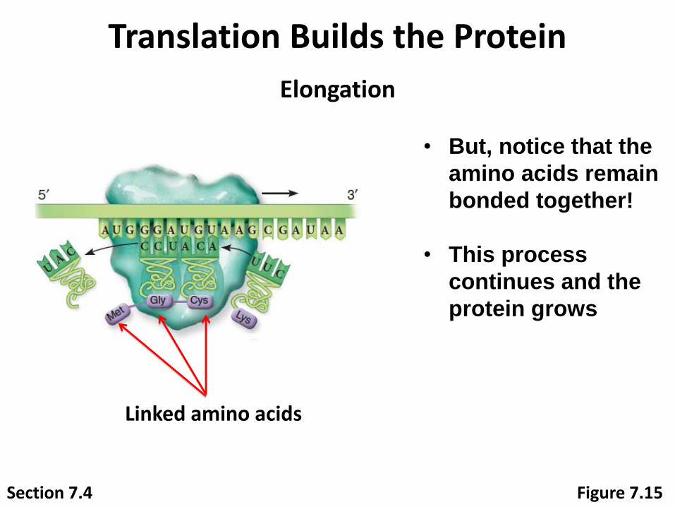
Translation Builds the Protein
Section 7.4
Elongation
• But, notice that the
amino acids remain
bonded together!
• This process
continues and the
protein grows
Linked amino acids
Figure 7.15
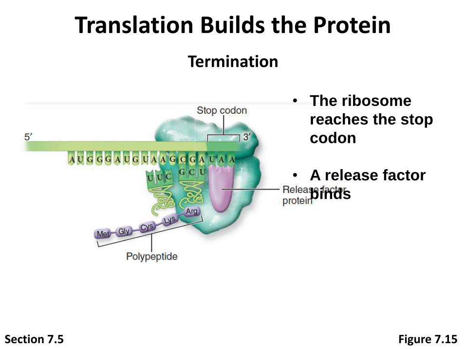
Translation Builds the Protein
Termination
• The ribosome
reaches the stop
codon
• A release factor
binds
Section 7.5 Figure 7.15
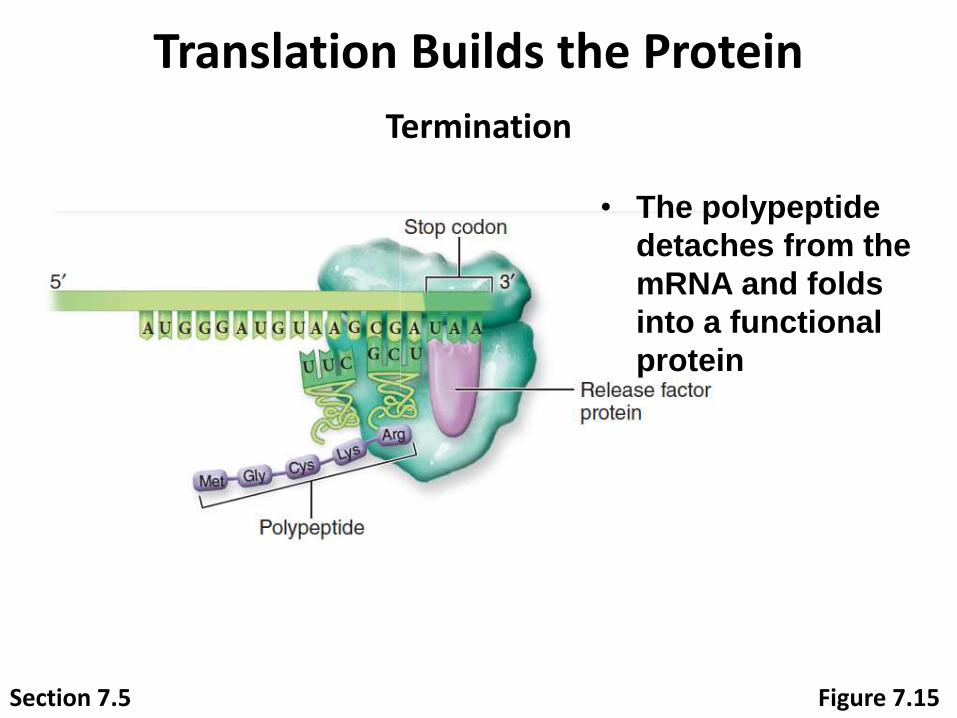
Translation Builds the Protein
Termination
• The polypeptide
detaches from the
mRNA and folds
into a functional
protein
Section 7.5 Figure 7.15

Translation Builds the Protein
PolypeptidemRNA Ribosome
SEM (false color) 50 nm
Ribosomes: © Kiseleva and Donald Fawcett/Visuals Unlimited
Translation is efficient when multiple ribosomes attach to an mRNA molecule simultaneously.
Section 7.5 Figure 7.16

10-78
Let’s review gene expression
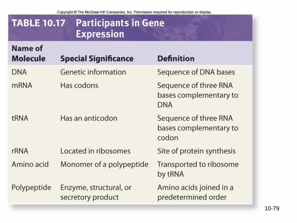
10-79
10-80
Connecting the Concepts:
Using all previously collected data concerning DNA structure, Watson and Crick were able to arrive at the legendary design of DNA—a double helix
Complementary base pairing explains the replication of DNA, how RNA molecules are made
Geneticists have confirmed that proteins are the link between the genotype (DNA combination) and the phenotype (physical appearance)
Food for thought?
How would you design an intelligent being to react to the
environment?
Consider that....
Every cell of the body contains ALL the DNA for the
organism.
Would you want to turn on all 25,000 genes in our body at
the same time? Why or why not?
Not all genes are necessary all the time
Cells must then have the ability to turn genes on and off

Francois Jacob and Jacques Monod Experiments with E. Coli showed that it is capable of
regulating the expression of its genes
An operon consists of the following elements
1. Promoter - where RNA polymerase attaches, signaling
the start of the gene
2. Operator - where a
repressor binds, stopping the
transcription of that gene
3. Structural Genes - genes
coding for the enzyme, they are
transcribed as a unit

The first well-understood example of how gene expression
is controlled
Prokaryotic gene expression
The lac Operon: The inducible operon

Logic of Lac operon
1. Lactose is an energy source but it is not always present
2. Genes encoding enzymes that metabolize lactose are
only needed (efficiency!!!) when lactose is present
3. If lactose and glucose are both present, glucose is the
preferred energy source (efficiency!!!)

The lac Operon: The inducible operon
This region is normally in the "off" position, it turns on
when lactose is present

1 2 3
Bacterial
cell
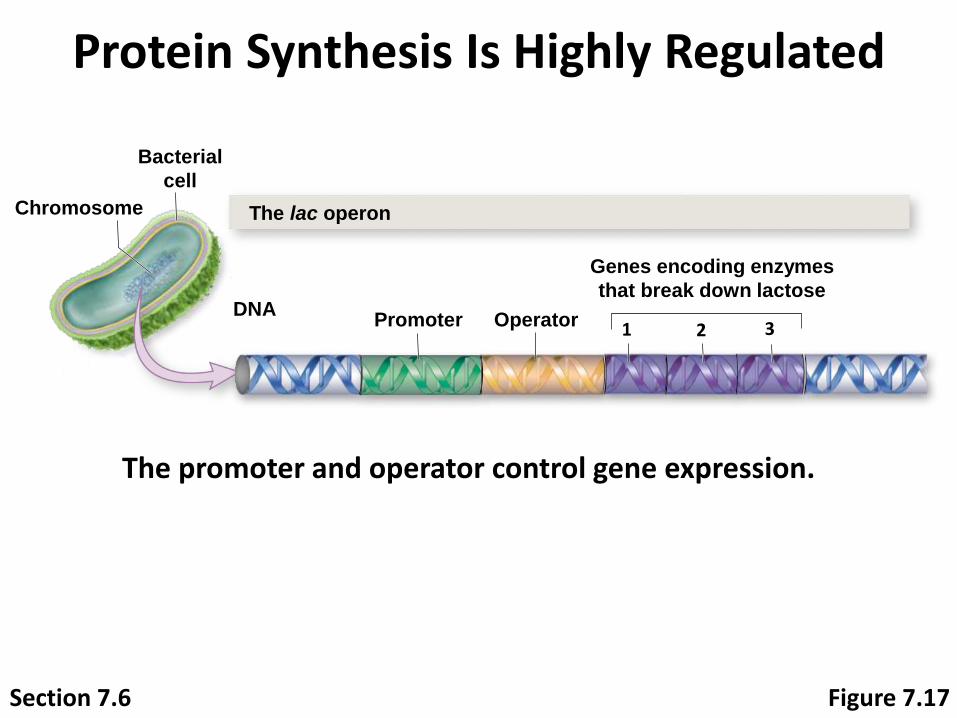
Chromosome The lac operon
DNAPromoter Operator
Genes encoding enzymes
that break down lactose
Protein Synthesis Is Highly Regulated
Figure 7.17
Genes in prokaryotes are organized into operons, groups of genes that are always transcribed
together.
Section 7.6
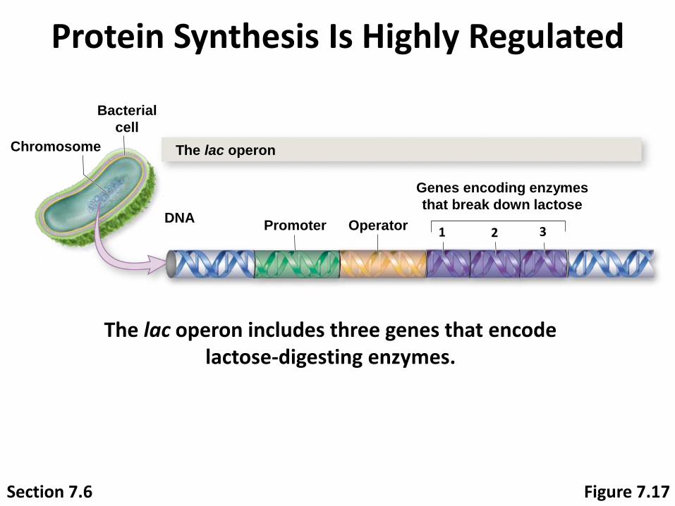
The lac operon includes three genes that encode lactose-digesting enzymes.
1 2 3
Bacterial
cell
Chromosome The lac operon
DNAPromoter Operator
Genes encoding enzymes
that break down lactose
Protein Synthesis Is Highly Regulated
Section 7.6 Figure 7.17
1 2 3
Bacterial
cell
Chromosome The lac operon
DNAPromoter Operator
Genes encoding enzymes
that break down lactose
Protein Synthesis Is Highly Regulated
The promoter and operator control gene expression.
Section 7.6 Figure 7.17
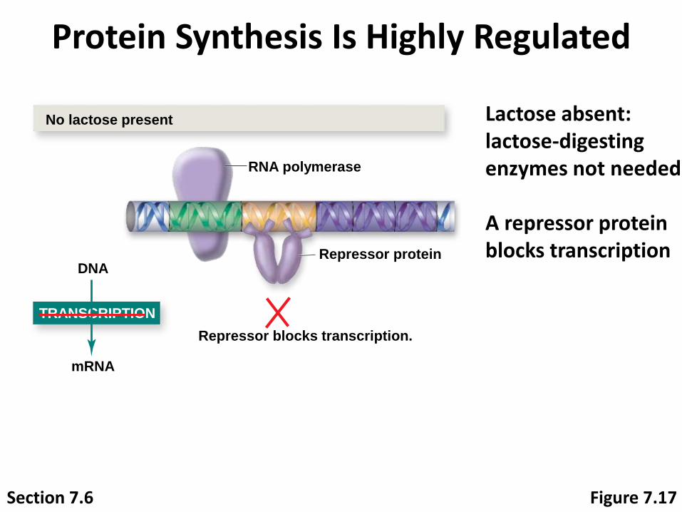
Protein Synthesis Is Highly Regulated
TRANSCRIPTION
No lactose present
RNA polymerase
DNARepressor protein
Repressor blocks transcription.
mRNA
Lactose absent: lactose-digesting enzymes not needed
A repressor protein blocks transcription
Section 7.6 Figure 7.17

Protein Synthesis Is Highly Regulated
Section 7.5
Lactose present
Lactose
Lactose binds
to repressor.Repressor protein
RNA polymerase
DNA
TRANSCRIPTION
mRNA
TRANSLATION
ProteinsEnzymes that break down
lactose are produced.
mRNA
Protein
Transcription proceeds.
Lactose present: repressor binds to lactose, changes shape, and releases the operator
Protein synthesis of lactose-digesting enzymes occurs
Figure 7.17

Protein Synthesis Is Highly Regulated
Section 7.5
Now let’s look at eukaryotes.
Protein synthesis requires lots of energy!
Cells save energy by only producing needed proteins.
Both prokaryotes and eukaryotes regulate protein synthesis, but in different ways.
Mushroom © Jacana/Science Source
Transcriptional control in
Eukaryotes
You may expect transcriptional control in eukaryotes to
1. Involve the organization of chromatin
2. Include regulatory proteins such as the repressor proteins
from the lac operon.
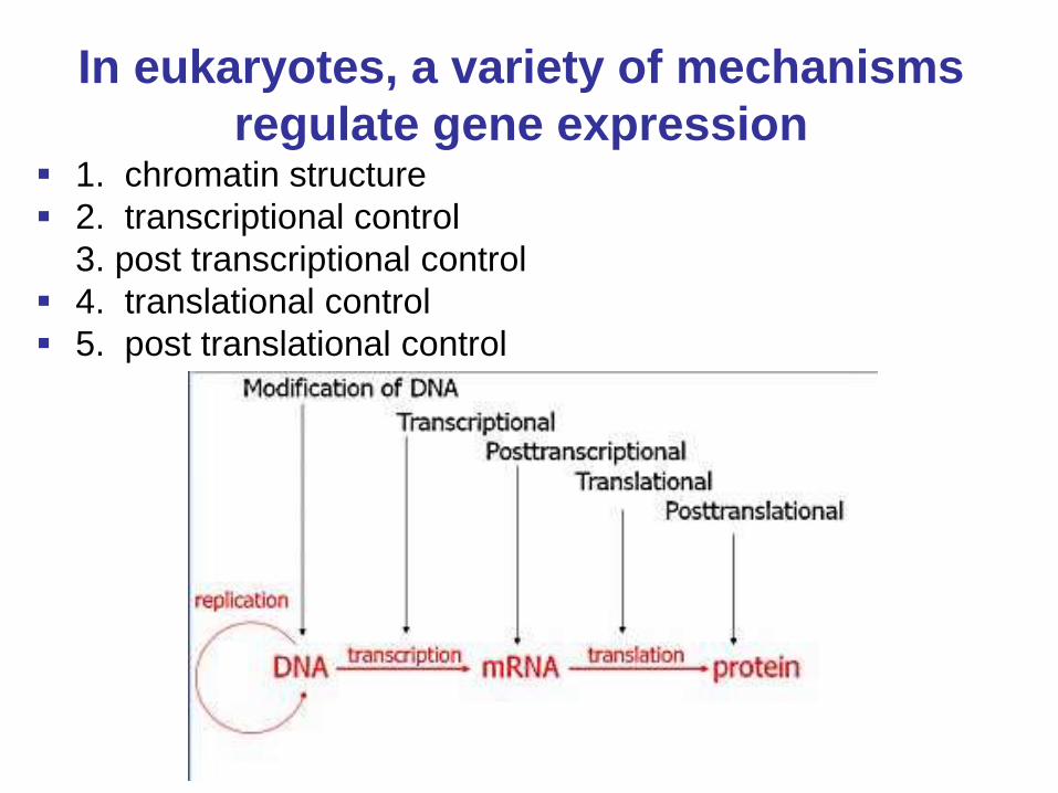
In eukaryotes, a variety of mechanisms
regulate gene expression 1. chromatin structure
2. transcriptional control
3. post transcriptional control
4. translational control
5. post translational control

Protein Synthesis Is Highly Regulated
Section 7.5
Gene regulation starts in the nucleus.
Figure 7.19
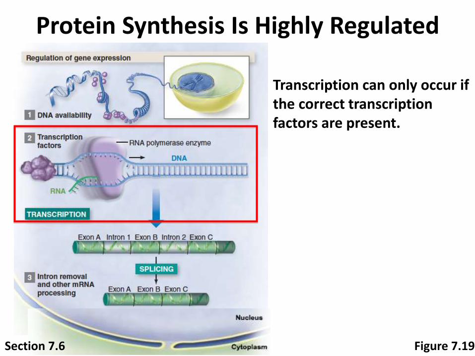
Protein Synthesis Is Highly Regulated
Transcription can only occur if the correct transcription factors are present.
Section 7.6 Figure 7.19
Protein Synthesis Is Highly Regulated
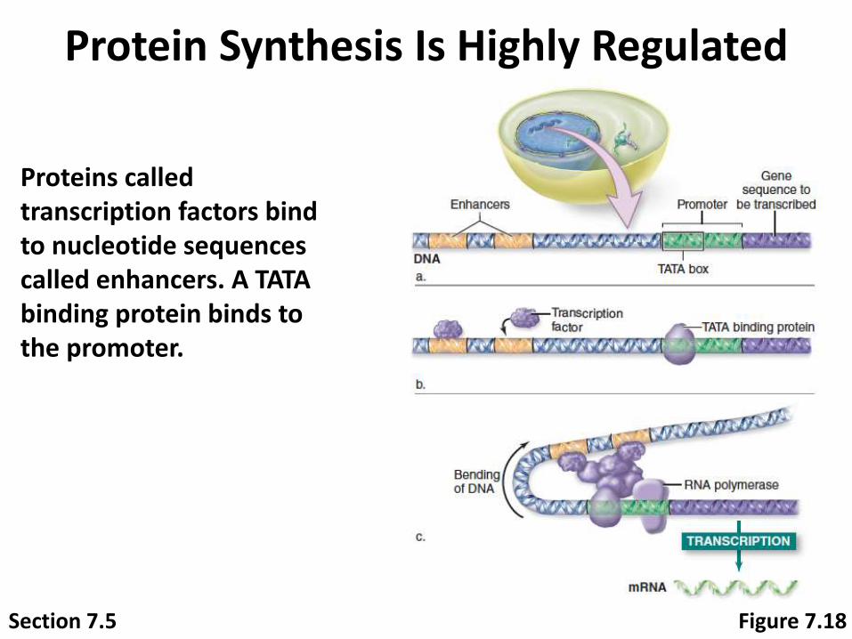
Section 7.5 Figure 7.18
Proteins called transcription factors bind to nucleotide sequences called enhancers. A TATA binding protein binds to the promoter.
Protein Synthesis Is Highly Regulated
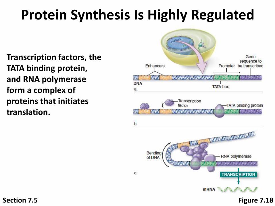
Section 7.5 Figure 7.18
Transcription factors, the TATA binding protein, and RNA polymerase form a complex of proteins that initiates translation.

Protein Synthesis Is Highly Regulated
One gene can encode multiple proteins if different introns are removed.
Section 7.6 Figure 7.19
Protein Synthesis Is Highly Regulated
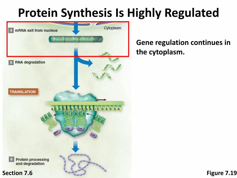
Gene regulation continues in the cytoplasm.
Section 7.6 Figure 7.19
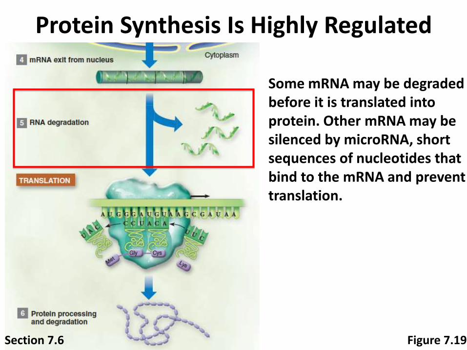
Protein Synthesis Is Highly Regulated
Some mRNA may be degraded before it is translated into protein. Other mRNA may be silenced by microRNA, short sequences of nucleotides that bind to the mRNA and prevent translation.
Section 7.6 Figure 7.19
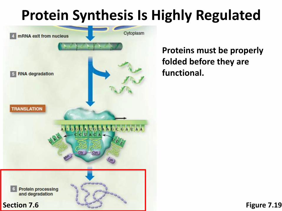
Protein Synthesis Is Highly Regulated
Proteins must be properly folded before they are functional.
Section 7.6 Figure 7.19
7.6 Mastering Concepts
Why do cells regulate which genes are expressed at any given time?
DNA bursting from bacterial cell © Dr. Gopal Murti/Science Source

10-105
Mutations Are Changes in the
Sequence of DNA Bases
10-106
DNA meets the criteria for the
genetic material
The genetic material must be:
Variable between species and able to store
information that causes species to vary from
one another
Constant within a species and able to be
replicated with high fidelity during cell division
Able to undergo rare changes, called
mutations, that provide the genetic variability
that allows evolution to occur

Mutations Change DNA
Figure 7.20Section 7.7 Wild fly: © Andrew Syred/Science Source; Mutant fly: © Science VU/Dr. F. R. Turner/Visuals Unlimited
A mutation in one gene causes a fly to develop legs where its antenna should be!
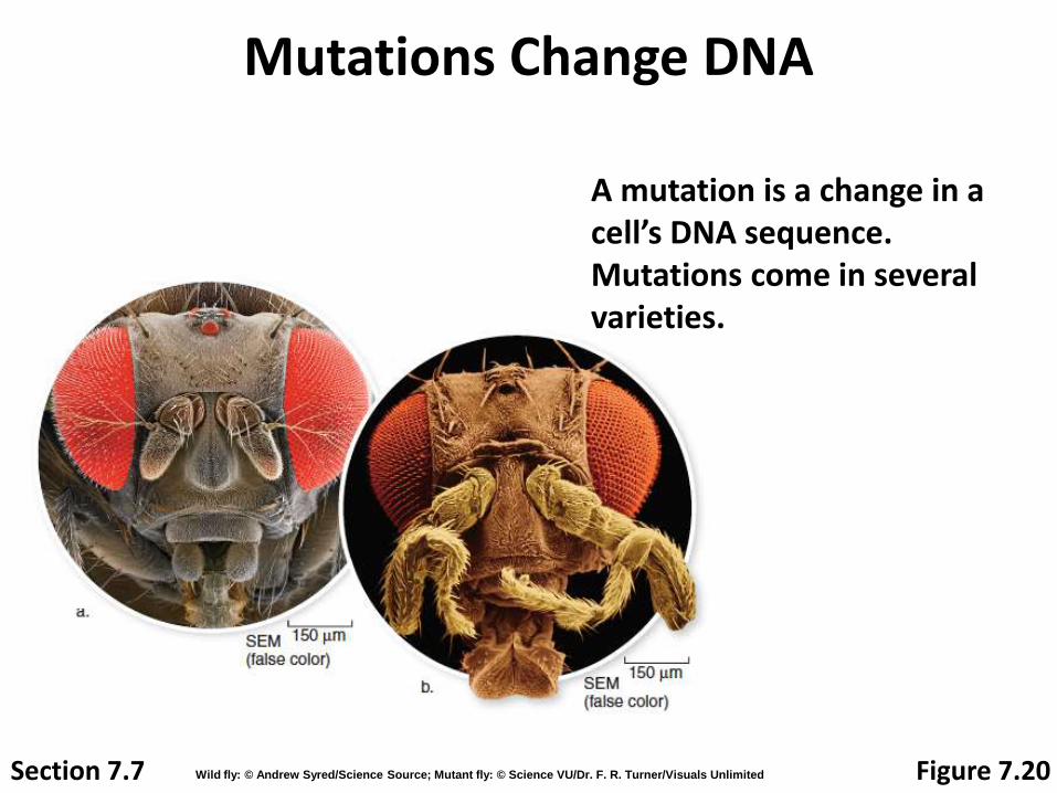
Mutations Change DNA
A mutation is a change in a cell’s DNA sequence. Mutations come in several varieties.
Section 7.7 Figure 7.20Wild fly: © Andrew Syred/Science Source; Mutant fly: © Science VU/Dr. F. R. Turner/Visuals Unlimited
Various Types of Mutations
Mistake made from polymerase
Mistake in DNA repair process
Recombination
Exogenous factors such as virus and
transposons
10-109
Mutation categories
Deleterious- mutation at splice junction or coding
region
Advantageous- any mutation that allows either
an increased rate of survival, replication rate or
ability to pass on genes
Neutral- mutation at a non-coding region
10-110
Definitions
Synonymous substitution- mutation that does not
change an amino acid (ex. Wobble base)
Non-synonymous substitution- mutation that
changes an amino acid
Are all proteins the same in regards to fitness?
10-111
10-112
Mutations affect genetic information
and expression
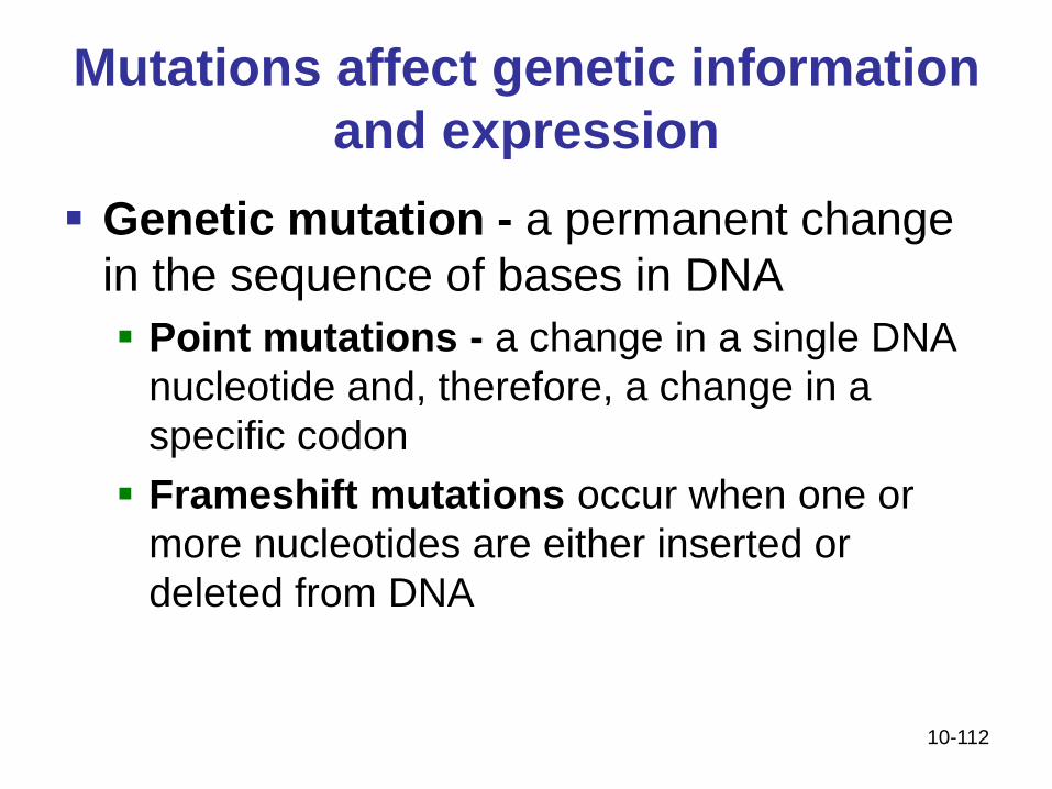
Genetic mutation - a permanent change
in the sequence of bases in DNA
Point mutations - a change in a single DNA
nucleotide and, therefore, a change in a
specific codon
Frameshift mutations occur when one or
more nucleotides are either inserted or
deleted from DNA
10-113
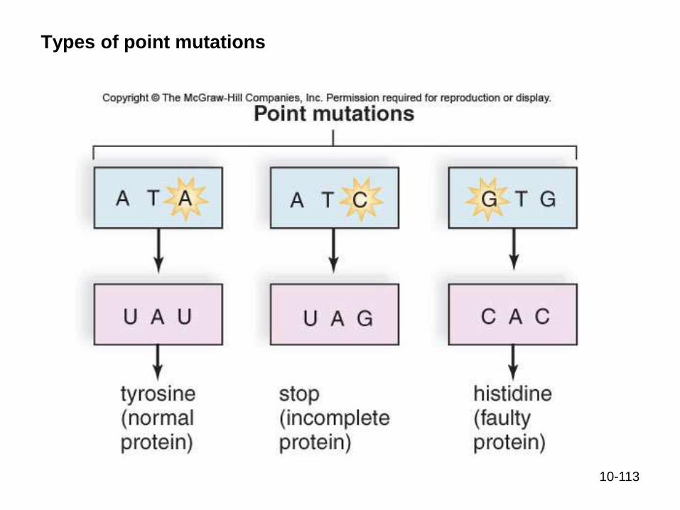
Types of point mutations

Mutations Change DNA
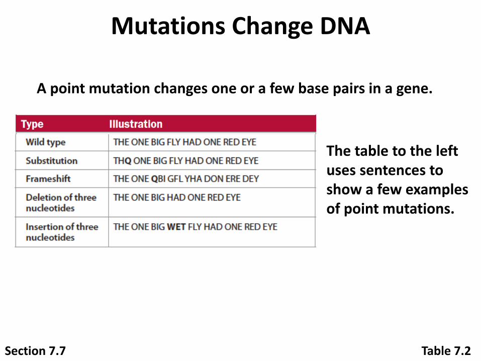
A point mutation changes one or a few base pairs in a gene.
Table 7.2Section 7.7
Mutations Change DNA
A point mutation changes one or a few base pairs in a gene.
The table to the left uses sentences to show a few examples of point mutations.
Table 7.2Section 7.7
Mutations Change DNA
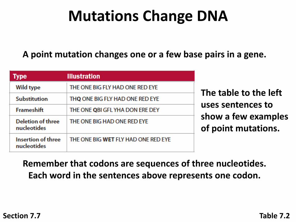
A point mutation changes one or a few base pairs in a gene.
The table to the left uses sentences to show a few examples of point mutations.
Remember that codons are sequences of three nucleotides. Each word in the sentences above represents one codon.
Table 7.2Section 7.7
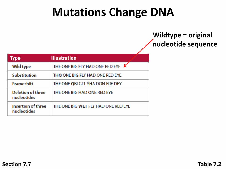
Mutations Change DNA
Wildtype = original nucleotide sequence
Table 7.2Section 7.7

Mutations Change DNA
Wildtype = original nucleotide sequence
Substitution = changed nucleotide(s)
Table 7.2Section 7.7
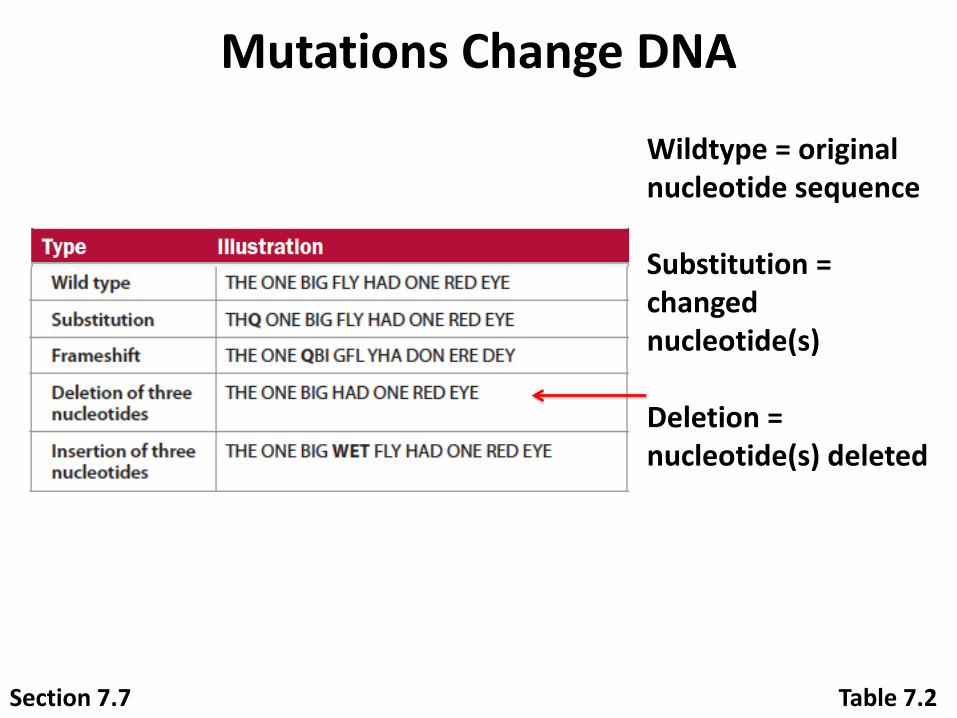
Mutations Change DNA
Wildtype = original nucleotide sequence
Substitution = changed nucleotide(s)
Deletion = nucleotide(s) deleted
Table 7.2Section 7.7
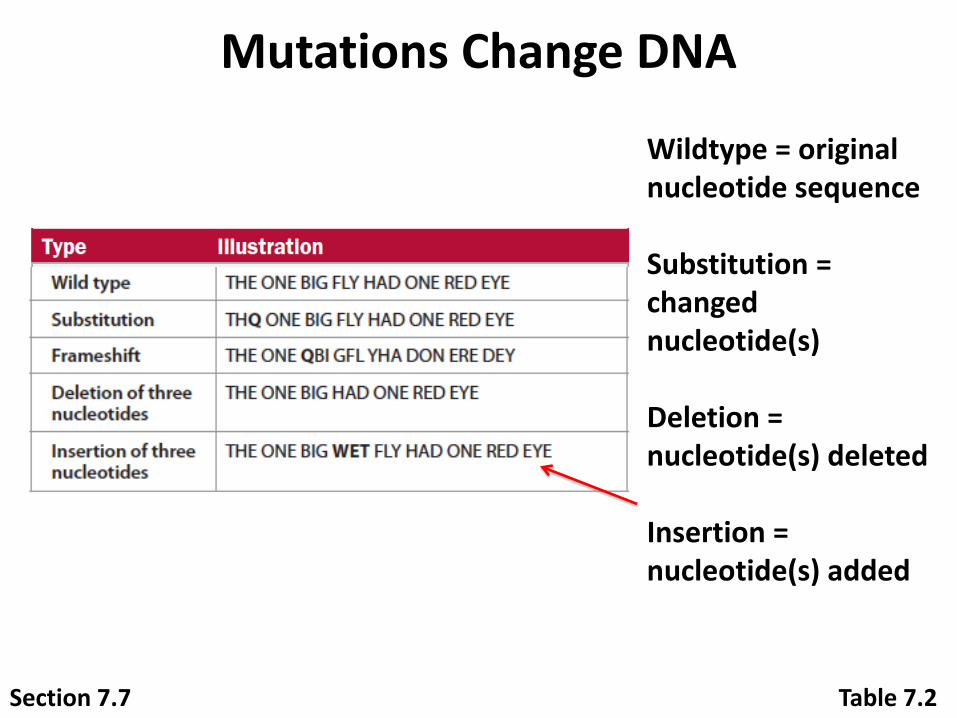
Mutations Change DNA
Wildtype = original nucleotide sequence
Substitution = changed nucleotide(s)
Deletion = nucleotide(s) deleted
Insertion = nucleotide(s) added
Table 7.2Section 7.7
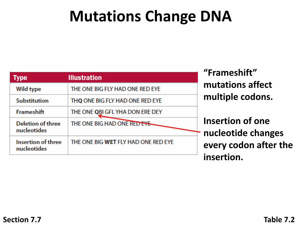
Mutations Change DNA
“Frameshift”mutations affect multiple codons.
Insertion of one nucleotide changes every codon after the insertion.
Table 7.2Section 7.7

Mutations Change DNA
Deletions or insertions of three nucleotides add or delete entire codons, but do not affect other codons.
Table 7.2Section 7.7

Mutations Change DNA
Figure 7.22Section 7.7

Normal cells: © Micro Discovery/Corbis; Sickled cells: © Dr. Gopal Murti/Science Source
Normal red blood cells
No aggregation
of hemoglobin
molecules
SEM
Pro Glu Glu
6 µm
Sickled red blood cells
Abnormal
aggregation
of hemoglobin
molecules
Pro Val Glu6 µm
G G A C T C C T T
C C U G A G G A A
G G A C A C C T T
C C U G U G G A A
SEM
A single base substitution in a hemoglobin gene causes blood cells to form abnormally, leading to sickle cell disease.
Figure 7.21
Mutations Change DNA
Section 7.7
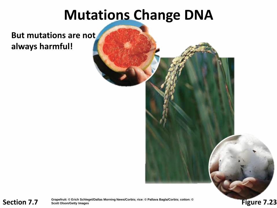
But mutations are not always harmful!
Figure 7.23
Mutations Change DNA
Grapefruit: © Erich Schlegel/Dallas Morning News/Corbis; rice: © Pallava Bagla/Corbis; cotton: ©
Scott Olson/Getty ImagesSection 7.7
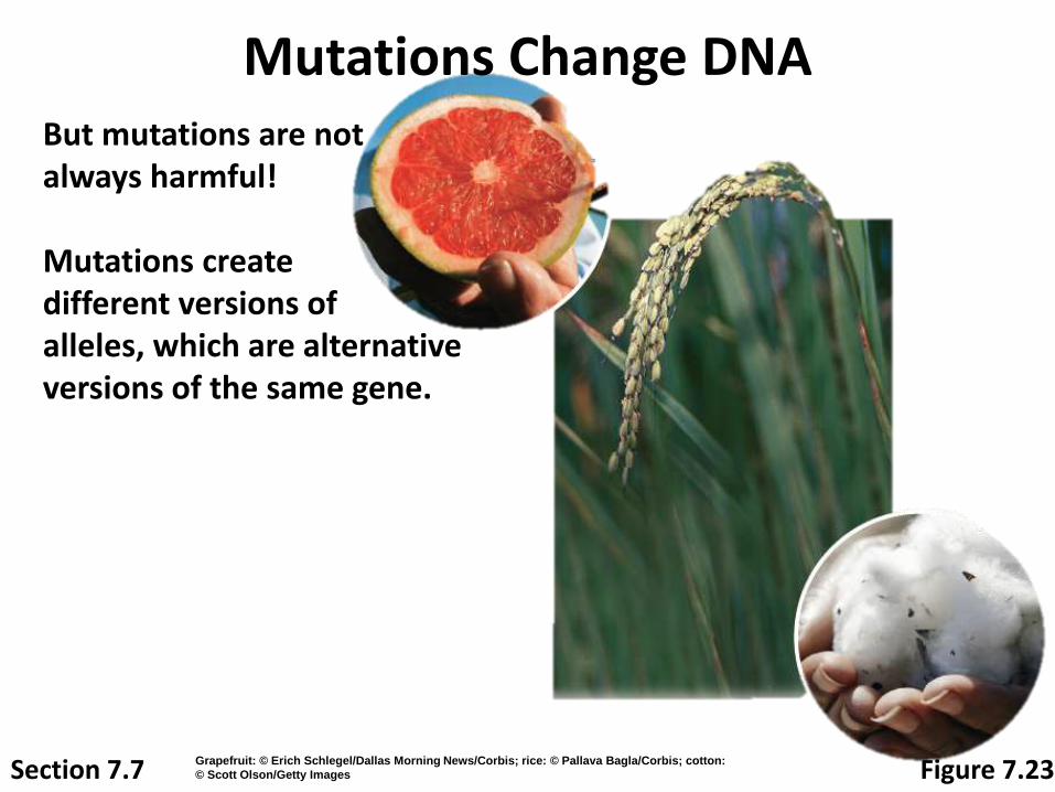
But mutations are not always harmful!
Mutations create different versions of alleles, which are alternative versions of the same gene.
Mutations Change DNA
Section 7.7 Figure 7.23Grapefruit: © Erich Schlegel/Dallas Morning News/Corbis; rice: © Pallava Bagla/Corbis; cotton:
© Scott Olson/Getty Images
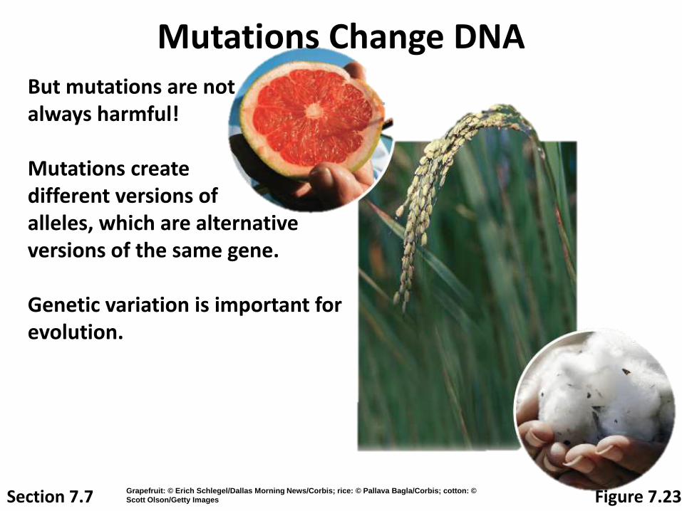
But mutations are not always harmful!
Mutations create different versions of alleles, which are alternative versions of the same gene.
Genetic variation is important for evolution.
Mutations Change DNA
Section 7.7 Figure 7.23Grapefruit: © Erich Schlegel/Dallas Morning News/Corbis; rice: © Pallava Bagla/Corbis; cotton: ©
Scott Olson/Getty Images

But mutations are not always harmful!
Mutations create different versions of alleles, which are alternative versions of the same gene.
Genetic variation is important for evolution.
Plant breeders even induce mutations to create new varieties of plants.
Mutations Change DNA
Section 7.7 Figure 7.23Grapefruit: © Erich Schlegel/Dallas Morning News/Corbis; rice: © Pallava Bagla/Corbis; cotton: ©
Scott Olson/Getty Images
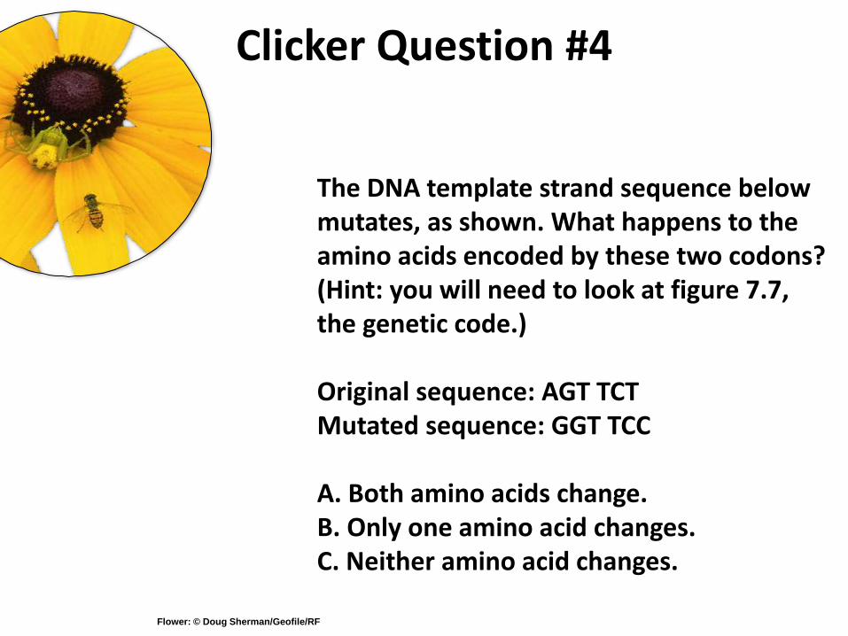
Clicker Question #4
The DNA template strand sequence below mutates, as shown. What happens to the amino acids encoded by these two codons? (Hint: you will need to look at figure 7.7, the genetic code.)
Original sequence: AGT TCTMutated sequence: GGT TCC
A. Both amino acids change.B. Only one amino acid changes.C. Neither amino acid changes.
Flower: © Doug Sherman/Geofile/RF
10-131
APPLYING THE CONCEPTS—HOW BIOLOGY IMPACTS OUR LIVES
Many agents can
cause mutations
Some mutations are spontaneous while
others are due to environmental mutagens
Environmental Mutagens
Mutagen - an environmental agent that
increases the chances of a mutation
Carcinogens - cancer-causing agents
Tobacco smoke contains a number of organic
chemicals that are known carcinogens

DNA Replication
10-132

133
Replication Facts
DNA has to be copied before a cell divides
DNA is copied during the S or synthesis phase of interphase
New cells will need identical DNA strands
134
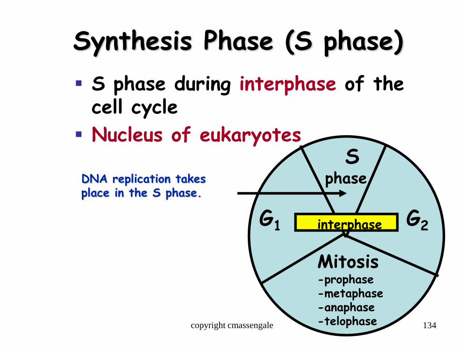
Synthesis Phase (S phase)
S phase during interphase of the cell cycle
Nucleus of eukaryotes
Mitosis-prophase-metaphase-anaphase-telophase
G1 G2
Sphase
interphase
DNA replication takesplace in the S phase.
copyright cmassengale

10-135
DNA replication is semiconservative
DNA replication - the process of copying a DNA molecule
Replication requires the following steps:1. Unwinding: Old strands are unwound and “unzipped”
2. Complementary base pairing or initiation: New complementary nucleotides are positioned by the process of base pairing
3. Joining or elongation: Complementary nucleotides join to form new strands
Each daughter DNA molecule contains a template strand, or old strand, and a new strand
Steps 2 and 3 are carried out by DNA polymerase
136
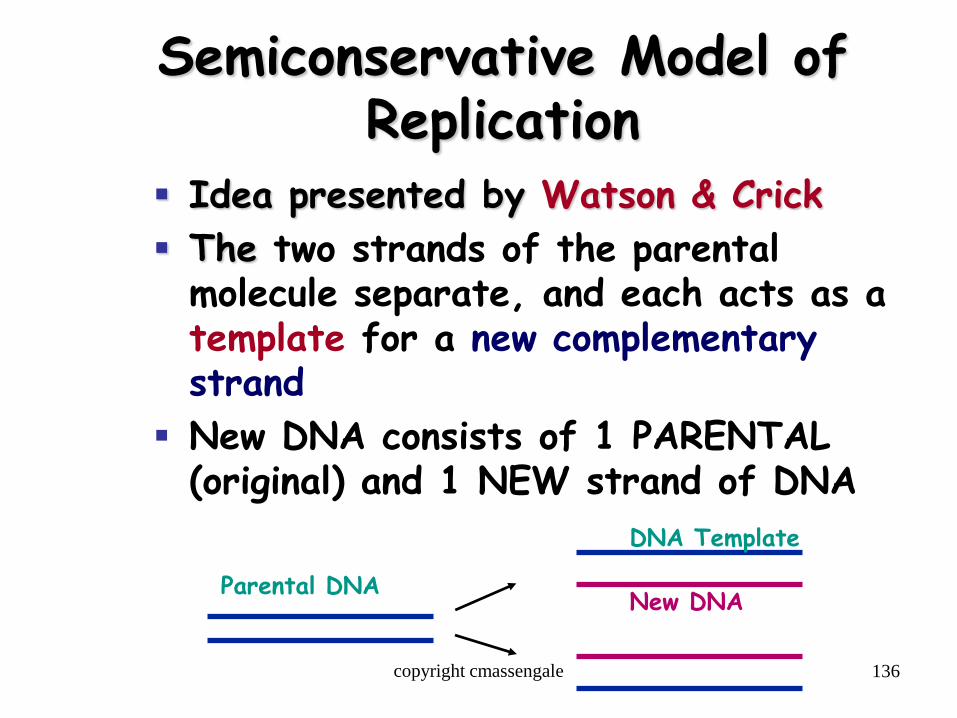
Semiconservative Model of Replication
Idea presented by Watson & Crick
The two strands of the parental molecule separate, and each acts as a template for a new complementary strand
New DNA consists of 1 PARENTAL (original) and 1 NEW strand of DNA
Parental DNA
DNA Template
New DNA
copyright cmassengale

137
DNA Replication
Begins at Origins of Replication
Two strands open forming Replication Forks (Y-shaped region)
New strands grow at the forks
ReplicationFork
Parental DNA Molecule
3’
5’
3’
5’copyright cmassengale

138
DNA Replication
As the 2 DNA strands open at the origin, Replication Bubblesform
Prokaryotes (bacteria) have a single bubble
Eukaryotic chromosomes have MANY bubbles
Bubbles Bubbles
copyright cmassengale
139
DNA Replication
Enzyme Helicase unwinds and separates the 2 DNA strands by breaking the weak hydrogen bonds
Single-Strand Binding Proteins attach and keep the 2 DNA strands separated and untwisted
copyright cmassengale
140
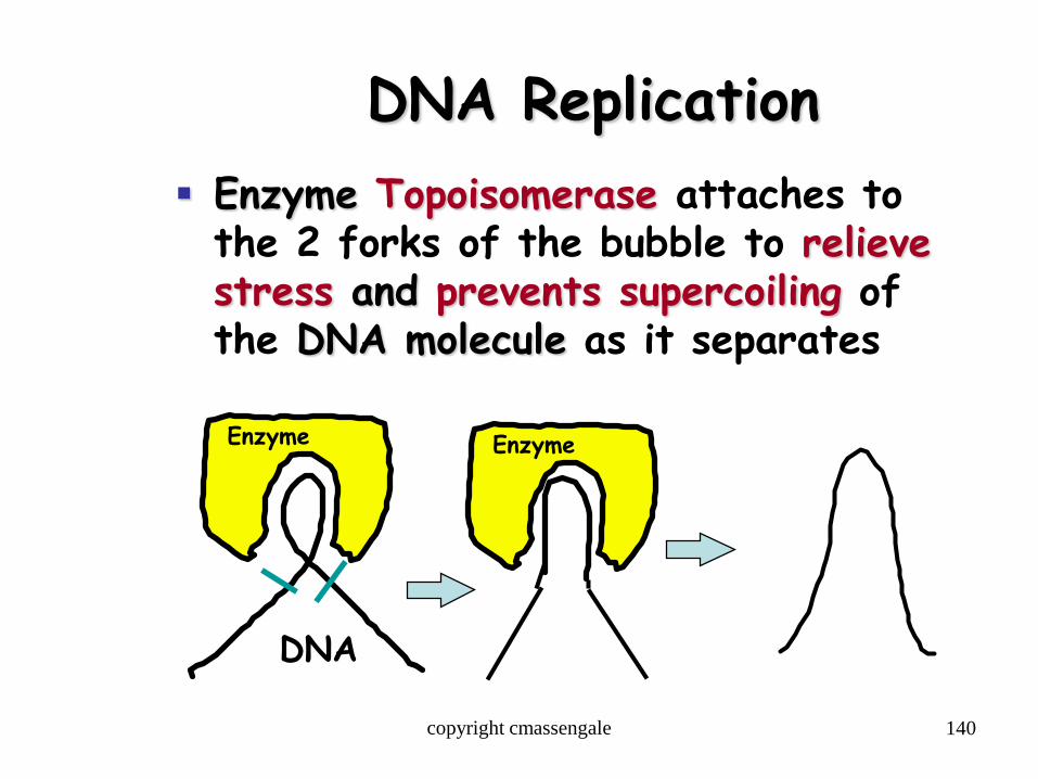
DNA Replication
Enzyme Topoisomerase attaches to the 2 forks of the bubble to relieve stress and prevents supercoiling of the DNA molecule as it separates
Enzyme
DNA
Enzyme
copyright cmassengale

141
DNA Replication Before new DNA strands can form,
there must be RNA primers present to start the addition of new nucleotides
Primase is the enzyme that synthesizes the RNA Primer
DNA polymerase can then add the new nucleotides
copyright cmassengale
142
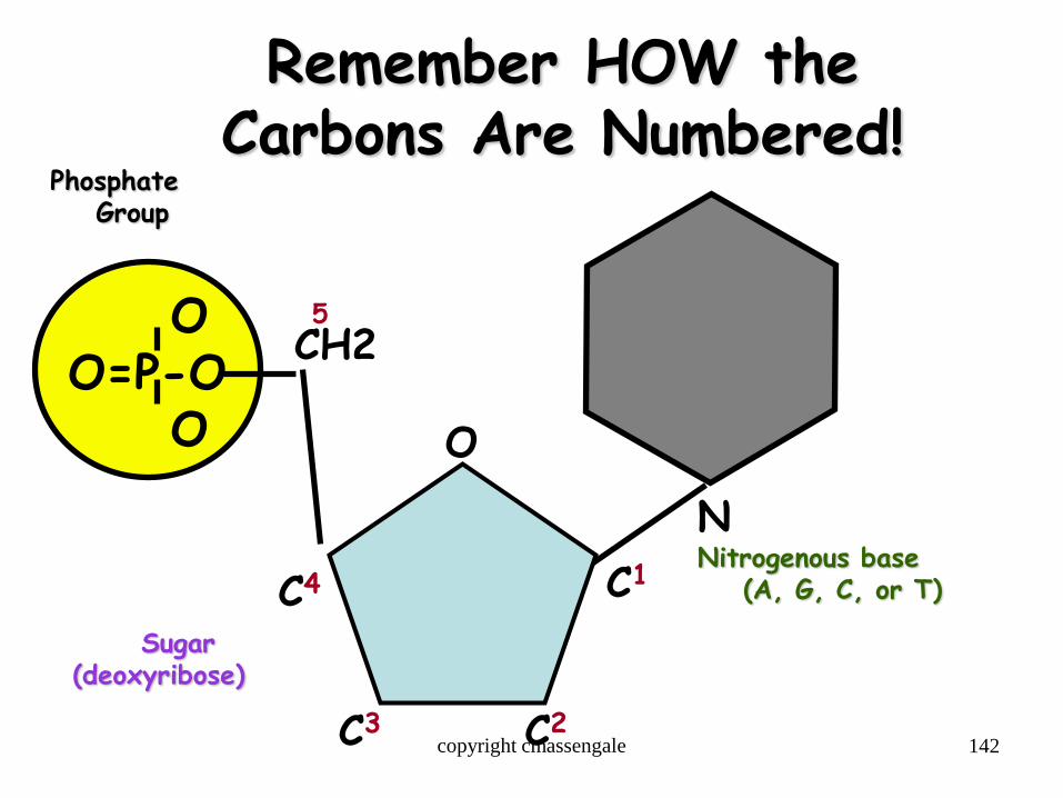
Remember HOW the Carbons Are Numbered!
OO=P-O
O
PhosphateGroup
NNitrogenous base
(A, G, C, or T)
CH2
O
C1C4
C3 C2
5
Sugar(deoxyribose)
copyright cmassengale
143
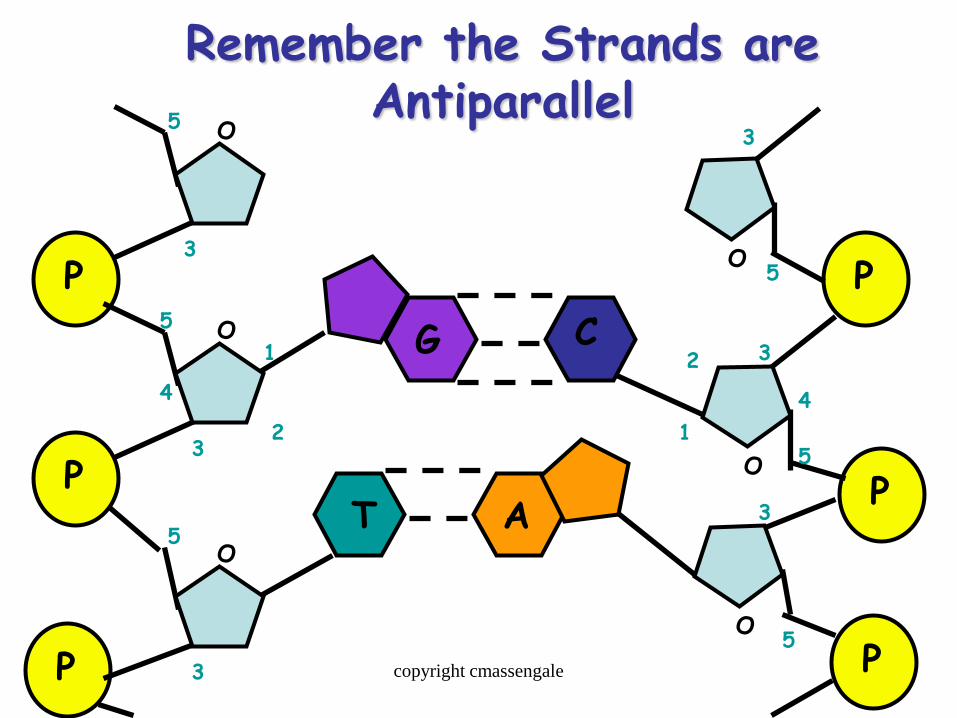
Remember the Strands are Antiparallel
P
P
P
O
O
O
1
23
4
5
5
3
3
5
P
P
PO
O
O
1
2 3
4
5
5
3
5
3
G C
T A
copyright cmassengale
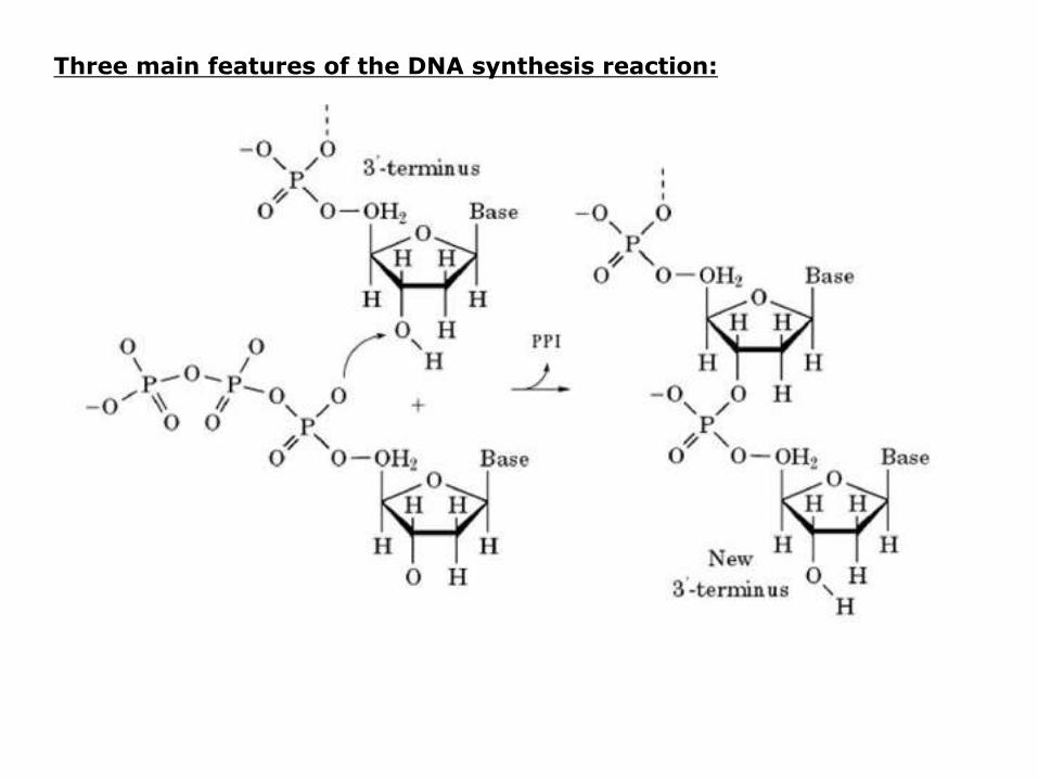
Three main features of the DNA synthesis reaction:
1. DNA polymerase catalyzes formation of phosphodiester bondbetween 3’-OH of the deoxyribose (on the last nucleotide) and the 5’-phosphate of the dNTP.
• Energy for this reaction is derived from the release of two of the three phosphates of the dNTP.
2. DNA polymerase “finds” the correct complementary dNTP at each step in the lengthening process.
3. Direction of synthesis is 5’ to 3’

DNA elongation (Fig. 3.3b):

146
Synthesis of the New DNA Strands
The Leading Strand is synthesized as a single strand from the point of origin toward the opening replication fork
RNAPrimerDNA PolymeraseNucleotides
3’5’
5’
copyright cmassengale
147
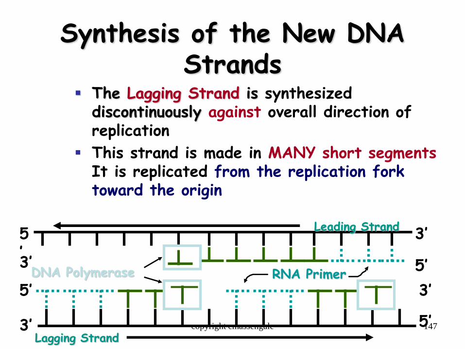
Synthesis of the New DNA Strands
The Lagging Strand is synthesized discontinuously against overall direction of replication
This strand is made in MANY short segmentsIt is replicated from the replication fork toward the origin
RNA Primer
Leading Strand
DNA Polymerase
5’
5’
3’
3’
Lagging Strand
5’
5’
3’
3’ copyright cmassengale

148
Lagging Strand Segments
Okazaki Fragments - series of short segments on the lagging strand
Must be joined together by an enzyme ligase
Lagging Strand
RNAPrimer
DNAPolymerase
3’
3’
5’
5’
Okazaki Fragment
copyright cmassengale

149
Joining of Okazaki Fragments
The enzyme Ligase joins the Okazaki fragments together to make one strand
Lagging Strand
Okazaki Fragment 2
DNA ligase
Okazaki Fragment 1
5’
5’
3’
3’
copyright cmassengale

150
Replication of Strands
Replication Fork
Point of Origin
copyright cmassengale
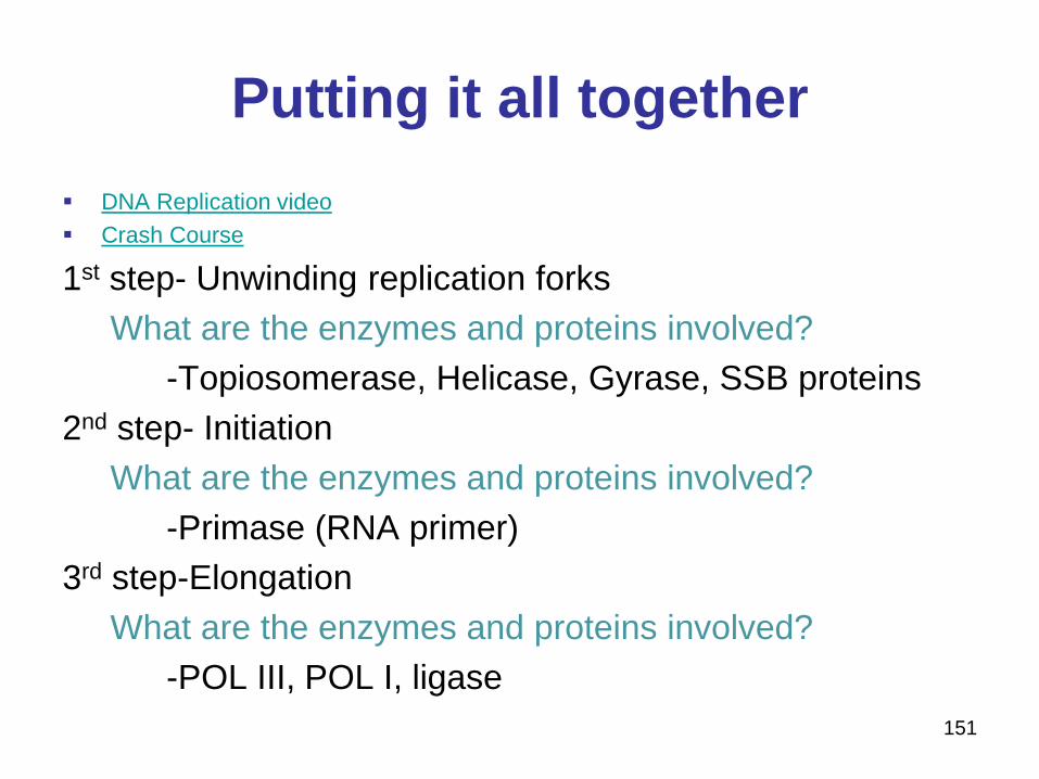
Putting it all together
DNA Replication video
Crash Course
1st step- Unwinding replication forks
What are the enzymes and proteins involved?
-Topiosomerase, Helicase, Gyrase, SSB proteins
2nd step- Initiation
What are the enzymes and proteins involved?
-Primase (RNA primer)
3rd step-Elongation
What are the enzymes and proteins involved?
-POL III, POL I, ligase
151
DNA Replication Errors
153
Proofreading New DNA
DNA polymerase initially makes about 1 in 10,000 base pairing errors
Enzymes proofread and correct these mistakes
The new error rate for DNA that has been proofread is 1 in 1 billion base pairing errors
copyright cmassengale
154
Causes of damaged DNA can be damage
Chemicals & ultraviolet radiationdamage the DNA in our body cells
Cells must continuously repair DAMAGED DNA
Excision repair occurs when any of over 50 repair enzymes remove damaged parts of DNA
DNA polymerase and DNA ligasereplace and bond the new nucleotides together
copyright cmassengale

155
Question:
What would be the complementary DNA strand for the following DNA sequence?
DNA 5’-CGTATG-3’
copyright cmassengale
156
Answer:
DNA 5’-CGTATG-3’
DNA 3’-GCATAC-5’
copyright cmassengale
10-157
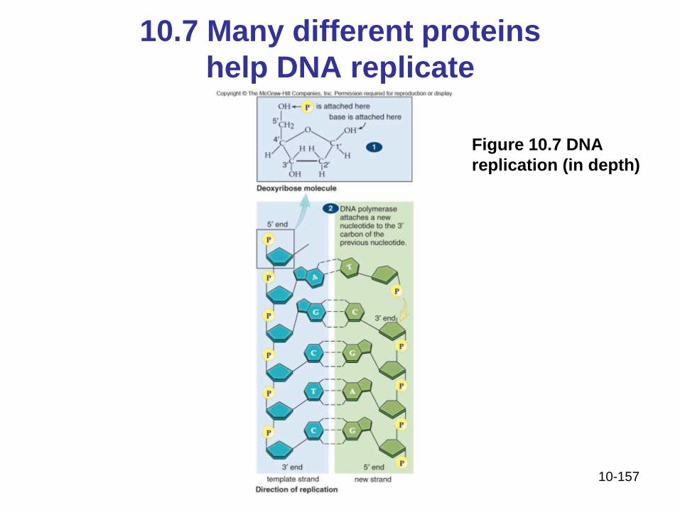
10.7 Many different proteins
help DNA replicate
Figure 10.7 DNA
replication (in depth)

10-158
10.7 Many different proteins
help DNA replicate
Figure 10.7 DNA
replication (in depth)
(Cont.)