the cerebral venous anatomy & management of postoperative venous...
TRANSCRIPT
THE CEREBRAL VENOUS THE CEREBRAL VENOUS ANATOMY & MANAGEMENT ANATOMY & MANAGEMENT
OF POSTOPERATIVE OF POSTOPERATIVE VENOUS INFARCTVENOUS INFARCT
Presented By : Dr.Rohit K Goel

Cerebral venous system : Cerebral venous system : Gross anatomyGross anatomy
C.V Drainage comprises of 3 segments:• 1. outer/superficial segment:-drains
scalp/muscles/tendons by scalp veins.• 2. intermediate segment:-draining
skull/diploe/duramater by diploe veins ,emissary veins, meningeal veins & duralvenous sinuses.
• 3. cerebral segment:-draining the brain proper by means of superficial cortical veins & deep venous system.

Cerebral venous system :Cerebral venous system :Gross anatomyGross anatomy
• Scalp veins:• Diploic veins:
The diploic veins & scalp veins can function as collateral pathways for venous outflow from I/C structures.
• Emissary veins:• The meningeal veins:• The bridging veins:

Cerebral venous system :Cerebral venous system :Gross anatomyGross anatomy
Cortical Veins– The superficial cortical veins are divided into three group based on
whether they drain the lateral, medial, or inferior surface of the hemisphere
– The cortical veins on the three surfaces are further subdivided on the basis of the lobe and cortical area that they drain.
– The largest group of cortical veins terminate by exiting the subarachnoid space to become bridging veins that cross the subdural space and empty into the venous sinuses in the duramater.
– A smaller group of cortical veins terminate by directly joining the deep venous system of the brain
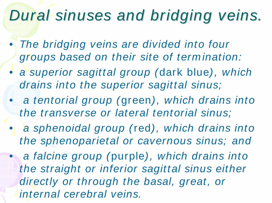
DuralDural sinuses and bridging veins.sinuses and bridging veins.
• The bridging veins are divided into four groups based on their site of termination:
• a superior sagittal group (dark blue), which drains into the superior sagittal sinus;
• a tentorial group (green), which drains into the transverse or lateral tentorial sinus;
• a sphenoidal group (red), which drains into the sphenoparietal or cavernous sinus; and
• a falcine group (purple), which drains into the straight or inferior sagittal sinus either directly or through the basal, great, or internal cerebral veins.
DuralDural sinuses and bridging sinuses and bridging veins.veins.
• The dural sinuses into which the cortical veins empty are :superior and inferior sagittal, straight, transverse,
tentorial,cavernous, sphenoparietal, sphenobasal, and sphenopetrosal sinuses.
• These sinuses form the terminal part of thesuperficial cortical venous system.

Major Major anastomoticanastomotic veinsveins.
• The vein of Trolard is the largest vein connecting he superficial sylvian vein with the superior sagittal sinus.
• The vein of Labbé is the largest vein connecting the superficial sylvian vein with the transverse sinus.
• The superficial sylvian vein drains the areas along the sylvian fissure and empties into the sinuses along the sphenoid ridge
A–D, different patterns. The dominant vein is darkly shaded.
• A, all three anastomotic veins are present, but the veins of Labbé and Trolard are dominant.
• B, dominant superficial sylvian and vein of Trolard. • C, dominant superficial sylvian vein. • D, dominant vein of Labbé.

Major Major anastomoticanastomotic veinsveins• According to DiChiro , the vein of
Labbé predominates in the dominant hemisphere nearly twice as often as it predominates in the nondominanthemisphere, and
• the vein of Trolard predominates in the nondominant hemisphere with approximately the same frequency

Cerebral venous system : Cerebral venous system : Gross anatomyGross anatomy
Deep cerebral veinsThe deep venous system of the brain consists of the internal cerebral,basal, and great vein and their tributaries.
These veins drain the deep white and gray matter surrounding the lateral and third ventricles and the basal cisterns.
The deep veins are divided into a ventricular group, composed of the veins draining the walls of the lateral ventricles, anda cisternal group, which includes the veins draining the walls of the basal cisterns.

• The deep cerebral veins may pose a major obstacle to operative approaches to deep-seated lesions, especially in the pinealregion, where multiple veins converge on the great vein.
• The fact that sacrifice of the major trunks of the deep venous system only infrequently leads to venous infarction with mass effect and neurological deficit is attributed to the diffuse anastomoses between the veins.
• Dandy noted that, not infrequently, one internal cerebral vein has been sacrificed without effect and, on a few occasions, both veins and even the great vein have been ligated with recovery without any apparent disturbance of function.
• On the other hand, injury to this complicated venous network has caused diencephalic edema, mental symptoms, coma, hyperpyrexia, tachycardia, tachypnea, miosis, rigidity of limbs, and exaggeration of deep tendon reflexes .
• Occlusion of the thalamostriate and other veins at the foramen of Monro may cause drowsiness, hemiplegia, mutism, and hemorrhagic infarction of the basal ganglia

Cerebral venous system : Cerebral venous system : functional anatomyfunctional anatomy
• The dural sinuses are designed to maintain the patency in the face of negative pressure.
• Their triangular shape make them relatively non compressible.
• Bridging vein entering the sinuses opposite the direction of sinus blood flow.
• There are no valves.• Their wall contain noradrenergic &
peptidergic fibres.

Cerebral venous system:Cerebral venous system:physiologyphysiology
Role in maintainance of ICP:-• of the 3 major intracranial
components,cerebral blood volume can change most rapidly.
• 70-80%of cerebral blood volume is located in venous system.
• Regulators of cerebral venous flow are:Pco2, sympathetic system
• At high ICP: sympathetic tone increase—veins constricts---cerebral bld. Vol. decrease---- tends to lower ICP

Cerebral venous system:Cerebral venous system:physiologyphysiology
Role in CSF absorption:-• CSF absorbed through arachnoid villi
into venous sinuses.• The flow across villus is
unidirectional & a pressure differential is required for flow.

The Cerebral venous systemThe Cerebral venous system--Applied aspectsApplied aspects
• The distribution of the superficial cortical veins is not as irregular and variable as is generally supposed, and their examination during the venous phase of the cerebral angiogram may prove helpful in localizing expanding lesions by revealing poor filling and displacement and alteration in the direction of flow.
• The fact that sacrifice of the individual cortical veins only infrequently leads to venous infarction, hemorrhage, swelling, and neurological deficit is attributed to the diffuse anastomoses between the individual cortical veins & b/n the sup. Cortical & the deep ventricular & cisternalveins

The Cerebral venous systemThe Cerebral venous system--Applied aspectsApplied aspects
• In Subtemporal approaches the various bridging veins encountered include the temporal, occipital, temporobasal, and occipitobasal veins and the vein of Labbé.
• Sacrifice of these veins, which pass from the lower part of the hemisphere to the transverse and tentorial sinuses, frequently causes some degree of venous infarction and edema of the temporal lobe
• . A contralateral hemiparesis, more marked in the face and arm than the leg, with an aphasia if the dominant hemisphere is affected, may follow occlusion of these veins

The Cerebral venous systemThe Cerebral venous system--Applied aspectsApplied aspects
• The ventricular veins provide valuable landmarks in directing the surgeon to the foramen of Monro and the choroidal fissure during operations on the ventricles.
• This is especially true if hydrocephalus, a common result of ventricular tumors, is present, because the borders between the neural structures in the ventricular walls become less distinct as the ventricles dilate.
• The thalamostriate vein is helpful in delimiting the junction of the caudate nucleus and the thalamus because it usually courses along the sulcus separating these structures.

The Cerebral venous systemThe Cerebral venous system--Applied aspectsApplied aspects
Surgical managementof sinus wall injuries:-
• 1.small laceration:-can often be closed with small interrupted sutures.
- upto 50%stenosis is well tolerated.2.Large tears/laceration:
- ligation in non critical sinuses like anterior 1/3rd
of SSS, non dominant transverse/sigmoid sinus, inferior saggital sinus & straight sinus.- autogenous venous graft in critical sinuses like posterior 2/3rd of SSS , torcula , dominant transverse/sigmoid sinus.

The Cerebral venous systemThe Cerebral venous system--Applied aspectsApplied aspects
Control of bleeding from venous sinuses:-
• Digital pressure in case of small h”rrhage.• Intraluminal balloon occlusion using fogarty
catheter.• Rarely a shunt fashioned from pediatric
endotracheal tube preoperatively, can be used to provide control of bleeding as well as diversion of blood.

The cortical venous systemThe cortical venous system--Applied aspectsApplied aspects
• VENOUS LACUNAE
•The lacunae may extend along the medial extent of the hemisphere adjacent to the falx and as far as 3 cm laterall over the convexity.
•Entering or occluding a lacuna at operation does not necessarily result in occlusion of the cortical veins or the superior sagittal sinus because most of the veins course deep to the lacunae and usually empty directly into the sinus.

The cortical venous systemThe cortical venous system--Applied aspectsApplied aspects
• In opening the dura mater adjoining the superior sagittalsinus, one should attempt to preserve the meningeal sinuses, which may arise as far as 2.5 cm lateral to the superior sagittal sinus (Fig.).
• These sinuses may receive the terminal end of numerous cortical veins.
• In removing a parasagittal tumor deep to these sinuses, the dura is opened along the edges of the sinus while preserving the sinus’ proximal junction with the cortical veins and its distal junction with the superior sagittalsinus.
• The tumor is then separated from the lower margin of the meningeal sinus without sacrificing the sinus.

The cortical venous systemThe cortical venous system--Applied aspectsApplied aspects
• The operative approach directed along the falx toward the anterior part of the corpus callosum may require the sacrifice of a bridging vein to the superior sagittal sinus.
• Occasionally,the corpus callosum may be reached in the area between theanterior and posterior frontal veins without
sacrificing any bridging veins

The cortical venous systemThe cortical venous system--Applied aspectsApplied aspects
• Obliteration of the bridging veins to the superior sagittal sinus in the region of the precentral, central, or postcentral gyrifrequently causes a contralateralhemiparesis that is more prominent in the lower than the upper extremity and is usually transient.
• Spontaneous occlusion of the veins in this region causes a hemiparesis that is commonly accompanied by headache and seizures

The cortical venous systemThe cortical venous system--Applied aspectsApplied aspects
• In occipital transtentorial operative approach, the occipital pole can usually be retracted from the straight sinus and the junction of the falx and the tentorium without sacrificing any veins to the superior sagittal or transverse sinuses (Fig.)
• The superior sagittal sinus is commonly devoid of bridgingveins in the area just in front of the torcular herophili

PostopPostop. Venous. Venous InfarctInfarctEtiologies:-1.inadvertant coagulation/tear of bridging
veins/meningeal sinuses during cranial surgeries.
2.resecton of malignant tm/meningiomainvading major venous sinus
3.repair of traumatic dural venous sinus injuries.
4.even following cannulation of neck veins5.intraop/postop dehydration.

PostopPostop. Venous infarct. Venous infarct
Various surgeries implicated in literature are:-
• Acoustic neuroma surgeries
• Clipping of ACA aneurysm
• Tumors firmly adherent to cortex
• Open surgical excision of cortex cyst of 3rd
ventricle.

PostopPostop. Venous infarct. Venous infarct• Pathophysiology:-
-extensive collateral in venous system leads to compensation in early stage of venous occlusions.
Schaller B in Cerebrovasc Dis. 2004has summarised the pathophysiological changes as follow:
1. Venous occlusion elevated cerebral venous pressure dilated venous/cap. beds
interstitial edema develops.2. Increased CSF production & decreased CSF
absorption3. Ultimately rupture of venous structure with
hematoma formation.

PostopPostop. Venous infarct. Venous infarct
Presentation:-Nakase et al in Acta Neurochir (Wien). 2005 has found that there are 2 types of infarcts:
• Severe onset[severe type]• Gradual onset[mild type]
The former needs immediate treatment from the intra-operative period onward, and the prevention of the ongoing venous thrombosis is essential in the latter.

PostopPostop. Venous infarct. Venous infarctVaried Clinical Features : acc. of specific
sites:-• Cavernous sinus thrombosis: chemosis,
proptosis & painful ophthalmoplegia.• Deep venous system: diencephalic dysfunction
& death, abulia, disorientation, vertical gaze paresis etc.
• Midportion of SSS:- spastic hemiparesis / quadriparesis
- raised ICT features• Posterior portion of SSS:-visual field defects
-cortical blindness-coma
• Diffuse sinus thrombosis:- sympt/signs of acutely raised ICT with herniation.

PostopPostop. Venous infarct. Venous infarctDiagnostic investigations:-1. NCCT:-postive delta sign in acute SSS thrombosis.
-haemorrhage in hemisphere.2. CECT:-negative delta sign in acute SSS thrombosis
-gyral/tentorial enhancement -cord sign in cortical venous thrombosis-hemispheric haemorrhage.
3. MRI:-investigation of choicespecial sequences used:-Gradient refocused echo {GRE}
-MRA-DWI
Ogami et al in No To Shinkei. 2001 has told that acute phase of venous infarct [diffusion hyperintensity] can be diagnosed by DWI.
4. IADSA:-detects venous sinus thrombosis as- filling defects.

PostopPostop. Venous infarct. Venous infarctTHERAPY:-
1. General measures:-avoid dehydration/hypotension/hyperglycemia-seizure prophylaxis.
2. Symptomatic treatment of raised ICT:--head elevation-hyperventilation-osmotic agent-mannitol / furosemide-ventricular drainage for hydrocephalous
3. Regular clinical & radiological follow up:-most of the venous infarcts will improve with
these measures & can be followed up. Thrombosedchannels shows recanalisation & collateral channels open up.

PostopPostop. Venous infarct. Venous infarct4.Role of anticoagulants::
– anticoagulants {heparin & acitrom} inhibits the progression of thrombosis but has the inherent danger of increasing haemorrhagiccomplications.
-Sepulveda JM et al in Neurologia. 2004 has advocated the use of anticoagulant Rx in h”gic brain infarcts due to large venous sinus thrombosis.

PostopPostop. Venous infarct. Venous infarct• Role of surgical treatment:-1.Ventricular drainage in cases
presenting as acute hydrocephalous.2.Decompressive hemicraniectomy:-if
severe mass effect is present & patient condition is deteriorating inspite of all medical measures.