the cerebro- placental ratio as a prognostic factor of
TRANSCRIPT

The Cerebro- Placental Ratio as a Prognostic
Factor of Fetal Outcome in Patients with
Hypertensive States of Pregnancy in Third Trimester
at Kenyatta National Hospital
Dissertation to be submitted in Part Fulfillment for the award of the
Degree of Masters of Medicine in Diagnostic Imaging of University of
Nairobi.
By
Dr. Parmar Linal Parshuram - MBChB (University of Nairobi)
Department of Diagnostic Imaging and Radiation Medicine
University of Nairobi 2013

ii
DECLARATION I, Dr. Parmar Linal Parshuram, declare that this dissertation has not been submitted for
another degree in this or any other University or Institution of Higher learning and that the views
expressed herein are mine unless otherwise stated, and where such has been the case
acknowledgement or reference has been quoted.
Signature ……………………………………………………...Date……………….
Approval by Supervisors
This research has been submitted with my approval as a University supervisor
Dr. Mwango G.N. MBChB (UON), Mmed Diagnostic Imaging (UON)
Lecturer Department of Diagnostic Imaging and Radiation Medicine, University of Nairobi
Signature………………………………………………… Date ……………….
Dr. Wambugu M.N. MBChB (Makerere University), Mmed Diagnostic Imaging (UON)
Senior Lecturer Department of Diagnostic Imaging and Radiation Medicine, University of
Nairobi
Signature………………………………………………… Date ……………….
Dr. Ong’ech J.O. MBChB (UON), Mmed Obstetrics and Gynecology (UON), MPH
(Tulane)
Lecturer Department of Obstetrics and Gynecology, University of Nairobi
Signature………………………………..……………… Date …………………

iii
ACKNOWLEDGEMENT
I am primarily thankful to the almighty God for the guidance and the direction through my life
and career.
I thank my beloved ones for their constant support, prayers and patience.
I am humbly grateful to the chairperson department of diagnostic imaging and radiation
medicine, university of Nairobi , my supervisors Dr Mwango G.N, Dr Wambugu M.N and Dr
On’gech J.O, University of Nairobi, for taking interest in my research and offering their
constant support and guidance.
I thank all the radiology residents, Obstetrics and Gynaecology residents, interns and
nurses in labor ward, Mrs. Zam zam and Hilda who all facilitated my data collection.
My heartfelt gratitude to Mr. Paul Mwai, who has been constantly helpful with data analysis
and formatting the research.

iv
Table of Contents DECLARATION ......................................................................................................................................... ii
Approval by Supervisors ...................................................................................................................... ii
ACKNOWLEDGEMENT ............................................................................................................................ iii
List of Tables .......................................................................................................................................... vi
List of Figures ........................................................................................................................................ vii
LIST OF ABBREVIATIONS ........................................................................................................................viii
ABSTRACT ............................................................................................................................................... 1
Introduction ........................................................................................................................................ 1
Study Objective ................................................................................................................................... 1
Study Design and Methodology ........................................................................................................... 1
Problem Statement ............................................................................................................................. 1
Findings .............................................................................................................................................. 1
Conclusion .......................................................................................................................................... 2
BACKGROUND ......................................................................................................................................... 3
Introduction ........................................................................................................................................ 3
Literature Review ................................................................................................................................ 5
Hypertensive States in Pregnancy .................................................................................................... 5
Fetal Monitoring .............................................................................................................................. 6
Doppler Ultrasound ......................................................................................................................... 8
Functional Doppler Studies ............................................................................................................ 12
STUDY DESIGN....................................................................................................................................... 19
Problem statement ............................................................................................................................ 19
Study Objective ................................................................................................................................. 19
Broad Objective ............................................................................................................................. 19
Specific Objectives ......................................................................................................................... 19
Study Justification.............................................................................................................................. 20
Research Question............................................................................................................................. 20
Hypothesis......................................................................................................................................... 20
Design and Methodology ................................................................................................................... 21
Sampling ........................................................................................................................................ 21
Method ......................................................................................................................................... 21
Sample size consideration .............................................................................................................. 23
Data management and statistical analysis ...................................................................................... 23

v
Ethical Considerations.................................................................................................................... 24
STUDY RESULTS ..................................................................................................................................... 25
DISCUSSION........................................................................................................................................... 36
CONCLUSION ......................................................................................................................................... 41
RECOMMENDATIONS ............................................................................................................................ 42
APPENDIX A ........................................................................................................................................... 43
Research Consent Form .................................................................................................................. 43
APPENDIX B ........................................................................................................................................... 44
Questionnaire ................................................................................................................................ 44
APPENDIX C ........................................................................................................................................... 46
Budget ........................................................................................................................................... 46
APPENDIX D........................................................................................................................................... 47
Study Database.................................................................................................................................. 47
BIBLIOGRAPHY ...................................................................................................................................... 48

vi
List of Tables Table 1 Demographic Characteristics of the study population .......................................................... 27 Table 2 Clinical Observations ......................................................................................................... 28 Table 3 Clinical Management and Fetal Outcome ........................................................................... 28 Table 4 Correlates of Fetal Outcome among hypertensive pregnant mothers .................................... 29 Table 5: Logistic regression for the correlates of Fetal Outcome among hypertensive pregnant
mothers ................................................................................................................................................. 30 Table 6 Correlates of Infant’s APGAR (5 minutes) Score of mothers with hypertensive disorder
during the index pregnancy .................................................................................................................... 31 Table 7 Logistic regression for the correlates of Infant’s APGAR (5 minutes) Score among mothers
with hypertensive disorder during the index pregnancy .......................................................................... 32 Table 8 Correlates of Infant’s birth weight among mothers with hypertensive disorder during the
index pregnancy .................................................................................................................................... 33 Table 9 Logistic regression for the correlates of Infant’s birth weight among mothers with
hypertensive disorder during the index pregnancy .................................................................................. 34 Table 10 Comparison of Prognostic Odds of neonate APGAR Score using CPR vs UA RI.................... 34 Table 11 Severity of PET and CPR……………………………………………………………………..35

vii
List of Figures Figure 1 Typical Waveform of umbilical artery and Calculation of Doppler Indices. (59) .................. 11 Figure 2 Waveforms of absent end diastolic flow (68)......................................................................... 11 Figure 3 Waveforms of reversed end diastolic flow (68) ...................................................................... 11 Figure 4 Characteristic Saw-tooth Appearance of Umbilical Arterial Flow (59)................................... 13 Figure 5 Reference Curves for the UARI (68) ..................................................................................... 13 Figure 6 Color Doppler View of middle cerebral arteries and Typical Doppler Waveform. (8) ............ 15 Figure 7 Reference Curves for MCARI (68) ....................................................................................... 15 Figure 8 Reference Curves for the Cerebro-Placental Ratio (68).......................................................... 18 Figure 9 Ultra-sound scan showing MCA RI of 0.683 ......................................................................... 25 Figure 10 Ultra-sound scan showing UA RI of 0.967 and absent end diastolic flow............................... 26 Figure 11 Ultra-sound scan showing MCA RI of 0.782 ......................................................................... 26 Figure 12 Ultra-sound scan showing UA RI of 0.596 ............................................................................ 27

viii
LIST OF ABBREVIATIONS
ABO Blood groups A, B and O system
AFV Amniotic Fluid Volume
ANC Antenatal Clinic
BP Blood Pressure
BPPS Biophysical Profile Score
CI Confidence Interval
CPR Cerebro-Placental ratio
CTG Cardiotocography
C/U RI Cerebral/umbilical resistive index
CW Continuous Wave
EDF End Diastolic Flow
EDV End Diastolic Velocity
ERC Ethics and Research Committee
FANC Focused Antenatal Care
FHR Fetal Heart Rate
FKC Fetal Kick Chart
Hb Haemoglobin
HIV Human Immunodeficiency Virus
HSP Hypertensive States of Pregnancy
IQR InterQuartile Range
IUFD Intrauterine Fetal Death
IUGR Intrauterine Growth Restriction
KNH Kenyatta National Hospital
KNH/UoN-ERC Kenyatta National Hospital/University of Nairobi-Ethics and
Research Committee
MCA Middle Cerebral Artery
MCA RI Middle Cerebral Artery Resistive Index
MCA PI Middle Cerebral Artery Pulsatility Index
MCH Maternal and Child Health clinic

ix
Mg/dl Milligrams per deciliter
MHz Mega Hertz
mmHg Millimeters of Mercury
NICU Neonatal Intensive Care Unit
NBU New Born Unit
NRFS Non reassuring Fetal Status
NST Non Stress Test
OR Odds ratio
PET Pre-eclamsic Toxaemia
PE Pre-eclampsia
PI Pulsatility Index/Gosling index
PSV Peak Systolic Velocity
PW Pulsed Wave
RI Resistive Index/Pourcelot index
SB Still Birth
S/D Systolic/ Diastolic Ratio
SD Standard Deviations
SVD Spontaneous Vaginal Delivery
UEC Urea, Electrolytes and Creatinine
UoN University of Nairobi
UA Umbilical Artery
UA RI Umbilical Artery Resistive Index
UA PI Umbilical Artery Pulsatility Index
U/S Ultrasound scan or Ultrasonography
1
ABSTRACT
Introduction Hypertensive states of pregnancy affect maternal and fetal circulations (1). The pathophysiology
can be assessed safely and non-invasively by Doppler `ultrasound using arterial Doppler indices of
umbilical and middle cerebral arteries thus attaining the cerebro- placental ratio (ratio of the middle
cerebral artery resistive index over that of the umbilical artery) (2)
Study Objective This study aimed at determining the role of the cerebro-placental ratio (CPR) as a prognostic factor of
fetal outcome in patients with hypertensive states of pregnancy delivered at or after 32 weeks of gestation
by dates.
Study Design and Methodology A prospective cohort study carried out at the Kenyatta National Hospital (KNH) over a period of nine
months. Gravid patients at least 32 weeks gestations by dates were recruited from labor ward after
obtaining informed consent. Consecutive sampling method was used. The cerebro-placental ratio was
then calculated. Follow up for fetal outcome, the 5 minute APGAR score and birth weight was made.
Ethical Approval was sought from and granted by the Kenyatta National Hospital/University of Nairobi-
Ethics and Research Committee (KNH/UoN-ERC).
Problem Statement Fetuses at greater risk of adverse perinatal outcome will have a high resistance umbilical artery waveform
and a low resistance waveform of the middle cerebral artery waveform thus a CPR <1
Findings A total of 160 patients were recruited into the study.
Median age was 28 years. Sixty two percent (62%) were primiparous. Median gestation at
admission was 34 weeks while at sonography, the average gestation was 31 weeks
Twenty nine percent (29%) had an abnormal CPR (<1.0) while seventy eight percent (78%) had
a normal CPR (≥1.0). Seventy eight percent (78%) were delivered via caesarean section while
twenty two percent (22%) were delivered vaginally.
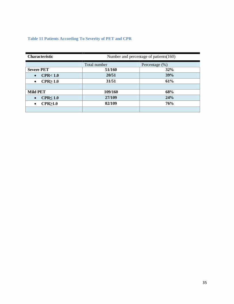
51 out of 160 (32%) had severe PET out of which 39% had CPR <1.0 and 61% had CPR ≥1.0.
while 109 out of 160 patients(68%) had mild PET out of which 24% had CPR <1.0 and 76% had
CPR ≥1.0.
Still births were 12.5 times more likely in mothers with CPR <1.0 than those with CPR ≥ 1.0,
An APGAR score < 7 was 66 times more often in mothers with CPR < 1.0 than mothers with
CPR≥ 1.0. Low birth weight was 4.7 times more likely among mothers with CPR < 1.0.as
compared to those with mothers with CPR≥1.0 (95% CI 2, 11.1; p< 0.001).
An APGAR score < 7 was 66 times more likely among neonates delivered vaginally as
compared to those born via caesarean section(95% CI 1.3, 23; p=0.02)
Still births were 14.5 times more often than among neonates born vaginally as compared to those
born via caesarean section (95% CI 3, 84; p<0.001).

2
The prognostic OR for CPR was 12.5 for live births (95% CI 2, 74; p=0.005), 66 for APGAR
score <7 (95% CI 13, 340; p< 0.001) and 4.7 for low birth weight (95% CI 2, 11.1; p< 0.001)
and 1.1 (95% CI 0.9, 1.4; p=0.327).
The prognostic OR of BPPS for live births was 1.7 (95% CI 1.1, 2.5; p=0.009), 1.3 for APGAR
score <7 (95% CI 0.9, 1.9; p=0.07) and 1.1 for low birth weight (95% CI 2, 11.1; p< 0.001) and
1.1 (95% CI 0.9, 1.4; p=0.327).
Conclusion CPR is significantly predictive of adverse perinatal outcome when used to monitor mothers with
hypertensive states of pregnancy than UA RI or BPPS used alone. CPR was predictive of adverse
perinatal outcome (live birth, APGAR score and low birth weight). Caesarian section should be
the recommended mode of delivery for hypertensive mothers.
3
BACKGROUND
Introduction Hypertensive states of pregnancy (HSP) are defined as conditions in which there is development
of new arterial hypertension in gravid women after 20 weeks of gestation without the presence of
protein in the urine (5). The HSP include pre-eclampsia/eclampsia (PE), chronic hypertension,
and chronic hypertension with superimposed pre-eclampsia and gestational hypertension (6).
HSP causes deficient infiltration of spiral arteries by trophoblast, failure of the spiral arteries to
convert into utero-placental arteries and therefore the tenfold increase in uterine perfusion occurs
and interferes with blood flow in the uterine artery on the maternal side (7).
On the fetal side, HSP cause poor vascularization of terminal villi, villous stromal hemorrhage
and hemorrhagic endovasculitis or even obliteration of stem villi (7).
Doppler ultrasound allows safe, non invasive and rapid investigation of the feto-placental
circulation (7) using three arterial indices devised (Pourcelot 1974) (59) to assess quality of flow
in vessels.
These indices are Resistive Index (RI)/Pourcelot index, Pulsatility Index (PI)/Gosling index and
Systolic/Diastolic ratio (S/D ratio)/Stuart and Drumm (2). The Resistive Index (RI); also known
as the Pourcelot index (Pourcelot 1974) is an indicator of resistance of an organ to perfusion. It is
the most commonly used index as it is easier to calculate and is reflective of vascular resistance
(2). Using Doppler ultrasonography, it is calculated by measuring peak systolic velocity (PSV)
and subtracting end diastolic velocity (EDV) from it and then dividing it by peak systolic
velocity (2.)Therefore, the Resistive Index is calculated as follows:
Peak Systolic Velocity PSV – End Diastolic Velocity EDV
Peak Systolic Velocity PSVRI
The Umbilical Artery Resistive Index (UA RI) reflects downstream placental vascular resistance
which has been found to correlate with intrauterine growth restriction (IUGR) and multisystem
effects of placental deficiency (8). Abnormalities of the umbilical artery (UA) Doppler
waveform are progressive beginning with reduction, loss and finally a reversal of diastolic
flow. When the blood flow pattern in the Umbilical Artery (UA) becomes abnormal, the
differentiation of fetal status requires Doppler information from systemic vessels (8) and one of
4
the systemic vessels that can be investigated is the Middle Cerebral Artery (MCA) which is used
for assessment of the fetal cerebral circulation. .It is easy to identify the MCA as the major
branch running antero-laterally from circle of Willis towards lateral edge of orbit (8 and 10). RI
of both the Middle Cerebral Artery (MCA) and Umbilical Artery (UA) can be obtained. A
comparison giving the cerebro-placental ratio (CPR).
A ratio >1.0 indicates preferential flow to vital structures like brain, heart and adrenal glands and
is therefore considered normal while that <1.0 is indicative of high resistance in utero-placental
circulation via Umbilical Artery (UA) Doppler waveform and inadequate supply to fetal brain
via Middle Cerebral Artery (MCA) Doppler waveform known as centralization and is considered
to be predictive of adverse perinatal outcome. (8).
The cerebro-placental resistive index ratio is the ratio of the MCA R.I to the UA R.I. It is
calculated as shown in the formula below.
Resistive index of middle cerebral artery MCA RI
Resistive index of umbilical artery UA RICPR
The brain sparing effect is seen when circulatory adaptation occurs with chronic hypoxia in form
of cerebral vasodilatation to preserve blood flow to the brain. This is demonstrated by a lower
value of MCA RI relative to gestational age and UA RI. Its disappearance is a critical event and
precedes fetal death (8).Also studies done have shown the CPR to be predictive of adverse
perinatal outcome. Doppler studies of multiple feto-placental vessels can be used to monitor
compromised fetus predicting adverse perinatal outcome and assisting in optimal time of
delivery (11-15). At the Kenyatta National Hospital (KNH), the biophysical profile score (BPPS)
and umbilical artery resistive index (UA RI) are used in management of patients with HSP to
determine method and optimal time of delivery. However there had been no study carried out at
locally to assess the role of the CPR as a prognostic factor in the prediction of fetal outcome in
patients with HSP in the third trimester.
This aim of this study was to determine the role of CPR in antenatal fetal monitoring in patients
with HSP so as to assess the fetus at greater risk of adverse perinatal outcome and thereby
greatly improve management and reduce neonatal mortality and morbidity.

5
Literature Review
Hypertensive States in Pregnancy
The incidence of HSP varies between 5-10% in different communities.(15) In our set up, Mati
1975 found an incidence of 7.1% with a range of between 1.5-9% (15) The prevalence of pre-
eclampsia in KNH in 1992 was found to be 5.4% (16). HSP are classified into 4 categories, as
recommended by the National High Blood Pressure Education Program Working Group (NHEP)
on High Blood Pressure in Pregnancy (17) and these are pre-eclampsia/eclampsia, chronic
hypertension, chronic hypertension with superimposed pre-eclampsia and gestational
hypertension. The predisposing factors to HSP include primigravidas, black race, and maternal
age below 20 or above 35 years, low socio-economic status and multiple gestations (57).
Pre-eclampsia is hypertension associated with proteinuria and edema occurring primarily in
nulliparas after the 20th gestational week and most frequently near term. Recent data supports the
elimination of edema as a diagnostic criterion because it is common in normal pregnancy and
pre-eclampsia can occur without edema which is called the dry type (18).Hypertension is defined
as blood pressure (BP) equal to or greater than 140/90mmHg. A rise of 30mmHg or more in
systolic blood pressure or a rise of 15mmHg or more in diastolic blood pressure in 2 occasions 6
hours apart is considered abnormal. A mean arterial blood pressure of >105mmHg or an increase
in mean arterial pressure of 20mmHg is also considered abnormal (1).
Proteinuria is defined as the excretion of 300 mg or more in a 24 hour specimen or 30 mg/dL in a
random specimen or >1+ on the dipstick. Heavy proteinuria is defined as the excretion of 2g or
more in a 24 hour specimen or >2+ on dipstick (1).The criteria for superimposed pre-eclampsia
are worsening hypertension (30mmHg systolic or 15mmHg diastolic above the values before 20
weeks gestation) together with non-dependent edema or proteinuria (1). Gestational hypertension
is divided into transient hypertension if pre-eclampsia is present at time of delivery and pressure
returns to normal by 12 weeks postpartum or chronic hypertension if the pressure remains
elevated beyond 12 weeks postpartum (1). Pre-eclampsia can be mild or severe. Pre-eclampsia
can be defined as Blood Pressure(BP) >160/110 mmHg on 2 occasions 6 hours apart and
proteinuria exceeding 2 grams in 24 hour period or 2-4+ on dipstick. (1, 18,19). Pre-eclampsia is
a disorder of placental dysfunction leading to a syndrome of endothelial dysfunction associated

6
with vasospasm. The pathology demonstrates evidence of placental insufficiency with associated
abnormalities such as diffuse placental thrombosis, an inflammatory placental decidual
vasculopathy, and/or abnormal trophoblastic invasion of the endometrium. Thus abnormal
placental development or placental damage from diffuse micro thrombosis is thought to be
central to the development of this disorder (21). The widespread endothelial dysfunction may
manifest as a maternal syndrome, fetal syndrome, or both. On the maternal side, it causes
deficient infiltration of the spiral arteries by the trophoblast, failing to convert it into utero-
placental arteries. Subsequently, the tenfold increase in uterine perfusion that occurs in normal
pregnancy, fails to occur. This interferes with blood flow in the uterine artery (21) and therefore
the mother may manifest with dysfunction of multiple organ systems, including the central
nervous, hepatic, pulmonary, renal, and hematological systems. The Endothelial damage leads to
pathologic capillary leak that can present in the mother as rapid weight gain, non-dependent
edema (face or hands), pulmonary edema, haemo-concentration, or a combination thereof
(21).On the fetal side, there is poor vascularization of the terminal villi, villous stromal
hemorrhage, hemorrhagic endovasculitis or obliteration of the stem villi. This results in a
decrease in fetal perfusion which manifests clinically as non-reassuring fetal heart rate testing,
low scores on a biophysical profile, oligohydramnios, or as intrauterine growth restriction
(IUGR) (22). Thus HSP can cause harm to the gravid patient and also to the fetus (22).It is one
of the leading causes of maternal and perinatal morbidity and mortality (23-29). HSP is a major
determinant of the timing of admission, timing of delivery and mode of delivery (30-33) It is
therefore vital to have reliable maternal and fetal monitoring methods that can be used
antenatally so as to reduce maternal and perinatal morbidity and mortality.
Fetal Monitoring At KNH, focused antenatal care (FANC) system is used. A gravid mother who comes to the
Maternal and Child Health clinic (MCH) or ante-natal clinic (ANC) is followed up at four
divided visits throughout her pregnancy.
At each ante-natal visit, the following is usually done
Checking of maternal weight
Blood pressure(BP) measurement
Urinalysis(UA)-for proteinuria or leucocytes
7
Checking the hemoglobin(Hb) level
Check the blood group(ABO and Rh status)
Check the patient’s serostatus (HIV status)
Assessment of fundal height and correlating it with gestation by dates.
Assessment of fetal presentation and lie.
Assessment of fetal heart rate using Pinnard fetoscope
Administration of ferrous and folic acid
Administration of tetanus toxoid (35)
For patients with high risk pregnancies, ante-natal follow up is then tailored to the individual
needs of the patient. The standard work up for patients with pre-eclampsia includes the
following:
Hemoglobin(Hb) and haematocrit level(Hct)
Serum urea, creatinine and uric acid level.(U/E/C)
Transaminase, albumin and lactic acid dehydrogenase levels.
Blood smear
Coagulation profile
Some of the tests mentioned above rule out haemolysis and hepatic involvement. They are also
useful for monitoring progression and specific organ involvement (36). Assessment of fetal well
being includes daily monitoring of fetal movements with more specific tests like non stress
test(NST), ultrasonographic assessment of fetal movements and amniotic fluid volume (AFV)
(36). Patients with mild pre-eclampsia may be followed up as outpatients bi-weekly and admitted
if this is not possible. The patient should be warned on the danger signs of pre-eclampsia and
eclampsia. Extra rest is advised and no dietary restrictions are necessary (36). When the fetal
well being/fetal status are in doubt, closer monitoring is required. There are various methods of
doing this.
Fetal kick chart (FKC) is a chart invented by Sadowsky in 1977 and the mother fills in
the chart over a period of 12 hours daily for a period of ten days and in this period, less
than 10 fetal kicks are considered abnormal (37).Studies have shown that the fetal kick
8
chart is not an accurate method of monitoring fetal status as a mother may exaggerate
kicks (38).
Cardiotocography (CTG) is an electronic method of simultaneously recording fetal heart
rate, fetal movements and uterine contractions to identify presence of fetal hypoxia (39).
Poor standardization in interpretation and disagreement about appropriate interventions
has resulted in a lack of reliable and valid data to demonstrate efficacy of CTG
monitoring (40).
Ultrasound scan (U/S) is performed to assess fetal presentation, placental position, and
fetal viability, attain fetal heart rate, ultrasonographic gestational age, and perform BPPS
and Doppler indices of UA and MCA (37). The BPPS has been found to be reflective of
the fetal condition (40, 41) and is better for detection of acute fetal distress as most of its
parameters measure acute asphyxia (42). The Umbilical Artery Resistive Index (UA RI)
is reflective of utero-placental circulatory status. The Doppler waveform changes have
been found to have good correlation with fetal compromise giving an earlier warning of
fetal distress than CTG or BPPS (44). It is however not reflective of the circulatory status
of fetal brain which requires assessment of Middle Cerebral Artery (MCA).
Doppler Ultrasound
Historical Perspective
The Doppler Shift Principle was discovered by Johann Christian Andreas Doppler in 1842 which
was initially used in astronomy and later in the military, in various branches of industry, and
more recently in medicine (45). The Doppler Shift Principle or Doppler Effect is based on the
perceived change in frequency of an energy waveform as the source and the receiver
move
toward or away from each other (46).
In biology, Doppler ultrasonography was used non-
invasively, to measure flow velocity in blood vessels. Flow velocity depends on volume and
speed of blood relative to diameter of the vessel. Estimating fetal vascular resistance through
study of Doppler flow velocity provides a means of indirectly assessing fetal physiology and
pathophysiology (8). Johnson and colleagues in 1965, made the earliest mention in the use of
Doppler Effect in obstetrics (47). The use of Doppler ultrasonography to
investigate the pattern
9
of waveforms in UA during pregnancy initially was reported in 1977 by Fitzgerald
and Drumm.
(48). Subsequently, Wladimiroff and associates reported on Doppler assessment of cerebral
blood flow in the human fetus (49). The addition of color mapping has enhanced the
utility of the
Doppler Shift Principle in sonographic assessment of the fetus. The fetal UA and MCA have
evolved as primary targets of fetal Doppler studies. Using Doppler systems in 1983, Campbell
published the assessment of the utero-placental circulation which showed that high resistance
waveforms were obtained in pre-eclampsia (49,50) With use of color Doppler, in 1987, it was
possible to study the MCA in fetuses and compare to UA RI to demonstrate centralization of
fetal circulation (51). Centralization is the process whereby there is high resistance in placental
circulation and circulatory adaptation in hypoxic states leads to preferential flow of blood to the
brain as compared to the peripheries. The use of Doppler velocimetry in pre-eclampsia and intra-
uterine growth restriction and its correlation with adverse pregnancy outcome is well established
(52). Doppler ultrasound provides a safe, non-invasive and rapid method to assess fetal well
being. Doppler velocimetry has an important contribution to the surveillance of fetal circulation.
These studies are used complementary to the traditional biophysical profile scores and non stress
test (53).
Technical Consideration Continuous Wave (CW), Pulsed Wave (PW), Color Flow (CF) and Power Doppler
instrumentation have all been used for fetal blood flow assessments. The most important for
Resistive index is the pulsed Doppler. Pulsed Doppler is defined as a technique in which the
transducer emits ultrasound in pulses and it is used to assess flow velocity patterns within the
arteries and veins that are simultaneously visualized by gray scale ultrasound. Blood flow
velocities in the placental and fetal circulations range between 10-80 cm/sec. (3, 54).
Doppler Wavelength Analysis
Quantitative Analysis A variety of arterial indices have been developed that use the ratio of systolic (S) and diastolic
(D) velocities so that the results are angle dependent. There are three arterial indices commonly
used to describe peak blood flow velocity waveforms and these are Systolic/Diastolic ratio (S/D

10
ratio), Resistive Index (RI), and Pulsatility Index (PI) (3). RI/ Pourcelot index (61) is a measure
of resistance of an organ to perfusion. It is measured by subtracting end- diastolic velocity
(EDV) from peak systolic velocity (PSV) and dividing that by peak- systolic velocity (3).With
vascular compliance, RI is dependent on resistance of the vessel and it therefore increases with
increase in vascular resistance. It approaches one when the diastolic velocity reaches zero. It is
preferred as it has been used worldwide for several years and is easy to measure (55).
PI/ Gosling index (61) is a measurement of variability of blood velocity in a vessel equal to the
difference between PSV and EDV divided by the mean velocity during one cardiac cycle (3).It is
a more accurate indicator of vascular resistance as it continues to show change even with no
diastolic flow. However, it is difficult to measure and therefore has not gained widespread use
(56). S/D ratio (61) is a measurement of the ratio of PSV over EDV (3).It reaches infinity once
diastolic velocity reaches zero. It is preferred by some investigators as it is simple and is less
prone to inter-observer and intra-observer errors (57). However all indices seem to show 100%
specificity and 80% sensitivity for prediction of fetal compromise (57) and as RI is most widely
used arterial index and is reflective of vascular resistance, it is used in this study.
Qualitative Analysis The UA Doppler waveforms can be:
Normal
Reduced diastolic flow
Absent end diastolic flow
Reversed end diastolic flow.
11
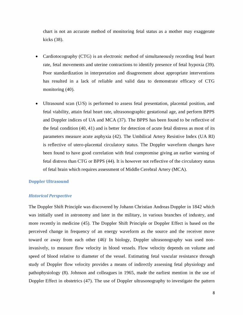
Figure 1 Typical Waveform of umbilical artery and Calculation of Doppler Indices. (59)
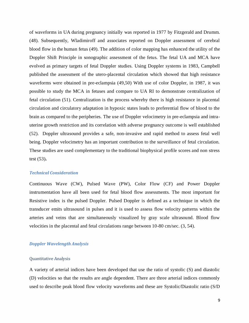
Figure 2 Waveforms of absent end diastolic flow (68)
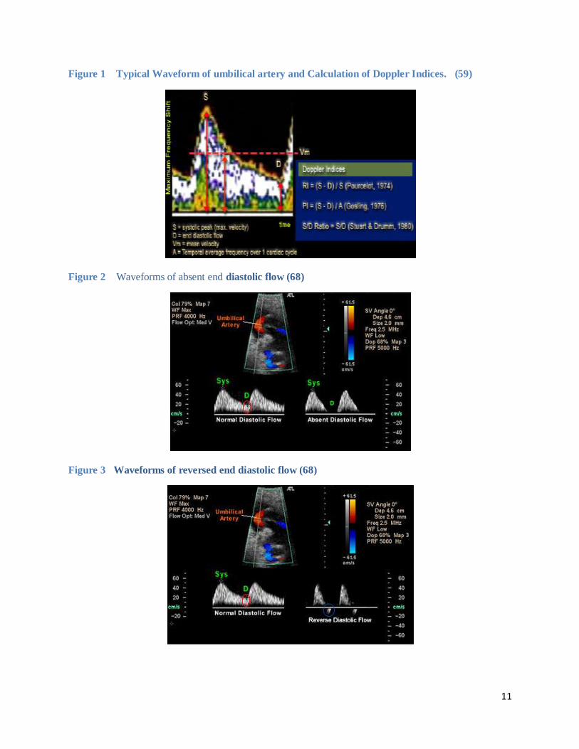
Figure 3 Waveforms of reversed end diastolic flow (68)
12
Functional Doppler Studies
The Physiology of Placental Blood Flow
Umbilical Artery.
The development of the fetal circulation begins with the coalescence of angioblasts in the
mesenchyme of the yolk sac at about day 17(60).The angioblasts begin to coalesce into blood
islands in the splanchnopleuric mesoderm of the embryo by day 18. The angioblasts flatten into
endothelial cells which join to form networks of endothelial channels that form the primitive
circulatory system (60). The endothelial heart tubes fuse to form a single heart tube by the end of
the third week and by day 22. The heart begins to beat and the embryo has a functional
circulatory system (60). The embryo connects to the developing feto-placental vasculature via
the umbilical cord at around 8 to 10 weeks of gestation.The oxygenated blood is delivered to the
fetus through the single umbilical vein and deoxygenated blood from the fetus is returned to the
placenta for re-oxygenation via two umbilical arteries (61).
Throughout the first trimester, the UA waveform is characterized by absent end diastolic flow.
Progressive growth of placental villous tree, together with an increase in fetal cardiac output,
increases both systolic and diastolic velocity in the umbilical artery. Therefore RI values
progressively fall as pregnancy advances (62). Diastolic velocities are typically present at 14-16
weeks thus absent or reversed end diastolic velocity in the umbilical artery is an abnormal
finding by 18-22 weeks. Absent end-diastolic flow in UA is indicative of more severe fetal
hypoxia and pathologically is associated with obliteration of capillaries in placental stem villi.
Reversed end-diastolic flow carries a very poor prognosis and is an indicator
of impending fetal
demise (62). The placentas of fetuses that exhibit abnormal Doppler flow velocity have slender
capillaries with decreased capillary loops in gas-exchanging terminal villi.
The UA is most commonly assessed fetal vessel and it gives a measure of fetal systemic and
placental vascular impedance (53). The abnormal UA Doppler waveforms are indicative of
placental insufficiency. Abnormal Doppler waveforms can be reduced end diastolic flow, absent
end diastolic flow or reversed end diastolic flow and these are indicative of fetal compromise.
The risk of perinatal mortality increases up to 60%, with increasing severity from reduced to
reversed end-diastolic flow velocity (63). The time period between identification of an

13
abnormal UA Doppler waveform and the development of fetal distress and/or death varies
widely – from days to weeks. Various studies show that there is a wide variability in the interval
between detection of UA absent or reverse end-diastolic flow velocities and the occurrence of
heart rate decelerations (64).
Figure 4 Characteristic Saw-tooth Appearance of Umbilical Arterial Flow (59)
Figure 5 Reference Curves for the UARI (68)
14
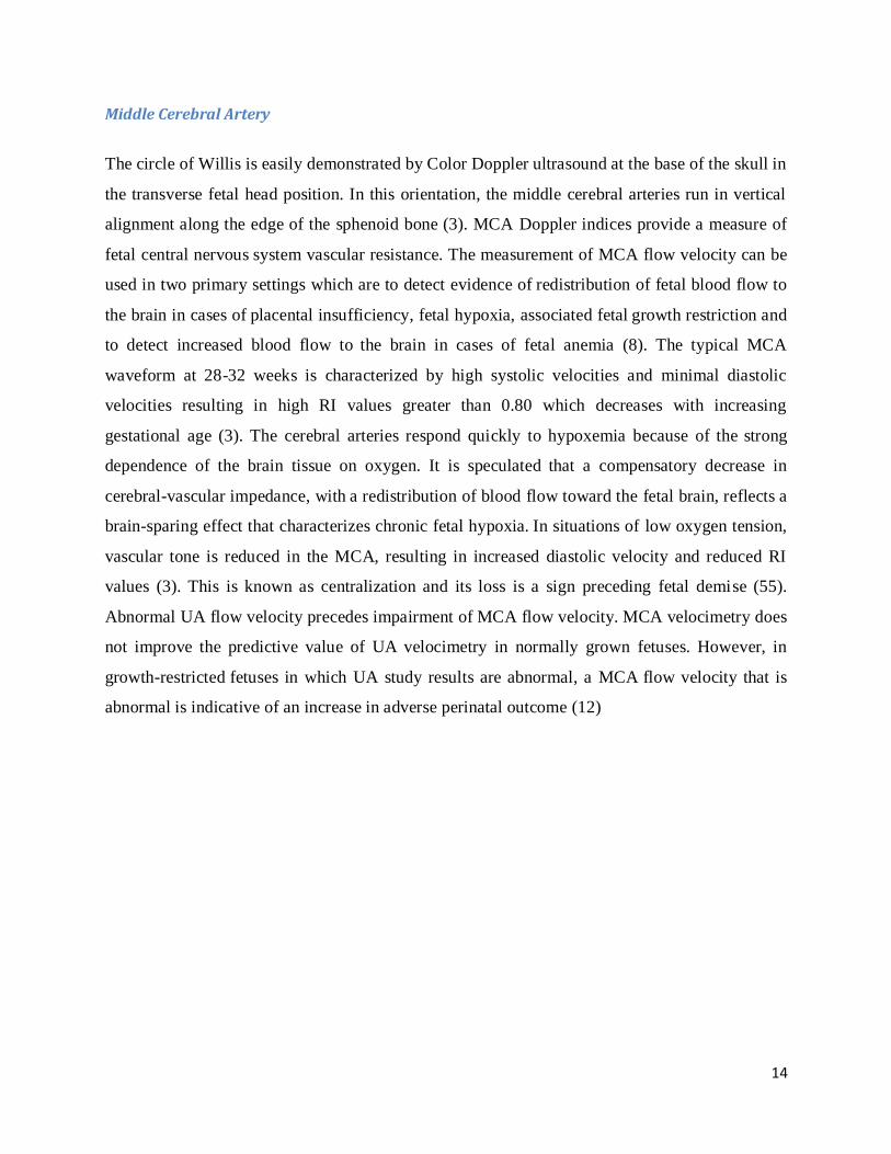
Middle Cerebral Artery
The circle of Willis is easily demonstrated by Color Doppler ultrasound at the base of the skull in
the transverse fetal head position. In this orientation, the middle cerebral arteries run in vertical
alignment along the edge of the sphenoid bone (3). MCA Doppler indices provide a measure of
fetal central nervous system vascular resistance. The measurement of MCA flow velocity
can be
used in two primary settings which are to detect evidence of redistribution of fetal blood flow to
the brain in cases of placental insufficiency, fetal hypoxia, associated fetal
growth restriction and
to detect increased blood flow to the brain in cases of fetal anemia (8). The typical MCA
waveform at 28-32 weeks is characterized by high systolic velocities and minimal diastolic
velocities resulting in high RI values greater than 0.80 which decreases with increasing
gestational age (3). The cerebral arteries respond quickly to hypoxemia because of the strong
dependence of the brain tissue on oxygen. It is speculated that a compensatory decrease in
cerebral-vascular impedance, with a redistribution of blood flow toward the fetal brain, reflects
a
brain-sparing effect that characterizes chronic fetal hypoxia. In situations of low oxygen tension,
vascular tone is reduced in the MCA, resulting in increased diastolic velocity and reduced RI
values (3). This is known as centralization and its loss is a sign preceding fetal demise (55).
Abnormal UA flow velocity precedes impairment of MCA flow velocity. MCA velocimetry does
not improve the predictive value of UA velocimetry in normally grown fetuses. However, in
growth-restricted fetuses in which UA study results are abnormal, a MCA
flow velocity that is
abnormal is indicative of an increase in adverse perinatal outcome (12)
15
Figure 6 Color Doppler View of middle cerebral arteries and Typical Doppler Waveform. (8)
Figure 7 Reference Curves for MCARI (68)

16
The Cerebro-Placental Ratio This is a ratio of the MCA RI over the UA RI and its change is an indicator of peripheral fetal
flow distribution (4). The main advantages of measuring the cerebral and placental vascular
resistance is that account is taken of placental vascular resistance which can be responsible for an
alteration of the maternal to fetal exchanges and also the cerebral hemodynamic consequences of
these abnormalities (4). In normal pregnancies, the diastolic component of the MCA waveform is
lower than in the UA waveform at any gestational age and therefore cerebral vascular resistance
remains higher than placental resistance resulting in a CPR more than 1.0 which is considered
normal (4).
According to Gramellini et al, the CPR remains constant in the last 10 weeks of pregnancy and
thus a single cut-off value of 1.0 is used in the study (63).When the CPR becomes <1.0 it is
considered abnormal because it is indicative of vascular redistribution to the brain and is shown
to be predictive of adverse perinatal outcome (6).
Various studies have shown the CPR to be predictive of adverse perinatal outcome With a CPR
threshold of less than 1.08, the sensitivity, specificity, and positive and negative predictive values
were 72%, 62%, 68%, and 67%, with an odds ratio (95% confidence interval) of 4.2
(1.2–15.3;
area under the receiver operating characteristic curve, 0.67. An abnormal CPR is associated with
adverse perinatal outcomes in growth-restricted fetuses. The accuracy of using gestational age–
specific reference levels was similar to that of using a categorical threshold. (11,12,13,14,15,66)
The authors found that
A longitudinal study of 80 patients with severe preeclampsia (PA >160/110, Proteinuria 3+) was
performed. Doppler study done by one operator was performed every 72 hours at the beginning
and within 7 days before delivery. Resistance index of middle cerebral artery (MCA) and
umbilical artery (UA) were used to calculate the CPR. CPR<1 was considered abnormal.
Abnormal CPR identifies 60% of newborns with severe neonatal morbidity (11)
CPR had 64.1% sensitivity, 72.7% specificity, 89.2% positive predictive value, and 36.3%
negative predictive value for neonatal morbidity. There was a strong correlation between the
CPR and neonatal outcome in women with preeclampsia. CPR <1.0 may be helpful in the
17
identification of newborns at risk of morbidity, irrespective of whether they are small or
appropriate for their gestational age (12).
A longitudinal study of 80 patients with severe pre-eclampsia(BP>160/110, proteinuria 3+) was
performed, Doppler study was performed by one operator was performed every 72 hours in the
beginning and before 7 days before delivery. Resistance of MCA and UA were used to calculate
the CPR. A CPR<1.0 was considered abnormal. The CPR had 64.1% sensitivity, 72.7%
specificity, 89.2% positive predictive value, and 36.3% negative predictive value for neonatal
morbidity. There was a strong correlation between the CPR and neonatal outcome in women
with preeclampsia. A conclusion made from the results of the study was that an abnormal CPR
identifies 60% of newborns with severe neonatal morbidity in pregnancies with severe
preeclampsia (13).
Eleven studies involving nearly 7000 women were included in meta-analysis trials. The trials
were generally of good quality. Doppler ultrasound in high risk pregnancy (especially those
complicated by hypertension or presumed impaired fetal growth) was associated with a reduction
in perinatal deaths (odds ratio 0.71, 95% confidence interval 0.50 to 1.01) as compared to no
Doppler sonography. The use of Doppler ultrasound was also associated with fewer inductions of
labour (odds ratio 0.83, 95% confidence interval 0.74 to 0.93) and fewer admissions to hospital
(odds ratio 0.56, 95% 0.43 to 0.72), without reports of adverse effects. No difference was found
for fetal distress in labour (odds ratio 0.81, 95% confidence interval 0.59 to 1.13) or caesarean
delivery (odds ratio 0.94, 95% 0.82 to 1.06). (14)
The CPR was 83% sensitive and 75% specific in predicting adverse perinatal outcome.
Sensitivity is increased to 91.6 % with multi-vessel Doppler study which include the fetal
umbilical artery,fetal ductus venosus, fetal aorta, maternal uterine artery, fetal middle cerebral
artery and fetal renal arteries(15).
A meta-analysis of randomized controlled trials suggests that incorporation of umbilical artery
Doppler waveform analysis into management protocols for high risk pregnancies significantly
decreases perinatal mortality. Compared to no Doppler ultrasound, Doppler ultrasound in high
risk pregnancy (especially those complicated by hypertension or presumed impaired fetal

18
growth) was associated with a trend to a reduction in perinatal deaths (odds ratio 0.71, 95%
confidence interval 0.50 to 1.01) (16).
An abnormal CPR is associated with adverse perinatal outcomes in growth restricted fetuses. The
accuracy of using gestational age specific reference levels was similar to that of using a
categorical threshold. The CPR has been shown to be a good predictor of the fetal oxygenation
status at birth and can be used to identify pregnancies that are at risk for adverse outcomes. (66).
Figure 8 Reference Curves for the Cerebro-Placental Ratio (68)
19
STUDY DESIGN
Problem statement The incidence of hypertensive disorders of pregnancy is 7.1% in Kenya (19) and a major cause
of maternal and perinatal morbidity and mortality. Currently, fetal monitoring in patients with
HSP at KNH is done via Pinnard fetoscope and CTG. An ultrasound scan is usually done
between 32-38 weeks of gestation including biometric profile, and an UA RI). Currently in
KNH, Management on time of delivery and mode of delivery is determined by BPPS and the UA
RI (34)
It has been shown that the BPPS is similar to CTG in fetal monitoring and does not add any
benefit to fetal monitoring (64). Whereas UA RI is a good indicator of fetal compromise, it is not
adequate as it is only reflective of placental circulation and is not indicative of the state of fetal
central nervous system circulation. Therefore it does not help in determining which fetus is at
greatest risk of adverse perinatal outcome (8). Assessment of MCA Doppler waveform and
calculation of CPR allows identification of fetuses at greater risk of adverse perinatal outcome
when abnormal UA waveforms are present. This study assesses the CPR in third trimester
gestation so as correlate with thee perinatal outcome. It forms a useful resource in development
of management protocols or standards for patients with hypertensive states of pregnancy.
Study Objective
Broad Objective To assess the role of the CPR as a prognostic factor in predicting fetal outcome in patients with
hypertensive disorders of pregnancy in the third trimester(at or more than 32 weeks).
Specific Objectives
To determine the socio-demographic characteristics of patients with hypertensive
disorders of pregnancy at Kenyatta National Hospital.
To classify the severity of hypertensive states of pregnancy using blood pressure and
urinalysis at admission or prior to entry into study.
To attain ultrasonographic fetal heart rate to assess for Non-Reassuring Fetal Status
(NRFS).

20
To evaluate the CPR for all patients with hypertensive states of pregnancy in the third
trimester(at or more than 32 weeks)
To correlate the CPR with fetal outcome in terms of live or still birth, the 5 minute Apgar
score and the birth weight of the live births.
Study Justification At KNH, the practice is to use the BPPS and UA RI in the management of patients with
hypertensive states in pregnancy as concerns mode and time of delivery (34). Investigators
suggest that the time period between identification of an abnormal umbilical artery Doppler
waveform and the development of fetal distress and/or death varies widely(48) Studies show that
there is a wide variability in the interval between detection of umbilical absent or reverse end-
diastolic flow velocities and occurrence of heart rate decelerations(48). Abnormal umbilical
artery Doppler waveforms (reduced diastolic flow, absent end diastolic flow and reversed end
diastolic flow) alone do not determine fetal status. However they are an indicator of impending
fetal distress (53) Assessing the Doppler waveform of the MCA and obtaining the
cerebroplacental ratio gives us an indication of fetuses that are at greatest risk of adverse
outcome(53). This study assessed the Doppler waveform of the middle cerebral artery and
umbilical artery and thereby obtaining the CPR. No such study had been published in this region
and therefore it was important to assess the role of the CPR in the prediction of fetal outcome in
mothers with hypertensive states of pregnancy locally. A positive correlation would greatly
enhance antenatal monitoring of the fetus and also improve on the management of these patients.
Research Question What is the prognostic value of the CPR as a predictor of adverse fetal outcome in patients with
hypertensive disorders of pregnancy?
Hypothesis The CPR is an accurate predictor of adverse fetal outcome in patients with hypertensive states of
pregnancy.

21
Design and Methodology
This is a prospective cohort study conducted at Kenyatta National Hospital over a period of nine
months (November 2010 to August 2011) following approval by the KNH/UON- Ethics and
research Committee.
Sampling A cohort of women with hypertensive disorders of pregnancy with or without concomitant IUGR
was recruited over the 9 month period after obtaining informed consent. Informed consent was
taken from the patient herself or next of kin in patients who were not able to give consent
(eclamptics or underage patients). Recruitment took place from the labor ward over a period of
24 hours a day by the principal investigator or research assistant.
Inclusion criteria include
Consent giving patients
Singleton pregnancy.
Gestational age at the time of study entry should be at least 32 completed weeks as this is
the time most patients with hypertensive states of pregnancy are admitted to labor ward
for closer monitoring/delivery.
Normal fetal anatomic survey.
Absence of maternal metabolic disease or any other vascular disease.
Exclusion criteria include
Non consent giving patients.
Multiple pregnancies as they have additional risk factors that can bias the study results.
Patients presenting for the ultrasound scan with IUFD or fetal anomalies.
Known maternal metabolic disease or other vascular diseases.
There was no discrimination based on race, religion, age, political background or socio-
economic status in this study.
Method After obtaining informed consent from the patient or next of kin, a structured questionnaire was
filled out by the principal investigator or research assistant. Blood pressure recording and
urinalysis report was recorded to classify severity and type of hypertension. An obstetric

22
ultrasound scan was carried out on request by the clinician and coded for fetal presentation,
placental position, fetal heart rate, approximate ultrasonographic age, BPPS, UA RI and MCA
RI. The ultrasound machines used were real time machines, the Phillips HD11 and GE Logic 7.
The transducer frequency was 3.5 – 5.0 MHz, the Doppler sample volume was 2 mm and the
wall filter was 50–100 Hz. The examination was performed with the mother in a semi-
recumbent position during relative fetal inactivity and apnea. This is because the end diastolic
flow (EDF) decreases with decreasing fetal heart rate and fetal breathing movements increase
variability in the Doppler measurements.
The UA was sampled at the middle of a free loop of umbilical cord. This is because the EDF is
higher near the umbilical cord insertion into the fetal abdomen than near the placental insertion
and vice versa at the placental end. It could also be assessed at the level of the fetal bladder. For
MCA, a transverse image of the fetal head was obtained at the level of the sphenoid bones. Color
Flow imaging was used to display the circle of Willis. The MCA in the near field was isonated
about 1 cm distal to its origin from the internal carotid artery. By using the optimal spectral trace
from each artery, the Resistive Index was calculated from the mean of a minimum of five
consecutive waveforms on a frozen image. A series of three readings were taken for each artery
to avoid errors. The cerebral/placental ratio was calculated from the MCA RI and UA RI.
Patients were divided into groups of two depending on the CPR. A CPR of > 1.0 was considered
normal and a CPR of < 1.0 abnormal.
U/S fetal heart rate, biometric measurements, BPPS and MCA/UA Doppler results were used to
determine management. Follow up was made for fetal outcome whether it was a live birth or still
birth. The 5 minute Apgar score and birth weight were recorded.

23
Sample size consideration
The study population included pregnant women with hypertensive disorders. The study arms
were defined as
Group 0: Mothers with hypertensive disorders and with a Doppler ultrasound indicative of an
abnormal cerebral/placental ratio (CPR<1.0).
Group 1: Mothers with hypertensive disorders with a Doppler ultrasound indicative of a normal
cerebral/placental ratio (CPR>1.0).
The study outcome variables were
1. Neonate condition (Live birth or still birth)
2. The 5 minute APGAR score-5 min (< 7 or ≥ 7)
3. Birth weight – (<10th
percentile of the expected weight for gestation)
For each of the outcome variable, let p be the smallest of the proportions of negative or positive
cases in the population and k be the number of predictor variables (either clinically or
statistically significant), then the minimum number of subjects to be enrolled is given by
10kN
p , Peduzzi et al. (1996) (65)
The maximum number of predictor variables that were included in the model was three while the
smallest proportion of outcome was p=0.2. This yields a total sample size of 150 patients but the
sample size achieved was 160 patients.
Data management and statistical analysis A structured questionnaire was used to collect data. The data was entered manually into a
database (Ms Access 2007) via user-defined forms. The data entry forms had quality control
checks in order to ensure accuracy of the data. The data base security features ensured
confidentiality and access to the participants’ data. All data was cleaned and exported for
statistical analysis. Statistical Package for Social Sciences (SPSS 17.0) was used for the analysis.
Descriptive statistics-mean (95% CI), standard deviation, median (IQR) and frequencies were
used to characterize the study population. Dummy tables are used to present the study findings.
Mann-Whitney U, chi-square and Fisher exact tests was used for comparative analysis. Logistic

24
regression was used to estimate the odds of each outcome variable associated with every
predictor variable, that is:
0
1
log ( )k
i
i
it p b x
Where p is the probability of presence of the characteristic of interest.
( of a characteristic)
1 ( of a characteristic)
p prob presenceodds
p prob absence
Unadjusted odds ratio of each outcome variable associated with CPR was determined; all
clinically and statistically significant variables were adjusted for in the multivariate analysis. A
5% level of significance was used to determine if a variable contribute significantly to the
prediction of the outcome variable.
Ethical Considerations
This study employed the use of ultrasound which is a sound waveform unlike other methods that
use ionizing radiation and has been confirmed to have no biological effect on patients or the fetus
at intensities typical of present diagnostic instruments. Permission to carry out the research was
sought from and granted by KNH/UON-ERC. Permission was sought from director of clinical
services, KNH. Authorities from Department of Diagnostic Imaging and Radiation Medicine and
Department of Obstetrics and Gynecology, KNH were consulted and informed.
The following ethical guidelines were used in line with the Helsinki Declaration (World Medical
Association – June 1964).
Patients name, religion and racial backgrounds were not documented in this study.
Patients were identified by Hospital numbers to safeguard confidentiality. Information
obtained from the study was treated with total confidentiality and results used for
academic and clinical improvement purposes only.
No additional imaging examination was done other than the one requested by the
referring clinician.
The patient did not incur any additional cost.
No blood sample was collected as it was not indicated in this study

25
All patients were managed at the optimal standards depending on clinical state.
No victimization or preferential treatment was offered to patients as a result of
participating or refusing to participate in the study.
A copy of the research findings would be given to the KNH/UON-Ethics and Research
Committee for future references and to facilitate any possible improvement in patient
management.
Informed written consent was obtained from all the participants.
The results of the ultrasound scan were availed to the clinician immediately to allow for
timely and appropriate management
STUDY RESULTS ULTRSOUND SCANS
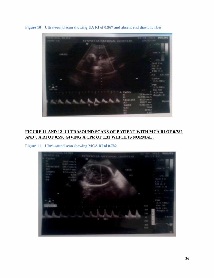
FIGURE 9 AND FIGURE 10-ULTRASOUND SCANS OF PATIENT WITH MCA RI OF
0.683 AND UA RI OF 0.967 GIVING A CPR OF 0.71 WHICH IS LESS THAN ONE
PREDICTIVE OF ADVERSE PERINATAL OUTCOME. Figure 9 Ultra-sound scan showing MCA RI of 0.683
26
Figure 10 Ultra-sound scan showing UA RI of 0.967 and absent end diastolic flow
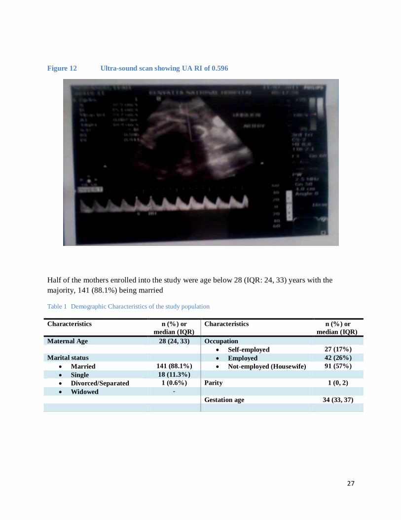
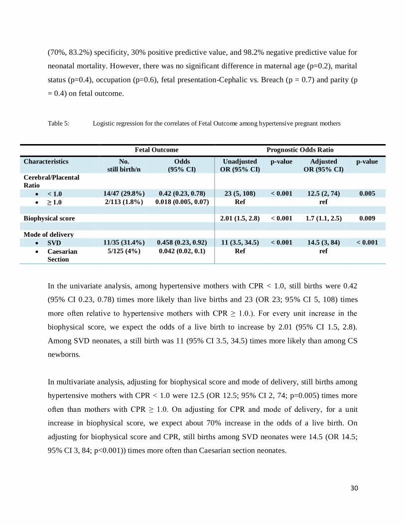
FIGURE 11 AND 12- ULTRASOUND SCANS OF PATIENT WITH MCA RI OF 0.782
AND UA RI OF 0.596 GIVING A CPR OF 1.31 WHICH IS NORMAL .
Figure 11 Ultra-sound scan showing MCA RI of 0.782
27
Figure 12 Ultra-sound scan showing UA RI of 0.596
Half of the mothers enrolled into the study were age below 28 (IQR: 24, 33) years with the
majority, 141 (88.1%) being married
Table 1 Demographic Characteristics of the study population
Characteristics n (%) or
median (IQR)
Characteristics n (%) or
median (IQR)
Maternal Age 28 (24, 33) Occupation
Self-employed 27 (17%)
Marital status Employed 42 (26%)
Married 141 (88.1%) Not-employed (Housewife) 91 (57%)
Single 18 (11.3%)
Divorced/Separated 1 (0.6%) Parity 1 (0, 2)
Widowed -
Gestation age 34 (33, 37)

28
Seventeen percent were self-employed, 42 (26%) were in formal employment while 91 (57%)
were not employed. Half of them had at least one (IQR: 0, 2) child and presented for care at 34
(IQR: 33, 37) weeks gestation.
Table 2 Clinical Observations
Characteristics n (%)
median (IQR)
Characteristics n (%)
median (IQR)
Clinical findings
Blood pressure- Fetal heart rate 138 (132, 140)
Systolic 154 (143, 170) Urinalysis 2 (2, 3)
Diastolic 104 (95, 113 )
PET Biophysical profile score 8 (6, 8)
Severe 51 (32%)
Mild 109 (68%) Approximate ultrasound age 31 (33, 35)
Ultra-sound findings
Umblical Artery resistive index (UARI) 0.64 (0.548, 0.72)
Fetal presentation
Cephalic 143 (89%) Middle Cerebral Artery resistive index 0.74 (0.646, 0.817)
Breech 17 (11%)
Cerebral/Placental Doppler Ratio
Placenta < 1.0 47 (29%)
1. Fundo-anterior 86 (54%) ≥ 1.0 113 (71%)
2. Fundo-posterior 74 (46%)
3. Low-lying -
The median Biophysical Profile score was 8 (IQR: 6, 8); the median umbilical artery resistive
index was 0.64 (IQR: 0.548, 0.72) while the middle cerebral artery resistive index was 074 (IQR:
0.646, 0.817). The CPR < 1.0 was observed in 47 (29%) of the study population.
Table 3 Clinical Management and Fetal Outcome
Characteristics n (%)
median (IQR)
Characteristics n (%)
median (IQR)
Steroid administered Mode of delivery
Yes 99 (62%) Caesarian Section 125 (78%)
No 61 (38%) SVD 35 (22%)
Fetal Outcome APGAR Score (5 minutes) 9 (8, 10)
4. Alive 144 (90%) < 7 30 (19%)
5. Still birth 16 (10%) ≥ 7 130 (81%)
Infant weight (at birth) 2,100 (1,700, 2638) Infant admitted in NBU
Low 27 (17%) Yes 42 (26)
Normal 133 (83%) No 118 (74)

29
Steroid was administered to 99 (62%) of the mothers. Caesarian section was performed on 125
(78%) of the mothers compared to 35 (22%) who had normal delivery. 10% of the deliveries
were still births, 27 (17%) of the infants weighed less than 1,500 grams with median weight
2,100 (IQR: 1,700, 2,638). The median APGAR score (at 5 minutes) was 9 (IQR: 8, 10); 30
(19%) has an APGAR score of less than 7. Forty two percent neonates were admitted in the
newborn unit.
Table 4 Correlates of Fetal Outcome among hypertensive pregnant mothers
Fetal Outcome
Still birth Live birth
Characteristics n (%) or
Median (IQR)
n (%) or
Median (IQR)
p-value
Cerebral/Placental Ratio
< 1.0 14 (87.5) 33 (23) < 0.001
≥ 1.0 2 (12.5) 111 (77)
Maternal age 31 (26, 34) 28 (24, 32) 0.2m
Marital Status
Married 13 (81) 128 (89) 0.4*
Not married 3 (19) 16 (11)
Occupation
With Employed 6 (37.5) 63 (44) 0.6
Not employed 10 (62.5) 81 (56)
Presentation
Cephalic 14 (87.5) 129 (89.6) 0.7*
Breech 2 (12.5) 15 (10.4)
Biophysical score 4 (4, 6) 8 (6, 8) < 0.001m
Parity
1 or none 10 (62.5) 105 (73) 0.4*
≥ 1 6 (37.5) 39 (27)
Mode of delivery
SVD 11 (69) 24 (17)
Caesarian Section 5 (31) 120 (83) < 0.001*
16 (100%) 144 (100%) Estimated conditional probabilities for fetal outcome prognosis
Estimated 95% Confidence Interval for sensitivity and specificity computed using Wilson Score Method
* Fisher Exact Tests used
m –Mann Whitney test used
There was evidence of association between the Cerebral/Placental Ratio (p<0.001), mode of
delivery (p<0.001), Biophysical Profile score (p<0.001) and fetal outcome among mothers in the
study population. The CPR (C/U RI ratio) had 87.5% (95% CI 64%, 96.5%) sensitivity, 77%
30
(70%, 83.2%) specificity, 30% positive predictive value, and 98.2% negative predictive value for
neonatal mortality. However, there was no significant difference in maternal age (p=0.2), marital
status (p=0.4), occupation (p=0.6), fetal presentation-Cephalic vs. Breach (p = 0.7) and parity (p
= 0.4) on fetal outcome.
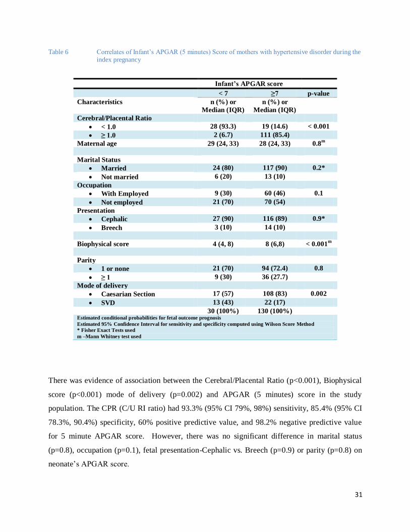
Table 5: Logistic regression for the correlates of Fetal Outcome among hypertensive pregnant mothers
Fetal Outcome Prognostic Odds Ratio
Characteristics No.
still birth/n
Odds
(95% CI)
Unadjusted
OR (95% CI)
p-value Adjusted
OR (95% CI)
p-value
Cerebral/Placental
Ratio
< 1.0 14/47 (29.8%) 0.42 (0.23, 0.78) 23 (5, 108) < 0.001 12.5 (2, 74) 0.005
≥ 1.0 2/113 (1.8%) 0.018 (0.005, 0.07) Ref ref
Biophysical score 2.01 (1.5, 2.8) < 0.001 1.7 (1.1, 2.5) 0.009
Mode of delivery
SVD 11/35 (31.4%) 0.458 (0.23, 0.92) 11 (3.5, 34.5) < 0.001 14.5 (3, 84) < 0.001
Caesarian
Section
5/125 (4%) 0.042 (0.02, 0.1) Ref ref
In the univariate analysis, among hypertensive mothers with CPR < 1.0, still births were 0.42
(95% CI 0.23, 0.78) times more likely than live births and 23 (OR 23; 95% CI 5, 108) times
more often relative to hypertensive mothers with CPR ≥ 1.0.). For every unit increase in the
biophysical score, we expect the odds of a live birth to increase by 2.01 (95% CI 1.5, 2.8).
Among SVD neonates, a still birth was 11 (95% CI 3.5, 34.5) times more likely than among CS
newborns.
In multivariate analysis, adjusting for biophysical score and mode of delivery, still births among
hypertensive mothers with CPR < 1.0 were 12.5 (OR 12.5; 95% CI 2, 74; p=0.005) times more
often than mothers with CPR ≥ 1.0. On adjusting for CPR and mode of delivery, for a unit
increase in biophysical score, we expect about 70% increase in the odds of a live birth. On
adjusting for biophysical score and CPR, still births among SVD neonates were 14.5 (OR 14.5;
95% CI 3, 84; p<0.001)) times more often than Caesarian section neonates.
31
Table 6 Correlates of Infant’s APGAR (5 minutes) Score of mothers with hypertensive disorder during the
index pregnancy
There was evidence of association between the Cerebral/Placental Ratio (p<0.001), Biophysical
score (p<0.001) mode of delivery (p=0.002) and APGAR (5 minutes) score in the study
population. The CPR (C/U RI ratio) had 93.3% (95% CI 79%, 98%) sensitivity, 85.4% (95% CI
78.3%, 90.4%) specificity, 60% positive predictive value, and 98.2% negative predictive value
for 5 minute APGAR score. However, there was no significant difference in marital status
(p=0.8), occupation (p=0.1), fetal presentation-Cephalic vs. Breech (p=0.9) or parity (p=0.8) on
neonate’s APGAR score.
Infant’s APGAR score
< 7 ≥7 p-value
Characteristics n (%) or
Median (IQR)
n (%) or
Median (IQR)
Cerebral/Placental Ratio
< 1.0 28 (93.3) 19 (14.6) < 0.001
≥ 1.0 2 (6.7) 111 (85.4)
Maternal age 29 (24, 33) 28 (24, 33) 0.8m
Marital Status
Married 24 (80) 117 (90) 0.2*
Not married 6 (20) 13 (10)
Occupation
With Employed 9 (30) 60 (46) 0.1
Not employed 21 (70) 70 (54)
Presentation
Cephalic 27 (90) 116 (89) 0.9*
Breech 3 (10) 14 (10)
Biophysical score 4 (4, 8) 8 (6,8) < 0.001m
Parity
1 or none 21 (70) 94 (72.4) 0.8
≥ 1 9 (30) 36 (27.7)
Mode of delivery
Caesarian Section 17 (57) 108 (83) 0.002
SVD 13 (43) 22 (17)
30 (100%) 130 (100%) Estimated conditional probabilities for fetal outcome prognosis
Estimated 95% Confidence Interval for sensitivity and specificity computed using Wilson Score Method
* Fisher Exact Tests used
m –Mann Whitney test used
32
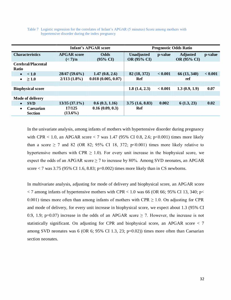
Table 7 Logistic regression for the correlates of Infant’s APGAR (5 minutes) Score among mothers with
hypertensive disorder during the index pregnancy
In the univariate analysis, among infants of mothers with hypertensive disorder during pregnancy
with CPR < 1.0, an APGAR score < 7 was 1.47 (95% CI 0.8, 2.6; p<0.001) times more likely
than a score ≥ 7 and 82 (OR 82; 95% CI 18, 372; p<0.001) times more likely relative to
hypertensive mothers with CPR ≥ 1.0). For every unit increase in the biophysical score, we
expect the odds of an APGAR score ≥ 7 to increase by 80%. Among SVD neonates, an APGAR
score < 7 was 3.75 (95% CI 1.6, 8.83; p=0.002) times more likely than in CS newborns.
In multivariate analysis, adjusting for mode of delivery and biophysical score, an APGAR score
< 7 among infants of hypertensive mothers with CPR < 1.0 was 66 (OR 66; 95% CI 13, 340; p<
0.001) times more often than among infants of mothers with CPR ≥ 1.0. On adjusting for CPR
and mode of delivery, for every unit increase in biophysical score, we expect about 1.3 (95% CI
0.9, 1.9; p=0.07) increase in the odds of an APGAR score ≥ 7. However, the increase is not
statistically significant. On adjusting for CPR and biophysical score, an APGAR score < 7
among SVD neonates was 6 (OR 6; 95% CI 1.3, 23; p=0.02)) times more often than Caesarian
section neonates.
Infant’s APGAR score Prognostic Odds Ratio
Characteristics APGAR score
(< 7)/n
Odds
(95% CI)
Unadjusted
OR (95% CI)
p-value Adjusted
OR (95% CI)
p-value
Cerebral/Placental
Ratio
< 1.0 28/47 (59.6%) 1.47 (0.8, 2.6) 82 (18, 372) < 0.001 66 (13, 340) < 0.001
≥ 1.0 2/113 (1.8%) 0.018 (0.005, 0.07) Ref ref
Biophysical score 1.8 (1.4, 2.3) < 0.001 1.3 (0.9, 1.9) 0.07
Mode of delivery
SVD 13/35 (37.1%) 0.6 (0.3, 1.16) 3.75 (1.6, 8.83) 0.002 6 (1.3, 23) 0.02
Caesarian
Section
17/125
(13.6%)
0.16 (0.09, 0.3) Ref
33
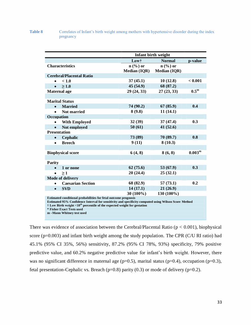
Table 8 Correlates of Infant’s birth weight among mothers with hypertensive disorder during the index
pregnancy
There was evidence of association between the Cerebral/Placental Ratio (p < 0.001), biophysical
score (p=0.003) and infant birth weight among the study population. The CPR (C/U RI ratio) had
45.1% (95% CI 35%, 56%) sensitivity, 87.2% (95% CI 78%, 93%) specificity, 79% positive
predictive value, and 60.2% negative predictive value for infant’s birth weight. However, there
was no significant difference in maternal age (p=0.5), marital status (p=0.4), occupation (p=0.3),
fetal presentation-Cephalic vs. Breach (p=0.8) parity (0.3) or mode of delivery (p=0.2).
Infant birth weight
Low† Normal p-value
Characteristics n (%) or
Median (IQR)
n (%) or
Median (IQR)
Cerebral/Placental Ratio
< 1.0 37 (45.1) 10 (12.8) < 0.001
≥ 1.0 45 (54.9) 68 (87.2)
Maternal age 29 (24, 33) 27 (23, 33) 0.5m
Marital Status
Married 74 (90.2) 67 (85.9) 0.4
Not married 8 (9.8) 11 (14.1)
Occupation
With Employed 32 (39) 37 (47.4) 0.3
Not employed 50 (61) 41 (52.6)
Presentation
Cephalic 73 (89) 70 (89.7) 0.8
Breech 9 (11) 8 (10.3)
Biophysical score 6 (4, 8) 8 (6, 8) 0.003m
Parity
1 or none 62 (75.6) 53 (67.9) 0.3
≥ 1 20 (24.4) 25 (32.1)
Mode of delivery
Caesarian Section 68 (82.9) 57 (73.1) 0.2
SVD 14 (17.1) 21 (26.9)
30 (100%) 130 (100%) Estimated conditional probabilities for fetal outcome prognosis
Estimated 95% Confidence Interval for sensitivity and specificity computed using Wilson Score Method
† Low Birth weight <10th
percentile of the expected weight for gestation
* Fisher Exact Tests used
m –Mann Whitney test used
34
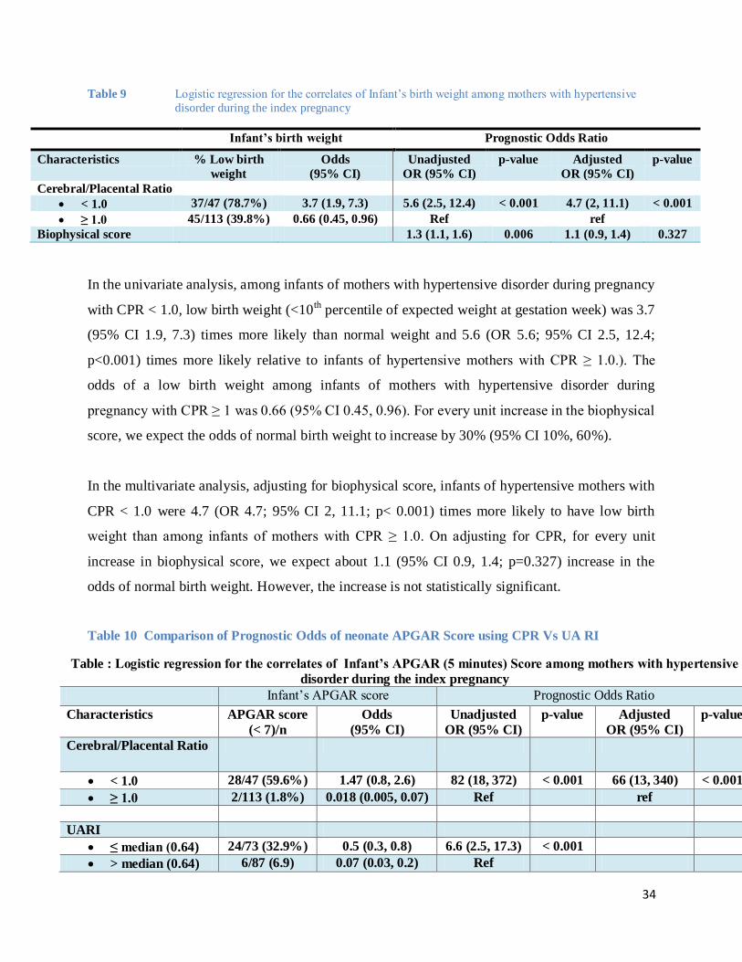
Table 9 Logistic regression for the correlates of Infant’s birth weight among mothers with hypertensive
disorder during the index pregnancy
Infant’s birth weight Prognostic Odds Ratio
Characteristics % Low birth
weight
Odds
(95% CI)
Unadjusted
OR (95% CI)
p-value Adjusted
OR (95% CI)
p-value
Cerebral/Placental Ratio
< 1.0 37/47 (78.7%) 3.7 (1.9, 7.3) 5.6 (2.5, 12.4) < 0.001 4.7 (2, 11.1) < 0.001
≥ 1.0 45/113 (39.8%) 0.66 (0.45, 0.96) Ref ref
Biophysical score 1.3 (1.1, 1.6) 0.006 1.1 (0.9, 1.4) 0.327
In the univariate analysis, among infants of mothers with hypertensive disorder during pregnancy
with CPR < 1.0, low birth weight (<10th percentile of expected weight at gestation week) was 3.7
(95% CI 1.9, 7.3) times more likely than normal weight and 5.6 (OR 5.6; 95% CI 2.5, 12.4;
p<0.001) times more likely relative to infants of hypertensive mothers with CPR ≥ 1.0.). The
odds of a low birth weight among infants of mothers with hypertensive disorder during
pregnancy with CPR ≥ 1 was 0.66 (95% CI 0.45, 0.96). For every unit increase in the biophysical
score, we expect the odds of normal birth weight to increase by 30% (95% CI 10%, 60%).
In the multivariate analysis, adjusting for biophysical score, infants of hypertensive mothers with
CPR < 1.0 were 4.7 (OR 4.7; 95% CI 2, 11.1; p< 0.001) times more likely to have low birth
weight than among infants of mothers with CPR ≥ 1.0. On adjusting for CPR, for every unit
increase in biophysical score, we expect about 1.1 (95% CI 0.9, 1.4; p=0.327) increase in the
odds of normal birth weight. However, the increase is not statistically significant.
Table 10 Comparison of Prognostic Odds of neonate APGAR Score using CPR Vs UA RI
Table : Logistic regression for the correlates of Infant’s APGAR (5 minutes) Score among mothers with hypertensive
disorder during the index pregnancy
Infant’s APGAR score Prognostic Odds Ratio
Characteristics APGAR score
(< 7)/n
Odds
(95% CI)
Unadjusted
OR (95% CI)
p-value Adjusted
OR (95% CI)
p-value
Cerebral/Placental Ratio
< 1.0 28/47 (59.6%) 1.47 (0.8, 2.6) 82 (18, 372) < 0.001 66 (13, 340) < 0.001
≥ 1.0 2/113 (1.8%) 0.018 (0.005, 0.07) Ref ref
UARI
≤ median (0.64) 24/73 (32.9%) 0.5 (0.3, 0.8) 6.6 (2.5, 17.3) < 0.001
> median (0.64) 6/87 (6.9) 0.07 (0.03, 0.2) Ref
35
Table 11 Patients According To Severity of PET and CPR
Characteristic Number and percentage of patients(160)
Total number Percentage (%)
Severe PET 51/160 32%
CPR< 1.0 20/51 39%
CPR≥ 1.0 31/51 61%
Mild PET 109/160 68%
CPR≤ 1.0 27/109 24%
CPR≥1.0 82/109 76%
36
DISCUSSION
In this study comprising of 160 patients with hypertensive states in pregnancy, the aim was to
determine the CPR as a prognostic factor of fetal outcome. The CPR incorporates data of both
the placental status using the umbilical artery and the fetal response using the middle cerebral
artery as the brain is a vital structure in the prediction of adverse outcomes. In this study,
gestational age–specific thresholds were compared with categorical thresholds of the CPR </≥1.
All the 160 patients were referred by clinicians from labor ward, antenatal clinics and casualty
departments, Kenyatta National Hospital.
The study population had a mean age of 28 (95% CI 27, 29). This is in keeping with Odibo
Anthony et al where the mean age was 28 years (66). It is also in keeping with Lakhkar B N et al
where the mean age was 27 years (13). It is however in contrast with Zavala-Coca Carlos et al
where the mean age was 35 years (11). This could mean that the reproductive age in our study
population is younger or that PET affects a younger age group in our study population. The
marital status showed that majority 141 (88.1%) were married, 18 (11.3%) were single and
1(0.6%) separated or divorced. These have not been documented in other comparative studies.
The occupation showed 27(17%) to be self employed, 42(26%) to be employed and majority of
91(57%) to be not employed (housewives). They however did attend all antenatal clinics and
received proper management despite low socioeconomic status.
I found 62% of study population to be primigravida which is in keeping with Lakhkar B N et al
showing that 60% were primigravidas (13). The median gestation was 34 weeks which is a
gestation where the fetus has achieved a good fetal weight. This is a younger gestation than that
Odibo A et al where mean gestation was 35 weeks (66) and that of Lakhkar BN et al where
gestation was also 35 weeks meaning either patients show up for care earlier or that intervention
is carried out earlier.
Thirty two percent (32%) of the patients were found to have severe PET as evidenced by blood
pressure on admission and proteinuria on urinalysis. Out of these 39% were found to have a CPR
<1.0 and 61% had CPR≥ 1.0. This is much higher than the population studied by Lakhkar BN et
al showing 11% to have severe PET thus we have a higher population presenting with severe

37
PET. This was not explained by any of the etiological causes of PET though it would be worth
carrying out other studies to find out why our patients have severe PET. The number of patients
with severe PET that had an abnormal CPR was not studied in other studies and is important as it
is significant number with sever PET that have an abnormal CPR. Sixty eight percent had mild
PET out of which 24% had an abnormal CPR (<1.0) while 76% had a normal CPR (≥1.0).this is
also significant as even a significant number of patients with mild PET had an abnormal CPR
(<1.0) so they also require close monitoring.
Eighty nine percent (89%) had fetuses in cephalic presentation while and eleven percent (11%)
had fetuses in breech presentation which can bring additional complications.
Twenty nine percent (29%) had a CPR <1.0 which was abnormal and seventy one percent (71%)
had a CPR ≥1.0 which was normal. This was not documented in the comparative studies. This is
an important finding as it shows that a third of the sample size presented with an abnormal CPR
which would imply that we have patients with severe PET. It also demonstrates the high number
of patients with abnormal CPR which results in higher neonatal morbidity and mortality
therefore proper and timely management is necessary.
The ultrasonographic fetal heart rate was found to be 138 beats per minute which is within
normal range (120-160).
86 patients (54%) had a placenta in fundo anterior location while 74 patients (46%) in fundo
posterior location. None of the patients had a low lying placenta which comes with its own
additional complications.
The median umbilical artery resistive index was 0.64 and middle cerebral artery resistive index
was 0.74 as expected according to the normogram.
About 47 patients (29%) had an abnormal CPR<1 while 113 (71%) had a normal CPR (≥1). 125
(78%) had delivery via caesarean section which is much higher than the 62% demonstrated by
Lakhkar BN et al. (35). 22% delivered SVD which is also higher than Lakhkar BN et al who
showed 5% delivering vaginally. This could be due to the smaller sample size which was 58 in
Lakhkar BN et al study as opposed to 160 patients in this study. It could also mean that we have

38
more patients with hypertensive states in pregnancy as compared to other comparative studies.
Most caesarean sections were carried out on these patients due to failed induction of labor.
The neonates delivered via cesarean section had better outcome as compared to those delivered
vaginally. This was not a study variable in the other two comparative studies. Therefore, delivery
via caesarean section is best as it improves on perinatal outcome.
The CPR </≥ 1 had a 93.3% (95% CI 79%, 98%) sensitivity, 85.4% (95% CI 78.3%, 90.4%)
specificity, 60% positive predictive value, and 98.2% negative predictive value for adverse
perinatal outcome. This contrasts to Lakhkar BN et al who had a CPR sensitivity of 47.2%,
specificity of 86.3%, PPV of 85% and NPV of 50% (13) which could be due to a smaller sample
size of 58 in the Lakhkar BN study as contrasts to this study which had a sample size of 160
while that of Odibo A et al showed the CPR to have a sensitivity of 67%, specificity of 66%,
PPV of 69% and NPV of 64%(66). Among neonates of mothers with hypertensive disorder
during pregnancy with UARI ≤ median (0.64), an APGAR score < 7 was 0.5 (95% CI 0.3, 0.8;
p<0.001) times more likely than a score > 7 and 6.6 (OR 6.6; 95% CI 2.5, 17.3; p<0.001) times
more likely relative to hypertensive mothers with UARI > median (0.64). Combining UA RI and
MCA RI improves the prognostic odds ratio from 6.6 to 82. (Table 10.). This implies that the
CPR is a better predictor than UA RI alone for APGAR score < 7. This refutes Lakhkar BN et al
findings that the UA PI is more sensitive than CPR in predicting adverse perinatal outcome (13)
but it is in keeping with findings of Gramellini et al (63).
The CPR is predictive of adverse perinatal outcome and therefore very good in antenatal
monitoring of mothers with hypertensive states of pregnancy. This should be used in antenatal
and perinatal centres for antenatal monitoring of patients with HSP.
CPR is better than BPPS in predicting adverse perinatal outcome. This was a new finding in this
study as it was not looked at in the other comparative studies.
This is important as obtaining biophysical profile score only shows the acute hypoxic status of
the fetus. Obtaining the CPR would be vital to see chronic hypoxic status and decide on proper
timing and method of management so as to reduce on perinatal morbidity and mortality.

39
The percentage of low birth weight infants in this study was 17% which is much higher than BN
Lakhkar BN et al which had a percentage of 6.8%. This could be due to patients being delivered
at an earlier gestation (at 32-34 weeks) as compared to 36 weeks in Lakhkar BN et al study or
more patients with severe PET causing low birth weight in these fetuses. Further studies should
be carried out to determine why our sample population has a severe PET.

40
41
CONCLUSION The following conclusions were drawn from this study:
1. The CPR is predictive of adverse perinatal outcome and therefore very good in antenatal
monitoring of mothers with hypertensive states of pregnancy.
2. Algorithm for management of hypertensive mothers should recommend caesarean section
as the mode of delivery as it improves perinatal outcomes among mothers with CPR < 1.
3. Combining the UA RI and MCA RI improves the prognostic odds ratio of APGAR
score from 6.6 to 82(Table 10). This implies that the CPR is a better predictor than UA
RI alone to improve on neonatal condition.
4. The biophysical profile is shown to predict acute fetal distress but the CPR is predictive
of chronic fetal distress and it is predictive of adverse fetal outcome. Therefore CPR
should be used for antenatal monitoring of patients with hypertensive states and not
biophysical profile score.

42
RECOMMENDATIONS
1. The CPR should be used by antenatal/ perinatal centres for antenatal monitoring in
mothers with hypertensive states of pregnancy to reduce perinatal morbidity and
mortality.
2. Clinical decisions on time of delivery should be based on CPR and clinical state of
patient.
3. Mothers with hypertensive states of pregnancy should be delivered at appropriate time
via caesarean section as it improves on perinatal outcome.
4. Further studies should be carried out to assess factors causing severe PET in our
population and also assess the neonatal umbilical artery pH in fetuses born from mothers
with hypertensive states of pregnancy so as to correlate with hypoxic status.

43
APPENDIX A
Research Consent Form My name is Dr. Parmar Linal Parshuram, a postgraduate student in the department of Diagnostic
Imaging and Radiation Medicine at the University of Nairobi.
I am conducting a study on the effect of high blood pressure on the unborn child. This study
involves answering some questions and performing an ultrasound scan with a special study of the
blood vessels of the unborn child. This will help to check on the well being of the unborn child
while in the uterus. This does not pose any danger to you or the unborn child. Follow up will be
made to assess the new born baby.
Your name will not be included in the report. Only ultrasound and file umbers will be used. Your
information shall be kept confidential.
I would like to recruit you in this study. You are not forced to participate and your failure to
consent will not deny you any treatment.
Patient ultrasound number ________________________________________________
Signature _________________________________________________________
Date _____________________________________________________________
I certify that the patient has understood and consented participation in the study.
Dr. Parmar Linal Parshuram
Signature __________________________________________________________
Contact - 0722-888991
Supervisors
Dr. Mwango G.N – Supervisor/lecturer
Department of Diagnostic Imaging and Radiation Medicine, University of Nairobi
Telephone – 020-2724722
Dr. On’gech J.O – Supervisor/honorary lecturer
Department of Obstetrics and Gynecology, University of Nairobi
Telephone – 020-2715765
Dr Wambugu M.N - Supervisor/ Senior lecturer
Department of Diagnostic Imaging and Radiation Medicine, University of Nairobi
Telephone – 020-2715765
KNH/UON-Ethics and Research Committee
KNH/UON-ERC
P.O Box 20723, Nairobi
Telephone – 020-726300-9

44
APPENDIX B
Questionnaire Patient ID: Research Asst ID Date of the Interview Social demographic characteristics
1. Mother’s age (years)
2. Marital status
a. Married
b. Single
c. Divorce/separated
d. Widowed
3. What is your occupation?
a. Self-employed
b. Employed
c. Not employed (Housewife)
4. Parity
5. LMP
6. Estimated date of delivery (E.D.D)
7. Gestation age (weeks)
Clinical findings 8. Blood pressure (at admission) /
9. Urinalysis
10. Fetal heart rate
Ultra-sound findings 11. Presentation
a. Cephalic
b. Breech
12. Placenta
a. Fundoanterior
b. Fundoposterior
c. Low lying
13. Fetal heart rate reading
14. Biophysical profile
Biophysical profile Score
a. Movement of the baby b. Tone
c. Aminotic fluid index (AFI) d. Breathing

45
15. Approximate ultrasound age
a. BPD
b. Head circumference
c. AC
d. Femur length
16. UARI -Umbilical artery resistance index ▪
17. MCARI-Middle cerebral artery resistance index ▪
Clinical management and fetal outcomes 18. Has steroid been administered? Yes No
19. Mode of delivery
a. Caesarian section
b. SVD
20. Fetal outcome
a. Alive
b. Still birth
21. Infant weight at birth (grams)
22. APGAR Score (5 minutes)
23. Has the infant been admitted to NBU? Yes No

46
APPENDIX C
Budget
Item Estimated Amount (KHz)
Stationary 6,000
Secretarial services 15,000
Printing 12,000
Ethics fee 1,000
Data collection and analysis 20,000
Selected imaging 8,000
Compiling report 20,000
Total 82,000

47
APPENDIX D
Study Database

48
BIBLIOGRAPHY
1. Reynolds Courtney, Mabie William C and Saibai Bah M
Hypertensive Disorders in Pregnancy
Currents Obstetric and Gynecologic Diagnosis and Treatment- Tenth edition-2004-
Chapter 19 pages 338-353.
Alan DeCherney and Lauren Nathan (Editors)
2. Nimrod Carl A and Nicholson Stuart F
Diagnostic Ultrasound – 3rd
editions –2006 volume 2 – Chapter 34 – Pages 1033-
1042 –
Remake Carol (Editor).
3. Arbeille Philippe
Cerebral and Umbilical Doppler in the Prediction of Fetal Outcome
Doppler Ultrasound in Obstetrics and Gynecology-2nd edition-2005-Chapter 13
Dev Maulik(Editor).
4. Williams KP, Wilson S
Maternal Cerebral Blood Flow Changes Associated with Eclampsia
Am J Perinatology 1995; 12: 189-91
5. Justin Kirkham
Wikipedia Encyclopedia
Gestational Hypertension
http://en.wikipedia.org/wiki/gestational_hypertension
6. Itshovitz-Eldor Joseph and Thaler Israel
Fetal and Maternal Cardiovascular Physiology
Doppler Ultrasound in Obstetrics and Gynecology - 2nd
edition – 2005 - Chapter 9 –
Dev Maulik(Editor)
7. Odendaal Hein
Doppler Velocimetry and Hypertension
Doppler Ultrasound in Obstetrics and Gynecology – 2nd
edition – 2005 - Chapter 19 –
Dev Maulik(Editor).
8. Taslimi Mark M
Neoreviews - Volume 5 number 6- 2004 e247
American Academy of Pediatrics
Doppler Ultrasonography in the Assessment of Fetal Well Being.
9. Kurmanavicius J,Florio I, Wisser, J. G. Hesbisch, R. Zimmerman, R. Muller, R. Huch
and Huch A
Reference Resistance Indices of Umbilical, Fetal Middle Cerebral and Uterine
arteries at 24-42 weeks of Gestation
Ultrasound Obstet Gynecology 10-1997 – pages 112-120
10. Ebrashy A, Azmy O, Ibrahim M, Waly M and Edris A. –
Middle Cerebral/ Umbilical Artery Resistance Index Ratio as a sensitive Parameter
for Fetal well being and Neonatal Outcome in Patients with pre-eclampsia- a case
control study
Croatian Medical Journal – 2005 October; 46(5):821-825

49
11 Zavala- Coca Carlos and Pacora Percy
American Journal Obstetrics and Gynecology – Volume 189, Issue 6, Supplement 1
– December 2003, page S181
12 Piazze J, Padula F, Cerekja A, E.V. Cosmi E V and Anceschi M M
Institute of Gynecological Sciences, Perinatology and Child Health, University,
Rome, Italy - 24 August 2005
13 Lakhkar B N, Rajagopal KV and Gourisankar PT
Indian Journal of Radiological Imaging 2006 – 16:1:109-116.
14 Neilson JP, Alfirevic Z. Doppler ultrasound for fetal assessment in high risk
pregnancies. Cochrane Database of Systematic Reviews 2000; (2):CD000073.
15 Mati J.K.G – P
Pregnancy hypertension in Kenya
Mmed Thesis U.O.N 1975 (unpublished).
16 Kibaru J.G
Outcome of Pregnancies in Patients with Hypertensive Disease
Mmed Thesis – U.O.N 1992
East African Medical Journal 2002
17 National High Blood Pressure Education Program Classification (NHEP) - Report of
the National High Blood Pressure Education Program Working Group on High Blood
Pressure in Pregnancy - Am J Obstetric Gynecol. 2000 Jul;183(1):S1-S22.
18 Prof Mohamed Imam - Hypertensive Disorders in Pregnancy - Mansoura Department
of Medicine – Egypt.
19 Tsimbiri P. Fedha
Severe preeclampsia – emergency caesarean section – live baby.
Mmed Thesis U.O.N 1999; 41-51 (unpublished).
20 Mwakirungu Cromwell M.
Current Reported Practical Trends among Obstetricians regarding the care of
pregnancies complicated by pregnancy induced hypertension.
Mmed Thesis U.O.N. 1999; 157-219 (unpublished).
21 Gibson Paul
Hypertension and Pregnancy
E-medicine Specialties – Obstetrics and Gynecology
Medical Problems in Pregnancy – Updated 30th
July 2009.
American College of Obstetricians and Gynecologists
Management of preeclampsia – Technical bulletin No. 91 Feb 1986
22 Nyansera J.K
Perinatal mortality and maternal mortality at Provincial General Hospital Kakamega,
Kenya.
Mmed Thesis U.O.N 1988; 166-221 (unpublished)
23 Dissanayake Lekha S.
Obstetric variables predicting survival of the newborn 2000 grams or less.
Mmed Thesis U.O.N 1989; 163 (unpublished)

50
24. Pritchard J.A, MacDonald P.C, Gaul N.F
Hypertensive disorders in pregnancy
William’s Obstetrics 18th
edition -1989-chapter 35 – page 653 – 690.
Appletone and Lange – Connecticut USA
25. Anderson G.D, Sibai B.M
Hypertension in pregnancy. In: Novel and problem pregnancies – Chapter 12 – 1st
edition-1986
Churchill Livingstone
26. Lawson J.B
Pre-eclampsia and eclampsia in obstetrics and gynecology in the tropics and
developing countries – 6th
edition –1988- chapter 8
Hodder and Stroughton (Editors)
27. Makokha A.E – Maternal mortality at Kenyatta National Hospital –
East African Medical Journal 57:451, 1980.
28. Hibbard B.M
Hypertensive disorders in pregnancy – In: Principles of obstetrics – 1st edition –1988-
chapter 14 – page 194
Butterworth and Co.
29. Waudo Wangwe
Low dose aspirin therapy in patients at high risk for pregnancy induced hypertension
and preeclampsia at KNH and Pumwani Maternity Hospital –
Mmed Thesis U.O.N 1995; 150 – 182 (unpublished)
30. Mati J.K.G -MRCOG – 1968
Pre-eclampsia: ARM: low cavity forceps delivery 1968:20 - Essential hypertension in
pregnancy: accidental hemorrhage : low uterine segment caesarian section – Thesis
U.ON 1968: 89-94.
31. Irungu Mwangi
Demographic and obstetric factors associated with delivery of preterm infants at
KNH
Mmed Thesis U.O.N 2001; 140-168 (unpublished)
32. Egziabher Tekle G
A retrospective analysis of caesarean section - incidence, indications and
complications at Coast Provincial General Hospital Mombasa, Kenya between 1st
January 1996 and December 1996.
Mmed Thesis U.O.N 1998; 154-190 (unpublished)
33. Collaboration between professional societies of Kenya and United Kingdom
Standards of Maternal Care in Kenya
rchive.k4health.org/toolkits/kenya.../standards-maternal-care-kenya
December 2002.
34. Qureshi Z P
Current Management of Hypertensive Disease in Pregnancy
Editorial – East African Medical Journal – April 2002.
35. Awoust J, Levi S
New Aspects of Fetal Dynamics with a Special Emphasis on Eye Movements –
Ultrasound in Medicine and Biology –2008-Volume 10, Issue 1 – 107-116.

51
36. Unterscheider J, Horgan R, O’Donoghue K, Greene R.
Reduced fetal movements - The Obstetricians and Gynecologists-2009- 11: 245-51.
37. Pattison N, McCowan L
Cardiotocography for Ante partum Fetal Assessment
Cochrane Review
The Cochrane Library, Issue 3, 2006.
38. Alfirevic Z, Devane D, Gyte GML
Continuous Cardiotocography (CTG) as a Form of Electronic Fetal Monitoring
(EFM) for Fetal Assessment During Labour - Cochrane Database of Systematic
Reviews 2006, Issue 3.
39. Platt LD, Manning FA, Lemey M, Sipas L
Human Fetal Breathing; Relationship to Fetal Condition
AM I Obstetric Gynecology 1978;112:514-518
40. Harman C, Menticoglou S, Manning F et al
Prenatal Fetal Monitoring; Abnormalities of Fetal Behavior. In James DK, Steer PJ,
Weiner CP et al, eds. High Risk Pregnancy Management Options. London :WB
Saunders:1994
41. Nguku S.W
The Biophysical Profile Scores and Resistance Indices of the Umbilical Artery as Seen
in Patients with Pregnancy Induced Hypertension at Two Nairobi Hospitals
Mmed Thesis Radiology 2003.
42. Trudinger BJ, Cook CM, Jones L et al
A Comparison of Fetal Heart Rate Monitoring and Umbilical Artery Waveforms in The
Recognition of Fetal Compromise
Br J Obstet Gynecology 1986;3:171-175
43. Doppler JCA.
On the colored light of the double stars and certain other stars of the heavens.
Presentation at the Royal Bohemian Society of Sciences-1842.
44. Deane Colin
Doppler Ultrasound
Principles and Practice.
45. Johnson WL, Stegall HF, Lein JN, Rushmer RF
Detection of Fetal Life in Early Pregnancy with an Ultrasonic Doppler Flow Meter.
Obstet Gynecol.1965; 26 :305 –307[Medline]
46. Fitzgerald DE, Drumm JE.
Non-invasive measurement of the human circulation using ultrasound: a new method.
BMJ-1977; 2 :1450 –1451
47. Wladimiroff JW, Tonge HM, Steward PA.
Doppler assessment of cerebral blood flow in human fetus.
Br J Obstet Gynaecol-1986; 93 :471 –475[Medline]
48. Campbell S, Diaz-Recasens J, Griffin DR, Cohen-Overbeek TE, Pearce JM, Willson K,
Teague MJ
New Doppler Technique for Assessing Uteroplacental Blood Flow
Lancet-1983 Mar 26; 1(8326 Pt 1):675-7. [PubMed]

52
49. Berkowitz GS, Mehalek KE, Chitkara U, Rosenberg J, Cogswell C, Berkowitz RL -
Doppler Umbilical Velocimetry in the Prediction of Adverse Outcome in Pregnancies at
Risk for Intrauterine Growth Retardation
Obstet Gynecol-1988 May;71(5):742-6. [PubMed]
50. Wladimiroff JW, Wijngaard JA, Degani S, Noordam MJ, van Eyck J, Tonge HM -
Cerebral and Umbilical Arterial Blood Flow Velocity Waveforms in Normal and
Growth-Retarded Pregnancies
Obstet Gynecol- 1987 May;69(5):705-9.[PubMed]
51. Aquilina J, Harrington K
Pregnancy, Hypertension and Uterine Artery Doppler Ultrasound
Current Opinion Obstet Gynecology - 1996 Dec; 8(6):435-40. Review. [PubMed]
52. Dr. Cristiane Barbieri
Doppler Ultrasound in High Risk Pregnancies
Protocol for a Cochrane Systematic Review-copy right 2008
Department of Obstetrics and gynecology of the Center for Integral Attention to
Women’s Health (CAISM), state University of Campinas (UNICAMP).
Brazil Gale Encyclopedia of Medicine
53. Bude Ronald O, Rubin Jonathan M
Relationship between Resistive Index and Vascular Compliance and Resistance
May 1999 Radiology 211; 411-417
54. Dev Maulik
Spectral Doppler Sonography-Waveform Analysis and Hemodynamic Interpretation
Doppler Ultrasound in Obstetrics and Gynecology – 2nd
edition – 2005 – Chapter 4
Dev Maulik (Editor)
55. William D Middleton, Alfred B Kurtz
Ultrasound - The Requisites – 2nd
Edition-2006
56. Hoskins PR, Haddad NE, Johnstone FD et al
The choice of Index for Umbilical Artery Doppler Waveform
Ultrasound Med Biol – 1989; 15:107-111
57. Dr. Abdalla Mohammed
Doppler ultrasound in pregnancy-12/01/2012
Domiat Hospital – Egypt
www.obgyn.net 58. Nick Manville
Discuss Fetal Circulation and The Changes in it at birth-1998
www.fortunecity.com
59. Arduini D, Rizzo G, Boccolini MR, et al
Functional assessment of uteroplacental and fetal circulations by means of color Doppler
ultrasonography
J ultrasound Med 1990;9:249-253
60. Hendricks SK, Sorensen TK, Wang KY et al
Doppler umbilical artery waveform indices – normal values from fourteen to forty two
weeks
AM J Obstet Gynecology 1989;161:761-765.

53
61. Robert Gagnon, Michiel Van den Hof
The Use of Fetal Doppler in Obstetrics
Society of Obstetricians and Gynecologists of Canada
Clinical Practice Guidelines – No. 130, July 2003.
62. Kurkinen – Raty M, Kivela A, Jouppila P
The Clinical Significance of an Absent End Diastolic Velocity in Umbilical Artery
detected before 34th
week of pregnancy
Acta Obstet Gynecology Scand. 1997 May; 76(5):398-404.
63. Gramellini D, Folli MC, Raboni S, Vadora E, Merialdi A.
Cerebral-umbilical Doppler ratio as a predictor of adverse perinatal outcome.
Obstet Gynecology 1992; 74:416–420.
64. Alfirevic Z, Neilson J.P
Biophysical Profile Score for Fetal Assessment in High Risk Pregnancies
Cochrane Review – Cochrane Library 1995.
65. Peduzzi P, Concato J, Kemper E, Holford TR, Feinstein AR (1996)
A simulation study of the number of events per variable in logistic regression analysis.
Journal of Clinical Epidemiology 49:1373-1379
66. Odibo Anthony O, Riddick Christopher, Pare Emmanuelle, Stamilio David M and
Macones George A
Cerebroplacental Doppler ratio and Adverse Perinatal Outcomes in Intrauterine Growth
Restriction- Evaluating the Impact of Using Gestational Age Specific References Values
Division of Maternal Fetal Medicine
Department of Obstetrics and Gynecology, university of Pennsylvania Medical Centre.
@2005 by American Institute of Ultrasound in Medicine
J Ultrasound Med 24:1223-1228.
67. De Vore Greggory R
Abnormal Umbilical Artery Waveform
Umbilical Artery Assessment
Fetal Diagnostic Centers-1988
www.obgyn.net/all_advisors/G_Devore.htm 68. Ertan AK, Hendrik HJ, Tossounidis I and Schmidt Werner
Normal Feto-maternal Doppler Indices in Second and Third Trimesters Of Pregnancy
Color Doppler Sonography in Gynecology and Obstetrics
Chapter 15-2005-page 132
Schmidt Werner and kurjak Asim (editors)