the changing face of mastectomy
TRANSCRIPT

The Changing Face of Mastectomy
Christine Laronga MD, FACSAssociate Member & Chief
Comprehensive Breast Program
H. Lee Moffitt Cancer Center & Research Institute

Historical Perspective
Ancient Egypt 1600 BC
No Treatment
Hippocrates 400 BC
“Hasten Death”
Historical Perspective
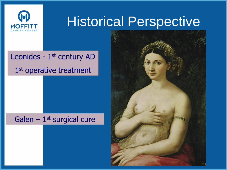
Leonides - 1st century AD
1st operative treatment
Galen – 1st surgical cure
Historical Perspective
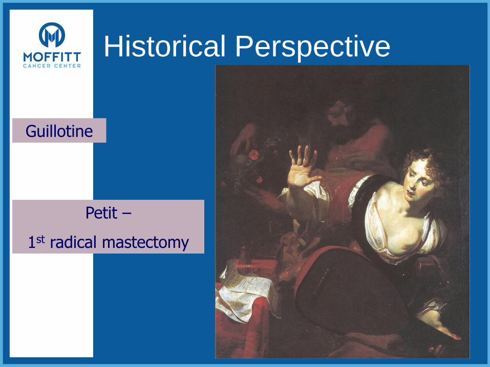
Guillotine
Petit –
1st radical mastectomy

Historical Perspective

Historical Perspective
Halsted
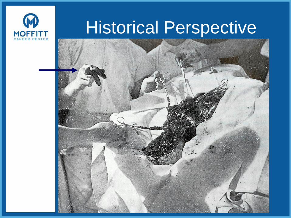
Historical Perspective

Mastectomy

Breast Reconstruction
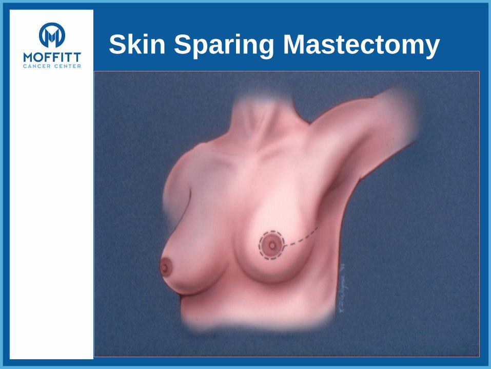
Skin Sparing Mastectomy
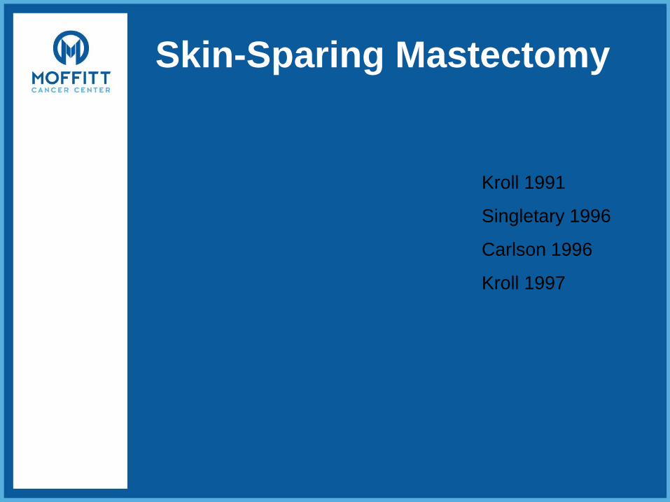
Skin-Sparing Mastectomy
Kroll 1991
Singletary 1996
Carlson 1996
Kroll 1997
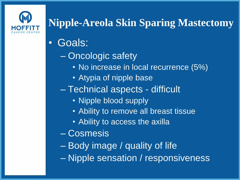
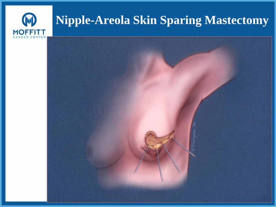
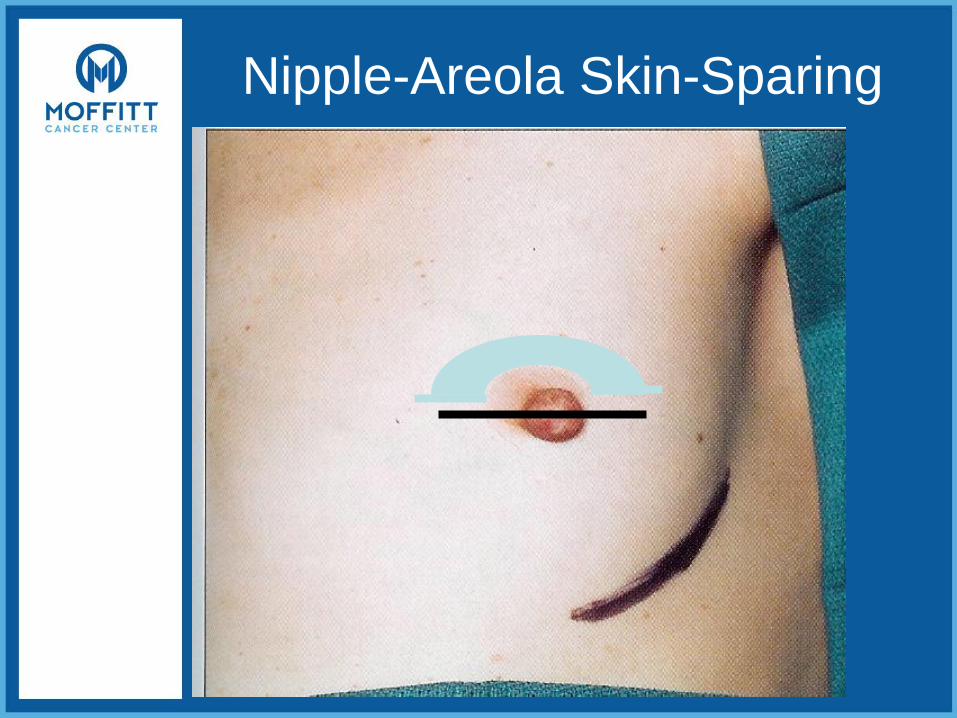
Nipple-Areola Skin Sparing Mastectomy
• Goals:
– Oncologic safety• No increase in local recurrence (5%)
• Atypia of nipple base
– Technical aspects - difficult• Nipple blood supply
• Ability to remove all breast tissue
• Ability to access the axilla
– Cosmesis
– Body image / quality of life
– Nipple sensation / responsiveness
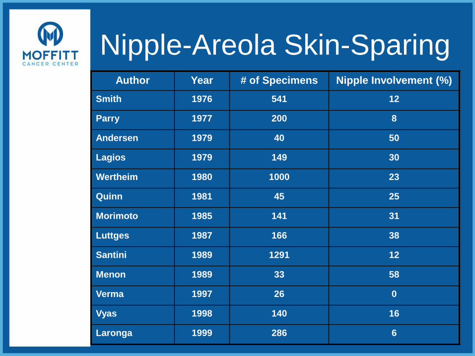
Nipple-Areola Skin-SparingAuthor Year # of Specimens Nipple Involvement (%)
Smith 1976 541 12
Parry 1977 200 8
Andersen 1979 40 50
Lagios 1979 149 30
Wertheim 1980 1000 23
Quinn 1981 45 25
Morimoto 1985 141 31
Luttges 1987 166 38
Santini 1989 1291 12
Menon 1989 33 58
Verma 1997 26 0
Vyas 1998 140 16
Laronga 1999 286 6
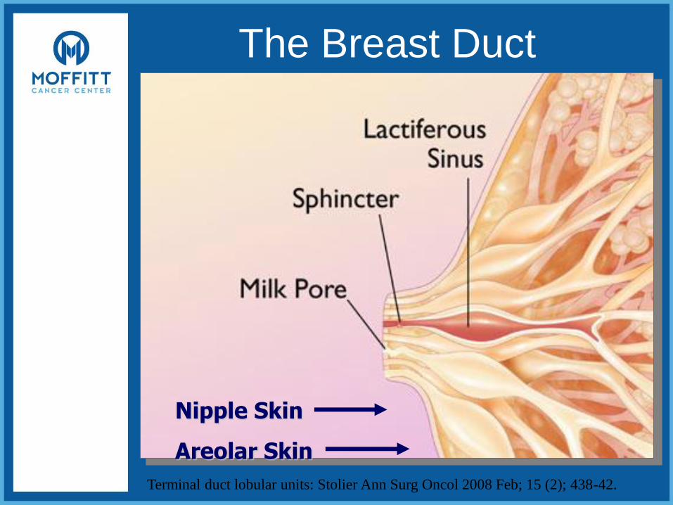
The Breast Duct
Nipple Skin
Areolar Skin
Terminal duct lobular units: Stolier Ann Surg Oncol 2008 Feb; 15 (2); 438-42.

Nipple-sparing Mastectomy
Nipple-Areola Skin Sparing Mastectomy
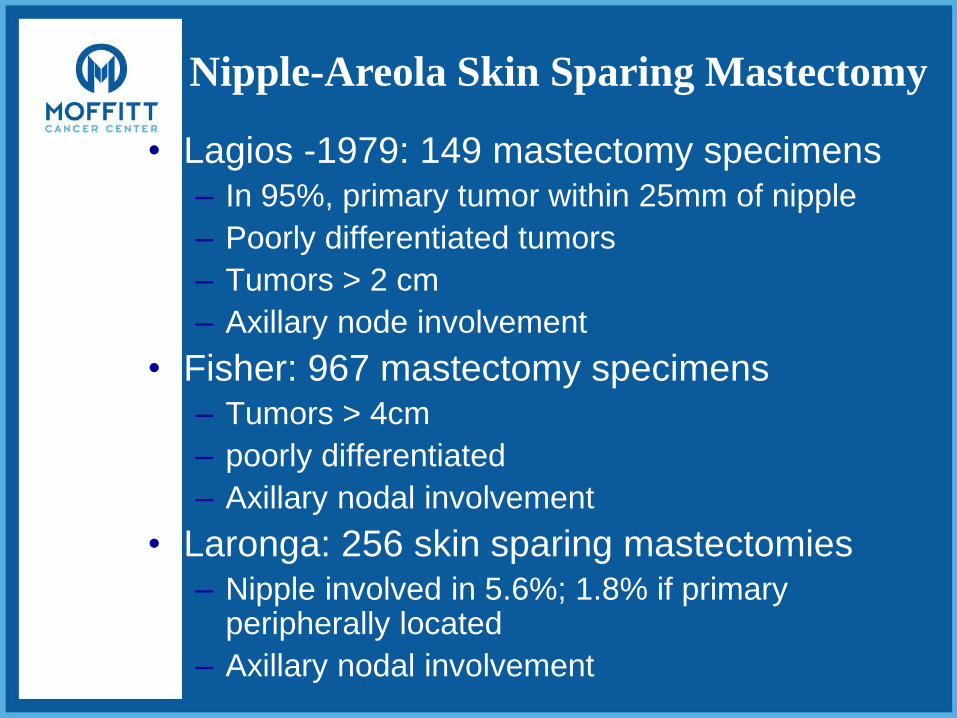
• Lagios -1979: 149 mastectomy specimens– In 95%, primary tumor within 25mm of nipple
– Poorly differentiated tumors
– Tumors > 2 cm
– Axillary node involvement
• Fisher: 967 mastectomy specimens – Tumors > 4cm
– poorly differentiated
– Axillary nodal involvement
• Laronga: 256 skin sparing mastectomies– Nipple involved in 5.6%; 1.8% if primary
peripherally located
– Axillary nodal involvement
Nipple-Areola Skin Sparing Mastectomy

Nipple-Areola Skin-Sparing

Nipple-Areola Skin Sparing Mastectomy
Author Year # of
Cases
Indication for
Surgery
Follow-Up LR or New
Hartmann 1999 575 SCM Prophylaxis 14 years 1%
Hartmann 2001 26 SCM Prophylaxis 13.4 years 0
Petit 2003 25 NSM Treatment 6 months NR
Gerber 2003 61 NSM Treatment 4.9 years 5%
Crowe 2004 44 NSM Both 6 weeks NR

Hartmann, LC et al. N Engl
J Med 340:77-84,1999
Subcutaneous Mastectomy
• Inframammary incision or infra-areola
incision
• Removes the majority of the breast
• Spares the skin and the nipple-areola
complex
• Requires reconstruction of breast mound
• Mayo clinic: 1/7 recurrences involved
nipple-areola complex

Pre-operative MRI
Nipple-Areola Skin Sparing Mastectomy
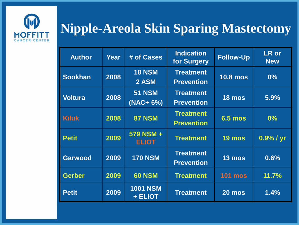
Author Year # of CasesIndication
for SurgeryFollow-Up
LR or
New
Sookhan 200818 NSM
2 ASM
Treatment
Prevention10.8 mos 0%
Voltura 200851 NSM
(NAC+ 6%)
Treatment
Prevention18 mos 5.9%
Kiluk 2008 87 NSMTreatment
Prevention6.5 mos 0%
Petit 2009579 NSM +
ELIOTTreatment 19 mos 0.9% / yr
Garwood 2009 170 NSMTreatment
Prevention13 mos 0.6%
Gerber 2009 60 NSM Treatment 101 mos 11.7%
Petit 20091001 NSM
+ ELIOTTreatment 20 mos 1.4%
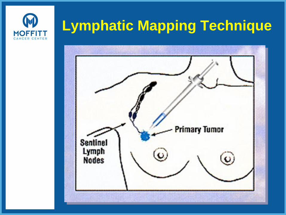
Lymphatic Mapping Technique
Nipple-Areola Skin-Sparing
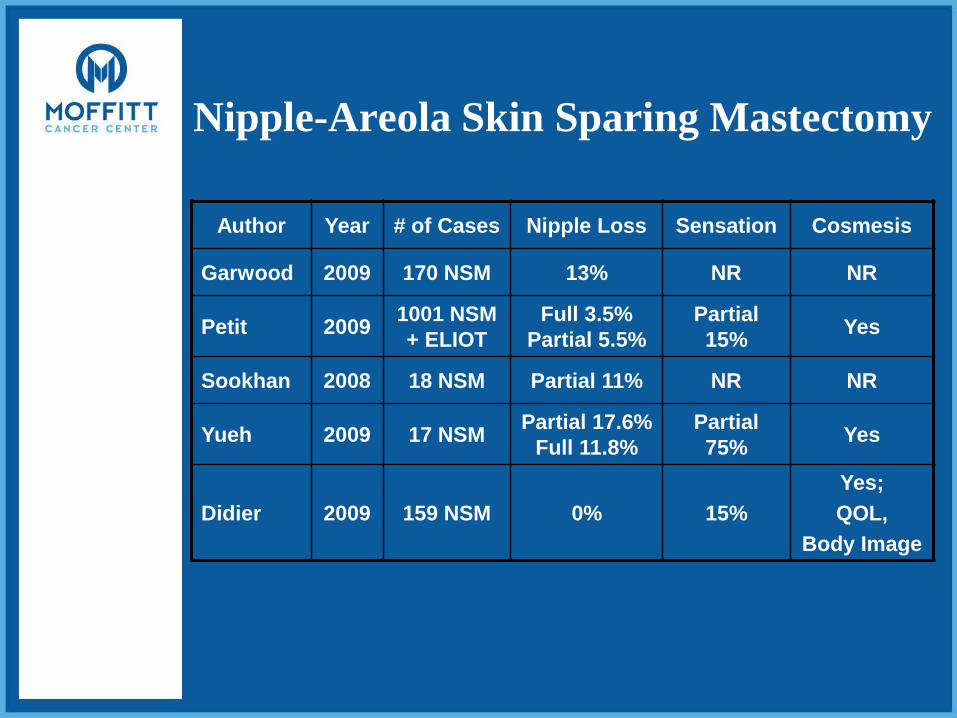
Nipple-Areola Skin Sparing Mastectomy
Author Year # of Cases Nipple Loss Sensation Cosmesis
Garwood 2009 170 NSM 13% NR NR
Petit 20091001 NSM
+ ELIOT
Full 3.5%
Partial 5.5%
Partial
15%Yes
Sookhan 2008 18 NSM Partial 11% NR NR
Yueh 2009 17 NSMPartial 17.6%
Full 11.8%
Partial
75%Yes
Didier 2009 159 NSM 0% 15%
Yes;
QOL,
Body Image
Performing NACSSM
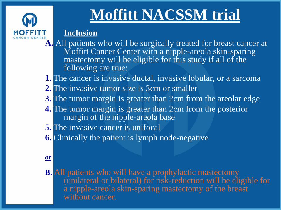
Moffitt NACSSM trialInclusion
A. All patients who will be surgically treated for breast cancer at Moffitt Cancer Center with a nipple-areola skin-sparing mastectomy will be eligible for this study if all of the following are true:
1. The cancer is invasive ductal, invasive lobular, or a sarcoma
2. The invasive tumor size is 3cm or smaller
3. The tumor margin is greater than 2cm from the areolar edge
4. The tumor margin is greater than 2cm from the posterior margin of the nipple-areola base
5. The invasive cancer is unifocal
6. Clinically the patient is lymph node-negative
or
B. All patients who will have a prophylactic mastectomy (unilateral or bilateral) for risk-reduction will be eligible for a nipple-areola skin-sparing mastectomy of the breast without cancer.
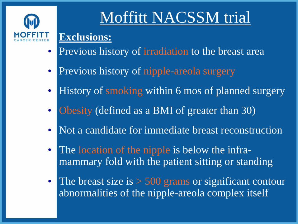
Moffitt NACSSM trial• Exclusions:
• Previous history of irradiation to the breast area
• Previous history of nipple-areola surgery
• History of smoking within 6 mos of planned surgery
• Obesity (defined as a BMI of greater than 30)
• Not a candidate for immediate breast reconstruction
• The location of the nipple is below the infra-mammary fold with the patient sitting or standing
• The breast size is > 500 grams or significant contour abnormalities of the nipple-areola complex itself

Nipple-Areola Skin-Sparing
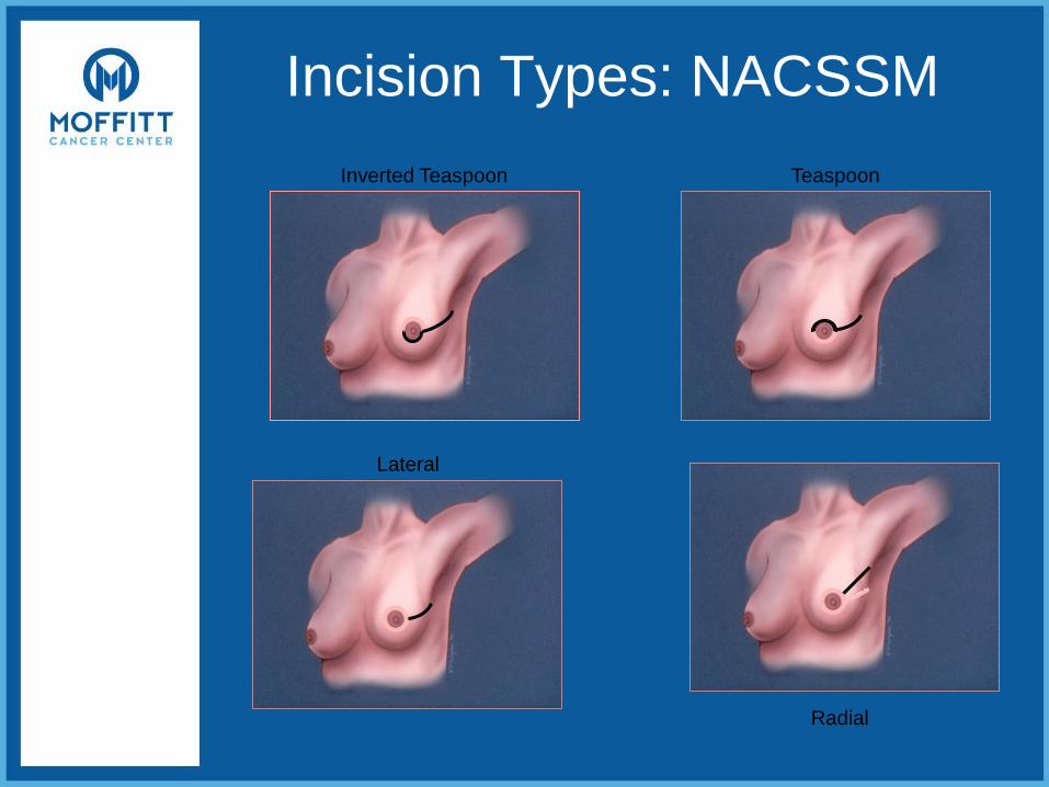
Incision Types: NACSSM
Radial
Inverted Teaspoon Teaspoon
Lateral
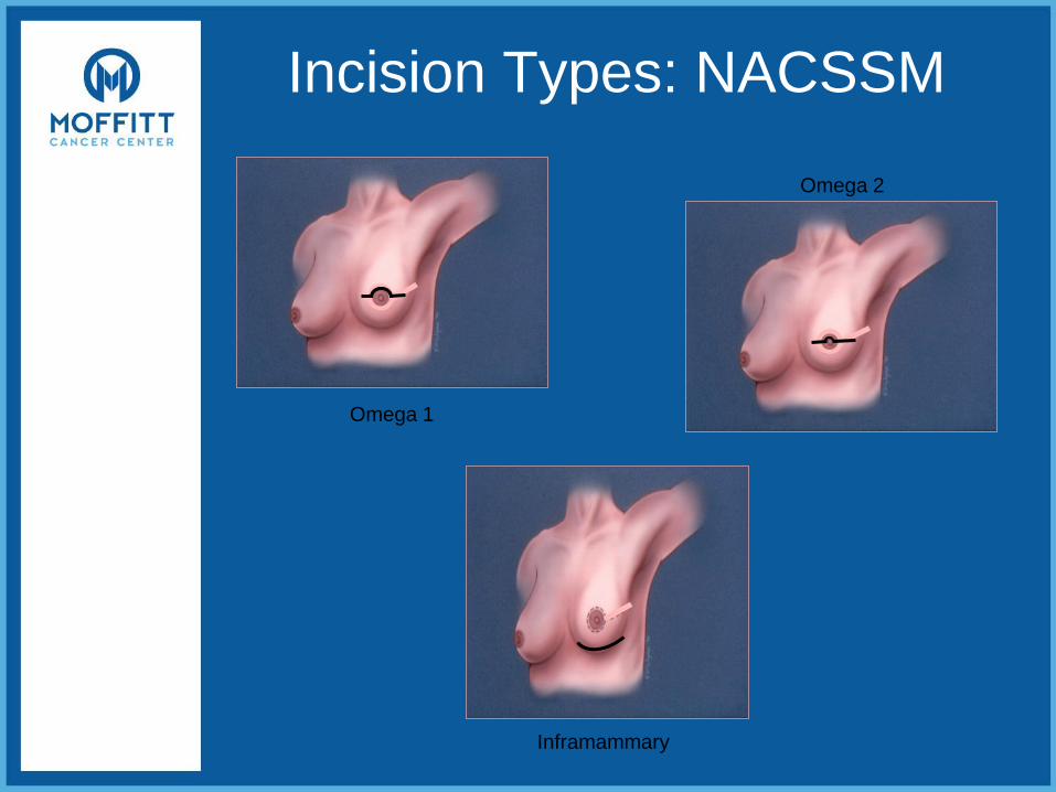
Incision Types: NACSSM
Omega 2
Omega 1
Inframammary

Body Image SurveyMake the following ratings based on how you feel about
your partner’s physical appearance
• 1 _ Strongly disagree
• 2 _ Disagree
• 3 _ Neither agree nor disagree
• 4 _ Agree
• 5 _ Strongly agree
• ___I am satisfied with the appearance of her left nipple
• ___I am satisfied with the appearance of her left breast
• ___I am satisfied with the appearance of her right nipple
• ___I am satisfied with the appearance of her right breast
• ___I think her left nipple is attractive
• ___I think her left breast is attractive
• ___I think her right nipple is attractive
• ___I think her right breast is attractive
Nipple-Areola Skin-Sparing
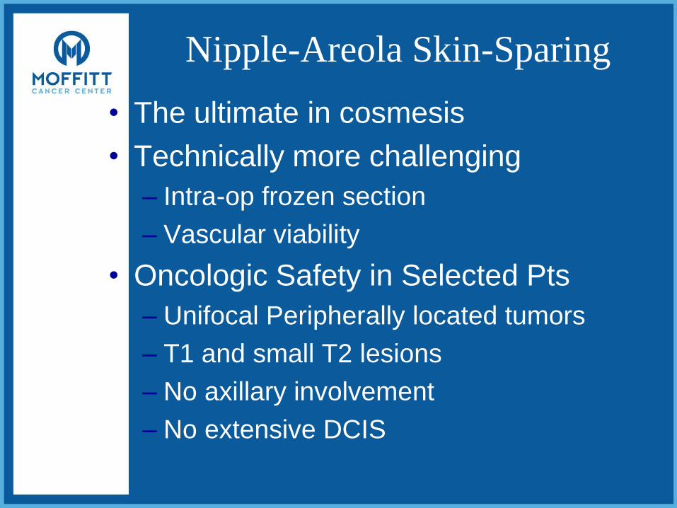
• The ultimate in cosmesis
• Technically more challenging
– Intra-op frozen section
– Vascular viability
• Oncologic Safety in Selected Pts
– Unifocal Peripherally located tumors
– T1 and small T2 lesions
– No axillary involvement
– No extensive DCIS