the circulatory system -...
TRANSCRIPT
Rajat Goyal and Michelle Fater
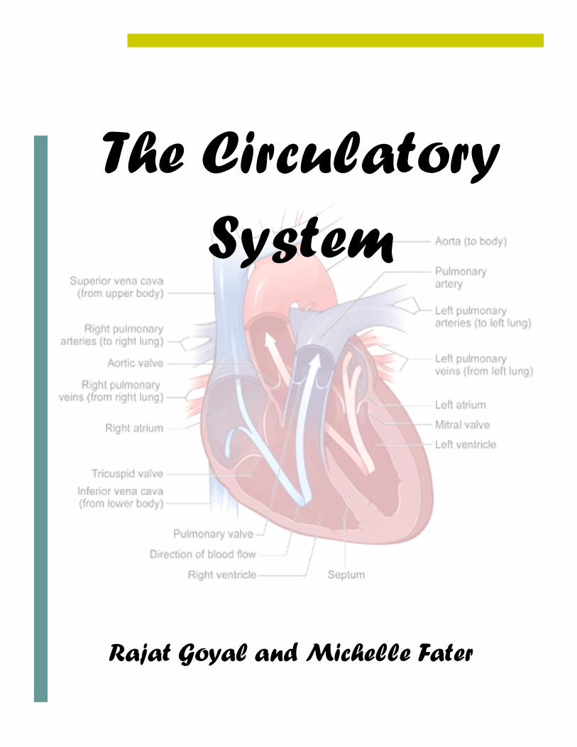
The Circulatory System
1
Table of Contents
Table of Contents 1 Human Organ Systems 2 The Circulatory System 3 Open Circulatory System 4 Closed Circulatory System 4 Functions of the System 5 Heart 6 Pulse 7 Chambers of the Heart 7 Arteries 8 Veins 9 Blood 10 Oxygenation of Blood 11 Red Blood Cells 12 White Blood Cells 13 Antigens 14 Plasma Cells and Platelets 15 Circulatory Systems of Other Organisms 16 Mammals and Birds 16 Amphibians and Reptiles 17 Fish 18 First Aid 19 Glossary 21 About the Authors 23 Illustration Credits 24
2
Human Organ Systems
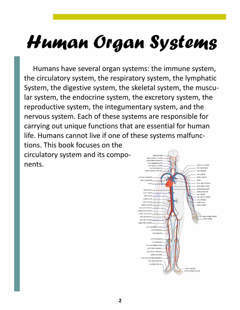
Humans have several organ systems: the immune system, the circulatory system, the respiratory system, the lymphatic System, the digestive system, the skeletal system, the muscu-lar system, the endocrine system, the excretory system, the reproductive system, the integumentary system, and the nervous system. Each of these systems are responsible for carrying out unique functions that are essential for human life. Humans cannot live if one of these systems malfunc-tions. This book focuses on the circulatory system and its compo-nents.
3
The Circulatory System
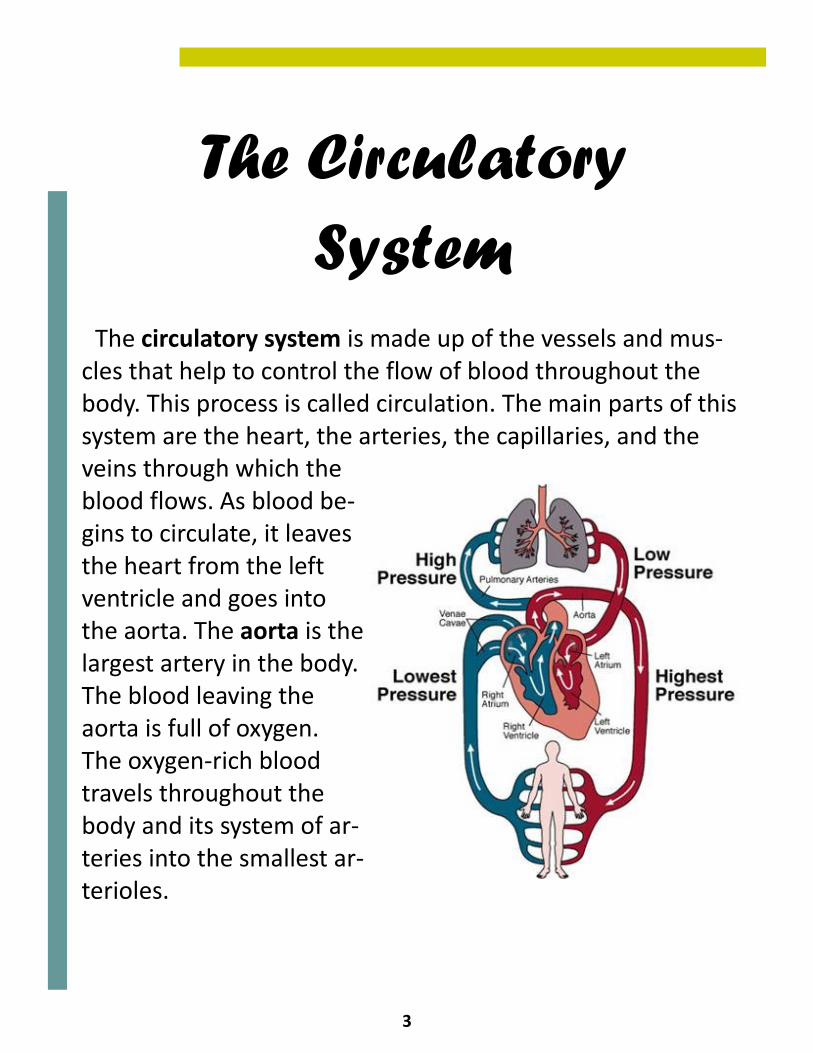
The circulatory system is made up of the vessels and mus-cles that help to control the flow of blood throughout the body. This process is called circulation. The main parts of this system are the heart, the arteries, the capillaries, and the veins through which the blood flows. As blood be-gins to circulate, it leaves the heart from the left ventricle and goes into the aorta. The aorta is the largest artery in the body. The blood leaving the aorta is full of oxygen. The oxygen-rich blood travels throughout the body and its system of ar-teries into the smallest ar-terioles.
4
Open and Closed Systems
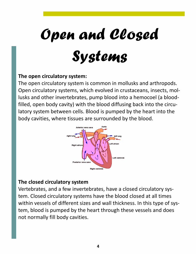
The open circulatory system: The open circulatory system is common in mollusks and arthropods. Open circulatory systems, which evolved in crustaceans, insects, mol-lusks and other invertebrates, pump blood into a hemocoel (a blood-filled, open body cavity) with the blood diffusing back into the circu-latory system between cells. Blood is pumped by the heart into the body cavities, where tissues are surrounded by the blood. The closed circulatory system Vertebrates, and a few invertebrates, have a closed circulatory sys-tem. Closed circulatory systems have the blood closed at all times within vessels of different sizes and wall thickness. In this type of sys-tem, blood is pumped by the heart through these vessels and does not normally fill body cavities.
5
Functions of the System
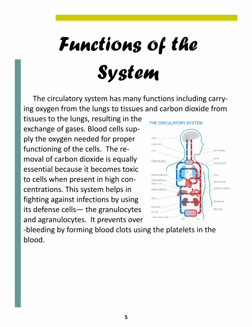
The circulatory system has many functions including carry-ing oxygen from the lungs to tissues and carbon dioxide from tissues to the lungs, resulting in the exchange of gases. Blood cells sup-ply the oxygen needed for proper functioning of the cells. The re-moval of carbon dioxide is equally essential because it becomes toxic to cells when present in high con-centrations. This system helps in fighting against infections by using its defense cells— the granulocytes and agranulocytes. It prevents over-bleeding by forming blood clots using the platelets in the blood.
6
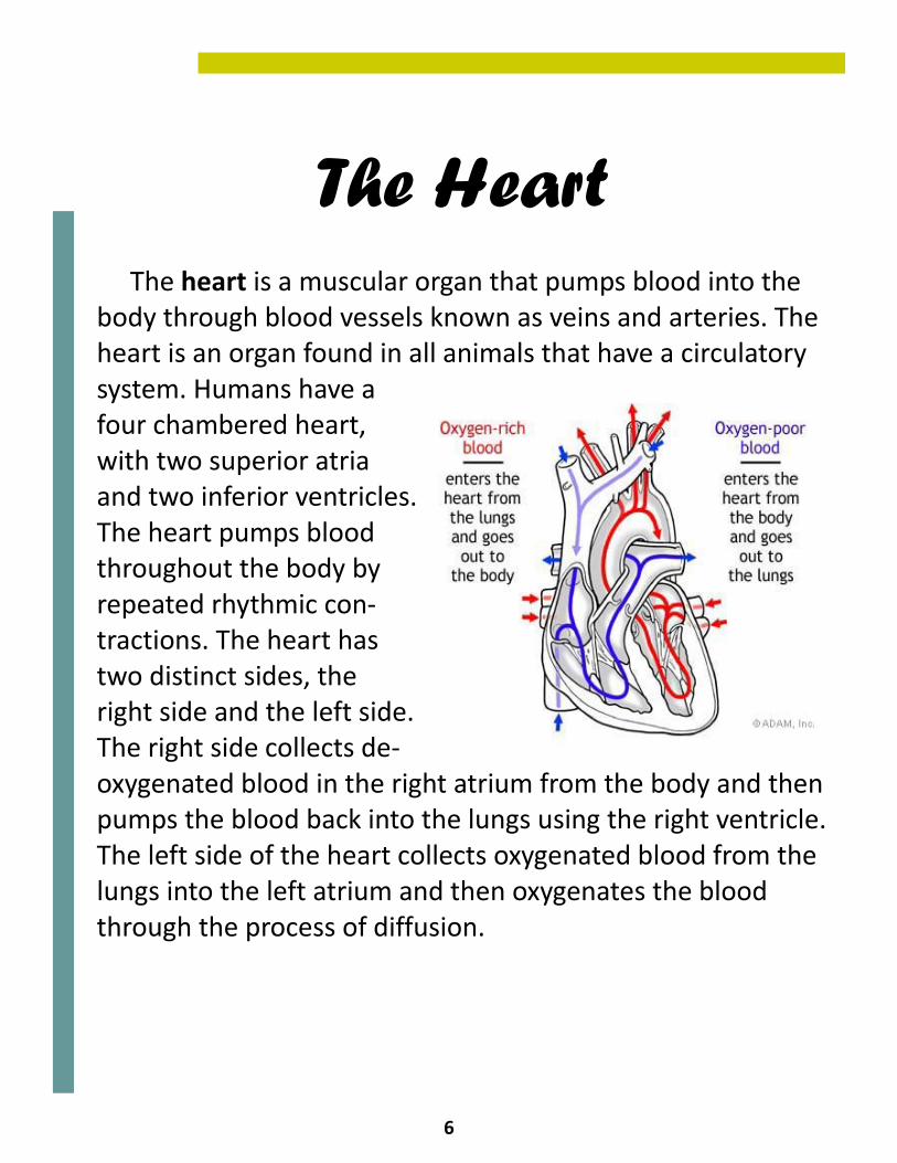
The Heart The heart is a muscular organ that pumps blood into the body through blood vessels known as veins and arteries. The heart is an organ found in all animals that have a circulatory system. Humans have a four chambered heart, with two superior atria and two inferior ventricles. The heart pumps blood throughout the body by repeated rhythmic con-tractions. The heart has two distinct sides, the right side and the left side. The right side collects de-oxygenated blood in the right atrium from the body and then pumps the blood back into the lungs using the right ventricle. The left side of the heart collects oxygenated blood from the lungs into the left atrium and then oxygenates the blood through the process of diffusion.
7
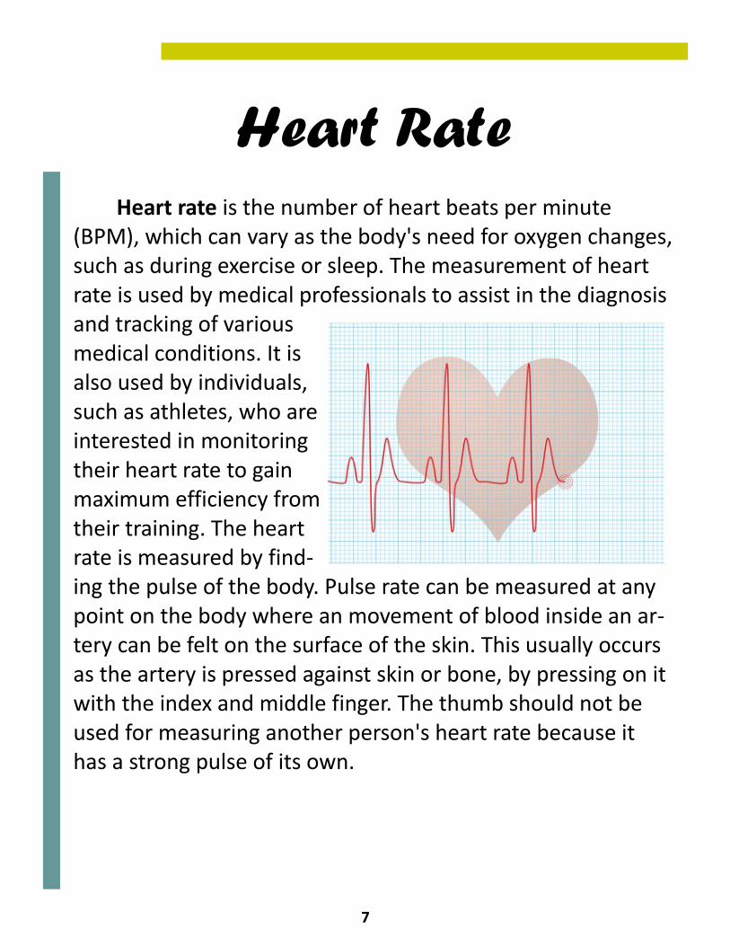
Heart Rate Heart rate is the number of heart beats per minute (BPM), which can vary as the body's need for oxygen changes, such as during exercise or sleep. The measurement of heart rate is used by medical professionals to assist in the diagnosis and tracking of various medical conditions. It is also used by individuals, such as athletes, who are interested in monitoring their heart rate to gain maximum efficiency from their training. The heart rate is measured by find-ing the pulse of the body. Pulse rate can be measured at any point on the body where an movement of blood inside an ar-tery can be felt on the surface of the skin. This usually occurs as the artery is pressed against skin or bone, by pressing on it with the index and middle finger. The thumb should not be used for measuring another person's heart rate because it has a strong pulse of its own.
8
Arteries
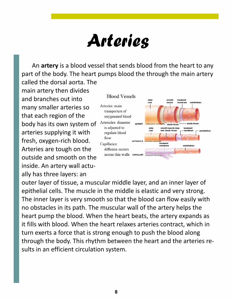
An artery is a blood vessel that sends blood from the heart to any part of the body. The heart pumps blood the through the main artery called the dorsal aorta. The main artery then divides and branches out into many smaller arteries so that each region of the body has its own system of arteries supplying it with fresh, oxygen-rich blood. Arteries are tough on the outside and smooth on the inside. An artery wall actu-ally has three layers: an outer layer of tissue, a muscular middle layer, and an inner layer of epithelial cells. The muscle in the middle is elastic and very strong. The inner layer is very smooth so that the blood can flow easily with no obstacles in its path. The muscular wall of the artery helps the heart pump the blood. When the heart beats, the artery expands as it fills with blood. When the heart relaxes arteries contract, which in turn exerts a force that is strong enough to push the blood along through the body. This rhythm between the heart and the arteries re-sults in an efficient circulation system.
9
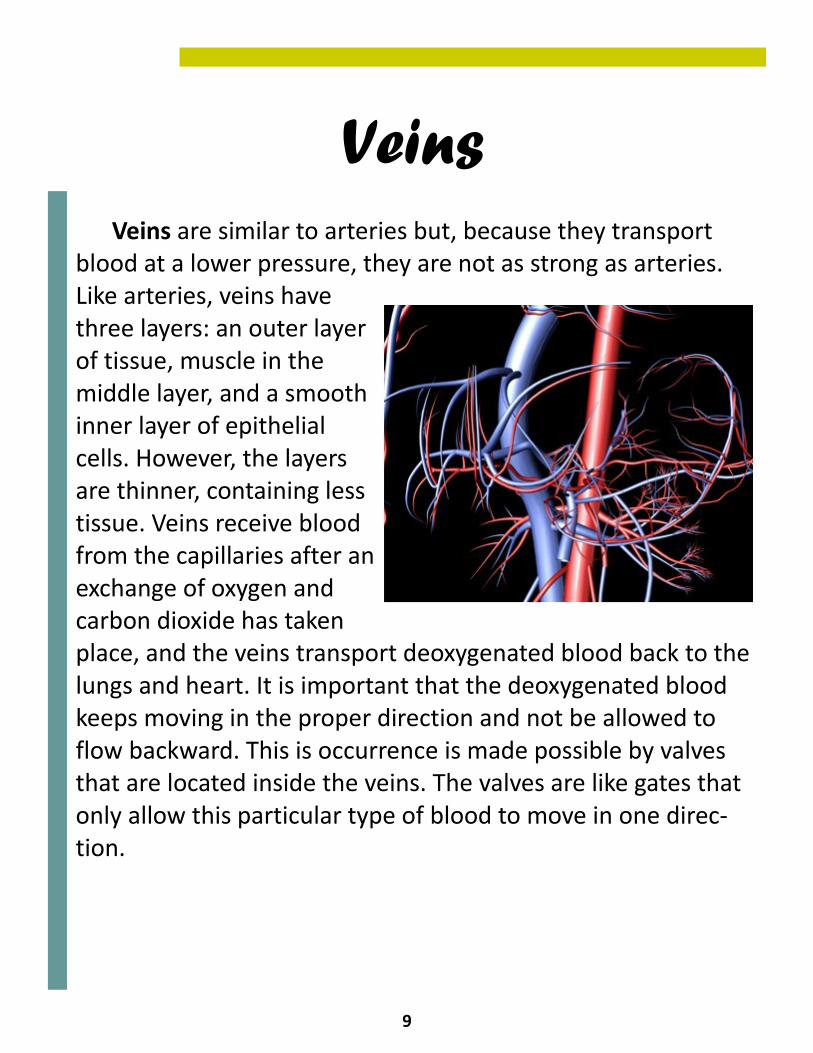
Veins Veins are similar to arteries but, because they transport blood at a lower pressure, they are not as strong as arteries. Like arteries, veins have three layers: an outer layer of tissue, muscle in the middle layer, and a smooth inner layer of epithelial cells. However, the layers are thinner, containing less tissue. Veins receive blood from the capillaries after an exchange of oxygen and carbon dioxide has taken place, and the veins transport deoxygenated blood back to the lungs and heart. It is important that the deoxygenated blood keeps moving in the proper direction and not be allowed to flow backward. This is occurrence is made possible by valves that are located inside the veins. The valves are like gates that only allow this particular type of blood to move in one direc-tion.
10
Blood
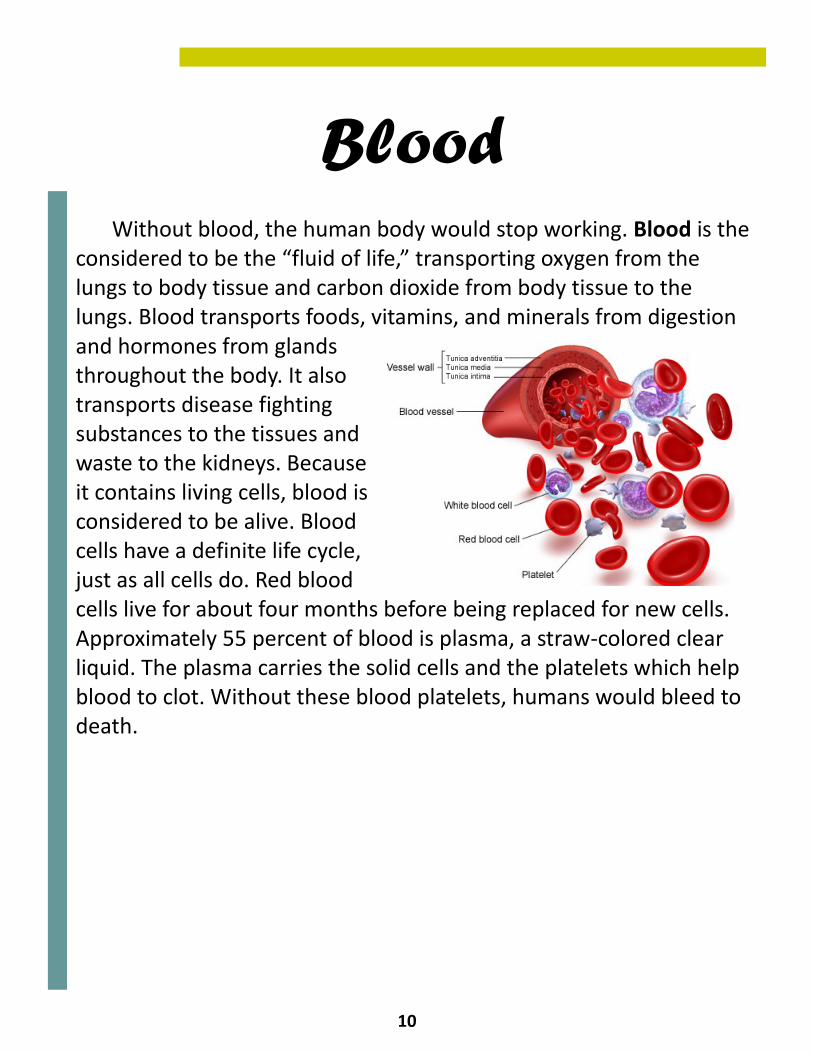
Without blood, the human body would stop working. Blood is the considered to be the “fluid of life,” transporting oxygen from the lungs to body tissue and carbon dioxide from body tissue to the lungs. Blood transports foods, vitamins, and minerals from digestion and hormones from glands throughout the body. It also transports disease fighting substances to the tissues and waste to the kidneys. Because it contains living cells, blood is considered to be alive. Blood cells have a definite life cycle, just as all cells do. Red blood cells live for about four months before being replaced for new cells. Approximately 55 percent of blood is plasma, a straw-colored clear liquid. The plasma carries the solid cells and the platelets which help blood to clot. Without these blood platelets, humans would bleed to death.
11
The Oxygenation of Blood
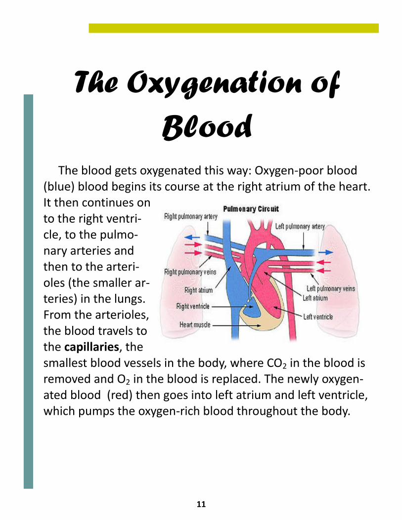
The blood gets oxygenated this way: Oxygen-poor blood (blue) blood begins its course at the right atrium of the heart. It then continues on to the right ventri-cle, to the pulmo-nary arteries and then to the arteri-oles (the smaller ar-teries) in the lungs. From the arterioles, the blood travels to the capillaries, the smallest blood vessels in the body, where CO2 in the blood is removed and O2 in the blood is replaced. The newly oxygen-ated blood (red) then goes into left atrium and left ventricle, which pumps the oxygen-rich blood throughout the body.
12
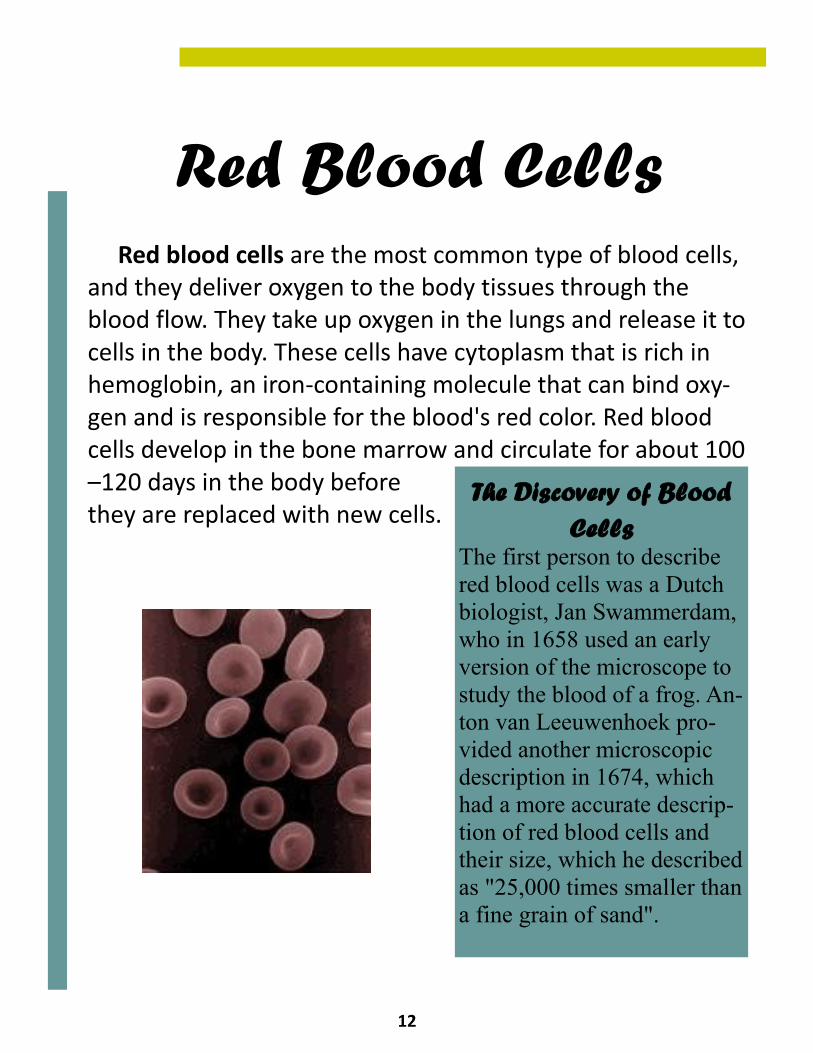
Red Blood Cells Red blood cells are the most common type of blood cells, and they deliver oxygen to the body tissues through the blood flow. They take up oxygen in the lungs and release it to cells in the body. These cells have cytoplasm that is rich in hemoglobin, an iron-containing molecule that can bind oxy-gen and is responsible for the blood's red color. Red blood cells develop in the bone marrow and circulate for about 100–120 days in the body before they are replaced with new cells.
The Discovery of Blood Cells
The first person to describe red blood cells was a Dutch biologist, Jan Swammerdam, who in 1658 used an early version of the microscope to study the blood of a frog. An-ton van Leeuwenhoek pro-vided another microscopic description in 1674, which had a more accurate descrip-tion of red blood cells and their size, which he described as "25,000 times smaller than a fine grain of sand".
13
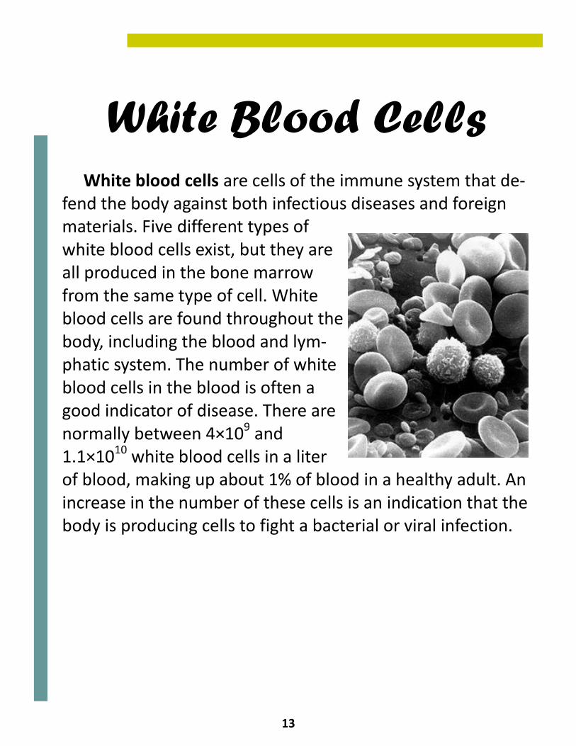
White Blood Cells White blood cells are cells of the immune system that de-fend the body against both infectious diseases and foreign materials. Five different types of white blood cells exist, but they are all produced in the bone marrow from the same type of cell. White blood cells are found throughout the body, including the blood and lym-phatic system. The number of white blood cells in the blood is often a good indicator of disease. There are normally between 4×10⁹ and 1.1×10¹⁰ white blood cells in a liter of blood, making up about 1% of blood in a healthy adult. An increase in the number of these cells is an indication that the body is producing cells to fight a bacterial or viral infection.
14
Blood Types and Antigens
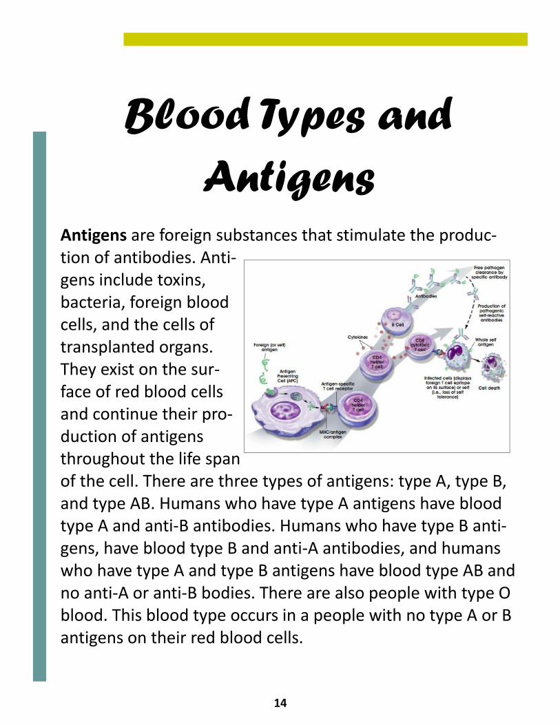
Antigens are foreign substances that stimulate the produc-tion of antibodies. Anti-gens include toxins, bacteria, foreign blood cells, and the cells of transplanted organs. They exist on the sur-face of red blood cells and continue their pro-duction of antigens throughout the life span of the cell. There are three types of antigens: type A, type B, and type AB. Humans who have type A antigens have blood type A and anti-B antibodies. Humans who have type B anti-gens, have blood type B and anti-A antibodies, and humans who have type A and type B antigens have blood type AB and no anti-A or anti-B bodies. There are also people with type O blood. This blood type occurs in a people with no type A or B antigens on their red blood cells.
15
Plasma Cells and Platelets
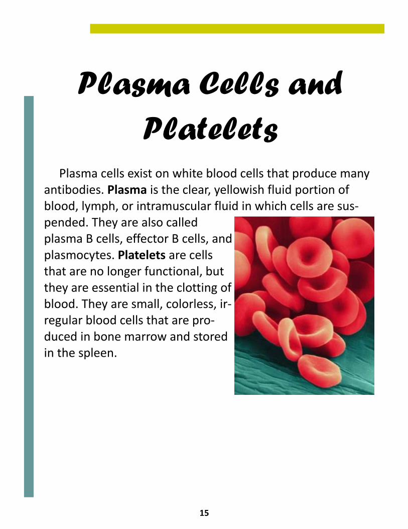
Plasma cells exist on white blood cells that produce many antibodies. Plasma is the clear, yellowish fluid portion of blood, lymph, or intramuscular fluid in which cells are sus-pended. They are also called plasma B cells, effector B cells, and plasmocytes. Platelets are cells that are no longer functional, but they are essential in the clotting of blood. They are small, colorless, ir-regular blood cells that are pro-duced in bone marrow and stored in the spleen.
16
The Circulatory System of Other Organisms
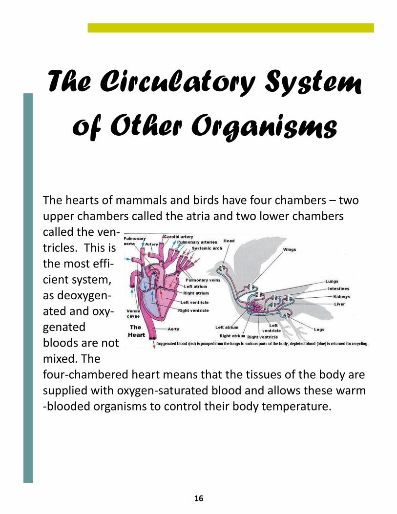
The hearts of mammals and birds have four chambers – two upper chambers called the atria and two lower chambers called the ven-tricles. This is the most effi-cient system, as deoxygen-ated and oxy-genated bloods are not mixed. The four-chambered heart means that the tissues of the body are supplied with oxygen-saturated blood and allows these warm-blooded organisms to control their body temperature.
17
Circulatory System of Other Organisms
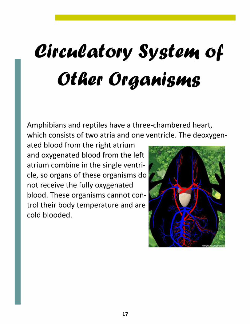
Amphibians and reptiles have a three-chambered heart, which consists of two atria and one ventricle. The deoxygen-ated blood from the right atrium and oxygenated blood from the left atrium combine in the single ventri-cle, so organs of these organisms do not receive the fully oxygenated blood. These organisms cannot con-trol their body temperature and are cold blooded.
18
Circulatory System of Other Organisms
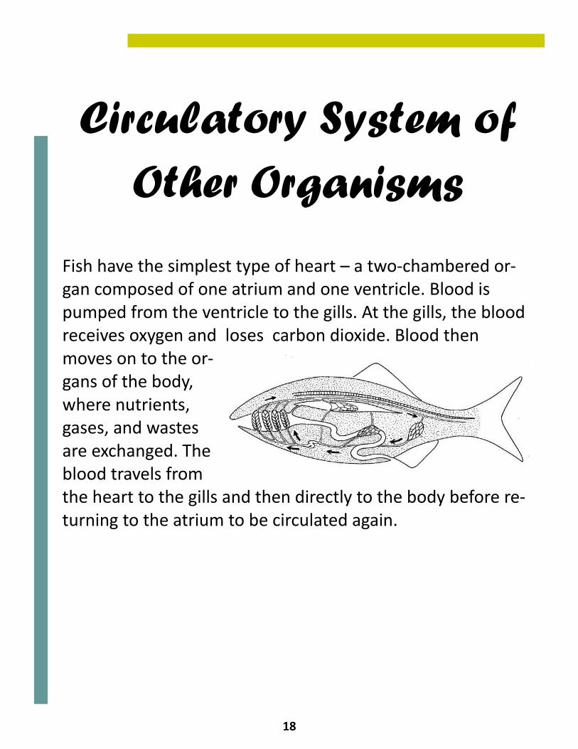
Fish have the simplest type of heart – a two-chambered or-gan composed of one atrium and one ventricle. Blood is pumped from the ventricle to the gills. At the gills, the blood receives oxygen and loses carbon dioxide. Blood then moves on to the or-gans of the body, where nutrients, gases, and wastes are exchanged. The blood travels from the heart to the gills and then directly to the body before re-turning to the atrium to be circulated again.
19
First Aid for the Circulatory System
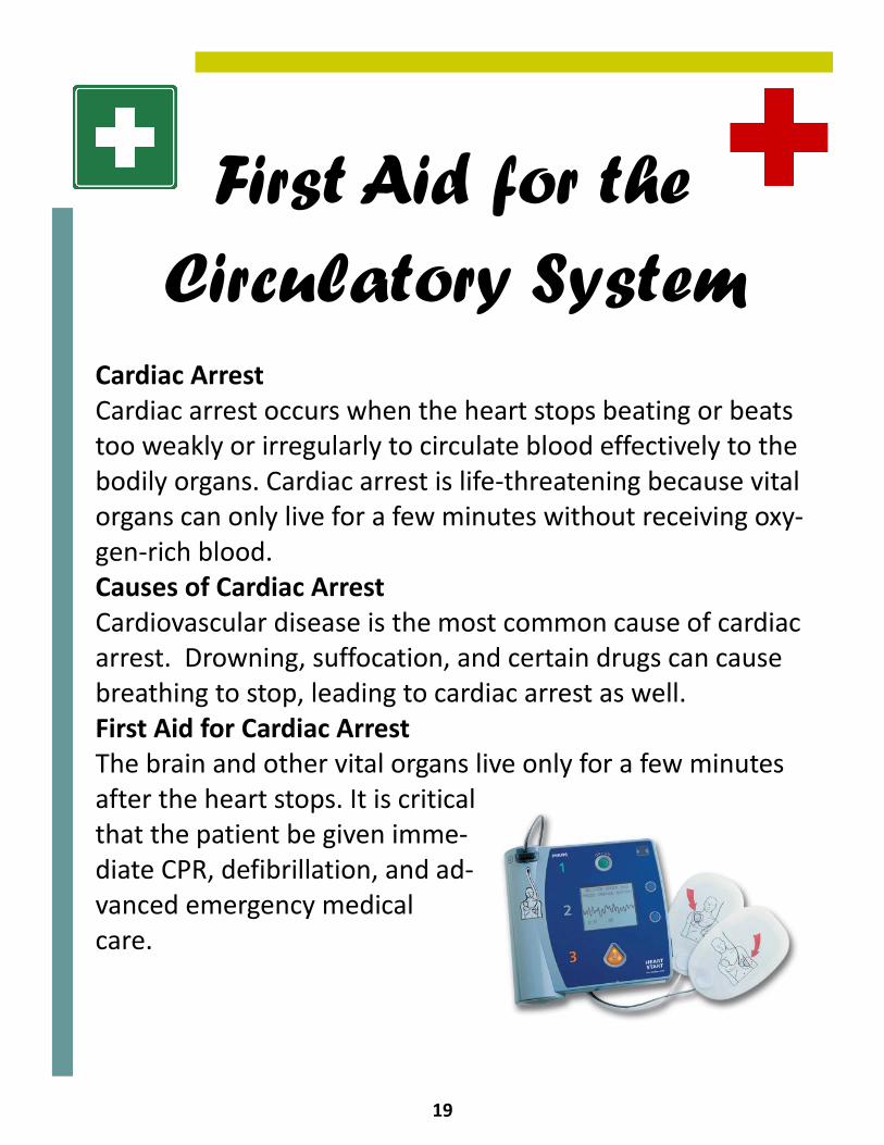
Cardiac Arrest Cardiac arrest occurs when the heart stops beating or beats too weakly or irregularly to circulate blood effectively to the bodily organs. Cardiac arrest is life-threatening because vital organs can only live for a few minutes without receiving oxy-gen-rich blood. Causes of Cardiac Arrest Cardiovascular disease is the most common cause of cardiac arrest. Drowning, suffocation, and certain drugs can cause breathing to stop, leading to cardiac arrest as well. First Aid for Cardiac Arrest The brain and other vital organs live only for a few minutes after the heart stops. It is critical that the patient be given imme-diate CPR, defibrillation, and ad-vanced emergency medical care.
20
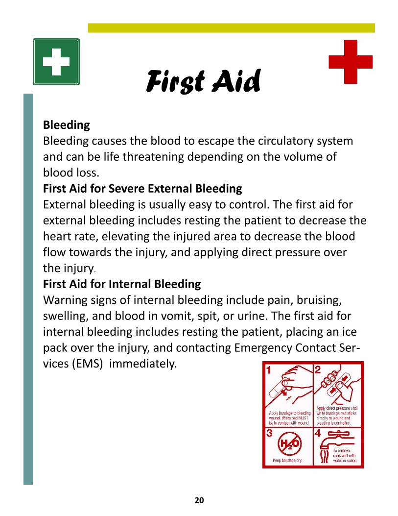
First Aid Bleeding Bleeding causes the blood to escape the circulatory system and can be life threatening depending on the volume of blood loss. First Aid for Severe External Bleeding External bleeding is usually easy to control. The first aid for external bleeding includes resting the patient to decrease the heart rate, elevating the injured area to decrease the blood flow towards the injury, and applying direct pressure over the injury.
First Aid for Internal Bleeding Warning signs of internal bleeding include pain, bruising, swelling, and blood in vomit, spit, or urine. The first aid for internal bleeding includes resting the patient, placing an ice pack over the injury, and contacting Emergency Contact Ser-vices (EMS) immediately.
21
Glossary
Antigens: Foreign substances that stimulate the production of antibodies. Aorta: The largest artery in the body. Artery: A blood vessel that sends blood from the heart to any part of the body. Capillaries: The smallest blood vessels in the body, where the nutrients in the blood are exchanged for new blood vessels. Circulatory system: The system made up of the vessels and muscles that help to control the flow of blood throughout the body. Heart: A muscular organ that pumps blood into the body through veins and arteries using blood vessels. Heart rate: The number of heart beats per minute (bpm), which can vary as the body's need for oxygen changes, such as during exercise or sleep.
22
Plasma: Clear, yellowish fluid portion of blood, lymph, or in-tramuscular fluid where cells are suspended. Platelets: Cells that are no longer functional, but are essen-tial in the clotting of blood. Red blood cells: The most common type of blood cells and are one of the principal means of delivering oxygen (O2) to the body tissues through the blood flow. Veins: While similar to arteries, they transport blood at a lower pressure, they are not as strong as arteries. the body tissues through the blood flow. White blood cells: Cells of the immune system that defend the body against both infectious diseases and foreign materi-als.

23
About the Authors
Rajat Goyal
Rajat Goyal is a junior at the Massachusetts Academy of Mathematics and Science in Worcester, Massachusetts. Recently he performed an inde-pendent research project that analyzed the effect of turmeric (a spice) on Escherichia coli . He has also recently written a book review on Doctor’s Diaries (a NOVA documentary) and coauthored an e-book chapter for Topics in Toxicology. In his spare time, Rajat enjoys skiing, biking, and ka-rate.
Michelle Fater
Michelle Fater is currently a junior at the Massachusetts Academy of Mathematics and Science in Worcester, Massachusetts. Recently she per-formed an independent research project as well as published several pieces of writing, including an e-book chapter for Topics in Toxicology. In her spare time, Michelle enjoys painting, musical theatre, and ball-room dancing.
24
Illustration Credits
Cover Picture: http://www.nhlbi.nih.gov/health/dci/Diseases/hb/ hb_understanding.html P.2: http://www.globalneighbourhood.org/k-presentation.php P.3: http://doctorgrasshopper.wordpress.com/2010/03/11/tools- for-the-toolbox-i-3-the-lub-dubber/ P.4: http://www2.gsu.edu/~bioasx/closeopen.html P.5: http://www.heartzine.com/anatomy-physiology/the-circulatory- system.html P.6: http://www.nytimes.com/imagepages/2007/08/01/health/ adam/19387Circulationofbloodthroughtheheart.html P.7: http://www.stormgrounds.com/wallpaper/Miscellaneous/ Heart-Rate/ P.8: http://www.kidney-hypertension.com/hypertension.htm P.9: http://www.landholt.com/3d/arteries_and_veins/ P.10: http://health.nytimes.com/health/guides/specialtopic/ physical-activity/exercise's-effects-on-the-heart.html P.11: http://en.wikipedia.org/wiki File:Diagram_of_the_human_heart_(cropped).svg P.12: http://en.wikipedia.org/wiki/Red_blood_cell P.13: http://en.wikipedia.org/wiki/White_blood_cell P.14: http://stemcells.nih.gov/info/scireport/chapter6.asp P.15: http://repairstemcell.wordpress.com/2009/02/ P.16: http://www.paulnoll.com/Oregon/Birds/Avian-Circulatory.html P.17:http://www.digitalfrog.com/resources/archives/circ.jpg P.18:http://www.biology-resources.com/drawing-fish-circulatory- system.html P.19:http://kytostat.com/Portals/0/howItWorksGraphic.jpg http://en.wikipedia.org/wiki/First_aid P.20:http://www.cbc.ca/22minutes/defibrillator-with-ecg-display-- 4.jpg http://en.wikipedia.org/wiki/First_aid