the comparative histology of the alimentary canal of
TRANSCRIPT

T H E C O M P A R A T I V E H I S T O L O G Y OF T H E A L I M E N T A R Y C A N A L OF C E R T A I N FRESH WATER TELEOST FISHES
BY AHSAN-UL-ISLAM, M.Sc. (HONS.) (Depa,'tment of'Zoology, University of the Panjab, Lahore)
Received December 31, 1949
(Communicated by Dr. Harold Khan Bhatti, V.A.SC.)
THE work done on the histology of the alimentary canal of fishes of Europe and America has been summarised by Dawes (1929). Recently some valuable work has been done by Al-Hussaini (1946, 1947, 1949 a, 1949 b) on the histology, cytology and physiology of the gut of Teleost fishes from the Red Sea. Very little work has, however, been done on the histology of the aii- mentary canal of fishes of this subcontinent, the only noticeable contribu- tions to the subject being those of Rahimullah (1935, 1936, 1939, 1941, 1943 and 1945), Dharmarajan (1936), Vanajakshi (1938), Sarbahi (1940) and Mohsin (1942, 1944--46).
The object of the present study is to find any possible correlatiol~ between the histological structure of the gut and the food and feeding habits of fishes. No work has so far been done with this object on the indigenous fishes. A study of the skin of the fishes has also been made as such a study greatly helps in the correct interpretation of the various structures in the lining of the buccal cavity and the pharynx. This factor has been completely neglected by previous workers on the histology of the alimentary canal and no use has been made of the extensive literature on the integument of fishes.
MATERIAL AND METHOD
Living specimens of Rita rita Hamilton, Cirrhina mrigala Hamilton and Ophicephalus gachua Hamilton were obtained from fishermen. Small pieces of the various regions of the alimentary canal were fixed in Bouin's fluid or in Zenker-Formol for 16 to 18 hours. Pieces of skin from the head region were either fixed in Jenkin's decalcifying and dehydrating fixative or in Bouin's fluid for 16 to 20 hours. Satisfactory results were obtained with all these fixatives, though Bouin's fluid was preferred on account of its being easy to make, store, use and wash out.
The portions of the alimentary canal studied were from the buccal cavity, pharynx, oesophagus, stomach and intestine along with the rectum.
m 297

298 Ahsan-ul-Isiam
Tongue and pyloric c~eca present in Ophicephalus gachua were also studied. Contents of the guts of all the available specimens of each fish were examined to verify the previous reports on the food of these fishes.
FOOD
Study of the contents of the stomach and intestine of Rita rita showed that two fishes had taken shrimp-like crustacea of fairly big size measuring over 1�88 inches in length, one had taken some insect larva, while the stomachs and intestines of two fishes were quite empty. Rita rita is carnivorous and as stated by Hamid Khan (1934) " i t feeds mostly on insects, their larvae and young fishes".
Ophicephalus gaehua too is carnivorous and " destroys eggs, and fry of other fishes " (Hamid Khan, 1934). Mookerjee, Ganguly and Islam (1946) find that in the case of Ophicephalus punctatus, the fish when very young takes only unicellular and multicellular alga ~. Later, they take to carnivo- rous diet consisting of Protozoa, Insecta, Copepoda, etc. As growth proceeds, the inclination towards fish-diet becomes evident, till in the adult, the per- centage of fish in the diet of this fish may be upto 100~. Ophicephalus gachua is said to have a food similar to that of O. punctatu~.
Cirrhina mrigala is a vegetarian fish but "has no objection to taking a bait of worms" (Hamid Khan, 1934). Mookerjee and Das (1945) de- scribe Cirrhina mrigala as a bottom feeder taking lots of mud a~d sand. Mookerjee and Ghosh (1945) find that although adult Cirrhina mr(gala "takes a mixed diet but the animal diet is almost negligible in comparison with vegetable . . . . . . The major portion of the diet is the higher plant bodies in semi rotten condition".
SKIN
The skin consists of epidermis and corium. A basement membrane usually separates the two regions.
Epidermis.--The epidermis of Rita rita is stratified and as described by Bhatti (1938) consists of ordinary epidermal cells, club-cells, mucus-cells, sensory cells and fibroblasts. The ordinary epidermal or epithelial cells (Fig. 1, epc.) are oval in shape with a large vesicular nucleus. In the super- ficial layers they tend to get flattened and lie parallel to the surface. The basal layer (epb.) rests on the basement membrane (b.m.) and consists of columnar squarish or rectangular cells. Club-cells (c.c.) are large oval or round cells having a central nucleus and cytoplasm which stains pink with eosin. In young cells the cytoplasm is homogeneous, but in older cells

Histology ofi Ali~nentayy Cenal o/'Fresk l, Kater l'deos! t~?shes 299
small vacuoles may appear inside the cells or near the periphery. The club- cells vary considerably in size, form and arrangement in the skin from dif- ferent regions of the body. Generally they are arranged in two or three rows. Some of the club-cells may be produced into a neck-like structure, but this neck never opens to the exterior. Mucus-cells or goblet cells (m.c.) are large round cells with basophilic reticular contents staining deeply with h0ema- toxylin. Each mucus-cell has a crescentic nucleus lying at the l:ottom of the goblet-shaped body. The mucus-cells are scattered all over the epidermis, but are more common in the upper layers, where they may be seen opening to the exterior by a very short neck. Sensory cells generally occur in the form of end-buds, which are identical with t;:c taste-buds found in the buccal cavity and pharynx. The taste-bud is an aggregation of sensory cells into a flask-shaped body situated on a finger-like projection of the corium. Fibroblasts are spindle-shaped cells with big granular nuclei lying in the basal layer of the epidermis.
In Ophicephalus gachua, the epidermis is thin and stratified, it co~sists of squarish or polygonal epithelial cells and numerous sac-cells (Fig. 2, s.c.). Club-cells are absent. The sac-cells are generally big cells, traversing more than three-fourth the thickness of the epidermal layer. Occasionally small sac-cells may be found in two or more layers. Very often the bigger cells can be seen to open to the outside by a short neck. These cells possess a very deeply-staining crescentic nucleus lying at the bottom of the sac. These cells differ from the typical mucus-cells in their staining reaction: the latter stain blue with h~ematoxylin, but the sac-cells fail to do so and pick up cyto- plasmic stains. These sac-cells resemble closely the "SackJblwzige serose Driisen" or "sac-cells" described by Bhatti (1938) in the integument of some Siluroidea. No sensory elements in the form of end buds ~eem to be present in the portion of the skin of Ophicepka&s gachua studied. A distinct base- ment membrane is not present.
In Cirrhina mrigala the epidermis contains, in addition to the ordinary epidermal cells (Fig. 3, epc), numerous club-cells (c.c.) which have the typical structure exactly like those on the skin of Rita rita. At places the club- cells are seen protruding out of the general surface of the epidermis and may sometimes be seen completely ejected out. A similar condition has been described by Bhatti (1938) in some Siluroids and has been interpreted as indicating an excretory function. Mucus-cells, sensory elements and fibroblasts are not distinguishable in the region of the skin studied.
Corium.--In Rita rita the upper region of the corium (Fig. 1 co.) is frequently raised into papillae projecting into the epidermis. The number,

300 Ahsan-ul-Isiam
height and diameter of these papill~ varies in the skin from different regions of the body. Lower down the fibres in the corium run parallel to the surface of the skin. In the lower region the lamellate character is better exhibted. A few bundles of vertical fibres may also be present in this region. Pigment cells and blood-vessels are often present below the basement membrane.
In Ophicephalus gachua the corium consists of highly vascular connec- tive tissue and in it lie numerous scales which are also covered over by the epidermis (Fig. 2, co.).
The corium in Cirrhina mrigala consists of two well defined regions, an upper layer, just below the epidermis, consisting of very thick and closely packed connective tissue fibres that run parallel to the surface of the epi- dermis; and a lower !ayer of very loose and thin connective tissue fibres that run in all directions and enclose numerous fat cells. Blood vessels are present all over in this layer (Fig. 3, co.).
BUCCAL CAVITY
The histological structure of the buccal cavity is essentially the same as that of the skin. The epidermis of the skin is represented here by the stratified mucous epithelium or mucosa and the corium by the submucous con- nective tissue.
Mucosa.--In Rim rita the bulk of the mucosa is formed of the ordinary epidermal cells (Fig. 4, epc.). Interspersed among these cells are the club- cells (c.e.), the mucus-cells (m.c.) and the sensory elements (t.b.). The ordi- nary epithelial cells are large and cubical on the surface, while in the region between the layers Of the mucus cells and the club cells they are oval and lie parallel to the surface. The basal layer consists of squarish or rectangular cells with large spherical nuclei and rests on a thin basement membrane. The club-cells are closely packed in two or three rows, but are relatively fewerin number than in the epidermis of the skin. The mucus-cells are much com- moner and form a more or less regular layer just below the surface. Some of the mucus-cells can be seen to open to the outside. Their contents stain light blue with h~matoxylin. The sensory cells are in the form of globular or flask-shaped end-buds or taste-buds, situated on projections of submucosa. They consist of a bundle of elongated ceils with their outer ends more deeply stained with eosin, and with an oval nucleus in each cell, and are similar in structure to taste buds described by Dawes (1929) and Rogick (1931) in fishes studied by them.
In Ophicephalus gach,,a the mucosa has numerous sac-cells (Fig. 5, s.c.) in addition to the oval ordinary epithelial cells (epc.). The basal layer rests

[~istology off M/imentary (_.'anal o f Fres/~ Water Te/eost Fishes 301
on a structureless basement membrane. The sensory clements take the form of flask-shaped taste-buds containing only a few sensory cells, and are very few in number.
In Cirrhina mrigala the mucus-cells are present in very large number in the superficial region of the mucosa. They may be arranged in two or more rows, but large areas may be completely devoid of them. The taste-buds and club-cells, though typical in structure are extremely rare. In size the club-cells do not reach the size of those found in Rita rita. Fibroblasts are numerous, and are found in the basal layer which rests on the basement membrane.
Submucosa.--In Rita rita the submucosa of the buccal cavity is a very thick coat, the upper region of which consists of compactly arranged con- nective tissue fibres running parallel to the surface immediately below the basement membrane and forming stratum compactum (Fig. 4, str. comp.), and with frequent vertical bundles. Lower down the submucosa becomes loose. Blood vessels are frequently seen all over the layer. A muscular coat, as a distinct part of the buccal lining, is absent.
In Ophicephalus gachua two layers can be distinguished in the submucosa : an upper consisting of loosely arranged thin connective tissue fibres and a lower consisting of thick fibres. A stratum compactum is not present. Some of the fibres may run down to separate muscle fibres from each other. The muscles of the buccal cavity are striated.
In Cirrhina mrigala immediately below the basement membrane is the stratum compactum (Fig. 6, str. comp.), consisting of a compact layer of thick connective tissue fibres. In the lower strata the fibres of the sub- mucosa enclose fat ceils. Blood vessels and nerves are also present.
Tongue.--Tongue is present in Ophicephalus gachua. It is an oval structure consisting of mueosa (Fig. 7) and submucosa forming almost com- plete coats round the central cartilaginous support. The histological struc- ture of these coats is the same as that of the buccal cavity, with the difference that a basement membrane (b.m.) and a stratum compactum (str. comp.) are yery clear and a few typical taste-buds are present. The taste-buds consist of flask-shaped structures formed by a few elongated cells and situated on slight elevations of stratum compactum. They open to the outside. The submucosa is richly supplied with blood vessels. Its middle region is full of fat cells.
PHARYNX
In general the histological structure of the pharynx is the same as that of the skin and the buccal cavity, except that a well developed muscularis is present in addition to the mueosa and submucosa,

epe
tlistologf o f Jllimenla~'y Canal o/" Fresh Water Te/eost Fishes 303
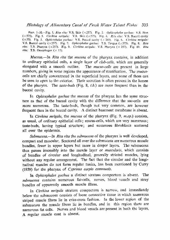
Ftc;s. 1-10.--Fig. 1. Rita rita: V.S. Skin (• Fig. 2. Ophicephahts gachua: V.S. Skin (• Fig. 3. Cirrhii~a mrigala: V.S. Skin (• Fig. 4. Rita rita: V.S. BuccaI cavity (x55). Fig. 5. Ophicephalus gachua: V.S. Buccal cavity (x 265). Fig. 6. Cirrhina mrigala: V.S. Buccal cavity (• 257). Fig. 7. Ophicephalus gachua: T.S. Tongue (x 575). Fig. 8. Rita rita: T.S. Pharynx (;4257). Fig. 9. Cirrhina mrigala: V.S. Pharynx (• 257). Fig. 10. Rita rita: T.S. Oesophagus (• 17).
Mucosa.--In Rita rita the mucosa of the pharynx contains, in addition to ordinary epithelial cells, a single layer of club-cells, which are generally elongated with a smooth outline. The mucus-cells are present in large numbers, giving in some regions the appearance of stratification. The mucus- cells are chiefly concentrated in the superficial layers, and some of them can be seen to open to the exterior. Their secretion is often present in the lumen of the pharynx. The taste-buds (Fig. 8, t.b.) are more frequent than in the buccal cavity.
In Ophicephalus gachua the mucosa of the pharynx has the same struc- ture as that of the buccal cavity with the difference that the sac-cells are more numerous. The taste-buds, though not very common, are however frequent than in the buccal cavity. A distinct basement membrane is absent.
In Cirrhina mrigala, the mucosa of the pharynx (Fig. 9, m.ep.) consists, as usual, of ordinary epithelial cells; mucus-cells, which are very numerous; taste-buds, having typical structure; and numerous fibroblasts scattered all over the epidermis.
Submucosa.--In Rita rita the submucosa of the pharynx is well developed, compact and muscular. Scattered all over the submucosa are numerous muscle bundles, fewer in upper layers but more in deeper layers. The submucosa thus passes insensibly into the muscle layer or muscularis, which consists of bundles of circular and longitudinal, generally striated muscles, lying without any regular arrangement. The fact that the circular and the longi- tudinal muscles do not form regular tunics, has been mentioned by Curry (1939) for the pharynx of Cyprinus carpio comrnunis.
In Ophicephalus gachua a distinct stratum comw.etum is absent. The submucosa contains numerous fat-cells, nerves, blood vessels and stray bundles of apparently smooth muscle fibres.
In Cirrhina mrigala stratum compactum is narrow, and immediately below the submucosa consists of loose connective tissue in which numerous striped muscle fibres lie in criss-cross fashion. In the lower region of the submucosa the muscle fibres lie in bundles, and in this region there are numerous fat cells. Nerves and blood vessels are present in both the layers,
A regular muscle coat is absent.

304 Ahsan-ul-Islam
OESOPHAGUS
The oesophagus, stomach and intestine have their wall composed of 4 coats, known respectively from without inwards as: Serosa, Muscularis, Submucosa and Mucosa or mucous membrane. The first three coats are of very similar structure in all parts of the gut while the mucosa has a characteristic structure in the different regions.
Sero,a.--ln Rita rita the serosa or the serous coat of the oesophagus (Fig. 10) is very thin and is almost indistinguishable at places. The great bulk of the serous coat is formed of the subserous connective tissue, the bundles of which even pass into the muscularis. The serous epithelium which usually consists of a single layer of cells (Dawts, 1929; Rogick, 1931, Curry, 1939), is not distinguishable in Rita rita. The serous coat gives the appearance of stratification, probably due to a resemblance between the nuclei of the serous epithelium and those of the subserous connective tissue. Many blood vessels supply the serosa.
In Ophicephalus gachua (Fig. 11) the serous coat is quite conspicuous and encloses nerves and blood vessels. In Cirrhina mrigala the serous coat is extremely thin. At places, the subserous connective tissue is well deve- loped and encloses nerves and blood vessels.
Muscularis.--In Rita rita both the muscle coats, an outer muscularis longitudinalis and inner muscalaris circularis, are fairly well developed and seem to be composed of unstriped muscle fibres (Fig. 10, mus.). Purser 0929) who describes a similar condition in Calamoichthvs calabaricus, says that the extent of the striated muscle fibres is used by many authors for determining the boundary between the pharynx and the cesophagus. Blood vessels are sometimes present between the two layers or in the muscularis longitudinalis.
In the oesophagus Ophicephalus gachua, contrary to the condition in other parts of the alimentary canal, the muscularis circularis lies outside the muscularis longitudinalis. The fibres of the longitudinal muscles often wander into the outer region of the submucosa. Both striped and unstriped muscles are met with. The muscularis passes insensibly into the submucosa.
In Cirrhina mrigala the muscularis consists of circular muscle coat only (Fig. 12, mus. c). The muscularis longitudinalis is absent as a distinct outer coat. Numerous longitudinal muscle fibres, however, lie in the meshes of the submucosa, a condition also reported by Curry (1939) in Cyprinus carpio communis, Mohsin (L 944--46) in Anabas testudineus and by Rogick (1931) in Campostoma anomalum. The muscularis circularis is well deve- loped and consists of very fine striated muscle fibres lying in quite a loose

Hislology of Aliraentary Canal of Fres/z Walee Teleosl Fishes 305
manner. Fibres of connective tissue are often present in this layer and enclose minute blood vessels.
Submucosa.--The submucosa in Rita rita (Fig. 10, s.m.) is extensive and vascialar, and enters,the various folds of the mucous membrane. Its outer portion is loose, but the portion adjacent to the mucosa is somewhat denser and more deeply stained. No muscle fibres are present in this coat. In Ophicephalus gachua the muscularis passes insensibly into the submucosa. The region of the submucosa adjacent to the mucosa is free of muscles and contains numerous blood vessels. In Cirrhina mrigala the submucosa (Fig. 12, s.m.) is poorly developed and consists of very loosely arranged connective tissue fibres, scattered among which lie numerous longitudinal muscle fibres (mus.f.).
Mucosa.--In Rita rita the mucosa or the mucous membrane (Fig. 10, m.) is deeply folded, the folds often simulating tubular glands. A muscularis mucos~e and a tunica propria are not present. A basement membrane is also not distinguishable. The mucous epithelium consists of long columnar cells with oval nuclei. Mucus-cells are round or sac-like and are present in large numbers and can sometimes be seen opening to the exterior.
In Ophicephalu~ gachua, too, the mucosa is thrown into very high folds, which exhibit a tendency to branch. Muscularis mucos~e is absent. The mucous epithelium (Fig. 11, m.ep.), though definitely simple at places especially at the top of the folds, appears to be stratified at most places. Almost the whole of the mucous membrane is formed of very closely packed sac-cells, (s.c.), which iie in one or more layers. At places unmodified epithelial cells lie as supporting cells below the sac-cells. The older sac-cells appear to be empty, but some typically goblet-shaped deeply-staining oxyphilic cells, probably younger sac-cells are also present.
In Cirrhina mrigala the mucosa Js simple, and is composed of a single, but greatly folded layer of ordinary epithelial cells (Fig. 12, epc.), with goblet - -o r mucus-cells (m.c.) and occasional taste-buds (t.b.). Some of the folds may be simple, others are branched. The ordinary epithelial cells are restricted to the tops and sides of certain folds. At other places they have either been replaced or squeezed in between the mucus-cells, which form the commonest element in the (esophageal mucosa of Cirrhina mrigala. Typical taste-buds are found here and there, especially near the top of the folds, and their presence in the oesophageal mucosa of this fish is a remark- able phenomenon and is presumably corelated with the feeding habits of the fish,

306 Ahsan-ul-Islam
STOMACH
Essentially the structure of the stomach is quite similar to that of the oesophagus except for the modification of the lining epithelium of the stomach to form the characteristic gastric epithelium and the gastric glands. Of the three fishes studied, Cirrhina mrigala does not posses a true stomach. In the other two fishes, the wall of the stomach consists of serosa, muscularis, submucosa and mucosa.
Serosa.--In Rita rita the serous coat of the stomach is similar to that of the eesophagus, and often encloses blood vessels and nerves. In Ophi- cephalus gachua, the serosa or the serous coat is extremely thin (Fig. 15, se.) and at most places consists of serous epithelium only.
Muscularis.--Serosa is'followed by Muscularis, which is composed of complete coats of longitudinal and circular muscles. The two coats are separated by an ill-defined layer of connective tissue enclosing blood vessels and a nerve-plexus called Auerbach's Plexus. The longitudinal coat of muscles is broken up into bundles separated by connective tissue. Fibres of connective tissue can also be seen running into the muscle layer. Muscu- laris longitudinalis is thin and in the region of the pyloric sphinctre it be- comes so much reduced as to be almost indistinguishable, while the muscula6s circularis increases greatly to form the sphincter.
In Ophicephalus gachua, the muscularis consists of almost equally thick muscularis longitudinalis (Fig. 15, mus. L) and muscularis cirrcularis (mus.c.) composed entirely of unstriated muscle-fibres. The two coats are separated by a layer of connective tissue, whose fibres penetrate both the coats and also enclose nerves.
Submucosa.--In Rita rita numerous bundles of longitudinal muscle fibres lie scattered in the submucosa, especially in the region adjacent to the mucosa. But these muscles do not form a muscularis mucos~e. The submucosa is rich in blood vessels. The submucosa, in Ophicephalus gachua is very well developed (Fig. 15, s.m.) richly vascular and invaded by numerous muscle fibres. These muscle fibres, mostly longitudinal, however, do not form a definite layer. The connective tissue fibres just below the submucous layer are said to comprise the tunica propria because they often penetrate into the mucous layer.
Mucosa.--The mucous membrane in Rita rita, consists of the lining epitheluim (Fig. 13, epc.), the gastric glands and the tunica propria. Stratum compactum is not present. The tunica propria is a vascular and well-defined layer of connective tissue, similar in structure to the submucosa. It

Histology of Alimentary Canal of Fres/z 14/'ater Teleosl Fishes 307
penetrates into the mucous layer, separating the gastric glands from each other, and supporting the lining epithelium. The epithelium consists of typical columnar ceils frequently turning inwards to form the crypts of gastric glands. The epithelial lining consists of very long and narrow cells. As the epithelium turns inwards to form the crypt, the cells become short and broad, till at the bottom of the crypt they assume a cubical or cuboid form. The outer extremity of the epithelial ceils stains deep blue with Mallory's stain. With mucicarmine, which is a specific mucous, stain, this zone stains red. The cytoplasm of these cells appears more or less non- granular when stained with eosin or picro-indigo-carmine, but Mallory's stain brings out fine granulations. This granulation does not increase in any perceptible degree in the crypt cells, but the secretory cells are heavily granulated. In the region of pyloric sphinctre, where the gastric glands are absent, the free ends of the epitheliar cells st.~in deep red with mucicarmine, thus exhibiting a mucoid nature. The folds of the epithelium iv. this region may be takep to represent the crypts of the glands, with the secretory por- tions missing.
The gastric glands are of the simple tubular type, each gland (Fig. 14) consisting of one or more secretory portions or tubules (s.t.), opening ,;nto the bottom of the crypt (or.). The lumen of the secretory tubule is very narrow and may even appear to be obliterated Secretory cells of only one type are present, and have been described as the chief or the peptic cells by Greene (1912) and Blake (1930 and 1936) in the fishes studied by them. The absence of any differentiation into peptic (chief) and oxyntic (parietal) cells in fishes, was made a generalization by Edinger in 1877, (Blake, 1936). The secretory cells are large, round, oval or pear shaped with a very big and round nucleus. The cytoplasm is choked with granules, called zymogen granules, which are particularly well stained by Mallory's stain.
The Mucous membrane or mueosa in Ophicephalus gachua consists of gastric epithelial cells (Fig. 15, epc.), lining the lumen of the stomach, and gastric glands (g.g.), several of which lie in the region between the tunica propria and the gastric epithelium. The gastric epithelium consists of elongated columnar cells, narrow proximally, broad distally, and having most of the stainable protoplasm, accumulated at the free, distal end. The protoplasm is faintly granular. The gastric glands consist of secretory por- tions or funduses and ducts of crypts. Many secretory tubules open to the exterior by one crypt. At the base of some crypts the cells remain unstained with Mallory's stain and pick up a very light stain with eosin.

epc

Hisgology o/" Al imentary Canal o f Fresh Wager Teleosg Fishes 309
FIGs. l l -19.--Flg. 11. Ophicephalus gachua: T.S. Oesophagus (• Fig. 12. Cirrhina mrigala: T.S. Oesophagus (• Fig. 13. Rita rita: T.S. Stomach (• 257). Fig. 14. Rita rita: A Gastric Gland (x265). Fig. 15. Ophieephalus gaehua: T.S. Stomach (• 17). Fig. 16. Rita rita: T.S. Intestine (• 55). Fig. 17. Ophicephalus gachua: T.S. Intestine (• Fig. 18. Ophicephalus gachua: T.S. Pyloric caecum (• Fig. 19. Cirrhina mrigala: L.S. Intestine (proximal part) ( • 29).
Similar cells in Pleuronectes platessa have been called "undoubtedly mucus producing cells, which can be compared to the mucoid cells scattered among the so-called peptic cells of the mammalian fundic glands" (Dawes, 1929). The cytoplasm of the secretory cells is full of zymogen granules, which stain red with Mallory's stain.
INTESTINE
The histological structure of the intestine is simple and resembles that of the ~:esophagus. The wall of the intestine consists of serosa, muscularis submucosa, and mucosa.
Serosa.--In Rita rita the serous coat of the intestine is vascular, (Fig. 16, se.). In Ophicephalus gachua, the serosa is very thin, being dis- tinguishable only at places where it gets separated from muscularis to enclose blood vessels (Fig. 17, se.). In Cirrhina mrigala, the serous coat is extremely thin, w i t h o u t a n y subserous connective tissue and probably incomplete, (Fig. 19 and 20, se.).
Muscularis.--In Rita rita, both the muscle coats are present. The muscularis circularis (Fig. 16 mus.c.) is only slightly thicker than the muscu- laris longitudinalis (mus. l.). All the muscles are unstriped. ~Ihe thin layer of loose connective tissue separating the two muscle coats encloses blood vessels and the ~erve plexus of Auerbach. In Ophicephalus gaci~ua, both the coats of muscularis are well developed, though the muscularis circularis (Fig. 17, mus.c.) is about four times as thick as muscularis longitudinalis mus. l.). Numerous blood vessels and nerves lie between the two coats, though there is no defi~ite layer of connective tissue between them. In Cirrhina mrigala, muscularis circularis is generally four to five times thicker than the museularis longitudinalis (Figs. 19 and 20, mus.c, and mu~. l.). Atl the muscles are unstriped. The two coats are separated from each other by a zone which is traversed by protoplasmic threads and contains numerous blood vessels and nerves. These threads do not stain blue with M~dlory's connective tissue stain.
Submucosa.--In Rita rita the submucosa (Fig. 16, .~.m.) is not very well developed and consists of loose connective tissue containiLg numerous blood vessels. It is prodv~ced into finger-like processes covered over by the

310 Ahsan-ul-islarn
lining epithelium to form the so-called "villi" True "villi" do not exist in any fish as is shown by the absence of the lacteals and the granular cells such as those of Paneth" (Rahimullah, 1945). "['he submucosa has also been designated as the "tunica propria", "combined parts of a true tunica propria and a submucosa" or merely as "areolar tissue" by various authors iti the fishes described by them. In Ophicephalus gachua, the submucosa is loose, fairly well developed and richly supplied with blood vessels. In Cirrhina mrigala, too, submucosa is well developed and highly vscular (Figs. 19 and 20, s.m.).
Mucosa.---In Rita rita, the mucous membrane or mucosa consists only of a single layer of columnar epithelial cells (Fig. 16, epc.), interspersed amongst which are the mucus-cells (m.c.). A muscularis mucos~e, a tunica propria, a stratum granulosum al~d a stratum compactum are not distinguish- able. The cells of the intestinal epithelium are of the simple columnar type, many times longer than broad, with slightly granular cytoplasm, and each with a well-defined top-plate. The nucleus lies towards the basal or attached end of the cell. The mucus-cells are present throughout the epithelium but their distributior is not uniform. They are round, oval or pear shaped with a filamentous basal portion which stains deeply with eosin and contains the nucleus. This indicates the evolution of the mucus-cells from the ordi- nary epithelial cells. The mucus-cell itself stains blue with hzematoxylil~ or Mallory's stain.
In Ophicephahts gachua the mucosa is simple, consisting of a single layer of columnar epithlial cells (Fig. 17, epc.), resting on a well defined tunica propria (t.p.). No muscularis mucos~e, stratum compactum or stratum granulosum are present. The mucous membrane is greatly folded to form "villi", the core of which is formed by the submucosa as in Rita rita. The columnar epithelial cells are very narrow, have a deeply staining cytoplasm, and are provided with top-plates. Interspersed among the columnar epithelial cells are numerous mucus-cells.
In Cirrhina mrigala, the mucosa is thrown into folds, and the mucosal folds in the proximal region of the intestine proper differ from those in the greater length of the intestine by being transverse rather than longitudinal. The epithelium of the mucosa is simple consisting of ordinary epithelial cells (Fig. 21, epc.), in addition to the mucus-cells (m.c.). The ordinary epithelial cells are many times longer than broad, more markedly so in the distal parts of the intestine proper. Their nuclei are, consequently, com- pressed and oval. Top plates are present, though they are not so prominent in the distal part of the intestine proper. The mucus-cells are fairly

Histology of Alimentary Canal o/Presh I/Valet Teleosl Fishes 311
numerous and may be goblet-shaped, sac-shaped or tube-like, sometimes ex- tending through the whole thickenss of the epithelium. The cells of the latter type indicate that the mucus-cells are formed by the transformation of the ordinary coulmnar epithelial cells. Numerous leucocytes are also strewn across the epithelium.
PYLORIC C~CUN
Two py!oric c~eca are present in Ophicephalus gaehua and their histo- logical structure is very similar to that of the intestine (duodenum) of which they are the outgrowths (Fig. 18). Their mucosa consists of a layer of epi- thelial cells (m.ep.), resting on welt-defined tunica propria (t.p.). The tuniea propria consists of a compact layer of connective tissue. Rahimullah (1945) regards this layer as differentiated part of the submucosa, but Greene (1912) and others consider it a part of the mucous membrane and treat it accord- ingly. The mucosa is raised into finger-like folds, the core of which is formed by the submucosa. These folds have been termed as the "ceca l villi" by RahimuUah (1943) in contrast with the true villi. The cecal villi are simple, unbranched and very long. The cecal epithelium is simple and exactly like the intestinal epithelium. The stratified condition of the cecal epittrelium described by Rahimullah (1943) for Ophicephalus striatus and other species of this genus was, however, not observed.
INTESTINAL BULB
In Cirrhina mrigala, the ~esophagus passes into a long tube, slightly wider than the o~sophagus and termed " the intestinal bulb " by Rogick (1931), or " the large arm of the intestine " by Curry (1939). The intestinal bulb gradually narrows down to the intestine proper, which forms by far the greater length of the gut.
Essentially, the histological structure of the intestinal bulb is similar to that of the rest of the intestine, the chief difference being in the degree of folding of mucosa. The serosa of the intestinal bulb (Figs. 22 and 23, se.) is very thin. In the muscularis, the circular (mus.c~) and the longitudinal (muse. l.)coats, though well developed, form only an insignificant part of the wall. The two coats, formed chiefly of unstriped muscle fibres, are separated by a thick pad of connective tissue contairiing nerves and blood vessels and a few striped muscles. A similar observation has been made by Curry (1939) in Cyprinus carpio communis.
The submucosa (s.m.) of the intestinal bulb is a very thin layer of coarse fibrous connective tissue, the processes of which pass into the mucosal folds

//Z/IS

Histology of Alimentary Canal of Fres/z Water Teieos! Fishes 313
FIGS. 20--27.--Fig. 20. Cirrhina mrigala: T.S, Intestine (middle and last part) ( , 55). Fig. 21. Cirrhina mrigala" T.S. intestine (middle and last part) (• Fig. 22. Cllrhina mrigala: Intestinal Bulb (• 11). Fig. 23, Cirrhina mrigala: T.S. Intestinal Bulb (257). Fig. 24. Rita rita: T.S. Rectum (• Fig. 25. Ophicephalus gachua: T.S, Rectum (• Fig. 26. Cirrhina m..rigala" T.S. Rectum (• 29). Fig. 27. Cirrhina mrigala: T.S. Rectum (•
and carry numerous blood vessels. The mucosa shows an extensive degree of folding, the folds or villi being more than ten times as high as the three outer coats taken together. Posteriorly, however, the folds gradually decrease in size. Only ordinary columnar epithelial cells (epc.) and mucus- cells are present. The ordinary epithelial ceils are extremely narrow and have most of their granular protoplasm aggregated near the free end of the cell. Top plates are present. Mucus-cells, though quite common, are not profuse.
RECTUM
The histological structure of the rectum resembles that of the intestine so closely that many authors do not describe rectum as a region distinct from the rest of the intestine.
Muscularis.--In all the three fishes studied, both the coats in the muscularis of the rectum are better developed than the corresponding coats in the intestine. The muscularis circularis (Figs. 24 and 26, mus.c.) is about three times as thick as the muscularis longitudinalis (musc. 1.). Blood vessels and small nerves are present between the two coats. A definite layer of connective tissue does not exist between the two coats in the rectum.
Submucosa.--Submucosa in the rectum in all the fishes studied is well developed, but as in the case of the intestine, this layer may be regarded as representing the tunica propria or tunica propria and submucosa combined.
Mucosa.--The epithelial cells are exactly like the corresponding cells in the intestine. The mucus-cells are much more numerous than in the intestine. In Ophicephalus gachua, the mucous membrane of the rectum differs from that of the intestine in the fact that the " rectal villi " show a slight tendency towards branching. In Cirrhina mrigala, just below the mucosa, the connective tissue is compact and stains more deeply and may be regarded as tunica propria. The mucus-cells in the mucosa (Fig. 27, m.c.) are narrow, long and finger like with a narrow neck, but sometimes their middle or basal region may be swollen. They stair dark blue with h~ema- toxylin, but fail to stain with Mallory's stain.
DISCUSSION
Skin.--Different types of gland-cells are met with in the skin of the three species studied. In Rita rita there are mucus-cells and club-cells; in
B2

314 Ahsan-ul-Islam
Ophicephal:a gachua there are the sac-cells and in Cirrhina mrigala there are the club-cells. So far as the club-cells are concerned, it was pointed out by Oxner (1905) and later by Bhatti (1938) that the presence of club- cells is indicative of genetic relationship because these cells are absent in all Physoclisti and are present in all Physostomi, except the Salmonida~ and Loficariida~. The present study corroborates this generalization in a striking manner. Thus Rita rita and Cirrhina mrigala both belonging to the group physostomi possess club-cells and Ophicephalus gachua belonging to the group Physoclisti is devoid of them, though Rim rita and Ophicephalus gachua are both carnivorous whereas Cirrhina mrigala is herbivorous. This shows that the club-cells in the skin, and hence in the buccal cavity and pharynx, indicate genetic relationship rather than an adaptation to feeding habits.
In Cirrhina mrigala some club-cells can be seen protruding out of the epidermis or even bodily ejected out, indicating the possibility of an excretory function of these cells. Wright (1884) attributed this condition to "defects in superficial layers of epidermis and due to the action of hardening reagents". Later observers (Oxner, 1905; Bhatti, 1938, and others) have, however, established the excretory function of these cells. Support or protection for the soft epidermis, is also advanced as a possible function of these cells.
Mucus-cells do not appear to be present in the portion of the skin of Ophicephalus gachua studied, and the sac-cells are the only type of gland- cells present. These sac-cells are exactly like the mucus-cells except for the staining reaction of the contents. Bhatti (1938) in the course of his studies on the integument of Siluroidea found that the sac-cells formed a definite generic character as they were present or absent in all the species of a genus. Presumably these cells also indicate a genetic relationship and are not concerned with feeding habits.
Terminal buds (taste-buds) have been observed in the skin of Rita rita, whose skin is naked, and are apparently absent in Cirrhina mrigala and Ophicephalus gachua, which have an exoskeleton of scales. Importance of taste-buds in feeding was first pointed out Herrick (1902) who wrote that " i t may be regarded as established that fishes which possess terminal buds in the outer skin taste by means of these organs and habitually find their food by their means". This was amply confirmed by Hamid Khan (1934) who described a totally blind but robust and healthy specimen of Rita rita from the Ravi, proving that in this fish taste, and not sight, played the major role in feeding. Ophicephalus gachua is piscivorous in the adult condition and obviously sight is much more important than the sense of taste in hunting. Cirrhina mrigala has been described by Mookerjee and

Histology of Alimenlary Canal o f Fresh I, Vater Teleosl 1;i~hes 315
Das (1945) as a Bottom-feeder taking lot of sand and mud. This mode of feeding does not require the presence of taste-buds in the skin of this fish.
Alimentary CanaL--The buccal cavity being a part of the embryonic stomodzeum, has essentially the same structure as the skin. In Cirrhina rnrigala mucus-cells appear in the buccal lining in l~rge numbers, but the club-cells record a decrease in number in this fish as also in Rita rita. Taste- buds are present in the buccal cavity of all the three fishes, but there are comparatively very few in Ophicephalus gachua. This may easily be cor- related with the nature of the food and the feeding habits.
A tongue is present in Ophicephalus gachua. As pointed out by Owen (1866) and Wiedersheim (1907), the tongue in fishes serves only as a tactile organ. The presence of a tactile orgar, in the buccal cavity of Ophicephalus gaehua may be interpreted as a compensation for the absence of barbels and other integumentary sense-organs.
The pharynx, being a continuation of the buccal cavity, has the same structure as the latter. However, the mucus-cells in Rita rita and Cirrhina rnrigala, and the sac-cells in Ophicephalus gachua, as well as the taste-buds ir~ all the three fishes, show a marked increase in number in this region. The mucous membrane is thrown into low broad folds, probably in order to lodge a great number of gland-cells and taste-buds. An increase in gland-cells has been reported in many fishes, e.g., C)Trinus carpio (Curry, 1939), Campostoma anornalum (Rogick, 1931). Probably the secretion of these gland-cells serves to wash the food down the oesophagus.
The ~esophagus has the same four coats as characterise the rest of the alimentary canal. In the cesophagus there is a transition from the stratified to simple epithelium. Moreover, the folding of the mucosa becomes very well marked, "the complexity of foldi,ag is charactristic of this coat" (Blake, 1930). The ~esophagus is richly supplied with gland-cell so much so that at places the gland-cells completely replace the ordinary~epithelial cells. No taste-buds are present in the oesophagus except in Cirrhina rnrigala. Prob- ably this backward extension of the taste-buds can be correlated with the mode of feeding and the nature of food of this fish.
In Rita rita and Ophicephalus gachua there is a typical c~ecal stomach provided with gastric glands. A true stomach, however, is absent in Cir- rhina mrigala. The absence of a true stomach in Cirrhina mrigala has been correlated with its food. Absence of a true stomach in the Cyprinida~ has also been recorded by Wiedersheim (1907). Jacobshagen (1937) pointed out that a true stomach is absent, among others, in "pure carnivorous fishes

316 Ahsan-ul-Islam
of which the intestine is shorter than the total length of the animal", and in "herbivorous fishes with an intestinal length of 5-6 times the length of the fish". In Rita rim, Ophicephalus gachua and Cirrhina mrigala the length of the gut is, respectively, 0" 6, 0" 7 and 11.25 times the length of the body. According to Jacobshagen's generalisation, all the three fishes should be stomach-less, but only Cirrhina mrigaIa comes up to expectations. Some- thing more than the mere ratio between the lengths of the gut and the whole body should be sought for to explain the absence of a true stomach in some fishes.
In Cirrhina mrigala the function of storage, performed in other fishes by the stomach, has been taken ovcr by the intestinal bulb which lies imme- diately behind the oesophagus and simulates a stomach. The intestinal bulb has been mistaken for a stomach by many authors (Mookerjee and Ghosh, 1945). However, its histological structure which is exactly like that of intestine, the opening of the bile duct immediately behind the oesophagus, and the absence of gastric glands, prove definitely that this organ is a part of the intestine.
The gastric glands of Rita rita and Ophicephalus gachua have the struc- ture usual in the fishes, i.e., they consist of (a) a crypt formed by an inturn- ing of the gastric epithelium, and (b) a fundus or the secretory portion. The cells of the gastric epithelium have their outer ends stained red with mucicar- mine, thus confirming the view of Stirling (1884), Jordan (1937) and others that these cells are mucoid in nature. The secretory cells are all of ohe type, confirming the statement of Edinger (1877) that in fishes in general there is no differentiation into peptic and oxyntic ceils. The only type of secretory cells present is comparable, according to Edinger (1877), neither to the central nor to the parietal cells of Mammalia. Recent authors (Greene, 1912; Blake, 1930 and 1936; and others) compare these cells with the chief or peptic (Central) cells in mammalian gastric glands. The question of the source of acid is still unsettled. According to Stirling (1884) the superficial cells, in addition to producing mucus, also produce acid. This problem, however, needs further investigation.
The correlation between the nature of food and the length of the intestine is clearly shown by the fishes studied. The intestine of the two carnivorous fishes studied is shorter than the total length of the body. Cirrhina mrigala, which lives on organic particles in mud and sand, has an intestine about 11 times as long as the length of the body, probably because such food requires a very large surface for its absorption. Similar conclusions have been arrived at A1-Hussaini (1947) for other herbivorous fishes.

Histology o/'Aliraenlary Canal of Fresh PUater Teleosl Fishes 317
In Rita rita and Ophieephalus gachua, the intestine has, throughout, the structure typical of piscine intestine. In Cirrhina mrigala however, the intestine is differentiated into various regions. The first part has a wide diameter. It is called the i.~atestinal bulb and its mucosa is thrown into very long folds. A short portion immediately behind the intestinal bulb has transverse folds, whereas in the rest of the intestine and the rectum the mucosal folds are longitudinal as is the usual condition.
Since the structure of the epithelium in all the three fishes is the same in all the regions of the intestine, no adaptive value can be attached to the intestinal mucosa. Goblet-cells or mucus-cells are present in the intestine of all the three fishes. In Ophieephalus gachua the mucus-cells are not met with in any other region of the alimentary canal except in the intestine.
In the fishes studied, pyloric c~eca are found only in Ophieephalus gaehua. As pointed out by Rahimullah (1945) "histologically, in general, the pyloric cmaza are very similar to the intestine ( = duodenum) of which they are out- growths". As regards their function, Rahimullah (1945) says, "it is not yet certain what exact correlation exists between diet and the significance of these c~eca in fishes. I think pyloric c~eca have arisen in those fishes where the abdominal space has to be economised owing to the influence of some environmental factors". What factors have lead Ophicephalus gachua to have a short straight intestine and then to have two pyloric c~eca as com- pensation, it is difficult to guess.
ACKNOWLEDGEMENT
I am gratefully indebted to Dr. Hamid Khan Bhatti, Ph.D. (Cantab.), F.N.I., F.A.Sc., Head of the University Teaching in Zoology, and Dr. Nazir Ahmad, P.h.D.(London), D.I.C., P.E.S., Professor of Zoology, Govern- ment College, Lahore, both of whom took a very keen interest in my work and helped me at every step. I also owe thanks to Dr. M. Rahimullah Qureshi, D.Sc., F.Z.S., and Dr. S. L. Hora, D.Sc., F.R.S.E., F.L.S., F.R.A.S., F.N.I., who gave me much valuable information regarding the current literature on the subject.
SUMMARY
1. The comparative histology of the alimentary canal of Rita rita Ham., Ophicephalus gachua Ham., and Cirrhina mrigala Ham., has been studied to correlate the histological structure with the nature of food.
2. In addition to the ordinary epithelial cells, the epidermis of the skin contains mucus-cells and club-eels in Rita rita; sac-cells in Ophicepha!u~

318 Ahsan-ul-Islam
gachua; and only club-cells in Cirrhina mrigala. Taste-buds are present only in the skin of Rita rita. In Cirrhina mrigala the club-cells have been seen being extruded bodily from the epidermis, thus providing a strong evidence for the view that the club-cells have an excretory function.
3. The histological structure of the buccal cavity and the pharynx is the same as that of the integument. Taste-buds and mucus-cells are present in the buccal cavity and pharynx of Cirrhina mrigala. In Ophice- phalus gachua a few taste-buds are present on the tongue but none in the buccal and pharyngeal epithelia.
4. In the oesophagus the epithelium is simple. No club-cells are present in Rita rita and Cirrhina mrigala, but in Ophicephalus gachua sac- cells are profusely abundant. In Cirrhina mrigala a few taste-buds extend even upto the oesophagus. This has been correlated with the food of this fish.
5. A true stomach is present in the two carnivorous fishes Rita rita and Ophicephalus gachua, but in the herbivorous fish Cirrhina mrigala it is absent as shown by (i) the absence of gastric glands, (ii) opening of bile- duct just behind the oesophagus, and (iii) the part following the oesophagus having the typical structure of intestine. The absence of a stomach in Cir- rhina mrigala has been correlated with its food. The cells forming the gastric epithelium in Rita rita and Ophicephalus gachua are mucoid in nature. Oxyntic cells are absent.
6. In Cirrhina mrigala the intestine is exceedingly long and coiled but in the other two fishes it is short and straight. This difference has been correlated with the difference in food in these fishes. In Cirrhina mrigala the first part of the intestine is dilated and is called the 'intestinal bulb'. In the region just behind the intestinal bulb the mucous membrane is trans- versely folded. In the rest of the intestine, as also in the intestinal bulb, the mucosa is longitudinally folded. No multicellular glands are present in the epithelium of the intestine. The mucus-cells (goblet-cells)are present in the intestine of all the three fishes studied. The mucus-cells make their appearance for the first time in the intestine in Ophicephalus gachua.
7. Pyloric c~eca, present only in Ophicephalus gachua amongst the fishes studied, have the same histological structure as the intestine.
8. The rectum differs from the intestine only in having thicker muscular coats and more numerous mucus-cells,

ttistology of .41imentary Canal of Fresh 14/aler Teleost Fishes 319
AI-Hussaini, A H.
Biaatti, H. K.
Blake, I. H . . .
Chan. V. M.
Curry, E. . .
Dawes, B. ..
Dharmarajan, M. ..
Edinger, L . . . . .
Greene, C. W.
Hamid Khan, M. ..
Herrick, C . J . . .
Jacobshagen, E, , .
LITERATURE CITED
.. "The Anatomy and Histology of the Alimentary Tract of the bottom-feeder, Mulloides auriflamma (Forsk.)," J. Morph. Philad., 1946, 78.
.. "The feeding habits and morphology of the alimentary tract of some teleosts," Pub. Mar. BioL Stat., Ghardaqa (Red Sea), 1947, 5.
. . "Funct ional Morphology of the Alimentary Tract of some fish in relation to differences in their feeding habits: Anatomy and Histology," Quart. Jour. Mier. Sci., 1949, 90, pt. 2.
. . "Funct ional Morphology of the Alimentary Tract of some fish in relation to differences in their feeding habits: Cytology and Physiology," Ibid., 1949, 90, pt. 4.
. . " T h e Integument and Dermal Skeleton of Siluroidea," Trans. Zool. Soc., 1938, 24, pt. 1.
. . "Studies on the Comparative Histology of the Digestive Tube of certain Teleost Fishes, I. The Sea Bass (Centro- pristes striatus)," J. Morph. Philad., 1930, 50.
.. "Studies on the Comparative Histology of the Digestive Tube of certain Teleost Fishes, II. A bottom-feeding Fish, the Sea robin (Prionoms carolinus)," Ibid., 1936, 60.
.. "The Histology of the Alimentary Tract of the Deep-water Gurnard, Peristedion longispatha (Goode and Bean) ," Univ. Nebraska Stud., 1941, 41, No. IL.
. . "The Histology of the Digestive Tube of the Carp (Cyprinus caroio communis)," J. Morph. Philad., 1939, 65.
�9 "The Histology of the Alimentary Tract of the Plaice (Pleuro- nectes ptatossa)," Quart. Journ. Micr. ScL, London, 1929, 73.
. . "The Anatomy and Histology of t,le Alimentary System of Otolithus ruber," Proc. Ind. Sci. Congr., 1936, 23.
.. "Uebe r die Schleimhaut des Fischdarmes nebst Bemerkungen zur Phylogenese der Drusen des Darmrohres," Archly..fur Mikrosk. Anat,, 1877, 13.
. . "Ana tomy and Histology of the Alimentary Tract of the King Salmon," Washington, D.C,, Dept. Comm. Bull., U.S. Bur. Fish., 1912 32.
.. "Habi t s and Habitats of Food-Fishes of the Punjab," Journ. Bombay Nat. Hist. Soc., 1934, 3 7 , No. 3.
. . " O n the relative value of certain Larvivorous Fisbes from the Punjab, with Notes on their Habits and Habitats ," lndian Journ. Veter. Sci., 1943, 13, pt. iv.
. . " T h e Organ and Sense of Taste in Fishes," Bull. U.S. Fish. Comm., 1903.
. . "Mit te l-und-Enddarm," in Bolk and others' Handbuch der vergl. Anat. der Wirbeltiere, 1937, 3, Berlin and Wien,

320 Ahsan-ul-Islam
Jordan, H. E . . . . . " A Text-Book of Histology, New York, 1937, 7th Ed.
Mohsin, S. M . . . . . " A preliminary on the Morphology and Histology of an Air-breathing Fish, Anabas testudineus (BI.)," Proc. lnd. Sci. Congr., 1941, 28.
. . . . . . "The Morphology and Histology of the Alimentary Tract of Anabas testudineus," Journ. Osm. Univ. (Science Faculty), 1944-46, 12.
. . . . . . "Histology of the Alimentary Tract of a Fresh-water Goby, Glossogobius guiris (Ham.)," Proc. lnd. Sci. Congr., 1946, 33.
Mookerjee, H. K. and Das, B.K. "Gu t of carnivorous and herbivorous fishes in relation to their food at different stages of their life," ibid., 1945, 32.
Mookerjee, H. K. and "Food of major Carps," Ibid., 1945, 32. Ghosh, S. N.
Mookerjee, H. K., Ganguly, D .N. "On the composition of food and their correlation with weight and Islam, M. and length of the body in the development of Ophicephalus
punctatus Bloch.," lbM., 1946, 33.
Oxrler, M . . . . . . . "Ueber die Kolbenzellen in der Epidermis der Fische, ihre Form, Verteilung, Entstehlung und Bedeutung," Jena Zeitschr. Naturw:, 1905, 40.
Owen, R . . . . . . . On the Anatomy of Vertebrates, Vol. I, Fishes and Reptiles, London, 1866.
Purser, G. L . . . . . "Calamoichthys calabaricus, Part II. The Alimentary and Respiratory Systems," Trans. Roy. Soc. Edin., 1929, 56,
Rahimullah, M . . . . . " A contribution to the structure and probable functions of the Pyloric C~eca in the family Opicephalidae," Anat. Anz., Jena, 1935, 80.
. . . . . . "On the Pyloric Caeca in the Notopteridae" (Abstract), Proc. lnd. Sci. Congr., Calcutta, 1935, 22.
. . . . . . "The structure of the Pyloric C~ecea in the family Masta- cembelidae," Ibid., 1936, 23.
. . . . . . "On the structure and functions of the so-called Pyloric Caeca in two genera of Fishes, Lactarius and Osphromenus'" Ibid., Calcutta, 1938, 25.
. . . . " O n the disposition of the so-called Pyloric Caeca in a Brotu. lid fish, Sirembo imberbis (Tern. and Sch.)," Ibid., 1941, 28.
. . . . . . "Contributions to our knowledge of the Pyloric C~eca o f three families of Fresh-water Indian Fishes (Ophicephalidae, Notopteridae and Mastacembelidae) together with some remarks on their probable functions," Proe. lnd. Acad. Sci., 1943, 18 B.
. . . . . . " A comparative study of the Morphology, Histology and probable Functions of the Pyloric Caeca in Indian Fishes, together with a Discussion on their Homology," 1bid., 1945, 21 B.
. . . . "Studies on the Comparative Histology of the Digestive Tube of certain Teleost Fishes. I1. A Minnow Campa. stoma anomalum,'" J. Morph. Philad,, 1931, 52.
Rogick, M. D,

Histology of ,4 hmentary Canal of F, peslt Water Teleost Fishes 32 i
Sarbahi, D. S.
Stirling, W . . .
Vanajakshi, T.P. ..
Wiedersheim, R. ..
Wright, R .R . . .
�9 . "Alimentary Canal of Labeo rohita," Journ. Asiat. Soc, Bengal (Science), 1940, 5, 2.
.. "On the Chemistry and Histology of the digestive organs of Fishes," 2nd Annual Report of the Fishery Board for Scotland, 1884 (Quoted by Dawes, 1929).
.. "Histology of the Digestive Tract of Saccobranchus vittatus," Pcoe. hTd. Acad. Set., 1938, 7 B, 2.
. . Comparative Anatomy of Vertebrates, adapted by W . N . Parker, 1907.
.. "On the Skin arid Cutaneous Sense Organs of Amiurus," Proc. Canad. Inst., n.s., 1884.
KEY TO LETTERING
b.m., Basement membrane; c.c., Club-celi; co., Corium; cr., Crypt; epb., Basal layer of epithelium; epc., Ordinary epithelial cell; g.g., Gastric gland; m., Mucous membrane; m.c., Mucusw.eU (goblet oell); m.ep., Mucous epithelium; mus., Muscularis; mus.e., Muscularis circu- laris; mus.]., Muscle-fibre; mus.L, Muscularis longitudinalis; s.c., Sac-cell; se., Serosa; s.m., Submueosa; s.t.,Secretory Tubule; str.comp., Stratum compactum; t.b., Taste-bud; t.p., Tunica propria,