the conformation of the prion domain of sup35 p in isolation and in the full-length protein
TRANSCRIPT
Solid-State NMR SpectroscopyDOI: 10.1002/anie.201304699
The Conformation of the Prion Domain of Sup35p in Isolation and inthe Full-Length Protein**Nina Luckgei, Anne K. Sch�tz, Luc Bousset, Birgit Habenstein, Yannick Sourigues,Carole Gardiennet, Beat H. Meier,* Ronald Melki,* and Anja Bçckmann*
The yeast protein Sup35p has prion properties,[1] and itaggregates into fibrillar assemblies.[2] It is at the origin of the[PSI+] trait in baker�s yeast, Saccharomyces cerevisiae.[3] TheSup35p yeast prion is an important model system to inves-tigate the structure–function relationship of prions. To reducethe complexity of the problem, the Sup35pNM fragment isoften used as a convenient model to document the assemblyand infectious properties of the full-length prion, as fibrillarSup35pNM is biologically relevant in the sense that it induces[PSI+] when introduced into [psi�] cells.[4] The notion thatfibrillar Sup35pNM perfectly mimics Sup35p fibrils suggeststhat the N and M domains of Sup35p adopt identicalconformations in Sup35pNM and Sup35p fibrils.[5] We ques-tion this assumption in the following: Using solid-state NMRmeasurements performed on Sup35pNM and full-lengthSup35p fibrils assembled under identical physiological con-ditions, and both inducing [PSI+] strains (Supporting Infor-mation, Figure S1), we demonstrate that fibrillar Sup35pNMand full-length Sup35p show significant structural differences,although both have a high b-sheet content.
Sup35p is a three-domain polypeptide (Supporting Infor-mation, Table S1); the N-terminal domain is critical for prionpropagation, while the C-terminal domain has GTPaseactivity and is involved in translation termination.[6] Themiddle domain connects the two domains. The N domainalone or together with the M domain, and also the full-length
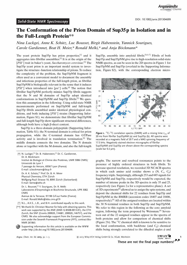
Sup35p, assemble into amyloid fibrils.[5a,b,7] Fibrils of bothSup35p and Sup35pNM give rise to high-resolution solid-stateNMR spectra, as can be seen in the 2D spectra in Figure 1 forSup35pNM and Sup35p (overlaid in the Supporting Informa-tion, Figure S2), with the corresponding electron micro-
graphs. The narrow and resolved resonances points to thepresence of highly ordered structures in both fibrils. Toincrease spectral resolution, we recorded 3D NCACB spectrain which each amino acid residue shows a (N, Ca, Cb)frequency triple. Surprisingly, although 253 and 685 signals forSup35pNM and Sup35p, respectively would be expected, thenumber of intense peaks in the 3D spectra is only 35 and 25,respectively (see Figure 2a for a representative plane). A setof 3D experiments[8] allowed us to assign the spin systems, anddeposit the chemical shifts for 22 residues from Sup35p andSup35pNM in the BMRB (accession codes 18407 and 18406,respectively).[9] All of the assigned residues are located withinthe 30 N-terminal residues in both Sup35p and Sup35pNM.We refer to this region in the following as the core amyloidregion, following the term proposed by Weissman.[4] Seven-teen out of the 22 assigned residues appear in the spectra ofboth proteins and allow for comparison of chemical shifts(Figure 2b). The 13C chemical shift is known to be sensitive tothe local conformation, with backbone (and Cb) chemicalshifts being strongly correlated to the dihedral angles f and
Figure 1. 13C–13C correlation spectra (DARR) with a mixing time tmix of20 ms from fibrillar Sup35pNM (a) and Sup35p (b). All spectra wererecorded at a magnetic field of 20 T and a MAS spinning frequency of17.5 kHz. Negatively stained electron micrographs of fibrillarSup35pNM and Sup35p are shown above the corresponding spectra(scale bar 0.2 mm).
[*] N. Luckgei,[+] Dr. B. Habenstein,[+] Dr. C. Gardiennet,Dr. A. BçckmannInstitut de Biologie et Chimie des Prot�ines, UMR 5086 CNRS/Universit� de Lyon 17 passage du Vercors, 69367 Lyon (France)E-mail: [email protected]
Dr. A. K. Sch�tz,[+] Prof. Dr. B. H. MeierPhysical Chemistry, ETH Z�richWolfgang-Pauli Strasse 10, 8093 Z�rich (Switzerland)E-mail: [email protected]
Dr. L. Bousset,[+] Y. Sourigues, Dr. R. MelkiLaboratoine d’Enzymologie et Biochimie Structurale, UPR 3082CNRSAvenue de la Terrasse, 91198 Gif-sur-Yvette (France)E-mail: [email protected]
[+] N.L., A.K.S., L.B., and B.H. contributed equally to this work.
[**] We thank Dr. Christian Wasmer for help with obtaining spectra. Thiswork was supported by the ANR (ANR-12-BS08-0013-01), the ETHZurich, the SNF (Grants 200020_134681, 200020_146757), and theCNRS. We also acknowledge support from the European Commis-sion under the Seventh Framework Programme (FP7), contract Bio-NMR 261863.
Supporting information for this article is available on the WWWunder http://dx.doi.org/10.1002/anie.201304699.
AngewandteChemie
1Angew. Chem. Int. Ed. 2013, 52, 1 – 5 � 2013 Wiley-VCH Verlag GmbH & Co. KGaA, Weinheim
These are not the final page numbers! � �
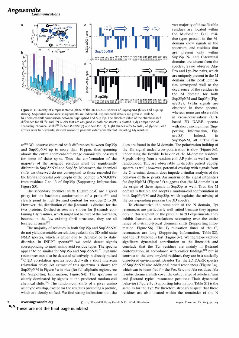
y.[10] We observe chemical-shift differences between Sup35pand Sup35pNM up to more than 10 ppm, thus spanningalmost the entire chemical-shift range canonically observedfor some of these spins. Thus, the conformation of themajority of the assigned residues must be significantlydifferent in Sup35pNM and Sup35p. Moreover, the chemicalshifts we observed do not correspond to those recorded forthe fibril and crystal polymorphs of the peptide GNNQQNYfrom residues 7 to 13 of Sup35p (Supporting Information,Figure S3).
The secondary chemical shifts (Figure 2c,d) are a goodproxy for the backbone conformation of a protein[11] andclearly point to high b-strand content for residues 2 to 30.However, the distribution of the b-strands is distinct for thetwo proteins. Dashed arrows are shown for b-strands con-taining Gly residues, which might not be part of the b-strands,because in the few existing fibril structures, they are alllocated in turns.[12]
The majority of residues in both Sup35p and Sup35pNMdo not yield detectable correlation peaks in the 3D solid-stateNMR spectra, which is either due to dynamic or to staticdisorder. In INEPT spectra[13] we could detect signalscorresponding to most amino acid residue types. The spectraappear to be similar for Sup35p and Sup35pNM.[9] Dynamicresonances can also be detected selectively in directly pulsed13C 2D correlation spectra recorded with a short interscanrelaxation delay. An extract of this spectrum is shown forSup35pNM in Figure 3 a in blue (for full aliphatic regions, seethe Supporting Information, Figure S4). The spectrum isclearly dominated by signals at the predicted random-coilchemical shifts.[14] The random-coil shifts of a given aminoacid type overlap, except for the residues preceding a proline,which are clearly shifted. We find strong indications that the
vast majority of these flexibleresidues are located withinthe M-domain: 1) all resi-due-types present in the M-domain show signals in thespectrum, and residues thatare present only withinSup35p N- and C-terminaldomains are absent from thespectra; 2) we observe Ala-Pro and Lys-Pro pairs, whichare uniquely present in the Mdomain; 3) the peak intensi-ties correspond well to theoccurrence of the residues inthe M domain for bothSup35pNM and Sup35p (Fig-ure 3e); 4) Thr signals areobserved in these spectra,whereas none are observablein cross-polarization (CP)-based 2D DARR spectrawith short mixing times (Sup-porting Information, Fig-ure S5). Indeed, inSup35pNM, all 11Thr resi-
dues are found in the M domain. The polarization buildup ofthe Thr signal under cross-polarization is slow (Figure 3c),underlining the flexible behavior of the M-domain residues.Signals arising from a random-coil AP pair, as well as fromrandom-coil Thr, are observable in directly pulsed Sup35pspectra as well; however, potential overlap with signals fromthe C-terminal domain does impede a similar analysis of thebehavior of these peaks. An analysis of the signal intensitiesfor Sup35pNM (Figure 3 f) suggests that the M-domain is atthe origin of these signals in Sup35p as well. Thus, the Mdomain is flexible and adopts a random-coil conformation inboth Sup35pNM and Sup35p, which explains the missing ofthe corresponding peaks in the 3D spectra.
To characterize the remainder of the N domain, Tyrresonances are particularly well suited because they appearonly in this segment of the protein. In 2D experiments, theyexhibit featureless correlations resonating over the entirerange of b-strand-typical chemical shifts (Supporting Infor-mation, Figure S6). The T2 relaxation times of the Ca
resonances are long (Supporting Information, Table S2),and the CP buildup is fast (Figure 3c). We therefore excludesignificant dynamical contribution to the linewidth andconclude that the Tyr residues are mainly in b-strandconformation, in accordance with earlier findings,[15] but incontrast to the core amyloid residues, they are in a staticallydisordered environment. Besides Tyr, the 2D DARR spectraof Sup35pNM also additional broad resonances (Figure 3a),which can be identified for the Pro, Ser, and Ala residues. Alaresidue chemical shifts cover the entire range of a-helical/turnand b-strand typical resonance positions. Their dynamicalbehavior (Figure 3c; Supporting Information, Table S1) is thesame as for the Tyr. We therefore strongly suspect that theseresidues are also located within the remainder of the N
Figure 2. a) Overlay of a representative plane of the 3D NCACB spectra of Sup35pNM (blue) and Sup35p(black). Sequential resonance assignments are indicated. Experimental details are given in Table S3.b) Chemical-shift comparison between Sup35pNM and Sup35p. The absolute value of the chemical-shiftdifference for all 13C and 15N nuclei that are assigned in both constructs is plotted. c,d) Comparison ofsecondary chemical shifts[11] for Sup35pNM (c) and Sup35p (d). Light shades refer to DdCa of glycine. Solidarrows refer to b-strands, dashed arrows to possible extensions thereof, including Gly residues.
.AngewandteCommunications
2 www.angewandte.org � 2013 Wiley-VCH Verlag GmbH & Co. KGaA, Weinheim Angew. Chem. Int. Ed. 2013, 52, 1 – 5� �
These are not the final page numbers!

domain. For the full-length protein, the unequivocal assign-ment of isolated peaks to the remaining N domain is moredifficult, but the spectra are compatible with the assumptionthat the parts of the N domain, which do not form part of theordered core amyloid, are statically disordered in the sensethat the conformation of each residue in this part is variable.Nevertheless, these residues are organized in b-sheets.Whether the disorder is within one fibril, which we suspect,or between fibrils in a way that may be described as anextensive polymorphism in this part cannot be decided by anensemble method like NMR spectroscopy.
The majority of Sup35p residues belong to the C-terminaldomain, which remains biochemically functional in fibrils,[5b]
suggesting that it retains its fold. We created a homologymodel from the X-ray structure of the homologous proteinfrom S. pombe[16] and predicted chemical shifts using Sparta[17]
(Supporting Information, Figure S7), which show a similardispersion and distribution of signals. The resolved signalsthat do not have a counterpart in the Sup35pNM spectra canthus tentatively be assigned to the C-terminal domain. Theyshow narrow line width and many resolved signals, which
confirms that the C-ter-minal domain is orderedand well-structured. Asshown in Figure 3 d (inblack), the CP buildup ofthe signals from the C-terminal domain is verysimilar to one of the coreamyloid residues, indi-cating that no large-scale amplitude motionstake place. Nevertheless,these residues are notobserved in the 3D spec-tra, indicating that subtledynamic effects, proba-bly a not yet fully char-acterized overall motionof the globular domain,are at the origin of signalattenuation in the 3Dspectra.
Our study providesinsight into the globalstructure and thedynamics of Sup35p N,M, and C domains infibrils and demonstratesthat the differentdomains feature differ-ent behavior. Theordered core amyloid ofthe fibrils is locatedwithin the first thirty res-idues and shows b-strandsecondary structure inagreement with the slowH/D exchange rates
observed for residues 4-37 in Sup35pNM fibrils assembledat 4 8C.[4] The faster proton–deuterium exchange ratesrecorded for the remainder of Sup35pNM[4] also agree withthe absence of regular, well-ordered secondary structure weobserve, although the observation by Toyama et al.[4] ofmoderate protection in some parts may hint at residuesforming more stable hydrogen-bonded interactions. Thedisordered but static nature of a significant fraction ofSup35pNM suggests that non-residue-specific interactionsbetween aromatic residues located outside the core amyloidmay play an important role in the initial steps of aggrega-tion.[18] Also, the presence of this possibly polymorphic regionmight add to the observation of various strains in Sup35p,because although the core amyloid shows clearly a well-defined structure, these immobilized segments of the Ndomain may have different conformations in different strains.
These findings shed new light on the surprisingly diverseworld of prion assemblies, where conformational variabilityplays a staggering and confusing role. It supports theemerging picture that prions are complex structural units.Indeed, even if displaying a highly defined structure, a given
Figure 3. a) Overlay of Sup35pNM 100 ms DARR spectrum (gray) and directly pulsed 100 ms DARR spectrum(blue) with a T1-filter on 13C by use of a short relaxation delay (4 s). Isolated signals of amino acids from both N-and M-domain are labeled in black, those appearing only in the M- domain in blue. b) only the 100 ms DARRspectrum. c,d) Cross-polarization buildup dynamics for selected residues (of Ca–Cb cross-peak). Residues fromthe core amyloid are colored dark blue, residues from the remainder of the N domain light blue, from the Mdomain in gray, and from the C domain in black. e,f) Comparison of the Ca–Cb cross-peak volume from thedirectly pulsed PDSD spectra (Supporting Information, Figure S3), with the amino acid distribution in the Mdomain, normalized on Lys residues. The error is the standard deviation of the noise in direct-pulsed PDSDspectra.
AngewandteChemie
3Angew. Chem. Int. Ed. 2013, 52, 1 – 5 � 2013 Wiley-VCH Verlag GmbH & Co. KGaA, Weinheim www.angewandte.org
These are not the final page numbers! � �
domain can adopt different conformations dependent on thesetting (in isolation, in the context of a larger fragment, or thefull-length protein) or the environment (buffer conditions,chaperones). For functional prions, such as the HET-s/HET-Ssystems, these properties can give rise to a functional switchthat triggers apoptosis.[19] Our findings give a molecular-levelexplanation for the contrasting assembly propensity andinfectivity of Sup35pNM and Sup35p, and stress the para-mount importance of a molecular-level structural character-ization of the aggregates employed in functional studies.
Experimental SectionThe proteins were expressed and purified as described previously,using M9 medium with 15N and 13C labeling.[5a,b] In detail, Sup35p inbuffer solution (20 mm Tris·HCl, pH 8.0, 200 mm NaCl, 5% glycerol,5 mm b-mercaptoethanol, 10 mm MgCl2) was incubated at 10 8C underorbital shaking at 30 rpm, 0.5 cm amplitude, for 3 weeks. The fibrilswere spun at 100000 g in a TL-100 tabletop centrifuge for 20 min at4 8C. The pellets were resuspended in distilled water, washed twice,and filled into 3.2 mm rotors by ultracentrifugation as described.[20]
All spectra were recorded on a Bruker Avance II + 850 MHzspectrometer operating at a static field of 20 T. A 3.2 mm Brukertriple-resonance MAS probe equipped with an LLC coil was used.The sample temperature was about 7 8C. The pulse sequences wereimplemented as recently reported,[8a] and the experimental parame-ters are given in the Supporting Information, Table S3. All spectrawere processed using TopSpin 2.0 (Bruker Biospin) with zero fillingand apodization by a squared cosine function. Spectra were analyzedand annotated using the CCPNmr Analysis package.[21]
Received: May 31, 2013Revised: July 24, 2013Published online: && &&, &&&&
.Keywords: fibrils · prions · proteins ·solid-state NMR spectroscopy · Sup35p
[1] R. B. Wickner, Science 1994, 264, 566.[2] J. R. Glover, A. S. Kowal, E. C. Schirmer, M. M. Patino, J. J. Liu,
S. Lindquist, Cell 1997, 89, 811.[3] B. S. Cox, Heredity 1965, 20, 505.[4] B. Toyama, M. Kelly, J. Gross, J. Weissman, Nature 2007, 449,
233.[5] a) J. Krzewska, R. Melki, EMBO J. 2006, 25, 822; b) J. Krzewska,
M. Tanaka, S. G. Burston, R. Melki, J. Biol. Chem. 2007, 282,1679; c) M. Kabani, B. Cosnier, L. Bousset, J.-P. Rousset, R.Melki, C. Fabret, Mol. Microbiol. 2011, 81, 640.
[6] a) G. Zhouravleva, L. Frolova, X. Legoff, R. Leguellec, S.Ingevechtomov, L. Kisselev, M. Philippe, EMBO J. 1995, 14,4065; b) I. Stansfield, K. Jones, V. Kushnirov, A. Dagkesaman-skaya, A. Poznyakovski, S. Paushkin, C. Nierras, B. Cox, M.Teravanesyan, M. Tuite, EMBO J. 1995, 14, 4365.
[7] a) C. King, R. Diaz-Avalos, Nature 2004, 428, 319; b) M. Tanaka,P. Chien, N. Naber, R. Cooke, J. Weissman, Nature 2004, 428,323; c) F. Shewmaker, D. Kryndushkin, B. Chen, R. Tycko, R. B.Wickner, Biochemistry 2009, 48, 5074.
[8] a) A. Schuetz, C. Wasmer, B. Habenstein, R. Verel, J. Green-wald, R. Riek, A. Bçckmann, B. H. Meier, ChemBioChem 2010,11, 1543; b) B. Habenstein, C. Wasmer, L. Bousset, Y. Sourigues,A. Sch�tz, A. Loquet, B. H. Meier, R. Melki, A. Bçckmann, J.Biomol. NMR 2011, 51, 235.
[9] a) A. Sch�tz, B. Habenstein, N. Luckgei, L. Bousset, Y.Sourigues, A. B. Nielsen, R. Melki, A. Bçckmann, B. H. Meier,Biomol. NMR Assignments 2013, DOI: 10.1007/s12104-013-9515-1; b) N. Luckgei, A. Sch�tz, B. Habenstein, L. Bousset, Y.Sourigues, R. Melki, B. H. Meier, A. Bçckmann, Biomol. NMRAssignments 2013, DOI: 10.1007/s12104-013-9518-y.
[10] D. S. Wishart, B. D. Sykes, F. M. Richards, J. Mol. Biol. 1991, 222,311.
[11] Y. Wang, O. Jardetzky, Protein Sci. 2002, 11, 852.[12] a) H. van Melckebeke, C. Wasmer, A. Lange, E. Ab, A. Loquet,
A. Bçckmann, B. H. Meier, J. Am. Chem. Soc. 2010, 132, 13765;b) C. Wasmer, A. Lange, H. van Melckebeke, A. B. Siemer, R.Riek, B. H. Meier, Science 2008, 319, 1523.
[13] A. Siemer, A. Arnold, C. Ritter, T. Westfeld, M. Ernst, R. Riek,B. H. Meier, J. Am. Chem. Soc. 2006, 128, 13224.
[14] K. Tamiola, B. Acar, F. A. A. Mulder, J. Am. Chem. Soc. 2010,132, 18000.
[15] F. Shewmaker, R. B. Wickner, R. Tycko, Proc. Natl. Acad. Sci.USA 2006, 103, 19754.
[16] C. Kong, K. Ito, M. A. Walsh, M. Wada, Y. Liu, S. Kumar, D.Barford, Y. Nakamura, H. Song, Mol. Cell 2004, 14, 233.
[17] Y. Shen, A. Bax, J. Biomol. NMR 2007, 38, 289.[18] Y. Ohhashi, K. Ito, B. H. Toyama, J. S. Weissman, M. Tanaka,
Nat. Chem. Biol. 2010, 6, 225.[19] C. Seuring, J. Greenwald, C. Wasmer, R. Wepf, S. J. Saupe, B. H.
Meier, R. Riek, PLoS Biol. 2012, 10, e1001451.[20] A. Bçckmann, C. Gardiennet, R. Verel, A. Hunkeler, A. Loquet,
G. Pintacuda, L. Emsley, B. H. Meier, A. Lesage, J. Biomol.NMR 2009, 45, 319.
[21] a) W. Vranken, W. Boucher, T. Stevens, R. Fogh, A. Pajon, P.Llinas, E. Ulrich, J. Markley, J. Ionides, E. Laue, Proteins Struct.Funct. Bioinf. 2005, 59, 687; b) T. J. Stevens, R. H. Fogh, W.Boucher, V. A. Higman, F. Eisenmenger, B. Bardiaux, B.-J.van Rossum, H. Oschkinat, E. D. Laue, J. Biomol. NMR 2011,51, 437.
.AngewandteCommunications
4 www.angewandte.org � 2013 Wiley-VCH Verlag GmbH & Co. KGaA, Weinheim Angew. Chem. Int. Ed. 2013, 52, 1 – 5� �
These are not the final page numbers!
Communications
Solid-State NMR Spectroscopy
N. Luckgei, A. K. Sch�tz, L. Bousset,B. Habenstein, Y. Sourigues,C. Gardiennet, B. H. Meier,* R. Melki,*A. Bçckmann* &&&&—&&&&
The Conformation of the Prion Domain ofSup35 p in Isolation and in the Full-Length Protein
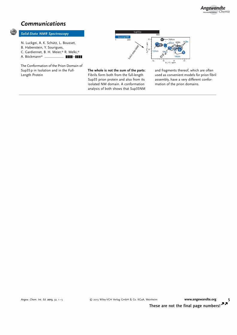
The whole is not the sum of the parts :Fibrils form both from the full-lengthSup35 prion protein and also from itsisolated NM domain. A conformationanalysis of both shows that Sup35NM
and fragments thereof, which are oftenused as convenient models for prion fibrilassembly, have a very different confor-mation of the prion domains.
AngewandteChemie
5Angew. Chem. Int. Ed. 2013, 52, 1 – 5 � 2013 Wiley-VCH Verlag GmbH & Co. KGaA, Weinheim www.angewandte.org
These are not the final page numbers! � �