the control of ventricular function in man*
TRANSCRIPT

Brit. Heart J., 1965, 27, 1.
THE CONTROL OF VENTRICULAR FUNCTION IN MAN*
BY
EUGENE BRAUNWALD
From the Cardiology Branch, National Heart Institute, National Institutes of Health, Bethesda, Maryland, U.S.A.
Received May 12,1964
Since William Harvey's discovery of the prime function of the heart as a pump, clinicians andphysiologists have marvelled at the extraordinary ability of this organ to adjust its performancealmost instantaneously to meet the rapidly changing requirements of the peripheral tissues. Pro-bably no other aspect of cardiovascular research has received as much effort and attention as theattempts to gain an understanding of the systems that control the heart's ability to propel bloodthrough the circulatory system. During the past century, this problem has been investigated atmany levels; from isolated extracts of contractile proteins, to isolated atrial and papillary musclepreparations, to isolated hearts and heart-lung preparations, and anesthetized open-chest dogs, aswell as closed-chest, unanxsthetized dogs.
Among the most significant contributions to these investigations were those that came from thelaboratories of Professors Frank (1895) and Starling (1918). These investigators extended to theheart the earlier observations of Fick (1882), Kries (1892), and Blix (1895) who had already estab-lished for skeletal muscle that the length and tension of the muscle at the time of stimulation deter-mined the magnitude of its contractile response. The immediate impact of this work, as presentedby Starling in his Linacre lecture (1918), requires little emphasis at this time. However, during thepast 15 years some investigators have challenged the importance, indeed even the applicability, of theFrank-Starling principle in the regulation of cardiac performance in intact man. Until quite recentlythe techniques available to the clinical investigator have not permitted a definitive answer to theimportant question of the control of ventricular function in man, and prevailing opinions have ofnecessity been based primarily upon extrapolations from animal experiments, as well as on indirectmeasurements in patients. During the past few years, however, the refinement of methods thatpermit a detailed study of the circulation in man has made possible a direct attack on this funda-mental question. It is the objective of this presentation to review the results of a series of experi-ments directed towards elucidating the role of two basic mechanisms of cardiac adjustment; (1) theFrank-Starling principle, i.e. the dependence of the mechanical activity of the heart on the ventricularend-diastolic volume and pressure, and (2) the adrenergic nervous system, changes in the activity ofwhich alter the humoral environment of the heart muscle and thereby permit variations in ventricularperformance at any given level of end-diastolic volume or pressures.
These investigations were begun by asking a simple question: "How does changing the length of asegment of human myocardium affect its contractile activity?" It was possible to obtain an answerby sewing a specially modified Walton-Brodie strain gauge arch to the right ventricle of 10 patientsand to the left ventricle of 1 patient, at the time of cardiac operation. This instrument permits thecontrolled alteration of the length of the myocardial segment to which it is attached, as well as mea-surement of the tension exerted by this segment of myocardium throughout the cardiac cycle. In all
* The third Haile Selassie Lecture delivered at the Royal College of Physicians, London, on April 20, 1964, underthe auspices of the National Heart Hospital.B I
on March 18, 2022 by guest. P
rotected by copyright.http://heart.bm
j.com/
Br H
eart J: first published as 10.1136/hrt.27.1.1 on 1 January 1965. Dow
nloaded from

EUGENE BRAUNWALD
11 patients studied, it was noted that as myocardial length was increased, the end-diastolic tensiontended to rise slightly at first, but with further stretching the tension increased more strikingly(Fig. 1). As the segment was stretched the maximal systolic tension that it developed increasedmore than did its resting tension. Thus, within limits, the active or developed tension rose as themyocardial segment increased in length. In 5 patients the active tension declined after the myo-cardial segment had been stretched to values ranging between 15 and 50 per cent above baselinelength, i.e. a descending limb of the length-active tension relation was evident (Fig. 1). No changein the duration of the active tension state occurred with progressive stretching of the myocardialsegment (Fig. 2), and changes in the rate of development of tension paralleled the changes in theactive tension (Fig. 3). Fig. 3 also shows the relation between resting tension and active (developed)tension. The ascending limb of the curve is steep, i.e. the active tension rises abruptly with rela-tively small rises in resting tension. These observations indicated that functioning human myo-cardium behaves in accordance with the Frank-Starling principle (Aygen and Braunwald, 1962).
More recently we have extended these studies to an examination of the mechanical activity of thepapillary muscle excised from the left ventricle of patients at the time of complete replacement of themitral valve. When mounted in a suitable bath, these isolated portions of the human heart canrespond to electrical stimulation and maintain excellent function for more than 12 hours. The rest-ing and active length-tension curves obtained from such tissue resemble those obtained from theheart in situ (Sonnenblick et al., 1964).
Inspection of the shape of the curves in Fig. 1 and 3 also provided information of potentialclinical significance. The relative flatness of human myocardial resting length-tension curves withsmall degrees of stretching (Fig. 1) is of importance in limiting the rise of ventricular end-diastolicpressure, and therefore of atrial and venous pressures, for any given augmentation of myocardiallength. Conversely, the relative steepness of the curve relating resting tension to active tension(Fig. 3) indicates that substantial augmentation of the force developed by the ventricle duringsystole is possible with relatively small increases in resting tension. It is 'likely that ventricularhypertrophy can modify the resting length-tension curve in a manner so that larger rises of ventri-cular end-diastolic pressure are necessary for any given increase in the ventricular end-diastolicvolume. From other experiments (Braunwald, Frye, and Ross, 1960b), it seems unlikely that thelinear external dimensions of the mammalian heart in situ can increase acutely by more than 15 to20 per cent, and it is therefore suggested that only the initial portion of the curves illustrated in Fig. 1and 3 describes the relation that exists in most physiological circumstances.
It might properly be asked whether the function of an isolated papillary muscle, or of a segmentof myocardium in situ, in which the length is controlled while the activity of the remainder of theheart remains unchanged, is of any relevance to the activity of the entire heart. In order to answerthis question, studies were carried out on patients in whom beat-to-beat alterations in left ventricularend-diastolic volume and pressure occurred. In patients with mitral stenosis, the obstruction to leftatrial emptying prevents the left ventricle from filling rapidly during early diastole.
Accordingly, it might be anticipated that in patients with mitral stenosis, and irregular ventricularrates due to atrial fibrillation, beat-to-beat variations in the filling of the left ventricle take place dueto alterations in the duration of the filling period. It was considered that if Starling's law of theheart operated in patients with mitral stenosis, the characteristics of each left ventricular con-traction should be a function of the previous end-diastolic fibre length or end-diastolic pressure,i.e. beats evidencing a relatively greater force of ventricular contraction should be preceded bygreater end-diastolic fibre lengths and filling pressures than beats evidencing a relatively smallerforce of ventricular contraction. On the other hand, if Starling's law did not operate in thesepatients, then no clear relation should be evident between ventricular end-diastolic fibre length orend-diastolic pressure and the characteristics of the subsequent contraction.A Whitney mercury-in-rubber gauge was sewn to the left ventricle of 13 patients with mitral
stenosis and atrial fibrillation at the time of mitral commissurotomy (Braunwald et al., 1960a). Thelength of the segment of left ventricle between the points of attachment of the gauge was recorded
2
on March 18, 2022 by guest. P
rotected by copyright.http://heart.bm
j.com/
Br H
eart J: first published as 10.1136/hrt.27.1.1 on 1 January 1965. Dow
nloaded from

CONTROL OF VENTRICULAR FUNCTION IN MAN
U) 200O4
zw
S Inn I.
22 23 24 25 26
MYOCARDIAL SEGMENT LENGTH (mm.)
FiG. 1.-Relationships between myocardial segmentlength, resting tension (Tr), maximal tension (Tm),and active tension (Ta), from the right ventricle ofa patient with a ventricular septal defect (Aygenand Braunwald, 1962).
170 640
160 600
150 560
2 140 5S20
130- 480
z
i 1201 A440z
110 4001
> 100 1 -360
4 901320
34"° 1 j 4 24 mm.
~23 mm.z
FI.2 uprmoe 22crda rrniontai.
w
40-
20-
0-2 C04 0-6 0-8 5-0
SECONDS
FIG. 2.-Superimposed myocardial tension tracinfgsobtained sequentially from various lengths. Notethat the time of peak tension, and the duration ofsystole remain constant (Aygen and Braunwald,1962).
150
I%S7
0:
U)
U)
20 40 60 80 100 120 140 160 180 200RESTING "'TENSION,CTr) GRAMS
FIG. 3.-Relationships between resting tension (Tr),active tension (Ta), and the mean rate of develop-ment of active tension (Talsec.) (Aygen andBraunwald, 1962).
140
130
120
I1I LO
-0 /7
0
.
r = 0-84
41 42 43 44 45 46 47
LV END- DIASTOLC SEGMENT LENGTH (mm.)
FIG. 4.-Graph depicting relation between the left ven-tricular (LV) end-diastolic segment length and thesystolic pressure of the subsequent beat. Eachpoint represents a single cardiac cycle (Braunwaldet al., 1960a).
3
on March 18, 2022 by guest. P
rotected by copyright.http://heart.bm
j.com/
Br H
eart J: first published as 10.1136/hrt.27.1.1 on 1 January 1965. Dow
nloaded from

4 EUGENE BRAUNWALD
5 sec.
--~~~~~~~~~~~~~~~-LA,LV, B.A. X LV SEG
CmmN.Hg3)Cm.
FIG. 5.-Simultaneous recording of electrocardiogram, left ventricular (LV), brachial artery (BA),and left atrial (LA) pressure pulses, as well as left ventricular segment length (LV Seg. Length)(Braunwald et al., 1960a).
BO** 60.B.I .(vE 75 5so< n~~~~~65 * 45-
.60s 40. .
U555 r=*r:88 35 * r:0-9S
3 4 6 7 a 9 8 10 12 14 16 IBLV END -DIASTOLIC PRESSURE Cmm.Hg)
FIG. 6.-Typical relations between left ventricular end-diastolic pressure and the brachial arterypulse pressure in two patients studied by transseptal left heart catheterization (Braunwald et al.,1960a).
continuously, along with the left atrial, left ventricular, and systemic arterial pressures (Fig. 5). Itwas seen that the mechanical activity of the left ventricle during each contraction could be related tothe left ventricular end-diastolic segment length, or to the end-diastolic pressure, just preceding theonset of the contraction under consideration. The mechanical activity of the left ventricle wasassessed by measuring the left ventricular peak systolic pressure, the brachial artery pulse pressure,the tension-time index, and the duration of systole. The relation between ventricular end-diastolicsegment lengths and one of these indices during a sequence of consecutive cardiac cycles is shown inFig. 4. Similar relations were observed between both end-diastolic segment length and pressure andall four indices of the mechanical activity of the left ventricle. Thus, from the studies carried out atthe time of operation, it is evident that regardless of the manner in which the characteristics of ventri-cular contractions are assessed, the left ventricular end-diastolic fibre length, as reflected by the leftventricular end-diastolic segment length, appears to be a fundamental determinant of the force ofventricular contraction.
In order to determine whether the close correlation between the characteristics of ventricularcontraction and of the left ventricular end-diastolic pressure which were apparent in the open-chestanesthetized patients studied at operation, exist also in intact unanwsthetized patients, observationswere then carried out on patients in the course of transseptal left heart catheterization (Ross,Braunwald, and Morrow, 1959). It was seen that the correlations between left ventricular end-diastolic pressure and the characteristics of the subsequent ventricular contraction in the intact
on March 18, 2022 by guest. P
rotected by copyright.http://heart.bm
j.com/
Br H
eart J: first published as 10.1136/hrt.27.1.1 on 1 January 1965. Dow
nloaded from

CONTROL OF VENTRICULAR FUNCTION IN MAN
2S <~~~~~B.A.
LV,s/d 233/30 225/21 202/15 194/12
BA,s/d 142/79 139/80 128/82 120/72GRAD. 91 86 74 74
DUR. SYS. 0-43 0 40 0'36 0-32
TTI 72 65 48 42
FIG. 7.-Simultaneously recorded left ventricular (LV) and brachial artery (BA) pressure pulses in apatient with aortic stenosis and atrio-ventricular dissociation. The vertical arrows near thetop designate the P waves of the cardiogram; the oblique arrows indicate the LV end-diastolicpressure. The figures below each beat indicate the pertinent hamodynamic measurements.S/d=systolic/diastolic in mm. Hg. GRAD. =pressure gradient in mm. Hg between the peakleft ventricular and peakbrachialarterypressures. DUR. SYST. =duration ofsystole in seconds,between onset of ventricular contraction and aortic valve closure. TTI=tension-time index inmm. Hg-sec. (Braunwald and Frahm, 1961).
patients were similar to those observed in the patients studied at operation (Fig. 6; Braunwaldet al., 1960a).
In order to exclude the possibility that the duration of diastole rather than the degree of leftventricular filling controlled the mechanical activity of the ventricle, studies were also carried out onpatients in whom the ventricular rhythm was regular but in whom variations in ventricular end-diastolic pressure occurred (Braunwald and Frahm, 1961). A representative tracing is reproducedin Fig. 7, obtained from a patient with aortic stenosis and atrio-ventricular dissociation. The leftventricular end-diastolic pressure was highest during the first beat, in which the atrial contributionto ventricular filling was maximal, and as the temporal relation between atrial and ventricularsystole became progressively more abnormal, the end-diastolic pressure fell. The strength of ventri-cular contraction (as reflected in the left ventricular peak systolic pressure, the brachial arterysystolic and pulse pressures, the peak left ventricular-brachial artery pressure gradient, the durationof mechanical systole, and the tension-time index) declined as left ventricular end-diastolic pressurefell. It is apparent, therefore, that even at a constant ventricular rate, the characteristics of ventri-cular contraction are apparently determined by the ventricular end-diastolic pressure which, in thepatient with atrio-ventricular dissociation, is a function of the contribution of atrial systole to ventri-cular filling, which in turn is dependent upon the temporal relation between atrial and ventricularsystole.
Though the investigations on intact unanesthetized patients with atrial fibrillation and withatrio-ventricular dissociation, using left ventricular end-diastolic pressure, supported the contentionthat Starling's law is operative in man, it was felt that it was also necessary to relate the strength ofcontraction to the end-diastolic dimensions or volume of the ventricle. Two techniques were used.In the first, small silver-tantalum clips were sutured to the external surfaces of ventricular chambersat the time of cardiac operations (Fig. 8). Following recovery from operation, and with the patients
5
on March 18, 2022 by guest. P
rotected by copyright.http://heart.bm
j.com/
Br H
eart J: first published as 10.1136/hrt.27.1.1 on 1 January 1965. Dow
nloaded from

EUGENE BRAUNWALD
FIG. 8.-Radiograph showing radio-opaque clips sutured to the surface of the left ventricle.
in the intact, unanwsthetized state, cineradiographs were exposed. The distances between any twoclips on the individual frames of the cineradiographs were measured, permitting determination ofchanges in ventricular dimensions throughout a large number of cardiac cycles (Harrison, Goldblatt,and Braunwald, 1963). With this technique, it was shown that phasic changes in ventriculardimensions occur during the respiratory cycle, the changes being more prominent in the rightventricle than in the left. Alterations in ventricular dimensions were more prominent duringforced respiration and during the Valsalva manceuvre than during quiet respiration (Goldblatt et al.,1963). The relatively large changes in right ventricular end-diastolic dimensions were considered toresult from the alterations in the filling of the right ventricle consequent to changes in intrapleuralpressure. Since relatively large beat-to-beat changes in ventricular end-diastolic dimensionsoccurred during the Valsalva manceuvre, Starling's law could be assessed by relating the end-diastolic dimension to the subsequent degree of shortening of the segment of heart muscle betweenthe points of attachment of the clips (Fig. 9). When end-diastolic dimensions diminished, as venousreturn to the right ventricle was impeded during the period of positive intrathoracic pressure of theValsalva manoeuvre, the extent of shortening during systole decreased, and vice versa. Similarobservations were made in four other patients, and the results again support the view that Starling'slaw is operative in intact man.
The second technique involved the measurement of ventricular end-systole volume, end-diastolicvolume, and stroke volume by a biplane angiocardiographic technique (Dodge et al., 1960). Duringthe performance of left ventricular angiocardiograms, beat-to-beat variations in end-diastolicvolume were sometimes noted, and these were related to the stroke volume of the subsequent beat
6
on March 18, 2022 by guest. P
rotected by copyright.http://heart.bm
j.com/
Br H
eart J: first published as 10.1136/hrt.27.1.1 on 1 January 1965. Dow
nloaded from

CONTROL OF VENTRICULAR FUNCTION IN MAN
z5*0 ~~~~~~~~~~~~~~~~~~100
UW4057
0 ,)40 223 50 7
0> ~~~~~~~~~~~~~~~w
3-020 21 22 23 24 50 75 tOO 125
RV END-DIASTOLIC DIMENSION LV END-DIASTOLIC VOLUME Cml.)
FIG. 9.-Beat-to-beat relation between right ventricular (RV) end- FIG. 10.-Beat-to-beat relation be-diastolic dimensions and systolic shortening during the subsequent tween left ventricular (LV) end-cycle in a patient with sinus rhythm during and after release of a diastolic volume and strokeValsalva manceuvre. volume determined angio-
cardiographically.
(Gleason and Braunwald, 1962). In every patient in whom such variations in end-diastolic volumeoccurred the subsequent stroke volume always varied with the end-diastolic volume (Fig. 10).In some instances the R-R intervals did not change in the same direction as the end-diastolicvolume, but the stroke volume always correlated with the latter variable. When no beat-to-beatchanges in left ventricular end-diastolic volume occurred, stroke volume remained constant. Thus,the results of these experiments again support the concept that Starling's law of the heart operates inintact unanmsthetized human subjects.
In the classic experiments of Patterson, Piper, and Starling (1914) upon which the Linacre lecturewas primarily based (Starling, 1918), the ventricle was stressed by increasing venous return (raisingthe venous reservoir leading to the right atrium) or by augmenting the resistance to left ventricularejection (constricting the outflow cannula). Under both conditions the isolated heart-lung prepara-tion responded by at first increasing its end-diastolic volume and then augmenting its contractileactivity to meet the increased load placed upon it. In view of the fundamental importance of thesetwo experimental interventions (increased preload and increased afterload) in the evolution of Star-ling's law, it was thought necessary to simulate these stresses in order to examine the Starlingprinciple in a more definitive manner in man.
In the past, infusion of intravenous fluids has been employed in attempts to reproduce in man theincrease in venous return to the Starling heart-lung preparation. A variety of fluids has been usedin such studies, including saline, dextran, glucose solutions, and human serum albumin. In generalthe results have been conflicting, with a few investigators noting an increase both in filling pressureand cardiac output, while in the majority of studies no consistent relation between these variableswas noted. Indeed, in most human subjects the augmentation of the total blood volume has notbeen associated with a consistent increase in cardiac output. The possibility was considered that thepresence of an actively functioning autonomic nervous system in the intact state and its absence in theheart-lung preparation accounts for the difference in the response to infusion in man and to increasedvenous return in the heart-lung preparation.
In order to study this possibility, the effects of a relatively large rapid blood transfusion (1500 ml.in 90 minutes) on the circulatory dynamics of normal subjects were compared in the control state, andafter ganglionic blockade with trimethaphan (arfonadR). In the control state, the transfusionproduced relatively small and inconsistent changes in the so-called central blood volume (rightatrium to brachial artery) (average change= + 1%) cardiac output (av.= + 100%) stroke volume
7
on March 18, 2022 by guest. P
rotected by copyright.http://heart.bm
j.com/
Br H
eart J: first published as 10.1136/hrt.27.1.1 on 1 January 1965. Dow
nloaded from

EUGENE BRAUNWALD
510IS 20 25 30 Uz 3 5 10IS2053
ZE 08f*u Z35000u ~ ~ ~~*0.rn E.4.-C 0 E-
F3 E4 E 2500 34 -g
L Ccm.1- 2000 320)
FI.1.Rlto etenefcielf vetrcuaen-d.toi 28ssr(LEP)adanme
7. 1~~~45 r.7 ~ ~~~j300 2
of .vaiales thade9bth 24hnclatvtfteL. esrmnswr aei
6 0 70,.,70< 22750w . 6% 0-250=E 3.4 W0 1u22 -to
2001 2t rn-~~~~~~40%`* 2-%;;
-J 0 Z
510 152027530 wi& lb1052702530LVEDP ~ ~ 2LED cmH0CVDcm.H20) LEPCmH0
FIG. 11I.-Relation between effective left ventricular end-diastolic pressure (LVEDP) and a numberof variables that describe the mechanical activity of the LV. Measurements were made in asteady state, and changes occurred consequent to blood infusion (Braunwald et al., 1961).
(av.=-3%O, and left ventricular stroke work (av.=+20%/). However, when these studies werecarried out in identical fashion in the same subjects following inhibition of the activity of the autono-mic nervous system by means of ganglionic blockade, the transfusion now resulted in consistentincreases of "central" blood volume (av.=+36%/), cardiac output (av.=+44%), stroke volume(av.= +53%Y.), and stroke work (av.= + 1080%) (Frye and Braunwald, 1960). These observationssuggested that when hypervolemia is induced acutely in intact man, striking alterations in circulatorydynamics are buffered by the activity of the autonomic nervous system. Perhaps reflex venodila-tation and reflex depression of the heart's contractile state prevent the acute expansion of the subject'sblood volume from having an effect resembling that occurring after an increase of venous returnto the heart-lung preparation. Thus, Starling's law of the heart cannot be readily tested bychanging the blood volume in subjects with an intact autonomic nervous system. However, afterthe activity of the autonomic nervous system has been inhibited, striking hiemodynamic changesoccur following transfusion, changes that resemble those noted when venous return to the Starlingheart-lung preparation is increased.
The experiments just described served to set the stage for an examination of the applicability ofStarling's law of the heart in man by augmenting blood volume, and thereby increasing venous return(Braunwald, Frahm, and Ross, 1961). In six subjects, changes in effective left ventricular filling pres-sure were assessed by simultaneously measuring left ventricular end-diastolic pressure by thetransseptal method and intra-cesophageal pressure with a balloon. Again, the autonomic nervoussystem was inhibited with trimethaphan, and large volumes of blood were infused. In the controlstate, i.e. during trimethaphan infusion, but just before the blood transfusion, the cardiac indicesaveraged 3 -15 l./min./m.2 and,the effective left ventricular end-diastolic pressure averaged 4-8 cm. H20.At the completion of the transfusion this pressure had risen to an average level of 22-2 cm. H20 andthe cardiac indices averaged 4-93 l./min./m.2. In every instance the stroke volume, left ventricularstroke work, left ventricular power, duration of ejection, and the mean rate of left ventricular ejectionalso rose progressively during the infusion. In addition, the tension-time index, i.e. the haemo-dynamic variable that correlates most closely with myocardial oxygen consumption (Sarnoff et al.,1958), also rose along with left ventricular end-diastolic pressure (Fig. 11). These data are againconsistent with the hypothesis that under the conditions of these experiments, the effective ventricularend-diastolic pressure is an important determinant of the characteristics of ventricular contraction,and that Starling's law of the heart is applicable to man.
Thus, after having shown that when end-diastolic volume or pressure, i.e. the preload, is increasedthe human heart is capable of reacting in a manner similar to that of the heart-lung preparation, it
8
on March 18, 2022 by guest. P
rotected by copyright.http://heart.bm
j.com/
Br H
eart J: first published as 10.1136/hrt.27.1.1 on 1 January 1965. Dow
nloaded from
CONTROL OF VENTRICULAR FUNCTION IN MAN
remained to be determined how an increase in the afterload affects the dynamics of ventricular con-traction. Accordingly, the resistance to left ventricular ejection was raised by means of the infusionof angiotensin, a pressor agent that, in the concentrations employed, primarily constricts the peri-pheral vascular bed and has only minimal direct effects upon the heart (Downing and Sonnenblick,1963).
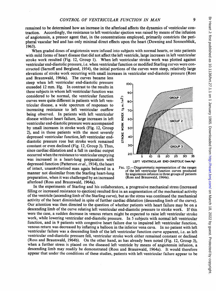
When graded doses of angiotensin were infused into subjects with normal hearts, or into patientswith mild forms of heart disease that did not affect the left ventricle, large increases in left ventricularstroke work resulted (Fig. 12, Group 1). When left ventricular stroke work was plotted againstventricular end-diastolic pressure, i.e. when ventricular function or modified Starling curves were con-structed (Sarnoff and Berglund, 1954), the initial portions of the curves were steep, relatively largeelevations of stroke work occurring with small increases in ventricular end-diastolic pressure (Rossand Braunwald, 1964a). The curves became lesssteep when left ventricular end-diastolic pressure 1ooexceeded 12 mm. Hg. In contrast to the results inthese subjects in whom left ventricular function was 90considered to be normal, the ventricular function N/curves were quite different in patients with left ven- E 80-tricular disease, a wide spectrum of responses to Eincreasing resistance to left ventricular outflow > 70/being observed. In patients with left ventricular /disease without heart failure, large increases in left Z 60ventricular end-diastolic pressure were accompanied-by small increases in stroke work (Fig. 12, Group 0so- -2), and in those patients with the most severely idepressed ventricular function, left ventricular end- 6 40ldiastolic pressure rose but stroke work remained tconstant or even declined (Fig. 12, Group 3). Thus, 30since cardiac dilatation and a fall in cardiac outputoccurred when the resistance to ventricular emptying 20 5 20 25 30 35was increased in a heart-lung preparation with LEFT VENTRICULAR END-DIASTOLICCmm.H§)depressed function (Patterson et al., 1914), the heart LEFTDVENticUr tOI rangeof intact, unamesthetized man again behaved in a FIG. 12.-Diagramatic representation of the rangesofintct,uancTshetizd man gain ehave in a of the left ventricular function curves producedmanner not dissimilar from the Starling heart-lung by angiotensin infusion in three groups of patientspreparation, when it was challenged by an increased (Ross and Braunwald, 1964a).afterload (Ross and Braunwald, 1964a).
In the experiments of Starling and his collaborators, a progressive mechanical stress (increasedfilling or increased resistance to ejection) resulted first in an augmentation of the mechanical activityof the ventricle (ascending limb of the Starling curve), but as the stress was continued the mechanicalactivity of the heart diminished in spite of further cardiac dilatation (descending limb of the curve).Our attention was then directed to the question of whether patients with heart failure may be on adescending limb of the curve relating left ventricular end-diastolic pressure to stroke work. If thiswere the case, a sudden decrease in venous return might be expected to raise left ventricular strokework, while lowering ventricular end-diastolic pressure. In 5 subjects with normal left ventricularfunction, and in 9 patients with congestive heart failure due to impaired left ventricular function,venous return was decreased by inflating a balloon in the inferior vena cava. In no patient with leftventricular failure was a descending limb of the left ventricular function curve apparent, i.e. as leftventricular end-diastolic pressure fell, ventricular stroke work either remained constant or declined(Ross and Braunwald, 1964b). On the other hand, as has already been noted (Fig. 12, Group 3),when a further stress is placed on the diseased left ventricle by means of angiotensin infusion, adescending limb may readily be demonstrated (Ross and Braunwald, 1964a). It would thereforeappear that under the conditions of these studies, patients with left ventricular failure appear to be
9
on March 18, 2022 by guest. P
rotected by copyright.http://heart.bm
j.com/
Br H
eart J: first published as 10.1136/hrt.27.1.1 on 1 January 1965. Dow
nloaded from

operating near the peak of their Starling curves, which none the less are flattened or depressed whencompared with the normal. Similarly, a descending limb was never seen in the patients in whombeat-to-beat alterations in ventricular dimensions were recorded (Braunwald et al., 1960a). Fromthe studies using the myocardial strain gauge arch, it was also apparent that considerable stretchingof the ventricle was required before the descending limb of the length-active tension curve wasreached (Aygen and Braunwald, 1962).
Since the classic studies of A. V. Hill, it has been known that the fundamental mechanicalproperties of skeletal muscle can be expressed by the relation between the force generated and thevelocity of shortening, i.e., the force-velocity relation (Hill, 1938). Recently, this concept has beenextended to isolated mammalian cardiac tissue (Abbott and Mommaerts, 1959; Sonnenblick, 1962)and currently, attempts are being made in our laboratory to extend the force-velocity relation to thehuman heart (Sonnenblick et al., 1964). These studies have been carried out with isolated papillarymuscles obtained from patients undergoing mitral valve replacement, as well as by determining thevelocity of myocardial shortening with radio-opaque clips sewn to the surface of the ventricles ofunanesthetized patients. Preliminary observations suggest that the force-velocity relation, studiedboth in vitro and in the intact states, apply to the human heart, i.e. at any given muscle length orventricular dimension, the velocity of shortening varies inversely with the load, and that the force-velocity relation is useful in characterizing the heart's contractile properties.
The investigations described above, when viewed as a whole, demonstrated the fundamentalimportance of ventricular end-diastolic volume or pressure in the regulation of the contractile activityof the human heart, and thereby extended the applicability of the Frank-Starling mechanism to thehuman heart. In addition, evidence was obtained that fundamental similarities existed betweenmammalian skeletal muscle and human myocardium, in respect to the relation between length, restingtension, and active tension, as well as to the relation between the force of contraction and thevelocity of shortening. However, these studies should not be interpreted to imply that these intrinsicmechanisms are the only ones that regulate the force of ventricular contraction. Indeed, Starling(1920) appreciated the great importance ofneurohumoral adjustments in adapting the heart's activityto the body's requirements. As Sarnoff and Mitchell (1962) have pointed out, alterations of theheart's neurohumoral environment, particularly changes in the catecholamine background, willdetermine the level of the function curve on which the ventricle is operating. Considerable recentwork has emphasized the importance of the activity of the adrenergic nervous system in the regulationof myocardial performance (Rushmer, Smith, and Franklin, 1959; Sarnoff and Mitchell, 1962).The efforts of our laboratory have been directed towards assessing the importance of this system inman.
Evidence for the fundamental importance of the adrenergic nervous system can be obtained bydetermining the effects of muscular exercise on cardiac dimensions. In studies on unanxsthetizeddogs, Rushmer and his associates noted that the diameter of the left ventricle usually decreased whilerunning on a treadmill, but they commented on the variability of this response (Rushmer et al.,1959). We have now determined the effects of pedalling a bicycle ergometer in the supine positionon the dimensions ofthe left and/or right ventricles of 15 patients by measuring the distances betweenradio-opaque clips on sequential frames ofcineradiographs (Braunwald et al., 1963b). Thesepatientshad only trivial abnormalities of cardiac function and their responses might be considered analogousto what might be expected in normal subjects investigated under similar conditions. In everypatient studied the ventricular dimensions decreased throughout the entire cardiac cycle and thechanges were of approximately the same magnitude in both ventricles (Fig. 13). The observeddiminutions in external ventricular dimensions during exercise approximated 50 per cent of thestroke volume.
The increased mechanical activity of the heart that occurs during exercise, coupled with a fall inventricular end-diastolic size and an increase in the rate of development of ventricular pressure(dp/dt) and in the rate of emptying (Fig. 13), clearly suggested the possibility that augmented activityof the adrenergic nervous system was involved in this response. Further evidence for the potential
10 EUGENE BRAUNWALD
on March 18, 2022 by guest. P
rotected by copyright.http://heart.bm
j.com/
Br H
eart J: first published as 10.1136/hrt.27.1.1 on 1 January 1965. Dow
nloaded from

CONTROL OF VENTRICULAR FUNCTION IN MAN
E
1-a.
zw-J
cc
REST EXERCISE
RVmm.Hg20-
*0.RV-15 -
~600 'ro
d/t RV -400 wdpldt 200~dpdt
5
L0 115.
14.
13.
12-
'I 0-2 sc
FIG. 13.-Simultaneous recording of RV (right ventricular) pressure, rate of pressure change (dp/dt)and length, at rest, and during exercise. RV pressure measured by means of intracardiacmanometer, RV length measured by radio-opaque clip method.
importance of the adrenergic nervous system in the regulation of myocardial contractility was provi-ded by determining the direct effects of electrical stimulation of the stellate ganglion in man (Fig. 14).This was carried out by Dr. A. G. Morrow at the time of left thoracotomy, and it was observedconsistently that the force of ventricular contraction, as recorded with a myocardial strain gaugearch, increased significantly, even though the length of the segment ofmyocardium to which the strain
H. R.-130
B, A.
(mm.OR) .j
0CONTRACTI LE io tFORCE >mm,) I]0i
CONTROL
I50
6Jsec °LEFT STELLATE STIMULATION
FIG. 14.-Effect of stimulating the left stellate ganglion on brachial artery (BA) pressure and onleft ventricular (LV) contractile force, measured with a strain gauge arch.
11
130
on March 18, 2022 by guest. P
rotected by copyright.http://heart.bm
j.com/
Br H
eart J: first published as 10.1136/hrt.27.1.1 on 1 January 1965. Dow
nloaded from

EUGENE BRAUNWALD
gauge arch was sewn remained constant. From this study it seemed clear that stimulation ofnervouspathways in the sympathetic nervous system might result in profound augmentation of the con-tractile state of the myocardium. However, it cannot be concluded from such stimulation experi-ments that the ordinary activity of the adrenergic nervous system has a significant effect on myocar-dial function in intact man.
This problem was approached by studying the circulatory effects of muscular exercise before andafter pharmacological inhibition of the autonomic nervous system. The investigations were carriedout on a group of healthy male subjects, studied in the supine position (Kahler, Gaffney, andBraunwald, 1962). Measurements of heart rate, cardiac output, arterial blood pressure, and oxy-gen consumption were made at rest and during exercise. The control study was performed withoutprior drug administration. Oral guanethidine was then begun and the dose was progressivelyincreased every other day for a period of 21 to 25 days. The maximum daily dose of guanethidineranged from 50 to 85 mg. while atropine was given intravenously in a dose of 2 mg. just before thesecond study. Exercise consisted of pedalling a bicycle ergometer at a constant rate. The effects ofexercise were determined after guanethidine administration and then under the infliuence of bothatropine and guanethidine.
The conditions of exercise were identical during the control period and after administration ofguanethidine and atropine. This was reflected in the finding that the increase in total body oxygenconsumption associated with the exercise was identical under both conditions. It was observed thatpharmacological inhibition of the autonomic nervous system greatly reduced the magnitude of thecirculatory response to exercise. During the control study, a level of exercise that resulted in a four-to fivefold increase in oxygen consumption produced average increases above resting values of 68per cent in heart rate, 17 per cent in the stroke volume, 96 per cent in the cardiac output, and 129 percent in left ventricular minute work. After the administration of atropine and guanethidine,identical levels of exercise resulted in average increases above resting values of only 28 per cent in theheart rate, 1 per cent in the stroke volume, 30 per cent in cardiac output, and 5 per cent in leftventricular minute work (Fig. 15). The absolute values of cardiac output, systemic arterial pressure,and ventricular work per minute and per beat were significantly lower and the values of the arterio-venous oxygen differences were significantly higher during exercise following atropine and guanethi-dine than during the control study.
Blockade of the parasympathetic nervous system alone raised the heart rate, cardiac output, andleft ventricular minute work at rest and did not interfere with the circulatory response to exercise.Guanethidine alone slowed the resting heart rate, but did not affect cardiac output or arterial pres-sure at rest. The cardiac output, mean arterial pressure, and left ventricular work during exercisewere lower after guanethidine than during the control study. These studies demonstrate the impor-tant contribution made by the autonomic nervous system to the circulatory response to exercise inman and indicate that the sympathetic division plays the major role in this regard.
The logical next step was to attempt to determine whether this system played a compensatoryrole in maintaining ventricular contractility when the function of the myocardium was depressed inpatients with heart disease. The availability of drugs, such as guanethidine, that selectively blockthe activity of the adrenergic nervous system has made it possible to estimate the role of this systemin the maintenance ofmyocardial function in man. The effects of the administration of guanethidineto patients with reduced cardiovascular reserve were first determined (Gaffney and Braunwald,1963). In addition to the theoretical interest of such studies, the widespread clinical use of adre-nergic blocking drugs in patients with heart disease makes an evaluation of the effects of these drugson myocardial function of immediate and practical importance. Ten adult patients with inactiverheumatic valvular or primary myocardial disease were studied. Each patient had signs or symp-toms of right and/or left-sided congestive heart failure at the time of the investigation. The admini-stration of guanethidine increased the clinical manifestations of heart failure in each of five patientswho were in functional classes III or IV. In these patients guanethidine resulted in an increase ofdyspncea and orthopncea, a decrease in urinary sodium excretion, and a rise in venous pressure and
12
on March 18, 2022 by guest. P
rotected by copyright.http://heart.bm
j.com/
Br H
eart J: first published as 10.1136/hrt.27.1.1 on 1 January 1965. Dow
nloaded from

CONTROL OF VENTRICULAR FUNCTION IN MAN 13
o 14-O tX 12 - ' 125_ GUANETHIDINE
12 125 80\DMINISTRATIONE10 20
- ES EXRIE CNTOETO
a,.P * 24O0~Q75-> 8~~~~~~ix220-a
6- Z5 Idg 200-
-*--C0NTROL x
-ATROPINE & " 84- GUANETHIDINE 50
0~~~~~2REST EXERCISE CONTROL ATRO- >
GUANETHIIDNE a, S
FIG. 15.-Mean and range of values for left ventricular I so5minute work made at rest and during exercise in 7 fnormal subjects before and after treatment with w 49-atropine and guanethidine (Kahler et al., 1962).On the left values, and on the right the percentage 48increase. 47I
C32 X3 I4SO/d20
E
z zz 2 NORMAL RANGE r
o
TI. 27-Chne inpam oeieprn uigDY
0.za:065zI
AK ~~~~~~~' 60-
exercise in patients with congestive heart failure FIG. 16.-Changes in venous pressure, body weight,(CHF). C=control or resting values. The 02 sodium balance, and heart rate, before, during, andconsumption during the exercise period is expressed after the administration of guanethidine (Gaffneyin multiples of the resting oxygen consumption. and Braunwald, 1963).The normal range is represented by the stippledarea (Chidsey et a!., 1962).
weight (Fig. 16). The administration of guanethidine to the other five patients, who were infunctional classes II or III, did not result in any worsening of the congestive heart failure.
It is evident from these studies that interference with the activity of the adrenergic nervoussystem is capable of aggravating congestive heart failure in some patients with cardiac decom-pensation. This effect occurred despite the fact that the adrenergic nerve blockade was almostcertainly incomplete. These observations, therefore, suggest that the adrenergic nervous systemplays an important compensatory role in the circulatory adjustments of patients to congestive heartfailure; and they emphasize the need for caution in the use of anti-adrenergic drugs, such as reserpineand guanethidine, in the treatment of patients with limited cardiac reserve.
It then became of interest to assess the activity of the sympathetic nervous system in patients withheart disease during the stress of muscular exercise and to compare it with the activity in normalsubjects. An index of the activity of the sympathetic nervous system at rest and during exercisewas obtained by measuring the concentration of norepinephrine in arterial blood. In normalsubjects, very small increases in the arterial norepinephrine concentrations were noted during
on March 18, 2022 by guest. P
rotected by copyright.http://heart.bm
j.com/
Br H
eart J: first published as 10.1136/hrt.27.1.1 on 1 January 1965. Dow
nloaded from

EUGENE BRAUNWALD
exercise (Chidsey, Harrison, and Braunwald, 1962). Though the levels of exercise were com-parable, the augmentations of plasma norepinephrine induced by exercise in patients with congestiveheart failure exceeded those observed in the normal subjects (Fig. 17). This excessive rise in thecirculating norepinephrine concentration is considered to reflect an increased activity of the sym-pathetic nervous system. Since, as already noted, increased activity of the sympathetic nervoussystem augments myocardial contractility, it is possible that the heightened sympathetic activityobserved during exercise in patients with heart failure plays an important role in supporting theirmyocardial function.
In view of these findings, we wondered if this massive barrage of sympathetic nerve impulse overprolonged periods affected the concentration of the neurohumoral transmitter, norepinephrine, in the
heart. The concentration of norepinephrine ino 4 biopsy specimens of atrial appendage was
measured in 29 patients with various cardiac5 . lesions who had not suffered from congestivez 3 * heart failure and was found to averageq .. 1 82+077 ,ug./g. In 17 patients who had ex-z2- I perienced chronic congestive heart failure, theffi - - concentration was significantly lower and< averaged 053±047 Vg./g. (Chidsey et al.,
1-X ~ ^g1963). Indeed, in 6 of the 17 patients the con-
S* . centrations of norepinephrine in the atrial______________________.appendage were less than 030 ug./g. This re-CONTROL RESERPINE CONGESTIVE duction was similar to that observed whenPATIENTS PTIENTS FAILUREPATIENTS patients without heart failure had been pre-
treated with reserpine (Fig. 18) (Braunwald etFIG. 18.-Concentration of norepinephrine in the atrialappendages excised at operation in 29 "control" al., 1963a).patients who had not been in congestive heart It was conceivable that the norepinephrinefailure, in 5 patients without failure who had ben per unit weight of tissue was reduced in thetreated with reserpine, and in 17 patients who hadniwegtotsue asrdcdnthbeen in congestive heart failure (Braunwald et al., patients with heart failure simply because the1963). number of adrenergic nerve endings was diluted
by the presence of increased amounts of fibroustissue or hypertrophied myocardium or both. In order to exclude this possibility the total nore-pinephrine content of papillary muscles excised at the time of mitral valve replacement wasdetermined and reductions in the total quantity of norepinephrine, not merely the concentra-tion, in patients with heart failure has been confirmed (Chidsey and Morrow, 1964). In addition,in guinea-pigs with left heart failure produced by supravalvular aortic constriction, the totalquantity of norepinephrine in the heart is substantially reduced (Spann, Chidsey, and Braunwald,1964). Similar results have been obtained in analyses of the hearts of dogs with congestiveheart failure produced by tricuspid regurgitation and pulmonary constriction (Chidsey et al., 1964).
The clinical significance of this biochemical abnormality in the hearts of patients with congestiveheart failure remains to be elucidated. It does not appear to be a primary abnormality: it seemsmore likely that the reduction in the norepinephrine concentration and content in the hearts ofmanypatients with congestive heart failure results secondarily from a prolonged intensive barrage of sym-pathetic nerve impulses which serve to bolster the activity of the failing heart. However, sincenorepinephrine depletion alone is capable of reducing myocardial contractility (Lee and Shideman,1959), it seems quite likely that the norepinephrine depletion that occurs in clinical heart failurefurther intensifies the heart failure state.
From the investigations reviewed above the following tentative hypothesis regarding the role ofthe autonomic nervous system in circulatory regulation might be suggested. The adrenergicnervous system increases its stimulation ofthe myocardium at a time when an imbalance, or a poten-tial imbalance, exists between the cardiac output and the perfusion requirements of the peripheral
14
on March 18, 2022 by guest. P
rotected by copyright.http://heart.bm
j.com/
Br H
eart J: first published as 10.1136/hrt.27.1.1 on 1 January 1965. Dow
nloaded from

CONTROL OF VENTRICULAR FUNCTION IN MAN
tissues. Such an imbalance may occur in the patient with congestive heart failure at rest, in whomwithdrawal of sympathetic stimulation by guanethidine may result in an intensification of heartfailure. However, the withdrawal or reduction of sympathetic impulses does not induce cardiacdecompensation in normal subjects, or in patients with heart disease who are not suffering from con-gestive heart failure. One possible explanation for this finding is that the sympathetic nervoussystem contributes little to the maintenance of cardiac activity in such subjects when they are atrest. An alternative possible explanation is that in the absence of cardiac failure an augmentation ofthe force of cardiac contraction through the operation of the Frank-Starling mechanism can stilltake place in spite of any depression of myocardial function evoked by the withdrawal of sym-pathetic impulses.
During the stress of muscular exercise the function of the adrenergic nervous system assumesgreater importance. In patients with congestive heart failure the intense activity of this system isreflected in the extremely high levels of circulating catecholamines which may occur under theseconditions. Even in normal subjects, however, the autonomic nervous system plays a significantrole in stimulating the myocardium during muscular exercise. This increased activity is reflectedby the augmentation of the urinary excretion of norepinephrine and the tendency for the circulatingnorepinephrine concentration to increase slightly. Inhibition of the adrenergic nervous systemalone prevents the small increase in stroke volume that normally occurs during exercise. Adre-nergic blockade, however, still permits an increase in cardiac output to occur as a result of a greaterincrease in heart rate, presumably mediated through the withdrawal of vagal impulses. When theparasympathetic nervous system is blocked in addition to the sympathetic, the circulatory responseto exercise is further reduced, and the mixed arteriovenous oxygen difference during exercise risessignificantly above the levels encountered in the subjects in whom the autonomic nervous system isintact.
CONCLUSIONSOur present concepts of the control of ventricular function in the normal and diseased human
heart point to the importance of two fundamental regulatory mechanisms. It might be appropriateto consider one of these, the Frank-Starling mechanism, to be intrinsic to the heart muscle, the mech-anical activity of which is determined to a major extent by its length at the onset of ventricular con-traction. On the other hand, the release of catecholamines by sympathetic nerve endings and by theadrenal medulla might be considered an extrinsic control mechanism, which can modulate themechanical activity of the heart at any given end-diastolic fibre length. It seems fruitless at thistime to engage in polemics by championing the relative importance of either the intrinsic or theextrinsic mechanism. As already demonstrated, it is possible to design experiments that willemphasize the importance of either one. In normal intact man, however, all the evidence nowavailable suggests that both mechanisms are continuously operative, and that sufficient reserve isavailable to both the Frank-Starling mechanism and the adrenergic nervous system so that theorganism can fall back on one if the other is interfered with. On the other hand, in the presence ofserious myocardial disease the reserve of both systems is encroached upon as the heart dilates andresting sympathetic tone increases. Under these conditions removal of the stimulation of myo-cardial contraction afforded either by an augmented end-diastolic fibre length or by the sympatheticnerves cannot be tolerated.
The author acknowledges with pleasure the collaboration of a number of colleagues in the design, execution, andinterpretation of the experiments reviewed above. Particular attention is directed to the contributions of Dr. J. Ross,Jr., C. A. Chidsey, M. M. Aygen, R. L. Kahler, T. E. Gaffney, E. H. Sonnenblick, and A. G. Morrow.
REFERENCESAbbott, B. C., and Mommaerts, W. F. H. M. (1959). A study of inotropic mechanisms in the papillary muscle
preparation. J. gen. Physiol., 42, 533.Aygen, M. M., and Braunwald, E. (1962). Studies on Starling's law of the heart. VIII. Mechanical properties of
human myocardium studied in vivo. Circulation, 26, 516.Blix, M. (1895). Die Lange und die Spannung des Muskels. Skand. Arch. Physiol., 5, 173.
15
on March 18, 2022 by guest. P
rotected by copyright.http://heart.bm
j.com/
Br H
eart J: first published as 10.1136/hrt.27.1.1 on 1 January 1965. Dow
nloaded from

16 EUGENE BRAUNWALD
Braunwald, E., Chidsey, C. A., Mason, D. T., and Morrow, A. G. (1963a). Effects of reserpine and of congestiveheart failure on the myocardial norepinephrine concentration in man. Trans. Ass. Amer. phycns, 76, 254.and Frahm, C. J. (1961). Studies on Starling's law of the heart. IV. Observations on the hemodynamicfunctions of the left atrium in man. Circulation, 24, 633.
, and Ross, J., Jr. (1961). Studies on Starling's law of the heart. V. Left ventricular function in man.J. clin. Invest., 40, 1882.
, Frye, R. L., Aygen, M. M., and Gilbert, J. W., Jr. (1960a). Studies on Starling's law of the heart. III. Obser-vations in patients with mitral stenosis and atrial fibrillation on the relationships between left ventricular end-diastolic segment length, filling pressure, and the characteristics of ventricular contraction. J. clin. Invest., 39,1874.- and Ross, J., Jr. (1960b). Studies on Starling's law of heart. II. Determinants of the relationship
between left ventricular end-diastolic pressure and circumference. Circulat. Res., 8, 1254.Goldblatt, A., Harrison, D. C., and Mason, D. T. (1963b). Studies on cardiac dimensions in intact, unanesthe-tized man. III. Effects of muscular exercise. Circulat. Res., 13, 460.
Chidsey, C. A., Braunwald, E., Morrow, A. G., and Mason, D. T. (1963). Myocardial norepinephrine concentrationin man: effects of reserpine and of congestive heart failure. New Engl. J. Med., 269, 653.
-, Harrison, D. C., and Braunwald, E. (1962). Augmentation of the plasma nor-epinephrine response to exercisein patients with congestive heart failure. New Engi. J. Med., 267, 650.Kaiser, G., Sonnenblick, E. H., Spann, J. F., and Braunwald, E. (1964). Cardiac norepinephrine stores inexperimental heart failure in the dog. J. clin. Invest. In the press.
, and Morrow, A. G. (1964). Analysis of sympathetic activity and cardiac norepinephrine stores in congestiveheart failure. Clin. Res., 12, 178.
Dodge, H. T., Sandler, H., Ballew, D. W., and Lord, J. D. (1960). The use of biplane angiocardiography for themeasurement of left ventricular volume in man. Amer. Heart J., 60, 762.
Downing, S. E., and Sonnenblick, E. H. (1963). Effects of continuous administration of angiotensin II on ventricularperformance. J. appl. Physiol., 18, 585.
Fick, A. (1882). Mechanische Arbeit und Warmeent-wickelung bei der Muskeltkatigheit. Brockhaus, Leipzig.Frank, 0. (1895). Zur Dynamik des Herzmuskels. Z. Biol., 32, 370.Frye, R. L., and Braunwald, E. (1960). Studies on Starling's law of the heart. I. The circulatory response to acute
hypervolemia and its modification by ganglionic blockade. J. clin. Invest., 39, 1043.Gaffney, T. E., and Braunwald, E. (1963). The importance of the adrenergic nervous system in the support of cir-
culatory function in patients with congestive heart failure. Amer. J. med., 34, 320.Gleason, W. L., and Braunwald, E. (1962). Studies on Starling's law of the heart. VI. Relationships between left
ventricular end-diastolic volume and stroke volume in man with observations on the mechanisms of pulsusalternans. Circulation, 25, 841.
Goldblatt, A., Harrison, D. C., Glick, G., and Braunwald, E. (1963). Studies on cardiac dimensions in intact, un-anesthetized man. II. Effects of respiration. Circulat. Res., 13, 455.
Harrison, D. C., Goldblatt, A., and Braunwald, E. (1963). Studies on cardiac dimensions in intact, unanesthetizedman. I. Description of techniques and their validation. Circulat. Res., 13, 448.
Hill, A. V. (1938). The heat of shortening and the dynamic constants of muscle. Proc. roy. Soc. Lond. B., 126, 136.Kahler, R. L., Gaffney, T. E., and Braunwald, E. (1962). The effects of autonomic nervous system inhibition on the
circulatory response to muscular exercise. J. clin. Invest., 41, 1981.Kries, J. von (1892). Untersuchungen zur Mechanik des quergestreiften Muskels. Arch. Anat. Physiol. (Lp. 3),
Physiol. Abt., P. 1.Lee, W. C., and Shideman, F. E. (1959). Role of myocardial catecholamines in cardiac contractility. Science,
129, 967.Patterson, S. W., Piper, H., and Starling, E. H. (1914). The regulation of the heartbeat. J. Physiol. (Lond.), 48,465.Ross, J., Jr., and Braunwald, E. (1964a). The study of left ventricular function in man by increasing resistance to
ventricular ejection with angiotensin. Circulation. In the press.-, and (1964b). Studies on Starling's law of the heart. IX. The effects of impeding venous return on perfor-
mance of the normal and failing human left ventricle. Circulation. In the press.and Morrow, A. G. (1959). Transseptal left atrial puncture: New technique for the measurement of left
atrial pressure in man. Amer. J. Cardiol., 3, 653.Rushmer, R. F., Smith, O., and Franklin, D. (1959). Mechanisms of cardiac control in exercise. Circulat. Res.,
7, 602.Sarnoff, S. J., and Berglund, E. (1954). Ventricular function. I. Starling's law of the heart studied by means of
simultaneous right and left ventricular function curves in the dog. Circulation, 9, 706.Braunwald, E., Welch, G. H., Jr., Case, R. B., Stainsby, W. N., and Macruz, R. (1958). Hemodynamic deter-minants of oxygen consumption of the heart with special reference to the tension-time index.. Amer. J. Physiol.,192, 148.and Mitchell, J. H. (1962). The control of the function of the heart. In Handbook ofPhysiology. Section 2:Circulation, Vol. 1, p. 490. Washington, D.C. American Physiological Society.
Sonnenblick, E. H. (1962). Force-velocity relations in mammalian heart muscle. Amer. J. Physiol., 202, 931.Glick, G., Morrow, A. G., and Braunwald, E. (1964). Force-velocity relations in the human heart. J. clin.Invest., 43, 1245.
Spann, J. F., Chidsey, C. A., and Braunwald, E. (1964). Reduction of norepinephrine in experimental heart failure.Science. In the press.
Starling, E. H. (1918). The Linacre Lecture on the Law of the Heart. (Cambridge, 1915). Longmans, Green,London.(1920). On the circulatory changes associated with exercise. J. roy. Army med. Cps, 34, 258.
on March 18, 2022 by guest. P
rotected by copyright.http://heart.bm
j.com/
Br H
eart J: first published as 10.1136/hrt.27.1.1 on 1 January 1965. Dow
nloaded from