the controversial role of glucose-6-phosphate
TRANSCRIPT
Review ArticleThe Controversial Role of Glucose-6-Phosphate DehydrogenaseDeficiency on Cardiovascular Disease: A Narrative Review
Maria Pina Dore ,1,2 Guido Parodi ,1 Michele Portoghese,3 and Giovanni Mario Pes 1,4
1Dipartimento di Scienze Mediche, Chirurgiche e Sperimentali, University of Sassari, Viale San Pietro 8, 07100 Sassari, Italy2Baylor College of Medicine, One Baylor Plaza Blvd., Houston Texas, USA3Heart Surgery Unit, AOU Sassari, Via Enrico de Nicola, Sassari, Italy4Sardinia Longevity Blue Zone Observatory, Ogliastra, Italy
Correspondence should be addressed to Maria Pina Dore; [email protected]
Received 8 January 2021; Revised 27 March 2021; Accepted 21 April 2021; Published 3 May 2021
Academic Editor: Jayeeta Ghose
Copyright © 2021 Maria Pina Dore et al. This is an open access article distributed under the Creative Commons AttributionLicense, which permits unrestricted use, distribution, and reproduction in any medium, provided the original work isproperly cited.
Cardiovascular disorders (CVD) are highly prevalent and the leading cause of death worldwide. Atherosclerosis is responsible formost cases of CVD. The plaque formation and subsequent thrombosis in atherosclerosis constitute an ongoing process that isinfluenced by numerous risk factors such as hypertension, diabetes, dyslipidemia, obesity, smoking, inflammation, and sedentarylifestyle. Among the various risk and protective factors, the role of glucose-6-phosphate dehydrogenase (G6PD) deficiency, themost common inborn enzyme disorder across populations, is still debated. For decades, it has been considered a protectivefactor against the development of CVD. However, in the recent years, growing scientific evidence has suggested that thisinherited condition may act as a CVD risk factor. The role of G6PD deficiency in the atherogenic process has been investigatedusing in vitro or ex vivo cellular models, animal models, and epidemiological studies in human cohorts of variable size andacross different ethnic groups, with conflicting results. In this review, the impact of G6PD deficiency on CVD was criticallyreconsidered, taking into account the most recent acquisitions on molecular and biochemical mechanisms, namely, antioxidativemechanisms, glutathione recycling, and nitric oxide production, as well as their mutual interactions, which may be impaired bythe enzyme defect in the context of the pentose phosphate pathway. Overall, current evidence supports the notion that G6PDdownregulation may favor the onset and evolution of atheroma in subjects at risk of CVD. Given the relatively high frequencyof this enzyme deficiency in several regions of the world, this finding might be of practical importance to tailor surveillanceguidelines and facilitate risk stratification.
1. Introduction
Atherosclerotic cardiovascular diseases (CVDs) are a groupof disorders that include coronary heart disease, cerebrovas-cular disease, peripheral arterial disease, and aortic athero-sclerosis [1]. Cardiovascular disorders are common in thegeneral population worldwide and are the leading cause ofmortality, representing 31% of total deaths, the majoritydue to heart attack and stroke [2, 3]. Atherosclerosis isresponsible for almost all cases of CVD, with plaque forma-tion, ulceration, and the consequent thrombotic occlusionforming a slow, continued process that is influenced by sev-eral risk factors [4, 5]. Among these, hypertension, diabetes,
dyslipidemia, obesity, a sedentary lifestyle, smoking, andinflammation may contribute to endothelial dysfunction,erosion, and plaque instability [6]. For these reasons, actingon modifiable risk factors such as tobacco use, diet, obesity,physical inactivity, and addiction to alcohol can prevent pre-mature CVD events.
Among the nonmodifiable determinants, genetic factorsplay a considerable role, and previous epidemiological stud-ies [7–10] as well as animal models [11–13] have suggestedthat the deficiency of the enzyme glucose-6-phosphate dehy-drogenase (G6PD; EC 1.1.1.49) may act as a protective factoragainst CVD. However, recent evidence from animal models,ex-vivo studies on cells isolated from deficient subjects,
HindawiOxidative Medicine and Cellular LongevityVolume 2021, Article ID 5529256, 19 pageshttps://doi.org/10.1155/2021/5529256

in vitro studies where deficiency was induced by gene silenc-ing, and large human cohorts indicate that G6PD can lead toadverse physiological effects in response to increased oxida-tive stress [14] acting as a cardiovascular risk factor [15–18].
The purpose of this review is to critically discuss the cur-rent knowledge about the metabolic modifications inducedinto cells by G6PD deficiency and the evidence for andagainst its possible involvement in atherogenesis and its clin-ical consequences, with the aim of understanding the extentto which this common enzyme disorder may impact CVDrisk.
The sources used in this literature review are original arti-cles published in PubMed, posted on public repositories, orlisted in clinical trial databases in addition to databases refer-ring to the World Health Organization and Centers for Dis-ease Control.
2. Pathophysiology of Glucose-6-PhosphateDehydrogenase Deficiency
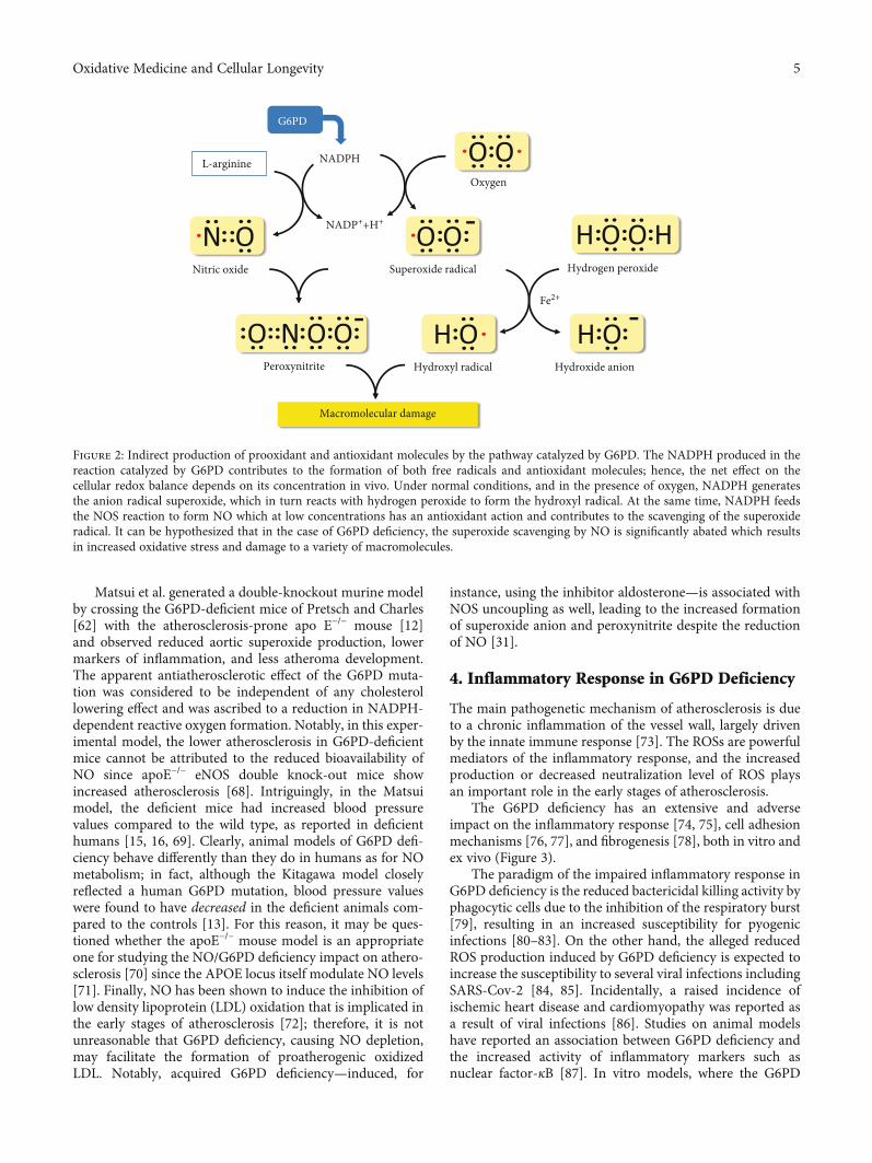
The G6PD is a cytosolic enzyme that catalyzes the first andrate-limiting step in the oxidative branch of the pentose phos-phate pathway (PPP), which converts glucose-6-phosphateinto 6-phosphoglucono-δ-lactone. The PPP reactions cata-lyzed by G6PD and the 6-phospho-gluconate dehydrogenase(EC 1.1.1.44) generate the reduced form of the pyridinic coen-zyme nicotinamide adenine dinucleotide phosphate(NADPH). The NADPH supplies high-energy electrons (i.e.,reducing equivalents) to cells to maintain their oxidoreductivebalance and feed reductive biosynthesis. The G6PD is funda-mental for the cell’s defense against the toxicity of reactiveoxygen species (ROSs) (Figure 1). Moreover, the nonoxidativebranch of PPP synthesizes the ribose-5-phosphate necessaryto sustain the synthesis of the DNA backbone.
The evolutionary origin of the PPP is ancient, possiblydating back to the prebiotic world [19]. In cells devoid ofmitochondria, such as erythrocytes, PPP is essential sincethe reaction catalyzed by G6PD is the only source ofNADPH. The coenzyme is also important for the elongationand desaturation of fatty acids [20, 21], biosynthesis of cho-lesterol [22], hydroxylation of steroids and other polycyclicmolecules including vitamin D [23], drugs metabolism bythe cytochrome P450 [24], and ROS generation in phagocyticand inflammatory cell to counteract pathogens [25]. Thegene coding for G6PD maps to long arm of the X chromo-some [26]; therefore, its inheritance is X-linked. The humanG6PD gene was cloned by Takizawa et al. from a human hep-atoma cDNA library [27]; it spans 18 kb, divided into 13exons [28], and the gene product encompasses 515 aminoacids with a molecular mass of 58 kD [29]. The transcribedregion from the initiation site to the poly(A) addition sitecovers 15,860 bp [30].
The human G6PD gene is remarkable for its allelic vari-ability, and some gene variants can be considered as loss-of-function mutations causing a lower catalytic activity and,in turn, global PPP downregulation. In reality, G6PD defi-ciency can be due to genetic (primary) or secondary causes[31, 32]. Clinical manifestations of primary G6PD deficiency,based on the severity of the mutation, may result in nonim-
mune hemolytic anemia in response to bacterial or viralinfections, or the ingestion of certain drugs or plants suchas Vicia faba (favism). Favism was known in antiquity, as evi-denced by the ban on eating or even naming beans during thetime of Pythagoras [33] and among Roman priests [34]. Incases of severe deficiency, intravascular hemolysis may occur,causing hemoglobin release and resulting in kidney failure[35]. The clinical outcomes are mainly related to theimpaired antioxidant activity due to NADPH depletion inred blood cells, which are exclusively dependent on PPP forglutathione regeneration [36].
Due to X-chromosome inheritance, males carrying themutant allele are hemizygotes with total enzyme deficiency;female carriers can be, rarely, homozygotes or more fre-quently, heterozygotes with a milder form of deficiency.However, due to the random inactivation of the X chromo-some (mosaicism), the degree of deficiency can be variablein females [36]. The enzyme deficiency occurs most fre-quently in those parts of the world where endemic malariawas prevalent in the past, such as sub Saharian Africa, South-east Asia, the Mediterranean basin, and the Middle East,including Israel [37]. In the United States, it affects about10% of African-American males [38]. In North Europe, it israre, even though it has been reported in newborns [39].Although the geographic distribution of G6PD deficiencysuggests a protective role against malaria, this hypothesishas been questioned (as reviewed in the meta-analysis byMbanefo et al. [40]).
In populations where G6PD deficiency is widespread, thecondition is usually diagnosed in childhood, but clinicalmanifestations may appear throughout a person’s lifetimeunder conditions of oxidative stress. In addition to hemolyticdisorders, G6PD deficiency may predispose to a wide rangeof conditions including neonatal jaundice [41]. In a smallsubset of patients with severe G6PD deficiency, a chronicnonspherocytic hemolytic anemia may occur [42].
According to the World Health Organization, mutantenzymes have been classified into five classes in the order ofdescending severity [43]. The G6PD B is the normal (wildtype) isoenzyme. The G6PD A– (point mutations at nucleo-tides 202A/376G) is the most common variant amongAfrican-Americans and is associated with mild to moderateenzyme activity (class III) [44]. The G6PD Mediterraneanvariant (C→T transition at nucleotide 563 of the codinggene, amino acids Ser188Phe) is the most common variantin Caucasians entailing enzyme instability and classicallyassociated with favism. There is no known null mutantamong the more than 200 spontaneous G6PDmutants foundin humans (Table 1). In addition to the common mutationslisted in Table 1, more than 400 different variants have beendescribed [45], making G6PD deficiency the most polymor-phic common inherited error of metabolism.
3. The Role of G6PD in the Antioxidant Defense
For a long time, G6PD was considered only a component ofthe mechanisms that counteract ROS toxicity. Recently, it hasbeen hypothesized that, under specific conditions, inefficientG6PD can generate free radicals, making its overall action
2 Oxidative Medicine and Cellular Longevity

more complex than previously understood [14, 46]. The firsttwo PPP redox reactions (catalyzed by G6PD and 6GPD)generate NADPH that provides high-energy electrons forglutathione (GSH) recycling. The importance of this mecha-nism is underscored by the heavy membrane damage andsubsequent hemolysis upon oxidative stress in cells thatdepend solely on PPP as a NADPH source. In cells harboringmitochondria with active tricarboxylic acid cycle (including
platelets), the situation is more complex because mitochon-dria are themselves a source of endogenous ROS as bypro-ducts of the electron transport chain [47]. ROS can damagecell membranes, lipoproteins, and DNA, leading to celldeath. However, the cytoplasm contains isocitrate dehydro-genase, and the mitochondria contain the malic enzymeand other enzymes that also yields NADPH to cope with oxi-dative stress.
Glucose
G6PD
Pentose phosphate pathway
NADPH
NADPH
GSSG
2 GSH6PGD
NADP+
GSR
Glucose-6-phosphate
6-phosphoglucono-𝛿-lactone
6-phosphogluconate
Ribulose-5-phosphate
L-arginineNO•
NADP+
Glutathione recycling
NOS
O•-
peroxynitrite
O2
NADP+H+
NADPH
NOX
H2O2OH•
O OON
Figure 1: Pentose phosphate pathway (PPP) and glucose 6-phosphate dehydrogenase (G6PD) in nucleated cells. The NADPH providesreducing equivalents for antioxidant defense and reductive biosynthesis and NADPH oxidase for generation of superoxide anions. TheG6PD is required to maintain a normal NADPH/NADP ratio which in turn regulates the glutathione (γ-L-glutamyl-L-cysteinylglycine,GSH) biosynthesis. The GSH is a sulfhydryl-containing compound present in all mammalian cells. The redox-active thiol group in GSH isessential in the regulation of disulfide bonds of proteins and to detoxify oxidant compounds. The function of GSH as an antioxidant isefficient when the free thiol is maintained. This is accomplished by the reaction catalyzed by the NADPH-dependent glutathione-disulfidereductase (GSR) that reduces glutathione disulfide (GSSG) into the form with a free thiol (GSH). In G6PD deficiency, the decreasedsupply of NADPH limits the GSH regeneration and in turn the disposal of oxidants. The NADPH produced in the PPP is also a substratefor nitric oxide synthase (NOS) for the release of nitric oxide (NO) and for NADPH oxidase for the release of superoxide anion. In G6PDdeficiency, NO depletion leads to the decreased neutralization of superoxide anion and other free radicals.
Table 1: Most common mutations causing G6PD deficiency worldwide.
Exon/intron location Nucleotidic substitution in cDNA Aminoacid substitution Designation Class KM NADP+ (μM) Reference
Exon 5, nt 376 A→G N126D G6PD A III 12.97 [29]
Exon 4, nt 202Exon 5, nt 376
G→AA→G
V68MN126D
G6PD A– III 15 [155]
Exon 6, nt 563 C→ T S188F G6PD Med II 2.43 [29]
Exon 8, nt 844 G→ C D282H G6PD Seattle III 2.4–2.8 [29]
Exon 11, nt 1260 C→ T R454C G6PD Union II 8.6 [40]
Exon 12, nt 1376 G→ T R459L G6PD Canton II 14.7 [40]
A lower level of enzyme activity in the erythrocytes of genetically deficient individuals might be due to a normal rate of synthesis of an enzyme of low catalyticefficiency, a decreased rate of synthesis of a normally active enzyme, an increased lability of the variant enzyme or a combined mechanism. The clinicalphenotype depends on the mutation location in the 3D structure of the protein. G6PD A– is a more labile enzyme with normal rate of synthesis.
3Oxidative Medicine and Cellular Longevity
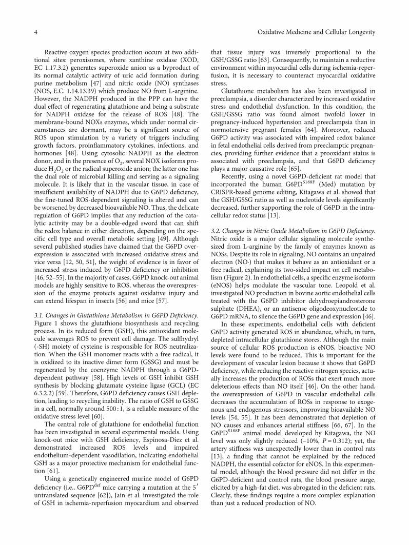
Reactive oxygen species production occurs at two addi-tional sites: peroxisomes, where xanthine oxidase (XOD,EC 1.17.3.2) generates superoxide anion as a byproduct ofits normal catalytic activity of uric acid formation duringpurine metabolism [47] and nitric oxide (NO) synthases(NOS, E.C. 1.14.13.39) which produce NO from L-arginine.However, the NADPH produced in the PPP can have thedual effect of regenerating glutathione and being a substratefor NADPH oxidase for the release of ROS [48]. Themembrane-bound NOXs enzymes, which under normal cir-cumstances are dormant, may be a significant source ofROS upon stimulation by a variety of triggers includinggrowth factors, proinflammatory cytokines, infections, andhormones [48]. Using cytosolic NADPH as the electrondonor, and in the presence of O2, several NOX isoforms pro-duce H2O2 or the radical superoxide anion; the latter one hasthe dual role of microbial killing and serving as a signalingmolecule. It is likely that in the vascular tissue, in case ofinsufficient availability of NADPH due to G6PD deficiency,the fine-tuned ROS-dependent signaling is altered and canbe worsened by decreased bioavailable NO. Thus, the delicateregulation of G6PD implies that any reduction of the cata-lytic activity may be a double-edged sword that can shiftthe redox balance in either direction, depending on the spe-cific cell type and overall metabolic setting [49]. Althoughseveral published studies have claimed that the G6PD over-expression is associated with increased oxidative stress andvice versa [12, 50, 51], the weight of evidence is in favor ofincreased stress induced by G6PD deficiency or inhibition[46, 52–55]. In the majority of cases, G6PD knock-out animalmodels are highly sensitive to ROS, whereas the overexpres-sion of the enzyme protects against oxidative injury andcan extend lifespan in insects [56] and mice [57].
3.1. Changes in Glutathione Metabolism in G6PD Deficiency.Figure 1 shows the glutathione biosynthesis and recyclingprocess. In its reduced form (GSH), this antioxidant mole-cule scavenges ROS to prevent cell damage. The sulfhydryl(-SH) moiety of cysteine is responsible for ROS neutraliza-tion. When the GSH monomer reacts with a free radical, itis oxidized to its inactive dimer form (GSSG) and must beregenerated by the coenzyme NADPH through a G6PD-dependent pathway [58]. High levels of GSH inhibit GSHsynthesis by blocking glutamate cysteine ligase (GCL) (EC6.3.2.2) [59]. Therefore, G6PD deficiency causes GSH deple-tion, leading to recycling inability. The ratio of GSH to GSSGin a cell, normally around 500 : 1, is a reliable measure of theoxidative stress level [60].
The central role of glutathione for endothelial functionhas been investigated in several experimental models. Usingknock-out mice with GSH deficiency, Espinosa-Diez et al.demonstrated increased ROS levels and impairedendothelium-dependent vasodilation, indicating endothelialGSH as a major protective mechanism for endothelial func-tion [61].
Using a genetically engineered murine model of G6PDdeficiency (i.e., G6PDdef mice carrying a mutation at the 5′untranslated sequence [62]), Jain et al. investigated the roleof GSH in ischemia-reperfusion myocardium and observed
that tissue injury was inversely proportional to theGSH/GSSG ratio [63]. Consequently, to maintain a reductiveenvironment within myocardial cells during ischemia-reper-fusion, it is necessary to counteract myocardial oxidativestress.
Glutathione metabolism has also been investigated inpreeclampsia, a disorder characterized by increased oxidativestress and endothelial dysfunction. In this condition, theGSH/GSSG ratio was found almost twofold lower inpregnancy-induced hypertension and preeclampsia than innormotensive pregnant females [64]. Moreover, reducedG6PD activity was associated with impaired redox balancein fetal endothelial cells derived from preeclamptic pregnan-cies, providing further evidence that a prooxidant status isassociated with preeclampsia, and that G6PD deficiencyplays a major causative role [65].
Recently, using a novel G6PD-deficient rat model thatincorporated the human G6PDS188F (Med) mutation byCRISPR-based genome editing, Kitagawa et al. showed thatthe GSH/GSSG ratio as well as nucleotide levels significantlydecreased, further supporting the role of G6PD in the intra-cellular redox status [13].
3.2. Changes in Nitric Oxide Metabolism in G6PD Deficiency.Nitric oxide is a major cellular signaling molecule synthe-sized from L-arginine by the family of enzymes known asNOSs. Despite its role in signaling, NO contains an unpairedelectron (NO·) that makes it behave as an antioxidant or afree radical, explaining its two-sided impact on cell metabo-lism (Figure 2). In endothelial cells, a specific enzyme isoform(eNOS) helps modulate the vascular tone. Leopold et al.investigated NO production in bovine aortic endothelial cellstreated with the G6PD inhibitor dehydroepiandrosteronesulphate (DHEA), or an antisense oligodeoxynucleotide toG6PD mRNA, to silence the G6PD gene and expression [46].
In these experiments, endothelial cells with deficientG6PD activity generated ROS in abundance, which, in turn,depleted intracellular glutathione stores. Although the mainsource of cellular ROS production is eNOS, bioactive NOlevels were found to be reduced. This is important for thedevelopment of vascular lesion because it shows that G6PDdeficiency, while reducing the reactive nitrogen species, actu-ally increases the production of ROSs that exert much moredeleterious effects than NO itself [46]. On the other hand,the overexpression of G6PD in vascular endothelial cellsdecreases the accumulation of ROSs in response to exoge-nous and endogenous stressors, improving bioavailable NOlevels [54, 55]. It has been demonstrated that depletion ofNO causes and enhances arterial stiffness [66, 67]. In theG6PDS188F animal model developed by Kitagawa, the NOlevel was only slightly reduced (–10%, P = 0:312); yet, theartery stiffness was unexpectedly lower than in control rats[13], a finding that cannot be explained by the reducedNADPH, the essential cofactor for eNOS. In this experimen-tal model, although the blood pressure did not differ in theG6PD-deficient and control rats, the blood pressure surge,elicited by a high-fat diet, was abrogated in the deficient rats.Clearly, these findings require a more complex explanationthan just a reduced production of NO.
4 Oxidative Medicine and Cellular Longevity
Matsui et al. generated a double-knockout murine modelby crossing the G6PD-deficient mice of Pretsch and Charles[62] with the atherosclerosis-prone apo E–/– mouse [12]and observed reduced aortic superoxide production, lowermarkers of inflammation, and less atheroma development.The apparent antiatherosclerotic effect of the G6PD muta-tion was considered to be independent of any cholesterollowering effect and was ascribed to a reduction in NADPH-dependent reactive oxygen formation. Notably, in this exper-imental model, the lower atherosclerosis in G6PD-deficientmice cannot be attributed to the reduced bioavailability ofNO since apoE–/– eNOS double knock-out mice showincreased atherosclerosis [68]. Intriguingly, in the Matsuimodel, the deficient mice had increased blood pressurevalues compared to the wild type, as reported in deficienthumans [15, 16, 69]. Clearly, animal models of G6PD defi-ciency behave differently than they do in humans as for NOmetabolism; in fact, although the Kitagawa model closelyreflected a human G6PD mutation, blood pressure valueswere found to have decreased in the deficient animals com-pared to the controls [13]. For this reason, it may be ques-tioned whether the apoE–/– mouse model is an appropriateone for studying the NO/G6PD deficiency impact on athero-sclerosis [70] since the APOE locus itself modulate NO levels[71]. Finally, NO has been shown to induce the inhibition oflow density lipoprotein (LDL) oxidation that is implicated inthe early stages of atherosclerosis [72]; therefore, it is notunreasonable that G6PD deficiency, causing NO depletion,may facilitate the formation of proatherogenic oxidizedLDL. Notably, acquired G6PD deficiency—induced, for
instance, using the inhibitor aldosterone—is associated withNOS uncoupling as well, leading to the increased formationof superoxide anion and peroxynitrite despite the reductionof NO [31].
4. Inflammatory Response in G6PD Deficiency
The main pathogenetic mechanism of atherosclerosis is dueto a chronic inflammation of the vessel wall, largely drivenby the innate immune response [73]. The ROSs are powerfulmediators of the inflammatory response, and the increasedproduction or decreased neutralization level of ROS playsan important role in the early stages of atherosclerosis.
The G6PD deficiency has an extensive and adverseimpact on the inflammatory response [74, 75], cell adhesionmechanisms [76, 77], and fibrogenesis [78], both in vitro andex vivo (Figure 3).
The paradigm of the impaired inflammatory response inG6PD deficiency is the reduced bactericidal killing activity byphagocytic cells due to the inhibition of the respiratory burst[79], resulting in an increased susceptibility for pyogenicinfections [80–83]. On the other hand, the alleged reducedROS production induced by G6PD deficiency is expected toincrease the susceptibility to several viral infections includingSARS-Cov-2 [84, 85]. Incidentally, a raised incidence ofischemic heart disease and cardiomyopathy was reported asa result of viral infections [86]. Studies on animal modelshave reported an association between G6PD deficiency andthe increased activity of inflammatory markers such asnuclear factor-κB [87]. In vitro models, where the G6PD
Nitric oxide
Macromolecular damage
L-arginine
Superoxide radical
Peroxynitrite
Hydrogen peroxide
Hydroxyl radical Hydroxide anion
Oxygen
NADPH
NADP++H+
G6PD
Fe2+
Figure 2: Indirect production of prooxidant and antioxidant molecules by the pathway catalyzed by G6PD. The NADPH produced in thereaction catalyzed by G6PD contributes to the formation of both free radicals and antioxidant molecules; hence, the net effect on thecellular redox balance depends on its concentration in vivo. Under normal conditions, and in the presence of oxygen, NADPH generatesthe anion radical superoxide, which in turn reacts with hydrogen peroxide to form the hydroxyl radical. At the same time, NADPH feedsthe NOS reaction to form NO which at low concentrations has an antioxidant action and contributes to the scavenging of the superoxideradical. It can be hypothesized that in the case of G6PD deficiency, the superoxide scavenging by NO is significantly abated which resultsin increased oxidative stress and damage to a variety of macromolecules.
5Oxidative Medicine and Cellular Longevity

expression was downregulated through a siRNA-mediatedRNA interference, revealed the overexpression of adhesionmolecules [76, 77] and a shift in the polarization of monocy-tes/macrophages towards a profibrotic phenotype [78].Increased adhesiveness of leukocytes to endothelial cells isalso stimulated by hemoglobin/heme released during hemo-lytic episodes and the NO exhaustion via high-affinity bind-ing to free plasma hemoglobin [88, 89]. Moreover, ex vivomonocytes from individuals with the G6PDA202A/376G vari-ant were found to produce 50% less anti-inflammatory inter-leukin 10 (IL-10) in response to lipopolysaccharide (LPS)and >90% less IL-10 in response to phorbol esters two dayspostinjury, when compared with nondeficient patients [38,90], a finding that was also reported in Sardinian G6PD-deficient patients with the Mediterranean variant [91]. Theseresults are in contrast with those of Wilmanski et al., whoobserved that activated G6PD-deficient macrophages displayan augmented production of cytokines with a prominentimpact on IL-10 production [74]. Moreover, macrophagesfrom G6PDmut mice exhibited the attenuated proinflamma-tory response to LPS stimulation [92]. Clearly, G6PD mayexert a proinflammatory as well as anti-inflammatory effect,depending on the model and the tissue involved. Despitethe value of experimental animal models, a conclusion onthe impact of G6PD deficiency on inflammation deservessome caution. The new animal models incorporating humanmutations [13] may be more promising than the old G6PD-deficient models that had different characteristics.
5. The Impact of G6PD Deficiency on Major CVRisk Factors
5.1. Serum Lipid Profile. Early studies on G6PD deficiencyspeculated about an alleged “statin-like” effect resulting fromNADPH shortage [10]. The inhibition of the NADPH-dependent hydroxymethylglutaryl-CoA (HMG-CoA) reduc-tase (EC 1.1.1.88) which catalyzes the rate-limiting step ofcholesterol biosynthesis in steroidogenic cells [93] may actas a natural statin. Several in vitro and in vivo experimentalmodels have highlighted a decreased cholesterol synthesisin deficient G6PD cells or animals [12, 14, 49] as well as aconcomitant increase in lipid peroxidation, which suggeststhat the impairment of the antioxidant defense prevails overthe cholesterol-lowering effect [94]. Notably, mice that aregenetically deficient in G6PD show hypertriglyceridemia fol-lowing a high fructose intake, indicating that the geneticdefect can alter fat metabolism [94].
As for studies in humans, the possible decrease in choles-terol synthesis to explain the alleged cardioprotective effect ofG6PD deficiency was proposed by Long et al. in a studyaddressing the relationship between G6PD deficiency andCVD risk; however, the cholesterol levels were not assessedin the cohort analyzed [7]. During the same period, a studyof serum cholesterol levels in healthy African-Americanmales had even reported higher values in those individualswith G6PD deficiency than in those with normal enzymeactivity [95].
Respiratory burst inhibition
O2
O2‒
H2O2
HOCl
NADPH
NADP+
Cl
Monocyte
Free Hb
Endothelium
VCAM-1
Red blood cell
NF-𝜅BICAM-1
Overexpression of adhesion molecules
E-selectin
GSSG
GSH
IL-10
Chronic, low-grade hemolysis
Membranefragmentation
Activation ofcoagulation cascade
Lipidperoxidation
Meth-Hb
Plateletsaggregation
G6PDdeficiency
NADP+
NADPH
Microparticlesrelease
NOX
Figure 3: Main mechanisms underlying vascular damage in G6PD deficiency. G6PD deficiency may act as a cardiovascular risk factor byactivating a number of mechanisms that involve numerous cell types and tissues, such as triggering an inflammatory response inmonocytes/macrophages and endothelial cells, causing hemolysis in red blood cells with the release of membrane fragments and freehemoglobin that activate the coagulation cascade and platelets aggregation.
6 Oxidative Medicine and Cellular Longevity
Subsequent studies, mostly performed in Sardinia, Italy,where G6PD deficiency occurs in nearly 10–12% of the popula-tion, provided discordant results with significant or no differ-ences in the lipid profile between subjects with and withoutG6PD deficiency. In a small study (five G6PD-deficient and fivenormal subjects), Batetta et al. found a significantly reducedlevels of both total and LDL cholesterol in G6PD-deficient sub-jects [96]. Similarly, in a study conducted among 2275 malesfrom Sardinia, including 13.1% of G6PD-deficient individuals,Muntoni et al. found a 6.73% and 8.82% reduction in the totaland LDL cholesterol, respectively, but no differences were foundin partially deficient females [9]. In contrast, in a study per-formed on diabetic patients with and without G6PD deficiency,Cappai et al. did not detect any difference between the total andhigh density lipoprotein (HDL) cholesterol [97]. Pinna et al.,investigating atherosclerotic retinal disease, claimed to haveobserved a protective effect of G6PD deficiency, but no differ-ence was reported in the cholesterol levels [98, 99].More impor-tantly, in the studies conducted in Sardinian subjects withG6PD deficiency, the possible copresence of beta thalassemia—with a prevalence of up to 12% in the general population[100]—was not taken into account. This condition is able toreduce the total and LDL cholesterol levels by 40% [101]. Inaddition, reticulocytosis in carriers of double defect (beta-thal-assemia + G6PD deficiency) may increase the G6PD activityabove the threshold, and deficient subjects may be falsely classi-fied as normal [17]. Other studies outside Sardinia havereported a reduction of lipid levels in G6PD-deficient subjects,but the possible interference by uncontrolled confounders hasrarely been addressed [18, 102, 103].
5.2. Blood Pressure. Chronic arterial hypertension is one of themain cardiovascular risk factors and may increase the lifetimerisk of developing CVD by 20%. Studies on animal modelsand humans have shown that G6PD deficiency increases bloodpressure, potentially contributing to the premature develop-ment of arterial lesions [13, 15, 16]. A large-scale study by Zhaoet al. showed that pregnant and prepregnant females withG6PD deficiency have higher blood pressure values, especiallythat of systolic blood pressure, compared with females with anormal enzymatic activity [16]. A study conducted by Thomaset al. among American military personnel has shown that vas-cular risk was increased in G6PD-deficient subjects in additionto a significant increase in the frequency of hypertension (22.4%vs 15.1%, P < 0:0001) [15]. The mechanisms underlying highblood pressure in G6PD deficiency mostly refer to alterationsin NO metabolism. However, in the G6PDS188F-deficient ratmodel developed by Kitagawa et al., a mild protective effecton the blood pressure levels was observed. Following theadministration of a high-fat diet, the basal pressure values didnot differ between deficient and wild rats; however, the bloodpressure increased significantly only in the wild type and notin the G6PD-deficient rats [13]. Similarly, increased arterialstiffness was observed only in the wild-type rats treated withthe high fat diet but not in the G6PD-deficient ones [13]. Thesefindings were explained by the reduction of NO due to theexhaustion of the pyridine coenzyme [13, 66, 67]. Arguably,the inhibition of NO synthase with L-NG-nitroarginine methylester (L-NAME) was expected to exacerbate hypertension and
arterial stiffness in G6PDS188F versus wild-type rats, whereas,actually, the reverse was observed. Therefore, either some com-pensatory mechanisms are involved or the G6PD deficiencycauses smooth muscle relaxation, which is rather unlikely, asexplained later. Moreover, other experimental models haveshown an increase rather than a decrease in blood pressure inG6PD-deficient animals [11].
5.3. Diabetes. The role of the PPP, and in particular of G6PD,in diabetes and its complications is manifold and varied, asmechanisms favoring or delaying the disease onset (forreview, see Ge et al. [104]) exists. There is evidence that anextreme reduction or excess in the G6PD activity canadversely affect glucose metabolism [105]. A recent meta-analysis by Lai et al. pooling the results from seven studiesfor a total of 893,408 participants observed 2.37 increasedodds of developing diabetes in patients with G6PD deficiencycompared to individuals with normal enzyme activity [106].Males were more likely to be affected compared to femalesaccording to the X-linked inheritance. In line with the find-ings of this meta-analysis, increased fasting glucose levelsand diabetes were detected among G6PD-deficient subjectsfrom Singapore [107], the western Amazon [108], and SaudiArabia [109]. In addition to the increased likelihood of devel-oping diabetes, G6PD deficiency accelerates the occurrenceof microvascular complications, resulting in long-term detri-mental consequences [97]. This can be explained by lookinginto the basic molecular mechanisms involved: a highNADPH/NADP+ ratio within beta cells facilitates the cou-pling of glucose stimulus with insulin release, as demon-strated by the increased insulin secretion in knock-out micefor NOX2 [110]. Conversely, a decline of NADPH/NADP+
ratio induced by G6PD deficiency blocks the glucose-stimulated insulin secretion, triggering oxidative stress andbeta-cell apoptosis [111, 112]. Accordingly, a blunted insulinresponse to glucose was reported in nondiabetic G6PD-deficient subjects from West African descent [113]. On theother hand, the overexpression of G6PD may be detrimentalas well since it fuels NOXs (NADPH oxidases) resulting inROS accumulation and reduced glucose-stimulated insulinsecretion [114]. As the virtues lie in the middle, the activityof G6PDmust remain within a narrow range to achieve max-imum efficiency, and it is highly probable that the presence ofa functional genetic defect in G6PD most likely perturbs thisdelicate balance [115]. Another aspect to take into account isthat chronic hyperglycemia may induce a reduction in theG6PD activity by nonenzymatic glycation, creating a self-reinforcing loop [116–118]. In addition, the protein levelsof adhesion molecules (ICAM-1 and VCAM-1) and inflam-matory cytokines (MCP-1 and TNF) were found significantlyincreased in G6PD-deficient cells exposed to high levels ofglucose when compared to G6PD-normal cells [119, 120].
6. Metabolic Implications of G6PDDeficiency inVarious Organs and Tissues
Earlier studies on G6PD deficiency have made it clear thatthe protein is a ubiquitous “housekeeping” enzyme presentin all mammalian cells and tissues [121]. In the human
7Oxidative Medicine and Cellular Longevity

tissues, the G6PD gene expression varies widely, with a max-imum in circulating leukocytes, adrenal, thyroid, testis, andovary [https://www.proteinatlas.org/ENSG00000160211-G6PD/tissue] while activity is lower in endothelial and mus-cle cells. Thus, in principle, its deficiency would have variablemetabolic consequences depending on the specific biochem-istry of each cell type. However, while in red cells, the reac-tion catalyzed by G6PD is the only source of NADPH, inmitochondria-containing cells, the coenzyme can originatefrom at least three additional pathways:
(i) Malic enzyme (ME1, EC.1.1.1.40) [122]
(ii) NADP+-dependent isocitrate dehydrogenase (IDH2,EC 1.1.1.42) [122]
(iii) Nicotinamide nucleotide transhydrogenase (NNT,EC 1.6.1.3) [123]
In adipose and liver cells, IDH2 is the major source ofNADPH [124]. In these cells, NADPH is packaged into cyto-sol and mitochondria compartments between which a signif-icantly limited crosstalk exists. Using a CRISPR-mediateddeletion of NADPH-dependent enzyme, Chen et al. dissectedintracellular NADPH sources, finding that PPP is the largestcontributor to NADPH production, while ME1 and IDH2appeared to be “backups” [125]. On the contrary, silencingIDH2 orME1 was found to have a minimal impact on the cellsensitivity to stressors. However, in the case of G6PD defi-ciency, the decreased NADPH supply is partially compen-sated by these alternative pathways providing fuel to theenzymes generating superoxide anion and other free radicals.Therefore, the inability of deficient G6PD to supply enoughNADPH has the potential for either increasing (by loweringGSH) or decreasing (by reducing NOS activity) the oxidativestress. The net effect depends on the specific metabolism ofthe cells and their redox status.
An unexpected finding of Chen et al.’s study was thatG6PD knock-out animals have impaired folate metabolismsince methylenetetrahydrofolate reductase (MTHFR) con-verts 5,10-methylenetetrahydrofolate to 5-methyl-tetrahy-drofolate, a cosubstrate for homocysteine remethylation tomethionine [125]. Although ME1 and IDH2 may compen-sate the G6PD deficiency by providing NADPH, this occursat the expenses of high NADP levels and further inhibitionof folate biosynthesis [125]. Several epidemiological studieshave reported that folate deficiency might increase the CVDrisk by increasing the circulating homocysteine levels [126].
Fatty acid biosynthesis and desaturation are alsoimpaired in G6PD-deficient cells. Earlier studies havereported that deficient red blood cells have a lower concen-tration of palmitic (C16:0) and stearic (C18:0) saturated fattyacids and an increased concentration of arachidonic acid(C20:4); in animal models of G6PD deficiency, an increasein lipid peroxidation has been demonstrated [94]. However,no differences in fatty acids were detected in the plasmabetween deficient subjects and those with a fully functionalenzyme [127]. In the rat liver, de novo synthesis of fatty acidsdepends on 50-75% of NADPH produced in the G6PD reac-tion [128].
It is worth mentioning that G6PD deficiency can down-regulate another enzyme that requires NADPH: the thyroidNADPH oxidase necessary for the synthesis of triiodothyro-nine (T3) [129], whose lower levels could increase cardiovas-cular risk [130].
6.1. Red Blood Cells and the Coagulation Cascade. The G6PDdeficiency, similarly to other hemolytic anemias, may cause achronic or intermittent low-grade hemolysis, contributing toa prothrombotic state through various plausible pathophysi-ological mechanisms (see, for reference, Ataga et al. [131]). Inerythrocytes, the reduced glutathione exhausted upon expo-sure to oxidative stressors is regenerated by NADPH. Sincethese cells have no mitochondria and PPP is the only sourceof NADPH, they are particularly sensitive to oxidativestressors. Although peroxide formation in erythrocytes maybe partially counteracted by the newly identified antioxidantmechanism based on enzyme peroxiredoxin 2 [132], its inter-action with G6PD is largely unknown and is unlikely to pro-vide resistance against severe oxidative challenge sufficient toprevent hemolysis [133].
Cell membranes of G6PD-deficient erythrocytes displaydifferent distribution of fatty acids [134], increased forma-tion of peroxides [135], and alteration of disulfide bondsbetween proteins [136]. As the reduced availability ofNADPH cannot be surrogated by alternative pathways, redblood cells are easily fragmented with release of polynegativemicroparticles and free hemoglobin—which is rapidly oxi-dized to methemoglobin (MetHb)—that can activate thecoagulation cascade [137] (Figure 3). Consistently, in freshfrozen plasma of subjects with G6PD deficiency, an increasedprocoagulant state was documented by metabolomic tech-niques [138]. The NO depletion induced by G6PD defi-ciency, and worsened via high-affinity binding to cell-freehemoglobin [139], may trigger vasoconstriction and plateletaggregation [140] facilitating thrombi formation on unstableatherosclerotic plaques.
Although hemolysis of deficient red cells may occurintravascularly, rarely leading to jaundice [141], oxidizedred cells are mostly recognized by the macrophages in thespleen and liver and are subsequently removed from circula-tion. Despite the damaged components of G6PD-deficientred cells that are likely to confer unfavorable rheologicalproperties and increased agglutinability, an early articlereported that G6PD-deficient erythrocytes have enhancedflexibility probably due to the higher level of unsaturatedfatty acids [134].
6.2. Platelets and Antiplatelet Therapy. After identification ofG6PD deficiency as a causal factor of chronic hemolytic ane-mia, the enzymatic defect was also demonstrated in tissuesother than red cells, including platelets [142, 143]. Theseearly studies revealed that, although the G6PD activity inthe platelets of deficient subjects was reduced, the ability ofplatelets to aggregate was apparently intact probably becausethese cells, unlike red blood cells, contain mitochondria thatcan supply NADPH [143]. However, more recent analyses ofthe platelets from subjects with G6PD deficiency have shownthat these cellular elements, in response to oxidative stress,
8 Oxidative Medicine and Cellular Longevity

can also undergo vesciculation, shedding microparticles withpotential procoagulant activity. For example, a study by Nan-takomol et al., which measured the number of microparticlesin blood samples from normal and G6PD-deficient subjectsusing flow cytometry analysis, showed significantly increasedmicroparticles in G6PD-deficient subjects compared withcontrols, as well as a strong inverse correlation between theirconcentration and the G6PD enzyme activity [144]. Using adouble immunofluorescence procedure with antibodiesagainst specific membrane antigens of red blood cells (glyco-phorin A) and platelets (CD41a), the authors demonstratedthat nearly 50% of the microparticles were derived fromerythrocytes, 30% from platelets, and the remainder frompoorly identified cells [144]. Although the increased numberof microparticles derived from red cells could simply reflectthe severity of G6PD deficiency, without functional conse-quences, the increased concentration of platelet-derivedmicroparticles could activate the coagulation cascadethrough phosphatidylserine exposure in the outer leaflet ofthe cell membrane [144]. These findings partially contrastwith those of Noulsri et al. who did not detect an increasein the platelet-derived microparticles in G6PD-deficient sub-jects but confirmed the increase in red cell-derived particles[145]. Clearly, these discrepancies may result from the differ-ent ethnicities of the investigated cohorts as well as from theresidual enzyme activity that depends on the specific molec-ular defect. Further investigations are, therefore, necessary tounderstand the clinical significance of the release of micro-particles. In any case, the hypothesis that hemolytic disorderscan activate the coagulation cascade is well established [146],and an association between G6PD deficiency and thrombosishas been described in some case reports [147, 148].
The potential prothrombotic impact of G6PD-deficientplatelets may be particularly relevant in the elderly wherean altered redox homeostasis often occurs. A study by Jainet al. examined an aging human cohort with several cardio-vascular risk factors and demonstrated that the changes inthe platelet redox phenotype with age do not follow a lineartrend but rather an inverted U-shaped curve [149]. Com-pared to young subjects, the platelets from elderly subjectsunder the age of 80 years are repleted by ROS and show a sig-nificant reduction in the GSH/GSSG ratio. After the age of80, however, the situation is reverted, i.e., the prooxidant loadand glutathione depletion decrease, while the endowment ofantioxidant mechanisms in platelets is apparently increased.This observation might be explained by a progressive selec-tion of aging subjects with time; the survivors are thosewho have a genetic makeup that enables them to preserve ahigh level of biological homeostasis for a long time despitethe presence of adverse factors. From this perspective, itcan be argued that G6PD deficiency has a maximum negativeimpact on cardiovascular risk in the elderly who are relativelyyoung [150], whereas the impact would become much lowerin the oldest population. In other terms, G6PD deficiencywould act similarly to several clotting disorders in centenar-ians, whose frequency is paradoxically raised despite theseindividuals have achieved exceptional longevity [151].
Aspirin (ASA) is used as an antiplatelet agent to preventacute coronary syndromes [152]; however, it has long been
believed that this molecule, at high doses, is occasionallycapable of triggering a hemolytic crisis in G6PD-deficientsubjects [153]. Despite the association between the use ofASA and hemolytic crises in deficient patients has beenreported—more often in case reports than in systematic stu-dies—many physicians still hesitate to prescribe ASA to thecarriers of the enzymopathy [154]. Nonetheless, for decades,several studies have tried to overturn this common beliefamong physicians [153, 155].
Moreover, since dual antiplatelet therapy (DAPT) withASA and P2Y12 receptor blockers has become increasinglycommon in coronary revascularization using drug-elutingstents, it is important to establish the safety of this drug inG6PD-deficient patients. Zuin et al. reviewed the scant liter-ature on this topic and concluded that the individual variabil-ity in drugs catabolism, as well as G6PD geneticheterogeneity, make it practically impossible to predict whena hemolytic event may occur [156]. The authors recommendto start a full loading dose of P2Y12 receptor blockers beforepercutaneous revascularization, immediately followed by75mg of ASA, and 100mg of ASA from the first postopera-tive day [156].
An aspect of the utmost importance is to establish themaximum dose of ASA compatible with the absence ofhemolysis. Eight in 12 studies from 1960 to 1998, examiningnoncardiac G6PD-deficient patients treated with ASA(reviewed by Li et al. [157]) reported hemolytic crises withdoses ranging from 81mg/day to 12 g/day. In nine furtherstudies from 1991 to 2020, that recruited G6PD-deficient car-diac patients treated with ASA at doses between 75 and250mg/day, reviewed by the same authors, three studiesreported hemolytic crises. However, the small number ofstudies does not allow a definitive conclusion about the safetyof ASA [157]. Recently, Sanna et al., examining an unselectedpopulation of acute coronary syndrome patients fromNorth-ern Sardinia with normal or deficient G6PD activity, treatedwith low doses (100mg/day) of ASA, did not observe anycase of hemolysis [158]. Unfortunately, these results cannotbe generalized, as the risk of hemolysis is linked to the sever-ity of the deficient phenotype and, ultimately, to the specificgene variant that caused it [154]. Finally, the use of100mg/day ASA for three months in the treatment of ische-mic stroke was evaluated by Chen et al. in 81 G6PD-deficientpatients finding an increased risk of a moderate-to-severebleeding and death from all causes [159].
6.3. Peripheral Blood Leukocytes. Circulating lymphomono-nuclear cells display the highest G6PD enzymatic activity[121]. Phagocytic leukocytes have a key role in antimicrobialimmune defense through ROS generation against invadingmicroorganisms; this occurs via the NADPH-oxidaseenzyme and myeloperoxidase during the oxidative bursts[81, 160] (Figure 3). The mutation causing G6PD deficiencyreduces the enzymatic activity in all tissues equally; however,in the case of the Mediterranean variant (S188F), the muta-tion makes the protein unstable; although, the short half-life of leukocytes (1–2 days) and their ability to synthesizeproteins, can maintain reasonably high levels of G6PD [79].Nevertheless, in the lymphomonocytes of G6PD-deficient
9Oxidative Medicine and Cellular Longevity
male subjects with the Mediterranean mutation, the residualenzyme activity was found to be <10% compared with lym-phomonocytes from normal male subjects [96]. Mononu-clear cell from G6PD-deficient subjects show an increasedDNA damage, e.g., a higher rate of apoptosis after UV irradi-ation owing to deficient ROS detoxification [161]. In contrastto most evidence, Ham et al., using the G6PD-deficientmouse model (G6PDmut) developed by Matsui et al. [62],demonstrated that G6PDmut deficient macrophages bluntoxidative stress and proinflammatory responses, potentiallyleading to lower tissue inflammation especially in high fat-fed G6PD-deficient mice [51].
6.4. Endothelium. The difference between blood cells—suchas leukocytes and erythrocytes—and endothelial cells isfundamental. The half-life of the former is shorter; there-fore, cells containing a mutated, structurally unstableG6PD enzyme are rapidly replaced by younger cells con-taining a newly synthesized functional enzyme [162].Unlike proliferating cells, endothelial cells are quiescentfor most of the time, except during angiogenesis and ath-eroma growth. The endothelium depends more on glycol-ysis than on oxidative metabolism, which has theadvantage of maintaining low ROS production in themitochondria, given the exposure to high concentrationsof blood O2. For all these reasons, quiescent endothelialcells are extremely sensitive to any weakening of antioxi-dant mechanisms such as glutathione recycling, and oxi-dant stressors may damage the endothelial barrier withleakage of blood components into the perivascular space.More importantly, the reductant NADPH in the endothe-lium is used to fuel the eNOS producing NO in the pres-ence of O2. In this scenario, the crucial point is that adysfunctional eNOS generates superoxide radical anion(•O2
–) rather than producing NO. Although the exactmechanism for eNOS uncoupling is unknown, S-glutathionylation at specific cysteine residues has been pro-posed [163]. In a series of elegant experiments, Leopoldet al. demonstrated that bovine aortic endothelial cells(BAECs) with deficient G6PD activity, which consequentlylowers NADPH levels, exhibit increased oxidative stress inparallel with NO depletion [46]. The same author alsoshowed that G6PD overexpression by gene transfer abro-gates the H2O2-induced oxidative stress and increases bio-available NO [54]. These findings strongly suggest thatG6PD deficiency in endothelial cells shifts the redox bal-ance towards enhanced oxidative stress coupled with insuf-ficient vasoprotection by NO. Oxidative stress is known tobreach the endothelial barrier leading the inflammatorycells to filter into the perivascular space. Therefore, it isnot surprising that G6PD mutations in humans are associ-ated with endothelial dysfunction [164].
6.5. Cardiomyocytes and Smooth Muscle Cells. The G6PDenzyme is the major source of NADPH in cardiac myocytesas well. In experimental models, the inhibition of G6PD byepiandrosterone and DHEA has caused contractile dysfunc-tion, probably due to the suppression of Ca2+ influx [49]. Inadult cardiomyocytes, the role of G6PD as a source of
NADPH is critical as the mitochondrial pool of NADPH isnot capable of surrogating the cytoplasmic pool [165]. Con-sequently, the pharmacological inhibition of G6PD in cardi-omyocytes is followed by a strong reduction in theGSH/GSSG ratio, increase in ROS production, and attenu-ated cardiomyocyte contractility via impaired calcium trans-port [165]. These functional alterations were reverted bytreatment with antioxidants (superoxide-dismutasemimetics) thus demonstrating that the contractile dysfunc-tion essentially depends on the weakening of the glutathionesystem [165].
While the pharmacological inhibition of G6PD clearlyshows a detrimental effect on cardiomyocyte function, theeffect of a reduction in the enzyme activity due to geneticcauses is unknown, although it may presumably be lessintense. It can only be hypothesized that a reduced, but notabolished, residual activity is sufficient to impair glutathionerecycling but not to attenuate ROS production via uncoupledNOS and NADH/NADPH oxidase that utilize NADPH togenerate superoxide [94]. Hecker et al. specifically testedthe hypothesis of a protective effect of G6PD deficiency onthe development of postischemic heart failure in a mousegenetically deficient in G6PD [94]. Contrary to expectations,the enzyme defect accelerated the onset of heart failure.Notably, peroxide generation was slightly decreased in defi-cient animals, albeit without attenuating oxidative stress,especially under a fructose challenge test [94]. This mecha-nism may explain the onset of heart failure in individualswith G6PD deficiency [166]. The injury of cardiomyocytesduring experimental myocardial infarction seems to bedirectly dependent on the activity of G6PD; in a murinemodel of myocardial ischemia, the administration of benfo-tiamine, a PPP activator capable of increasing the activity ofG6PD and transketolase, improves the vascularization ofthe peri-infarct area and limits the extension of myocardialdamage [167].
Vascular smooth muscle cells represent two thirds of allcells in the atherosclerotic lesions [168], where they play animportant pathogenetic role. Their contractility is regulatedby G6PD [169]. The molecular mechanisms involved inG6PD-dependent contraction of coronary artery have beenstudied in detail in bovine hearts, and an increased constric-tion or relaxation upon G6PD upregulation or downregula-tion, respectively, was observed [170].
6.6. Adipose Tissue. Adipocytes play a key role both in lipo-genesis and lipolysis. The G6PD, together with ME1, partic-ipate in the reductive biosynthesis of fatty acids andcholesterol with NADPH as the source of reducing power[171]. Although IDH is a potential source of NADPH, itsmajor function is irrelevant to lipogenesis [172]. The G6PDexpression has been found to be increased in animal modelsof obesity [171], as well as in humans [173], suggesting thatthe abnormal enzyme overexpression in fat tissues mayinduce lipid metabolism disorders, and, in particular, anincreased lipogenesis/lipolysis ratio. Accordingly, a reductionin the G6PD catalytic activity is expected to increase lipolysiswith release of free fatty acids, contributing to aggravate insu-lin resistance [174].
10 Oxidative Medicine and Cellular Longevity
7. Role of G6PD Deficiency in CVD:Epidemiological and Clinical Evidence
7.1. Studies Claiming a Protective Role of G6PD Deficiency onCVD. Epidemiological studies claiming that G6PD deficiencyhas a cardiovascular protective effect date back over half acentury, but their results remain largely controversial. Tothe best of our knowledge, Long et al. provided the first epi-demiological report of a significant effect of G6PD deficiencyto reduce CVD prevalence [7]. This study recruited 1382African-Americans from the University of Texas Southwest-ern Medical School in Dallas and 91 patients from the BenTaub Hospital, Houston. Based on hospital admissionrecords, the authors found that the proportion of coronaryartery disease, but not cerebrovascular disease, was signifi-cantly less frequent in men with G6PD deficiency (A andA– variants) than in those with normal G6PD (B variant)[7]. They also detected a higher incidence of cardiomyopathyin the G6PD-deficient group, although much less than coro-nary artery disease. However, G6PD deficiency was assessedby the methemoglobin reduction test [175] which has areported positive predictive value as low as 61-65% [176],making results and conclusions questionable.
Cocco et al., in a cohort of 1756 Sardinian men withG6PD deficiency, reported a standardized mortality ratiofor CVD of 0.28 versus the 1.0 reference value for ischemicheart disease and 0.22 versus 1.0 for cerebrovascular disease[8]. The major limitations of this study were the lack of ran-domization and underpowered statistics. A second study wasconducted in patients admitted to the hospital for CVD(cases) or for non-CVD (controls). After adjusting for familyhistory, hypertension, diabetes, and smoking, a 40% CVDrisk reduction associated with G6PD deficiency was detected[9]. However, the disproportionately higher frequency ofG6PD deficiency observed in the controls—nearly twofoldthan usually reported in Sardinia [29]—compared with thecases probably widened the frequency gap between studygroups resulting in an artificially inflated effect size.
A third study, conducted in Sardinia by Pinna et al., eval-uated the relation between G6PD deficiency, ascertained witha quantitative assay, and retinal vein occlusion [98]. The oddsratio (OR) for the atherosclerotic retinopathy, after adjust-ment for hypertension, diabetes and hypercholesterolemiabut not for age, was 0.39 (95% CI 0.24–0.64). Unexpectedly,the effect was stronger in heterozygous females (OR 0.35, P= 0:003) than in hemizygotic males (0.46, P = 0:06); theopposite would have been expected based on X-chromosome linkage [98]. A subsequent study by the sameauthors that examined the association between nonarteriticanterior ischemic optic neuropathy and G6PD deficiencyfound a barely detectable statistically significant association(univariate, P = 0:02; multivariate, P = 0:071) with the dis-ease [99].
A study conducted in the Iranian population by Seyedianet al. [102] on 420 patients with coronary artery disease, 28 ofwhom were G6PD deficient, reported a nonsignificant reduc-tion in the OR (0.87, 95% CI 0.56–1.35). However, the use ofthe fluorescent spot method, which is qualitative and notquantitative [177], to screen for G6PD deficiency and failure
to control for confounders make the interpretation of theseresults problematic.
7.2. Studies Claiming a Harmful Role of G6PD Deficiency onCVD. More recent surveys in various populations, includingSardinia, seem to support an association of G6PD deficiencywith increased CVD risk [15–18]. Thomas et al. analyzed themedical records of 17,338 military personnel, dependents,retirees, and civilians within the North Atlantic RegionalMedical Command. A significantly higher incidence (OR1.396, 95% CI 1.044–1.867) of composite CVD was foundin individuals with G6PD deficiency [15]. However, thesefindings may have been biased due to the relatively higherproportion of subjects with cardiomyopathy, which has apathogenesis different from that of myocardial ischemia.
In a large series of patients from Sardinia undergoingupper endoscopy with a known G6PD status and a completeclinical history, including established CVD risk factors andH. pylori infection, after adjusting for potential confounders,an overall increased CVD risk in subjects with G6PD defi-ciency (OR 3.24; 95% CI 2.44–4.30) was confirmed [150].Notably, the CVD risk was similar in subjects with and with-out G6PD deficiency before the age of 60, whereas after thatage, it was found to be significantly higher (P < 0:0001) espe-cially in deficient males. Despite the strength of the results,the study design was retrospective—a major limitation perse—and it is preferable to await confirmation from large-scale prospective studies [178]. In line with these results, arecent propensity score-matched study showed a 71% greaterrisk of developing CVD in patients with G6PD-deficiencycompared with controls after adjusting for established riskfactors [17]. This finding may be due to the fact that the prev-alent mutation in Sardinia (Mediterranean variant, S188F) ismore severe (Class II) than the one affecting African-Americans (G6PD A–, 202A/376G) (Class III) and is charac-terized by a significantly lower residual activity [17].Although the lack of randomization was circumvented byusing the propensity-matched score, in this study there werea number of potential methodological weaknesses such as theobservational design and the possibility of uncontrolled con-founding factors such as family history, diet, and physicalactivity [17].
In a multicenter observational study recruiting 1251patients with acute ischemic stroke in a Southern area of Chi-na—where the prevalence of G6PD deficiency varies from 4%to 10%—Ou et al. reported a higher proportion of large-artery atherosclerosis (OR 1.53, 95% CI 1.09-2.17) in patientswith enzyme deficiency compared with the control group[18]. Among the limitations of this study, however, it shouldbe noted that the G6PD assessment was not standardizedacross the participating groups, and due to a lack of geneticanalysis, there is a non-negligible possibility of false negativesamong heterozygous females. Moreover, in a small sample ofthe Saudi population, currently the one with the highest fre-quency of the genetic defect in the world, G6PD deficiencywas reported to be associated with the risk of stroke (themutation was found in 47.9% of patients and in 16.7% ofcontrols), and this difference was statistically significant(P = 0:0017) [179].
11Oxidative Medicine and Cellular Longevity
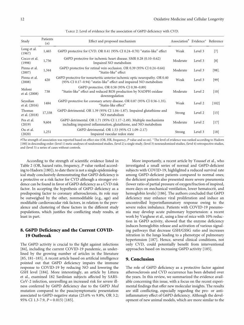
According to the strength of scientific evidence listed inTable 2 (OR, hazard ratio, frequency, P value ranked accord-ing toHadorn [180]), to date there is not a single epidemiolog-ical study conclusively demonstrating that G6PD deficiency isa protective or a risk factor for CVD although a stronger evi-dence can be found in favor of G6PD deficiency as a CVD riskfactor. In accepting the hypothesis of G6PD deficiency as apredisposing factor to coronary atherosclerosis, its role maybe outweighed by the other, nonmodifiable (e.g., age) andmodifiable cardiovascular risk factors, in relation to the prev-alence and clustering of these factors in the different studypopulations, which justifies the conflicting study results, atleast in part.
8. G6PD Deficiency and the Current COVID-19 Outbreak
The G6PD activity is crucial to the fight against infections[84], including the current COVID-19 pandemic, as under-lined by the growing number of articles in the literature[85, 181–185]. A recent article based on artificial intelligencepointed out that G6PD deficiency impairs the immuneresponse to COVID-19 by reducing NO and lowering theGSH level [184]. More interestingly, an article by Litteraet al., examined 182 Sardinian subjects affected by SARS-CoV-2 infection, unravelling an increased risk for severe ill-ness conferred by G6PD deficiency due to the G6PD Medmutation compared to the paucisymptomatic presentationassociated to G6PD-negative status (25.6% vs 9.8%, OR 3.2;95% CI 1.3-7.9, P = 0:015) [185].
More importantly, a recent article by Youssef et al., whoinvestigated a small series of normal and G6PD-deficientsubjects with COVID-19, highlighted a reduced survival rateamong G6PD-deficient patients compared to normal ones;the deficient patients also presented more severe pneumonia(lower ratio of partial pressure of oxygen/fraction of inspired,more days on mechanical ventilation, lower hematocrit, andhemoglobin levels) [186]. The authors concluded that G6PDdeficiency may enhance viral proliferation and induce anuncontrolled hyperinflammatory response owing to thesevere redox imbalance. Patients with COVID-19 pneumo-nia may develop acute pulmonary hypertension: a recentwork by Varghese et al., using a line of mice with 10% reduc-tion in G6PD activity, showed that the enzyme deficiencyinduces hemoglobin release and activation of various signal-ing pathways that decrease GSH/GSSG ratio and increasesnitration in the lungs leading to a phenotype of pulmonaryhypertension [187]. Hence, several clinical conditions, notonly CVD, could potentially benefit from interventionalapproaches based on increasing G6PD activity [185].
9. Conclusion
The role of G6PD deficiency as a protective factor againstatherosclerosis and CVD occurrence has been debated overthe years. In this review, we summarized the evidence avail-able concerning this issue, with a focus on the recent experi-mental findings that offer new molecular insights. The resultsare still conflicting, especially regarding the pro- or anti-inflammatory effect of G6PD deficiency. Although the devel-opment of new animal models, which are more similar to the
Table 2: Level of evidence for the association of G6PD deficiency with CVD.
StudyPatients(n)
Effect and proposed mechanism Association# Evidence∗ Reference
Long et al.(1967)
1,465 G6PD protective for CVD. OR 0.41 (95% CI 0.24–0.70) “statin-like” effect Weak Level 3 [7]
Cocco et al.(1998)
1,756G6PD protective for ischemic heart disease. SMR 0.28 (0.10–0.62)
Impaired NO metabolismModerate Level 3 [8]
Pinna et al.(2007)
1,344G6PD protective for retinal vein occlusion. OR 0.39 (95% CI 0.24–0.64)
“Statin-like” effectModerate Level 3 [98].
Pinna et al.(2008)
420G6PD protective for nonarteritic anterior ischemic optic neuropathy. OR 0.40
(95% CI 0.17–0.94) “statin-like” effect and impaired NO metabolismWeak Level 3 [99]
Meloniet al. (2008)
738G6PD protective. OR 0.58 (95% CI 0.38–0.89)
“Statin-like” effect and reduced ROS production by NADPH oxidasedownregulation
Moderate Level 2 [10]
Seyedianet al. (2016)
1484G6PD protective for coronary artery disease. OR 0.87 (95% CI 0.56-1.35).
“Statin-like effect”Weak Level 2 [102]
Thomaset al. (2018)
17,338G6PD detrimental. OR 1.39 (95% CI 1.04–1.87). Impaired glutathione and
NO metabolismStrong Level 2 [15]
Pes et al.(2019)
9,604G6PD detrimental. OR 1.71 (95% CI 1.17–2.49). Multiple mechanismsincluding impaired inflammation, glutathione, and NO metabolism
Moderate Level 2 [17]
Ou et al.(2020)
1,251G6PD detrimental. OR 1.53 (95% CI 1.09–2.17)
Impaired vascular redox stateStrong Level 3 [18]
#The strength of association was reported based on effect size (OR, HR, frequency, P value and so on). ∗The level of evidence was ranked according to Hadorn[180] in descending order: (level 1) meta-analyses of randomized studies, (level 2) a single study, (level 3) nonrandomized studies, (level 4) retrospective studies,and (level 5) a series of cases without controls.
12 Oxidative Medicine and Cellular Longevity

human G6PD mutations, may shed light on the overall effectof G6PD deficiency on the cardiovascular system, the find-ings obtained in these models cannot always be translatedto humans. Throughout their long evolutionary history,human beings have developed molecular mechanisms andcellular mediators more complex than those of lower mam-mals. The awareness of this diversity recommends a cautiousinterpretation before generalizing the results obtained in ani-mal models. The dominant paradigm of a protective effect ofthe enzyme deficiency was based on the results obtained inerythrocytes and leukocytes, without considering thatG6PD deficiency may have a different impact in endothelialand vascular muscle cells, which are deeply involved in ath-erogenesis. The G6PD remains a key enzyme in antioxidantdefense and possesses a broader protective role in several tis-sues and organs irrespective of providing reducing equiva-lents. Since G6PD plays an essential role in modulating thevascular redox state, the deficiency may result in vasculardysfunction which is crucial for the onset and progressionof the atherosclerotic process.
Data Availability
Not Applicable.
Conflicts of Interest
The authors declare that they have no conflicts of interest.
Authors’ Contributions
M.P.D., G.P., M.P., and G.M.P. were responsible for concep-tualization. M.P.D. and G.M.P. contributed in formal analy-sis. M.P.D. and G.M.P. contributed in investigation. M.P.D.,G.P., M.P., and G.M.P. contributed in writing, review, andediting. G.M.P. contributed in visualization. M.P.D. wasresponsible for supervision. M.P.D. and G.M.P. were respon-sible for project administration. M.P.D. was responsible forfunding acquisition.
References
[1] R. Ross, “Atherosclerosis — An inflammatory disease,” TheNew England Journal of Medicine, vol. 340, no. 2, pp. 115–126, 1999.
[2] GBD 2013 Mortality and Causes of Death Collaborators,“Global, regional, and national age-sex specific all-cause andcause-specific mortality for 240 causes of death, 1990-2013:a systematic analysis for the Global Burden of Disease Study2013,” The Lancet, vol. 385, pp. 117–171, 2015.
[3] E. J. Benjamin, P. Muntner, A. Alonso et al., “Heart diseaseand stroke statistics-2019 update: a report from the AmericanHeart Association,” Circulation, vol. 139, no. 10, pp. e56–e528, 2019.
[4] J. F. Bentzon, F. Otsuka, R. Virmani, and E. Falk, “Mecha-nisms of plaque formation and rupture,” CirculationResearch, vol. 114, no. 12, pp. 1852–1866, 2014.
[5] B. Emini Veseli, P. Perrotta, G. R. A. de Meyer et al., “Animalmodels of atherosclerosis,” European Journal of Pharmacol-ogy, vol. 816, pp. 3–13, 2017.
[6] A. Hafiane, “Vulnerable plaque, characteristics, detection,and potential therapies,” Journal of Cardiovascular Develop-ment and Disease, vol. 6, no. 3, p. 26, 2019.
[7] W. K. Long, S. W. Wilson, and E. P. Frenkel, “Associationsbetween red cell glucose-6-phosphate dehydrogenase vari-ants and vascular diseases,” American Journal of HumanGenetics, vol. 19, no. 1, pp. 35–53, 1967.
[8] P. Cocco, P. Todde, S. Fornera, M. B. Manca, P. Manca, andA. R. Sias, “Mortality in a cohort of men expressing theglucose-6-phosphate dehydrogenase deficiency,” Blood,vol. 91, no. 2, pp. 706–709, 1998.
[9] S. Muntoni, B. Batetta, S. Dessi, S. Muntoni, and P. Pani,“Serum lipoprotein profile in the Mediterranean variant ofglucose-6-phosphate dehydrogenase deficiency,” EuropeanJournal of Epidemiology, vol. 8, no. S1, pp. 48–53, 1992.
[10] L. Meloni, M. R. Manca, I. Loddo et al., “Glucose-6-phos-phate dehydrogenase deficiency protects against coronaryheart disease,” Journal of Inherited Metabolic Diseases,vol. 31, no. 3, pp. 412–417, 2008.
[11] R. Matsui, S. Xu, K. A. Maitland et al., “Glucose-6 phosphatedehydrogenase deficiency decreases the vascular response toangiotensin II,” Circulation, vol. 112, no. 2, pp. 257–263,2005.
[12] R. Matsui, S. Xu, K. A. Maitland et al., “Glucose-6-phosphatedehydrogenase deficiency decreases vascular superoxide andatherosclerotic lesions in apolipoprotein E-/- mice,” Arterio-sclerosis, Thrombosis, and Vascular Biology, vol. 26, no. 4,pp. 910–916, 2006.
[13] A. Kitagawa, I. Kizub, C. Jacob et al., “CRISPR-mediated sin-gle nucleotide polymorphism modeling in rats reveals insightinto reduced cardiovascular risk associated with Mediterra-neanG6PDVariant,” Hypertension, vol. 76, no. 2, pp. 523–532, 2020.
[14] P. A. Hecker, J. A. Leopold, S. A. Gupte, F. A. Recchia, andW. C. Stanley, “Impact of glucose-6-phosphate dehydroge-nase deficiency on the pathophysiology of cardiovascular dis-ease,” American Journal of Physiology-Heart and CirculatoryPhysiology, vol. 304, no. 4, pp. H491–H500, 2013.
[15] J. E. Thomas, S. Kang, C. J. Wyatt, F. S. Kim, A. D. Mangels-dorff, and F. K. Weigel, “Glucose-6-phosphate dehydroge-nase deficiency is associated with cardiovascular disease inU.S. military centers,” Texas Heart Institute Journal, vol. 45,no. 3, pp. 144–150, 2018.
[16] J. Zhao, X. Zhang, T. Guan et al., “The association betweenglucose-6-phosphate dehydrogenase deficiency and abnor-mal blood pressure among prepregnant reproductive-ageChinese females,” Hypertension Research, vol. 42, no. 1,pp. 75–84, 2019.
[17] G. M. Pes, G. Parodi, and M. P. Dore, “Glucose-6-phosphatedehydrogenase deficiency and risk of cardiovascular disease:a propensity score-matched study,” Atherosclerosis, vol. 282,pp. 148–153, 2019.
[18] Z. Ou, Y. Chen, J. Li et al., “Glucose-6-phosphate dehydroge-nase deficiency and stroke outcomes,” Neurology, vol. 95,no. 11, pp. e1471–e1478, 2020.
[19] A. Stincone, A. Prigione, T. Cramer et al., “The return ofmetabolism: biochemistry and physiology of the pentosephosphate pathway,” Biological Reviews of the CambridgePhilosophical Society, vol. 90, no. 3, pp. 927–963, 2015.
[20] D. B. Jump, “Mammalian fatty acid elongases,” in Lipidomics,D. Armstrong, Ed., vol. 579 of Methods in Molecular Biology
13Oxidative Medicine and Cellular Longevity
(Methods and Protocols), pp. 375–389, Humana Press,Totowa, NJ, 2009.
[21] J. Shen, G. Wu, A.-L. Tsai, and M. Zhou, “Structure andmechanism of a Unique Diiron Center in mammalianstearoyl-CoA desaturase,” Journal of Molecular Biology,vol. 432, no. 18, pp. 5152–5161, 2020.
[22] M. D. Siperstein, I. L. Chaikoff, and S. S. Chernick, “Signifi-cance of endogenous cholesterol in arteriosclerosis: synthesisin arterial tissue,” Science, vol. 113, no. 2948, pp. 747–749,1951.
[23] J. L. Omdahl, H. A. Morris, and B. K. May, “Hydroxylaseen-zymes of THEVITAMIND pathway: expression, function,and regulation,” Annual Review of Nutrition, vol. 22, no. 1,pp. 139–166, 2002.
[24] D. S. Riddick, X. Ding, C. R. Wolf et al., “NADPH-cyto-chrome P450 oxidoreductase: roles in physiology, pharma-cology, and toxicology,” Drug Metabolism & Disposition,vol. 41, no. 1, pp. 12–23, 2013.
[25] D. C. Thomas, “The phagocyte respiratory burst: Historicalperspectives and recent advances,” Immunology Letters,vol. 192, pp. 88–96, 2017.
[26] B. Childs, W. Zinkham, E. A. Browne, E. L. Kimbro, and J. V.Torbert, “A genetic study of a defect in glutathione metabo-lism of the erythrocyte,” Bulletin of the Johns Hopkins Hospi-tal, vol. 102, no. 1, pp. 21–37, 1958.
[27] T. Takizawa, I.-Y. Huang, T. Ikuta, and A. Yoshida, “Humanglucose-6-phosphate dehydrogenase: primary structure andcDNA cloning,” Proceedings of the National Academy of Sci-ences of the United States of America, vol. 83, no. 12,pp. 4157–4161, 1986.
[28] G. Martini, D. Toniolo, T. Vulliamy et al., “Structural analysisof the X-linked gene encoding human glucose 6-phosphatedehydrogenase,” EMBO Journal, vol. 5, no. 8, pp. 1849–1855, 1986.
[29] M. D. Cappellini and G. Fiorelli, “Glucose-6-phosphate dehy-drogenase deficiency,” The Lancet, vol. 371, no. 9606, pp. 64–74, 2008.
[30] E. Y. Chen, A. Cheng, A. Lee et al., “Sequence of humanglucose-6-phosphate dehydrogenase cloned in plasmids anda yeast artificial chromosome,” Genomics, vol. 10, no. 3,pp. 792–800, 1991.
[31] J. A. Leopold, A. Dam, B. A. Maron et al., “Aldosteroneimpairs vascular reactivity by decreasing glucose-6-phosphate dehydrogenase activity,” Nature Medicine,vol. 13, no. 2, pp. 189–197, 2007.
[32] T. A. Gheita, S. A. Kenawy, R. W. el Sisi, H. A. Gheita, andH. Khalil, “Subclinical reduced G6PD activity in rheumatoidarthritis and Sjögren's Syndrome patients: relation to clinicalcharacteristics, disease activity and metabolic syndrome,”Modern Rheumatology, vol. 24, no. 4, pp. 612–617, 2014.
[33] D. Laertius, Lives of eminent philosophers, R. D. Hicks, Ed.,Oxford University Press, II edition, 2018.
[34] J. Bostock and H. T. Riley, The natural history of Pliny, AlphaEditions, 2019.
[35] D. I. Montasser, Y. Zajjari, A. Alayoud et al., “Acute renal fail-ure in favism revealing familial glucose-6-phosphate dehy-drogenase deficiency,” Indian Journal of Nephrology, vol. 22,no. 1, pp. 67-68, 2012.
[36] E. Beutler, “Glucose-6-phosphate dehydrogenase deficiency:a historical perspective,” Blood, vol. 111, no. 1, pp. 16–24,2008.
[37] L. Luzzatto and R. Notaro, “Malaria: protecting against badair,” Science, vol. 293, no. 5529, pp. 442-443, 2001.
[38] Z. Spolarics, M. Siddiqi, J. H. Siegel et al., “Increased inci-dence of sepsis and altered monocyte functions in severelyinjured type A– glucose-6-phosphate dehydrogenase-deficient African American trauma patients,” Critical CareMedicine, vol. 29, no. 4, pp. 728–736, 2001.
[39] A. Ohlsson, K. Rehnholm, K. Shubham, and U. von Döbeln,“Incidence of glucose-6-phosphate dehydrogenase deficiencyamong Swedish newborn infants,” International Journal ofNeonatal Screening, vol. 5, no. 4, p. 38, 2019.
[40] E. C. Mbanefo, A. M. Ahmed, A. Titouna et al., “Associationof glucose-6-phosphate dehydrogenase deficiency andmalaria: a systematic review and meta-analysis,” ScientificReports, vol. 7, no. 1, article 45963, 2017.
[41] E. Atay, A. Bozaykut, and I. O. Ipek, “Glucose-6-phosphatedehydrogenase deficiency in neonatal indirect hyperbilirubi-nemia,” Journal of Tropical Pediatrics, vol. 52, no. 1, pp. 56–58, 2006.
[42] L. Luzzatto, A. Mehta, and T. Vulliamy, “Glucose-6-phosphatedehydrogenase deficiency,” in The metabolic basis of inheriteddisease, C. R. Scriver, A. L. Beaudet, and W. S. Sly, Eds., vol. 3,pp. 4517–4553, McGraw-Hill, New York, NY, 2001.
[43] K. Betke, E. Beutler, G. H. Brewer et al., Standardization ofprocedures for the study of glucose-6-phosphate dehydroge-nase. Report of a WHO scientific group, WHO TechnicalReport Series, 1967.
[44] E. T. Nkhoma, C. Poole, V. Vannappagari, S. A. Hall, andE. Beutler, “The global prevalence of glucose-6-phosphatedehydrogenase deficiency: a systematic review and meta-analysis,” Blood Cells, Molecules and Diseases, vol. 42, no. 3,pp. 267–278, 2009.
[45] L. Luzzatto and G. Battistuzzi, “Glucose-6-phosphate dehy-drogenase,” in Advances in Human Genetics, H. Harris andK. Hirschhorn, Eds., vol. 14, pp. 217–329, Springer, Boston,MA, 1985.
[46] J. A. Leopold, A. Cap, A. W. Scribner, R. C. Stanton, andJ. Loscalzo, “Glucose‐6‐phosphate dehydrogenase deficiencypromotes endothelial oxidant stress and decreases endothe-lial nitric oxide bioavailability,” FASEB Journal, vol. 15,no. 10, pp. 1771–1773, 2001.
[47] A. V. Snezhkina, A. V. Kudryavtseva, O. L. Kardymon et al.,“ROS generation and antioxidant defense systems in normaland malignant cells,” Oxidative Medicine and Cellular Lon-gevity, vol. 2019, Article ID 6175804, 17 pages, 2019.
[48] C. Peiró, T. Romacho, V. Azcutia et al., “Inflammation, glu-cose, and vascular cell damage: the role of the pentose phos-phate pathway,” Cardiovascular Diabetology, vol. 15, no. 1,2016.
[49] D. K. Rawat, P. Hecker, M.Watanabe et al., “Glucose-6-phos-phate dehydrogenase and NADPH redox regulates cardiacmyocyte L-type calcium channel activity and myocardialcontractile function,” PLoS One, vol. 7, no. 10, articlee45365, 2012.
[50] J. Park, H. K. Rho, K. H. Kim, S. S. Choe, Y. S. Lee, and J. B.Kim, “Overexpression of glucose-6-phosphate dehydroge-nase is associated with lipid dysregulation and insulin resis-tance in obesity,” Molecular and Cellular Biology, vol. 25,no. 12, pp. 5146–5157, 2005.
[51] M. Ham, J. W. Lee, A. H. Choi et al., “Macrophage glucose-6-phosphate dehydrogenase stimulates proinflammatory
14 Oxidative Medicine and Cellular Longevity

responses with oxidative stress,”Molecular and Cellular Biol-ogy, vol. 33, no. 12, pp. 2425–2435, 2013.
[52] Z. Zhang, Z. Yang, B. Zhu et al., “Increasing glucose 6-phosphate dehydrogenase activity restores redox balance invascular endothelial cells exposed to high glucose,” PLoSOne, vol. 7, no. 11, article e49128, 2012.
[53] L. Cao, D. Zhang, J. Chen et al., “G6PD plays a neuroprotec-tive role in brain ischemia through promoting pentose phos-phate pathway,” Free Radical Biology and Medicine, vol. 112,pp. 433–444, 2017.
[54] J. A. Leopold, Y. Y. Zhang, A. W. Scribner, R. C. Stanton, andJ. Loscalzo, “Glucose-6-Phosphate dehydrogenase overex-pression decreases endothelial cell oxidant stress andincreases bioavailable nitric oxide,” Arteriosclerosis, Throm-bosis, and Vascular Biology, vol. 23, no. 3, pp. 411–417,2003.
[55] J. A. Leopold, J. Walker, A. W. Scribner et al., “Glucose-6-phosphate Dehydrogenase Modulates Vascular EndothelialGrowth Factor-mediated Angiogenesis,” Journal of BiologicalChemistry, vol. 278, no. 34, pp. 32100–32106, 2003.
[56] S. K. Legan, I. Rebrin, R. J. Mockett et al., “Overexpression ofGlucose-6-phosphate Dehydrogenase Extends the Life Spanof Drosophila melanogaster,” Journal of Biological Chemistry,vol. 283, no. 47, pp. 32492–32499, 2008.
[57] S. Nóbrega-Pereira, P. J. Fernandez-Marcos, T. Brioche et al.,“G6PD protects from oxidative damage and improves health-span in mice,” Nature Communications, vol. 7, no. 1, article10894, 2016.
[58] M. Kurata, M. Suzuki, and N. Agar, “Glutathione regenera-tion in mammalian erythrocytes,” Comparative HematologyInternational, vol. 10, no. 2, pp. 59–67, 2000.
[59] S. C. Lu, “Regulation of glutathione synthesis,” MolecularAspects of Medicine, vol. 30, no. 1-2, pp. 42–59, 2009.
[60] D. Giustarini, D. Tsikas, G. Colombo et al., “Pitfalls in theanalysis of the physiological antioxidant glutathione (GSH)and its disulfide (GSSG) in biological samples: an elephantin the room,” Journal of Chromatography B: Analytical Tech-nologies in the Biomedical and Life Sciences, vol. 1019, pp. 21–28, 2016.
[61] C. Espinosa-Díez, V. Miguel, S. Vallejo et al., “Role of gluta-thione biosynthesis in endothelial dysfunction and fibrosis,”Redox Biology, vol. 14, pp. 88–99, 2018.
[62] W. Pretsch, D. J. Charles, and S. Merkle, “X-linked glucose-6-phosphate dehydrogenase deficiency inMus musculus,” Bio-chemical Genetics, vol. 26, no. 1-2, pp. 89–103, 1988.
[63] M. Jain, L. Cui, D. A. Brenner et al., “Increased myocardialdysfunction after ischemia-reperfusion in mice lackingglucose-6-phosphate dehydrogenase,” Circulation, vol. 109,no. 7, pp. 898–903, 2004.
[64] I. Németh, H. Orvos, and D. Boda, “Blood glutathione redoxstatus in gestational hypertension,” Free Radical Biology andMedicine, vol. 30, no. 7, pp. 715–721, 2001.
[65] I. Afzal-Ahmed, G. E. Mann, A. H. Shennan, L. Poston, andR. J. Naftalin, “Preeclampsia inactivates glucose-6-phosphate dehydrogenase and impairs the redox status oferythrocytes and fetal endothelial cells,” Free Radical Biologyand Medicine, vol. 42, no. 12, pp. 1781–1790, 2007.
[66] R. M. Fitch, R. Vergona, M. E. Sullivan, and Y. X. Wang,“Nitric oxide synthase inhibition increases aortic stiffnessmeasured by pulse wave velocity in rats,” CardiovascularResearch, vol. 51, no. 2, pp. 351–358, 2001.
[67] I. B. Wilkinson, S. S. Franklin, and J. R. Cockcroft, “Nitricoxide and the regulation of large artery Stiffness,” Hyperten-sion, vol. 44, no. 2, pp. 112–116, 2004.
[68] J. W. Knowles, R. L. Reddick, J. C. Jennette, E. G. Shesely,O. Smithies, and N. Maeda, “Enhanced atherosclerosis andkidney dysfunction in eNOS–/–Apoe–/– mice are amelioratedby enalapril treatment,” The Journal of Clinical Investigation,vol. 105, no. 4, pp. 451–458, 2000.
[69] S. L. Wiesenfeld, N. L. Petrakis, B. J. Sams, M. F. Collen, andJ. L. Cutler, “Elevated blood pressure, pulse rate and serumcreatinine in negro males deficient in glucose-6-phosphatedehydrogenase,” The New England Journal of Medicine,vol. 282, no. 18, pp. 1001-1002, 1970.
[70] S. Oppi, T. F. Lüscher, and S. Stein, “Mouse models for ath-erosclerosis research-which is my line?,” Frontiers in Cardio-vascular Medicine, vol. 6, p. 46, 2019.
[71] C. M. Brown, E. Wright, C. A. Colton, P. M. Sullivan, D. T.Laskowitz, and M. P. Vitek, “Apolipoprotein E isoformmedi-ated regulation of nitric oxide release1, 2,” Free Radical Biol-ogy and Medicine, vol. 32, no. 11, pp. 1071–1075, 2002.
[72] N. Hogg, B. Kalyanaraman, J. Joseph, A. Struck, andS. Parthasarathy, “Inhibition of low-density lipoprotein oxi-dation by nitric oxide Potential role in atherogenesis,” FEBSLetters, vol. 334, no. 2, pp. 170–174, 1993.
[73] D.Wolf and K. Le, “Immunity and inflammation in atheroscle-rosis,” Circulation Research, vol. 124, no. 2, pp. 315–327, 2019.
[74] J. Wilmanski, E. Villanueva, E. A. Deitcha, and Z. Spolarics,“Glucose-6-phosphate dehydrogenase deficiency and theinflammatory response to endotoxin and polymicrobial sep-sis,” Critical Care Medicine, vol. 35, no. 2, pp. 510–518, 2007.
[75] H. C. Yang, M. L. Cheng, Y. S. Hua et al., “Glucose 6-phosphate dehydrogenase knockdown enhances IL-8 expres-sion in HepG2 cells via oxidative stress and NF-κB signalingpathway,” Journal of Inflammation, vol. 12, no. 1, 2015.
[76] R. Parsanathan and S. K. Jain, “L-Cysteine in vitro can restorecellular glutathione and inhibits the expression of cell adhe-sion molecules in G6PD-deficient monocytes,” Amino Acids,vol. 50, no. 7, pp. 909–921, 2018.
[77] R. Parsanathan and S. K. Jain, “Glucose-6-phosphate dehy-drogenase deficiency increases cell adhesion molecules andactivates human monocyte-endothelial cell adhesion: Protec-tive role of l-cysteine,” Archives of Biochemistry and Biophys-ics, vol. 663, pp. 11–21, 2019.
[78] R. Parsanathan and S. K. Jain, “G6PD deficiency shifts polar-ization of monocytes/macrophages towards a proinflamma-tory and profibrotic phenotype,” Cellular & MolecularImmunology, vol. 18, no. 3, pp. 770–772, 2021.
[79] B. Wolach, M. Ashkenazi, R. Grossmann et al., “Diurnal fluc-tuation of leukocyte G6PD activity. A possible explanationfor the normal neutrophil bactericidal activity and the lowincidence of pyogenic infections in patients with severeG6PD deficiency in Israel,” Pediatric Research, vol. 55,no. 5, pp. 807–813, 2004.
[80] D. Kwiatkowski, “Science, medicine, and the future: suscepti-bility to infection,” British Medical Journal, vol. 321, no. 7268,pp. 1061–1065, 2000.
[81] Z. Rostami-Far, K. Ghadiri, M. Rostami-Far, F. Shaveisi-Zadeh, A. Amiri, and B. Rahimian Zarif, “Glucose-6-phos-phate dehydrogenase deficiency (G6PD) as a risk factor ofmale neonatal sepsis,” The Journal of Medicine and Life,vol. 9, no. 1, pp. 34–38, 2016.
15Oxidative Medicine and Cellular Longevity
[82] U. Siler, S. Romao, E. Tejera et al., “Severe glucose-6-phosphate dehydrogenase deficiency leads to susceptibilityto infection and absent NETosis,” Journal of Allergy and Clin-ical Immunology, vol. 139, no. 1, pp. 212–219.e3, 2017.
[83] S. Zamani, A. Z. Hoseini, and A. M. Namin, “Glucose-6-phosphate dehydrogenase (G6PD) activity can modulatemacrophage response to Leishmania major infection,” Inter-national Immunopharmacology, vol. 69, pp. 178–183, 2019.
[84] A. Peretz, M. Azrad, and A. Blum, “Influenza virus and ath-erosclerosis,” QJM, vol. 112, no. 10, pp. 749–755, 2019.
[85] D. J. Vick, “Glucose-6-phosphate dehydrogenase deficiencyand COVID-19 infection,” Mayo Clinic Proceedings, vol. 95,no. 8, pp. 1803-1804, 2020.
[86] J. C. Kwong, K. L. Schwartz, M. A. Campitelli et al., “Acutemyocardial infarction after laboratory-confirmed influenzainfection,” The New England Journal of Medicine, vol. 378,no. 4, pp. 345–353, 2018.
[87] Y. Xu, Z. Zhang, and J. Hu, “Glucose-6-phosphatedehydrogenase-deficient mice have increased renal oxidativestress and increased albuminuria,” FASEB Journal, vol. 24,no. 2, pp. 609–616, 2010.
[88] G. Balla, H. S. Jacob, J. W. Eaton, J. D. Belcher, and G.M. Ver-cellotti, “Hemin: a possible physiological mediator of lowdensity lipoprotein oxidation and endothelial injury,” Arte-riosclerosis, Thrombosis and Vascular Biology, vol. 11, no. 6,pp. 1700–1711, 1991.
[89] J. D. Belcher, J. D. Beckman, G. Balla, J. Balla, andG. Vercellotti, “Heme degradation and vascular injury,” Anti-oxidants & Redox Signaling, vol. 12, no. 2, pp. 233–248, 2010.
[90] A. M. Liese, M. Q. Siddiqi, J. H. Siegel, E. A. Deitch, andZ. Spolarics, “Attenuated monocyte IL-10 production inglucose-6-phosphate dehydrogenase-deficient traumapatients,” Shock, vol. 18, no. 1, pp. 18–23, 2002.
[91] B. Mordmuller, F. Turrini, H. Long, P. G. Kremsner, andP. Arese, “Neutrophils and monocytes from subjects withthe Mediterranean G6PD variant: effect of plasmodium falci-parum hemozoin on G6PD activity, oxidative burst and cyto-kine production,” European Cytokine Network, vol. 9, no. 3,pp. 239–245, 1998.
[92] J. Wilmanski, M. Siddiqi, E. A. Deitch, and Z. Spolarics,“Augmented IL-10 production and redox-dependent signal-ing pathways in glucose-6-phosphate dehydrogenase-deficient mouse peritoneal macrophages,” Journal of Leuko-cyte Biology, vol. 78, no. 1, pp. 85–94, 2005.
[93] E. S. Istvan and J. Deisenhofer, “Structural mechanism forstatin inhibition of HMG-CoA reductase,” Science, vol. 292,no. 5519, pp. 1160–1164, 2001.
[94] P. A. Hecker, V. Lionetti, R. F. Ribeiro et al., “Glucose 6-phosphate dehydrogenase deficiency increases redox stressand moderately accelerates the development of heart failure,”Circulation: Heart Failure, vol. 6, no. 1, pp. 118–126, 2013.
[95] A. R. Tarlov, G. J. Brewer, P. E. Carson, and A. S. Alving, “Pri-maquine sensitivity,” Archives of Internal Medicine, vol. 109,no. 2, pp. 209–234, 1962.
[96] B. Batetta, R. R. Bonatesta, F. Sanna et al., “Cell growth andcholesterol metabolism in human glucose-6-phosphate dehy-drogenase deficient lymphomononuclear cells,” Cell Prolifer-ation, vol. 35, no. 3, pp. 143–154, 2002.
[97] G. Cappai, M. Songini, A. Doria, J. D. Cavallerano, andM. Lorenzi, “Increased prevalence of proliferative retinopa-thy in patients with type 1 diabetes who are deficient in
glucose-6-phosphate dehydrogenase,” Diabetologia, vol. 54,no. 6, pp. 1539–1542, 2011.
[98] A. Pinna, C. Carru, G. Solinas, A. Zinellu, and F. Carta, “Glu-cose-6-phosphate dehydrogenase deficiency in retinal veinocclusion,” Investigative Ophthalmology & Visual Science,vol. 48, no. 6, pp. 2747–2752, 2007.
[99] A. Pinna, G. Solinas, C. Masia, A. Zinellu, C. Carru, andA. Carta, “Glucose-6-Phosphate dehydrogenase (G6PD) defi-ciency in nonarteritic anterior ischemic optic neuropathy in aSardinian population, Italy,” Investigative Ophthalmology &Visual Science, vol. 49, no. 4, pp. 1328–1332, 2008.
[100] A. Cao, R. Galanello, M. Furbetta et al., “Thalassaemia typesand their incidence in Sardinia,” Journal of Medical Genetics,vol. 15, no. 6, pp. 443–447, 1978.
[101] M. Maioli, S. Pettinato, G. M. Cherchi et al., “Plasma lipids inbeta-thalassemia minor,” Atherosclerosis, vol. 75, no. 2-3,pp. 245–248, 1989.
[102] S. M. Seyedian, F. Ahmadi, P. Ghorbani, S. M. H. Adeland,and S. M. H. Aletayeb, “Glucose-6-phosphate dehydrogenasedeficiency and coronary artery disease,” International Journalof Tropical Medicine, vol. 11, no. 5, pp. 186–188, 2016.
[103] M. K. Kwok, G. M. Leung, S. L. A. Yeung, and C. M. School-ing, “Glucose-6-phosphate dehydrogenase deficiency andmetabolic profiling in adolescence from the Chinese birthcohort: ‘children of 1997’,” International Journal of Cardiol-ogy, vol. 281, pp. 146–149, 2019.
[104] T. Ge, J. Yang, S. Zhou, Y. Wang, Y. Li, and X. Tong, “Therole of the pentose phosphate pathway in diabetes and can-cer,” Frontiers in Endocrinology, vol. 11, p. 365, 2020.
[105] M. B. Adinortey, R. K. Owusu, I. K. A. Galyuon, W. Ekloh,I. N. Owusu, and D. Larbi, “G-6-PDdeficiency - a potential riskfactor for development of diabetesmellitus,” Journal ofMedicineand Medical Sciences, vol. 2, no. 8, pp. 1017–1021, 2011.
[106] Y. K. Lai, N. M. Lai, and S. W. Lee, “Glucose-6-phosphatedehydrogenase deficiency and risk of diabetes: a systematicreview and meta-analysis,” Annals of Hematology, vol. 96,no. 5, pp. 839–845, 2017.
[107] N. Saha, “Association of glucose-6-phosphate dehydrogenasedeficiency with diabetes mellitus in ethnic groups of Singa-pore,” Journal of Medical Genetics, vol. 16, no. 6, pp. 431–434, 1979.
[108] M. A. M. Brito, V. S. Sampaio, W. M. Monteiro et al., “Highfrequency of diabetes and impaired fasting glucose in patientswith glucose-6-phosphate dehydrogenase deficiency in theWestern brazilian Amazon,” American Journal of TropicalMedicine and Hygiene, vol. 91, no. 1, pp. 74–76, 2014.
[109] G. A. Niazi, “Glucose-6-phosphate dehydrogenase deficiencyand diabetes mellitus,” International Journal of Hematology,vol. 54, no. 4, pp. 295–298, 1991.
[110] N. Li, B. Li, T. Brun et al., “NADPH oxidase NOX2 defines anew antagonistic role for reactive oxygen species andcAMP/PKA in the regulation of insulin secretion,” Diabetes,vol. 61, no. 11, pp. 2842–2850, 2012.
[111] P. Spégel, V. V. Sharoyko, I. Goehring et al., “Time-resolvedmetabolomics analysis of β-cells implicates the pentose phos-phate pathway in the control of insulin release,” BiochemicalJournal, vol. 450, no. 3, pp. 595–605, 2013.
[112] Z. Zhang, C. W. Liew, D. E. Handy et al., “High glucoseinhibits glucose-6-phosphate dehydrogenase, leading toincreased oxidative stress and beta-cell apoptosis,” FASEBJournal, vol. 24, no. 5, pp. 1497–1505, 2010.
16 Oxidative Medicine and Cellular Longevity
[113] S. Monte Alegre, S. T. Saad, E. Delatre, and M. J. Saad, “Insu-lin secretion in patients deficient in glucose-6-phosphatedehydrogenase,” Hormone and Metabolic Research, vol. 23,no. 4, pp. 171–173, 1991.
[114] J. W. Lee, A. H. Choi, M. Ham et al., “G6PD up-regulationpromotes pancreatic β-Cell dysfunction,” Endocrinology,vol. 152, no. 3, pp. 793–803, 2011.
[115] E. Sobngwi, J. F. Gautier, J. P. Kevorkian et al., “High preva-lence of glucose-6-phosphate dehydrogenase deficiency with-out gene mutation suggests a novel genetic mechanismpredisposing to ketosis-prone diabetes,” The Journal of Clin-ical Endocrinology & Metabolism, vol. 90, no. 8, pp. 4446–4451, 2005.
[116] A. Leong, V. J. Y. Lim, and C. Wang, “Association of G6PDvariants with hemoglobin A1c and impact on diabetes diag-nosis in East Asian individuals,” BMJ Open Diabetes Research& Care, vol. 8, no. 1, article e001091, 2020.
[117] S. K. Jain, “Glutathione and glucose-6-phosphate dehydroge-nase deficiency can increase protein glycosylation,” Free Rad-ical Biology & Medicine, vol. 24, no. 1, pp. 197–201, 1998.
[118] S. K. Jain andM. Palmer, “Effect of glucose-6-phosphate dehy-drogenase deficiency on reduced and oxidized glutathione andlipid peroxide levels in the blood of African-Americans,” Clin-ica Chimica Acta, vol. 253, no. 1-2, pp. 181–183, 1996.
[119] R. Parsanathan and S. K. Jain, “Glucose-6-phosphate dehy-drogenase deficiency activates endothelial cell and leukocyteadhesion mediated via the TGFβ/NADPH oxidases/ROS sig-naling pathway,” International Journal of Molecular Sciences,vol. 21, no. 20, article 7474, 2020.
[120] R. Parsanathan and S. K. Jain, “Glucose-6-phosphate dehy-drogenase (G6PD) deficiency is linked with cardiovasculardisease,” Hypertension Research, vol. 43, no. 6, pp. 582–584,2020.
[121] G. Battistuzzi, M. D'Urso, D. Toniolo, G. M. Persico, andL. Luzzatto, “Tissue-specific levels of human glucose-6-phosphate dehydrogenase correlate with methylation of spe-cific sites at the 3' end of the gene,” Proceedings of theNational Academy of Sciences of the United States of America,vol. 82, no. 5, pp. 1465–1469, 1985.
[122] F. Corpas and J. B. Barroso, “NADPH-generating dehydroge-nases: their role in the mechanism of protection against nitro-oxidative stress induced by adverse environmental condi-tions,” Frontiers in Environmental Science, vol. 2, 2014.
[123] L. A. Metherell, J. A. Guerra-Assunção, M. J. Sternberg, andA. David, “Three‐dimensional model of human nicotinamidenucleotide transhydrogenase (NNT) and sequence‐structureanalysis of its disease‐causing variations,” Human Mutation,vol. 37, no. 10, pp. 1074–1084, 2016.
[124] H. J. Koh, S. M. Lee, B. G. Son et al., “Cytosolic NADP+-dependent isocitrate dehydrogenase plays a key role in lipidmetabolism,” Journal of Biological Chemistry, vol. 279,no. 38, pp. 39968–39974, 2004.
[125] L. Chen, Z. Zhang, A. Hoshino et al., “NADPH production bythe oxidative pentose-phosphate pathway supports folatemetabolism,” Nature Metabolism, vol. 1, no. 3, pp. 404–415,2019.
[126] P. Durand, M. Prost, and D. Blache, “Folate deficiencies andcardiovascular pathologies,” Clinical Chemistry and Labora-tory Medicine, vol. 36, no. 7, pp. 419–429, 1998.
[127] M. R. Clemens, H. Einsele, and H. D. Waller, “The fatty acidcomposition of red cells deficient in glucose-6-phosphate
dehydrogenase and their susceptibility to lipid peroxidation,”Wiener Klinische Wochenschrift, vol. 63, no. 13, pp. 578–582,1985.
[128] D. L. Hodge and L. M. Salati, “Nutritional regulation of theglucose-6-phosphate dehydrogenase gene is mediated by anuclear posttranscriptional mechanism,” Archives of Bio-chemistry and Biophysics, vol. 348, no. 2, pp. 303–312, 1997.
[129] K. Hashizume, T. Onaya, and A. Sato, “The role of the pen-tose phosphate shunt in thyrotropin-induced thyroid hor-mone secretion:in vivoandin vitrostudies with 6-aminonicotinamide in mouse thyroids,” Endocrinology,vol. 97, no. 4, pp. 962–968, 1975.
[130] M. von Hafe, J. S. Neves, C. Vale, M. Borges-Canha, andA. Leite-Moreira, “The impact of thyroid hormone dysfunc-tion on ischemic heart disease,” Endocrine Connections,vol. 8, no. 5, pp. R76–R90, 2019.
[131] K. I. Ataga, “Hypercoagulability and thrombotic complica-tions in hemolytic anemias,” Haematologica, vol. 94, no. 11,pp. 1481–1484, 2009.
[132] F. M. Low, M. B. Hampton, and C. C. Winterbourn, “Perox-iredoxin 2 and peroxide metabolism in the erythrocyte,”Antioxidants & Redox Signaling, vol. 10, no. 9, pp. 1621–1630, 2008.
[133] L. Luzzatto and P. Arese, “Favism and glucose-6-phosphatedehydrogenase deficiency,” The New England Journal of Med-icine, vol. 378, no. 1, pp. 60–71, 2018.
[134] W. Tillmann, M. Gahr, N. Labitzke, and W. Schröter, “Mem-brane deformability of erythrocytes with glucose-6-phosphate dehydrogenase,” Acta Haematologica, vol. 57,no. 3, pp. 162–167, 1977.
[135] D. T. Chiu and T. Z. Liu, “Free radical and oxidative damagein human blood cells,” Journal of Biomedical Science, vol. 4,no. 5, pp. 256–259, 1997.
[136] D. C. McMillan, L. J. Bolchoz, and D. J. Jollow, “Favism: effectof divicine on rat erythrocyte sulfhydryl status, hexose mono-phosphate shunt activity, morphology, and membrane skele-tal proteins,” Toxicological Sciences, vol. 62, no. 2, pp. 353–359, 2001.
[137] M. D. Cappellini, “Coagulation in the pathophysiology ofhemolytic anemias,” Hematology, pp. 74–78, 2007.
[138] V. L. Tzounakas, F. Gevi, H. T. Georgatzakou et al., “Redoxstatus, procoagulant activity, and metabolome of fresh frozenplasma in glucose 6-phosphate dehydrogenase deficiency,”Frontiers in Medicine, vol. 5, p. 16, 2018.
[139] C. Donadee, N. J. Raat, T. Kanias et al., “Nitric oxide scaveng-ing by red blood cell microparticles and cell-free hemoglobinas a mechanism for the red cell storage lesion,” Circulation,vol. 124, no. 4, pp. 465–476, 2011.
[140] Y. Y. Chirkov, N. N. Belushkina, I. A. Tyshchuk, I. S. Sever-ina, and J. D. Horowitz, “Increase in reactivity of humanplatelet guanylate cyclase during aggregation potentiates thedisaggregating capacity of sodium nitroprusside,” Clinicaland Experimental Pharmacology and Physiology, vol. 18,no. 7, pp. 517–524, 1991.
[141] Z. M. Ezz El-Deen, N. F. Hussin, T. A. Abdel Hamid, O. R.Abdel Migeed, and R. M. Samy, “G6PD deficiency andG6PD (Mediterranean and silent) polymorphisms in Egyp-tian infants with neonatal Hyperbilirubinemia,” LaboratoryMedicine, vol. 44, no. 3, pp. 228–234, 2013.
[142] B. Ramot, A. Szeinberg, A. Adam, C. Sheba, and D. Gafni, “Astudy of subjects with erythrocyte glucose-6-phosphate
17Oxidative Medicine and Cellular Longevity
dehydrogenase deficiency: investigation of platelet enzymes,”The Journal of Clinical Investigation, vol. 38, no. 9, pp. 1659–1661, 1959.
[143] J. C. Doery, J. Hirsh, and G. C. de Gruchy, “Erythrocyte andplatelet glucose 6-phosphate dehydrogenase in normal andmutant Caucasians,” Scandinavian Journal of Haematology,vol. 6, no. 1, pp. 5–9, 1969.
[144] D. Nantakomol, A. Palasuwan, M. Chaowanathikhom,S. Soogarun, and M. Imwong, “Red cell and platelet-derivedmicroparticles are increased in G6PD-deficient subjects,”European Journal of Haematology, vol. 89, no. 5, pp. 423–429, 2012.
[145] E. Noulsri, S. Lerdwana, D. Palasuwan, and A. Palasuwan,“Cell-derived microparticles in blood products from blooddonors deficient in glucose-6-phosphate dehydrogenase,”Laboratory Medicine, vol. 7, 2021.
[146] C. L'Acqua and E. Hod, “New perspectives on the thromboticcomplications of haemolysis,” British Journal of Haematol-ogy, vol. 168, no. 2, pp. 175–185, 2015.
[147] P. A. Thompson, E. Chew, and J. Szer, “Deep vein thrombosisin association with acute intravascular haemolysis in glucose-6-phosphate dehydrogenase deficiency: a unique case,” Inter-nal Medicine Journal, vol. 43, no. 10, pp. 1164-1165, 2013.
[148] J. Albertsen, H. B. Ommen, A. Wandler, and K. Munk, “Fatalhaemolytic crisis with microvascular pulmonary obstructionmimicking a pulmonary embolism in a young African manwith glucose-6-phosphate dehydrogenase deficiency,” BMJCase Reports, vol. 2014, 2014.
[149] K. Jain, T. Tyagi, K. Patell et al., “Age associated non-linearregulation of redox homeostasis in the anucleate platelet:Implications for CVD risk patients,” EBioMedicine, vol. 44,pp. 28–40, 2019.
[150] M. P. Dore, M. Portoghese, and G. M. Pes, “The Elderly withGlucose-6-Phosphate Dehydrogenase Deficiency Are MoreSusceptible to Cardiovascular Disease,” Journal of Atheroscle-rosis and Thrombosis, vol. 28, no. 6, 2021.
[151] D. Mari, P. M. Mannucci, R. Coppola, B. Bottasso, K. A.Bauer, and R. D. Rosenberg, “Hypercoagulability in centenar-ians: the paradox of successful aging,” Blood, vol. 85, no. 11,pp. 3144–3149, 1995.
[152] B. Ibanez, S. James, S. Agewall et al., “2017 ESCGuidelines forthe management of acute myocardial infarction in patientspresenting with ST-segment elevation: the Task Force forthe management of acute myocardial infarction in patientspresenting with ST-segment elevation of the European Soci-ety of Cardiology (ESC),” European Heart Journal, vol. 39,no. 2, pp. 119–177, 2018.
[153] O. Shalev, “Long-term, low-dose aspirin is safe in glucose-6-phosphate dehydrogenase deficiency,” DICP, vol. 25, no. 10,pp. 1074-1075, 1991.
[154] N. V. Kafkas, C. I. Liakos, and A. G. Mouzarou, “Antiplateletand invasive treatment in patients with glucose-6-phosphatedehydrogenase (G6PD) deficiency and acute coronary syn-drome. The safety of aspirin,” Journal of Clinical Pharmacyand Therapeutics, vol. 40, no. 3, pp. 349–352, 2015.
[155] E. Beutler, “Glucose-6-Phosphate dehydrogenase deficiency,”The New England Journal of Medicine, vol. 324, no. 3,pp. 169–174, 1991.
[156] M. Zuin, G. Rigatelli, M. Carraro et al., “Antiplatelet therapyin patients with glucose-6-phosphate dehydrogenases defi-ciency after percutaneous coronary intervention: a reap-
praisal for clinical and interventional cardiologists,”Cardiovascular Revascularization Medicine, vol. 18, no. 3,pp. 226–229, 2017.
[157] J. Li, Y. Chen, Z. Ou et al., “Aspirin therapy in cardiovasculardisease with glucose-6-phosphate dehydrogenase deficiency,safe or not?,” American Journal of Cardiovascular Drugs,2020.
[158] G. D. Sanna, S. Deriu, M. Uras et al., “Aspirin adherence insubjects with glucose-6-phosphate-dehydrogenase deficiencyhaving an acute coronary syndrome,” European HeartJournal-Cardiovascular Pharmacotherapy, 2020.
[159] Y. Chen, J. Li, Z. Ou et al., “Safety and efficacy of low-doseaspirin in ischemic stroke patients with different G6PD con-ditions,” International Journal of Stroke, 2020.
[160] K. Bedard, B. Lardy, and K. Krause, “NOX family NADPHoxidases: not just in mammals,” Biochimie, vol. 89, no. 9,pp. 1107–1112, 2007.
[161] T. Efferth, U. Fabry, and R. Osieka, “DNA damage and apo-ptosis in mononuclear cells from glucose-6-phosphatedehydrogenase-deficient patients (G6PD Aachen variant)after UV irradiation,” Journal of Leukocyte Biology, vol. 69,no. 3, pp. 340–342, 2001.
[162] X. Li, X. Sun, and P. Carmeliet, “Hallmarks of endothelial cellmetabolism in health and disease,” Cell Metabolism, vol. 30,no. 3, pp. 414–433, 2019.
[163] D. M. Townsend, “S-Glutathionylation: indicator of cellstress and regulator of the unfolded protein response,”Molec-ular Interventions, vol. 7, no. 6, pp. 313–324, 2007.
[164] M. A. Forgione, J. Loscalzo, M. Holbrook et al., “TheA376G(A+) variant of the glucose-6-phosphate dehydroge-nase gene is associated with endothelial dysfunction in Afri-can Americans,” Journal of the American College ofCardiology, vol. 41, no. 6, p. 249, 2003.
[165] M. Jain, D. A. Brenner, L. Cui et al., “Glucose-6-phosphatedehydrogenase modulates cytosolic redox status and contrac-tile phenotype in adult cardiomyocytes,” CirculationResearch, vol. 93, no. 2, pp. e9–16, 2003.
[166] M. Drent, A. P. Gorgels, and A. Bast, “Cardiac failure associ-ated with G6PD deficiency,” Circulation Research, vol. 93,no. 8, p. e75, 2003.
[167] R. Katare, A. Caporali, C. Emanueli, and P. Madeddu, “Ben-fotiamine improves functional recovery of the infarcted heartvia activation of pro-survival G6PD/Akt signaling pathwayand modulation of neurohormonal response,” Journal ofMolecular and Cellular Cardiology, vol. 49, no. 4, pp. 625–638, 2010.
[168] A. Misra, Z. Feng, R. R. Chandran et al., “Integrin beta3 reg-ulates clonality and fate of smooth muscle-derived athero-sclerotic plaque cells,” Nature Communications, vol. 9,no. 1, article 2073, 2018.
[169] R. S. Gupte, H. Ata, D. Rawat et al., “Glucose-6-phosphatedehydrogenase is a regulator of vascular smooth muscle con-traction,” Antioxidants & Redox Signaling, vol. 14, no. 4,pp. 543–558, 2011.
[170] H. Ata, D. K. Rawat, T. Lincoln, and S. A. Gupte, “Mechanismof glucose-6-phosphate dehydrogenase-mediated regulationof coronary artery contractility,” American Journal ofPhysiology-Heart and Circulatory Physiology, vol. 300, no. 6,pp. H2054–H2063, 2011.
[171] J. Park, S. S. Choe, A. H. Choi et al., “Increase in glucose-6-phosphate dehydrogenase in adipocytes stimulates oxidative
18 Oxidative Medicine and Cellular Longevity
stress and inflammatory signals,” Diabetes, vol. 55, no. 11,pp. 2939–2949, 2006.
[172] L. M. Salati and B. Amir-Ahmady, “Dietary regulation ofexpression of glucose-6-phosphated dehydrogenase,” AnnualReview of Nutrition, vol. 21, no. 1, pp. 121–140, 2001.
[173] T. Takamura, H. Misu, N. Matsuzawa-Nagata et al., “Obesityupregulates genes involved in oxidative phosphorylation inlivers of diabetic patients,” Obesity, vol. 16, no. 12,pp. 2601–2609, 2008.
[174] M. Ham, S. S. Choe, K. C. Shin et al., “Glucose-6-phosphatedehydrogenase deficiency improves insulin resistance withreduced adipose tissue inflammation in obesity,” Diabetes,vol. 65, no. 9, pp. 2624–2638, 2016.
[175] C. J. Brewer, A. R. Tarlov, and A. S. Alving, “The methemo-globin reduction test for primaquine-type sensitivity of eryth-rocytes,” Journal of the American Medical Association,vol. 180, no. 5, pp. 386–388, 1962.
[176] S. Sanpavat, I. Nuchprayoon, A. Kittikalayawong, andW. Ungbumnet, “The value of methemoglobin reduction testas a screening test for neonatal glucose 6-phosphate dehydro-genase deficiency,” Journal of the Medical Association ofThailand, vol. 84, Supplement 1, pp. S91–S98, 2001.
[177] L. Thielemans, G. Gornsawun, B. Hanboonkunupakarn et al.,“Diagnostic performances of the fluorescent spot test for G6PDdeficiency in newborns along the Thailand-Myanmar border: acohort study,”Wellcome Open Research, vol. 3, article 1, 2018.
[178] Y. Arai, “G6PD deficiency: a possible cardiovascular risk fac-tor in older people,” Journal of Atherosclerosis and Thrombo-sis, 2020.
[179] A. Hellani, S. Al-Akoum, and K. K. Abu-Amero, “G6PDMediterranean S188F codon mutation is common amongSaudi sickle cell patients and increases the risk of stroke,”Genetic Testing and Molecular Biomarkers, vol. 13, no. 4,pp. 449–452, 2009.
[180] D. C. Hadorn, D. Baker, J. S. Hodges, and N. Hicks, “Ratingthe quality of evidence for clinical practice guidelines,” Jour-nal of Clinical Epidemiology, vol. 49, no. 7, pp. 749–754, 1996.
[181] B. D. Jamerson, T. H. Haryadi, and A. Bohannon, “Glucose-6-phosphate dehydrogenase deficiency: an actionable riskfactor for patients with COVID-19?,” Archives of MedicalResearch, vol. 51, no. 7, pp. 743-744, 2020.
[182] S. K. Jain, R. Parsanathan, S. N. Levine, J. A. Bocchini, M. F.Holick, and J. A. Vanchiere, “The potential link betweeninherited G6PD deficiency, oxidative stress, and vitamin Ddeficiency and the racial inequities in mortality associatedwith COVID-19,” Free Radical Biology and Medicine,vol. 161, pp. 84–91, 2020.
[183] D. Aydemir, G. Dağlıoğlu, A. Candevir et al., “COVID-19may enhance risk of thrombosis and hemolysis in theG6PD deficient patients,” Nucleosides Nucleotides NucleicAcids, pp. 1–12, 2021.
[184] Y. Buinitskaya, R. Gurinovich, C. G. Wlodaver, andS. Kastsiuchenka, “Centrality of G6PD in COVID-19: thebiochemical rationale and clinical implications,” Frontiers inMedicine, vol. 7, article 584112, 2020.
[185] R. Littera, M. Campagna, S. Deidda et al., “Human leukocyteantigen complex and other immunogenetic and clinical fac-tors influence susceptibility or protection to SARS-CoV-2infection and severity of the disease course. The SardinianExperience,” Frontiers in Immunology, vol. 11, article605688, 2020.
[186] J. G. Youssef, F. Zahiruddin, G. Youssef et al., “G6PD defi-ciency and severity of COVID19 pneumonia and acute respi-ratory distress syndrome: tip of the iceberg?,” Annals ofHematology, vol. 100, no. 3, pp. 667–673, 2021.
[187] M. V. Varghese, J. James, O. Rafikova, and R. Rafikov, “Glu-cose-6-phosphate dehydrogenase deficiency contributes tometabolic abnormality and pulmonary hypertension,” Amer-ican Journal of Physiology-Lung Cellular and Molecular,vol. 320, no. 4, pp. L508–L521, 2021.
19Oxidative Medicine and Cellular Longevity