the crl4cdt2 ubiquitin ligase mediates the proteolysis of cyclin
TRANSCRIPT

MOLECULAR AND CELLULAR BIOLOGY, Sept. 2010, p. 4120–4133 Vol. 30, No. 170270-7306/10/$12.00 doi:10.1128/MCB.01135-09Copyright © 2010, American Society for Microbiology. All Rights Reserved.
The CRL4Cdt2 Ubiquitin Ligase Mediates the Proteolysis ofCyclin-Dependent Kinase Inhibitor Xic1 through a
Direct Association with PCNA�
Dong Hyun Kim,1 Varija N. Budhavarapu,1# Carlos R. Herrera,1§ Hyung Wook Nam,2†Yu Sam Kim,2 and P. Renee Yew1*
Department of Molecular Medicine, Institute of Biotechnology, University of Texas Health Science Center at San Antonio,San Antonio, Texas 78245,1 and Department of Biochemistry and Protein Network Research Center, Yonsei University,
Seoul 120-749, South Korea2
Received 20 August 2009/Returned for modification 24 September 2009/Accepted 25 June 2010
During DNA polymerase switching, the Xenopus laevis Cip/Kip-type cyclin-dependent kinase inhibitor Xic1associates with trimeric proliferating cell nuclear antigen (PCNA) and is recruited to chromatin, where it isubiquitinated and degraded. In this study, we show that the predominant E3 for Xic1 in the egg is theCul4-DDB1-XCdt2 (Xenopus Cdt2) (CRL4Cdt2) ubiquitin ligase. The addition of full-length XCdt2 to theXenopus extract promotes Xic1 turnover, while the N-terminal domain of XCdt2 (residues 1 to 400) cannotpromote Xic1 turnover, despite its ability to bind both Xic1 and DDB1. Further analysis demonstrated thatXCdt2 binds directly to PCNA through its C-terminal domain (residues 401 to 710), indicating that thisinteraction is important for promoting Xic1 turnover. We also identify the cis-acting sequences required forXic1 binding to Cdt2. Xic1 binds to Cdt2 through two domains (residues 161 to 170 and 179 to 190) directlyflanking the Xic1 PCNA binding domain (PIP box) but does not require PIP box sequences (residues 171 to178). Similarly, human p21 binds to human Cdt2 through residues 156 to 161, adjacent to the p21 PIP box. Inaddition, we identify five lysine residues (K180, K182, K183, K188, and K193) immediately downstream of theXic1 PIP box and within the second Cdt2 binding domain as critical sites for Xic1 ubiquitination. Our studiessuggest a model in which both the CRL4Cdt2 E3- and PIP box-containing substrates, like Xic1, are recruitedto chromatin through independent direct associations with PCNA.
The eukaryotic cell cycle is positively regulated by cyclin-dependent kinases (CDKs) and negatively regulated by CDKinhibitors (CKIs) (22, 25, 27, 28). A complete knockout of allCDK inhibitor function, although as yet not attained in mam-malian cells, has been accomplished in Saccharomyces cerevi-siae and is shown to result in genomic instability due to pre-mature entry into S phase (19). Conversely, the overexpressionof cyclin E in mammalian cells has also been observed toinduce chromosome instability (31). These studies suggest thatCDK inhibitor function can play a critical role in maintaininggenomic stability through the proper regulation of DNA rep-lication initiation. Mammalian Cip/Kip-type CDK inhibitorsp27 and p21 are stoichiometric inhibitors of CDK2-cyclins thatregulate the entry into S phase and are targeted by ubiquitin-and proteasome-dependent proteolysis during the G1-to-S-phase transition (4, 5, 33, 35). In the frog, Xenopus laevis, threetypes of CDK inhibitors have been identified that share se-
quence and functional similarities with mammalian p27 andp21. The first type of CDK inhibitor includes the Xenopusinhibitor of CDK (p27Xic1 or Xic1) and kinase inhibitor fromXenopus (p28Kix1 or Kix1), which share �90% amino acidsequence identity with each other, preferentially inhibit theactivity of CDK2-cyclin E or A and bind all CDK-cyclins andproliferating cell nuclear antigen (PCNAs) (30, 32). The sec-ond and third types of Xenopus CDK inhibitors are p16Xic2 andp17Xic3, which share sequence homology with p21 and p27,respectively, and exhibit restricted developmental expressionbut have not been extensively characterized biochemically (9).
In an effort to study the molecular mechanism of Cip/Kip-type CDK inhibitor proteolysis in the context of the temporalevents of DNA replication initiation, we utilize the biochemi-cally tractable Xenopus egg extract system. This extract canrecapitulate all of the events of semiconservative DNA repli-cation and fully support protein ubiquitination and degrada-tion in the context of DNA replication initiation (3, 36). Usingthis system, we have shown that during DNA polymeraseswitching, Xic1 is recruited to sites of DNA replication initia-tion through its association with proliferating cell nuclearantigen (PCNA) and is targeted for ubiquitination and degra-dation (6). Using a strategy of PCNA reconstitution to PCNA-depleted extracts, our studies showed that Xic1 ubiquitinationand turnover required not only PCNA binding but also theability of PCNA to be loaded at a site of DNA replicationinitiation by replication factor C (RFC) (6). Our previous studyindicated that like mammalian p27 and p21, Xic1 could beubiquitinated in vitro by SCFXSkp2 (21), but our subsequent
* Corresponding author. Mailing address: Department of MolecularMedicine, Institute of Biotechnology, 15355 Lambda Drive, Universityof Texas Health Science Center at San Antonio, San Antonio, TX78245. Phone: (210) 567-7263. Fax: (210) 567-7277. E-mail: [email protected].
# Present address: Baylor College of Medicine, Department ofMedicine, Section of Hematology/Oncology, Houston, TX 77030.
§ Present address: Department of Psychiatry, University of TexasHealth Science Center at San Antonio, San Antonio, TX 78245.
† Present address: Department of Molecular Pharmacology and Ex-perimental Therapeutics, Mayo Clinic College of Medicine, 200 FirstStreet, Rochester, MN 55905.
� Published ahead of print on 6 July 2010.
4120
Dow
nloa
ded
from
http
s://j
ourn
als.
asm
.org
/jour
nal/m
cb o
n 17
Oct
ober
202
1 by
219
.100
.37.
245.

studies suggested that Xenopus Skp2 (XSkp2) levels were verylow in the early embryo, and XSkp2 immunodepletion did notstabilize Xic1 in the Xenopus egg extract (our unpublishedobservations). Therefore, we postulated that in the interphaseegg extract, Xic1 was targeted for ubiquitination by an alter-nate ubiquitin ligase.
In this study, we identify Cul4-DDB1-XCdt2 (CRL4Cdt2) asthe ubiquitin ligase for Xic1 in the egg. We also identify boththe critical residues of Xic1 required for association to Cdt2and the critical lysine residues of Xic1 ubiquitinated byCRL4Cdt2. Importantly, we report a direct interaction betweenthe C-terminal domain of Cdt2 and PCNA and show that theC-terminal domain of Cdt2 is required to promote the prote-olysis of Xic1. Our studies suggest a model for Xic1 ubiquiti-nation and proteolysis which requires the Xic1 PIP box forassociation with PCNA and Xic1 chromatin recruitment, theXic1 sequences flanking the PIP box for association with Cdt2,specific lysine residues within the Cdt2 binding domain of Xic1for efficient Xic1 ubiquitination, and a direct association be-tween the Cdt2 C terminus and PCNA.
MATERIALS AND METHODS
Cloning of Xenopus Cdt2. To identify the putative full-length cDNA sequencefor Xenopus Cdt2, a BLAST search of the National Center for BiotechnologyInformation (NCBI) database was performed using the human Cdt2 (hCdt2)amino acid sequence (gi 7012714). This resulted in the identification of anmRNA sequence (gi 147904833) encoding the Xenopus laevis hypothetical pro-tein MGC114697, which exhibited an overall 55% amino acid identity and 65%amino acid similarity to the human Cdt2 protein. The entire open reading frameencoding amino acids 1 to 710 was then PCR amplified from a Xenopus cDNAlibrary (embryonic stage 11.5) using Pfu DNA polymerase and primers 5�-ATAAGCTTGGCCGGCCACCATGTTGTTTCGCTCTGTGATG and 5�-CCCTCGAGGGCGCGCCTTACTCAGACTTCTTGAAAAAGTAGGTGC. The PCRproduct was subcloned into the FseI/AscI sites of the pCS2�FA vector (kindlyprovided by Ethan Lee, Vanderbilt University) and was verified by DNA se-quencing.
Generation of the XCdt2, Xic1, and p21 mutants. XCdt2 deletion mutantsXCdt21–400 (residues 1 to 400 of XCdt2) and XCdt2401–710 were generated byPCR mutagenesis using pCS2�/XCdt2 as the template plasmid, followed bysubcloning into pCS2�FA and pGEX-FA. For the generation of Xic1 and p21deletion mutants, pCS2�/Xic1, pCS2�FA/XCdt2, pCS2�/p21, and pGEM2/p21�156–161 were used as the template for PCR, and the products were clonedinto the BamHI/EcoRI or BamHI/XhoI sites of pCS2� and pGEX-4T. Thefollowing point mutants Xic1 (K2R, K2R-2, K5R, K6R, K8R, K11R, and K13R),XCdt2R247A, and p21F150A were generated using the pCS2�/wild-type Xic1(Xic1-WT) and pCS2�/Xic1-NLS2 (also called Xic1-K3R), pCS2�FA/XCdt2,or pCS2�/p21 plasmids, respectively, and the QuikChange site-directed mu-tagenesis kit (Stratagene). Xic1WT-NPIP1, Xic1I174A-NPIP1, and Xic1N160-NPIP1
were generated by PCR mutagenesis, using pCS2�/Xic1 as the template plas-mid, followed by subcloning into the BamHI/EcoRI sites of pCS2� and pGEX-4T. For the generation of Xic1WT-NPIP2, Xic1I174A-NPIP2, and Xic1N160-NPIP2,pGEM2/p21�156–161 was used as the template for PCR, and the products werecloned into the BamHI/EcoRI sites of pCS2� and pGEX-4T. Subsequently, thePCR products of Xic1WT(2–210), Xic1I174A(2–210), and Xic1N160(2–160) were clonedinto the EcoRI and XhoI sites of pCS2�/p21135–164(�156–161) or pGEX-4T/p21135–164(�156–161). All mutations were verified by DNA sequencing. The primersequences are available upon request.
Preparation of Xenopus interphase egg extracts and demembranated spermchromatin. Xenopus interphase egg extracts (low-speed supernatant [LSS]),membrane-free high-speed supernatant (HSS), and demembranated Xenopussperm chromatin (XSC) were prepared as described previously (6, 7, 21).
Analysis of proteins by mass spectrometry. The GST-Xic1 fusion proteinswere expressed in BL21(pLysS)DE3, as described previously (21). The GST andGST-Xic1 proteins (5 �g) were coupled to glutathione-Sepharose 4B (GEHealthcare) and incubated in 250 �l of LSS for 1 h at 4°C. The beads werewashed extensively with NETN buffer (50 mM Tris at pH 8, 250 mM NaCl, 5 mMEDTA, and 0.5% Nonidet P-40). The GST or GST-Xic1 binding proteins wereseparated by sodium dodecyl sulfate-polyacrylamide gel electrophoresis (SDS-
PAGE), and the corresponding acrylamide gel lanes were sliced into 8 pieces anddigested with 10 ng/�l trypsin (Promega) at 37°C for 18 h. The peptides wereextracted in 5% formic acid and 50% acetonitrile and dried in a SpeedVac(Savant). The tryptic peptides were separated by UltiMate Nano liquid chroma-tography (LC) systems (LC Packings) and sequenced using a QStar mass spec-trometer (Applied Biosystems) as described previously (23). Acquired mass datawere searched against the NCBI protein database using the MASCOT softwarepackage (Matrix Sciences). Only sequences exhibiting P values of �0.05 wereconsidered. Notably, XCdt2 was identified by mass spectrometry as a Xic1-interacting protein, but the P value was above the 0.05 cutoff point. This may bedue to a difficulty in eluting the Cdt2 peptides from the gel, a failure to recovernegatively charged peptides of Cdt2, or posttranslational modification of Cdt2,resulting in phosphorylated Cdt2-derived peptides.
In vitro transcription and translation and antibody preparation. Wild-typeand mutant Xic1 and XCdt2 were in vitro transcribed and translated from theSP6 promoter in pCS2� using the TNT-coupled reticulocyte lysate system (Pro-mega) and [35S]methionine (PerkinElmer Life Sciences). The generation ofantibodies to XPCNA, XCdt2, and XDDB1 was described previously (2, 6, 15).
Xic1 degradation assay and p21 ubiquitination assay. Xic1 degradation assayswere conducted as described previously (6, 7). To examine the ability of exog-enously added XCdt2 to promote Xic1 degradation, 2.4 �l in vitro-translatedXCdt2WT, XCdt2R247A, XCdt21–400, XCdt2401–710, unprogrammed rabbit reticu-locyte lysate (RRL), or Xenopus extracts supplemented with buffer (XB�; 100mM KCl, 0.1 mM CaCl2, 1 mM MgCl2, 10 mM HEPES at pH 7.7) was added toHSS (12 �l). The samples were incubated with a 0.1 volume of [35S]methionine-labeled Xic1 in the presence or absence of 15 ng/�l �X174 single-stranded DNA(New England BioLabs) for 0, 45, 90, 135, or 180 min at 23°C. The samples werethen analyzed by SDS-PAGE, followed by PhosphorImager analysis (MolecularDynamics), and quantitation was performed using ImageQuant software (GEHealthcare). In general, the addition of RRL alone inhibited Xic1 turnover bybetween �20 to 25% compared to that of XB�. p21 ubiquitination assays wereperformed as described above for the Xic1 degradation assay, except that invitro-translated human Cdt2 (hCdt2) was added. We performed a statisticalanalysis of the percentage of Xic1 remaining at the 1.5- and 3-h time points (LSS)or at the 1- and 2-h time points (HSS) for the Xic1-K5R mutant compared to thatof the Xic1 wild type (WT), K6R, K8R, or K13R using a dependent sample or“paired” t test analysis, where the P values for the two time points were averaged.
Xic1 ubiquitination assay. In vitro-translated [35S]methionine- and [35S]cys-teine-labeled wild-type Xic1 and mutants (K2R, K3R, K5R, K6R, K8R, andK13R) (0.5 �l) were incubated in 3 �l HSS in the presence of 15 ng/�l �X174single-stranded DNA (New England BioLabs) and 3 �g/�l methyl-ubiquitin(Boston Biochem, Inc.) for 120 min at 23°C, followed by SDS-PAGE (4 to 20%precise protein gel; Pierce) and PhosphorImager analysis.
Recombinant protein expression and purification. The MBP-Xic1 fusion pro-teins and His6-XPCNA cloned into pET28a were expressed in BL21(pLysS)DE3and purified with amylose agarose (NEB) or nickel-nitrilotriacetic acid-Sepha-rose (Qiagen), respectively (6). Recombinant proteins were dialyzed into buffercontaining 50 mM Tris, 150 mM NaCl, and 5 mM EDTA (pH 7.5) and concen-trated before use.
In vitro binding assays. For glutathione S-transferase (GST) pulldown assays,GST-Xic1, GST-hp21, and GST-hp27 fusion proteins (5 �g) were bound toglutathione-Sepharose 4B (GE Healthcare) and incubated with [35S]methionine-labeled XCdt2 (4 �l) for 1.5 h at 23°C. For GST-XCdt21–400, GST-XCdt2401–710,and GST-XPCNA, beads were incubated with purified MBP-Xic1 or XPCNA (5,15, 25, or 50 �g). The beads were washed with NETN buffer (50 mM Tris, 250mM NaCl, 5 mM EDTA at pH 7.5, and 0.1% NP-40) and subjected to SDS-PAGE and phosphorimager analysis or Coomassie blue staining. Protein bandsfrom Coomassie blue staining were analyzed by matrix-assisted laser desorptionionization–time of flight (MALDI-TOF) mass spectrometry (Voyager-DE Pro;Applied Biosystems) as described previously (18). To examine the binding be-tween p21 and XPCNA, 2.5 �g GST or GST-p21 wild-type or mutant proteinsimmobilized on glutathione-Sepharose beads was incubated with 10 �l of HSS inNETN buffer, followed by immunoblotting with anti-hPCNA antibody (PC-10;Santa Cruz). To examine the binding between XPCNA and Xic1, GST-PCNAfusion proteins (5 �g) were bound to glutathione-Sepharose 4B and incubatedwith [35S]methionine-labeled Xic1-NPIP mutants (4 �l) for 1.5 h at 23°C. Thebeads were washed with NETN buffer and subjected to SDS-PAGE andPhosphorImager analysis. For the coimmunoprecipitation assay, antibody toXPCNA, XCdt2, or XDDB1 was incubated with 15 �l of protein A-Sepharose(GE Healthcare) for 1.5 h at 23°C. The beads were washed with NETN andincubated with [35S]methionine-labeled Xic1 or XCdt2 for 1.5 h at 23°C inNETN, followed by SDS-PAGE and PhosphorImager analysis.
VOL. 30, 2010 CRL4Cdt2 BINDS PCNA TO MEDIATE Xic1 PROTEOLYSIS 4121
Dow
nloa
ded
from
http
s://j
ourn
als.
asm
.org
/jour
nal/m
cb o
n 17
Oct
ober
202
1 by
219
.100
.37.
245.

Immunodepletion and rescue assays. Specific antibody to XDDB1 or XCdt2was covalently coupled to protein A-Sepharose and then incubated in the eggextract for 1 h at 4°C. The depleted extracts were separated from the beads bycentrifugation at 4°C, and the immunodepletion protocol was repeated. Controldepletions were performed using normal rabbit serum (NRS). To restore XCdt2activity to the immunodepleted extract, in vitro-translated wild-type or mutantXCdt2 or unprogrammed RRL was added.
Sequence alignment analysis. AlignX of Vector NTI (Invitrogen) software wasused for sequence alignment analysis. The Xenopus laevis hypothetical proteinMGC115611 (gi 71679818) was compared to human Cul4a, and MGC114697 wascompared to human Cdt2.
RESULTS
Identification of Xic1-associated proteins from the Xenopusinterphase egg extract. In an effort to identify E3 of Xic1 in theegg, we purified Xic1-associated proteins from the egg extractusing recombinant GST-Xic1 and identified the proteins bymass spectrometry (Fig. 1). The proteins identified as Xic1-associating proteins included known Xic1 partners, such ascyclins A and B, CDK2, and PCNA. In addition, Hsp70 andimportin family members were found to be associated withXic1. Our previous studies have indicated that the nuclearlocalization of Xic1 requires both CDK2-cyclin binding and thethree basic nuclear localization sequences within the Xic1 Cterminus (7). We had also previously noted an interactionbetween Xic1 and importin . Together, these findings suggestthat importin family members may play an important role inXic1 nuclear localization. Interestingly, Xic1 was also found tobe associated with DDB1, a component of the Cul4 ubiquitin
ligase (CRL4), implying that Xic1 might be a substrate ofCul4-DDB1. Our previous studies demonstrated that Xic1turnover in the egg extract was absolutely dependent upon itsbinding to PCNA (6). Recent studies indicated that the repli-cation licensing factor Cdt1 was targeted for degradation in theXenopus egg and mammalian cells in a PCNA- and CRL4Cdt2-dependent manner (2, 15). In fact, the molecular mechanism ofCdt1 turnover in the egg appeared remarkably similar to themechanism of Xic1 turnover in the egg. Based on these find-ings, we tested whether CRL4Cdt2 mediated the turnover ofXic1 in the egg extract.
Xic1 interacts with DDB1 and the N-terminal domain ofXCdt2. Based on studies of Cdt1 in the Xenopus extract, wewondered whether XCdt2 might function as the substrate-binding component to recruit Xic1 to the CRL4 ubiquitinligase. To examine this possibility, we performed a BLASTsearch of the NCBI database using the human Cdt2 amino acidsequence and identified a related Xenopus cDNA sequenceencoding hypothetical protein MGC114697 (gi 66911503). TheXenopus MGC114697 and human Cdt2 proteins exhibited 55%amino acid identity and 65% amino acid similarity, suggestingthat MGC114697 is the Xenopus paralog of human Cdt2 (seeFig. 7A). Further sequence analysis of the putative XCdt2coding sequence indicated that it was organized into 6 WD40domains and 2 WDXR motifs, similar to its human counterpart(Fig. 2A). Studies of human Cdt2 suggested that the WDXRmotifs and, in particular, residue R246 are essential for bindingto DDB1 (15). We generated the conservative mutation in theputative XCdt2 protein at residue R247 and tested the abilityof the wild-type XCdt2 (XCdt2WT) and R247A mutant(XCdt2R247A) to bind to DDB1 from the Xenopus egg extract.Immunopurified DDB1 from the egg extract was incubatedwith 35S-labeled XCdt2WT, XCdt2R247A, XCdt21–400 (N-termi-nal amino acids [aa] 1 to 400), or XCdt2401–710 (C-terminal aa401 to 710). Consistent with studies of human Cdt2, DDB1 as-sociated with XCdt2WT and XCdt21–400 but did not appreciablyassociate with XCdt2R247A or XCdt2401–710 (Fig. 2C). These stud-ies strongly suggest that the hypothetical MGC114697 gene en-codes the Xenopus Cdt2 protein.
To investigate a role for DDB1 and XCdt2 in the turnover ofXic1, we first studied whether Xic1 could associate with DDB1,and in particular with XCdt2, as would be expected if XCdt2functioned as the substrate-binding domain of Cul4-DDB1.For these studies, we added to the egg extract either GST orGST-tagged Xic1 bound to glutathione-Sepharose and immu-noblotted the bound fraction with anti-DDB1 antibody. Theimmunoblot revealed weak binding between Xic1 and DDB1from the extract, despite Xic1’s ability to efficiently bind PCNA(Fig. 2B). We next immunopurified XCdt2 from the egg ex-tract and examined its ability to associate with 35S-labeled Xic1(Fig. 2D). Our results suggested that Xic1 readily associatedwith XCdt2, and to further study the nature of this interaction,we studied the binding of XCdt2WT, XCdt2R247A, XCdt21–400,and XCdt2401–710 to GST-Xic1 (Fig. 2E). Our studies indicatedthat Xic1 efficiently associated with the XCdt2 amino-terminaldomain (15.6% binding) and much less efficiently bound to theXCdt2 C-terminal domain (1.6% binding) (Fig. 2E). Never-theless, Xic1 still retained some specific binding to the C ter-minus compared to background binding (Fig. 2E). These stud-ies suggest that both Xic1 and DDB1 associate with the
FIG. 1. DDB1 associates with Xic1 in the egg extract. (A) GST orGST-Xic1 binding proteins purified from the Xenopus interphase eggextract (LSS) and stained with Coomassie blue. GST-Xic1, GST, andyolk proteins are indicated, along with molecular mass markers inkilodaltons (M). (B) Xic1-interacting proteins identified by LC-tan-dem mass spectrometry (MS-MS). Proteins are listed according totheir molecular weights (MW). Each protein is also identified by itsgene identification number and common name.
4122 KIM ET AL. MOL. CELL. BIOL.
Dow
nloa
ded
from
http
s://j
ourn
als.
asm
.org
/jour
nal/m
cb o
n 17
Oct
ober
202
1 by
219
.100
.37.
245.

N-terminal residues of XCdt2 and that Xic1 might be a sub-strate of CRL4XCdt2.
Xic1 residues 161 to 170 and 179 to 190 are important forXCdt2 binding. To better understand the requirements forXCdt2 binding to Xic1, we generated and tested a number
of Xic1 mutants in order to identify the cis-acting sequences ofXic1 that were necessary and sufficient for XCdt2 binding (Fig.3A). Using XCdt2 immunopurified from the egg extract and35S-labeled Xic1 N- and C-terminal truncation mutants, wefound that the amino-terminal half of Xic1 was not absolutelyrequired, nor was it sufficient for XCdt2 binding (Fig. 3A, B,and D, left). Upon further examination, we found that C-terminal sequences within Xic1 residues 161 to 190 were im-portant for XCdt2 binding. Analysis of the ability of GST-Xic1mutants to bind XCdt2 revealed that point mutants CK� (de-fective for CDK2-cyclin binding) and I174A (defective forPCNA binding) were able to associate with XCdt2 as well aswild-type Xic1 (Fig. 3C and D, right). Moreover, while Xic1residues 161 to 190 appeared to represent the minimal domainof Xic1 that could bind to XCdt2, a deletion of the residuescomprising the PIP box (PCNA binding motif, aa 171 to 178)in the Xic1 mutant, �171 to 178 (�PIP), indicated that the PIPbox was not required for XCdt2 binding since this mutantbound Cdt2 similar to wild-type Xic1 (Fig. 3C and D). Theseresults suggested that Xic1 residues 161 to 170 and 179 to 190were the amino acids that were necessary and sufficient forXCdt2 binding. However, residues upstream and downstreamof amino acids 161 and 190 appeared to contribute towardestablishing an efficient interaction between Xic1 and XCdt2,as evidenced by the robust binding of Xic1161–210 and Xic11–190
to XCdt2 compared to the �7-fold-lower binding of Xic1161–190
to XCdt2 (Fig. 3D). Consistent with this finding, our previousstudies indicated that although Xic1161–190 could be modestlyubiquitinated, it was not efficiently degraded (8). These studiessuggest that the XCdt2 and PCNA binding regions of Xic1 areimmediately adjacent to one another, perhaps suggesting thatsome type of cooperation may occur between PCNA andXCdt2 to mediate Xic1 ubiquitination and degradation.
XCdt2 promotes DNA-dependent Xic1 proteolysis in theXenopus interphase egg extract. The interaction between Xic1and DDB1-Cdt2 suggested that Xic1 might be a substrate ofthe CRL4XCdt2 ubiquitin ligase and that XCdt2 might functionas the Xic1 substrate-binding component of Cul4-DDB1. IfXCdt2 functions as an important mediator of Xic1 turnover inthe egg extract, then it is predicted that if XCdt2 is a limitingcomponent in the extract, the addition of XCdt2 will promotethe degradation of Xic1. To examine this possibility, we addedin vitro-translated XCdt2WT, XCdt2R247A, XCdt21–400, orXCdt2401–710 (Fig. 4A, left) to the interphase extract in thepresence or absence of DNA and analyzed the degradation of35S-labeled Xic1 over time. Our results indicated that while thehalf-life of Xic1 in the control sample supplemented with un-programmed reticulocyte lysate was �105 min, when the ex-tract was supplemented with lysate programmed with theXCdt2WT protein, the half-life of Xic1 was substantially re-duced to �30 min. Further, the addition of XCdt21–400 andXCdt2401–710 did not promote Xic1 turnover, even though theXCdt21–400 polypeptide was sufficient to bind both Xic1 andDDB1 (Fig. 4A and B). Similarly, the DDB1 binding-deficientmutant XCdt2R247A and the XCdt21–400 and XCdt2401–710
polypeptides added together did not promote Xic1 turnover(Fig. 4A and B). Taken together, these results suggest, first,that XCdt2 promotes Xic1 degradation through Cul4-DDB1-mediated ubiquitination and, second, that XCdt2 binding toXic1 and DDB1 is not sufficient to support efficient Xic1 turn-
FIG. 2. Xic1 interacts with DDB1 and XCdt2. (A) Schematic rep-resentation of XCdt2. XCdt2 contains two WDXR motifs (gray boxes),six WD40 domains (white boxes), and a conserved arginine residue(R247) essential for DDB1 binding. (B) GST pulldown assay. GST orGST-Xic1 immobilized on glutathione-Sepharose beads was incubatedwith Xenopus interphase egg extract and immunoblotted with antibodyagainst Xenopus DDB1 and PCNA (Western blot). GST and GST-Xic1 proteins (20% of Western blot reaction) were stained with Coo-massie brilliant blue. The input (4%) is shown in lane 1. , anti; *,nonspecific bacterial protein. (C) Coimmunoprecipitation assay. Im-munoprecipitated DDB1 (IP) from the egg extract was bound to pro-tein A beads and incubated with 35S-labeled wild-type XCdt2 (WT),XCdt2R247A (R247A), XCdt21-400 (1-400), or XCdt2401-710 (401-710).As a control, nonspecific normal rabbit serum (NRS) was used in theplace of DDB1 antiserum. Efficient immunoprecipitation of XDDB1was confirmed by immunoblotting with anti-DDB1 antibody (top).Binding of XCdt2 proteins (35S-Cdt2) was analyzed by SDS-PAGE andphosphorimaging, and 5% of the input proteins is shown (5% input).(D) Coimmunoprecipitation assay. Immunoprecipitated XCdt2 (anti-CDT2, IP) from the egg extract was incubated with 35S-labeled Xic1and subjected to SDS-PAGE and phosphorimager analysis. Efficientimmunoprecipitation of XCdt2 was confirmed by immunoblotting withanti-Cdt2 antibody (top). Immunoprecipitation with normal rabbit se-rum (NRS) was included as a control, and input samples are indicated.(E) GST pulldown assay. Bacterially expressed GST or GST-Xic1 (5�g) was immobilized on glutathione-Sepharose beads and incubatedwith 35S-labeled XCdt2 proteins, as indicated. A total of 5% of theinput reaction is shown. The percentage of Cdt2 bound by all GST-Xic1 proteins (% binding) is an average value obtained from 2 inde-pendent experiments.
VOL. 30, 2010 CRL4Cdt2 BINDS PCNA TO MEDIATE Xic1 PROTEOLYSIS 4123
Dow
nloa
ded
from
http
s://j
ourn
als.
asm
.org
/jour
nal/m
cb o
n 17
Oct
ober
202
1 by
219
.100
.37.
245.

over. One possibility is that in addition to Xic1 binding andrecruitment to Cul4A-DDB1, XCdt2 may mediate an addi-tional as-yet-uncharacterized function encoded within theXCdt2 C terminus that is required for efficient Xic1 turnover.This possibility is examined below (see Fig. 9).
DDB1 and XCdt2 are required for Xic1 degradation duringDNA polymerase switching. Our studies suggest that XCdt2 isa limiting factor in the interphase egg extract for Xic1 ubiq-uitination, and the addition of XCdt2 to the extract promotesXic1 turnover. To more directly study a requirement forCRL4XCdt2 in Xic1 turnover, we performed immunodepletionstudies of the Xenopus interphase extract called the high-speedsupernatant (HSS), an extract that fully supports Xic1 ubiq-uitination and degradation as well as DNA polymerase switch-ing, the step of replication initiation required for Xic1 turnover(6). The interphase extract was either not depleted or immu-nodepleted with control antibody or DDB1 antibody coupled
to beads and was then analyzed for the ability to support Xic1degradation (Fig. 5A and B). The results indicated that uponthe depletion of DDB1, Xic1 was significantly stabilized in theextract. Similarly, upon the immunodepletion of XCdt2 fromthe egg extract, Xic1 turnover was inhibited (Fig. 5C and E).Importantly, when extract depleted of XCdt2 protein was sub-sequently supplemented with the in vitro-translated XCdt2protein, Xic1 turnover was completely restored compared tothat with the addition of unprogrammed reticulocyte lysate(Fig. 5D and E). This indicated that although the depletionof XCdt2 might have codepleted another regulator of Xic1turnover, the addition of XCdt2 to the extract was sufficientto fully restore Xic1 turnover. These studies demonstratethe requirement for DDB1 and XCdt2 in Xic1 turnover inthe egg extract and strongly suggest that CRL4XCdt2 is theubiquitin ligase for Xic1 in the replicating Xenopus inter-phase egg extract.
FIG. 3. Xic1 residues immediately upstream and downstream of its PCNA binding domain are important for Cdt2 binding. (A) Schematicrepresentation of full-length Xic1 and Xic1 deletion mutants, with CDK/cyclin and PCNA binding domains indicated. Amino- or carboxy-terminalserial deletion mutants of Xic1 were in vitro-translated (35S-Xic1) or bacterially expressed as GST-Xic1 fusion proteins (GST-Xic1). The Xic1 wildtype (WT), point mutant I174A deficient for PCNA binding (I174A), CK� mutant deficient for CDK2-cyclin binding (CK�), or Xic1 deletionmutants indicated by the residues contained within the mutant or deleted (�) within the mutant are shown. (B) Coimmunoprecipitation assay.Immunoprecipitated XCdt2 (anti-CDT2, IP) from the egg extract was incubated with the 35S-Xic1 wild type (WT) or mutants as indicated.Equivalent immunoprecipitation of XCdt2 for each sample was confirmed by immunoblotting with anti-Cdt2 antibody (data not shown).Immunoprecipitation with normal rabbit serum (NRS) was conducted as a control, and 5% of the input 35S-Xic1 is shown (5% input). (C) GSTpulldown assay. GST or GST-Xic1 wild-type or mutant proteins as indicated were immobilized on glutathione-Sepharose beads and incubated with35S-CDT2. A total of 5% of the input XCdt2 for each reaction is shown (5% input). (D) Quantitation of the results shown in panels B and C. Therelative XCdt2 binding value (% relative Cdt2 binding) for each Xic1 mutant is shown, where wild-type Xic1 (WT) binding was normalized to 100%for each experiment. Each sample was tested at least 2 or 3 times, and the standard error of the mean (SEM) is shown as an error bar for samplestested at least three times. IVT, in vitro transcribed.
4124 KIM ET AL. MOL. CELL. BIOL.
Dow
nloa
ded
from
http
s://j
ourn
als.
asm
.org
/jour
nal/m
cb o
n 17
Oct
ober
202
1 by
219
.100
.37.
245.

Five lysine residues of Xic1 directly downstream of thePCNA binding domain and within the XCdt2 binding domainare critical for efficient Xic1 turnover. Our studies suggest thatboth PCNA and XCdt2 bind to Xic1 residues within the Xic1C terminus and perhaps cooperate to target Xic1 for ubiquiti-nation. To further examine the molecular mechanism of Xic1ubiquitination by XCdt2, we wanted to identify the lysine res-idues of Xic1 that are critical for efficient Xic1 degradation.Xic1 contains a total of 17 lysine residues spanning the entirelength of the Xic1 polypeptide, although the majority of theXic1 lysine residues are clustered within the C terminus ofXic1 (Fig. 6A). Our past studies suggested that Xic1 was ubiq-uitinated on �3 to 4 lysine residues within the C-terminaldomain of Xic1 (8). In order to identify the precise lysineresidues of Xic1 essential for efficient Xic1 turnover, lysineresidues were mutated to arginine to prevent possible ubiquiti-nation at that site but to preserve the overall charge of Xic1(Fig. 6A). The stability of Xic1 mutants was then examined inboth the membrane-containing LSS and membrane-free HSSextracts. We found that mutation of three lysine residues(K3R) or two lysine residues (K2R, or in the HSS only, K2R-2)resulted in only subtle stabilizations of Xic1 in the LSS whileroughly doubling the half-life of Xic1 in the HSS (Fig. 6B). In
contrast, upon the mutation of five of these lysine residues(K5R; K180, K182, K183, K188, and K193), Xic1 turnover wassubstantially inhibited in both extracts (Fig. 6B). Additionalmutation of Xic1 lysine residues to include a total of six lysineresidues (K6R) or eight lysine residues (K8R) had only amodest effect in the LSS (K5R versus K8R; P 0.13), whileXic1-K8R was statistically more stabilized in the HSS (K5Rversus K8R; P 0.04). Mutation of all 13 lysine residues withinthe Xic1 C-terminal domain (K13R) also further stabilizedXic1 beyond the mutation of five lysine residues in both ex-tracts (K5R versus K13R; P 0.04) (Fig. 6A and B). Studiesto examine Xic1 ubiquitination confirmed that the K5R, K6R,K8R, and K13R Xic1 mutants were less efficiently ubiquiti-nated in the egg extract, as expected (Fig. 6D). To ensure thatthe lysine mutants of Xic1 were not grossly misfolded, weconfirmed that Xic1WT, Xic1K6R, and Xic1K13R were able tocomparably associate with XCdt2 and PCNA from the eggextract (Fig. 6C). These studies suggest that CRL4XCdt2 medi-ates the ubiquitination of at least five critical lysine residues ofXic1 and that ubiquitination of these sites is required for theefficient degradation of Xic1. Interestingly, the critical lysineresidues for Xic1 turnover are located directly downstream ofthe PCNA binding domain and within the second XCdt2 bind-ing domain of Xic1, suggesting that this might be a generalfeature of ubiquitination by CRL4Cdt2. We postulate that thisorganization of PCNA and XCdt2 binding to Xic1 helps to linkXic1 ubiquitination to PCNA binding and DNA polymeraseswitching.
p21 is ubiquitinated in a DNA-, PCNA-, and Cdt2-depen-dent manner during the events of DNA polymerase switching/elongation in the Xenopus egg extract. Our previous studies hadindicated that human p21 and p27 were not readily ubiquiti-nated or degraded in the Xenopus interphase egg extract in thepresence or absence of DNA (our unpublished results). Thisled us to speculate that the ubiquitination machinery in theXenopus egg extract might not support mammalian CDK in-hibitor turnover or that the mechanisms regulating frog andmammalian CDK inhibitor turnover were different. In light ofour current studies and recent studies (1, 17, 24), we nowpostulate that the mechanisms regulating Cip/Kip-type CDKinhibitor turnover in vertebrates are actually highly conserved,while the substrate-binding domains of CRL-type E3s aremore divergent. Examination of the amino acid sequences ofhuman and frog Cul4A, DDB1, and PCNA demonstrate thatall share �90% amino acid similarity. In comparison, the con-servation between human and Xenopus Cdt2 is significantlylower (�65% similarity) (Fig. 7A). Because our previous andcurrent studies indicate that Xic1 is targeted for ubiquitinationand proteolysis during DNA polymerase switching by CRL4XCdt2,we wanted to examine whether human p21 was also targetedfor proteolysis in a DNA-dependent manner during DNApolymerase switching using the Xenopus extract system. Con-sistent with recent studies, we found that in an in vitro bindingassay, p21 readily associated with human Cdt2 (hCdt2), whilehuman p27 did not (Fig. 7B) (1, 17, 24). Our studies alsosuggested that while the PIP box of p21 (residues 144 to 151)and residue F150, both being regions of p21 shown to beessential for PCNA binding, were not required for Cdt2 bind-ing, residues 156 to 161 of p21 directly downstream of the PIPbox were critical for human Cdt2 binding (Fig. 7C and D).
FIG. 4. XCdt2 promotes the turnover of Xic1 in the egg extract.(A, right) Xic1 degradation assay. 35S-Xic1 was incubated in the HSSsupplemented with XB� buffer, unprogrammed reticulocyte lysate(Unprog. Lysate), XCdt2WT (WT), XCdt2R247A (R247A), XCdt21–400
(1–400), XCdt2401–710 (401–710), or both XCdt21–400 and XCdt2401–710
with (�) or without (�) single-stranded DNA (ssDNA), and sampleswere analyzed at time points between 0 and 180 min as indicated.(Left) Input amounts of unlabeled in vitro-translated XCdt2 proteinsadded were quantitated by Western blotting (WB) using anti-Cdt2antibody. 35S-labeled Cdt2 (1–400) was also quantitated by phosphor-imager analysis since it is not recognized by the anti-Cdt2 antibodywhich was generated against the C-terminal fragment of Cdt2.(B) Quantitation of Xic1 turnover. The mean percentage of Xic1remaining from two or three independent experiments as described inthe legend to panel A is shown, where the 0 h time point was normal-ized to 100% of Xic1 remaining for each sample. SEMs are shown aserror bars.
VOL. 30, 2010 CRL4Cdt2 BINDS PCNA TO MEDIATE Xic1 PROTEOLYSIS 4125
Dow
nloa
ded
from
http
s://j
ourn
als.
asm
.org
/jour
nal/m
cb o
n 17
Oct
ober
202
1 by
219
.100
.37.
245.

Additionally, residues 135 to 164 were sufficient for wild-typelevels of binding to both PCNA and human Cdt2. Because ourprevious studies indicated that the Xenopus extract could notsupport p21 ubiquitination or proteolysis, we decided to “hu-manize” the Xenopus extract by supplementing human Cdt2under the assumption that Xenopus Cdt2 did not readily sup-port p21 ubiquitination. As predicted, Xenopus extracts sup-plemented with buffer (XB�) or unprogrammed rabbit reticu-locyte lysate did not support p21 ubiquitination, while extractssupplemented with in vitro-translated human Cdt2 exhibitedDNA-dependent ubiquitination and modest proteolysis of p21(Fig. 7E). Further studies indicated that the DNA-dependentubiquitination of p21 observed in the Xenopus extract supple-mented with human Cdt2 required both PCNA and Cdt2 bind-ing since the p21 mutants, F150A and �156 to 161, defectivefor PCNA and Cdt2 binding, respectively, were not ubiquiti-nated (Fig. 7F). When the extract was supplemented withadditional Xenopus Cdt2, p21 ubiquitination was observed,suggesting that at higher concentrations, Xenopus Cdt2 can
support p21 ubiquitination (Fig. 7F). This study suggests thatthe ubiquitination of Xenopus Xic1 and that of human p21 areremarkably similar, both requiring DNA, Cdt2, and binding toPCNA. This study also suggests that like Xic1, the timing ofp21 ubiquitination may also be during DNA polymeraseswitching.
Adjacent localization of Cdt2 and PCNA binding domainson Xic1 is not required for efficient Xic1 turnover. Our studiessuggest that within the Xic1 C-terminal domain, the Cdt2 bind-ing region directly flanks the PIP box (PCNA binding motif) oneither side. This places the Cdt2 binding domain, the PCNAbinding domain, and the critical lysine residues of Xic1 withina small 33-amino-acid region (Xic1 aa 161 to 193) we call the“Xic1 ubiquitination domain” (Fig. 8A). Similarly, our resultsfor p21 also indicate that the Cdt2 and PCNA binding domainsare directly adjacent to each other. These studies suggest thatperhaps the organization of the Xic1 ubiquitination domainmay be important to mediate some type of cooperation be-tween PCNA and Cdt2 in the ubiquitination of Xic1. Our
FIG. 5. Both DDB1 and XCdt2 are required for Xic1 turnover in the interphase egg extract. (A, top) Xic1 degradation assay. HSS that wasnot depleted (NOT DEPL), control depleted using nonspecific rabbit serum (CTRL DEPL), or immunodepleted (DDB1 DEPL) of DDB1 wassupplemented with (�) or without (�) single-stranded DNA, 35S-Xic1, and assayed at the time points indicated. (Bottom) Samples of the treatedextracts were immunoblotted with anti-DDB1 antibody. (B) Quantitation of Xic1 turnover. The mean percentage of Xic1 remaining from two orthree independent experiments described in the legend to panel A is shown, where the 0-h time point was normalized to 100% of Xic1 remaining.SEMs are shown as error bars. (C, left) Xic1 degradation assay. HSS that was not depleted (NOT DEPL), control depleted (CTRL DEPL), orimmunodepleted (CDT2 DEPL) of XCdt2 was supplemented with (�) or without (�) single-stranded DNA, 35S-Xic1, and assayed at the timepoints indicated. (Right) Samples of the treated extracts were immunoblotted with anti-XCdt2 antibody. (D, top) Unprogrammed reticulocytelysate (Unprog. Lysate) or lysate programmed with XCdt2 was immunoblotted with anti-XCdt2 antibody. (Bottom) HSS immunodepleted ofXCdt2 as described in the legend to panel C was supplemented with 35S-Xic1, single-stranded DNA, and unprogrammed reticulocyte lysate(�Unprog. Lysate) or lysate programmed with XCdt2 (�CDT2 IVT) as indicated. Samples were analyzed at the indicated time points.(E) Quantitation of Xic1 turnover. The mean percentage of Xic1 remaining from two or three independent experiments as described in the legendsto panels C and D is shown, where the 0-h time point was normalized to 100% of Xic1 remaining. SEMs are shown as error bars for the sampletime points shown in panel C.
4126 KIM ET AL. MOL. CELL. BIOL.
Dow
nloa
ded
from
http
s://j
ourn
als.
asm
.org
/jour
nal/m
cb o
n 17
Oct
ober
202
1 by
219
.100
.37.
245.

studies suggest that the minimal region of Xic1 for wild-typebinding to PCNA includes residues 161 to 190, while the min-imal region of Xic1 for wild-type binding to Cdt2 includesresidues 161 to 190 as well as additional N-terminal or C-terminal residues of Xic1. Additionally, based on the crystal
structure of the C-terminal domain of p21 with PCNA, a con-served isoleucine residue within Xic1 (I184) predicted to me-diate efficient binding to PCNA lies within the second Cdt2binding domain of Xic1 (11). This implies that the bindingregions for PCNA and Cdt2 are overlapping and suggests ei-
FIG. 6. Xic1 lysine residues adjacent to the PCNA binding domain are critical for efficient Xic1 turnover. (A) Schematic representationof Xic1, the PCNA and Cdt2 binding regions of Xic1, all the lysine residues of Xic1, and the targeted lysine-to-arginine mutations generatedwithin Xic1. The shaded portions indicate the CDK2-cyclin binding domain (left) and the PCNA binding domain (right). Wild-type Xic1 andseven Xic1 mutants were generated containing lysine (K)-to-arginine (R, circled) mutations at specific residues as indicated. (B) Quantitationof Xic1 degradation assays. The 35S-labeled Xic1 wild type and mutants were incubated in LSS supplemented with Xenopus sperm chromatin orHSS supplemented with �X174 single-stranded DNA and analyzed at the time points indicated. The mean percentage of Xic1 remaining from atleast three independent experiments is shown, where the 0-h time point was normalized to 100% of Xic1 remaining. SEMs are shown as error barsfor each sample. Dependent sample or paired t test analysis between K5R and WT, K6R, K8R, or K13R was performed. K5R versus WT, P valuesof 0.01 (LSS) and 0.02 (HSS); K5R versus K6R, P value of 0.2 (LSS and HSS); K5R versus K8R, P values of 0.13 (LSS) and 0.04 (HSS); K5R versusK13R, P value of 0.04 (LSS and HSS). (C) PCNA and Cdt2 coimmunoprecipitation assay. Immunoaffinity-purified PCNA (anti-PCNA) and XCdt2(anti-CDT2) from the egg extract were incubated with 35S-labeled WT Xic1, K6R, and K13R. Normal rabbit serum was used as a negative controlfor the immunoprecipitations, and samples were immunoblotted with antibody to Cdt2 or PCNA to confirm efficient immunoprecipitation of theseproteins. Input samples for the Cdt2 and PCNA immunoblots and for the 35S-labeled Xic1 are shown. (D) Xic1 ubiquitination assay. 35S-labeledWT Xic1 or mutants (K2R, K3R, K5R, K6R, K8R, K13R) were incubated in the HSS supplemented with 3 �g/�l methyl ubiquitin at 0 and 120min. Monoubiquitinated Xic1 species (Ubn) are shown on the right, and molecular mass markers are shown in kilodaltons on the left. The asteriskindicates a nonspecific in vitro translation product.
VOL. 30, 2010 CRL4Cdt2 BINDS PCNA TO MEDIATE Xic1 PROTEOLYSIS 4127
Dow
nloa
ded
from
http
s://j
ourn
als.
asm
.org
/jour
nal/m
cb o
n 17
Oct
ober
202
1 by
219
.100
.37.
245.

FIG. 7. p21 is ubiquitinated during the events of DNA polymerase switching/elongation in the Xenopus egg extract. (A) Amino acid sequencesimilarity between Xenopus and human Cul4a, Cul4b, DDB1, Cdt2, and PCNA. Xenopus residue numbers are indicated at the bottom of thesequence alignments, and the percentages of similarity (S) and identity (I) between the Xenopus and human proteins are shown on the right.Xenopus Cul4a, the MGC115611 protein (gi 71679818), contains 200 additional residues in the N terminus compared to human Cul4a, so onlyresidues 200 to 858 of Xenopus Cul4a were compared in the alignment. (B) GST pulldown assay. GST, GST-p21, or GST-p27 proteins wereimmobilized on glutathione-Sepharose beads and incubated with 35S-hCDT2. A total of 5% of the input hCdt2 is shown (5% input). (C) Schematicrepresentation of p21 mutants. CDK-cyclin and PCNA binding domains for untagged and GST-tagged p21 mutants are indicated. In the p21 pointmutant F150A, phenylalanine is replaced by alanine at residue 150. Mutant �156–161 contains a deletion of residues 156 to 161, while otherdeletion mutants are named by the remaining residues of p21. (D) GST pulldown assay. (Top) GST or GST-p21 wild-type or mutant proteins werebound to glutathione-Sepharose beads, followed by incubation with 10 �l of HSS in NETN buffer. The bead fraction was analyzed by immuno-blotting with anti-hPCNA antibody (Santa Cruz), and 0.5 �l HSS was included as an input control (5% input). (Bottom) GST or GST-p21 wild-typeor mutant proteins were immobilized onto glutathione-Sepharose beads, followed by incubation with 35S-hCDT2 and analysis by SDS-PAGE andphosphorimaging. The average percentage of hCdt2 bound (ave % CDT2 binding) was calculated using results from 2 independent experimentsand was normalized to the level of hCdt2 binding to wild-type p21, which was set at 100%. (E) p21 ubiquitination and degradation assay. 35S-labeledwild-type p21 was incubated in HSS supplemented with 2.5 �l XB� buffer, unprogrammed reticulocyte lysate (unprog; lysate programmed withvector DNA), or in vitro-translated hCdt2 with (�) or without (�) single-stranded DNA (ssDNA). Samples were analyzed at time points between0 and 180 min as indicated. Ubiquitinated p21 species (Ubn) are shown on the right, and molecular mass markers are shown in kilodaltons on theleft. The percentage of p21 remaining at each time point was calculated as a percentage of the amount of p21 at the zero time point, which wasnormalized to 100%. (F) p21 ubiquitination assay. 35S-labeled wild-type p21 (WT), the p21F150A point mutant (F150A), or the p21�156–161 deletionmutant (�156–161) was incubated in HSS supplemented with 2.5 �l in vitro-translated Xenopus Cdt2 (XCdt2) or human Cdt2 (hCdt2) as indicatedin the presence (�) or absence (�) of single-stranded DNA (ssDNA), followed by analysis at 0 and 120 min. Ubiquitinated p21 species (Ubn) areshown on the left, and molecular mass markers are shown in kilodaltons on the right.
4128 KIM ET AL. MOL. CELL. BIOL.
Dow
nloa
ded
from
http
s://j
ourn
als.
asm
.org
/jour
nal/m
cb o
n 17
Oct
ober
202
1 by
219
.100
.37.
245.

ther that this particular arrangement may be important forcooperation between PCNA and Cdt2 or that the binding ofthese two Xic1 regulators may be mutually exclusive. To studythe organization of the Xic1 33-amino-acid ubiquitination do-main, we first asked a simple question of whether it was critical
for the PCNA and Cdt2 binding regions to be directly adjacentto one another by separating the two domains. We reengi-neered Xic1 so that the C-terminal PCNA binding region ofXic1 was eliminated through a point mutation (I174A) whileleaving the C-terminal Cdt2 binding region of Xic1 intact (Fig.
FIG. 8. Xic1 turnover does not require the tandem arrangement of PCNA and Cdt2 binding domains. (A) Amino acid sequence alignment ofp21 (p21Cip1) and Xic1 (p27Xic1). Cdt2 binding regions indicated by italicized amino acid residues and bold lines, the PCNA binding element(PIP box) indicated by gray box, and critical lysine residues of Xic1 indicated by underlining, italicizing, and boldfacing of amino acid residues.(B) Schematic representation of mutant Xic1 proteins. CDK2-cyclin and wild-type PCNA binding domains are indicated by dark gray shading,while the I174A PCNA binding mutant is indicated by a white box. Xic1 residue numbers are indicated below each schematic. The NPIP1 andNPIP2 domains are fused to the N terminus of wild-type Xic1 (WT-NPIP), Xic1-I174A (I174A-NPIP), or amino acids 1 to 160 of Xic1(N160-NPIP) as indicated and includes Xic1 amino acids 171 to 186 (TTPITDYFPKRKKILS) for NPIP1 and p21 residues 135 to 164 with aninternal deletion of residues 156 to 161 for NPIP2. The NPIP2 domain serves solely as a PCNA binding domain and does not retain the abilityto efficiently bind Cdt2. (C) GST pulldown assay. GST or GST-Xic1 wild-type and mutant proteins (top, NPIP1; bottom, NPIP2) were immobilizedon glutathione-Sepharose beads and incubated with 35S-labeled Xenopus Cdt2 (35S-XCDT2). The 35S-XCdt2 input control (5% input) is shown inlane 1. (D) Xic1 degradation assay. (Top and middle) 35S-labeled Xic1 wild-type (WT) and mutant proteins (WT-NPIP2, I174A, I174A-NPIP2,and N160-NPIP2) as indicated were incubated in HSS with (�) or without (�) single-stranded DNA for the indicated times, followed bySDS-PAGE and phosphorimager analysis. Asterisks indicate internal initiation translation products. (Bottom) Quantitation of Xic1 degradation.The mean percentage of Xic1 remaining from two (WT, WT-NPIP1, I174A-NPIP1, and N160-NPIP1) or three (WT-NPIP2, I174A, I174A-NPIP2,and N160-NPIP2) independent experiments as described above is shown, where the 0-h time point was normalized to 100% of Xic1 remaining foreach sample. SEMs are shown as error bars. (E) Quantitation of Xic1 binding to PCNA. GST or GST-PCNA proteins were immobilized onglutathione-Sepharose beads and incubated with 35S-labeled Xic1 wild-type (WT) or mutant proteins (I174A, WT-NPIP1, I174A-NPIP1, N160-NPIP1, WT-NPIP2, I174A-NPIP2, and N160-NPIP2). The average percentage of Xic1 bound by GST-PCNA (% PCNA binding) is shown, wherevalues for WT Xic1 and I174A are averages of results from 4 independent experiments, and the values of the NPIP mutants (WT-NPIP1,I174A-NPIP1, N160-NPIP1, WT-NPIP2, I174A-NPIP2, and N160-NPIP2) are averages of results from 2 independent experiments. SEMs areshown as error bars.
VOL. 30, 2010 CRL4Cdt2 BINDS PCNA TO MEDIATE Xic1 PROTEOLYSIS 4129
Dow
nloa
ded
from
http
s://j
ourn
als.
asm
.org
/jour
nal/m
cb o
n 17
Oct
ober
202
1 by
219
.100
.37.
245.

8B, C, and E). We next added an efficient PCNA binding siteto the N terminus of Xic1, which in the end we could moreeasily achieve by using residues from p21 than by using resi-dues from Xic1 (Fig. 8B, C, and E). Initially, we utilized Xic1residues 171 to 186 for fusion to the Xic1 N terminus (NPIP1),reasoning that this region included all the sequences predictedto mediate PCNA binding (Fig. 8A and B). However, whenfused to Xic1-I174A or Xic1-N160 (containing the N-terminal160 amino acids of Xic1 and used as a control), this Xic1mutant was not able to efficiently bind to PCNA, and as ex-pected, it was not degraded (Fig. 8C to E). We then proceededto fuse p21 residues 135 to 164/�156 to 161 to the N terminusof Xic1 (NPIP2) and finally obtained a Xic1 mutant whichcould efficiently bind to both Cdt2 within its C-terminal do-main and PCNA within its N-terminal domain (Fig. 8B, C, andE). Contrary to our expectations, when the PCNA bindingdomain was physically separated from the Cdt2 binding do-main in the mutant I174A-NPIP2, Xic1 was still efficientlydegraded (Fig. 8D). The Xic1 mutant N160-NPIP2, expressingthe PCNA binding domain from p21 at the Xic1 N terminusbut missing the Xic1 Cdt2 binding domain, was stabilized asexpected (Fig. 8D). These studies suggest that the specificorganization of adjacent or overlapping PCNA and Cdt2 bind-ing domains within Xic1 is not essential for Xic1 turnoverduring DNA polymerase switching. However, because Cip/Kip-type CDK inhibitors are thought to be generally unstruc-tured (26) and that it is possible that the Xic1 protein couldbend to bring the amino and carboxyl termini into close prox-imity and because trimeric PCNA has 3 binding domains forXic1, our studies cannot completely rule out some type ofcooperation between PCNA and Cdt2 in mediating Xic1 turn-over.
The C-terminal domain of Cdt2 binds directly to PCNA.While the studies shown in Fig. 8 suggested that on the Xic1molecule, the binding domains for PCNA and Cdt2 need notexist adjacent to one another, we wanted to better understandthe binding dynamics between Xic1 and PCNA or Cdt2 byperforming competitive binding studies with PCNA, Cdt2, andXic1. From these studies, we made the surprising discoverythat in the absence of Xic1, in vitro-translated Cdt2 can bind toPCNA (Fig. 9B, middle, lane 2). Using bacterially expressedand purified proteins, we show that PCNA binds directly to theC terminus of Cdt2 (residues 401 to 710) (Fig. 9A, left, lanes 7and 8). In contrast, Xic1 appears to bind preferentially to theCdt2 N-terminal domain, when normalized to the Cdt2 input(Fig. 4 and 9A, right). The studies shown in Fig. 4 suggestedthat while the N-terminal domain of Cdt2 was sufficient forboth Xic1 and DDB1 binding, it could not promote Xic1 turn-over when added to the egg extract, suggesting that anotherfunction of Cdt2 was required to mediate Xic1 ubiquitinationand degradation. We postulate that this other function of Cdt2required for Xic1 turnover is direct binding to PCNA. Ourfurther studies indicate that Xic1 can compete with Cdt2 forbinding to PCNA (Fig. 9B and C) and that PCNA can competewith Xic1 for binding to Cdt2 (Fig. 9D). Additionally, thestudies of the Xic1-I174A mutant, defective for PCNA binding,suggest that Cdt2 bound to PCNA may not be able to bind toXic1 (Fig. 9D) since the addition of PCNA could reduce Cdt2binding to Xic1, despite our previous finding that Xic1-I174Abinds to Cdt2 like WT Xic1 (Fig. 3C and D). Our studies
suggest not only that Xic1 binds to both PCNA and Cdt2directly but also that there is a previously undescribed directassociation between PCNA and the C terminus of Cdt2. Ad-ditionally, the competitive binding studies suggest that certaininteractions among Xic1, Cdt2, and PCNA are mutually exclu-sive.
DISCUSSION
Based on our studies, we postulate that the ubiquitinationand degradation of Xic1 requires the following steps: (i) PCNAbinding to the Xic1 PIP box, (ii) PCNA binding to the Cdt2C-terminal domain, (iii) Cdt2 binding to Xic1 through twoelements located directly upstream and downstream of theXic1 PIP box, and (iv) ubiquitination of Xic1 by CRL4XCdt2 atfive critical lysine residues within the second Cdt2 bindingdomain. In our working model, we hypothesize that Xic1 andCdt2 initially associate with different subunits of the PCNAtrimer in solution and on DNA (Fig. 10). It is unclear whetherCRL4 components can themselves associate with DNA in theabsence of PCNA. Our studies have shown that Xic1 can freelyassociate with soluble PCNA and Cdt2 in the absence of DNA,but the presence of DNA is essential to trigger Xic1 proteol-ysis. In our model, PCNA serves as a platform to nucleate boththe substrate and CRL4Cdt2, while we postulate that DNA mayserve to trigger a conformational shift in PCNA. We hypoth-esize that chromatin-bound PCNA is conformationally distinctand provides the structural constraints necessary to bring a PIPbox-containing substrate like Xic1 into functional proximitywith the CRL4Cdt2 ubiquitin ligase.
Our past and current studies provide support for this model.Our previous studies demonstrated that the proteolysis of Xic1required DNA and binding to trimeric PCNA (6). AlthoughXic1 could bind to monomeric PCNA, this did not supportXic1 proteolysis in the Xenopus extract (6). Importantly, weidentified a direct interaction between PCNA and the C-ter-minal domain of Cdt2. This finding is significant because pre-vious models of PCNA- and CRL4Cdt2-mediated ubiquitina-tion of PIP box-containing substrates indicated that it was thesubstrate, bound to PCNA on chromatin, that was required forrecruitment of the E3 (12, 15), while our studies suggest thatPCNA serves to recruit both substrate and CRL4Cdt2 directlyto DNA. Additionally, studies have demonstrated that Xic1can associate directly with both PCNA and Cdt2 in the absenceof DNA, yet Xic1 is ubiquitinated and degraded only whenbound to chromatin (6, 7, 10). This suggests that perhaps Xic1cannot associate with both PCNA and Cdt2 simultaneously insolution. Our studies identified overlapping binding domainsfor PCNA and Cdt2, and our competitive binding studies sug-gested that the binding dynamics among Xic1, PCNA, andCdt2 are complex and that certain associations may be mutu-ally exclusive. While the role of DNA in the molecular mech-anism of Xic1 turnover is still unclear, one possibility is thatDNA binding alters the conformation of PCNA, CRL4, orboth of these. Another possibility is that a chromatin-boundfactor mediates a posttranslational modification of one ofthese components, thereby triggering a conformational changeand favoring a functional association between the substrateand CRL4Cdt2. Further studies are necessary to fully under-
4130 KIM ET AL. MOL. CELL. BIOL.
Dow
nloa
ded
from
http
s://j
ourn
als.
asm
.org
/jour
nal/m
cb o
n 17
Oct
ober
202
1 by
219
.100
.37.
245.

stand the molecular dynamics of substrate ubiquitination byPCNA and CRL4Cdt2 during DNA replication.
Subsequent to our finding that Xic1 binding to PCNA wasrequired for its proteolysis during DNA replication initiation(6), the turnover of other proteins, including the prereplicationcomplex protein Cdt1 and the mammalian Cip-type CDK in-hibitor p21, was shown to require PCNA binding during UVdamage and DNA replication (1, 2, 17, 24). Several of thesefindings also indicated that these proteins were targeted forubiquitination by the CRL4Cdt2 ubiquitin ligase (1, 15, 17).Initially, our finding that Xic1 proteolysis strictly requiredPCNA binding suggested that the mechanism of Xic1 turnoverwas different than the mechanism of mammalian CDK inhib-itors such as p21. However, our studies and the recent studieson p21 ubiquitination by CRL4Cdt2 clearly demonstrate that
the turnover of Cip-type CDK inhibitors is remarkably con-served among higher eukaryotes (1, 17, 24).
In Xenopus, studies have shown that both Xic1 and Cdt1 aretargeted for proteolysis by the same PCNA- and DNA-depen-dent mechanism during DNA replication initiation (2, 6).However, recent studies suggest that there are notable differ-ences between the ubiquitination of Xenopus Cdt1 (12) andXic1 by CRL4Cdt2. First, Cdt1 does not appreciably bind tofree soluble PCNA or Cdt2, while as mentioned above, Xic1does bind efficiently to both soluble PCNA and Cdt2 (Fig. 2, 3,7, and 9) (6). Second, efficient binding of Xic1 to Cdt2 does notrequire a PIP box but does require sequences both upstreamand downstream of the PIP box (Fig. 3 and 7), while Cdt1binding to Cdt2 requires a specialized PIP box “degron” (PIPdegron), containing a specialized PIP box and a basic amino
FIG. 9. PCNA directly interacts with the C-terminal domain of XCdt2. (A) GST pulldown assay. Bacterially expressed GST, GST-XCdt21–400,or GST-XCdt2401–710 was bound to glutathione-Sepharose and incubated with purified XPCNA (0, 5, 25, and 50 �g) or bovine serum albumin(BSA; 0 and 50 �g) (left) as indicated and MBP-Xic1 (0, 5, 25, and 50 �g) (right), followed by staining with Coomassie blue. Protein bands wereidentified by mass spectrometry and are labeled accordingly. Several bacterial contaminants were identified. “�” was identified as the bacterialDnaK protein, and “*” was identified as the bacterial GroEL protein. (B) GST pulldown and competitive binding assay. Bacterially expressed GSTor GST-PCNA (5 �g) was bound to glutathione-Sepharose beads and incubated with 0 to 50 �g of purified MBP-Xic1 or GST as indicated and35S-labeled wild-type XCdt2. (C) GST pulldown and competition study. GST or GST-PCNA was bound to glutathione-Sepharose beads andincubated with 0 to 50 �g of purified MBP-Xic1 as indicated. Following a washing step, samples were incubated with 35S-labeled XCdt2. (D) GSTpulldown assay and competitive binding assay. GST, GST-Xic1WT, or GST-Xic1I174A bound to glutathione-Sepharose beads was incubated with0 to 50 �g of purified XPCNA and 35S-labeled wild-type XCdt2. (B to D) Samples were analyzed by Coomassie blue staining and phosphorimaging.(Left) Schematic representation of proteins analyzed in binding assays. (Right) The average relative Cdt2 binding values [relative Cdt2 binding(%)] of results from at least 2 independent experiments are shown, where the “zero competitor” value was normalized to 100%.
VOL. 30, 2010 CRL4Cdt2 BINDS PCNA TO MEDIATE Xic1 PROTEOLYSIS 4131
Dow
nloa
ded
from
http
s://j
ourn
als.
asm
.org
/jour
nal/m
cb o
n 17
Oct
ober
202
1 by
219
.100
.37.
245.
acid 4 residues downstream of the PIP box. Additionally, whilethe PIP degron was sufficient for ubiquitination and degrada-tion of Cdt1 by CRL4Cdt2, our studies have shown that thisminimal PIP degron is not sufficient for Xic1 turnover (Fig. 8,see the studies of Xic1-I174A-NPIP1 and Xic1-N160-NPIP2).Thus, our data are more consistent with those studies thatdemonstrate that Cdt1 and p21 can bind to free soluble PCNAin the absence of DNA and that the binding of Cdt1 and p21to Cdt2 does not require a PIP box (1, 13, 14, 17, 24).
During DNA replication, it appears that PCNA serves as thehub for proteolysis of CRL4Cdt2 substrates, but how the orderof substrate ubiquitination is determined is not known. More-over, many more substrates have been shown to be targeted forproteolysis by CRL4Cdt2 in response to UV damage or duringa checkpoint, including p21, Cdt1, PCNA, fly E2F, worm DNApolymerase �, and possibly Chk1 (1, 15–17, 20, 24, 29, 34).Again, how the ubiquitination of these various CRL4Cdt2 sub-strates is orchestrated by PCNA during DNA repair is unclear.Because the ubiquitination of a CRL4Cdt2 substrate has notbeen reconstituted from purified components, it is not knownwhether other essential regulators may be required. However,based on our studies, it is now clear that PCNA plays a directcentral role in mediating the ubiquitination of substrates byCRL4Cdt2.
ACKNOWLEDGMENTS
We thank Johannes Walter and Emily Arias (Harvard MedicalSchool) for providing the XCdt2 and DDB1 antibodies, J. WadeHarper and Jianpin Jin (Harvard Medical School) for providing theHomo sapiens Cdt2 cDNA, Martin J. Allday (Ludwig Institute forCancer Research, Imperial College of Science) for providing thep21�156 to 161 cDNA, Guem Hee Baek for the generation of theXic1K11R and Xic1K13R mutants, Angelica Hernandez for the genera-tion of the XCdt2R247A mutant, and Ethan Lee (Vanderbilt University)for providing FA cloning plasmids. We also thank Ikjin Kim and NamHee Kim (UTHSCSA) for helpful comments and advice and MichaelJ. Parker (UTHSCSA) for excellent technical support.
This work was supported by a Career Development Award to P.R.Y.(award DAMD17-02-1-0589) from the U.S. Army Department of De-fense and by the National Institute of Health (grant RO1-GM066226to P.R.Y.).
REFERENCES
1. Abbas, T., U. Sivaprasad, K. Terai, V. Amador, M. Pagano, and A. Dutta.2008. PCNA-dependent regulation of p21 ubiquitylation and degradation viathe CRL4Cdt2 ubiquitin ligase complex. Genes Dev. 22:2496–2506.
2. Arias, E. E., and J. C. Walter. 2006. PCNA functions as a molecularplatform to trigger Cdt1 destruction and prevent re-replication. Nat. CellBiol. 8:84–90.
3. Blow, J. J., and R. A. Laskey. 1986. Initiation of DNA replication in nucleiand purified DNA by a cell-free extract of Xenopus eggs. Cell 47:577–587.
4. Bornstein, G., J. Bloom, D. Sitry-Shevah, K. Nakayama, M. Pagano, and A.Hershko. 2003. Role of the SCFSkp2 ubiquitin ligase in the degradation ofp21Cip1 in S phase. J. Biol. Chem. 278:25752–25757.
5. Carrano, A. C., E. Eytan, A. Hershko, and M. Pagano. 1999. SKP2 is re-
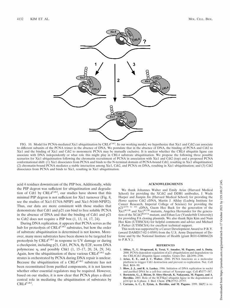
FIG. 10. Model for PCNA-mediated Xic1 ubiquitination by CRL4Cdt2. In our working model, we hypothesize that Xic1 and Cdt2 can associateto different subunits of the PCNA trimer in the absence of DNA. We postulate that in the absence of DNA, the binding of PCNA and Cdt2 toXic1 and the binding of Xic1 and Cdt2 to monomeric PCNA may be mutually exclusive. It is unclear whether the CRL4 ubiquitin ligase canassociate with DNA independently or what role this might play in CRL4 substrate ubiquitination. We propose the following three possiblescenarios for Xic1 ubiquitination following the chromatin recruitment of PCNA in association with Xic1 and Cdt2 (top) and a proposed PCNAconformational shift: (1) Xic1 dissociates from PCNA and binds to the N-terminal domain of PCNA-bound Cdt2, resulting in Xic1 ubiquitination;(2) chromatin-bound PCNA mediates a stable interaction among Xic1, Cdt2, and PCNA on DNA, resulting in Xic1 ubiquitination; and (3) Cdt2dissociates from PCNA and binds to Xic1, resulting in Xic1 ubiquitination.
4132 KIM ET AL. MOL. CELL. BIOL.
Dow
nloa
ded
from
http
s://j
ourn
als.
asm
.org
/jour
nal/m
cb o
n 17
Oct
ober
202
1 by
219
.100
.37.
245.

quired for ubiquitin-mediated degradation of the CDK inhibitor p27. Nat.Cell Biol. 1:193–199.
6. Chuang, L. C., and P. R. Yew. 2005. Proliferating cell nuclear antigen recruitscyclin-dependent kinase inhibitor Xic1 to DNA and couples its proteolysis toDNA polymerase switching. J. Biol. Chem. 280:35299–35309.
7. Chuang, L. C., and P. R. Yew. 2001. Regulation of nuclear transport anddegradation of the Xenopus cyclin-dependent kinase inhibitor, p27Xic1.J. Biol. Chem. 276:1610–1617.
8. Chuang, L. C., X. N. Zhu, C. R. Herrera, H. M. Tseng, C. M. Pfleger, K.Block, and P. R. Yew. 2005. The C-terminal domain of the Xenopus cyclin-dependent kinase inhibitor, p27Xic1, is both necessary and sufficient forphosphorylation-independent proteolysis. J. Biol. Chem. 280:35290–35298.
9. Daniels, M., V. Dhokia, L. Richard-Parpaillon, and S. Ohnuma. 2004. Iden-tification of Xenopus cyclin-dependent kinase inhibitors, p16Xic2 andp17Xic3. Gene 342:41–47.
10. Furstenthal, L., C. Swanson, B. K. Kaiser, A. G. Eldridge, and P. K. Jackson.2001. Triggering ubiquitination of a CDK inhibitor at origins of DNA rep-lication. Nat. Cell Biol. 3:715–722.
11. Gulbis, J. M., Z. Kelman, J. Hurwitz, M. O’Donnell, and J. Kuriyan. 1996.Structure of the C-terminal region of p21(WAF1/CIP1) complexed withhuman PCNA. Cell 87:297–306.
12. Havens, C. G., and J. C. Walter. 2009. Docking of a specialized PIP box ontochromatin-bound PCNA creates a degron for the ubiquitin ligaseCRL4Cdt2. Mol. Cell 35:93–104.
13. Higa, L. A., I. S. Mihaylov, D. P. Banks, J. Zheng, and H. Zhang. 2003.Radiation-mediated proteolysis of CDT1 by CUL4-ROC1 and CSN com-plexes constitutes a new checkpoint. Nat. Cell Biol. 5:1008–1015.
14. Hu, J., and Y. Xiong. 2006. An evolutionarily conserved function of prolif-erating cell nuclear antigen for Cdt1 degradation by the Cul4-Ddb1 ubiquitinligase in response to DNA damage. J. Biol. Chem. 281:3753–3756.
15. Jin, J., E. E. Arias, J. Chen, J. W. Harper, and J. C. Walter. 2006. A familyof diverse Cul4-Ddb1-interacting proteins includes Cdt2, which is requiredfor S phase destruction of the replication factor Cdt1. Mol. Cell 23:709–721.
16. Kim, S. H., and W. M. Michael. 2008. Regulated proteolysis of DNA poly-merase eta during the DNA-damage response in C. elegans. Mol. Cell 32:757–766.
17. Kim, Y., N. G. Starostina, and E. T. Kipreos. 2008. The CRL4Cdt2 ubiquitinligase targets the degradation of p21Cip1 to control replication licensing.Genes Dev. 22:2507–2519.
18. Kwon, C., and I. K. Chung. 2004. Interaction of an Arabidopsis RNA-binding protein with plant single-stranded telomeric DNA modulates telom-erase activity. J. Biol. Chem. 279:12812–12818.
19. Lengronne, A., and E. Schwob. 2002. The yeast CDK inhibitor Sic1 preventsgenomic instability by promoting replication origin licensing in late G(1).Mol. Cell 9:1067–1078.
20. Leung-Pineda, V., J. Huh, and H. Piwnica-Worms. 2009. DDB1 targets Chk1
to the Cul4 E3 ligase complex in normal cycling cells and in cells experienc-ing replication stress. Cancer Res. 69:2630–2637.
21. Lin, H. R., L. C. Chuang, H. Boix-Perales, A. Philpott, and P. R. Yew. 2006.Ubiquitination of cyclin-dependent kinase inhibitor, Xic1, is mediated by theXenopus F-box protein xSkp2. Cell Cycle 5:304–314.
22. Morgan, D. O. 1995. Principles of CDK regulation. Nature 374:131–134.23. Nam, H. W., R. Simpson, and Y. S. Kim. 2005. N-terminal isotope tagging
with propionic anhydride: proteomic analysis of myogenic differentiation ofC2C12 cells. J. Chromatogr. B Analyt. Technol. Biomed. Life Sci. 826:91–107.
24. Nishitani, H., Y. Shiomi, H. Iida, M. Michishita, T. Takami, and T. Tsuri-moto. 2008. CDK inhibitor p21 is degraded by a proliferating cell nuclearantigen-coupled Cul4-DDB1Cdt2 pathway during S phase and after UVirradiation. J. Biol. Chem. 283:29045–29052.
25. Roberts, B. T., C. Y. Ying, J. Gautier, and J. L. Maller. 1999. DNA replica-tion in vertebrates requires a homolog of the Cdc7 protein kinase. Proc. Natl.Acad. Sci. U. S. A. 96:2800–2804.
26. Russo, A. A., P. D. Jeffrey, A. K. Patten, J. Massague, and N. P. Pavletich.1996. Crystal structure of the p27Kip1 cyclin-dependent-kinase inhibitorbound to the cyclin A-Cdk2 complex. Nature 382:325–331.
27. Sherr, C. J. 1994. G1 phase progression: cycling on cue. Cell 79:551–555.28. Sherr, C. J., and J. M. Roberts. 1999. CDK inhibitors: positive and negative
regulators of G1-phase progression. Genes Dev. 13:1501–1512.29. Shibutani, S. T., A. F. de la Cruz, V. Tran, W. J. Turbyfill III, T. Reis, B. A.
Edgar, and R. J. Duronio. 2008. Intrinsic negative cell cycle regulationprovided by PIP box- and Cul4Cdt2-mediated destruction of E2f1 during Sphase. Dev. Cell 15:890–900.
30. Shou, W., and W. G. Dunphy. 1996. Cell cycle control by Xenopus p28Kix1,a developmentally regulated inhibitor of cyclin-dependent kinases. Mol.Biol. Cell 7:457–469.
31. Spruck, C. H., K. A. Won, and S. I. Reed. 1999. Deregulated cyclin E induceschromosome instability. Nature 401:297–300.
32. Su, J. Y., R. E. Rempel, E. Erikson, and J. L. Maller. 1995. Cloning andcharacterization of the Xenopus cyclin-dependent kinase inhibitor p27XIC1.Proc. Natl. Acad. Sci. U. S. A. 92:10187–10191.
33. Sutterluty, H., E. Chatelain, A. Marti, C. Wirbelauer, M. Senften, U. Muller,and W. Krek. 1999. p45SKP2 promotes p27Kip1 degradation and induces Sphase in quiescent cells. Nat. Cell Biol. 1:207–214.
34. Terai, K., T. Abbas, A. A. Jazaeri, and A. Dutta. 2010. CRL4(Cdt2) E3ubiquitin ligase monoubiquitinates PCNA to promote translesion DNA syn-thesis. Mol. Cell 37:143–149.
35. Tsvetkov, L. M., K. H. Yeh, S. J. Lee, H. Sun, and H. Zhang. 1999. p27(Kip1)ubiquitination and degradation is regulated by the SCF(Skp2) complexthrough phosphorylated Thr187 in p27. Curr. Biol. 9:661–664.
36. Yew, P. R., and M. W. Kirschner. 1997. Proteolysis and DNA replication: theCDC34 requirement in the Xenopus egg cell cycle. Science 277:1672–1676.
VOL. 30, 2010 CRL4Cdt2 BINDS PCNA TO MEDIATE Xic1 PROTEOLYSIS 4133
Dow
nloa
ded
from
http
s://j
ourn
als.
asm
.org
/jour
nal/m
cb o
n 17
Oct
ober
202
1 by
219
.100
.37.
245.