the ecm protein nephronectin promotes kidney...
TRANSCRIPT
DEVELO
PMENT
2501RESEARCH ARTICLE
INTRODUCTIONThe metanephric kidney arises through instructive signaling betweentwo cell populations, a mesenchymal population known as thenephric cord and an epithelial population termed the nephric duct(ND). Both of these populations arise from the intermediatemesoderm on approximately embryonic day (E) 9.0 in the mouse.Within the nephric cord, in a region adjacent to the posteriorhindlimb bud, there develops a special group of cells known as themetanephric mesenchyme (MM). At ~E11, a signal(s) from the MMelicits the formation of the ureteric bud (UB) from the ND. Onceformed, the UB extends toward and then invades the MM, and by~E11.5 the UB begins branching, thus giving rise to a tubularnetwork that will eventually mature into the collecting ducts of theadult kidney. As the UB invades and branches, it expresses genesthat induce the MM to condense and differentiate into various celltypes that comprise the nephron. In the absence of invasion by theUB into the MM, the metanephric kidney does not develop(reviewed in Saxen, 1987).
Glial cell line-derived neurotrophic factor (GDNF), a member ofthe transforming growth factor-� (TGF�) superfamily, is a keysignal in the initiation of UB formation and subsequent branching(Costantini and Shakya, 2006). GDNF is expressed in the MM andsignals to the UB through a receptor complex consisting of thereceptor tyrosine kinase RET and a coreceptor, GDNF familyreceptor alpha1 (GFR�1). GDNF has been shown to induce ectopicUB formation in culture, and mice lacking either the Gdnf, Ret or
Gfra1 genes display renal agenesis at high penetrance (Cacalano etal., 1998; Moore et al., 1996; Pichel et al., 1996; Sainio et al., 1997;Sanchez et al., 1996; Schuchardt et al., 1994). Gdnf expression in theMM is regulated by a highly conserved network of transcriptionfactors, and can be activated or inhibited by extracellular signalingmolecules such as WNT11, GDF11 and SLIT2 at discrete stages ofdevelopment (Brodbeck and Englert, 2004; Esquela and Lee, 2003;Grieshammer et al., 2004; Majumdar et al., 2003). It is also knownthat GDNF signaling is dependent on heparan sulphateglycosaminoglycans, which interact with or are constituents of theextracellular matrix (ECM) (Barnett et al., 2002).
Integrins are cell adhesion receptors that serve as a link betweenthe ECM and the cytoskeleton. Integrins are heterodimers,consisting of two single-pass transmembrane proteins designated asthe � and � subunits. Integrins are thought to occupy either aninactive or active state depending on cues received from theextracelluar environment in the form of an appropriate ECM ligandor growth factor signal. They communicate information from theECM to the cell interior through recruitment of cytoskeletal proteinsand kinases. In addition, they are able to communicate informationfrom the cell interior to the ECM, resulting in its proper depositionand remodeling (Delon and Brown, 2007; ffrench-Constant andColognato, 2004; Giancotti and Ruoslahti, 1999; Hynes, 2002).
Previously, we showed that the integrin subunit �8 (Itga8) isexpressed throughout the nephric cord, including the MM (Mulleret al., 1997). Furthermore, we demonstrated that loss of Itga8function invariably results in a delay of invasion of the MM by theUB, which in turn results in a high frequency of kidney agenesis.The molecular mechanism by which Itga8 function in the MMinfluences the UB has yet to be determined. Several ECMconstituents are known ligands for �8�1 integrin, includingfibronectin, osteopontin, tenascin C and vitronectin (Denda et al.,1998a; Denda et al., 1998b; Varnum-Finney et al., 1995). However,data from expression and loss-of-function analyses indicated that
The ECM protein nephronectin promotes kidneydevelopment via integrin �8�1-mediated stimulation ofGdnf expressionJames M. Linton1, Gail R. Martin2 and Louis F. Reichardt1,3,*
Development of the metanephric kidney crucially depends on proper interactions between cells and the surrounding extracellularmatrix. For example, we showed previously that in the absence of �8�1 integrin, invasion by the ureteric bud into the metanephricmesenchyme is inhibited, resulting in renal agenesis. Here we present genetic evidence that the extracellular matrix proteinnephronectin is an essential ligand that engages �8�1 integrin during early kidney development. We show that embryos lacking afunctional nephronectin gene frequently display kidney agenesis or hypoplasia, which can be traced to a delay in the invasion ofthe metanephric mesenchyme by the ureteric bud at an early stage of kidney development. Significantly, we detected no defects inextracellular matrix organization in the nascent kidneys of the nephronectin mutants. Instead, we found that Gdnf expression wasdramatically reduced in both nephronectin- and �8 integrin-null mutants specifically in the metanephric mesenchyme at the time ofureteric bud invasion. We show that this reduction is sufficient to explain the agenesis and hypoplasia observed in both mutants.Interestingly, the reduction in Gdnf expression is transient, and its resumption presumably enables the nephronectin-deficientureteric buds to invade the metanephric mesenchyme and begin branching. Our results thus place nephronectin and �8�1 integrinin a pathway that regulates Gdnf expression and is essential for kidney development.
KEY WORDS: Kidney, Integrin, Extracellular matrix, GDNF, Mouse, Ureteric bud
Development 134, 2501-2509 (2007) doi:10.1242/dev.005033
1Department of Physiology, 2Department of Anatomy and 3Howard Hughes MedicalInstitute, 1550 Fourth Street, University of California, San Francisco, San Francisco,CA 94143, USA.
*Author for correspondence (e-mail: [email protected])
Accepted 2 May 2007
DEVELO
PMENT
2502
these ligands are unlikely to be mediators of �8�1 integrin functionin the developing kidney. Using an expression cloning strategy weidentified a gene that encodes a novel ECM molecule, nephronectin(Npnt), that is expressed by the UB and the epithelia of severaldeveloping organs (Brandenberger et al., 2001). We demonstratedthat �8�1 integrin recognizes nephronectin in binding assays andassociates with nephronectin in vivo. In addition, we found that thelocalization of nephronectin in the kidney is consistent with itmediating �8�1 integrin function during development.
Here we report that mice lacking nephronectin frequently displaykidney agenesis. We show that the phenotype arises during the earlystages of metanephric development, when the UB is beginning toinvade the MM, similar to the phenotype of mice lacking Itga8 (andtherefore �8�1 integrin function). Thus, nephronectin is an ECMprotein expressed by the UB that is required for �8�1 integrinfunction during early stages of UB invasion and branching.Significantly, we demonstrate that Gdnf expression is reduced inboth Npnt and Itga8 mutants at the time when the invasion of theMM by the UB is delayed. Finally, we present genetic dataindicating that Gdnf dosage as well as signaling from the receptortyrosine kinase, RET, impact the penetrance of the Itga8 mutantphenotype. Taken together, our results suggest that the observedreduction of Gdnf expression in the MM is sufficient to explain thephenotypes observed in mice lacking either nephronectin or the �8integrin subunit.
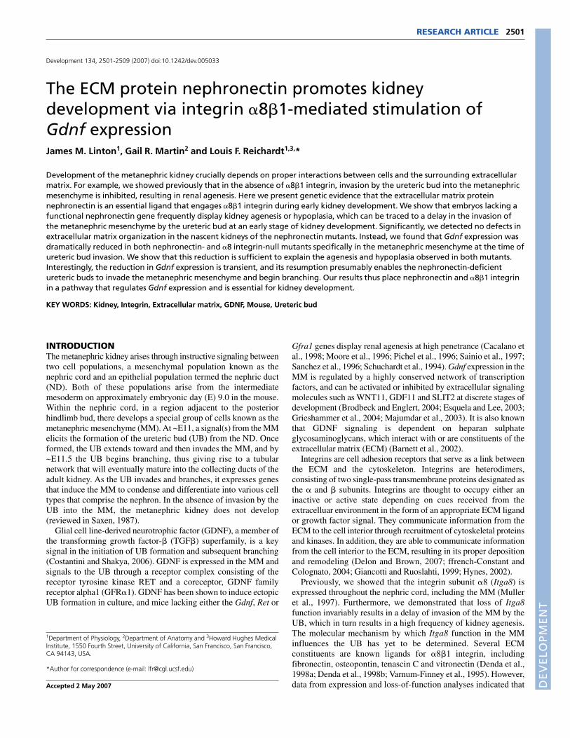
MATERIALS AND METHODSTargeting of the Npnt locusA probe derived from the 5� region of the Npnt locus was used to screen amouse 129Sv/J BAC library, RPCI-22 (Roswell Park Cancer Institute). Oneof 9 clones isolated was used to generate the floxneo targeting construct (Fig.1B-D) by recombineering (Zhang et al., 1998). Clones harboring BACs thatwere correctly modified were identified by Southern blot hybridization,which revealed the presence of both the 5� and 3� arms. One clone,designated 273P10 NN-1, was electroporated into E14 embryonic stem (ES)cells.
Because of the large size of the targeting construct (300 kb), we usedTaqMan (Applied Biosystems) real-time quantitative PCR to identify EScells in which there was a reduction in copy number of a region that wastargeted for insertion (Valenzuela et al., 2003). This involved the use ofspecific PCRs that amplify the wild-type allele, but fail to amplify themutated allele because of the increase in sequence length. Candidate ES cellclones that were identified by sequential PCR screens were assessed forreduced wild-type copy number by quantitative Southern hybridizationusing densitometry. Two clones isolated from the screen were injected intoC57BL/6 blastocysts (Transgenic/Targeted Mutagenesis Core, University ofCalifornia, San Francisco, CA), and both were incorporated into the germline.
We generated a null allele for nephronectin by crossing animalsheterozygous for the Npnt floxneo allele with animals expressing CRErecombinase under the �-actin promoter (Lewandoski et al., 1997). Thiscross produced mice carrying the Npnt�ex1 allele in which the first exon ofNpnt was excised without deletion of the neomycin-resistance geneexpression cassette (Fig. 1D,E). All the analysis described here wasperformed on mice carrying this allele on a mixed genetic background(129Sv/J; C57BL/6; FVB/N).
In situ hybridizationTo stage embryos, noon of the day on which a vaginal plug was detected wasconsidered E0.5. Embryos were collected at various stages and the regioncontaining the hindlimb buds was fixed in 4.0% paraformaldehyde(PFA)/phosphate-buffered saline (PBS) overnight at 4°C and cryosectionedat 14 �m. Analysis of gene expression using in situ hybridization with RNAprobes was performed according to standard protocols. Data using thefollowing probes are presented: Gdnf (Srinivas et al., 1999), Eya1 (Xu et al.,1999), Six2 (Xu et al., 2003), Pax2 (Dressler et al., 1990).
Histology and immunofluorescenceEmbryos at various stages and kidneys from newborn animals were fixed in4.0% PFA/PBS overnight at 4°C. Tissues were cryosectioned and stainedwith hematoxylin and eosin according to standard protocols. Sections werestained with the following antibodies: anti-nephronectin (1:100)(Brandenberger et al., 2001), anti-EHS laminin (1:500) (Sigma L9393), anti-fibronectin (1:300) (Sigma F-6140), anti-calbindin D28K (1:600) (SwantCB-38a), anti-collagen type IV (1:500) (LB-1403; Cosmo Bio., Tokyo,Japan), and anti-pax2 (1:100) (PRB-276P; Covance, Princeton, NJ).Confocal imaging was performed on a Zeiss LSM 5 Pascal.
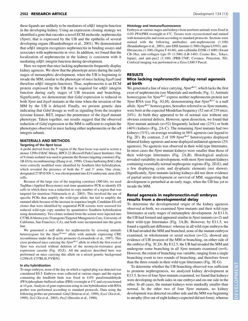
RESULTSMice lacking nephronectin display renal agenesisat birthWe generated a line of mice carrying, Npnt�ex1, which lacks the firstexon of nephronectin (see Materials and methods; Fig. 1). Animalshomozygous for Npnt�ex1 express neither nephronectin protein norNpnt RNA (see Fig. 1G,H), demonstrating that Npnt�ex1 is a nullallele. Npnt�ex1 homozygotes, hereafter referred to as Npnt mutants,were born at the expected Mendelian frequency (52 out of 219 mice;24%). At birth they appeared to be of normal size without anyobvious external defects. However, upon dissection, we found thatNpnt mutant animals (n=52) frequently lacked one (23%) or both(46%) kidneys (Fig. 2A-C). The remaining Npnt mutants had twokidneys (31%), on average resulting in 58% agenesis (see legend toFig. 2F). By contrast, 2 of 109 Npnt�ex1 heterozygotes displayedbilateral kidney agenesis and none displayed unilateral agenesis (2%agenesis). No agenesis was observed in their wild-type littermates.In most cases the Npnt mutant kidneys were smaller than those oftheir wild-type littermates (Fig. 2A,B). Histological analysisrevealed variability in development, with most Npnt mutant kidneyscontaining essentially normal nephrogenic regions (Fig. 2D,E), anda few displaying cystic and dysplastic elements (not shown).Significantly, Npnt mutants lacking kidneys did not show evidenceof partial ureter development or survival of MM, suggesting thatdevelopment is perturbed at an early stage, when the UB has yet toinvade the MM.
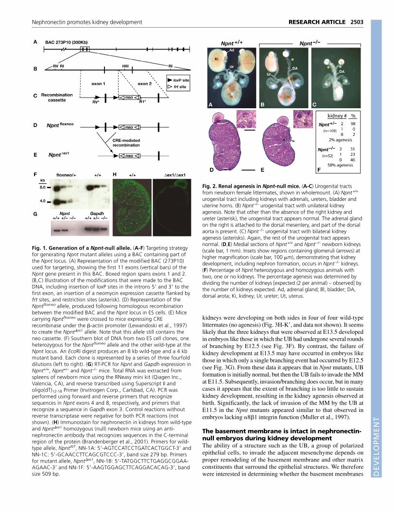
Renal agenesis in nephronectin-null embryosresults from a developmental delayTo determine the developmental origin of the kidney agenesisobserved at birth, we examined Npnt mutants and their wild-typelittermates at early stages of metanephric development. At E11.0,the UB had formed and appeared similar in Npnt mutants (n=2) andtheir wild-type littermates (Fig. 3A,B). However, by E11.5, wefound a significant difference: whereas in all wild-type embryos theUB had invaded the MM and branched, none of the mutant embryosexamined, in wholemount or serial section (n=12), showed anyevidence of UB invasion of the MM or branching, on either side ofthe embryo (Fig. 3C,D). By E12.5, the UB had invaded the MM andundergone some branching in all Npnt mutants examined (n=4).However, the extent of branching was variable, ranging from a singlebranching event to two rounds of branching, and therefore fewerthan the three rounds in their wild-type littermates (Fig. 3E-G).
To determine whether the UB branching observed was sufficientto promote nephrogenesis, we analyzed kidney development atE13.5. In two of four Npnt mutants examined, we found that kidneyswere developing on both sides in one embryo and on one side in theother. In all cases, the mutant kidneys were markedly smaller thannormal. In the other two of four Npnt mutants, no kidneydevelopment was detected on either side and the MM was beginningto atrophy (five out of eight kidneys expected did not form), whereas
RESEARCH ARTICLE Development 134 (13)
DEVELO
PMENT
kidneys were developing on both sides in four of four wild-typelittermates (no agenesis) (Fig. 3H-K�, and data not shown). It seemslikely that the three kidneys that were observed at E13.5 developedin embryos like those in which the UB had undergone several roundsof branching by E12.5 (see Fig. 3F). By contrast, the failure ofkidney development at E13.5 may have occurred in embryos likethose in which only a single branching event had occurred by E12.5(see Fig. 3G). From these data it appears that in Npnt mutants, UBformation is initially normal, but then the UB fails to invade the MMat E11.5. Subsequently, invasion/branching does occur, but in manycases it appears that the extent of branching is too little to sustainkidney development, resulting in the kidney agenesis observed atbirth. Significantly, the lack of invasion of the MM by the UB atE11.5 in the Npnt mutants appeared similar to that observed inembryos lacking �8�1 integrin function (Muller et al., 1997).
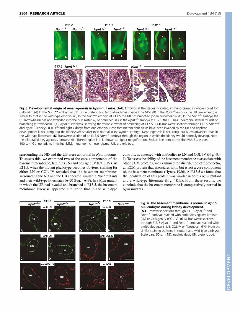
The basement membrane is intact in nephronectin-null embryos during kidney developmentThe ability of a structure such as the UB, a group of polarizedepithelial cells, to invade the adjacent mesenchyme depends onproper remodeling of the basement membrane and other matrixconstituents that surround the epithelial structures. We thereforewere interested in determining whether the basement membranes
2503RESEARCH ARTICLENephronectin promotes kidney development
Fig. 1. Generation of a Npnt-null allele. (A-F) Targeting strategyfor generating Npnt mutant alleles using a BAC containing part ofthe Npnt locus. (A) Representation of the modified BAC (273P10)used for targeting, showing the first 11 exons (vertical bars) of theNpnt gene present in this BAC. Boxed region spans exons 1 and 2.(B,C) Illustration of the modifications that were made to the BACDNA, including insertion of loxP sites in the introns 5� and 3� to thefirst exon, an insertion of a neomycin expression cassette flanked byfrt sites, and restriction sites (asterisk). (D) Representation of theNpntfloxneo allele, produced following homologous recombinationbetween the modified BAC and the Npnt locus in ES cells. (E) Micecarrying Npntfloxneo were crossed to mice expressing CRErecombinase under the �-actin promoter (Lewandoski et al., 1997)to create the Npnt�ex1 allele. Note that this allele still contains theneo cassette. (F) Southern blot of DNA from two ES cell clones, oneheterozygous for the Npntfloxneo allele and the other wild-type at theNpnt locus. An EcoRI digest produces an 8 kb wild-type and a 4 kbmutant band. Each clone is represented by a series of three fourfolddilutions (left to right). (G) RT-PCR for Npnt and Gapdh expression inNpnt+/+, Npnt+/– and Npnt–/– mice. Total RNA was extracted fromspleens of newborn mice using the RNeasy mini kit (Qiagen Inc.,Valencia, CA), and reverse transcribed using Superscript II andoligo(dT)12-18 Primer (Invitrogen Corp., Carlsbad, CA). PCR wasperformed using forward and reverse primers that recognizesequences in Npnt exons 4 and 8, respectively, and primers thatrecognize a sequence in Gapdh exon 3. Control reactions withoutreverse transcriptase were negative for both PCR reactions (notshown). (H) Immunostain for nephronectin in kidneys from wild-typeand Npnt�ex1 homozygous (null) newborn mice using an anti-nephronectin antibody that recognizes sequences in the C-terminalregion of the protein (Brandenberger et al., 2001). Primers for wild-type allele, NpntWT, NN-1A: 5�-AGTCCATCCTGATCACTGGCT-3� andNN-1C: 5�-GCAACCTTCAGCGTCCC-3�, band size 279 bp. Primersfor mutant allele, Npnt�ex1, NN-1B: 5�-TATGGCTTCTGAGGCG GAA -AG AAC-3� and NN-1F: 5�-AAGTGGAGCTTCAGGACACAG-3�, bandsize 509 bp.
Fig. 2. Renal agenesis in Npnt-null mice. (A-C) Urogenital tractsfrom newborn female littermates, shown in wholemount. (A) Npnt+/+
urogenital tract including kidneys with adrenals, ureters, bladder anduterine horns. (B) Npnt–/– urogenital tract with unilateral kidneyagenesis. Note that other than the absence of the right kidney andureter (asterisk), the urogenital tract appears normal. The adrenal glandon the right is attached to the dorsal mesentery, and part of the dorsalaorta is present. (C) Npnt–/– urogenital tract with bilateral kidneyagenesis (asterisks). Again, the rest of the urogenital tract appearsnormal. (D,E) Medial sections of Npnt+/+ and Npnt–/– newborn kidneys(scale bar, 1 mm). Insets show regions containing glomeruli (arrows) athigher magnification (scale bar, 100 �m), demonstrating that kidneydevelopment, including nephron formation, occurs in Npnt–/– kidneys.(F) Percentage of Npnt heterozygous and homozygous animals withtwo, one or no kidneys. The percentage agenesis was determined bydividing the number of kidneys [expected (2 per animal) – observed] bythe number of kidneys expected. Ad, adrenal gland; Bl, bladder; DA,dorsal arota; Ki, kidney; Ur, ureter; Ut, uterus.
DEVELO
PMENT
2504
surrounding the ND and the UB were abnormal in Npnt mutants.To assess this, we examined two of the core components of thebasement membrane, laminin (LN) and collagen IV (COL IV). AtE11.5, when the mutant phenotype becomes obvious, staining foreither LN or COL IV revealed that the basement membranessurrounding the ND and the UB appeared similar in Npnt mutantsand their wild-type littermates (n=3) (Fig. 4A-F). In a Npnt mutantin which the UB had invaded and branched at E13.5, the basementmembrane likewise appeared similar to that in the wild-type
controls, as assessed with antibodies to LN and COL IV (Fig. 4G-J). To assess the ability of the basement membrane to associate withother ECM proteins, we examined the distribution of fibronectin,an ECM protein that associates with, but is not a core componentof, the basement membrane (Hynes, 1986). At E13.5 we found thatthe localization of this protein was similar in both a Npnt mutantand a wild-type littermate (Fig. 4K,L). From these results, weconclude that the basement membrane is comparatively normal inNpnt mutants.
RESEARCH ARTICLE Development 134 (13)
Fig. 3. Developmental origin of renal agenesis in Npnt-null mice. (A-G) Embryos at the stages indicated, immunostained in wholemount forCalbindin. (A) In the Npnt+/+ embryo at E11.0 the ureteric bud (arrowhead) has invaded the MM. (B) In the Npnt–/– embryo the UB (arrowhead) issimilar to that in the wild-type embryo. (C) In the Npnt+/+ embryo at E11.5 the UB has branched (open arrowheads). (D) In the Npnt–/– embryo theUB (arrowhead) has not extended into the MM (asterisk) or branched. (E) In the Npnt+/+ embryo at E12.5 the UB has undergone several rounds ofbranching (arrowheads). (F,G) Npnt–/– embryos, showing the variable extent of branching at E12.5. (H-J) Transverse sections through E13.5 Npnt+/+
and Npnt–/– kidneys. (I,J) Left and right kidneys from one embryo. Note that metanephric fields have been invaded by the UB and nephrondevelopment is occurring, but the kidneys are smaller than normal in the Npnt–/– embryo. Nephrogenesis is occurring, but is less advanced than inthe wild-type littermate. (K) Transverse section of an E13.5 Npnt–/– embryo through the region in which the kidney would normally develop. Notethe bilateral kidney agenesis (arrows). (K�) Boxed region in K is shown at higher magnification. Broken line demarcates the MM. Scale bars,100 �m. Go, gonad; In, intestine; MM, metanephric mesenchyme; UB, ureteric bud.
Fig. 4. The basement membrane is normal in Npnt-null embryos during kidney development.(A-F) Transverse sections through E11.5 Npnt+/+ andNpnt–/– embryos stained with antibodies against laminin(LN) or Collagen IV (COL IV). (G-L) Transverse sectionsthrough E13.5 Npnt+/+ and Npnt–/– embryos stained withantibodies against LN, COL IV or fibronectin (FN). Note thesimilar staining patterns in mutant and wild-type embryos.Scale bars, 50 �m. ND, nephric duct; UB, ureteric bud.
DEVELO
PMENT
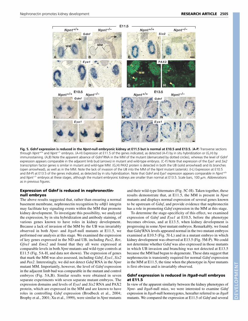
Expression of Gdnf is reduced in nephronectin-null embryosThe above results suggested that, rather than ensuring a normalbasement membrane, nephronectin recognition by �8�1 integrinmay facilitate key signaling events within the MM that promotekidney development. To investigate this possibility, we analyzedthe expression, by in situ hybridization and antibody staining, ofvarious genes known to have roles in kidney development.Because a lack of invasion of the MM by the UB was invariablyobserved in both Npnt- and Itga8-null mutants at E11.5, weperformed our analysis at this stage. We examined the expressionof key genes expressed in the ND and UB, including Pax2, Ret,Gfra1 and Emx2 and found that they all were expressed atcomparable levels in both Npnt mutants and wild-type controls atE11.5 (Fig. 5A-H, and data not shown). The expression of genesthat mark the MM was also assessed, including Gdnf, Eya1, Six2and Pax2. Interestingly, we did not detect Gdnf RNA in the Npntmutant MM. Importantly, however, the level of Gdnf expressionin the adjacent limb bud was comparable in the mutant and controlembryos (Fig. 5A,B). Similar results were obtained in sevenseparate experiments with seven separate mutant embryos. Theexpression domains and levels of Eya1 and Six2 RNA and PAX2protein, which are expressed in the MM and are known to haveroles in controlling Gdnf expression (Brodbeck et al., 2004;Brophy et al., 2001; Xu et al., 1999), were similar in Npnt mutants
and their wild-type littermates (Fig. 5C-H). Taken together, theseresults demonstrate that, at E11.5, the MM is present in Npntmutants and displays normal expression of several genes knownto be upstream of Gdnf, and provide evidence that nephronectinhas a role in promoting Gdnf expression in the MM at this stage.
To determine the stage-specificity of this effect, we examinedexpression of Gdnf and Eya1 at E10.5, before the phenotypebecomes obvious, and at E13.5, when kidney development isprogressing in some Npnt mutant embryos. Remarkably, we foundthat Gdnf RNA levels appeared normal in the two mutant embryosexamined at E10.5 (Fig. 5I-L) and in a mutant embryo in whichkidney development was observed at E13.5 (Fig. 5M-P). We couldnot determine whether Gdnf was also expressed in those mutantsin which UB invasion and branching was not detected at E13.5because the MM had begun to degenerate. These data suggest thatnephronectin is transiently required for normal Gdnf expressionin the MM at E11.5, the time when the phenotype in Npnt mutantsis first obvious and is invariably observed.
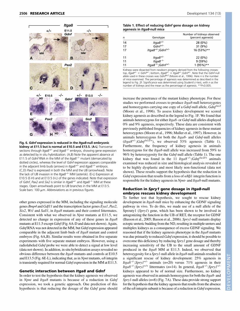
Gdnf expression is reduced in Itga8-null embryosat E11.5In view of the apparent similarity between the kidney phenotypes ofNpnt- and Itga8-null mice, we were interested to examine Gdnfexpression in Itga8-null homozygotes, hereafter referred to as Itga8mutants. We compared the expression at E11.5 of Gdnf and several
2505RESEARCH ARTICLENephronectin promotes kidney development
Fig. 5. Gdnf expression is reduced in the Npnt-null embryonic kidney at E11.5 but is normal at E10.5 and E13.5. (A-P) Transverse sectionsthrough Npnt+/+ and Npnt–/– embryos. (A-H) Expression at E11.5 of the genes indicated, as detected (A-F) by in situ hybridization or (G,H) byimmunostaining. (A,B) Note the apparent absence of Gdnf RNA in the MM of the mutant (demarcated by dotted circles), whereas the level of Gdnfexpression appears comparable in the adjacent limb bud (arrows) in mutant and wild-type embryos. (C-F) Note that expression of the Eya1 and Six2transcription factor genes is similar in mutant and wild-type MM. (G,H) PAX2 protein is detected in both the UB (solid arrowhead) and its branches(open arrowhead), as well as in the MM. Note the lack of invasion of the UB into the MM of the Npnt mutant (asterisk). (I-L) Expression at E10.5and (M-P) at E13.5 of the genes indicated, as detected by in situ hybridization. Note that Gdnf and Eya1 expression appears comparable in Npnt+/+
and Npnt–/– embryos at these stages, although the mutant embryonic kidneys are smaller than normal at E13.5. Scale bars, 100 �m. Abbreviationsas in previous figures.
DEVELO
PMENT
2506
other genes expressed in the MM, including the signaling moleculegenes Bmp4 and Gdf11 and the transcription factor genes Eya1, Pax2,Six2, Wt1 and Sall1, in Itga8 mutants and their control littermates.Consistent with what we observed in Npnt mutants at E11.5, wedetected no change in expression of any of these genes in Itga8mutants at E11.5 except Gdnf (Fig. 6A-D and data not shown). Again,Gdnf RNA was not detected in the MM, but Gdnf expression appearedcomparable in the adjacent limb buds of Itga8 mutant and controlembryos (Fig. 6A,B). Similar results were obtained in five separateexperiments with five separate mutant embryos. However, using aradiolabeled Gdnf probe we were able to detect a signal at low level(data not shown). In addition, in situ hybridization assays revealed noobvious difference between the Itga8 mutants and controls at E10.5and E13.5 (Fig. 6E-L), indicating that, as in Npnt mutants, �8 integrinis transiently required for normal Gdnf expression in the MM at E11.5.
Genetic interaction between Itga8 and GdnfIn order to test the hypothesis that the kidney agenesis we observedin Npnt and Itga8 mutants is because of a reduction in Gdnfexpression, we took a genetic approach. One prediction of thishypothesis is that reducing the dosage of the Gdnf gene should
increase the penetrance of the mutant kidney phenotype. For thesestudies we performed crosses to produce Itga8-null heterozygotesand homozygotes carrying one copy of a Gdnf-null allele, Gdnf lacZ
(Moore et al., 1996). To assess kidney development we scoredkidney agenesis as described in the legend to Fig. 1F. We found thatanimals heterozygous for either Itga8- or Gdnf-null alleles displayed0% and 9% agenesis, respectively. These data are consistent withpreviously published frequencies of kidney agenesis in these mutantheterozygotes (Moore et al., 1996; Muller et al., 1997). However, inanimals heterozygous for both the Itga8- and Gdnf-null alleles(Itga8+/–;Gdnf lacZ/+), we observed 53% agenesis (Table 1).Furthermore, the frequency of kidney agenesis in animalshomozygous for the Itga8-null allele was increased from 59% to96% by heterozygosity for the Gdnf-null allele (Table 1). The onekidney that was found in the 11 Itga8–/–;Gdnf lacZ/+ animalsexamined was reduced in size and histological analysis revealed itto be highly dysplastic and most likely non-functional (data notshown). These results support the hypothesis that the reduction inGdnf expression that results from a loss of �8�1 integrin function isresponsible for the kidney agenesis in Npnt- and Itga8-null mutants.
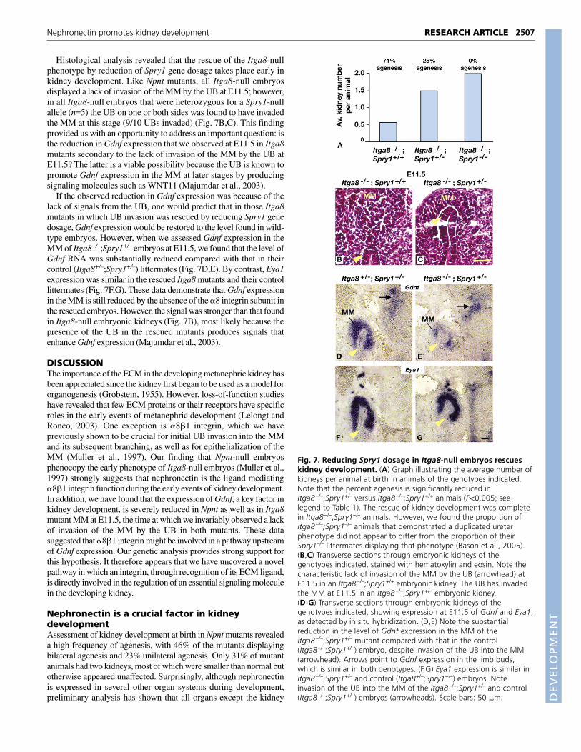
Reduction in Spry1 gene dosage in Itga8-nullembryos rescues kidney developmentTo further test that hypothesis, we sought to rescue kidneydevelopment in Itga8-null mice by enhancing the GDNF signalingpathway in vivo. To do this, we made use of a null allele of theSprouty1 (Spry1) gene, which has been shown to be involved inantagonizing the function in the UB of RET, the receptor for GDNF(Basson et al., 2005; Basson et al., 2006). Spry1-null mutants displayectopic ureteric budding from the ND, resulting in multiple ureters andmultiplex kidneys as a consequence of excess GDNF signaling. Wereasoned that if the kidney agenesis phenotype in the Itga8 mutantswas due primarily to reduced Gdnf expression, it should be possible toovercome this deficiency by reducing Spry1 gene dosage and therebyincreasing sensitivity of the UB to the small amount of GDNFproduced in the Itga8 MM at E11.5. Indeed, we observed thatheterozygosity for a Spry1-null allele in Itga8-null animals resulted ina significant rescue of kidney development: 25% agenesis inItga8–/–;Spry1+/– animals (n=20) versus 71% agenesis in theirItga8–/–;Spry1+/+ littermates (n=14). In general, Itga8–/–;Spry1+/–
kidneys appeared to be of normal size. Furthermore, no kidneyagenesis was observed in animals homozygous for both the Itga8- andSpry1-null alleles (n=8) (Fig. 7A). These data provide strong supportfor the hypothesis that the kidney agenesis that results from the absenceof the �8 integrin subunit is because of a reduction in Gdnf expression.
RESEARCH ARTICLE Development 134 (13)
Table 1. Effect of reducing Gdnf gene dosage on kidneyagenesis in Itga8-null mice
Number of kidneys observedn Genotype (percent agenesis)
14 Itga8+/– 28 (0%)17 Gdnf+/– 31 (9%)17 Itga8+/–;Gdnf+/– 16 (53%)**
11 Itga8+/– 22 (0%)11 Itga8–/– 9 (59%)11 Itga8–/–;Gdnf+/– 1 (95%)**
Kidneys were dissected from newborn progeny derived from the following crosses:top, Itga8+/– � Gdnf+/–; bottom, Itga8+/– � Itga8+/–;Gdnf+/–. Note that the Gdnf-nullallele used in these crosses was GdnflacZ (Moore et al., 1996). Here n is the numberof mice examined. The percentage of agenesis was determined as described in thelegend to Fig. 2F. Significance was determined using Student’s t-test, with n as thenumber of kidneys and the mean as the percentage of agenesis. **P<0.005.
Fig. 6. Gdnf expression is reduced in the Itga8-null embryonickidney at E11.5 but is normal at E10.5 and E13.5. (A-L) Transversesections through Itga8+/– and Itga8–/– embryos, showing gene expressionas detected by in situ hybridization. (A,B) Note the apparent absence atE11.5 of Gdnf RNA in the MM of the Itga8–/– mutant (demarcated bydotted circles), whereas the level of Gdnf expression appears comparablein the adjacent limb buds (arrows) in Itga8+/– and Itga8–/– embryos.(C,D) Pax2 is expressed in both the MM and the UB (arrowhead). Notethe lack of UB invasion in the Itga8–/– MM (asterisk). (E-L) Expression atE10.5 (E-H) and at E13.5 (I-L) of the genes indicated. Note that expressionof Gdnf, Pax2 and Six2 is similar in Itga8+/– and Itga8–/– MM at thesestages. Open arrowheads point to UB branches in the MM at E13.5.Scale bars: 100 �m. Abbreviations as in previous figures.
DEVELO
PMENT
Histological analysis revealed that the rescue of the Itga8-nullphenotype by reduction of Spry1 gene dosage takes place early inkidney development. Like Npnt mutants, all Itga8-null embryosdisplayed a lack of invasion of the MM by the UB at E11.5; however,in all Itga8-null embryos that were heterozygous for a Spry1-nullallele (n=5) the UB on one or both sides was found to have invadedthe MM at this stage (9/10 UBs invaded) (Fig. 7B,C). This findingprovided us with an opportunity to address an important question: isthe reduction in Gdnf expression that we observed at E11.5 in Itga8mutants secondary to the lack of invasion of the MM by the UB atE11.5? The latter is a viable possibility because the UB is known topromote Gdnf expression in the MM at later stages by producingsignaling molecules such as WNT11 (Majumdar et al., 2003).
If the observed reduction in Gdnf expression was because of thelack of signals from the UB, one would predict that in those Itga8mutants in which UB invasion was rescued by reducing Spry1 genedosage, Gdnf expression would be restored to the level found in wild-type embryos. However, when we assessed Gdnf expression in theMM of Itga8–/–;Spry1+/– embryos at E11.5, we found that the level ofGdnf RNA was substantially reduced compared with that in theircontrol (Itga8+/–;Spry1+/–) littermates (Fig. 7D,E). By contrast, Eya1expression was similar in the rescued Itga8 mutants and their controllittermates (Fig. 7F,G). These data demonstrate that Gdnf expressionin the MM is still reduced by the absence of the �8 integrin subunit inthe rescued embryos. However, the signal was stronger than that foundin Itga8-null embryonic kidneys (Fig. 7B), most likely because thepresence of the UB in the rescued mutants produces signals thatenhance Gdnf expression (Majumdar et al., 2003).
DISCUSSIONThe importance of the ECM in the developing metanephric kidney hasbeen appreciated since the kidney first began to be used as a model fororganogenesis (Grobstein, 1955). However, loss-of-function studieshave revealed that few ECM proteins or their receptors have specificroles in the early events of metanephric development (Lelongt andRonco, 2003). One exception is �8�1 integrin, which we havepreviously shown to be crucial for initial UB invasion into the MMand its subsequent branching, as well as for epithelialization of theMM (Muller et al., 1997). Our finding that Npnt-null embryosphenocopy the early phenotype of Itga8-null embryos (Muller et al.,1997) strongly suggests that nephronectin is the ligand mediating�8�1 integrin function during the early events of kidney development.In addition, we have found that the expression of Gdnf, a key factor inkidney development, is severely reduced in Npnt as well as in Itga8mutant MM at E11.5, the time at which we invariably observed a lackof invasion of the MM by the UB in both mutants. These datasuggested that �8�1 integrin might be involved in a pathway upstreamof Gdnf expression. Our genetic analysis provides strong support forthis hypothesis. It therefore appears that we have uncovered a novelpathway in which an integrin, through recognition of its ECM ligand,is directly involved in the regulation of an essential signaling moleculein the developing kidney.
Nephronectin is a crucial factor in kidneydevelopmentAssessment of kidney development at birth in Npnt mutants revealeda high frequency of agenesis, with 46% of the mutants displayingbilateral agenesis and 23% unilateral agenesis. Only 31% of mutantanimals had two kidneys, most of which were smaller than normal butotherwise appeared unaffected. Surprisingly, although nephronectinis expressed in several other organ systems during development,preliminary analysis has shown that all organs except the kidney
2507RESEARCH ARTICLENephronectin promotes kidney development
Fig. 7. Reducing Spry1 dosage in Itga8-null embryos rescueskidney development. (A) Graph illustrating the average number ofkidneys per animal at birth in animals of the genotypes indicated.Note that the percent agenesis is significantly reduced inItga8 –/–;Spry1+/– versus Itga8 –/–;Spry1+/+ animals (P<0.005; seelegend to Table 1). The rescue of kidney development was completein Itga8 –/–;Spry1–/– animals. However, we found the proportion ofItga8 –/–;Spry1–/– animals that demonstrated a duplicated ureterphenotype did not appear to differ from the proportion of theirSpry1–/– littermates displaying that phenotype (Bason et al., 2005).(B,C) Transverse sections through embryonic kidneys of thegenotypes indicated, stained with hematoxylin and eosin. Note thecharacteristic lack of invasion of the MM by the UB (arrowhead) atE11.5 in an Itga8 –/–;Spry1+/+ embryonic kidney. The UB has invadedthe MM at E11.5 in an Itga8 –/–;Spry1+/– embryonic kidney.(D-G) Transverse sections through embryonic kidneys of thegenotypes indicated, showing expression at E11.5 of Gdnf and Eya1,as detected by in situ hybridization. (D,E) Note the substantialreduction in the level of Gdnf expression in the MM of theItga8 –/–;Spry1+/– mutant compared with that in the control(Itga8+/–;Spry1+/–) embryo, despite invasion of the UB into the MM(arrowhead). Arrows point to Gdnf expression in the limb buds,which is similar in both genotypes. (F,G) Eya1 expression is similar inItga8 –/–;Spry1+/– and control (Itga8+/–;Spry1+/–) embryos. Noteinvasion of the UB into the MM of the Itga8 –/–;Spry1+/– and control(Itga8+/–;Spry1+/–) embryos (arrowheads). Scale bars: 50 �m.
DEVELO
PMENT
2508
appear grossly normal in Npnt mutants at birth. Consistent with thisfinding, Npnt mutants that survive beyond birth are healthy and fertile,and have an apparently normal life span. This suggests that in tissuesother than the developing kidney, the presence of other ECM proteinscompensates for the absence of nephronectin. Among the other ECMproteins that could replace nephronectin is Mam domain and EGfdomain-containing protein (MAEG) (Buchner et al., 2000), whichshares 41% overall amino acid identity with nephronectin and hasbeen shown to be a ligand for �8�1 integrin (Osada et al., 2005).However, at present, little is known about MAEG function andexpression, and it remains to be determined whether this ECM proteinhas roles in organogenesis.
Nephronectin is an essential ligand for �8�1integrin during the initial events of kidneydevelopmentOur analysis has revealed that although a UB forms in Npnt mutants,it consistently fails to invade the MM at E11.5. Significantly, thisphenotype very closely resembles the early phenotype of Itga8mutants (Muller et al., 1997) (Fig. 6D). Since its identification,nephronectin has been a candidate ligand for �8�1 integrin in thedeveloping kidney and this similarity in phenotype strongly pointsto nephronectin as an essential ligand for �8�1 integrin during thecrucial early process of UB invasion.
Although the Npnt and Itga8 mutant phenotypes appear verysimilar at E11.5, there are some important differences at later stages.One is that Npnt mutants display kidney agenesis at a lowerfrequency than Itga8 mutants, 58% versus 83% agenesis,respectively (Muller et al., 1997) (Fig. 2F). A possible explanationfor this is that there may be functional redundancy with anotherligand(s) expressed by the UB, which can be recognized by �8�1integrin and can mediate responsiveness of the MM. If so, theexpression of this ligand might be responsible for enabling the UBin Npnt mutants to undergo the delayed invasion and branching thatwe observed at E12.5, which in some cases must be sufficient forkidney formation. By contrast, Itga8 mutants should be unable torespond to any ligand, and therefore display complete agenesis. Thefinding that kidneys occasionally form in Itga8 mutants raises thepossibility that another integrin may compensate for the absence of�8�1 integrin.
Differences between the Npnt- and Itga8-null mutants might alsoreflect differences in the genetic backgrounds of the mice. Althoughthe background of the Itga8 mutants was largely C57BL/6, withsome minor contribution remaining from 129Sv/J, the Npnt allelehas been maintained on a mixed background with contributions fromC57BL/6, 129Sv/J and FvB/N. In support of this explanation, wehave observed that Itga8 mutant survival increases dramatically onan outbred background (J.M.L. and L.F.R., unpublished). Once theNpnt-null allele has been bred onto a pure background thepenetrance of the homozygous phenotype may more closelyresemble that in Itga8 mutants.
A role for nephronectin and �8�1 integrin inregulating Gdnf expression in the developingkidneyHere we have presented data that support a role for �8�1 integrin andits ligand nephronectin in a pathway that regulates the expression ofGdnf, an essential growth factor in kidney development. Using in situhybridization, we have shown that Gdnf expression is severely reducedin Npnt- and Itga8-null embryos at a time when we invariably foundthat the UB has not invaded the MM. We have demonstrated thatItga8;Gdnf-compound-null heterozygotes display kidney agenesis at
a fivefold higher frequency than is observed in Gdnf-nullheterozygotes, and that reducing the level of Gdnf gene dosageincreases the penetrance of the Itga8-null phenotype. Furthermore, wefound that by reducing the dosage of a gene that encodes an attenuatorof GDNF signaling, Sprouty1, and thus enhancing the sensitivity ofItga8-null mutants to GDNF, we decreased the penetrance of theItga8-null phenotype. Taken together, these data provide geneticevidence that �8�1 integrin and GDNF function in a commonpathway and suggest that �8�1 integrin and its ECM ligand play a rolein regulating the expression of Gdnf.
Of special interest, our results show that the severe reduction inGdnf expression in Npnt and Itga8 mutants is transient: in Npnt andItga8 mutants at E10.5, Gdnf RNA levels appeared normal, at E11.5Gdnf RNA was barely detectable, and at E13.5 Gdnf RNA wasreadily detected in those mutants in which sufficient UB branchinghad occurred such that kidney development proceeded. Thistransient effect may be indicative of multiple factors working atdifferent times during kidney development to produce the normalpattern of Gdnf expression. For example, WNT11, which has beenshown to maintain Gdnf expression in the MM, seems to be requiredonly after UB invasion (Majumdar et al., 2003). According to thishypothesis, lack of either nephronectin or �8�1 integrin results in asevere decrease in Gdnf expression, which causes a delay in UBinvasion that is subsequently overcome by the presence of otherfactors, possibly WNT11, or perhaps members of the fibroblastgrowth factor (FGF) or TGF-� families, which may have facilitatingroles in regulating Gdnf expression.
An alternative explanation for the reduction in Gdnf expression isthat it is a secondary effect of the absence of the UB from the MMin Npnt and Itga8 mutants. We have addressed this possibility byassaying for Gdnf expression in Itga8–/–;Spry1+/– embryos, in which�8�1 integrin function is lacking but the UB has invaded the MM atE11.5. We found that in these Itga8–/–;Spry1+/– embryos the level ofGdnf expression at E11.5 was substantially reduced compared withthat in their Itga8+/–;Spry1+/– littermates. This result demonstratesthat loss of �8�1 integrin causes a substantial decrease in Gdnfexpression in the MM even in the presence of a UB and, therefore,strongly supports our hypothesis that the recognition ofnephronectin by �8�1 integrin in the developing kidney is necessaryfor robust Gdnf expression.
A possible mechanistic explanation of �8�1integrin-mediated effects on Gdnf expression inthe developing kidneyHow might an integrin and its ECM ligand regulate Gdnfexpression? Integrins are classically known as adhesion receptors,which have been shown to play roles in organizing the cytoskeletonand activating intercellular signaling pathways (ffrench-Constantand Colognato, 2004; Humphries et al., 2004; Hynes, 2002). Thereis an extensive literature demonstrating that in cell culture, integrin-mediated cell adhesion together with growth factor signaling canpromote mitogenesis, cell viability and gene expression (ffrench-Constant and Colognato, 2004; Giancotti and Ruoslahti, 1999). Inmammary gland cultures, �1 integrins have been shown to synergizewith prolactin signaling to activate Stat5 and thus to play a role inmaintaining the differentiated state of the glandular epithelium andits expression of �-casein (Akhtar and Streuli, 2006; Faraldo et al.,1998; Naylor et al., 2005).
With respect to �8�1 integrin, it has been shown that its recognitionof fibronectin activates both the MAPK and PI3K pathways in cellculture systems (Farias et al., 2005). These data raise the possibilitythat in the kidney, �8�1 integrin activates the MAPK cascade in the
RESEARCH ARTICLE Development 134 (13)
DEVELO
PMENT
MM. In support of this, we have observed reduced levels of phospho-ERK in the MM of Itga8 mutants (J.M.L. and L.F.R., unpublished).Therefore, it is conceivable that signaling by �8�1 integrin synergizeswith a growth factor signal in the MM to activate the MAPK cascadethat then impinges on the transcriptional network involved inregulating Gdnf expression.
The signaling properties of integrins have been appreciated forsome time, but there is as yet very little in vivo evidencedemonstrating roles for integrin signaling in regulating geneexpression. The data presented here suggest that this is a keyfunction of integrin in the developing kidney. Further studies will beneeded to identify the specific signaling pathway(s) and target genesinvolved.
We thank Zhen Huang and Nigel Killeen for advice and guidance in workingwith BACs and quantitative PCR; Ayako Kuroda for ES cell culture andblastocyst injection and Natasha Shinsky-Bjorde for technical assistance; DeniseMarciano, Dean Sheppard, Uta Grieshammer and members of the Reichardtlaboratory for discussion and comments on the manuscript; Frank Costantini,Greg Dressler, Richard Maas and Pin Xu for providing the plasmids used toprepare in situ hybridization probes. This work has been supported by NIHgrants R37 HD25331 and 5 R01 DK64338. L.F.R. is an investigator of theHoward Hughes Medical Institute.
ReferencesAkhtar, N. and Streuli, C. H. (2006). Rac1 links integrin-mediated adhesion to the
control of lactational differentiation in mammary epithelia. J. Cell Biol. 173, 781-793.
Barnett, M. W., Fisher, C. E., Perona-Wright, G. and Davies, J. A. (2002).Signalling by glial cell line-derived neurotrophic factor (GDNF) requires heparansulphate glycosaminoglycan. J. Cell Sci. 115, 4495-4503.
Basson, M. A., Akbulut, S., Watson-Johnson, J., Simon, R., Carroll, T. J.,Shakya, R., Gross, I., Martin, G. R., Lufkin, T., McMahon, A. P. et al. (2005).Sprouty1 is a critical regulator of GDNF/RET-mediated kidney induction. Dev. Cell 8,229-239.
Basson, M. A., Watson-Johnson, J., Shakya, R., Akbulut, S., Hyink, D.,Costantini, F. D., Wilson, P. D., Mason, I. J. and Licht, J. D. (2006). Branchingmorphogenesis of the ureteric epithelium during kidney development iscoordinated by the opposing functions of GDNF and Sprouty1. Dev. Biol. 299,466-477.
Brandenberger, R., Schmidt, A., Linton, J., Wang, D., Backus, C., Denda, S.,Muller, U. and Reichardt, L. F. (2001). Identification and characterization of anovel extracellular matrix protein nephronectin that is associated with integrinalpha8beta1 in the embryonic kidney. J. Cell Biol. 154, 447-458.
Brodbeck, S. and Englert, C. (2004). Genetic determination of nephrogenesis: thePax/Eya/Six gene network. Pediatr. Nephrol. 19, 249-255.
Brodbeck, S., Besenbeck, B. and Englert, C. (2004). The transcription factor Six2activates expression of the Gdnf gene as well as its own promoter. Mech. Dev.121, 1211-1222.
Brophy, P. D., Ostrom, L., Lang, K. M. and Dressler, G. R. (2001). Regulation ofureteric bud outgrowth by Pax2-dependent activation of the glial derivedneurotrophic factor gene. Development 128, 4747-4756.
Buchner, G., Orfanelli, U., Quaderi, N., Bassi, M. T., Andolfi, G., Ballabio, A.and Franco, B. (2000). Identification of a new EGF-repeat-containing gene fromhuman Xp22: a candidate for developmental disorders. Genomics 65, 16-23.
Cacalano, G., Farinas, I., Wang, L. C., Hagler, K., Forgie, A., Moore, M.,Armanini, M., Phillips, H., Ryan, A. M., Reichardt, L. F. et al. (1998).GFRalpha1 is an essential receptor component for GDNF in the developing nervoussystem and kidney. Neuron 21, 53-62.
Costantini, F. and Shakya, R. (2006). GDNF/Ret signaling and the development ofthe kidney. BioEssays 28, 117-127.
Delon, I. and Brown, N. H. (2007). Integrins and the actin cytoskeleton. Curr. Opin.Cell Biol. 19, 43-50.
Denda, S., Muller, U., Crossin, K. L., Erickson, H. P. and Reichardt, L. F. (1998a).Utilization of a soluble integrin-alkaline phosphatase chimera to characterizeintegrin alpha 8 beta 1 receptor interactions with tenascin: murine alpha 8 beta 1binds to the RGD site in tenascin-C fragments, but not to native tenascin-C.Biochemistry 37, 5464-5474.
Denda, S., Reichardt, L. F. and Muller, U. (1998b). Identification of osteopontin asa novel ligand for the integrin alpha8 beta1 and potential roles for this integrin-ligand interaction in kidney morphogenesis. Mol. Biol. Cell 9, 1425-1435.
Dressler, G. R., Deutsch, U., Chowdhury, K., Nornes, H. O. and Gruss, P. (1990).Pax2, a new murine paired-box-containing gene and its expression in thedeveloping excretory system. Development 109, 787-795.
Esquela, A. F. and Lee, S. J. (2003). Regulation of metanephric kidney developmentby growth/differentiation factor 11. Dev. Biol. 257, 356-370.
Faraldo, M. M., Deugnier, M. A., Lukashev, M., Thiery, J. P. and Glukhova, M.A. (1998). Perturbation of beta1-integrin function alters the development ofmurine mammary gland. EMBO J. 17, 2139-2147.
Farias, E., Lu, M., Li, X. and Schnapp, L. M. (2005). Integrin alpha8beta1-fibronectin interactions promote cell survival via PI3 kinase pathway. Biochem.Biophys. Res. Commun. 329, 305-311.
ffrench-Constant, C. and Colognato, H. (2004). Integrins: versatile integrators ofextracellular signals. Trends Cell Biol. 14, 678-686.
Giancotti, F. G. and Ruoslahti, E. (1999). Integrin signaling. Science 285, 1028-1032.
Grieshammer, U., Le, M., Plump, A. S., Wang, F., Tessier-Lavigne, M. andMartin, G. R. (2004). SLIT2-mediated ROBO2 signaling restricts kidney inductionto a single site. Dev. Cell 6, 709-717.
Grobstein, C. (1955). Inductive interaction in the development of the mousemetanephros. J. Exp. Zool. 130, 319-340.
Humphries, M. J., Travis, M. A., Clark, K. and Mould, A. P. (2004). Mechanismsof integration of cells and extracellular matrices by integrins. Biochem. Soc. Trans.32, 822-825.
Hynes, R. O. (1986). Fibronectins. Sci. Am. 254, 42-51.Hynes, R. O. (2002). Integrins: bidirectional, allosteric signaling machines. Cell 110,
673-687.Lelongt, B. and Ronco, P. (2003). Role of extracellular matrix in kidney development
and repair. Pediatr. Nephrol. 18, 731-742.Lewandoski, M., Meyers, E. N. and Martin, G. R. (1997). Analysis of Fgf8 gene
function in vertebrate development. Cold Spring Harb. Symp. Quant. Biol. 62,159-168.
Majumdar, A., Vainio, S., Kispert, A., McMahon, J. and McMahon, A. P. (2003).Wnt11 and Ret/Gdnf pathways cooperate in regulating ureteric branching duringmetanephric kidney development. Development 130, 3175-3185.
Moore, M. W., Klein, R. D., Farinas, I., Sauer, H., Armanini, M., Phillips, H.,Reichardt, L. F., Ryan, A. M., Carver-Moore, K. and Rosenthal, A. (1996).Renal and neuronal abnormalities in mice lacking GDNF. Nature 382, 76-79.
Muller, U., Wang, D., Denda, S., Meneses, J. J., Pedersen, R. A. and Reichardt,L. F. (1997). Integrin alpha8beta1 is critically important for epithelial-mesenchymalinteractions during kidney morphogenesis. Cell 88, 603-613.
Naylor, M. J., Li, N., Cheung, J., Lowe, E. T., Lambert, E., Marlow, R., Wang, P.,Schatzmann, F., Wintermantel, T., Schuetz, G. et al. (2005). Ablation of beta1integrin in mammary epithelium reveals a key role for integrin in glandularmorphogenesis and differentiation. J. Cell Biol. 171, 717-728.
Osada, A., Kiyozumi, D., Tsutsui, K., Ono, Y., Weber, C. N., Sugimoto, N., Imai,T., Okada, A. and Sekiguchi, K. (2005). Expression of MAEG, a novel basementmembrane protein, in mouse hair follicle morphogenesis. Exp. Cell Res. 303, 148-159.
Pichel, J. G., Shen, L., Sheng, H. Z., Granholm, A. C., Drago, J., Grinberg, A.,Lee, E. J., Huang, S. P., Saarma, M., Hoffer, B. J. et al. (1996). Defects in entericinnervation and kidney development in mice lacking GDNF. Nature 382, 73-76.
Sainio, K., Suvanto, P., Davies, J., Wartiovaara, J., Wartiovaara, K., Saarma,M., Arumae, U., Meng, X., Lindahl, M., Pachnis, V. et al. (1997). Glial-cell-line-derived neurotrophic factor is required for bud initiation from ureteric epithelium.Development 124, 4077-4087.
Sanchez, M. P., Silos-Santiago, I., Frisen, J., He, B., Lira, S. A. and Barbacid, M.(1996). Renal agenesis and the absence of enteric neurons in mice lacking GDNF.Nature 382, 70-73.
Saxen, L. (1987). Organogenesis of the Kidney. Cambridge: Cambridge UniversityPress.
Schuchardt, A., D’Agati, V., Larsson-Blomberg, L., Costantini, F. and Pachnis, V.(1994). Defects in the kidney and enteric nervous system of mice lacking thetyrosine kinase receptor Ret. Nature 367, 380-383.
Srinivas, S., Wu, Z., Chen, C. M., D’Agati, V. and Costantini, F. (1999). Dominanteffects of RET receptor misexpression and ligand-independent RET signaling onureteric bud development. Development 126, 1375-1386.
Valenzuela, D. M., Murphy, A. J., Frendewey, D., Gale, N. W., Economides, A.N., Auerbach, W., Poueymirou, W. T., Adams, N. C., Rojas, J., Yasenchak, J.et al. (2003). High-throughput engineering of the mouse genome coupled withhigh-resolution expression analysis. Nat. Biotechnol. 21, 652-659.
Varnum-Finney, B., Venstrom, K., Muller, U., Kypta, R., Backus, C., Chiquet, M.and Reichardt, L. F. (1995). The integrin receptor alpha 8 beta 1 mediatesinteractions of embryonic chick motor and sensory neurons with tenascin-C.Neuron 14, 1213-1222.
Xu, P. X., Adams, J., Peters, H., Brown, M. C., Heaney, S. and Maas, R. (1999).Eya1-deficient mice lack ears and kidneys and show abnormal apoptosis of organprimordia. Nat. Genet. 23, 113-117.
Xu, P. X., Zheng, W., Huang, L., Maire, P., Laclef, C. and Silvius, D. (2003). Six1 isrequired for the early organogenesis of mammalian kidney. Development 130,3085-3094.
Zhang, Y., Buchholz, F., Muyrers, J. P. and Stewart, A. F. (1998). A new logic forDNA engineering using recombination in Escherichia coli. Nat. Genet. 20, 123-128.
2509RESEARCH ARTICLENephronectin promotes kidney development