the effect of an atmospheric pressure, dc nonthermal ... 1, number 2, 2011 the effect of an...
TRANSCRIPT

2151-805X/11/$35.00 © 2011 by Begell House, Inc. 143
Plasma Medicine, 1(2): 143–155 (2011)
The Effect of an Atmospheric Pressure, DC Nonthermal Plasma Microjet on Tooth Root Canal, Dentinal Tubules Infection and Reinfection PreventionRuixue Wang,1,* Haixia Zhou,1,2,* Peng Sun,3 Haiyan Wu,3 Jie Pan,1,4,** Weidong Zhu,5 Jue Zhang,1,3,** & Jing Fang1,3
1 Academy for Advanced Interdisciplinary Studies, Peking University, Beijing, China; 2West China College of Stomatology, Sichuan University, Chengdu, China; 3College of Engineering, Peking University, Beijing, China; 4School of Stomatology, Peking University, Beijing, China; 5Dept. of Applied Science and Technology, Saint Peter’s College, New Jersey, USA
* These authors have equal contribution to this work.** Address all correspondence to: Jue Zhang ([email protected]) and Jie Pan ([email protected]).
ABSTRACT: A direct current, cold, atmospheric-pressure Ar/O2 (2%) plasma microjet (PMJ) was used to disinfect root canals in single-rooted extracted human teeth. Results showed that 98.8% Enterococcus faecalis (E. faecalis) was inactivated in 8 min. However, the seemingly cleaned root canal was reinfected after a week, possibly due to the hard-to-reach infected dentinal tubules. It was found that a 30-min PMJ treatment could effectively prevent the re-infection. This presents a simple yet effective alternative to traditional treatment of root canal infections in endodontic therapy.
KEY WORDS: plasma microjet, root canal, disinfection
INTRODUCTIONI.
Bacterial infection has long been recognized as the primary etiologic factor in the development of pulp and periapical lesions.1 The purpose of root canal treatment is to eliminate the infection of the root canal system and prevent reinfection. For treatment of root canal infection, the primary methods include mechanical cleaning, irrigation, laser irradiation, ultrasound, and the application of hypochlorite and other antibacte-rial compounds.2 Standard intracanal antisepsis usually cannot achieve a complete elimination of microorganisms from endodontic sites.3 In fact, some studies have demonstrated that the incidence of negative cultures ranges from 40%–60% of the cases treated by those standard intracanal antisepsis strategies.4 The majority of these reinfection cases are attributed to E. faecalis, which can grow within dentinal tubules and reinfect the obturated root canal.5,6 Moreover, it is believed that the failures of disinfection are induced by a number of factors, including the complex anatomy of root canal systems, the diffused localization of endodontic infections, the limited dif-fusibility of antimicrobials, and the relative antimicrobial resistance of infecting bac-teria.3 For instance, calcium hydroxide, which was used as an intracanal medicament routinely in endodontic treatment, could not eliminate bacteria living in root canal

Plasma Medicine
144 Wang et al.
completely.7,8 Therefore, more effective methods to disinfect tooth root canals needed to be developed for endodontic therapy.
Recently, atmospheric-pressure nonthermal plasmas emerged as a novel tool in den-tistry,9,10,11 especially in tooth root canal disinfection.12,13,14 E. Stoffels and co-workers13 reported that nonthermal plasmas do not cause significant thermal burn or destruction. They also discussed the probability of applying nonthermal plasmas in the oral environ-ment. Lu and co-workers12 reported preliminary results on a real infected tooth root canal with a He (O2) plasma plume driven by a high-voltage (HV) submicrosecond pulsed direct-current (DC) power supply. Root canals were not completely disinfected in that experiment with 10 min of plasma treatment. Jiang and co-workers developed a He (O2) plasma plume14,15 that was powered by 4- to 6-kV ~100-ns electric pulses at a repetition rate up to 2 kHz. They stated a highly promising capability of these plasmas in endodontic disinfection. However, scanning electron microscope (SEM) results re-vealed that plasmas failed to reach below 1 mm in depth where biofilms were still pres-ent. More recently, R. Bussiahn and co-workers16 reported the generation of a hairline plasma with a length up to 1.5 cm, which could be adjusted to treat the tip of the root canal. Despite the variation of the devices, preliminary results from these groups show effective disinfection of root canal on extracted tooth or tooth models in a time ranging from 4 min to 5 min. However, to our best knowledge, no assessment on reinfection has been conducted up till now.
In this study, we used a DC-driven Ar/O2 (2%) plasma jet to disinfect E. faecalis in tooth root canal. E. faecalis was chosen as it was the dominant species recovered from the failed obturated root canal.5,6 The reinfection of E. faecalis was evaluated by com-paring the bactericidal efficiency between the plasma treatment and the standard clinical treatment with intracanal medicaments. SEM was employed to evaluate the existence of E. faecalis both in tooth root canal and dentinal tubules. Electron spin resonance (ESR) spectroscopy and optical emission spectroscopy (OES) were utilized to analyze the reactive species generated in the plasma.
MATERIALS AND METHODS II.
Experiment SetupA.
The plasma device comprises two coaxial copper tubes as the electrodes, separated by a dielectric layer with a thickness around 0.5 mm. A schematic diagram of the device is shown in Fig. 1 (a). Detailed information on the device and electrical circuit can be found in our previous papers,17,18 as well as in J. Kolb and co-workers’ publication.19 A premixed gas mixture (argon with 2% oxygen) was used as the working gas and was forced through the inner electrode at a flow rate of 5 slm. The sustaining voltage of the PMJ was in the range of 400–600 V, with an operating current of 20–35 mA. The length of the plasma microjet was typically 5 mm at these operating parameters. The teeth were placed at 5 mm below the PMJ nozzle during the plasma treatment process. Figure 1 (b) shows a picture of the plasma jet being applied to a real tooth.

Volume 1, Number 2, 2011
The Effect of an Atmospheric Pressure, DC Nonthermal Plasma Microjet on Tooth Root Canal, Dentinal Tubules Infection and Reinfection Prevention
145
Root Canal Samples and Bacterial GrowthB.
Single-rooted extracted intact permanent teeth were selected and stored at 4°C in 1% thymol solution before experiment. The root canals were prepared with Ni–Ti hand files (Mani Inc., Japan) up to size #40 following step-back technique, and irrigated each time file size was changed to remove debris. Each apical foramen was sealed with composite resin (Clearfill AP-X, Kuraray Dental, Japan); this step is necessary for the inoculation of bacteria inside the root canal. After this procedure, root canals form a cone-shaped cavity (volume ~ 10 μL) with the narrow bottom sealed. All specimens were sterilized by an autoclave before further treatment. Enterococcus faecalis (ATCC29212) was cultured in a Luria-Bertani (LB) medium for 18 h until it reached the logarithmic growth phase. Ten μL fresh diluted suspension (106 CFU/mL) was then injected into root canals. This concentration of bacteria was chosen as it is close to the real situation in clinics.
Experiment Regimen C.
The experiments were divided into two parts, as shown in Table 1. Part I evaluates the effectiveness of the PMJ on disinfection of root canals. Forty-eight samples were equally divided into two groups: Group A was treated with gas flow (without plasma) for 2, 4, 6, and 8 min, respectively, and used as negative control. Group B was treated by PMJ for the same time periods. No evaluation of reinfection was performed for these samples. After each treatment, the root canals were rinsed with LB medium five times. Ten μL me-dium was injected each time and the 50-μL collected suspension was mixed with 100 μL LB medium and spread onto LB agar culture medium on a Petri dish, which was then in-
FIGURE 1. (a) A schematic diagram of the atmospheric pressure Ar (2%) O2 plasma microjet (PMJ) and (b) a picture of the PMJ treatment on an extracted single-rooted human tooth.

Plasma Medicine
146 Wang et al.
cubated at 37°C for 24 h for CFU evaluation. The experiment was repeated at least three times in each group to obtain an average inactivation rate and the statistical error bars. CFU counting was performed with an image recognition program developed in house. The inactivation rate is defined as: (1 – CFUtreated/CFUcontrol) 100%.
Elimination of microorganism in the infected root canal is one of the goals of en-dodontic treatment. Other than biomechanical preparation and root canal shaping, in-tracanal medicament is necessary for the infected root canal before obturation. In Part II, the reinfection of E. faecalis after PMJ treatment was evaluated and compared with traditional intracanal medicament treatment.
Twenty-five samples were equally divided into five groups (Group C–Group G): Samples in all groups were injected with 10 μL bacterial suspension. Group C was treat-ed with gas flow and used as the negative control. Samples in groups D–F were treated with different intracanal medicament for 7 days: Group D with formocresol (FC); Group E with camphor phenol (CP) and Group F with Ca(OH)2. Group G was treated with PMJ for different times (8, 10, 20, 30, 40 min). All samples were temporarily sealed with Cavit (3M ESPE, St. Paul, MN, USA) and incubated at 37°C for 1 week. The root canals were then rinsed and incubated following the same procedure as described in part I. The reinfection rate was directly defined as: (CFUtreated/CFUcontrol) 100%.
TABLE 1. A List of the Treatment Groups
Part IGroup Treatment
MethodPlasma on/off Treatment Time Reinfection
EvaluationA Ar/O2 gas flow Off 0-8 minutes (2 min increment) NoB Ar/O2 PMJ On 0-8 minutes (2 min increment) No
Part II
C Ar/O2 gas flow Off 0 minute YesD Formocresol (FC) Off 7 days Yes
E Camphor Phenol (CP) Off 7 days Yes
F Ca(OH)2 Off 7 days YesG Ar/O2 PMJ On 10–40 min (10-min increment) Yes
Temperature EvaluationD.
A portable intelligent data logger (TH-210, High-chance Ltd., Beijing, China) was placed 5 mm away from the exit nozzle of the PMJ device to evaluate the PMJ temperature at different treatment times.
SEM Evaluation E.
Teeth from each group were split longitudinally with a bamboo knife. The tooth slices were then fixed overnight with 2.5% glutaraldehyde at 4°C, dehydrated sequentially in ethanol (30%, 50%, 70%, 80%, 90%, and 100%), dried naturally, and mounted on a sili-

Volume 1, Number 2, 2011
The Effect of an Atmospheric Pressure, DC Nonthermal Plasma Microjet on Tooth Root Canal, Dentinal Tubules Infection and Reinfection Prevention
147
con slice. The samples were sputter-coated with gold-palladium for 1 min and examined with a SEM (NOVA NANOSEM 430, US).
ESR SpectroscopyF.
ESR spectroscopy, which has been widely used for free-radical detection20,21 was applied to evaluate the reactive species (with unpaired electron) generated by PMJ in distilled wa-ter, to simulate the plasma-water interaction at the surfaces of the root canal. The persistent aminoxyl spin-trapped adduct of the radicals produced in the system has a much longer life-span and therefore can be detected easily by ESR spectroscopy. In this work, DMPO (5,5-dimethyl-1-pyrroline-N-oxid, Sigma Aldrich Co., Ltd) and TEMP (2,2,6,6-Tetram-ethylpiperidine, Sigma Aldrich Co., Ltd) were used as the spin-trap reagent for hydrox-yl radical (•OH) and singlet oxygen (1O2), with DMPO-OH and TEMPO as their spin-trapped adducts, respectively. DMPO-OH is characterized by quartet spectrum (peak ratio: 1:2:2:1), while TEMPO shows a triplet spectrum (peak ratio: 1:1:1). Evaluation of these spin-trap adducts were carried out on an ESR spectrometer operated at room tem-perature (ER-200D-SRC, Bruker Ltd, Germany; central magnetic field: 3420.00 Gauss; sweep width: 200.0 Gauss; frequency: 9.53 GHz; modulation frequency: 100 kHz; power: 20 mW). Detailed detection procedure can be found in our previous work.11,22,23
Optical Emission Spectroscopy (OES)G.
End-on light emission of the PMJ in air was collected through a quartz fiber-optics cable to the entrance slit (500 µm) of a 0.75-m spectrometer (Acton 2750) equipped with a 1800 groove/mm grating. The light was focused onto one end of the fiber-optics cable via a quartz convex lens (D = 12.7 mm, F = 20.0 mm). The dispersed emission spectra were recorded by an intensified CCD camera (Roper Scientific I-MAX-1024; Trenton, NJ, USA) at the exit plane of the spectrometer.
RESULTSIII.
Temperature EvaluationA.
The temperature increased with the PMJ treatment time and reached an equilibrium stage at approximately 40°C in 10 min. This temperature is not sufficient to inactivate E. faeca-lis24 and is also safe for human tooth (<42°C).25
Root Canal DisinfectionB.
Figure 2 shows the disinfection rates of E. faecalis in Group B with various PMJ treatment times. Clear reduction of the bacterial colonies is observed after a 2-min plasma treatment. The disinfection rate gradually increases with the treatment time and reaches 98.8% after an 8-min PMJ treatment. These data agree with the results reported in references.12,14

Plasma Medicine
148 Wang et al.
The reinfection of E. faecalis after PMJ treatment and the comparison between the PMJ treatment and intracanal medicaments was plotted in Fig. 3. Root canal treated with PMJ for 8 min showed a reinfection rate of about 62% after 7 d. The reinfection rate decreased gradually with the increase of the PMJ treatment time, reaching 0.8% for that with 30-min PMJ treatment. The samples treated with PMJ for 40 min presented
FIGURE 2. Inactivation of E. faecalis in human tooth canal at different PMJ treatment times.
FIGURE 3. Reinfection of root canal by E. faecalis in 7 d.

Volume 1, Number 2, 2011
The Effect of an Atmospheric Pressure, DC Nonthermal Plasma Microjet on Tooth Root Canal, Dentinal Tubules Infection and Reinfection Prevention
149
no reinfection, while the samples treated with intracanal medicaments for 7 consecutive days showed reinfection rates of 0% (Group D: FC), 2.8% (Group E: CP), and 4.2% (Group F: Ca(OH)2), respectively.
Scanning Electron MicroscopeC.
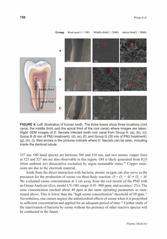
SEM examination was carried out to evaluate the effect of PMJ on E. faecalis–infected single-rooted teeth. The teeth from groups A, B, and G were prepared for observation im-mediately after plasma treatment. An illustrative picture of a tooth is shown on the left of Fig. 4, where three boxes are drawn to show three locations (root canal, the middle third, and the apical third of the root canal) where the SEM pictures were taken with magnifica-tions of 100, 5000, and 5000, respectively.
In the control group, bacteria cover the entire root canal surface from the top third to apical third [Figs. 4 (a), (b), and (c)]. After 8-min plasma treatment, the root canal is partially cleaned with a clear contrast line separating the zones with and without bacteria [arrow in Fig. 4 (d)], indicating the active species generated by plasma have a limited penetration depth in 8 min. This phenomenon is consistent with the findings in the work reported by Jiang and co-workers.14 The middle third of the tooth root canal had few bacteria observed around dentinal tubules’ opening [Fig. 4 (e)]. A considerable amount of bacteria still survived in the apical third of the root canal [Fig. 4 (f)]. This could explain the high reinfection rate in 7 d with a short PMJ treatment time in Group G. When the treatment time was extended to 30 min, the entire root canal was thoroughly cleaned, with no bacteria observed in SEM pictures [Figs. 4 (g), (h), and (i)], suggesting that appropriate PMJ treatment could prevent reinfection effectively.
Optical Emission Spectroscopy (OES)D.
Due to the strong electric field between the closely spaced electrodes, the electron energy distribution in the plasma is highly non-Maxwellian, with a relatively large concentration of high-energy electrons.26,27 These high-energy electrons are required for the single- or multiple-step ionization and excitation of species in the Ar/O2 or around aqueous media. OES was employed to investigate the active species generated in the Ar/O2 PMJ. Figure 5 shows an end-on optical emission spectrum of the PMJ from 200 to 860 nm when it was operated in air at a discharge current of 30 mA and a gas flow rate of 5 slm.
The spectrum is dominated by atomic argon emissions, mostly attributed to direct electron impact excitation. Strong emissions from excited atomic oxygen at 777 nm and 844 nm are also observed in the spectrum. The oxygen emission at 777 nm is due to dis-sociative excitation by electronic collisions from the ground state oxygen molecules.28 The oxygen emission at 844 nm, however, is due to mostly direct electron impact with ground state oxygen atoms. The excited state of oxygen can also be populated by dis-sociative excitation of O2 by the excited argon metastables: Ar* + O2 O + O* + Ar.29 Figure 5 inset shows in the UV-A and UV-B regions between 300 and 400 nm, emissions from the 2nd positive system of N2 (C
3Πu B3Πg) with the most intense band head at
Plasma Medicine
150 Wang et al.
337 nm. OH band spectra are between 306 and 310 nm, and two atomic copper lines at 325 and 327 nm are also observable in this region. OH is likely generated from H2O (from ambient air) dissociative excitation by argon metastable states.30 Copper emis-sions are due to the electrode material.
Aside from the direct interaction with bacteria, atomic oxygen can also serve as the precursor for the production of ozone via three-body reaction: O + O2 + M O3 + M. We evaluated ozone concentration at 1 cm away from the exit nozzle of the PMJ with an Ozone Analyzer (Eco, model UV-100; range: 0.01–900 ppm; and accuracy: 2%). The zone concentration reached about 40 ppm at the same operating parameters as men-tioned above. This is lower than the “high ozone concentration” threshold of 50 ppm.31 Nevertheless, one cannot neglect the antimicrobial effects of ozone when it is prescribed in sufficient concentration and applied for an adequate period of time.32 Further study of the inactivation of bacteria by ozone without the presence of other reactive species will be conducted in the future.
FIGURE 4. Left: Illustration of human tooth. The three boxes show three locations (root canal, the middle third, and the apical third of the root canal) where images are taken. Right: SEM images of E. faecalis infected tooth root canal from Group A: (a), (b), (c); Group B (8 min of PMJ treatment): (d), (e), (f); and Group G (30 min of PMJ treatment): (g), (h), (i). Red arrows in the pictures indicate where E. faecalis can be seen, including inside the dentinal tubule.

Volume 1, Number 2, 2011
The Effect of an Atmospheric Pressure, DC Nonthermal Plasma Microjet on Tooth Root Canal, Dentinal Tubules Infection and Reinfection Prevention
151
ESR SpectroscopyE.
Hydroxyl radical (•OH), superoxide anion radical (•O2–) and singlet oxygen (1O2) are
believed to play an important role during the oxidation of the fatty acid in the cell mem-brane, thus leading to the inactivation of microorganism.17,18,23,33 ESR spectroscopy was used to detect these reactive oxygen species (ROS) in the liquid solution. When DMPO was added into 1 mL distilled water and treated with PMJ for 20 sec, quartet DMPO-OH adduct spectrum was observed [as shown in Fig. 6 (a)]. The hyperfine splitting constants aN = aH = 1.5 mT are consistent with the reported value of DMPO-OH.34 DMPO was also used to capture superoxide anion radical (•O2
–). Superoxide dismutase (SOD, an enzyme that specifically depletes •O2
–) was added into the system before the PMJ treatment. A decrease of DMPO-OH signal was found (data not shown), indicating that •OH was probably derived from the reaction between •O2
– and the liquid. TEMP was used to spin-trap singlet molecular oxygen (1O2). A typical triplet TEMPO signal in ESR spectrum is shown in Fig. 6 (b) (aN = 1.72 mT), indicating the existence of 1O2 in the plasma-liquid system. Detailed discussion on the effect of •OH, ∙O2
–, and 1O2 can be found in our previ-ous work.18,33
DISCUSSION AND CONCLUSIONSIV.
The tooth root canal system has complicated structures, such as isthmuses, ramifications, deltas, irregularities, and in particular dentinal tubules. It has been reported that bacteria can enter the dentinal tubules as deep as 500–1000 micron.35 This is considered to be one of the major causes for failure in endodontic treatments.36 The commonly used intracanal
FIGURE 5. End on optical emission spectrum of Ar/O2 PMJ operated in air (200–850 nm). The inset shows emission spectrum in 200–400 nm range, where OH and N2 emissions are observed.

Plasma Medicine
152 Wang et al.
medicament, such as calcium hydroxide, could not eliminate bacteria completely in the root canal system.37 Nearly half of the cases treated by traditional intracanal antisepsis strategies suffer from E. faecalis reinfections.4 Plasma-generated reactive species in the gas stream, however, can effectively reach the places that are hard or even impossible to reach by traditional medicaments. These reactive species (including excited atoms and ions) produced in the plasma can directly interact with the bacteria via, for example, direct oxidation of fatty acid.38 Perni and co-workers found that atomic oxygen plays the most essential role in the plasma inactivation process by employing three single-deletion mutants of Escherichia coli.39 Feng and co-workers23 demonstrated that an oxi-dative stress pathway is directly involved in cell resistance to plasma processing. Charge accumulation on cell membrane from excessive ions produced in the downstream of the plasma can cause the rupture of membrane via coulombic force.40 However, D. Dobrynin and co-workers, in their work with a corona discharge, reported that charged particles alone do not play an important role.41 Some of reactive species (longer living) can dis-solve in water on the surface of the tooth (however thin a layer it may be) and interact with bacteria. They can also, upon interaction with water, produce powerful ROS such as ∙OH. The reactions of these species with water are rather complicated and beyond the scope of this study. However, whether dissolved or created in water, these reactive spe-cies may be transported to cytoplasm through a series of mechanisms.42,43 The reaction of these species with nearby organics leads to chain oxidation and thus destruction of DNA molecules as well as cellular membranes and other cell components.44
In conclusion, we investigated the disinfection of root canal by a direct current, atmospheric-pressure, cold Ar/O2 (2%) plasma microjet. The temperature of the plasma jet at the treatment distance was below the critical temperature in dental treatment. A 98.8% inactivation rate of E. faecalis was observed after an 8-min plasma treatment. However, a plasma treatment of at least 30 min was needed to prevent reinfection of the root canal. Compared to traditional treatment with intracanal medicaments, which usually takes 1–2 weeks, nonthermal plasmas provide a rapid, effective alternative for tooth root canal disinfection.
FIGURE 6. DMPO-OH and TEMPO signals detected by ESR: (a) DMPO-OH signal; (b) TEMPO signal.

Volume 1, Number 2, 2011
The Effect of an Atmospheric Pressure, DC Nonthermal Plasma Microjet on Tooth Root Canal, Dentinal Tubules Infection and Reinfection Prevention
153
ACKNOWLEDGMENTS
The authors would like to thank Ms. Hua Zhong for her kind help in revising the pa-per, Mr. Yongsheng Chen from Institute of Biophysics, Chinese Academy of Sciences, for his help with conducting the ESR experiments, and Mr. Jiayi Zeng, for his help with the schematic diagram. The research was supported by Bioelectrics Inc. (USA), MST Program of International Science and Technology Cooperation (under Grant # 2009DFB30370: “Cold Plasma Induced Biological Effect and Its Clinical Application Studies”), and National Basic Research Program (No. 2007CB935602). WZ would like to acknowledge the Air Force Office of Scientific Research (AFOSR) and American Chemical Society Petroleum Research Fund (ACS-PRF) for their partial support of the study of the PMJ.
REFERENCES
Jiang C, Chen M-T, Gorur A, Schaudinn C, Jaramillo DE, Costerton JW, Sedghizadeh PP, 1. Vernier PT, Gundersen MA. Nanosecond pulsed plasma dental probe. Plasma Processes Polym. 2009;6:479–483.Colak M, Evcil S, Bayindir YZ, Yigit NJ, Contemp J. The effectiveness of three instrumen-2. tation techniques on the elimination of Enterococcus faecalis from a root canal: an in vitro study. Dent Pract. 2005;136(1):80.Soares JA, Leonardo MR, Silva LA, Filho MT, Ito IY. Histomicrobiologic aspects of the root 3. canal system and periapical lesions in dogs’ teeth after rotary instrumentation and intracanal dressing with Ca(OH)2 pastes. J Appl Oral Sci. 2006;14(5):355–364.Siqueira JF Jr, Rôįas IN, Santos SRLD, Lima KC, Magalhaes FAC. de Uzeda M. Efficacy 4. of instrumentation techniques and irrigatation regimens in reducing the bacterial population within root canals. J Endod. 2002;28:181–184. Sundqvist G, Figdor D, Persson S, Sjögren U. Microbiologic analysis of teeth with failed en-5. dodontic treatment and the outcome of conservative re-treatment. Oral Surg Oral Med Oral Pathol Oral Radiol Endod. 1998;85:86–93.Dahlèn G, Samuelsson W, Molander A, Reit C. Identification and antimicrobial susceptibil-6. ity of enterococci isolated from the root canal. Oral Microbiology and Immun. 2000;15: 309–312.Peters LB, van Winkelhoff A-J, Buijs JF, Wesselink PR. Effects of instrumentation, irriga-7. tion and dressing with calcium hydroxide on infection in pulpless teeth with periapical bone lesions. Int J Endod. 2002;35:13–21.Sathorn C, Parashos P, Messer H. Antibacterial efficacy of calcium hydroxide intracanal 8. dressing: a systematic review and meta-analysis. Int Endod J. 2007;40: 2–10.Pan J, Sun P, Tian Y, Zhou H, Bai N, Wu H, Zhu W, Zhang J, Becker K, Fang J. A novel 9. method of tooth whitening using cold plasma microjet driven by direct current in atmospher-ic-pressure air. IEEE Trans Plasma Sci. 2010;38:3143.Lee HW, Nam SH, Mohamed AH, Kim GC, Lee JK. Atmospheric pressure plasma jet 10. composed of three electrodes: application to tooth bleaching. Plasma Process Polym. 2010;7:274.

Plasma Medicine
154 Wang et al.
Sun P, Pan J, Tian Y, Bai N, Wu H, Wang L, Yu C, Wei S, Zhang J, Zhu W, Fang J. Tooth-11. whitening with hydrogen peroxide assisted by a direct current, cold, atmospheric-pressure air plasma microjet. IEEE Trans Plasma Sci. 2010;38:1892–1896.Lu X, Cao Y, Yang P, Xiong Q, Xiong Z, Xian Y, Pan Y. An RC plasma device for steriliza-12. tion of root canal of teeth. IEEE Trans Plasma Sci. 2009;37:668–673.Sladek R, Stoffels E, Walraven R, Tielbeek P, Koolhoven R. Plasma treatment of dental cavi-13. ties: a feasibility study. IEEE Trans Plasma Sci. 2004;32:1540–1542.Jiang C, Chen M, Schaudinn C, Gorur A, Vernier PT, Costerton JW, Jaramillo DE, Sedghiza-14. deh PP, Gundersen MA. Pulsed atmospheric pressure cold plasma for endodontic disinfec-tion. IEEE Trans Plasma Sci. 2009;37:1190–1195.Jiang C, Chen M-T, Gorur A, Schaudinn C, Jaramillo DE, Costerton JW, Sedghizadeh 15. PP, Vernier PT, Gundersen MA. Nanosecond pulsed plasma dental probe. Plasma Process Polym. 2009;6:479–483.Bussiahn R, Brandenburg R, Gerling T, Kindel E, Lange H, Lembke N, Weltmann K-D, 16. von Woedtke Th, Kocher T. The hairline plasma: an intermittent negative dc-corona discharge at atmospheric pressure for plasma medical application. Appl Phys Lett. 2010;96:143701-3.Feng H, Sun P, Chai Y, Tong G, Zhang J, Zhu W, Fang J. The interaction of a direct-current cold 17. atmospheric-pressure air plasma with bacteria. IEEE Trans Plasma Sci. 2009;37:121–127.Liu F, Sun P, Bai N, Tian Y, Zhou H, Wei S, Zhang J, Zhu W, Becker K, Fang J. Atmospheric 18. pressure cold plasma as an antifungal therapy. Plasma Process Polym. 2010;7:231–233.Kolb JF, Mohamed A-AH, Price RO, Swanson RJ, Bowman A, Chiavarini RL, Stacey M, 19. Schoenbach KH. Cold atmospheric pressure air plasma jet for medical applications. Appl Phys Lett. 2008;92:241501. Halliwell B, Whiteman M. Measuring reactive species and oxidative damage in vivo 20. and in cell culture: how should you do it and what do the results mean? Brit J Pharm. 2004;142:231–255.Koppenol WH. The Haber-Weiss cycle: 70 years later. Redox Rep. 2001;6:229–234.21. Sun P, Sun Y, Wu H, Zhu W, Lopez J, Liu W, Zhang J, Li R, Fang J. Atmospheric pressure 22. cold plasma as an antifungal therapy. Appl Phys Lett. 2011;98:021501-3.Feng H, Wang R, Sun P, Wu H, Liu Q, J. Fang, W. Zhu, F. Li, J. Zhang,. Astudy of eukaryotic 23. response mechanisms to atmospheric pressure cold plasma by using Saccharomyces cerevi-siae single gene mutats. Appl. Phys. Lett. 2010, 97:131501–131503.C. H. Stuart, S.A. Schwartz, T. J. Beeson, C. B. Owatz. Enterococcus faecalis: its role in 24. root canal treatment failure and current concepts in retreatment.J. Endod. 2006 Feb, 32(2), 93–98.Plant CG, Jones DW, Darell BW. The heat evolved and temperatures attained during setting 25. of restorative materials. Brit Dent. J. 1974;137:233–238.Gill P, Webb CE. Electron energy distributions in the negative glow and their relevance to 26. hollow cathode lasers. J Phys D: Appl Phys. 1977;10:299–311.Boeuf JP, Marode E. Monte Carlo simulation of electron swarm motion in SF6. J Phys D: 27. Appl Phys. 1984;17:1133–1148.J.Jask, P.macko, V.Martisovits, P,.Luckac, P.Veis.Time Resolved Actinometirc Stud of Plused 28. RF Oxygen Discharge. Czechoslovak Journal of Physics, 2004, 54(6):661-676

Volume 1, Number 2, 2011
The Effect of an Atmospheric Pressure, DC Nonthermal Plasma Microjet on Tooth Root Canal, Dentinal Tubules Infection and Reinfection Prevention
155
Saloum S, Naddaf M, Alkhaled B. Properties of thin films deposited from HMDSO/O2 in-29. duced remote plasma: effect of oxygen fraction. Vacuum. 2008;82:742–747.Dodet B, Odic E, Goldman A, Goldman M, Renard D. Hydrogen peroxide. Formation by 30. discharges in argon/water vapor mixtures at atmospheric pressure. J Adv Oxid Technol. 2005;8:91–97.Bocci V. Oxygen-ozone therapy, a critical evaluation. 1st ed. Dordrecht, The Netherlands 31. Kluwer Academic Publishers, 2002, p. 144.Kowalski Wj, Bahnfleth WP, Striebig BA, Whittam TS. Demonstration of a hermetic air-32. borne ozone disinfection system: studies on E. coli. AIHA J. 2003;64:222–227.Bai N, Sun P, Zhou H, Wu H, Tian Y, Lopez J, Zhu W, Zhang J, Fang J. Inactivation of Staph-33. ylococcus aureus in water by a cold, He/O2 atmospheric pressure plasma microjet. Plasma Process Polym. 2011;8:424–431.Buettner GR. Spin trapping: ESR parameters of spin adducts. Free Radic Biol Med. 34. 1987;3:259–303.Haapasalo M, Orstavik D. In vitro infection and disinfection of dentinal tubules. J Dent Res. 35. 1987;66:1375–1379 . Lin LM, Skribner JE, Gaengler P. Factors associated with endodontic treatment failures. 36. J Endod. 1992;18:625–627.Waltimo T, Trope M, Haapasalo M, Ørstavik D. Clinical efficacy of treatment procedures 37. in endodontic infection control and one year follow-up of periapical healing. J Endod. 2005;31:863-866.Laroussi M. Low-temperature plasma for medicine. IEEE Trans Plasma Sci. 2009;37: 714–725.38. Perni S, Shama G, Hobman JL, Lund PA, Kershaw CJ, Hidalgo-Arroyo GA, Penn CW, 39. Deng XT, Walsh JL, Kong MG. Probing bactericidal mechanisms induced by cold atmso-pheric plasma with Escherichia coli mutants. Appl Phys Lett. 2007;90:073902-3.Laroussi M. Non-thermal decontamination of biological media by atmospheric pressure 40. plasma: review, analysis and prospects. IEEE Trans Plasma Sci. 2002;30:1409–1415.Dobrynin D, Starikovskiy A, Friedman G, Fridman A. Bacteria inactivation effect of ions 41. generated by dc corona discharge. 63rd Annual Gaseous Electronics Conference and 7th In-ternational Conference on Reactive Plasmas, Paris, France, 2010.Laroussi M, Richardson JP, Dobbs FC. Effects of non-equilibrium atmospheric pressure 42. plasmas on the heterotrophic pathways of bacteria and on their cell morphology. Appl Phys Lett. 2002;81:772–774.Deng X, Shi J, Kong MG. Physical mechanisms of inactivation of Bacillus subtilis spores 43. using cold atmospheric plasma. IEEE Trans Plasma Sci. 2006;34:1310–1315.Dobrynin D, Fridman G, Friedman G, Fridman A. Physical and biological mechanisms of 44. direct plasma interaction with living tissue. New J Phys. 2009;11:115020.
