the effect of green tea on prevention of mouth bacterial ... · certain mouth bacteria in dental...
TRANSCRIPT

Iranian Journal of Toxicology Volume 5, No 14, Autumn 2011
1-Department of Pharmaceutics, School of Pharmacy, Jundishapur University of Medical Sciences, Ahvaz, Iran. 2-Department of Microbiology, School of Medicine, Jundishapur University of Medical Sciences, Ahvaz, Iran. 3-Department of Pharmacognozy, Jundishapur University of Medical Sciences, Ahvaz, Iran. 4-Department of Medicinal Chemistry, School of Pharmacy, Jundishapur University of Medical Sciences, Ahvaz, Iran. *Corresponding Author Email: [email protected]
502
The Effect of Green Tea on Prevention of Mouth Bacterial Infection, Halitosis, and Plaque Formation on Teeth
Abdolhosein Moghbel 1*, Ahmad Farjzadeh 2, Nasrin Aghel 3, Homayoon Agheli 4, Nafiseh Raisi 1
Received:29.4.2011 Accepted:21.5.2011
ABSTRACT Background: Compounds present in both green and black teas have been shown to inhibit the growth and activity of bacteria associated with mouth infections. The aim of this study was to assess the effects of green tea leaves extract on the aerobic mouth bacterial load. Methods: A total of 25 volunteer female students, aged 20-25 years, were selected and then evaluated by green tea extract and mouthwashes containing 0.2, 0.5, and 1% tannin, as the most effective antibacterial complex in green tea. Then a comparative study was conducted on a green tea mouthwash containing 1% tannin with 10% ethanol, a alcohol free mouthwash, and a green tea herbal mouthwash with a chlorhexidine 0.2% sample, as and chemical brand. Results: Green tea mouthwash containing 1% tannin was more effective than other concentrations (P<0.05). There were no meaningful differences between the green tea mouthwashes containing 10% alcohol and alcohol free, as well as the herbal and chemical chlorhexidine 0.2% (P>0.05). Conclusion: The herbal green tea mouthwash could reduce the aerobic mouth bacterial load and may prevent plaque formation on teeth and come over halitosis due to infection of the bacteria. Also, it is a safe and nontoxic mouthwash especially for children and pregnant women. Keywords: Bacterial Infection, Green Tea, Halitosis, Mouthwash, Plaque Formation, Teeth.
INTRODUCTION Plant extracts have been widely used in
topical and oral applications for disease treatment. Examples of these include gingko biloba, echinacea, ginseng, grape seed, green tea, lemon, lavender, rosmary, thuja, sara, allantoin, fever wort, blood root, apache plume, papaya, and tragacanth (1). Black tea is the second most commonly drunk liquid on earth after water. Green tea Camellia sinensis (Figure 1), which is not fermented at all during
the drying process, has numerous medicinal benefits mainly due to its antibacterial and antioxidant properties (2). Tea was essentially native to China but then it was transferred to India and Japan. Later on, it gained popularity in Europe and Russia, and, finally, in Iran.
A short list of phenolic phytochemicals with promising properties to benefit human health includes a group of polyphenol compounds called catechins (3-5) which are found in green tea (Figure 2).
Dow
nloa
ded
from
ijt.a
rakm
u.ac
.ir a
t 7:4
6 +
0430
on
Sun
day
July
1st
201
8

Iranian Journal of Toxicology Abdolhosein Moghbel et al
Volume 5, No 14, Autumn 2011 http://www.ijt.ir 503
Figure1. Fresh leaves of green tea
Figure 2. Chemical compound of green tea
Compounds present in both green and black teas have been shown to inhibit the growth and activity of bacteria with tooth decay producing halitosis (6,7). Animals and humans that have tea compounds in their drinking water develop fewer dental caries and less plaque formation than those drinking plain water (8-11). Tooth decay is the gradual breakdown of the tooth beginning with the enamel surface and eventually progressing to the inner pulp. Tooth decay and, eventually, halitosis are caused by acids produced by certain mouth bacteria in dental plaque. Factors
that affect this process include oral hygiene, diet meal frequency, salvia production, and heredity. Teeth with significant decay are said to have caries or cavities (6,12-13).
The malefic bacteria in our mouths can cause tooth decay and bad breath (halitosis). Plaque is another contributor to bad breath. Pace University recently did a study on how effective green tea is in getting rid of bad breath. In this research project, they mixed green tea with the bacteria that cause tooth decay and strep throat. Results showed that green tea inhibited the bacterial growth by 30%
Dow
nloa
ded
from
ijt.a
rakm
u.ac
.ir a
t 7:4
6 +
0430
on
Sun
day
July
1st
201
8

TThhee EEffffeecctt ooff GGrreeeenn TTeeaa oonn PPrreevveennttiioonn ooff…….. Iranian Journal of Toxicology
http://www.ijt.ir Volume 5, No 14, Autumn 2011 504
and decreased the amount of bad breath producing compounds. Drinking green tea may also help prevent getting sore throats and colds since it helps fight the bacteria harboring in the throat (14).
Bad breath is caused by anaerobic sulfur producing bacteria that normally live on the surface of the tongue and in the throat. These bacteria are supposed to be there because they assist humans in digestion by breaking down the proteins found in specific foods, mucous, phlegm, blood, and diseased or “broken-down” oral tissues. Under certain conditions, these bacteria start to break proteins down at a very high rate. Proteins are made up of amino acids; two amino acids, cysteine and methionine, are dense with sulfur. When these “beneficial” bacteria come into contact with these compounds, the odorous and “lousy-tasting” sulfur compounds are released from the back of the tongue and throat as hydrogen sulfide, methyl mercaptan, and other odorous and bad tasting compounds. These “problem” compounds are often referred to as volatile sulfur compounds (14). Some reasons motivated us to examine the Iranian green tea antibacterial effect in order to use it later on as a mouthwash for blocking halitosis. As a matter of fact, we have been working on green tea for its healing effect on burns and wounds (15-16). In addition, it has been a traditional belief in Iran that when you have a bad breath due to alcoholic beverages, garlicky foods, or any other compounds which cause bad breath after digestion, you may chew some dry tea powder to block it. Based on one article, the phenolic compound in green tea may block the growth of bacteria responsible for teeth cavity and plaque formation (14). Therefore, the aim of this study was to evaluate the potency of different extract concentrations of Iranian green tea as a mouthwash on mouth microbial load and then approving its effect on preventing halitosis or plaque formation in a separate study.
MATERIALS AND METHODS Leaves drying method and preparation of dried extract powder
After the fresh leaves were picked from fields in Lahijan, a city in the north of Iran, they were spread out in hot air to wither. Once
they became soft and pliable, they were traditionally pan-fried in woks, and after scientific evaluation by the Pharmacognosy Department, a voucher specimen was deposited at the herbarium of the faculty. In the final step, the leaves were dried by firing whereby, the natural fragrances and flavors were stabilized so that the leaves preserve their green color. The dried leaves were powdered mechanically and 200g of the 18 mesh powder was extracted by 1000 ml of deionized water at 70-80°C for 30 minutes. The extract was filtered by a cloth filter and concentrated under low pressure conditions with the device installed in a laminar airflow (FARPAJOO-Fee 2120-2). The extract was finally lyophilized (-40°C, 0.03 Tor) by a freeze-dryer (ZIRBUS Za co-5, Germany) for about 6 hours (6,17).
Tannin assay, validation, and preparation of calibration curve
Stock solutions of standard tannic acid were prepared inside eleven 100 ml volumetric vessels each regent containing zero to 10 ml of standard tannic acid, respectively. To each vessel, 1.5 ml of Folin-Denise reagent, 10ml of sodium carbonate solution, and sufficient deionized water were added to make the 100ml solution. Then, each sample was mixed vigorously until a light to dark blue color gradually appeared. The first vessel (free tannic acid) with no color was used as a blank sample. In order to complete the color reaction, all samples were held at room temperature for 3 hours. This process was repeated for three to six consecutive days to find the inter- and intra-day variations. Absorbance levels were measured spectrophotometrically (Shimadzu uv/visible spectrophotometer 2100; Toko, Japan) at 760 nm. The mean data (n=10) were used for the preparation of the calibration curve (6,18). The tannin concentration for each sample (mg/100 ml) was calculated from the regression equation obtained from the calibration curve.
Preparation of different equivalent tannin concentrations from the green tea powder extract
Samples containing various concentrations of tannin (0.2, 0.5, and 1%) were prepared by equivalent amounts of 3.2,
Dow
nloa
ded
from
ijt.a
rakm
u.ac
.ir a
t 7:4
6 +
0430
on
Sun
day
July
1st
201
8

Iranian Journal of Toxicology Abdolhosein Moghbel et al
Volume 5, No 14, Autumn 2011 http://www.ijt.ir 505
8.02, and 16.05 mg of the dried green tea powder yielded from the 100mg dried extract, respectively (18). Each sample was deposited inside a 100 ml stoppered bottle which had already been sterilized by an autoclave at 121°c, 15 lb pressure for 20 minutes. The volume of each sample was adjusted at 100 ml with deionized water which was finally filtered through a 0.45 membrane filter to be sterilized.
Antimicrobial evaluation of green tea extracts containing 0.2, 0.5, and 1 % tannin
The participants, 25 female students aged between 20-25 years, were asked to wash their mouths with 10-15 ml deionized water for one minute at 12 p.m., and after brushing their teeth before 10 a.m. Mouth containing of each student was poured into sterile containers. Aliquots of 0.01 ml of each sample was diluted in 1ml deionized water under sterilization condition. Then an aliquot of each new sample was plated on petri dishes containing bloody agar. The plates were incubated inside a candle jar with 5-7 CO2 concentration at 35-37°c for 24 hours and, then, colonies were counted separately (19). This process was repeated for the extracts containing 0.2, 0.5, and 1% tannin with the time interval of at least one day for each concentration before the trial with deionized water and then, the green tea extract.
Preparation of mouthwash formulation No.1 (green tea mouthwash containing 1% tannin)
A quantity of 16.5 mg green tea dried extract equivalent to 1% tannin was dissolved in about 70 ml of 50°C deionized water with 20 mg sodium saccharin as a non-sugar sweetener. The final volume was adjusted to 100 ml by deionzed water after thorough mixing and paper filtering. The microbial activity of this formulation was evaluated as the method mentioned above for the extract.
Preparation of mouthwash formulation No.2 (green tea mouthwash containing 1% tannin with 10% alcohol)
All evaluations on this formulation were performed the same as those on No.1 formulation. In this formulation, the role of 10% alcohol, which had been incorporated along with green tea, was evaluated on the microorganism.
Comparison of the herbal green tea mouthwash with chemical chlorhexidine 0.2%
In this evaluation, the formulation No. 2 (with 10% alcohol) was compared with a commercial brand known as chlorhoxidine 0.2% on the mouth microbial content of the same students on separate tests.
Coagulase test This test was run to identify
Staphylococcus aureus species in a restrictive laboratory medium. Staphylococcus aureus cultures contain a coagulase enzyme which can coagulate blood plasma by action on fibrinogen. This enzyme does not exist in other staphylococcus which are known as negative staphylococcus. In this test, for preparation of the sample, one drop of normal saline was deposited on one side of a clean glass plate and with the culture beam a cycle of full microbial culture was dispersed in the normal saline drop. On the other side of the glass plate, one cycle full of microbial culture was deposited to make an emulsion after mixing. The mixture was agitated for about 3 minutes and when the plasma began to dry, one drop of saline was added. In this evaluation, if coagulase culture were positive, a granule mode would appear. This mode would not be seen in the mixture of culture in saline (control); therefore, in the case of negative result, no granule residue would appear (19).
Oxidase test This test was done for identifying
bacteria suspected to Neisseria. The tetramethylparaphenyl aminodehydrochloride 0.1% solution as the regent was prepared with deionized water. A filter paper was installed in a clean plate and then few drops of the regent were added to the surface of the filter. Finally, using a platinan loop, few colonies of the bacterium were spread on the wet side of the paper filter so that in case of positive result for live bacteria, an intensive purple color would appear after 10 seconds (19).
Statistical analysis and validation Statistical comparisons were carried out
by t-test and one-way analysis of variance (ANOVA) for paired and also independent samples using SPSS software (P<0.05). The
Dow
nloa
ded
from
ijt.a
rakm
u.ac
.ir a
t 7:4
6 +
0430
on
Sun
day
July
1st
201
8

TThhee EEffffeecctt ooff GGrreeeenn TTeeaa oonn PPrreevveennttiioonn ooff…….. Iranian Journal of Toxicology
http://www.ijt.ir Volume 5, No 14, Autumn 2011 506
results were expressed as mean SD. The inter and intra day variations data for validation of the calibration curve were applied, and the concentration of the equivalent dissolved drug was calculated from the regression equation obtained from the standard deviation.
RESULTS The inter and intra-day variations of
standard deviation and accuracy of the tannic acid assay (the most important active material) are shown in Tables 1 and 2.
Table1. The inter-day variation data of the standard curve and accuracy results for tannic acid assay
by Folin- Deinis method
Concentration mg/100 ml
Absorbance Mean (n=3) SD CV% Accuracy%
Mean SD
0.1 005.0066.0 8.66 81.857.91 0.2 005.0163.0 3.53 09.433.114 0.3 005.0193.0 2.98 72.240.90 0.4 00.029.0 0 00.007.102 0.5 011.0343.0 3.36 27.379.96 0.6 0152.0423.0 3.60 61.356.99 0.7 011.0493.0 2.34 33.252.99 0.8 005.0573.0 1.00 02.126.101 0.9 00.063.0 0 00.094.98 1 005.0706.0 0.81 81.092.99
Table 2. The intera-day variation data of the standard curve and accuracy results in 6 different days
and triplicate for tannic acid absorbance and assay by Folin –Denis method.
Concentration mg/100 ml
Absorbance Mean (n=3) SD CV% Accuracy%
Mean SD
0.1 002.0066.0 3.2 98.257.91 0.2 004.0175.0 2.7 55.339.110 0.3 002.0195.0 1.7 54.171.91 0.4 001.0291.0 0.5 04.225.103 0.5 006.0353.0 1.8 7.240.100 0.6 002.0421.0 0.6 77.091.98 0.7 004.0500.0 0.9 01.155.101 0.8 001.0574.0 0.2 30.046.101 0.9 005.0626.0 0.5 55.068.98 1 001.0706.0 0.9 19.084.99
The result of mean SD for the assay of
tannin in green tea extract by Folin-Denis
method repeated in three consecutive days in
triplicate was 6.23 0.19 mg of tannin in 100
mg of the dried powder extract of green tea
( Figure 3, Table 3).
Dow
nloa
ded
from
ijt.a
rakm
u.ac
.ir a
t 7:4
6 +
0430
on
Sun
day
July
1st
201
8

Iranian Journal of Toxicology Abdolhosein Moghbel et al
Volume 5, No 14, Autumn 2011 http://www.ijt.ir 507
Figure 3. Standard cure of tannic acid
Table 3. Tannin assay result of green tea leaves (n=3) Equivalent amount of tannin concentration
Tannin % in total extract
Tannin concentration Mg/100ml
Absorbance Weight of green tea extract, total (mg)
Sample No.
6. 03 0.86 0.61 14.28 1 6.23 0.89 0.63 14.28 2 6.37 0.91 0.65 14.28 3
19.023.6 025.088.0 02.063.0 14.28 Mean SD The effectiveness of water as a negative
control or drug free mouthwash in different stages of mouth washing (1and 2 hours after beginning the trial ) are shown in Figure 4
Figure 4. The effect of water on mean of mouth bacterial load at different times
The comparative results between water as a negative or drug free control and different concentrations of tannin (0.2 ,0.5, and 1% ) as
the most effective antibacterial agent in green tea leaves are demonstrated in Figure 5 (0.2 and 0.5% not shown).
Dow
nloa
ded
from
ijt.a
rakm
u.ac
.ir a
t 7:4
6 +
0430
on
Sun
day
July
1st
201
8
TThhee EEffffeecctt ooff GGrreeeenn TTeeaa oonn PPrreevveennttiioonn ooff…….. Iranian Journal of Toxicology
http://www.ijt.ir Volume 5, No 14, Autumn 2011 508
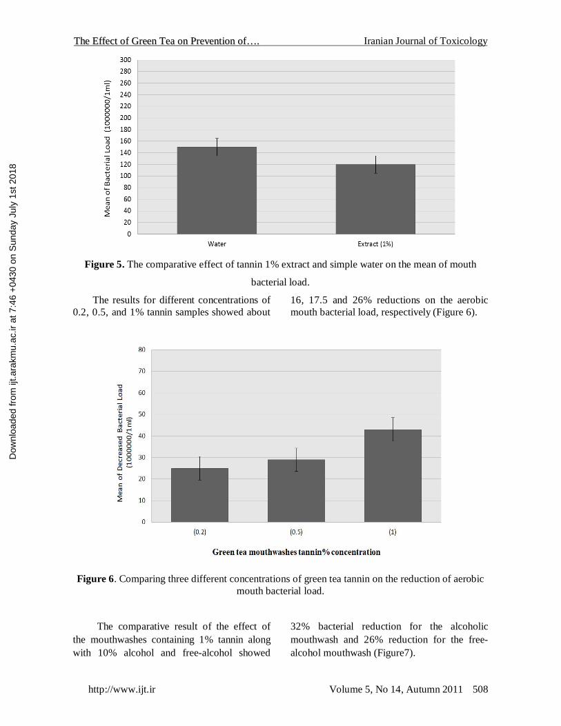
Figure 5. The comparative effect of tannin 1% extract and simple water on the mean of mouth
bacterial load.
The results for different concentrations of 0.2, 0.5, and 1% tannin samples showed about
16, 17.5 and 26% reductions on the aerobic mouth bacterial load, respectively (Figure 6).
Figure 6. Comparing three different concentrations of green tea tannin on the reduction of aerobic
mouth bacterial load.
The comparative result of the effect of the mouthwashes containing 1% tannin along with 10% alcohol and free-alcohol showed
32% bacterial reduction for the alcoholic mouthwash and 26% reduction for the free-alcohol mouthwash (Figure7).
Dow
nloa
ded
from
ijt.a
rakm
u.ac
.ir a
t 7:4
6 +
0430
on
Sun
day
July
1st
201
8

Iranian Journal of Toxicology Abdolhosein Moghbel et al
Volume 5, No 14, Autumn 2011 http://www.ijt.ir 509
Figure 7. Green tea effect of mouthwashes containing tannin 1% concentration with and without alcohol on the reduction of aerobic mouth bacterial load.
Green tea tannin 1% mouthwashes
Evaluation of the effectiveness of the 1% tannin green tea in comparison with the chemical chlorhoxidine 0.2% mouthwash
showed that both mouthwashes induced the same reduction (about 32%) in the bacteria (Figure 8).
Figure 8. Comparing mouthwashes effect of green tea containing alcoholic tannin 1% with
chlorhexidine 0.2% on the reduction of aerobic mouth bacterial load.
Dow
nloa
ded
from
ijt.a
rakm
u.ac
.ir a
t 7:4
6 +
0430
on
Sun
day
July
1st
201
8
TThhee EEffffeecctt ooff GGrreeeenn TTeeaa oonn PPrreevveennttiioonn ooff…….. Iranian Journal of Toxicology
http://www.ijt.ir Volume 5, No 14, Autumn 2011 510
The results of stability tests of the product as the pH changing, also the amount of tannin remained unchanged during 0 to 90 days holding at different temperatures for evaluation of the stability of green tea extract mouthwash showed a constant value (P<0.05) during study (Table 4-6 and Figures 9- 10) The coagulase and oxidase tests showed that no granule was formed; therefore, the coagulase test was negative; however, the oxidase test was positive because of the intensive purple color which was evidence of Neisseria in the presence of idophenoloxidos enzyme (19). DISCUSSION
Application of an appropriate mouthwash has the following benefits: having antiseptic effects on mouth, washing the food residue on the gingiva (gum) medium and teeth, reducing the mouth bacteria, masking and neutralizing halitosis, and introducing a good taste and sense of freshness, having anti-plaque, anti-stain, and anti-tartar properties, acting as a fluoride depot, preventing tooth cavities that help wound healing and gum injuries, decreasing the incidence of tooth rash and herpes, preventing bad breath, producing a fresh mint taste, helping the health of milk teeth, and protecting the permanent teeth of children and adults (12,13). In a broad field of medicine, several researchers have published various articles on the effects of green tea, such as reducing and adjusting the weight (20), curing dermal inflammation and disorders (21,22), healing wounds (15,16), preventing immature and ill time of human skin (23), preventing and curing some cancers, reducing blood pressure, cholesterol, and sugar, preventing heart diseases, and adjusting hormones which promote health and long life (24-29). One effect that no article has pointed out so far is the probability of showing dermal anti-sensitivity effects on bringing itching and urticaria to a stop. Also, a recent article introduced antibacterial and anti-caries effects for green tea (30). The main purpose of this study was to test this fact, in lab condition and also, clinically. Green tea contains flovonoids, tannin, vitamins, fluoride, and other mineral salts. Some antioxidant and antimicrobial agents of green tea could increase the life and efficiency of teeth (31-33). Tannins are biosynthetic materials which have a potent
antibacterial effect. In recent decay researches, it has been shown that green and black teas prevent gathering of bacteria and, therefore, result in the formation of plaques on teeth. This results in the decreasing construction of human amylase excretion, preventing glucosil transferase and, finally, limiting glucan biosynthesis which gets stuck on teeth. In addition, it was specified that the routine consumption of green tea in human studies could reduce the intensity of teeth caries (34-36). The results of mouth washing with different tannin concentrations of green tea for the participants showed a different reduction in bacterial colonies. For example, the tannin 1% green tea mouthwash caused a better and more significant reduction in bacteria (26%) compared with 0.5 and 0.2 concentrations (P<0.05). The findings of the study by Yamamoto et al. on green tea in the United States of America in 1997 showed that green tea extract containing 0.5% tannin with 2% alcohol decreased the different types of staphylococcus about 15% (37). However, in Iranian alcohol free tannin 0.5% green tea mouthwash, there was a 16% reduction in the bacteria. This shows that Iranian green tea may contain a higher amount of tannin. The yield value of tannin in 100 mg extract powder of Iranian green tea was calculated to be 6.23 0.19 mg. The assay of tannin was performed in triplicate with a mean CV-percentage of about 2.6 and accuracy percentage % of 100 3.33. This result indicates that Folin-Denis method can be accepted as a specific method for this assay. Another significant point in comparing Yamamoto’s research with this study is the difference in choosing the amount of tannin in the formula (37). This is an important factor which may affect the intensity of microbial death. Applying a sample with 1% tannin instead of 0.5% could increase the microbial death as much as two-fold (26%) and even more (32%) when 10% alcohol is charged. This arrangement of the formulation showed its preference and also the better idea of the formula. Although the mouthwash with 10% alcohol had a result of 6% more in reduction of mouth bacteria, this increase was not significant enough (P>0.05). Most pharmaceutical references have reported using
Dow
nloa
ded
from
ijt.a
rakm
u.ac
.ir a
t 7:4
6 +
0430
on
Sun
day
July
1st
201
8

Iranian Journal of Toxicology Abdolhosein Moghbel et al
Volume 5, No 14, Autumn 2011 http://www.ijt.ir 511
a range of 15-20% alcohol for its preservative effect of alcohol (ethanol) in pharmaceutical dosage forms and also 60-70% as an antibacterial or antiseptic dermal solution (36-38). Having in mind the above mentioned fact about alcohol its side effects and benefits, depending on the person who is using the mouthwash (adults, children, or pregnant women) and when it is used, during breast feeding or other times, green tea may be used with 10% alcohol or just as alcohol free green tea. Therefore, we decided not to omit alcohol from the final formulation but emphasize that children, pregnant women, and mothers who are at the breast feeding period should use the free-alcohol type.
In this study, a comparison was made between green tea extract and water as a negative control to make sure that water did not influence the death or removal of the bacteria during rinsing out the mouth with water. The results of mouth rinsing with water at different times (e.g. 0,1, and 2 hours) which were done to find out understand the time interval of the bacteria return to the mouth showed a significant difference between the loads of bacteria in mouth one and two hours after rinsing (P<0.05). There were no differences between stages one and three (P>0.05). Therefore, the time interval for the bacteria to come back to the same amount of the starting time was indicated to be about 2 hours (Figure 4). Hence, the reason of choosing this lag time between the evaluation of different concentrations of tannin and the stages of brushing or washing the mouth with water 2 hours before testing the mouthwashes was as mentioned above. The comparison of Figures 2 and 4 showed a clearly significant difference between all concentrations of the green tea extracts (0.2, 0.5, and 1% tannin) and water as negative control (P<0.05). This could be estimated from the 17-34% decrease in the mean bacterial load of mouth. However, there were no significant differences between the results of 0.2 and 0.5% tannin extract (P>0.05). In addition, the comparison of the mouthwash containing 1% tannin with water showed the same significant difference as tannin extract alone (P<0.05), but the mean decrease in the bacterial load of the mouthwashes containing 1% tannin with and without alcohol (Figure 7)
had small but not significant differences (P>0.05). The comparison of chlorhexidine 0.2% with water (Figure not shown) also showed a significant difference (P<0.05) but the difference in mean decrease for bacterial load between tannin 1% containing alcohol with chlorhexidine 0.2% (Figure 8) was not significant (P>0.05). However, it is not necessary for alcohol to be formulated with tannin extract in green tea mouthwash chemically and, moreover, toxicologically because of being safe for children and pregnant women or during breast feeding. However, it is possible and somehow preferable to use 10% alcohol in the formula as a protective or preservative agent during use at home and opening the door of the container several times. Of course, adding alcohol to the mouthwash, despite of being harmful for children or others, may have some benefits in the process of formulation. For example, acting alcohol as a co-solvent, especially in the case of oily materials (essential oils), promoting the expiry and stability date, refreshing the taste contraction and, therefore, severity of the gum and tongue, and, eventually, helping the sense of taste and teeth whitening. The results of coagulase and oxidase tests on culture medium showed that two kinds of bacteria showed better reductions. Purification of the sample and identification of the bacteria resulted in the negative coagulase and species of Neisseria It should be notified that limited identification of these bacteria of mouth only by above tests is not sufficient so biochemical tests and even the use of molecular biologic tests based on ribosomal PCR and RNA would be more valid. In case of having a safe and nontoxic mouthwash for human, allergic signs, such as throat, mouth, and tongue mucous burn, red surface and irritation of lips, gum, and tear glands, should not leak out. The prickly and heat sense in mouth due to using mouthwashes containing some alcohol is normal because of the contra-reaction between alcohol molecules and salvia which is also tolerable. To wash away the normal needed fluoride and salvia enzyme along with pathogen microorganism of mouth presents difficulties with some mouthwashes by chelating with chemical ions. In addition, the probability of staining the teeth by some chemical mouthwashes should be
Dow
nloa
ded
from
ijt.a
rakm
u.ac
.ir a
t 7:4
6 +
0430
on
Sun
day
July
1st
201
8

TThhee EEffffeecctt ooff GGrreeeenn TTeeaa oonn PPrreevveennttiioonn ooff…….. Iranian Journal of Toxicology
http://www.ijt.ir Volume 5, No 14, Autumn 2011 512
considered. The reason of this problem could be the trend of change of pH and, therefore, the stability of chemical mouth washes because of having reactive elements and materials.
To make sure that the stability of the mouthwash is constant after formulation, the evaluation of pH for green tea mouthwash must not have significant changes in pH in three stages (P>0.05) (Table 4).
Table 4. The result of pH changing after formulation (Mean SD ) Mean of pH SD Evaluation time after formulation 7.63 0.04 48 hours 7.62 0.03 One weak 7.58 0.026 One month 7.60 0.043 Three months
Furthermore, the study of chemical stability for green tea mouthwash was followed by calculating tannin remaining unchanged during zero to 90 days 3 times
after holding the mouthwash at 30 ,45, and 60 °C temperatures (38) in separate tests (Tables 5&6 and Figures 9&10).
Table 5. Tannin % remaining unchanged during zero to 90 days of different temperatures
Remaining tannin (%) in different temperatures Time (days) 60 °C 45 °C 30 °C 100 100 100 0
531.009.99 921.047.98 531.07.99 15 531.086.97 921.047.98 00.04.99 30 921.063.96 531.094.69 531.078.98 45 00.079.94 531.094.69 531.086.98 60 921.003.92 921.079.94 00.063.96 90
Table 6 Linear parameters data of figure 10
Temperature (°C) Linear specifications
60 45 30 0.9901 0.9874 0.9679 Correlation coefficient 0.0004 0.0003 0.0002 Regression coefficient 2.0018 2.007 2.0013 y-intercept
Figure 9. The log of remaining drug versus time.
Dow
nloa
ded
from
ijt.a
rakm
u.ac
.ir a
t 7:4
6 +
0430
on
Sun
day
July
1st
201
8

Iranian Journal of Toxicology Abdolhosein Moghbel et al
Volume 5, No 14, Autumn 2011 http://www.ijt.ir 513
Figure10. Log t90% curve versus of the reciprocal Kelvin temperature.
Generally, any dosage form which has its
active drug as much as 90% of the initial dose of the formulation is suggested to be stable and legally authorized to be used (38). Studies have shown that tea can eliminate bad breath (halitosis). Green tea helps toothpaste and mouthwashes fight viruses by eliminating the bacteria. It also helps remove plaque build-up within gums and teeth which is another contributor to bad breath. Therefore, green tea may combat bad breath by drinking it in large quantities, using it as a mouthwash before and after tooth brushing, or mixing it with toothpaste before tooth brushing.
CONCLUSION A herbal mouthwash formulation of
Iranian green tea extract containing 1% tannin could reduce the aerobic mouth bacterial load about 26-32% and also, due to this reduction it may prevent plaque formation on teeth and therefore, halitosis. These two need to be approved by more study later. Depending who is using the mouthwash, adults, child or pregnant women and, when it is using, during breast feeding or the other times, green tea with 10% alcohol or alcohol free green tea may be used. But children, pregnant women and mothers who are at breast feeding period should use the alcohol free mouthwash.
ACKNOWLEDGEMENTS This paper is issued from thesis of
Nafiseh Raisi, Pharm.D and financial support was provided by Ahvaz Jundishapur University of Medical Sciences.
REFERTENCES 1. Moghbel A, Hemmati A, Agheli H, Amraee K,
Rashidi I. The effect of tragacanth mucilage on the healing of full-thickness wound in rabbit. Archives of Iranian Medicine. 2005;8(4):257-62.
2. David W, Sifton R. PDR for herbal medicines. 4th ed: Thomason ; 2004 :408- 14.
3. Mukhtar H, Grupta H. Ahmad N. Inhibition of nuclear transcription factor NFKB by green tea constituent epigallocatechin 3- gallate in human epidermoid carcinoma cells A 431.J dermatol Sci 1998: 16:50-55.
4. Chopra D, Simon D. The Chopra Center Herbal Handbook. The Chopra Center Herbal Handbook. 2000.
5. Moghbel A, Abbaspoor H. A study on the factors affecting the compressibility of green tea leaves powder to make a herbal tablet. SCIENTIFIC MEDICAL JOURNAL. 2010;8(4):78-463.
6. Rasheed A, Haider M. Antibacterial activity ofCamellia sinensis extracts against dental caries. Archives of Pharmacal Research. 1998;21(3):348-52.
Dow
nloa
ded
from
ijt.a
rakm
u.ac
.ir a
t 7:4
6 +
0430
on
Sun
day
July
1st
201
8

TThhee EEffffeecctt ooff GGrreeeenn TTeeaa oonn PPrreevveennttiioonn ooff…….. Iranian Journal of Toxicology
http://www.ijt.ir Volume 5, No 14, Autumn 2011 514
7. Matsumoto M, Minami T, Sasaki H, Sobue S, Hamada S, Ooshima T. Inhibitory effects of oolong tea extract on caries–inducing properties of Mutans streptococci. Caries research. 2000;33(6):441-5.
8. Otake S, Makimura M, Kuroki T, Nishihara Y, Hirasawa M. Anticaries effects of polyphenolic compounds from Japanese green tea. Caries research. 1991;25(6):438-43.
9. Ooshima T, Minami T, Aono W, Izumitani A, Sobue S, Fujiwara T, et al. Oolong Tea Polyphenols Inhibit Experimental Dental Caries in SPF Rats Infected with Mutatis Streptococci. Caries research. 1993;27(2):124-9.
10. Ohshima T, Minami T, Matsumoto M, Fujiwara T, Sobue S, Hamada S. Comparison of the cariostatic effects between regimens to administer oolong tea polyphenols in SPF rats. Caries Res,1998; 32: 75-80.
11. Ooshima T, Minami T, Aono W, et al. Reduction of dental plaque deposition in humans by oolong tea extract caries Res.1994;28:146-9.
12. Lang Np, Lindhe J. Clinical periodentology and ampler dentistry. 5thed, Iowa (USA) Blackwell; 2008 :183-202.
13. Caranza F, Newman M. Clinical periodentology. 10th ed. Philadephia: WB saunders; 2006: 684-883.
14. Blasingame J. Green tea prevents bad breath.[cited 2009]. Available from: http://blog.therabreath.com/2009/08/green-tea-prevents-bad-breath.
15. Rahimzadeh F, Moghbel A Kalantar A. formulation of wound healing cream from Iranian green tea extract. A thesis presented for the pharmacy doctorate degree, Ahvaz Jundishapur University of Medical Sciences , Ahvaz, Iran 2006 :81 -92 (unpublished ).
16. Rahimzadeh F, Moghbel A. Apilot study of theraputic effect of water extract (infusion) of Iranian green tea on the healing of full-thickness wound in rabbit. No. 482 approved research project at research deputy of Ahvaz Jundishapur University of Medical Sciences Ahvaz, Iran 2004 :20-5 (unpublished).
17. Tory DB. Remington: The science and practice of pharmacy 21thed. Philadelphia, Lippincott William & Wilkins; 2005: 706- 1061.
18. Waterman PG, Mole S. Analysis of phenolic plant metabolits 1thed. Blackwell Scientific Publication; 1999:13-83.
19. Baron EJ, Bailley WR, Finegold SM, Bailey & Scott's. Diagnostic Microbiology 8thed. St. Louls: C.V. Mosby Company; 1990:72-95.
20. Westerterp MS, Lejeune MP, Kovacs EM. Body weight loss and weitght maintenance in relation to habitual caffeine intake and green tea supplementation. Obes Res ,2005; 13(7):1195-204. 21. Hsu S. Green tea and the skin. Journal of the American Academy of Dermatology. 2005;52(6):1049-59.
21. Vayalil PK, Elmets CA, Latiyar SK. Treatment of geent tea polyphenols in hydrophilic cream prevents UVB – induced oxidation of liplids and proteins depletion of antioxidant enzyme and phoshphorylation of MAPK proteins in SKH-1 hairless mouse skin. Carcinogensis, 2003; 24 (5):927- 36.
22. Chiu AE, Chan JL, Kern DG, Kohler S, Rehmus WE, Kimball AB. Double�Blinded, Placebo�Controlled Trial of Green Tea Extracts in the Clinical and Histologic Appearance of Photoaging Skin. Dermatologic surgery. 2005;31:855-60.
23. Fukino Y, Shimbo M, Aoki N, Okubo T, Iso H. Randomized controlled trial for an effect of green tea consumption on insulin resistance and inflammation markers. Journal of nutritional science and vitaminology. 2005;51(5):335-42.
24. Choan E, Segal R, Jonker D, Malone S, Reaume N, Eapen L, et al., editors. A prospective clinical trial of green tea for hormone refractory prostate cancer: an evaluation of the complementary/alternative therapy approach2005: Elsevier.
25. Seely D, Mills EJ, Wu P, Verma S, Guyatt GH. The effects of green tea consumption on incidence of breast cancer and recurrence of breast cancer: a systematic review and meta-analysis. Integrative cancer therapies. 2005;4(2):144.
26. Suzuki Y, Tsubono Y, Nakaya N, Koizumi Y, Tsuji I. Green tea and the risk of breast cancer: pooled analysis of two prospective studies in Japan. British journal of cancer. 2004;90(7):1361-3.
27. Ishikawa A, Kuriyama S, Tsubono Y, Fukao A, Takahashi H, Tachiya H, et al. Smoking, alcohol drinking, green tea consumption and the risk of esophageal cancer in Japanese men. Journal of epidemiology. 2006;16(5):185-92.
28. Kuriyama S, Shimazu T, Ohmori K, Kikuchi N, Nakaya N, Nishino Y, et al. Green tea consumption and mortality due to cardiovascular disease, cancer, and all causes in Japan. JAMA: the journal of the American Medical Association. 2006;296(10):1255.
29. Wolinsky L, Cuomo J, Quesada K, Bato T, Camargo P. A comparative pilot study of the effects of a dentifrice containing green tea bioflavonoids, sanguinarine or triclosan on oral
Dow
nloa
ded
from
ijt.a
rakm
u.ac
.ir a
t 7:4
6 +
0430
on
Sun
day
July
1st
201
8

Iranian Journal of Toxicology Abdolhosein Moghbel et al
Volume 5, No 14, Autumn 2011 http://www.ijt.ir 515
bacterial biofilm formation. The Journal of clinical dentistry. 2000;11(2):53.
30. Bérubé-Parent S, Pelletier C, Doré J, Tremblay A. Effects of encapsulated green tea and Guarana extracts containing a mixture of epigallocatechin-3-gallate and caffeine on 24 h energy expenditure and fat oxidation in men. British Journal of Nutrition. 2005;94(3):432.
31. Ferrara L, Montesano D, Senatore A. The distribution of minerals and flavonoids in the tea plant (Camellia sinensis)* 1. Il farmaco. 2001;56(5-7):397-401.
32. du Toit R, Volsteedt Y, Apostolides Z. Comparison of the antioxidant content of fruits, vegetables and teas measured as vitamin C equivalents. Toxicology. 2001;166(1-2):63-9.
33. Paye M, Barel AO, Maibach HI. Handbook of cosmetic and technology. 2th ed. New York.CRC press,2006:98.
34. Serafini M, Ghiselli A, Ferro-Luzzi A. In vivo antioxidant effect of green and black tea in man. European journal of clinical nutrition. 1996;50(1):28.
35. Martindale, Sweetman SC, Martindale W. The complete drug reference: Pharmaceutical Press (2002). Martindale: The complete drug reference, thirty-fifth edition, London; 2005.
36. Yamamoto T. Chemistry and applications of green tea: CRC; 1997.
37. Banker G, Anderson N, Lachman L. The theory and practice of industrial pharmacy. The theory and practice of industrial pharmacy. 1986.
Dow
nloa
ded
from
ijt.a
rakm
u.ac
.ir a
t 7:4
6 +
0430
on
Sun
day
July
1st
201
8