the effectiveness and limitations of triphenyltetrazolium
TRANSCRIPT

TitleThe effectiveness and limitations of triphenyltetrazoliumchloride to detect acute myocardial infarction at forensicautopsy.
Author(s)Kakimoto, Yu; Tsuruyama, Tatsuaki; Miyao, Masashi; Abiru,Hitoshi; Sumiyoshi, Shinji; Kotani, Hirokazu; Haga, Hironori;Tamaki, Keiji
Citation The American journal of forensic medicine and pathology(2013), 34(3): 242-247
Issue Date 2013-09
URL http://hdl.handle.net/2433/189846
Right © 2013 by Lippincott Williams & Wilkins.
Type Journal Article
Textversion author
Kyoto University
brought to you by COREView metadata, citation and similar papers at core.ac.uk
provided by Kyoto University Research Information Repository

The effectiveness and limitations of triphenyl tetrazolium chloride to detect acute
myocardial infarction at forensic autopsy
Yu Kakimoto1, Tatsuaki Tsuruyama 1, Masashi Miyao1, Hitoshi Abiru1, Shinji Sumiyoshi2,
Hirokazu Kotani1, Hironori Haga2, Keiji Tamaki1*
1 Department of Forensic Medicine and Molecular Pathology, Kyoto University Graduate
School of Medicine, Yoshida-Konoe-cho, Sakyo-ku, Kyoto, 606-8501, Japan
2 Department of Diagnostic Pathology, Kyoto University Hospital, 54 Shogoin-Kawahara-cho,
Sakyo-ku, Kyoto, 606-8507, Japan
Corresponding author. Tel.: +81 75 753 4474; Fax: +81 75 761 9591.
E-mail address: [email protected]
The authors declare that they have no conflict of interest.
This study receives no official funds or grants.

Abstract
Triphenyl tetrazolium chloride (TTC) is one of the most conventional stains to detect infarcted
area of the heart in animal experiments. However, its availability and limitations have not been
thoroughly discussed in the forensic field. Here, authors stained human hearts with TTC soon
after the harvest. Photographs of the samples were analyzed using image analysis software,
which evaluated the occupying ratio of the stained area on the surface of each slice. The results
showed that the stainability of TTC declines with the length of the postmortem interval (PMI).
Specimens reacted well to TTC within 1.5 days after death, and then decreased the stainability
logarithmically with PMI (y = - 0.294 In (x) + 1.0441; x = PMI, y = TTC-stained area / total
myocardial area, R2 = 0.5673). Samples with old myocardial infarction produced clear TTC
contrast; normal tissue is vivid red and fibrotic myocardium is white discoloration. In AMI
cases where death occurred within 9 hours after the attack, however, the detection of infarcted
area was very difficult even when PMI was less than 1.5 days. In summary, the TTC method
may be useful within 1.5 days after death, but short suffering period before death disturbs its
staining efficiency.
Keywords
TTC; myocardial infarction; sudden death; postmortem interval; postmortem diagnosis

INTRODUCTION
The most common cause of sudden, unexpected, natural deaths in adults is arteriosclerotic
cardiovascular disease, which accounts for as many as 34-55% of all such deaths (1-3).
Although most clinicians tend to label these deaths as acute myocardial infarction (AMI) by
their episodes and symptoms, some patients do not show histopathologically ischemic changes
at the very acute phase. In fact, most AMI cases have chronic stenosis with calcification in
coronary arteries; however, more than 50% of the fatal thrombi occur at sites with < 75%
cross-sectional stenosis by plaque (4). The infarcted area was difficult to identify according to
the extent of coronary arteriosclerosis, as severe stenosis is not always prerequisite for AMI.
Besides, about 20-40% of AMI failed to show direct evidence of thrombi (5-7). Microscopic
changes, including waviness of fibers or coagulation necrosis, appear in 30 minutes after
infarction, while macroscopic changes require more than 4 to 12 hours to be detected as a
reddish-blue or yellow-tan area of discoloration (8, 9). If the infarcted area is reperfused more
than 6 hours after the onset of chest pain, extensive hemorrhage is observed in that region (10).
However, sudden death within a few hours after the infarct without reperfusion shows little
histological changes. Therefore, the postmortem diagnosis of AMI can be difficult, particularly
when death has occurred soon after coronary occlusion.
Triphenyltetrazolium chloride (TTC) has been used to visually detect the infarcted area of
postmortem organs (11-16). TTC is reduced into triphenyltetrazolium formazan (TTF), which
imparts a brick-red color to intact myocardium, where dehydrogenase activity is preserved. On
the other hand, infarcted myocardia, in which the enzymes are inactivated or lacking, appear as
an unstained pale zone. Accordingly, TTC staining clearly shows the division between the
normal and the ischemic area. It has been possible to highlight the infarcted area by TTC if

more than 3 hours have passed since the artery occlusion before death in dog experiments
(11-13). Actually, this method has also been used in human pathological autopsies, and the
infarct was recognized in patients who died 8 hours after the onset of the clinical symptoms (14,
15). With a postmortem interval (PMI) shorter than 18 hours, the diagnostic sensitivity and
specificity are up to 77.4% and 92.6% respectively, in pathological cases (16). Although the
diagnostic sensitivity is not prominent, the specificity is higher than that of the commercial kits
detecting troponin T or heart-type fatty acid-binding protein (H-FABP) (17). TTC staining,
however, has been seldom used at forensic autopsies. It is not realistic to apply methods
confirmed in animal or pathological cases with short PMI to forensic samples under worse
conditions. Here, we examined the availability of the TTC method to diagnose AMI in forensic
samples of patients who died in the very acute phase and revealed the limitations of the method
for old samples, i.e., how long after death this staining is applicable, by using image analysis
software. This is the first attempt to evaluate the effectiveness and limitations of TTC staining in
the forensic field.
MATERIALS AND METHODS
Subjects
The heart samples were obtained from forensic autopsies performed at Kyoto University
Graduate School of Medicine between November 2010 and May 2011. This project was
approved by the Ethics Committee of Medicine at Kyoto University, and the studies were
conducted according to the principles of Helsinki’s Declaration. The samples were selected and
classified into three groups: acute myocardial infarction (AMI), old myocardial infarction

(OMI), and non-myocardial infarction (NMI).
AMI was diagnosed with the following criteria: (ⅰ) sudden death within 12 hours from the
onset of symptoms; (ⅱ) fresh thrombi, plaque erosion, or plaque rupture; (ⅲ) coronary
atherosclosis (> 75% occlusion); (ⅳ) microscopic findings, including waviness of fibers,
coagulation necrosis, myocyte hypereosinosis, contraction band, or neutrophilic infiltrate; and
(ⅴ) H-FABP in the blood ≥ 6.2 ng/ml.
OMI was diagnosed on the basis of the following criteria: (ⅰ) visually white fibrosis scarring
in myocardia; (ⅱ) coronary atherosclosis (> 50% occlusion); (ⅲ) microscopic findings,
including increased collagen deposition, fatty metaplasia, or decreased cellularity; and (ⅳ)
H-FABP in the blood < 6.2 ng/ml.
NMI was diagnosed after exclusion of AMI and OMI and in the following cases: (ⅰ) OMI
complicated by AMI; (ⅱ) other heart diseases, such as cardiomyopathy, myocarditis, and
congenital heart abnormality; (ⅲ) abnormal temperature circumstances, such as death by burns,
heatstroke, and cold; and (ⅳ) age < 20.
A total of 27 cases were collected (17 males and 10 females, 53.5 ± 19 years, PMI = 2.8 ± 2.3
days: Table 1). Four cases were AMI (all males, 53.5 ± 14 years, PMI = 1.4 ± 0.2 days), Three
cases were OMI (all males, 72.3 ± 13.8 years, PMI = 2.2 ± 0.8 days), and 20 cases were NMI
(10 males and 10 females, 50.7 ± 19 years, PMI = 3.1 ± 2.6 days). These results are expressed
as the means ± SD.
Tissue Preparation
In each case, two approximately 1-cm-thick transverse sections of the heart, located 1~2 cm
under the atrioventricular sulcus, were taken out for TTC staining. The blood on the surface of
the tissue was washed off to exclude hemoglobin mimicking the TTF color (18). After

macro-staining, the heart sections were fixed with 10% formaldehyde for more than 2 weeks
and embedded in paraffin. The blocks were sectioned to 4 μm thickness. Samples for
microscopy were taken from the left ventricular posterior, lateral, and anterior walls, the septum,
and the right ventricle wall of the paraffin blocks, including the main perfusion area of the main
coronary arteries.
Histochemical Staining
One of the sections from each heart was wrapped with gauze and incubated in 1%
2,3,5-triphenyltetrazolium chloride (TTC; Sigma, US) at 37℃ for 1 hour. Another section was
incubated in phosphate-buffered saline (PBS) as a negative control for the same period. To
avoid oxidative reaction with the air, each section was sealed with the buffer in a zipped freezer
bag after carefully removing the air from the bag. During the incubation, we gently shook the
bags a few times in a hot bath to obtain uniform staining. Microscopic staining was performed
with Azan as well as hematoxylin and eosin (HE). H-FABP in the blood was measured using a
commercial kit (Rapicheck; DS Pharm Biomedical, JP), which is effective within 6 hours,
especially within 3 hours, after AMI (17).
Planimetric Measurements
Photographs were taken using a single-lens reflex digital camera (EOS kiss x3; Canon, JP)
with a polarizing filter (CPL; Marumi, JP) to minimize reflecting light, which would present
white spots similar to the infracted area on the picture. Planimetry was performed with Adobe
Photoshop (version 6.0; Adobe, CA). On the surface of each slice, the total myocardial area
(MA), visually fibrosis area without TTC staining (FA), visually non-fibrotic area without TTC
(NFA), TTC-stained area (TA), and non-TTC-stained area (NTA) were classified by the color

gamut. Then, their pixels were counted and converted from pixels squared to millimeters
squared as previously reported (19, 20). We calculated the occupying ratios of each area to the
total myocardial area (FA/MA, TA/MA, and NTA/MA).
In NMI cases, we plotted the postmortem day and occupying ratio (TA/MA) to the scatter
diagram and approximated the graph to a logarithmic function by Excel (Microsoft Office 2010;
Microsoft Inc., US).
RESULTS
AMI
All AMI samples in this study were examined within 41 hours postmortem. They were
completely stained vivid red with TTC (TA/MA = 1.0, Fig.1a) despite the presence of thrombus
with more than 90% occlusion in the left ascending artery of Case 2 and in the left circumflex
artery of Case 4 (Fig. 1b), plaque erosion with more than 75% occlusion in the right coronary
artery of Case 1, and plaque rapture with more than 90% occlusion in the left ascending artery
of Case 3. Microscopic findings also showed coronary sclerosis with fresh thrombus (Fig. 2a,b)
and myocardial infarction in the acute phase (Fig. 2c).
OMI
OMI hearts were stained within 71 hours after cardiac arrest. In each case, the occupying ratio
of the visually fibrosis area before staining (FA/MA) and that of the non-TTC-stained area
(NTA/MA) were examined (Fig. 3). Cases 5 and 7 showed a smaller white area after staining
(FA/MA > NTA/MA), and Case 6 showed the opposite (FA/MA < NTA/MA). The color contrast

on the TTC-stained slice (TA versus NTA) was much clearer than that on the control slice (NFA
versus FA) in every case. In NTA and its peripheral area, dense collagenous scars and fatty
changes were observed.
NMI
In NMI cases, samples within 36 hours after death were completely stained with TTC (TA/MA
= 1.0), while some hearts harvested more than 40 hours after death were insufficiently stained
(TA/MA < 1.0, Fig. 4). In the regression analysis of all NMI cases, the ability of staining
decreased with the PMI, as shown below (Fig. 5): y = - 0.294 In (x) + 1.0441; y = TA/MA, x =
PMI > 1.2 days, R2 = 0.5673 (NMI cases, n=20).
DISCUSSION
Regarding the use of the TTC method to detect AMI, forensic samples, compared with animal
models or human pathological cases, present two main problems related to time. First, the time
from the critical attack to death is much shorter. Most forensic victims have no chance to call an
ambulance or die before arriving at the hospital. Second, PMI is much longer. The bodies of
individuals, who do not die at a hospital and do not have a witness, may undergo a degree of
damage and decomposition depending on the postmortem environment. Therefore, it is difficult
to directly apply the methods established for animal experiments or pathological autopsies to
forensic samples. In this first forensic trial, the results show that the first problem, i.e., the time
between the critical insult and death, plays a more significant role in the postmortem detection
of AMI than the second one, i.e., the time between death and autopsy. The cases, where death

had occurred within 9 hours after the attack and no macroscopic histological change had yet
appeared, did not show discoloration of TTC.
Some NMI cases whose PMI is more than 1.5 days have NTA, although no findings of AMI
were observed in each area microscopically. According to the staining results, plenty of
dehydrogenases are preserved in heart samples 1.5 days after death. In this observation, the PMI
of AMI cases is relatively short (≤ 1.7days), and the stainability of TTC to detect the infarcted
area might be well preserved. In fact, the AMI samples were completely stained without NTA
against their ischemic changes on microscopy. This contradiction is due to the brief period
between the critical myocardial infarction and death rather than to the postmortem effects. The
earliest time to detect AMI as a dehydrogenase defect in animal models is reportedly 3 hours
after a heart attack (11-13), while human cases require a longer period of at least 8 hours (14,
15). In our examination, the diagnosis of AMI by TTC within 9 hours of coronary occlusion was
difficult, which is consistent with previous pathological reports.
OMI area was macroscopically distinguished by TTC staining. The difference between FA/MA
and NTA/MA seems to depend on PMI and the degree of fibrosis in marginal areas. The longer
the time after death is, the more unclear the contrast between FA and NFA or TA and NTA
becomes in the samples. The fibrosis parts are supposed to lack dehydrogenase, given that
collagen fibers replace myocardial cells. It is difficult to conclude how much fibrosis could be
detected by the TTC method from this small number of samples. Moreover, the fibrosis area is
originally different in each slice, as the sections are serial and 1 cm in thickness.
The least stained case for PMI was case 19, the oldest patient in NMI. Its stainability was only
35.8% of the expected value based on the formula. Its cause of death, blood loss, does not seem
to contribute its inferior result, reviewing other same cases (Case 8, Case 10, and Case 14). The
second senior patient (Case 22) also showed less stainability, 86.4% of that expected. These

discolorations may reflect the ischemic condition caused by their high atherosclerosis or imply
the fragility and less mitochondrial activity of an aging heart. As senescent myocardium is more
vulnerable to stress or injury (21-23), the enzymes necessary for TTC might easily dissipate at
the time of death. Considering the dysfunction of the mitochondrial respiratory chain (24-26),
the activity of dehydrogenases or electron carriers may be diminished in elderly hearts.
In fact, even if a patient died within 30 minutes after the occlusion of the coronary artery
without visible thrombosis, it is possible to diagnose AMI based on electron microscopic
findings: sarcoplasmic edema, mitochondrial swelling, and loss of glycogen (8, 9).
Immunochemical staining against myoglobin, α-actin, vinculin, and desmin also reveals the
AMI area 1 hour after the onset (27). An in situ apoptosis (DNA fragmentation) assay shows the
AMI region 2 hours after the event (28, 29). However, such microscopic examinations without
macroscopic orientation require extensive sampling, and tissue processing is costly as well as
time-consuming. Macroscopic detection of the focus is essential to determine the area to sample
for microscopic examination. Whole staining, which can be easily performed at forensic autopsy,
will hopefully increase the efficiency and accuracy of postmortem diagnosis.
Sudden coronary death is the result of very complex pathophysiology with many parameters.
Not only atherosclerosis but also hypertension and hyperglycemia contribute to its deterioration
(30-33). However, forensic examiners often lack the information about such clinical risk factors.
Consequently, visible morphological findings are very important at autopsy. TTC is a very
inexpensive substance (about 3 dollars/g), and the staining method is quite simple. The pigment
of TTF stays only on the surface of the tissue and thus does not interfere with subsequent HE
staining (14). Even though the number of applicable cases is limited in forensic scenes, TTC can
be useful as a rapid screening technique when AMI is strongly suspected.
In this present study, we stained only one slice with TTC per heart in order to compare the
color tone with that of a slice incubated in PBS as a negative control. Although the stained slice
covered the area perfused by main coronary arteries, the risk that we missed the infarcted
myocardia outside the slice could not be definitively excluded. At practical autopsies, better
efficiency is expected by staining serial slices of a whole heart.
To summarize, the TTC method is available in cases within 1.5 days after death, except for
aged patients (≥ 80 years) and the deceased with abnormal conditions of high temperature. The
stainability of TTC logarithmically decreases with the PMI after 1.5 days. Considering another
limitation that TTC hardly detects the infarcted lesion in sudden death with ischemic duration
less than 9 hours, this staining method by itself cannot be a final diagnosis tool of AMI. When
the clinical history, blood test, or coronary occlusion of a fresh autopsy case suggests AMI with
some agonal period, TTC may be helpful to identify the infarcted parts for subsequent
microscopic examination.
REFERENCES
1. Schatzkin A, Cupples LA, Heeren T, et al. Sudden death in the Framingham Heart
Study. Differences in incidence and risk factors by sex and coronary disease status. Am
J Epidemiol. 1984;120:888-899.
2. Spitz W. Spitz and Fisher's Medicolegal Investigation of Death: Guidelines for the
Application of Pathology to Crime Investigation, forth ed. Charles C Thomas Pub Ltd
2005.
3. Adnet F, Renault R, Jabre P, et al. Incidence of acute myocardial infarction resulting in
sudden death outside the hospital. Emerg Med J. 2010.
4. Farb A, Burke AP, Tang AL, et al. Coronary plaque erosion without rupture into a lipid
core. A frequent cause of coronary thrombosis in sudden coronary death. Circulation.
1996;93:1354-1363.
5. Davies MJ. Anatomic features in victims of sudden coronary death. Coronary artery
pathology. Circulation. 1992;85:I19-24.
6. Farb A, Tang AL, Burke AP, et al. Sudden coronary death. Frequency of active
coronary lesions, inactive coronary lesions, and myocardial infarction. Circulation.
1995;92:1701-1709.
7. Burke AP, Farb A, Malcom GT, et al. Effect of risk factors on the mechanism of acute
thrombosis and sudden coronary death in women. Circulation. 1998;97:2110-2116.
8. Emanuel R, John L, F. Pathology, third edition Lippincott Williams & Wilkins; 1999.
9. Kumar V. Robbins & Cotran Pathologic Basis of Disease. Saunders; 8 edition 2009.
10. Allen B, Fabio T. Practical Cardiovascular Pathology. Lippincott Williams &
Wilkins; Har/Psc edition 2010.
11. Fishbein MC, Meerbaum S, Rit J, et al. Early phase acute myocardial infarct size
quantification: validation of the triphenyl tetrazolium chloride tissue enzyme staining
technique. Am Heart J. 1981;101:593-600.
12. Block MI, Said JW, Siegel RJ, et al. Myocardial myoglobin following coronary artery
occlusion. An immunohistochemical study. Am J Pathol. 1983;111:374-379.
13. Siegel RJ, Said JW, Shell WE, et al. Identification and localization of creatine kinase B
and M in normal, ischemic and necrotic myocardium. An immunohistochemical study.
J Mol Cell Cardiol. 1984;16:95-103.
14. Nachlas MM, Shnitka TK. Macroscopic identification of early myocardial infarcts by
alterations in dehydrogenase activity. Am J Pathol. 1963;42:379-405.
15. Ramkissoon RA. Macroscopic identification of early myocardial infarction by
dehydrogenase alterations. J Clin Pathol. 1966;19:479-481.
16. Adegboyega PA, Adesokan A, Haque AK, et al. Sensitivity and specificity of triphenyl
tetrazolium chloride in the gross diagnosis of acute myocardial infarcts. Arch Pathol
Lab Med. 1997;121:1063-1068.
17. Seino Y, Ogata K, Takano T, et al. Use of a whole blood rapid panel test for heart-type
fatty acid-binding protein in patients with acute chest pain: comparison with rapid
troponin T and myoglobin tests. Am J Med. 2003;115:185-190.
18. Pitts KR, Stiko A, Buetow B, et al. Washout of heme-containing proteins dramatically
improves tetrazolium-based infarct staining. J Pharmacol Toxicol Methods.
2007;55:201-208.
19. Pislaru SV, Ni Y, Pislaru C, et al. Noninvasive measurements of infarct size after
thrombolysis with a necrosis-avid MRI contrast agent. Circulation. 1999;99:690-696.
20. Ni Y, Pislaru C, Bosmans H, et al. Intracoronary delivery of Gd-DTPA and
Gadophrin-2 for determination of myocardial viability with MR imaging. Eur Radiol.
2001;11:876-883.

21. Abete P, Ferrara N, Cioppa A, et al. Preconditioning does not prevent postischemic
dysfunction in aging heart. J Am Coll Cardiol. 1996;27:1777-1786.
22. Bartling B, Friedrich I, Silber RE, et al. Ischemic preconditioning is not
cardioprotective in senescent human myocardium. Ann Thorac Surg. 2003;76:105-111.
23. Lakatta EG, Sollott SJ. Perspectives on mammalian cardiovascular aging: humans to
molecules. Comp Biochem Physiol A Mol Integr Physiol. 2002;132:699-721.
24. Bak MI, Wei JY, Ingwall JS. Interaction of hypoxia and aging in the heart: analysis of
high energy phosphate content. J Mol Cell Cardiol. 1998;30:661-672.
25. Jahangir A, Ozcan C, Holmuhamedov EL, et al. Increased calcium vulnerability of
senescent cardiac mitochondria: protective role for a mitochondrial potassium channel
opener. Mech Ageing Dev. 2001;122:1073-1086.
26. Navarro A, Boveris A. The mitochondrial energy transduction system and the aging
process. Am J Physiol Cell Physiol. 2007;292:C670-686.
27. Zhang JM, Riddick L. Cytoskeleton immunohistochemical study of early ischemic
myocardium. Forensic Sci Int. 1996;80:229-238.
28. Bardales RH, Hailey LS, Xie SS, et al. In situ apoptosis assay for the detection of early
acute myocardial infarction. Am J Pathol. 1996;149:821-829.
29. Nakatome M, Matoba R, Ogura Y, et al. Detection of cardiomyocyte apoptosis in
forensic autopsy cases. Int J Legal Med. 2002;116:17-21.
30. Weber MA, Smith DH, Neutel JM, et al. Cardiovascular and metabolic characteristics
of hypertension. Am J Med. 1991;91:4S-10S.
31. Nitenberg A, Antony I. Epicardial coronary arteries are not adequately sized in
hypertensive patients. J Am Coll Cardiol. 1996;27:115-123.
32. Iwakura K, Ito H, Ikushima M, et al. Association between hyperglycemia and the
no-reflow phenomenon in patients with acute myocardial infarction. J Am Coll Cardiol.
2003;41:1-7.
33. Tuo QH, Zeng H, Stinnett A, et al. Critical role of angiopoietins/Tie-2 in
hyperglycemic exacerbation of myocardial infarction and impaired angiogenesis. Am J
Physiol Heart Circ Physiol. 2008;294:H2547-2557.

FIGURE LEGENDS
.
FIGURE 1. AMI heart (Case 4).
a: The upper sample is negative control just in PBS, and the lower one is stained with
TTC. The TTC slice is light brick-red in color, while the control is brownish. No NTA
is found on the TTC slice.
b : Severe sclerosis of LCX (after fixation).
FIGURE 2. Histological findings of AMI (Case 4).
a: Coronary artery with massively thickened intima.
b: A higher magnification of the inset in a. Fresh thrombus accompanied with fibrin
precipitation
c: Waviness of fibers indicating ischemic lesioin at the lateral wall.
Original magnification a ×20, b ×200, c ×100. Azan stain.
FIGURE 3. OMI heart (Case 5).
The upper sample is the control, and the lower one is stained with TTC. FA is
observed on the endocardial side (arrows). That area is revealed clearly after TTC
staining (arrowheads). The scale is 5 mm.
FIGURE 4. NMI hearts.
The upper samples are the control, and the lower ones are stained with TTC.
a: Fresh heart; PMI = 1.3 days (Case 9). The TTC slice is vivid red in color, while the
control is dark.

b: Old heart; PMI = 5.5 days (Case 25). Some non-stained spots are found on the
TTC slice (arrows). The scale is 5 mm.
FIGURE 5. Regression analysis of the stainability of NMI after death.
The X-axis is the PMI, and the Y-axis is TA/MA. The curve shows the approximation
formula: y = - 0.294 In (x) + 1.0441, x > 1.2, R2 = 0.5673.

a b
FIGURE 1

a
b c
FIGURE 2

FIGURE 3

a b
FIGURE 4
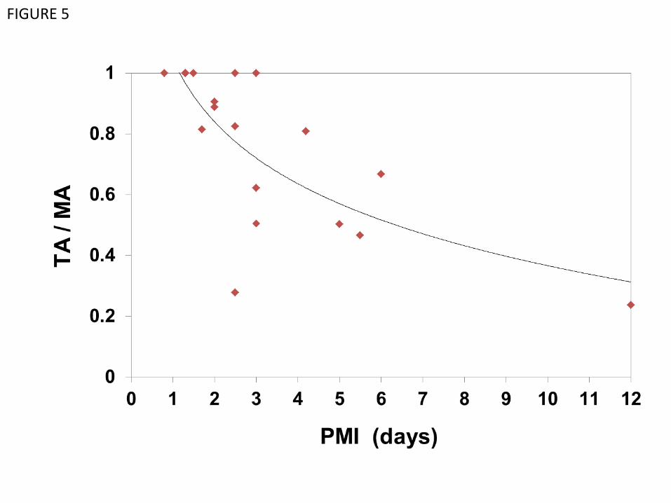
FIGURE 5

TABLE 1. Details of Patient
Case Sex Age Time from onset
to death
PMI
(day) Cause of death
Sclerosis
of coronary arteries TA / MA
1 M 72 < 9 hr 1.2 AMI High, Plaque erosion RA 1
2 M 40 < 7 hr 1.3 AMI Severe, Thrombus in LAD 1
3 M 48 < 2 hr 1.5 AMI Severe, Plaque rapture in LAD 1
4 M 54 < 20 min 1.7 AMI Severe, Thrombus in LCX 1
5 M 88 8 days 1.5 Brain contusion Mild 0.546 (NTA/MA = 0.391)
6 M 62 A few days 2 Meningoencephalitis Mild 0.482 (NTA/MA = 0.503)
7 M 67 Unknown 3 Acute respiratory failure Severe 0.315 (NTA/MA = 0.136)
8 F 47 < 30 min 0.8 Blood loss None 1
9 M 39 9 hr 1.3 Brain contusion Mild 1
10 F 79 < 2 hr 1.3 Blood loss Little 1
11 M 41 5 days 1.3 Brain contusion None 1
12 F 69 < 1 hr 1.3 Subarachnoid hemorrhage Little 1
13 F 37 < 30 min 1.5 Asphyxia None 1
14 M 29 9 hr 1.7 Blood loss None 0.814
15 M 33 Unknown 2 Pulmonary edema None 0.888
16 M 61 1 hr 2 Asphyxia Little 0.906
17 F 36 < 10 min 2.5 Asphyxia None 1
18 M 52 < 10 min 2.5 Brain contusion High 0.825
19 M 86 Unknown 2.5 Blood loss Mild 0.277
20 M 73 < 30 min 3 Brain contusion Little 1
21 F 39 22 hr 3 Hypoxic encephalopathy None 0.505
22 F 80 Unknown 3 Pancreatic carcinoma High 0.622
23 M 36 < 10 min 4.2 Asphyxia High 0.809
24 F 44 Unknown 5 Cirrhosis None 0.502
25 F 70 Unknown 5.5 Cerebral hemorrhage Mild 0.466
26 F 41 Unknown 6 Pneumonia None 0.667
27 M 21 Unknown 12 Unknown None 0.237
Sclerosis of coronary arteries is categorized according to the most serious occlusion at cross section;
Severe > 90%,High > 75%, Mild > 50%,Little > 25%, None ≤ 25%.
RA: right coronary artery, LAD: left anterior descending coronary artery, LCX: left circumflex coronary artery.