the effects of dihydroergotamine in patients with head injury and raised intracranial pressure
TRANSCRIPT
Intensive Care Med (1989) 15:523-527 Intensive Care Medicine © Springer-Verlag 1989
The effects of dihydroergotamine in patients with head injury and raised intracranial pressure P.-O. Gr~nde
Department of Anaesthesia, University Hospital of Lund, and Department of Physiology and Biophysics, University of Lund, Sweden
Received: 28 December 1988; accepted: 26 June 1989
Abstract. In the first study 6 patients with raised in- tracranial pressure due to brain oedema following head injury were given dihydroergotamine because of low perfusion pressure. The intracranial pressure fell simultaneously with the increase in arterial pressure. The intracranial pressure fell from 24_+2 mmHg by a maximum of 12_+ 1 mmHg after a single intravenous injection of 0.25 mg of dihydroergotamine and re- mained at a low level for 3 5 - 70 rain before stabilizing at a new level 5 + 1 mmHg below the baseline. The ini- tial rapid and marked decrease in intracranial pressure may be the result of a reduced intracranial blood volume, due predominantly to constriction of the more voluminous venous capacitance vessels (by anal- ogy with the corresponding vascular effect of dihydroergotamine on skeletal muscle and skin.) In the second study, experiments using sympathectomized cat skeletal muscle, showed that dihydroergotamine also reduced the hydrostatic capillary pressure, induc- ing absorption of fluid from the interstitial tissue to blood. It is suggested that a similar transcapillary ab- sorption effect in the damaged brain may be an expla- nation for the observation that the intracranial pres- sure stabilized at a level below the initial one following dihydroergotamine.
Key words: Dihydroergotamine - Intracranial pres- sure - Vascular control - Head injury
High intracranial pressure due to cerbebral contusions and brain oedema after acute brain injury is frequent- ly treated with hyperventilation and barbiturate infu- sion [1]. These are thought to reduce the intracranial pressure via constriction of the cerebral resistance ves- sels so causing a decrease in the blood volume. Theo- retically, raised intracranial pressure should be reduced more effectively by constriction of the venous capaci-
tance vessels as these hold a greater blood volume than the resistance vessels [2].
Dihydroergotamine is used clinically to treat or- thostatic hypotension and other hypotensive states and from experiments on skeletal muscle and skin, is considered to exert its clinical effects mainly via con- striction of the venous capacitance vessels [3, 4]. Pro- vided such vascular responses also occur in the brain, administration of dihydroergotamine may transiently reduce the cerebral blood volume and so lower a raised intracranial pressure perhaps even to a greater extent than that induced by hyperventilation or by barbitu- rate infusion.
The hypothesis that a raised intracranial pressure may be reduced by the administration of dihydroergot- amine in man was tested and confirmed in the present study. It is suggested that this may be due to an effect of reduced intracranial blood volume, predominantly on the venous side of the circulation. Experiments were also performed on the sympathectomized cat skeletal muscle in order to assess whether dihydro- ergotamine also may induce a slow but progressive de- crease in tissue volume via the mechanism of fluid ab- sorption due to reduced capillary pressure. This hy- pothesis was confirmed.
Material and methods
Studies in man
Observations were made on 6 previously healthy patients who had suffered traffic accidents with isolated severe head injuries on the basis of cerebral contusions and brain oedema. One of the patients (patient E in Table 1) also showed a small subdural haemorrhage demonstrated on the CT scan. All except two patients (patient B and E in Table 1), were simultaneously being treated with high doses of thiopental. In all cases the medical indication for treatment with dihydroergotamine was low cerebral perfusion pressure. Intracranial pressure was measured continuously via an intraventricular catheter using a Bentley transducer [5]. Dihydroergotamine (Orstanorm ®,
524 P.-O. Gr~inde: Dihydroergotamine and raised intracranial pressure
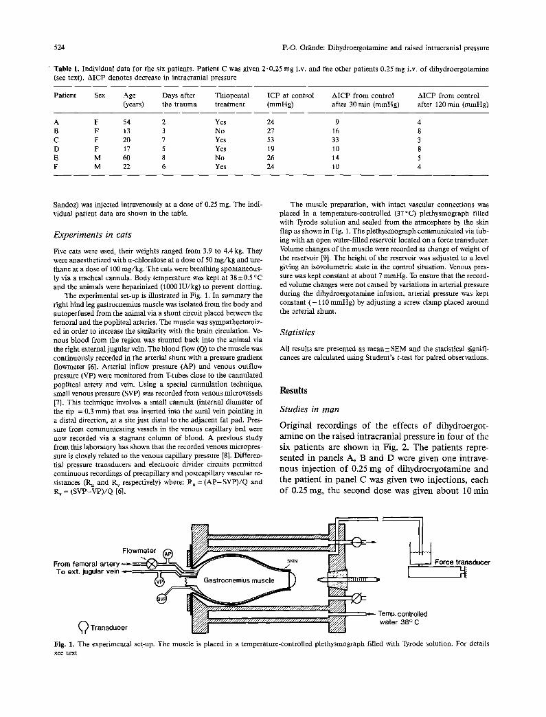
Table 1. Individual data for the six patients. Patient C was given 2.0.25 mg i.v. and the other patients 0.25 mg i.v. of dihydroergotamine (see text). AICP denotes decrease in intracranial pressure
Patient Sex Age Days after Thiopental ICP at control AICP f rom control AICP f rom control (years) the t rauma t reatment (mmHg) after 30 min (mmHg) after 120 min (mmHg)
A F 54 2 Yes 24 9 4 B F 13 3 No 27 16 8 C F 20 7 Yes 53 33 3 D F 17 5 Yes 19 10 8 E M 60 8 No 26 14 5 F M 22 6 Y e s 24 10 4
Sandoz) was injected intravenously at a dose of 0.25 mg. The indi- vidual patient data are shown in the table.
Experiments in cats
Five cats were used, their weights ranged from 3.9 to 4.4 kg. They were anaesthetized with n-chloralose at a dose of 50 m g / k g and ure- thane at a dose of 100 mg/kg. The cats were breathing spontaneous- ly via a tracheal cannula. Body temperature was kept a t 38 + 0.5 °C and the animals were heparinized (1000 IU/kg) to prevent clotting.
The experimental set-up is illustrated in Fig. 1. In summary the right hind leg gastrocnemius muscle was isolated from the body and autoperfused from the animal via a shunt circuit placed between the femoral and the popliteal arteries. The muscle was sympathectomiz- ed in order to increase the similarity with the brain circulation. Ve- nous blood from the region was shunted back into the animal via the right external jugular vein. The blood flow (Q) to the muscle was continuously recorded in the arterial shunt with a pressure gradient flowmeter [6]. Arterial inflow pressure (AP) and venous outflow pressure (VP) were monitored from T-tubes close to the cannulated popliteal artery and vein. Using a special cannulat ion technique, small venous pressure (SVP) was recorded from venous microvessels [7]. This technique involves a small cannula (internal diameter of the tip = 0.3 mm) that was inserted into the sural vein pointing in a distal direction, at a site just distal to the adjacent fat pad. Pres- sure from communicat ing vessels in the venous capillary bed were now recorded via a stagnant column of blood. A previous study from this laboratory has shown that the recorded venous micropres- sure is closely related to the venous capillary pressure [8]. Differen- tial pressure transducers and electronic divider circuits permitted continuous recordings of precapillary and postcapillary vascular re- sistances (R a and R v respectively) where: R a = ( A P - S V P ) / Q and R v = (SVP-VP) /Q [6].
The muscle preparation, with intact vascular connections was placed in a temperature-controlled (37°C) plethysmograph filled with Tyrode solntion and sealed from the atmosphere by the skin flap as shown in Fig, 1. The plethysmograph communicated via tub- ing with an open water-filled reservoir located on a force transducer. Volume changes of the muscle were recorded as change of weight of the reservoir [9]. The height of the reservoir was adjusted to a level giving an isovolumetric state in the control situation. Venous pres- sure was kept constant at about 7 mmHg. To ensure that the record- ed volume changes were not caused by variations in arterial pressure during the dihydroergotamine infusion, arterial pressure was kept constant ( - 1 1 0 mmHg) by adjusting a screw clamp placed around the arterial shunt.
Statistics
All results are presented as mean_+SEM and the statistical signifl- cances are calculated using Student 's t-test for paired observations.
Results
Studies in man
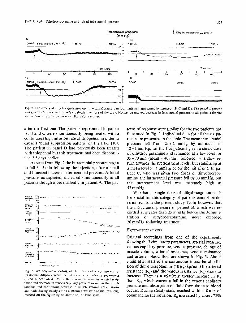
Original recordings of the effects of dihydroergot- amine on the raised intracranial pressure in four of the six patients are shown in Fig. 2. The patients repre- sented in panels A, B and D were given one intrave- nous injection of 0.25 mg of dihydroergotamine and the patient in panel C was given two injections, each of 0.25 mg, the second dose was given about 10 min
. . . . . . . - . . , , Temp. controlled I ~ ~ / / / / / / / / / / / / - ' / J J J / / A water 38 ° C
kll ) Transducer ~ ~ •
Fig. 1. The experimental set-up. The muscle is placed in a temperature-controlled plethysmograph filled with Tyrode solution. For details see text
Force transducer
P.-O. Gr/inde: Dihydroergotamine and raised intracranial pressure 525
A
100/65
In t racran ia l p r e s s u r e ~: Dihydroergotamine O.25mg i.v. ( rnm Hg)
B Blood pressure (rnm Hg) 135/70 110/65 T 110/55 115/55 105/55
40 t 0 J.
Time (rain) I I I ! I | I J I ~ ~ ~
0 20 40 60 60 100
c 110/60 Blood pressure (mm Hg) 115/60 108/60
0
I I I / I I I I I I 0 20 40 60 80
D
70150 90/55
Time (min) t I
lO0
80/50
I
Fig. 2. The effects of dihydroergotamine on intracranial pressure in four patients (represented b y p a n e l s A , B, C and D). The panel C patient was given two doses and the other patients one dose of the drug. Notice the marked decrease in intracranial pressure in all patients despite an increase in perfusion pressure. For details see text
after the first one. The patients represented in panels A, B and C were simultaneously being treated with a continuous high infusion rate of thiopental in order to cause a 'burst suppression pattern' on the EEG [~0]. The patient in panel D had previously been treated with thiopental but this treatment had been discontin- ued 3.5 days earlier.
As seen from Fig. 2 the intracranial pressure began to fall 3 - 5 min following the injection, after a small and transient increase in intracranial pressure. Arterial pressure, as expected, increased simultaneously in all patients though more markedly in patient A. The pat-
i~o
PRESSURE lOO (ram H{})
CAPILLAR Y 20 ] PF~ES~URE io . . . . . . . . . . . . . . . . . . . . . . . . . . . . . . . . . . . . . . . . . . . . . . . . . . . . . . . . . . . . . . . . . . . . . . . . . . . . . . . . . . . . . . . . . . . . . . . . . . . . . . . . . . . . . . . . . . . . . . . . . . . . . . . . . . . . (ram Hg) O APe = 30 mmHg
pRESSURE (ram H~)
VOLUME 0.5 (mr/loOg)
RESISTANCE (PRU~ ~0
RESISTANCE *~o%
(PRU) 0"
ARTERIAL ~ BLOOD FLOW (n~/m~/lOOg)
0 T..,~ ~ ,~ E i I ~ i i i L i I i l f i
i 1
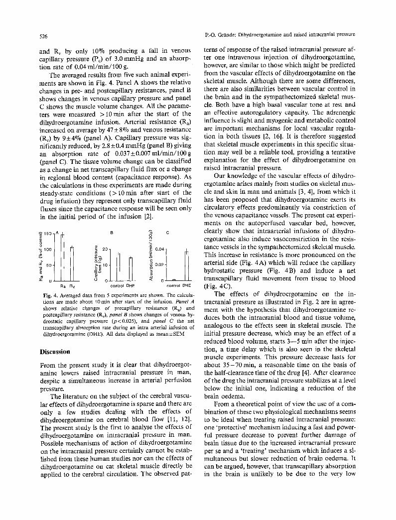
Fig. 3. An original recording of the effects of a continuous in- traarterial dihydroergotamine infusion on circulatory parameters (listed as ordinates). Notice the marked increase in arterial resis- tance and decrease in venous capillary pressure as well as the simul- taneous and continuous decrease in muscle volume. Calculations are made during steady-state (> 10 rain after start of the infusion), marked on the figure by an arrow on the time scale
terns of response were similar for the two patients not illustrated in Fig. 2. Individual data for all the six pa- tients are presented in the table. The mean intracranial pressure fell from 24___2mmHg by as much as 12_+ 1 mmHg, for the five patients given a single dose of dihydroergotamine and remained at a low level for 3 5 - ? 0 m i n (mean = 40rain), followed by a slow re- turn towards the pretreatment levels, but stabilizing at a mean level 5 + 1 mmHg below the initial one. In pa- tient C, who was given two doses of dihydroergot- amine, the intracranial pressure fell by 33 mmHg, but the pretreatment level was extremely high at 53 mmHg.
Whether a single dose of dihydroergotamine is beneficial for this category of patients cannot be de- termined from the present study. Note, however, that the intracranial pressure in patient B, which was re- corded at greater than 25 mmHg before the adminis- tration of dihydroergotamine, never exceeded 20 mmHg following treatment.
Experiments in cats
Original recordings from one of the experiments showing the ? circulatory parameters, arterial pressure, venous capillary pressure, venous pressure, change of muscle volume, arterial resistance, venous resistance and arterial blood flow are shown in Fig. 3. About 3 rain after start of the continuous intraarterial infu- sion of dihydroergotamine (10 ~tg/kg/min) the arterial resistance (Ra) and the venous resistance (Rv) starts to increase. There is a relatively greater increase in R~ than Rv, which causes a fall in the venous capillary pressure and absorption of fluid from tissue to blood occurs. During steady-state, reached within 10 min of commencing the infusion, R a increased by about 75°70
526 P.-O. Gr~nde: Dihydroergotamine and raised intracranial pressure
and R v by only 10% producing a fall in venous capillary pressure (Pc) of 3.0 mmHg and an absorp- tion rate of 0.04 ml/min/100 g.
The averaged results from five such animal experi- ments are shown in Fig. 4. Panel A shows the relative changes in pre- and postcapillary resistances, panel B shows changes in venous capillary pressure and panel C shows the muscle volume changes. All the parame- ters were measured > 10 min after the start of the dihydroergotamine infusion. Arterial resistance (Ra) increased on average by 47 +__ 8% and venous resistance (Rv) by 9+_4o70 (panel A). Capillary pressure was sig- nificantly reduced, by 2.8 + 0.4 mmHg (panel B) giving an absorption rate of 0 .037+0.007ml/min/100g (panel C). The tissue volume change can be classified as a change in net transcapillary fluid flux or a change in regional blood content (capacitance response). As the calculations in these experiments are made during steady-state conditions (> 10rain after start of the drug infusion) they represent only transcapillary fluid fluxes since the capacitance response will be seen only in the initial period of the infusion [2].
• ~ 150-A E o
100-
50.
2 o Ra Rv
B
o~ 20 1
a f ~1o o[
control DHE
C
2= E 0.041 v
~_~ 0102 1
0 J ~h control DHE
Fig. 4, Averaged data from 5 experiments are shown. The calcula- tions are made about 10 min after start of the infusion. Panel A shows relative changes of precapillary resistance (Ra) and postcapillary resistance (Rv), panel B shows changes of venous hy- drostatic capillary pressure (p<0.025), and panel C the net transcapillary absorption rate during an intra arterial infusion of dihydroergotamine (DHE). All data displayed as mean +_ SEM
Discussion
From the present study it is clear that dihydroergot- amine lowers raised intracranial pressure in man, despite a simultaneous increase in arterial perfusion pressure.
The literature on the subject of the cerebral vascu- lar effects of dihydroergotamine is sparse and there are only a few studies dealing with the effects of dihydroergotamine on cerebral blood flow [11, 12]. The present study is the first to analyse the effects of dihydroergotamine on intracranial pressure in man. Possible mechanisms of action of dihydroergotamine on the intracranial pressure certainly cannot be estab- lished from these human studies nor can the effects of dihydroergotamine on cat skeletal muscle directly be applied to the cerebral circulation. The observed pat-
terns of response of the raised intracranial pressure af- ter one intravenous injection of dihydroergotamine, however, are similar to those which might be predicted from the vascular effects of dihydroergotamine on the skeletal muscle. Although there are some differences, there are also similarities between vascular control in the brain and in the sympathectomized skeletal mus- cle. Both have a high basal vascular tone at rest and an effective autoregulatory capacity. The adrenergic influence is slight and myogenic and metabolic control are important mechanisms for local vascular regula- tion in both tissues [2, 16]. It is therefore suggested that skeletal muscle experiments in this specific situa- tion may well be a reliable tool, providing a tentative explanation for the effect of dihydroergotamine on raised intracranial pressure.
Our knowledge of the vascular effects of dihydro- ergotamine arises mainly from studies on skeletal mus- cle and skin in man and animals [3, 4], from which it has been proposed that dihydroergotamine exerts its circulatory effects predominantly via constriction of the venous capacitance vessels. The present cat experi- ments on the autoperfused vascular bed, however, clearly show that intraarterial infusions of dihydro- ergotamine also induce vasoconstriction in the resis- tance vessels in the sympathectomized skeletal muscle. This increase in resistance is more pronounced on the arterial side (Fig. 4A) which will reduce the capillary hydrostatic pressure (Fig. 4B) and induce a net transcapillary fluid movement from tissue to blood (Fig. 4C).
The effects of dihydroergotamine on the in- tracranial pressure as illustrated in Fig. 2 are in agree- ment with the hypothesis that dihydroergotamine re- duces both the intracranial blood and tissue volume, analogous to the effects seen in skeletal muscle. The initial pressure decrease, which may be an effect of a reduced blood volume, starts 3--5 min after the injec- tion, a time delay which is also seen in the skeletal muscle experiments. This pressure decrease lasts for about 3 5 - 7 0 rain, a reasonable time on the basis of the half-clearance time of the drug [4]. After clearance of the drug the intracranial pressure stabilizes at a level below the initial one, indicating a reduction of the brain oedema.
From a theoretical point of view the use of a com- bination of these two physiological mechanisms seems to be ideal when treating raised intracranial pressure: one 'protective' mechanism inducing a fast and power- ful pressure decrease to prevent further damage of brain tissue due to the increased intracranial pressure per se and a 'treating' mechanism which induces a si- multaneous but slower reduction of brain oedema. It can be argued, however, that transcapillary absorption in the brain is unlikely to be due to the very low
P.-O. Gr~inde: Dihydroergotamine and raised intracranial pressure 527
capillary permeability in this tissue compared to skele- tal muscle. Certainly this is the case in the normal brain, but there is much data to suggest that the capil- lary permeability is markedly increased after head trauma with damage of the blood-brain barrier [14].
According to the studies previously mentioned [11, 12], the cerebral blood flow is unaltered by dihydro- ergotamine, indicating that the vasconstrictor effect of this drug on cerebral blood flow is counteracted by the simultaneous increase in arterial blood pressure. This also indicates that the nutritional state of the brain is not significantly altered.
The mode of action of dihydroergotamine on smooth muscle is still unclear in spite of thorough analysis [15]. Dihydroergotamine seems to exert its lo- cal vasoconstrictor influence via a direct excitatory ef- fect on the vascular smooth muscle, perhaps mediated through stimulation of 5 - HT and alpha receptors [4].
This type of comparative analysis between differ- ent tissues, if made with caution, can be a useful in- strument to explain phenomena which are diffficult or impossible to analyse directly. We are reduced to this type of comparative study for detailed analysis of ce- rebral vascular control as no other organ of the body is less adapated to experimental study of its circulation than the brain [16].
This study suggests that dihydroergotamine is a drug which can reduce raised intracranial pressure. Further studies are required to precisely define the mechanisms behind this effect and before dihydroer- gotamine is used clinically for the treatment of raised intracranial pressure.
Acknowledgements. This study was supported by grants from the Swedish Medical Research Council (2210) and from the Faculty of Medicine, University of Lund. The author thanks Mrs. Helbn Han- sen and Mrs Christine Wikstrand for skilled technical and secretari- al assistance.
References
1. Piatt JH, Schiff SJ (1984) High dose barbiturate therapy in neurosurgery and intensive care. Neurosurgery 15:427-444
2. Mellander S, Johansson B (1968) Control of resistance, ex- change and capacitance functions in the peripheral circulation. Pharmacol Rev 20:117-196
3. Mellander S, Nordenfelt I (1970) Comparative effects of dihy- droergotamine and noradrenaline on resistance, exchange and capacitance functions in the peripheral circulation. Clin Sci 39:183-201
4. Maller-Schweinitzer E, Rosenthaler J (1987) Dihydroergot- amine: pharmacodynamics, and mechanism of venconstrictor action in beagle dogs. J Cardiovasc Pharmacol 9:686-693
5. Lundberg N (1960) Continuous recording and control of ven- tricular fluid pressure in neurosurgical practice. Acta Psychiatr Neurol Scand 36:1 - 193
6. Gr/inde PO, BorgstrOm P (1977) An electronic differential pressure flowmeter and a resistance meter for continuous mea- surement of vascular resistance. Acta Physiol Scand 102:224- 230
7. Gr~nde PO (1979) Influence of neural and humoral beta- adrenoceptor stimulation on dynamic myogenic microvascular reactivity in cat skeletal muscle. Acta Physiol Scand 106:457 - 465
8. Mellander S, BjOrnberg J, Maspers M, Myrhage R (1987) Meth- od for continuous recording of hydrostatic exchange vessel pressure in cat skeletal muscle. Acta Physiol Scand 129:325-335
9. Gr/inde PO, J/irhult J, Mellander S (1974) Method for gravimet- tic registration of changes in tissue volume. Acta Physiol Scand 91:211-215
10. Boarini DJ, Kassel NF, Coester HC (1984) Comparison of sodi- um thiopental and methoexital for high-dose barbiturate anaes- thesia. J Neurosurg 60:602-608
11. Andersen AR, Refelt-Hansen P, Lassen NA (1987) The effect of ergotamine and dihydroergotamine on cerebral blood flow in man. Stroke 18:120-123
12. Lindblad B, Bergquvist D (1983) Tissue blood flow and blood flow distribution after administration of dextran 70, dihydroer- gotamine and their combination. A study in dogs using the ra- dioactive microsphere technique. Acta Chir Scand 14:467-472
13. Gustafsson D, Gr~nde PO, Borgstr6m P, Lindberg L (1988) Ef- fects of calcium antagonists on myogenic and neurogenic con- trol of resistance and capacitance vessels in cat skeletal muscle. J Cardiovasc Pharmacol 12:413-422
14. BradburyM (1979) Theconcept ofablood-brainbarrier. Wiley, New York, p 465
15. Rothin E, Konzett H, Cerletti A (1954) The antagonism of ergot alkaloids towards the inhibitory response of the isolated rabbit intestine to epinephrine and norpinephrine. J Pharmacol Exp Ther 112:185-190
16. Heistad DD, Kontos AH (1983) Cerebral circulation. In: Hand- book of Physiology 2:137- 182
Dr. P.-O. Gr~nde Department of Anaesthesia University Hospital of Lund S-221 85 Lurid Sweden