the elmer and mamdouha bobst hospital | … . quarterly. veterinary community news from amc spring...
TRANSCRIPT
rDVM QUARTERLYVETERINARY COMMUNIT Y NEWS FROM AMC SPRING 2017
T H E E L M E R A N D M A M D OU H A B O B S T H O S P I TA L | C A S PA RY R E S E A R C H I N S T I T U T E | T H E I N S T I T U T E F O R P O S T G R A DUAT E E DU C AT ION
3 • CARDIOLOGY | 5 • DENTISTRY | 8 • INTERNAL MEDICINE | 12 • PATHOLOGY
15 • WHAT’S YOUR DIAGNOSIS? | 16 • UPDATES & EDUCATION

VETERINARY COMMUNIT Y NEWS FROM AMC | WINTER 201700 001
Dear Colleagues,
I am very pleased to present you with our first rDVM newsletter of 2017. In this edition, we are highlighting some of the services that don’t always make the front page like our Dentistry Service with Dan Carmichael and our Pathology Service with Taryn Donovan. I do love that this platform allows us to bring these practical tips to you, not only to highlight what we are doing here every day but also to give you our opinion on advances that everyone can incorporate into their own practice, especially in areas like dentistry, internal medicine and surgery.
As spring is just beginning, I wanted to devote a few words to some of the things that have happened in our community over the winter, since our last newsletter. First, we have had two outbreaks of infectious diseases in the city that have hit the national news. The first, of course, was the very large H7N2 avian flu outbreak among cats at a Manhattan shelter. Remember it is not unusual for influenza viruses to infect animals from another species (i.e., a cat getting an avian flu virus), but what is unusual is for the virus to then become established in the new species and to start being transmitted within it (i.e., cat to cat transmission of an avian flu virus). This clearly happened in this inci-dent and I would like to commend everyone involved in the rapid recognition and response to this new outbreak that clearly prevented a larger impact on cats in other shelters, shows, etc. The lesson to be learned for all of us is to stay vigilant and not be afraid to think outside the box – just because there is no cat flu doesn’t meant there couldn’t be, and when presented with an unusual cluster of cases, taking action via testing and isolation are key.
The second was the recent human Leptospirosis cases in the Bronx – with at least three people infected and one fatality. We have people with Lepto in the city every year, but with three people in the same location, this story became newsworthy and served as a reminder to all of us of how important this disease is for dogs in NYC and all over the country. Even Arizona is seeing plenty of cases this year.
It is such an honor to be part of a profession that is entrusted with the lives of animals, and in many ways humans as well. These last two outbreaks remind me of the tremendous privilege and responsibility it is to be a part of the veterinary community in NYC.
Thank you for your tremendous support of AMC in 2016, and I cannot wait to see what 2017 brings. As always, feel free to contact me with any comments, questions or concerns about AMC. I look forward to hearing from you.
Richard
Richard E. Goldstein DVM, DACVIM, [email protected] 212-329-8824cell 347-733-7338
A LETTER FROM OUR CMO
VETERINARY COMMUNIT Y NEWS FROM AMC | SPRING 2017 2
To Our Valued Partners In Care,
Spring is a time for growth and change and here at AMC, we are energized by what is on the horizon. We continue to make improvements in the hospital, creating better workflow and improving our space for patients, clients and staff. We soon will welcome our new intern class and incoming residents while saying goodbye to those moving on to the next chapter in their veterinary careers. We hope to see some of those faces in your practices as these skilled veterinarians advance in their careers.
I would like to thank you once again for providing such great feedback in our recent referral survey. You are a top priority to us and through our strategic planning process, which is well underway, we have designated a special task force to address the needs of you, our valued referring doctors. We strive for excellence when treating your patients and hope to ensure your experience and your clients’ experience with us is seamless. We thank you for entrusting AMC with your patients and clients.
I hope this newsletter serves as a beneficial publication for you and your prac-tice. I welcome your feedback and hope that you will let me know if there is any additional information you would like us to include.
I thank you for your continued support of the Animal Medical Center.
Sincerely,
Kate
Kathryn Coyne [email protected]
A LETTER FROM OUR CEO

00 VETERINARY COMMUNIT Y NEWS FROM AMC | SPRING 2017 43
CARDIOLOGY
Pacemakers are commonly implanted in dogs when a complete (3rd degree) atrioventricular (AV) block causes lethargy or collapse, but cats can some-times benefit from this therapy as well. AMCs cardiology and surgery teams worked together to save four cats whose normal 180–200 beats per minute heart rates gave out, resulting in bradycardia, hypotension and shock.
Three of these cats, aged 8 to 13, presented recently. They were previously healthy, but developed acute weakness or syncope and were referred to AMC’s Emergency and Cardiology Services. Their ECGs recorded complex heart rhythms that included transient, normal sinus rhythm, punctuated by advanced atrioventricular block and bradycardia, or atrial standstill. When in sinus rhythm, each cat appeared quiet but alert. However, when bradycar-dia developed, they became quickly hypotensive, and some collapsed whose rhythms regressed to advanced heart block. Three of these cats had high grade second degree AV block.
Though infrequently encountered in the dog, high grade 2nd degree AV block is the most common cause of symptomatic bradycardia in felines and can be devastating if not treated successfully. Unlike complete heart block where ventricular escape rates vary between 85–100 beats per minute, high grade 2nd degree AV block results in periods up to 15 seconds without a single heart beat. This is because high grade 2nd degree AV block does not usually permit normal slow ventricular escape rhythms—life saving heart beats—that are usually present in states of complete heart block.
Cardiology and critical care worked together to stabilize these cats in prepa-ration for anesthesia and pacemaker implantation. Where needed, their slow heart rates were overcome with the aid of a temporary, external trans-thoracic cardiac pacing system. This improved cardiac output and systemic blood pressure and stabilized them for subsequent pacemaker implantation.
Anesthesia, surgery, and cardiology teams worked together to reduce the risk in these critically ill cats. Dr. Daniel Spector and Dr. Robert Hart, AMC surgeons, utilized a ventral abdominal, transdiaphragmatic approach (a tech-nique first published at AMC for certain pacemaker implantation patients). This procedure provides access that permits the end of the pacemaker lead to be affixed to the cardiac apex, accessed through a diaphragmatic incision which is then closed. The pacemaker itself was attached and implanted subcu-taneously in the abdominal wall, and the abdominal incision was then closed. During and after these procedures, the pacemaker was adjusted remotely by Drs. Wiley, White, and Fox using a pacemaker interrogator instrument. This device checks the integrity of the pacemaker and lead and can program the system to optimize pacemaker sensing, output, and heart rate.
These patients recovered uneventfully. Pacemaker longevity varies from case to case, but can last up to 10 years in some instances.
Pacemaker Therapy for Cats
Philip R. Fox DVM, DACVIM/ECVIM, DACVECC Head of [email protected]
Right lateral and VD radiographs showing location and position of endocardial pacemaker lead and pulse generator (‘pacemaker’) placement in a cat with syncope and collapse caused by high grade 2nd degree AV block.
CARDIOLOGY

5 VETERINARY COMMUNIT Y NEWS FROM AMC | SPRING 2017 6
DENTISTRY
It has been several decades since veterinarians first became acquainted with the diagnosis and treatment of tooth resorption in cats. Back in the 1970s, these were referred to as cervical lines erosions; in the 1980s, neck lesions; the 1990s as feline odontoclastic resorptive lesions or FORLs; and most recently this common feline problem is simply referred to as tooth resorption. Veterinarians have learned to examine the oral cavity for the hallmark cherry-red localized gingivitis suggesting an underlying lesion, to probe the cat’s teeth under anesthesia, to always take dental radiographs to identify the type and stage of the lesion, and to make appropriate treatment decisions based on the results of the intraoral examination and radiographs. At this point in time, although the etiology of the condition in cats has yet to be elucidated, we remain confident that appropriate diagnosis and treatment decisions can be made. However, we are increasingly finding similar lesions in dog teeth. (Figure 1a and 1b)
Tooth resorption appears to be less prevalent in dogs than in cats – but not as rare as one might imagine. One study that considered the prevalence of tooth resorption in dogs (presented for dental treatment) identified 53% of patients as having radiological evidence of tooth resorption. This study noted a direct relationship between the incidence of canine tooth resorption and advanced age, as well as a greater prevalence in large-breed dogs. A theory that may explain why there is an apparent increase in our diagnosis of canine tooth resorption is that we are now obtaining dental radiographs with a greater frequency (as we should). In other words, canine tooth resorption has always been a significant disease but we are now just starting to recognize it.
Although there are various forms of tooth resorption with various classifica-tion systems used in both humans and in animals, the most common form of tooth resorption seen in dogs is external replacement resorption. Simply stated, the process of external replacement resorption is where tooth struc-ture is resorbed by odontoclasts, and replaced by osteoblasts with bone. For some types of tooth resorption, the cause is known. The cause of canine external replacement resorption is idiopathic. Some have suggested that masticatory forces over time could be responsible for the external replace-ment resorption seen in dogs, and this would explain why we see it more in older dogs (they have chewed longer) and also why it is more prevalent in large breed dogs (they chew harder). External replacement resorption usually falls under the heading of Type 2 resorption within the AVDC nomenclature committee classification (www.avdc.org).
Diagnosis
Canine tooth resorption is diagnosed by coupling the results of intraoral examination (with a dental probe) and the interpretation of correspond-ing dental radiographs. During intraoral examination on the anesthetized patient, a dental probe should be used to examine all surfaces of every tooth, especially probing below the gingival margin. Tooth resorption may be appre-ciated visually as a loss of tooth structure, or more commonly appreciated by
Canine Tooth Resorption
DENTISTRY
Dan Carmichael DVM, [email protected]
1a. This is a radiograph of the right mandible of an adult cat. Note the tooth resorption associated with the man-dibular molar (#409), with complete resorption of the distal root.
1b. This is a radiograph of the right caudal mandible in an adult dog. Note the tooth resorption associated with the second mandibular molar (#410), with complete resorp-tion of the distal root.
2a. This is a photograph of the left maxilla of an adult large-breed dog. There is moderate to severe gingivitis and cal-culus formation, but also note carefully the left maxillary fourth premolar (#208). The distal portion of the crown
appears to be missing, and when probed revealed a large cavity defect.
2b. This is a radiograph of the tooth #208 depicted in Fig-ure 2a. Note the resorption of the distal portion of the crown as well as the distal root of #208. This is external replacement resorption. Also notice root resorption associated with #207.
3. This is a radiograph of the left mandibular third (#307 - on left) and fourth (#308 – on right) premolars of an adult geriatric dog. Both teeth show tooth resorption, seen as indistinct crown and root structure, loss of a visible periodontal ligament space, and a “ghostly” appearance.
FIG. 1a
FIG. 2a
FIG. 3
FIG. 2b
FIG. 1b

VETERINARY COMMUNIT Y NEWS FROM AMC | SPRING 20177 8
probing a “cavity” in the dog’s tooth. (Figure 2 a and b) Since the process of external replacement resorption generally begins in the root cementum, all early lesions and many advanced lesions are only diagnosed by radiography. This is another reason why it is imperative to always take a full-mouth set of dental radiographs for every canine patient. Radiographically, the resorp-tive process causes areas of the tooth to become indistinct. The roots appear “ghostly.” (Figure 3) The loss of classic tooth structure is accompanied by production of bone tissue in the area that was previously tooth – giving the name replacement resorption. Periodontal ligament spaces become indistinct. The process of tooth resorption is not considered painful unless the resorp-tion erodes the crown of the tooth exposing the sensitive portion of the tooth to the oral cavity.
Treatment
The treatment options for dogs with teeth affected by resorption are dictated by the results of intraoral examination and dental radiography.
Monitor
In cases of tooth resorption where there are no clinically detectable lesions on the tooth – cases where the lesion is only a radiographic diagnosis – treatment may not be indicated. The process of tooth resorption is not considered pain-ful unless the resorption erodes the crown of the tooth exposing the sensitive portion of the tooth to the oral cavity. These teeth can remain functional. The radiographic evidence of tooth resorption should be noted in the patient’s dental chart, and yearly follow up with dental radiographs recommended.
Extract the tooth
In cases where tooth resorption has compromised the tooth to an extent that the lesion extends through the crown and into the oral cavity, treatment is indicated. Treatment for these teeth is extraction. Extraction of teeth experiencing tooth resorption is sometimes difficult due to the nature of the resorptive process and the inevitable ankyloses that occurs when tooth and periodontal ligament are replaced by bone. The goal of extraction should be to remove the entire root, but often times the entire root no longer exists. Any root structure coronal to the area of resorption should be extracted in its entirety. Any root remnants associated with radiographic pathology (lucency) should be extracted. Crown amputation can be performed for cases where roots are no longer radiographically distinct. Always perform post-operative radiographs for future monitoring purposes.
DENTISTRY
Elizabeth Appleman VMD, DACVIMHead of Internal [email protected]
Emma Stanley VMDResident, Internal [email protected]
Cobalamin (B12) is a water-soluble B-vitamin that serves as an important cofactor for two enzymes, methionine synthase and methylmalonyl CoA mutase, both of which are necessary for the synthesis of multiple proteins, neu-rotransmitters, and nucleic acids in the body.1–3 Cobalamin is involved in multi-ple fundamental biological processes including DNA synthesis, hematopoiesis, and neuron myelination. A deficiency in this B-vitamin in people can result in a broad range of gastrointestinal, hematological, and neurological disorders.1,4 Similarly, folate is a water-soluble B vitamin required for DNA synthesis, and therefore folate deficiency may lead to similar hematologic changes.5
Because cobalamin and folate cannot be synthesized endogenously by animals, exogenous sources must be obtained by the gastrointestinal tract from food.1 Natural sources of cobalamin are ingested as 5'-adenosylcobalamin, a protein-vitamin complex first digested by pepsin.1,2 Free cobalamin then binds to haptocorrin (R-protein) in gastric secretions, which is degraded to unbound cobalamin by pancreatic proteases in the distal duodenum.1,2,5 Intrinsic factor (IF), produced with chlorhydric acid by gastric parietal cells in humans and dogs and exclusively by the pancreas in cats, binds cobalamin in the duodenum and protects it from intestinal proteolysis.2,5 Distal ileal IF-cobalamin (cubam or cubilin) receptors located on enterocyte membranes specifically absorb cobalamin, transporting it to portal blood bound to transcobalamin.1,5 Approximately 1% of cobalamin also undergoes passive absorption through-out the gastrointestinal tract.17
Given the complexity of cobalamin absorption and metabolism, diverse gas-trointestinal disorders may lead to cobalamin deficiency in both human and veterinary patients. Human conditions associated with hypocobalaminemia are numerous and include atrophic gastritis (pernicious anemia), partial gas-trectomy or ileal resection, Helicobacter pylori infection, inflammatory bowel disease, alimentary lymphoma, congenital or acquired deficiencies of cubam receptors or IF, or even long-term antacid therapy (achlorhydria).5–8 Among vet-erinary patients, cobalamin deficiency may also develop in a variety of primary gastrointestinal disease states analogous to those reported in humans, as well as pancreatitis, exocrine pancreatic insufficiency, and hyperthyroidism.2,9–12
The most well-recognized manifestation of cobalamin deficiency in humans is megaloblastic anemia, a non-regenerative anemia believed to result from defective DNA synthesis, ineffective hematopoiesis, and maturation arrest of erythrocyte precursors.1,5,13 Leukopoiesis and megakaryopoiesis may be similarly disrupted, generating increased progenitor cells, granulocyte hyper-segmentation and impaired release into systemic circulation.5,15 Hematologic manifestations of B12 deficiency appear to be rare among veterinary patients.2,9 Concurrent anemia of chronic disease and hypocobalaminemia are rela-tively common clinicopathologic findings among cats and dogs with primary
Evaluation of the Prevalence of Cobalamin and Folate Deficiencies in Anemic Dogs
INTERNAL MEDICINE

VETERINARY COMMUNIT Y NEWS FROM AMC | WINTER 2017 0000 VETERINARY COMMUNIT Y NEWS FROM AMC | SPRING 2017 009
INTERNAL MEDICINE
VETERINARY COMMUNIT Y NEWS FROM AMC | SPRING 2017 10
gastrointestinal disease, but a causal relationship between hypocobalaminemia and hematologic disease has not been established in the veterinary literature.2,17
Hematologic abnormalities resulting from cobalamin deficiency in people are typically rapidly reversible with B12 supplementation. Mild reticulocytosis is evident after a few days of cobalamin supplementation, and pancytopenia may completely resolve within eight weeks.5,13 Often life-long supplementation is necessary; established protocols for oral or parenteral routes in humans appear to be equally effective for long-term use.1,16 A universally accepted supplemen-tation protocol is not established among veterinary patients, but weekly sub-cutaneous injections of cyanocobalamin for six weeks followed by injections every two to four weeks is routinely implemented in cats and dogs.2 Recently, a retrospective study out of Sweden evaluated the feasibility of oral B12 therapy in 51 dogs with chronic enteropathies and hypocobalaminemia, and found that it was quite effective.17 All dogs had a normal B12 level within 20–202 days after starting daily oral supplementation, and the mean increase in serum cobala-min concentration after treatment was 794 ± 462 ng/L. The authors theorized that the positive response to oral cobalamin may be due to an alternative pas-sive absorption pathway throughout the gastrointestinal tract, independent of receptor-mediated transport in the ileum. This oral protocol may improve owner compliance with long-term supplementation, and also reduce cost and morbidity associated with injections. To the authors’ knowledge, no similar studies evaluating oral B12 supplementation have been performed in cats.
A current clinical trial at the Animal Medical Center is prospectively evaluating the prevalence of cobalamin and folate deficiencies in anemic dogs. We hypothesize that dogs with low B12 and/or folate will have an increased prevalence of anemia, and that dogs with non-regenerative anemia will have a greater likelihood of these deficiencies as compared to dogs with regenerative anemias. If our hypothesis is proven true, this may serve as a basis for more active screening of B12 and folate in anemic dogs, and supplementation of these vitamins as a standard therapy in patients with challenging anemias.
INTERNAL MEDICINE
References:
1. Stabler SP. Vitamin B12 deficiency. New Engl J Med 2013; 368:149-160.
2. Ruaux CG. Cobalamin in companion animals: diagnostic marker, defi-ciency states and therapeutic impli-cations. Vet J 2013; 196:145-152.
3. Worhunsky P, Toulza O, Rishniw N, et al. The relationship of serum cobalamin to methylmalonic acid concentrations and clinical vari-ables in cats. J Vet Intern Med 2013; 27:1056-1063.
4. Ho CH, Thomas M, McGuire E, Yano S. Two-year-old girl with pancytopenia due to vitamin B12 (cobalamin) deficiency. J Paediatr Child H 2014; 1-3.
5. Briani C, Torre CD, Citton V, et al. Cobalamin deficiency: clinical picture and radiological findings. Nutrients 2013; 5:4521-4539.
6. Andres E, Loukili NH, Noel E, et al.
Vitamin B12 (cobalamin) deficiency
in elderly patients. Can Med Assoc J
2004; 171(3): 251-259.
7. Halfdanarson TR, Walker JA, Litzo
MR, Hanson CA. Severe vitamin B12
deficiency resulting in pancytope-
nia, splenomegaly and leukoeryth-
roblastosis. Eur J Haematol 2008;
80:448-451.
8. Anitha P, Sasitharan R, Thambarasi
T, et al. Vitamin B12 deficiency
presenting as pancytopenia and
retinopathy in a young boy – Heli-
cobacter pylori, a novel causative
agent. Australas Med J 2014;
7(3):143-148.
9. Simpson KW, Fyfe J, Cornetta A,
et al. Subnormal concentrations of
serum cobalamin (vitamin B12) in
cats with gastrointestinal disease.
J Vet Intern Med 2001; 15:26-32.
10. Thompson KA, Parnell NK, Hohen-haus AE, et al. Feline exocrine pancreatic insufficiency: 16 cases (1992-2007). J Feline Med Surg 2009; 11:935-940.
11. Cook AK, Suchodolski JS, Steiner JM, Robertson JE. The prevalence of hypocobalaminemia in cats with spontaneous hyperthyroidism. J Small Anim Pract 2011; 52:101-106.
12. Bailey S, Benigni L, Eastwood J, et al. Comparisons between cats with normal and increased fPLI concentrations in cats diagnosed with inflammatory bowel disease. J Small Anim Pract 2010; 51:484-489.
13. Andres E, Affenberger S, Zimmer J, et al. Current hematological find-ings in cobalamin deficiency. A study of 201 consecutive patients with documented cobalamin deficiency. Clin Lab Haematol 2006; 28:50-56.
14. Wickramasinghe S. Morphology,
biology and biochemistry of cobala-
min- and folate-deficient bone mar-
row cells. Baillieres Clin Haematol
1995; 8(3):441-459.
15. Bolaman Z, Kadikoylu G, Yukselen
V, et al. Oral versus intramuscular
cobalamin treatment in megaloblas-
tic anemia: a single center, prospec-
tive, randomized open-label study.
Clin Ther 2003; 25(12):3124-3134.
16. Berghoff N, Parnell NK, Hill SL, et al.
Serum cobalamin and methylmalo-
nic acid concentrations in dogs with
chronic gastrointestinal disease.
Am J Vet Res 2013; 74(1):84-89.
17. L. Toresson, J.M. Steiner, J.S.
Suchodolski, et al. Oral Cobalamin
Supplementation in Dogs with
Chronic Enteropathies and Hypo-
cobalaminemia. J Vet Intern Med
2016;30(1):101-107.
Doug Palma DVM, DACVIM Internal [email protected]
Narrow band imaging (NBI) is a form of modulating the images acquired with traditional endoscopy and/or high definition endoscopy. NBI alters the wavelength of the light and filters out different wavelengths resulting in a “narrow band” of wavelengths/colors when viewing an image.
NBI has become standard in human medicine for evaluating certain patho-logic conditions. The acquisition of images with NBI, allows for accentuation of certain topographic features. This form of optical image enhancement technology improves the visibility of vessels and other tissues on the muco-sal surface. These features have been correlated with a specific disease pro-cesses in human medicine. Additionally, the accentuation of certain criteria can be helpful at more reliably identifying lesions and guiding diagnostic biopsy acquisition.
Narrowband illumination is strongly absorbed by hemoglobin and only pen-etrates the superficial tissues allowing for contrast between the two. This results in capillaries within the mucosal surface appearing as brown while veins in the submucosa are displayed as cyan.
The major application in human medicine is in the identification of squa-mous metaplasia of the esophagus (Barrett’s esophagus), esophagitis, neoplasia (esophageal, bladder, colonic, oral, gastric, brain, bronchial, laryn-geal), laryngeal reflux, peritoneal metastasis, and endometriosis. However, NBI has the potential to be useful for various conditions in both veterinary and human medicine.
Application to veterinary patients may be difficult due to species differences with respect to clinical disease occurrence, progression and/or significance. However, novel application to alternative clinical scenarios may provide value in the future. Currently, only one case report demonstrated in dogs is present suggesting that narrowband imaging may provide better visualiza-tion of superficial lesions within both the esophagus and stomach, guiding biopsy acquisition.
Esophageal inflammation can go unnoticed in people based on clinical his-tory alone. Furthermore, normal white light endoscopy is common, making histopathologic assessment necessary to definitively rule out esophagitis. When esophagitis is documented, application of manometry and pH testing are more readily available/easy to perform in people to potentially further define the etiology. Unfortunately, histopathologic assessment, verbaliza-tion of our patients and access to advanced testing makes esophageal disease difficult to identify and characterize in animals. For all these reasons, a more complete assessment of esophageal inflammation and/or causes is possible in people. It is the hope that application of narrow band imaging may further shed light on esophageal disease in our patients as it has been utilized in people to detect subtle white light endoscopy negative esophagitis lesions.
An additional application worth exploring in our patient population is during routine gastroduodenoscopy. It is possible that narrow band imag-ing may help the clinician to sample histologically more severely affected
Narrow Band Imaging

VETERINARY COMMUNIT Y NEWS FROM AMC | WINTER 2017 000011
INTERNAL MEDICINE
VETERINARY COMMUNIT Y NEWS FROM AMC | SPRING 2017 12
areas. With the hope that this may more reliably provide a representa-tive microscopic evaluation and potentially help to localize/identify potential neoplastic changes (epith-eliotropism, epithelial plaques, etc.).
As stated above, narrow band imaging is frequently used for the identifica-tion and characterization of multiple tumors in people within many differ-ent body systems. It is hoped that the application of NBI may potentially identify neoplastic lesions before visible changes are noted on routine endoscopy. This may provide earlier diagnosis in our patients and poten-tially better outcomes.
Currently, the Animal Medical Center is investigationally evaluating poten-tial applications for this unique form of imaging. It is an exciting time in veterinary medicine where technol-ogy may provide new opportunities for early diagnosis, treatment and potentially improved prognosis.
Taryn Donovan DVM, DACVPHead of Anatomic [email protected]
PATHOLOGY
Signalment: 9 month old, male castrated Domestic Shorthair cat.
History: 2.5 months ago the owners noted weakness in the pelvic limbs and anorexia. A work-up was performed which did not identify the cause for anorexia. The owner began syringe feeding AD. The pelvic limb weakness improved, but there was progressive, concurrent thoracic limb weakness. Tet-raparesis rapidly progressed during a 2 week period, after which the patient was non-ambulatory, and euthanasia was elected.
Necropsy findings: The brain is removed. The cerebral cortical gyri are expanded (edema). The cerebellar vermis is flattened against the underlying medulla (cerebellar coning). The spinal cord is removed in its entirety. No gross external abnormalities are observed in the spinal cord.
The fixed brain is sectioned. The ventricles are diffusely dilated, (most prominently the lateral and third ventricles) and contain abundant, gelatinous, cerebrospinal fluid. The ventricular lining is markedly irregular and thick-ened (ependymitis). (Figures 1,2) Cerebral gyri are expanded (edema).
Please formulate differential diagnoses based upon the history, clinical find-ings and images before turning the page.
Gross Pathology: What’s Your Morphologic Diagnosis?
FIG. 1 FIG. 2

VETERINARY COMMUNIT Y NEWS FROM AMC | WINTER 2017 000013
PATHOLOGY
VETERINARY COMMUNIT Y NEWS FROM AMC | SPRING 2017 14
PATHOLOGY
References:
Addie DD, Jarrett O. Feline corona-virus infections. In: Greene CE, ed. Infectious Diseases of the Dog and Cat. 3rd ed. St. Louis, MO: Saunder; 2006:91-100.
Brown CC, Baker DC, Barker IK. Alimentary system. In: Maxie MG, ed. Jubb Kennedy, and Palmer’s Pathology of Domestic Animals. Vol 2. 5th ed. Philadelphia, PA: Elsevier Ltd; 2007:290-293.
JPC Veterinary Systemic Pathology Online, Nervous System, May 2014, N-V17 http://www.askjpc.org/vspo/index.php
Zachary JF. Nervous system. In: McGavin MD, Zachary JF, eds. Pathologic Basis of Veterinary Dis-ease. 5th ed. St. Louis, MO: Elsevier; 2012:771-870.
Histology: The gross images can be correlated with the subgross images, which show severe ependymal thickening and inflammation denoted by the dark purple cellular infiltrates (ependymitis), and accumulation of eosinophilic (proteinaceous) cerebrospinal fluid in the dilated ventricles. (Figures 3-8) Inflammation is intermixed with fibrin, necrotic cell debris and consists of neutrophils, macrophages, lymphocytes and plasma cells. There is regional edema and gliosis of the surrounding parenchyma. Smaller amounts of inflammation are observed in the leptomeninges. These changes are also present within the spinal cord.
Morphologic diagnosis: Ependymitis and meningitis, necrotizing, pyogranu-lomatous and lymphoplasmacytic with vacuolation, edema, gliosis, brain and spinal cord, consistent with Feline Infectious Peritonitis (FIP) virus infection.
Comments: The presence of ependymitis and proteinaceous cerebrospinal fluid is a classic pattern of FIP infection in the brain. In addition, significant white matter changes were observed in the cerebellum and spinal cord, likely caused by neuronal and axonal damage secondary to inflammation and necrosis caused by FIP infection.
FIP is a fatal, systemic disease caused by infection with mutated Feline coronavirus (FCoV). This virus can infect both domestic and wild felids, and is characterized by fibrinous, pyogranulomatous serositis and protein rich effusions in body cavities, with pyogranulomatous and lymphoplasmacytic inflammation occurring in multiple organ systems. FIP effusions typically have a very high protein content, but low cellularity, with a predominance of macrophages and neutrophils when sufficient cells are present. The prevalence of FCoV infection is high, but only a small percentage of cats will develop FIP. Predisposing factors include a young age, immune suppression and multiple cat households. Spontaneous viral genetic mutations during replication in the infected host is thought to represent the crucial step in obtaining virulence. The three key features necessary for development of FIP lesions include: 1) Systemic infection with virulent FCoV (FIPV), 2) Sustainable FIP replica-tion in monocytes and 3) Activation of FIP infected monocytes.
The two clinical forms of FIP include the effusive, or wet form, characterized by intracavitary effusions and the dry, or parenchymatous form, which also includes the neurological, (brain and eye) form. The host cell mediated immune response determines the severity of FIP lesions. With strong cell mediated immunity, viral replication is terminated. With partial cell medi-ated immunity, the dry form occurs, and no cell mediated immunity results in the wet form. Despite the presence of significant CNS involvement in this case, no eye lesions were found with histologic evaluation.
FIG. 3
FIG. 5
FIG. 7
FIG. 4
FIG. 6
FIG. 8
15 VETERINARY COMMUNIT Y NEWS FROM AMC | SPRING 2017 16
Please join us in welcoming Heather Daverio, DVM, DACVP, to our growing Anatomic Pathology Service. Dr. Daverio completed a rotating internship at AMC, after which time, she stayed on as an intern in Pathol-ogy. The following year, she was accepted into a combined anatomic pathology residency program with Cornell University and the Wildlife Conservation Society (Bronx Zoo). She passed her boards in 2016, and is excited to return to AMC. We are thrilled to have her back.
Cardiology Assessment of safety and effec-tiveness of Lasix administered by IV bolus compared with constant rate infusion to treat dogs with first time congestive heart failure
Dentistry Comparison of treating early canine periodontal disease with closed root planing alone versus concurrent use of doxycycline hyclate gel or clindamycin hydrochloride hydrogel
Integrative & Rehabilitative Medicine Investigation in use of stem cells to treat dogs with osteoarthritis
Internal Medicine Assessment of symmetric dimethylarginine (SDMA) and creatinine concentrations in cats with post-renal obstructions before and after decompression of the obstruction
Comparison of constant rate intravenous infusion and inter-mittent intramuscular adminis-tration of regular insulin in cats with diabetic ketoacidosis
Evaluation of the relationship between cobalamin and folate deficiencies and anemia in dogs
Interventional Radiology & Interventional Endoscopy Allogeneic stem cell delivery for cats with chronic kidney disease. Phase I: Safety
Autogenous stem cell delivery for chronic kidney disease. Phase II: Efficacy
Treatment of extrahepatic bili-ary duct obstruction (EHBDO) in dogs and cats by endoscopic retrograde cholangiopancreatog-raphy (ERCP) with biliary stent placement or the use of a rescue Subcutaneous Intestinal Biliary Bypass Device (SIBB)
Drug-eluting bead chemoem-bolization for non-resectable hepatocellular carcinoma (HCC) in dogs
Oncology Evaluation of efficacy and safety of feline interleukin-2 immu-nomodulator following surgical excision of feline fibrosarcoma
Leukocytes infiltrating canine solid tumors may harbor onco-genic mutations
Combination chemotherapy and immunotherapy for dogs with splenic hemangiosarcoma
Radiation Oncology Stereotactic Body Radiation Therapy (SBRT) for the treat-ment of nasal adenocarcinoma in the canine
Surgery Evaluation of preoperative CT imaging to predict surgical resection of liver tumors in dogs and cats
For additional details and contact information for these studies, please visit amcny.org/clinicaltrials.
These events are open to all area veterinarians and technicians and are FREE of charge. All lectures are held at AMC from 8:00–9:00 am, unless otherwise noted. AMC lecture topics are subject to change. Please visit amcny.org/celectures or email [email protected] for up-to-date information. You may also sign up to receive email updates. All Partners In Practice (PIP) lectures are free and CE accred-ited, but require registration. Visit our Partners In Practice page to register today at amcny.org/pipseminars, as these events fill up quickly.
Upcoming Lectures
April 20 Anesthesia Presented by Dr. Berit Fischer
April 24 Neurology Presented by Dr. Chad West
April 27 Basic Care of Rabbits Presented by Dr. Kathy Quesenberry
May 4 Gastroenterology Presented by Dr. Jörg Steiner
PIP COMPREHENSIVE CLINICAL CONFERENCES
Partners In Practice Compre-hensive Clinical Conferences are intended to provide several hours of comprehensive review and updates of important and contemporary topics in veteri-nary medicine. Upon comple-tion, participants should gain enhanced knowledge of the selected topic. Conferences are held at AMC on Sundays from 9:00 am–3:00 pm and are both RACE and NYSED approved.
Sunday, September 10 Behavior
CLINICAL TRIALS/ CURRENT STUDIES
CONTINUING EDUCATION LECTURE SERIES
STAFF UPDATES
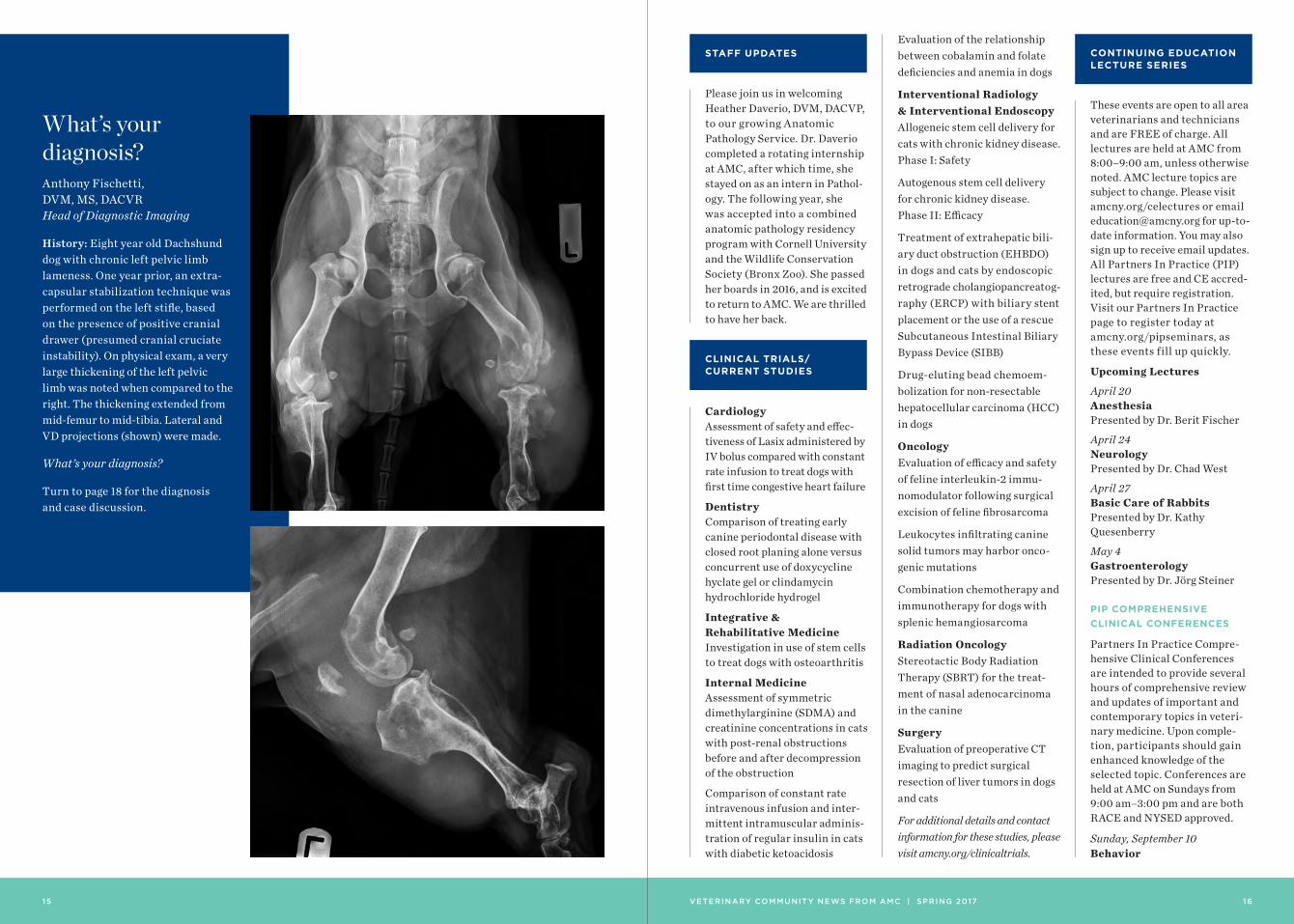
What’s your diagnosis?Anthony Fischetti, DVM, MS, DACVR Head of Diagnostic Imaging
History: Eight year old Dachshund dog with chronic left pelvic limb lameness. One year prior, an extra-capsular stabilization technique was performed on the left stifle, based on the presence of positive cranial drawer (presumed cranial cruciate instability). On physical exam, a very large thickening of the left pelvic limb was noted when compared to the right. The thickening extended from mid-femur to mid-tibia. Lateral and VD projections (shown) were made.
What’s your diagnosis?
Turn to page 18 for the diagnosis and case discussion.

17 VETERINARY COMMUNIT Y NEWS FROM AMC | SPRING 2017 18
What’s your diagnosis?Radiographic findings: The same lateral projection of the left stifle previously presented on another page. There is severe multifocal lysis of the distal femur, patella, and tibia. The lysis is localized to subchondral bone as well as areas of synovial joint attachment to bone (dark arrowheads). A very large soft tissue mass is centered on the stifle joint, completely effacing the infrapatellar fat pad and displacing the patella cranially (white arrows). Irregular periosteal reaction extends caudally from the proximal tibia. The adjacent proximal fibula is thin (partially lytic). A small lucency surrounded by a halo of scle-rosis is present in the proximal tibia, consistent with the pin tract from a prior extracapsular stabilization of the left stifle.
Radiographic diagnosis: 1. Large soft tissue mass with multi-focal regional bony lysis of the left stifle.
Comments: Synovial neoplasia is considered most likely, including histiocytic sarcoma, synovial cell sarcoma, and myxoma/myxosarcoma. Cranial cruciate instability may have been present at some point, but ligamentous instability alone would not produce these extensive radiographic changes.
The soft tissue component of the stifle mass was biopsied and confirmed to be an histiocytic sarcoma. Three views of the thorax were made at the time of these orthopedic radio-graphs and there was no evidence of pulmonary metastasis. The left pelvic limb was amputated and the dog recovered uneventfully. No other follow up is provided.
Sunday, November 12 LVT Lecture – New and Practical Updates
PIP PRACTICAL CLINICAL WORKSHOPS
Partners In Practice Practical Clinical Workshops are designed to promote sound diagnosis and effective therapies. Bring and share case materials if you wish! Participate in our time-honored teaching rounds and small group, interactive workshops. Space is limited to 15 participants, so register today! These PIP Work-shops are held at The AMC on Tuesday evenings from 7:00– 8:30 pm and are NYSED approved.
April 18 Endocrine Disease
May 16 Infectious Diseases
June 13 Radiology Reading
September 12 Dentistry
October 17 Cardiology
November 21 Common Emergency Toxicities
December 12 Exotic Medicine
Integrative MedicineDr. Leilani Alvarez conducted a large survey intended to identify referral patterns to small animal rehabilitation facilities and document veterinarians’ per-ceptions of these services. Most respondents (324/461 [70.3%]) had referred patients for post-operative rehabilitation therapy. Respondents ranked neurologic disorder as the condition they would most likely consider for referral for rehabilitation therapy. The most frequently cited rea-son for not referring a patient for
providing benefit for residents and emergency clinicians by providing immediate feedback.
Noel PG, Fischetti AJ, Moore GE, Le Roux AB. Off-site smartphone vs. standard workstation in the radiographic diagnosis of small intestinal mechanical obstruction in dogs and cats. Vet Radiol Ultra-sound. 2016 Sep;57(5):457-61.
Dr. Le Roux and colleagues studied the relationship between ex vivo canine small intestinal layering and histology because canine ultrasonographic intestinal layers have been reported to correlate with histological layering. They found no significant statistical differences between the ultrasono-graphic and histological measure-ments. Also, there was a strong to very strong positive correlation between for all layers, except for the serosa, which had a low mod-erate positive correlation.
Le Roux AB, Granger LA, Waka-matsu N, Kearney MT, Gaschen L. Ex vivo correlation of ultra-sonographic small intestinal wall layering with histology in dogs. Vet Radiol Ultrasound 2016 Sep;57(5):534-45.
This newsletter is distributed quar-terly to AMC’s network of refer-ring veterinarians and others who opt-in to receive this publication. If you have questions regarding this newsletter or would like to sign up to be included in our distribution, please email [email protected].
To receive our current staff directory, email [email protected].
For access to the AMC Patient Referral Form, visit amcny.org/referralform.
ABOUT THIS NEWSLETTER
rehabilitation therapy was per-ceived cost (54.4%) followed by distance to a rehabilitation facil-ity (29.3%); 87.4% of respondents felt that continuing education in the field of veterinary rehabilita-tion was lacking.
Alvarez LX, Fox PR, Van Dyke JB, Grigsby P. Survey of referring veterinarians’ perceptions of and reasons for referring patients to rehabilitation facilities. J Am Vet Med Assoc. 2016 Oct 1;249(7):807-13.
Interventional RadiologyDr. Allison Berent and colleagues described a balloon dilation procedure useful for dogs with nasopharyngeal stenosis. Com-puted tomography helps identify and define the nasal lesion. Their findings demonstrate that bal-loon valvuloplasty is an effective treatment for this condition.
Berent AC, Kinns J, Weisse C. Bal-loon dilatation of nasopharyngeal stenosis in a dog. J Am Vet Med Assoc. 2006 Aug 1;229(3):385-8.
Berent AC. Diagnosis and Man-agement of Nasopharyngeal Ste-nosis. Vet Clin North Am Small Anim Pract. 2016 Jul;46(4):677-89. Review.
Tong K, Weisse C, Berent AC. Rigid urethrocystoscopy via a percutaneous fluoroscopic-assisted perineal approach in male dogs: 19 cases (2005–2014). J Am Vet Med Assoc. 2016 Oct 15;249(8):918-925.
Lulich JP, Berent AC, Adams LG, Westropp JL, Bartges JW, Osborne CA. ACVIM Small Ani-mal Consensus Recommendations on the Treatment and Prevention of Uroliths in Dogs and Cats. J Vet Intern Med. 2016 Sep;30(5):1564-1574. doi: 10.1111/jvim.14559.
Berent AC. Interventional Radiol-ogy of the Urinary Tract. Vet Clin North Am Small Anim Pract. 2016 May;46(3):567-96, vii. doi: 10.1016/ j.cvsm.2015.12.011. Review.
Wilson KE, Berent AC, Weisse CW. Use of a percutaneously
controlled hydraulic occluder for treatment of refractory urinary incontinence in three female cats. J Am Vet Med Assoc. 2016 Mar 1;248(5):544-51. doi: 10.2460/javma.248.5.544.
Berent AC. Advances in Urinary Tract Endoscopy. Vet Clin North.
PathologyDr. Taryn Donovan and col-leagues at AMC and North Caro-lina State University discovered an association between dissemi-nated pyogranulomatous inflam-mation in a dog and Bartonella henselae infection. Bartonella infections have important One Health implications because diseases attributed to Bartonella include a wide range of condi-tions including osteomyelitis, CNS diseases, splenitis, endocar-ditis, hepatitis and lymphadenitis in dogs and humans. Additional research will help improve understanding of disease recog-nition and treatment options.
Donovan TA, Fox PR, Balakrish-nan N, Ericson M, Hooker V, Breitschwerdt EB. Pyogranulo-matous Pancarditis with Intra-myocardial Bartonella henselae San Antonio 2 (BhSA2) in a Dog. J Vet Intern Med. 2016 Nov 24. doi: 10.1111/jvim.14609. [Epub ahead of print]
RadiologyDrs. Noel, Fischetti and col-leagues compared the accuracy compressed images taken by smartphone devices and trans-mitted by email or text- versus standard radiographic abdominal radiographs- to assess diagnosis of small intestinal mechanical obstruction in vomiting dogs and cats. Accuracy of the smartphone vs standard abdominal image interpretation judged by radiolo-gists were not significantly differ-ent. They concluded that off-site expert consultation with a smart-phone provides an acceptable interface for accurate diagnosis of small intestinal mechani-cal obstruction in dogs and cat,
RESEARCH HIGHLIGHTS

510 East 62nd Street New York, NY 10065
VETERINARY COMMUNIT Y NEWS FROM AMC | SPRING 2017
AVIAN & EXOTICS Dr. Kathy Quesenberry Dr. Cyndi Brown 212-329-8888 Sunday – Friday 9 am – 5 pm
CARDIOLOGY Dr. Philip Fox Dr. Betsy Bond Dr. Dennis Trafny 212-329-8701 Monday – Sunday 9 am – 5 pm
DENTISTRY Dr. Dan Carmichael Dr. Stephen Riback Dr. Django Martel 212-329-8678 Monday – Friday 9 am – 5 pm
DERMATOLOGY Dr. Mark Macina 212-329-8777 Tuesday – Saturday 9 am – 5 pm
INTERNAL MEDICINE A Dr. Beth Appleman Dr. Carly Bloom 212-329-8619 Monday – Sunday 9 am – 5 pm
INTERNAL MEDICINE B Dr. Douglas Palma Dr. Dennis Slade 212-329-8675 Monday – Sunday 9 am – 5 pm
INTERVENTIONAL RADIOLOGY & INTERVENTIONAL ENDOSCOPY Dr. Chick Weisse Dr. Allyson Berent 212-329-8700 Monday – Friday 9 am – 5 pm
NEUROLOGY Dr. Chad West Dr. John McCue Dr. Abbie Lebowitz 212-329-8770 Monday – Sunday 9 am – 5 pm
ONCOLOGY Dr. Nicole Leibman Dr. Ann Hohenhaus Dr. Maria Camps 212-329-8797 Monday – Saturday 9 am – 5 pm
OPHTHALMOLOGY Dr. Alexandra van der Woerdt 212-329-8813 Monday 10 am – 6 pm Tuesday 10 am – 5 pm Thursday 2 pm – 9 pm Friday 9 am – 3 pm
RADIATION ONCOLOGY Dr. Rachel St-Vincent 212-329-8821 Monday – Friday 9 am – 5 pm
REHABILITATION & INTEGRATIVE MEDICINE Dr. Leilani Alvarez Dr. Barry Cherno 212-329-8860 Monday – Saturday 9 am – 5 pm
SURGERY SERVICE 2 Dr. Dan Spector 212-329-8863 Wednesday – Saturday 9 am – 5 pm
SURGERY SERVICE 3 Dr. Pamela Schwartz 212-329-8867 Monday – Friday 9 am – 5 pm
SURGERY SERVICE 4 Dr. Rob Hart 212-329-8674 Monday – Friday 9 am – 5 pm
A.M.C. PORTAL amcny.org/ referral-portal-login
DR. RICHARD GOLDSTEIN, CMO 347-733-7338 richard.goldstein @amcny.org
PRIORITY EMERGENCY/ CRITICAL CARE HOTLINE 212-329-8616 or 646-556-6411 (fax)
AMC Dedicated Phone Numbers for Referring Veterinarians