the endocrine system - linn–benton community...
TRANSCRIPT
Business
Reminder: Study for Quiz #2 Partial study guide in “Sample Exams” section of SG
Homework 11 in SG Omit #11
Due in lab this week
Part One
The Endocrine System
Overview
Acts with the nervous system to coordinate and integrate the
activity of body cells
Influences metabolic activities by means of hormones
Responses occur more slowly but tend to last longer than
those of the nervous system
Endocrine glands: pituitary, thyroid, thymus, pancreas,
parathyroid, adrenal, and pineal glands
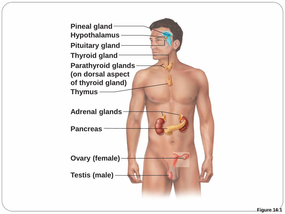
Figure 16.1
Pineal gland
Hypothalamus
Pituitary gland
Parathyroid glands
(on dorsal aspect
of thyroid gland)
Thymus
Thyroid gland
Adrenal glands
Pancreas
Ovary (female)
Testis (male)
Copyright © 2010 Pearson Education, Inc.
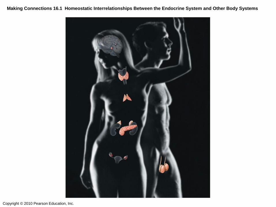
Making Connections 16.1 Homeostatic Interrelationships Between the Endocrine System and Other Body Systems

Overview
Nervous System Endocrine System
Nerve impulses
Neurotransmitters
Faster responses
Brief effects
Acts on specific target
Hormones
Slower responses
Longer effects
Broader influence
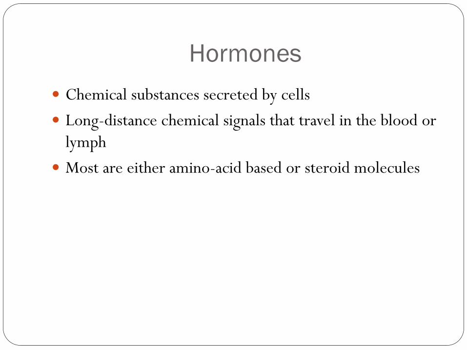
Hormones
Chemical substances secreted by cells
Long-distance chemical signals that travel in the blood or
lymph
Most are either amino-acid based or steroid molecules
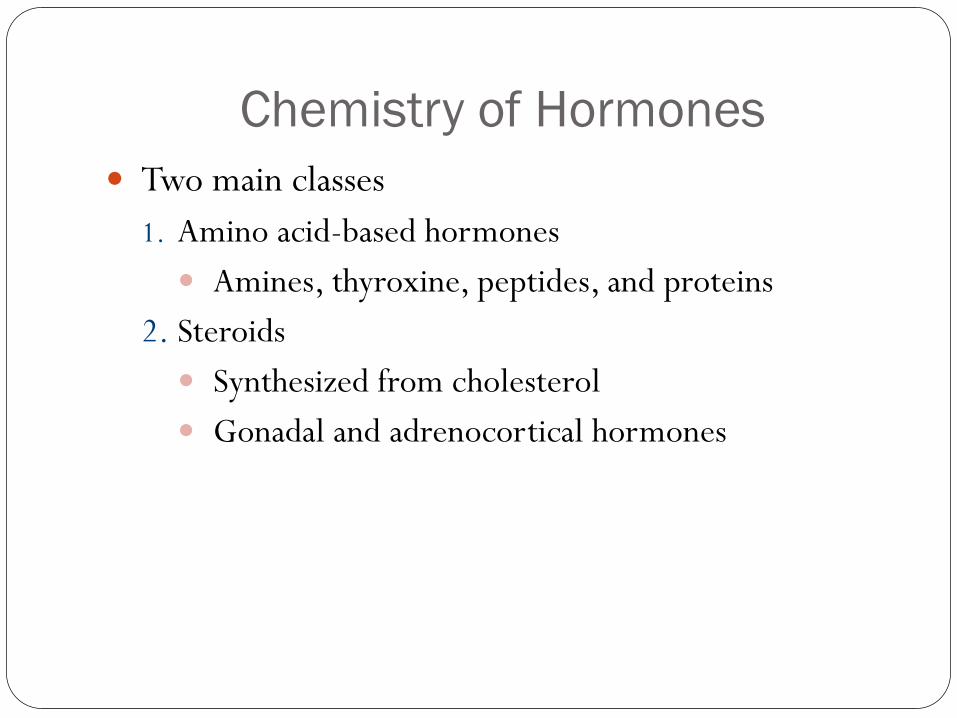
Chemistry of Hormones
Two main classes
1. Amino acid-based hormones
Amines, thyroxine, peptides, and proteins
2. Steroids
Synthesized from cholesterol
Gonadal and adrenocortical hormones
Copyright © 2010 Pearson Education, Inc.
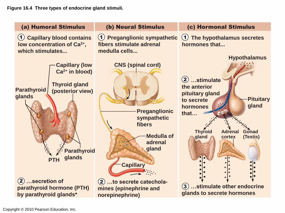
Figure 16.4 Three types of endocrine gland stimuli.
(a) Humoral Stimulus (b) Neural Stimulus (c) Hormonal Stimulus
Capillary (low
Ca2+ in blood)
Parathyroid
glands
Thyroid gland
(posterior view)
PTH
Parathyroid
glands
CNS (spinal cord)
Medulla of adrenal gland
Preganglionic
sympathetic
fibers
Capillary
Hypothalamus
Thyroid gland
Adrenal cortex
Gonad (Testis)
Pituitary
gland
1 Capillary blood contains
low concentration of Ca2+,
which stimulates...
1 Preganglionic sympathetic
fibers stimulate adrenal
medulla cells...
1 The hypothalamus secretes
hormones that...
2 …stimulate
the anterior
pituitary gland
to secrete
hormones
that…
2 …secretion of
parathyroid hormone (PTH)
by parathyroid glands*
2 …to secrete catechola-
mines (epinephrine and
norepinephrine)
3 …stimulate other endocrine
glands to secrete hormones
Target Cell Specificity
A hormone may have more than one type of target cell
Specific receptors
Hormone effects are due to alteration of cell’s activity Effects vary
Target Cell Activation
Target cell activation depends on three factors
1. Blood levels of the hormone
2. Relative number of receptors on or in the target cell
3. Affinity of binding between receptor and hormone
Target Cell Activation
Hormone concentration depends on
Rate of release and synthesis
Speed of inactivation
Hormones influence the number of their receptors
Up-regulation - target cells form more receptors in
response to the hormone
Down-regulation - target cells lose receptors in response
to the hormone
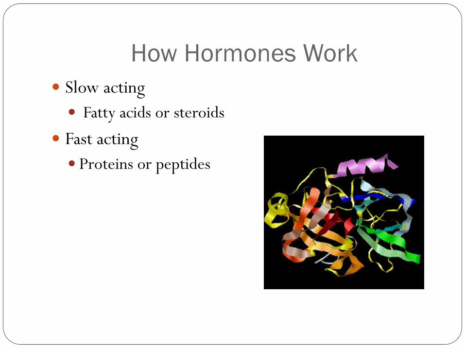
How Hormones Work
Slow acting
Fatty acids or steroids
Fast acting
Proteins or peptides
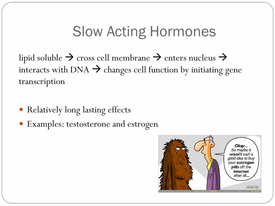
Slow Acting Hormones
lipid soluble cross cell membrane enters nucleus
interacts with DNA changes cell function by initiating gene
transcription
Relatively long lasting effects
Examples: testosterone and estrogen
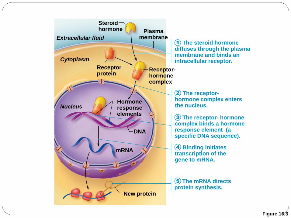
Figure 16.3
mRNA
New protein
DNA
Hormone response elements
Receptor- hormone complex
Receptor protein
Cytoplasm
Nucleus
Extracellular fluid
Steroid hormone
The steroid hormone diffuses through the plasma membrane and binds an intracellular receptor.
The receptor- hormone complex enters the nucleus.
The receptor- hormone complex binds a hormone response element (a specific DNA sequence).
Binding initiates transcription of the gene to mRNA.
The mRNA directs protein synthesis.
Plasma membrane
1
2
3
4
5
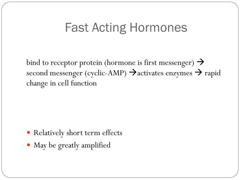
Fast Acting Hormones
bind to receptor protein (hormone is first messenger)
second messenger (cyclic-AMP) activates enzymes rapid
change in cell function
Relatively short term effects
May be greatly amplified
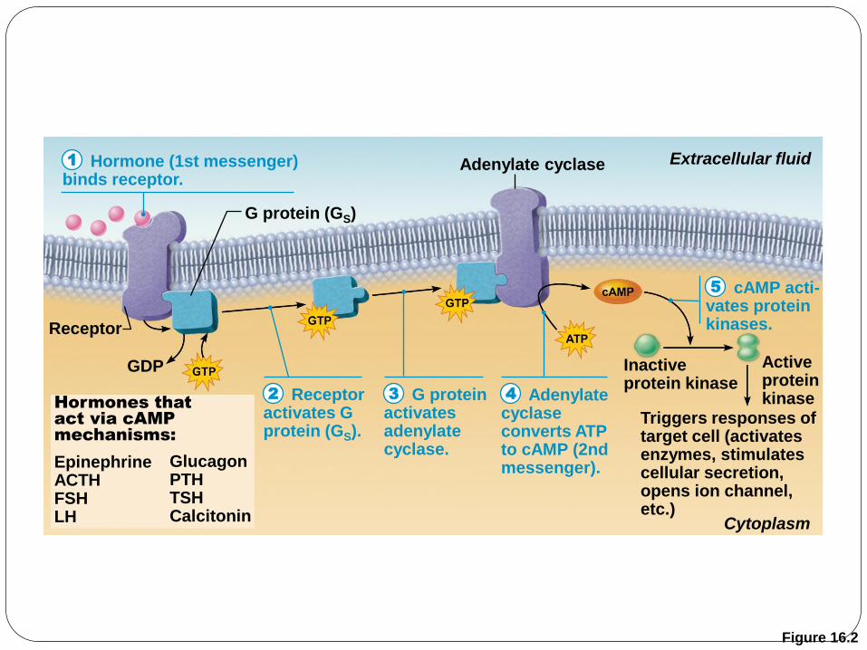
Figure 16.2
Hormone (1st messenger) binds receptor.
Receptor activates G protein (GS).
G protein activates adenylate cyclase.
cAMP acti- vates protein kinases.
Adenylate cyclase converts ATP to cAMP (2nd messenger).
Receptor
G protein (GS)
Adenylate cyclase
Triggers responses of target cell (activates enzymes, stimulates cellular secretion, opens ion channel, etc.)
Hormones that
act via cAMP
mechanisms:
Epinephrine ACTH FSH LH
Inactive protein kinase
Extracellular fluid
Cytoplasm
Active protein kinase
GDP
Glucagon PTH TSH Calcitonin
1
2 3 4
5
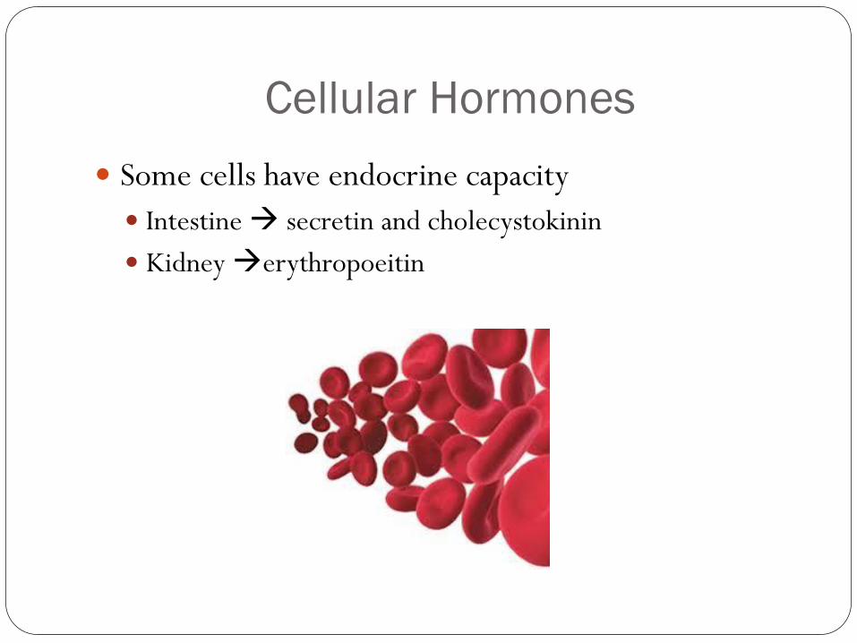
Cellular Hormones
Some cells have endocrine capacity
Intestine secretin and cholecystokinin
Kidney erythropoeitin
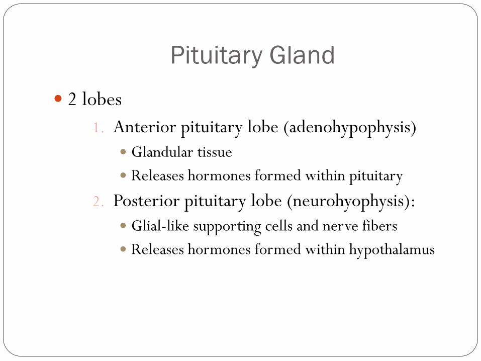
Pituitary Gland
2 lobes
1. Anterior pituitary lobe (adenohypophysis)
Glandular tissue
Releases hormones formed within pituitary
2. Posterior pituitary lobe (neurohyophysis):
Glial-like supporting cells and nerve fibers
Releases hormones formed within hypothalamus

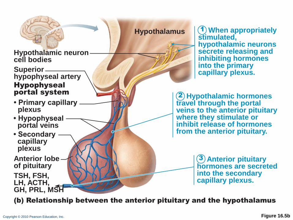
Copyright © 2010 Pearson Education, Inc. Figure 16.5b
1
2
3
When appropriately stimulated, hypothalamic neurons secrete releasing and inhibiting hormones into the primary capillary plexus.
Hypothalamic hormones travel through the portal veins to the anterior pituitary where they stimulate or inhibit release of hormones from the anterior pituitary.
Anterior pituitary hormones are secreted into the secondary capillary plexus.
Hypothalamus
Hypothalamic neuron cell bodies
Hypophyseal
portal system
Superior hypophyseal artery
(b) Relationship between the anterior pituitary and the hypothalamus
Anterior lobe of pituitary
TSH, FSH, LH, ACTH, GH, PRL, MSH
• Primary capillary plexus
• Hypophyseal portal veins
• Secondary capillary plexus
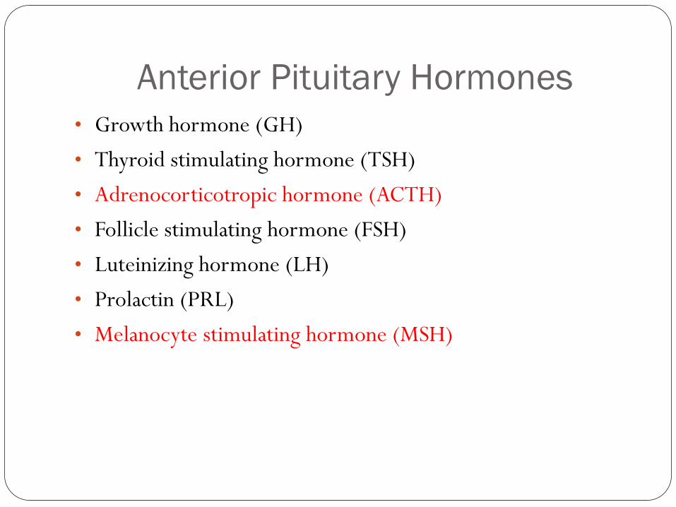
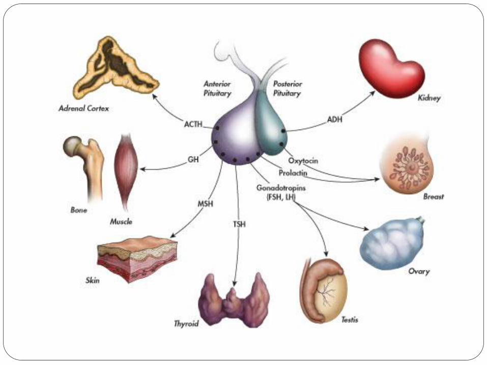
Anterior Pituitary Hormones
• Growth hormone (GH)
• Thyroid stimulating hormone (TSH)
• Adrenocorticotropic hormone (ACTH)
• Follicle stimulating hormone (FSH)
• Luteinizing hormone (LH)
• Prolactin (PRL)
• Melanocyte stimulating hormone (MSH)
Growth Hormone (GH)
Stimulates most cells, but targets bone and skeletal
muscle
Promotes protein synthesis, encourages use of fats for
fuel, and breakdown of glycogen
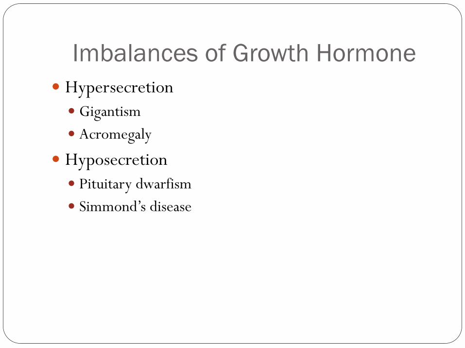
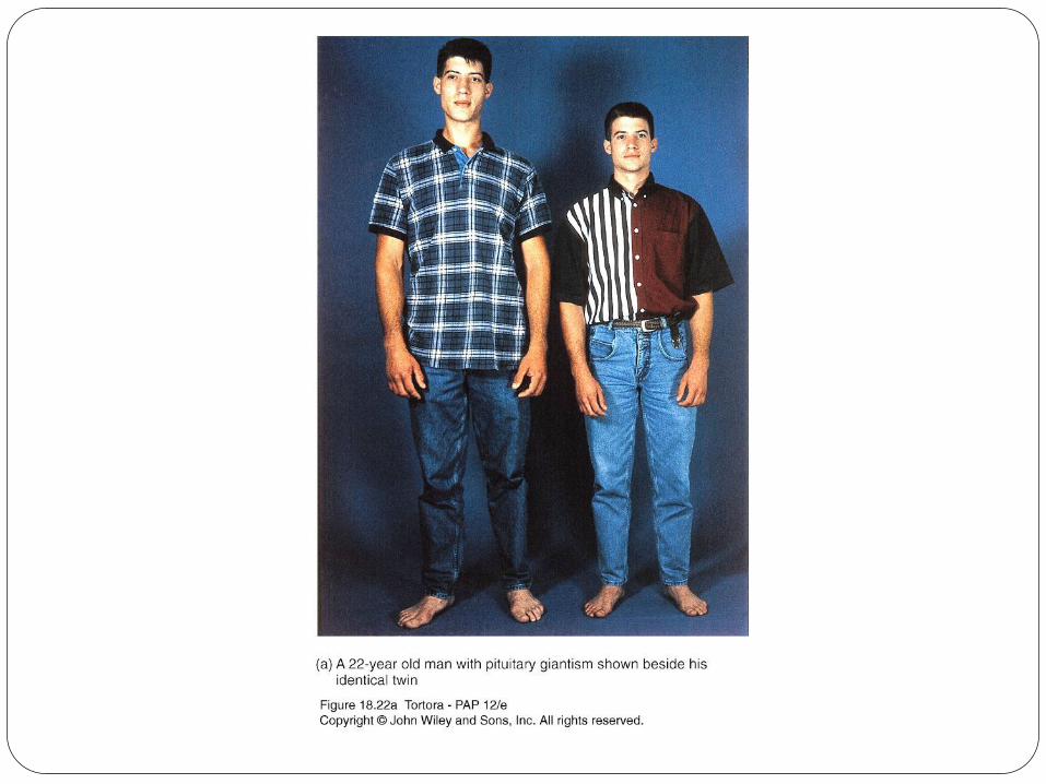
Imbalances of Growth Hormone
Hypersecretion
Gigantism
Acromegaly
Hyposecretion
Pituitary dwarfism
Simmond’s disease
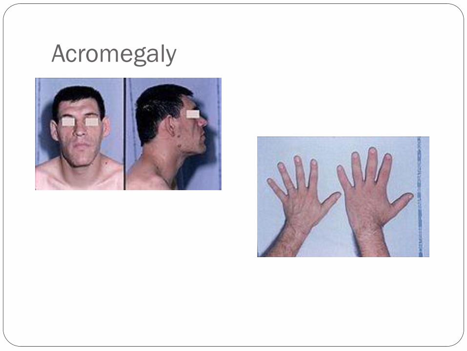
Acromegaly
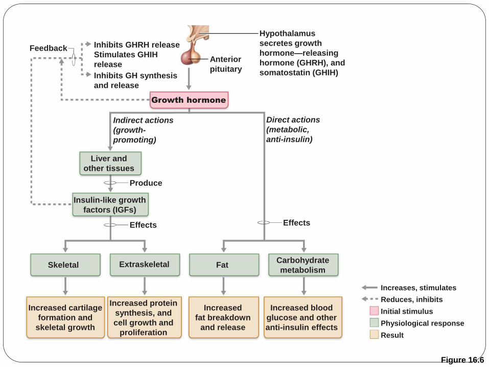
Figure 16.6
Growth hormone
Feedback Inhibits GHRH release
Stimulates GHIH
release
Inhibits GH synthesis
and release
Anterior
pituitary
Liver and
other tissues
Indirect actions
(growth-
promoting)
Direct actions
(metabolic,
anti-insulin)
Insulin-like growth
factors (IGFs)
Extraskeletal Skeletal Fat Carbohydrate
metabolism
Increased cartilage
formation and
skeletal growth
Increased protein
synthesis, and
cell growth and
proliferation
Increased
fat breakdown
and release
Increased blood
glucose and other
anti-insulin effects
Effects Effects
Produce
Hypothalamus
secretes growth
hormone—releasing
hormone (GHRH), and
somatostatin (GHIH)
Initial stimulus
Physiological response
Result
Increases, stimulates
Reduces, inhibits
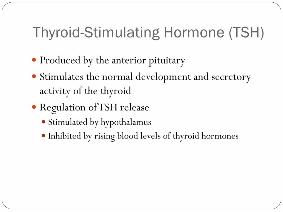
Thyroid-Stimulating Hormone (TSH)
Produced by the anterior pituitary
Stimulates the normal development and secretory
activity of the thyroid
Regulation of TSH release
Stimulated by hypothalamus
Inhibited by rising blood levels of thyroid hormones
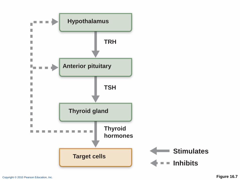
Copyright © 2010 Pearson Education, Inc. Figure 16.7
Hypothalamus
Anterior pituitary
Thyroid gland
Thyroid
hormones
TSH
TRH
Target cells Stimulates
Inhibits
Adrenocorticotropic Hormone (ACTH)
Secreted by the anterior pituitary
Stimulates the adrenal cortex to release corticosteroids
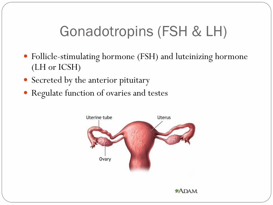
Gonadotropins (FSH & LH)
Follicle-stimulating hormone (FSH) and luteinizing hormone (LH or ICSH)
Secreted by the anterior pituitary
Regulate function of ovaries and testes
Gonadotropins
Regulation of gonadotropin release
Triggered by the gonadotropin-releasing hormone (GnRH)
Absent from the blood in prepubertal boys and girls
Suppressed by gonadal hormones
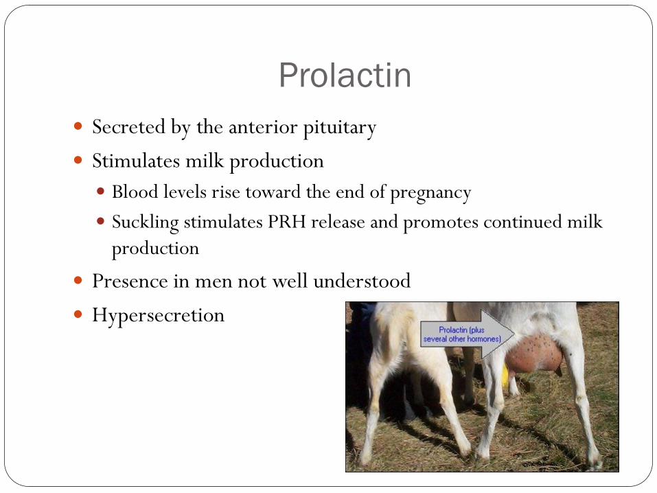
Prolactin
Secreted by the anterior pituitary
Stimulates milk production
Blood levels rise toward the end of pregnancy
Suckling stimulates PRH release and promotes continued milk
production
Presence in men not well understood
Hypersecretion
Melanocyte Stimulating Hormone
Stimulates melanocytes
• Unknown role in humans
• May influence brain activity
Control of Anterior Pituitary Hormones Pituitary-Hypothalamic Relationships
Hypophyseal portal system
Capillary plexuses
Hypophyseal portal veins
Copyright © 2010 Pearson Education, Inc. Figure 16.5b
1
2
3
When appropriately stimulated, hypothalamic neurons secrete releasing and inhibiting hormones into the primary capillary plexus.
Hypothalamic hormones travel through the portal veins to the anterior pituitary where they stimulate or inhibit release of hormones from the anterior pituitary.
Anterior pituitary hormones are secreted into the secondary capillary plexus.
Hypothalamus
Hypothalamic neuron cell bodies
Hypophyseal
portal system
Superior hypophyseal artery
(b) Relationship between the anterior pituitary and the hypothalamus
Anterior lobe of pituitary
TSH, FSH, LH, ACTH, GH, PRL, MSH
• Primary capillary plexus
• Hypophyseal portal veins
• Secondary capillary plexus
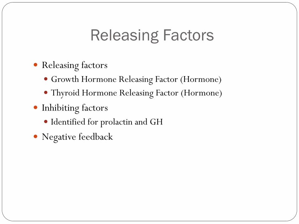
Releasing Factors
Releasing factors
Growth Hormone Releasing Factor (Hormone)
Thyroid Hormone Releasing Factor (Hormone)
Inhibiting factors
Identified for prolactin and GH
Negative feedback
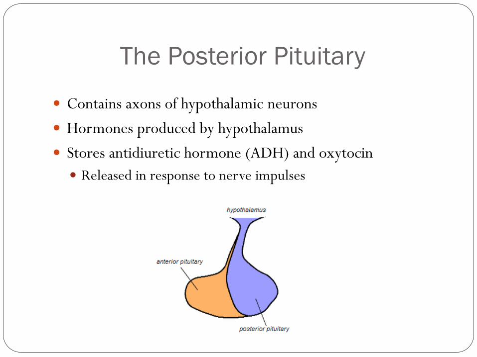
The Posterior Pituitary
Contains axons of hypothalamic neurons
Hormones produced by hypothalamus
Stores antidiuretic hormone (ADH) and oxytocin
Released in response to nerve impulses
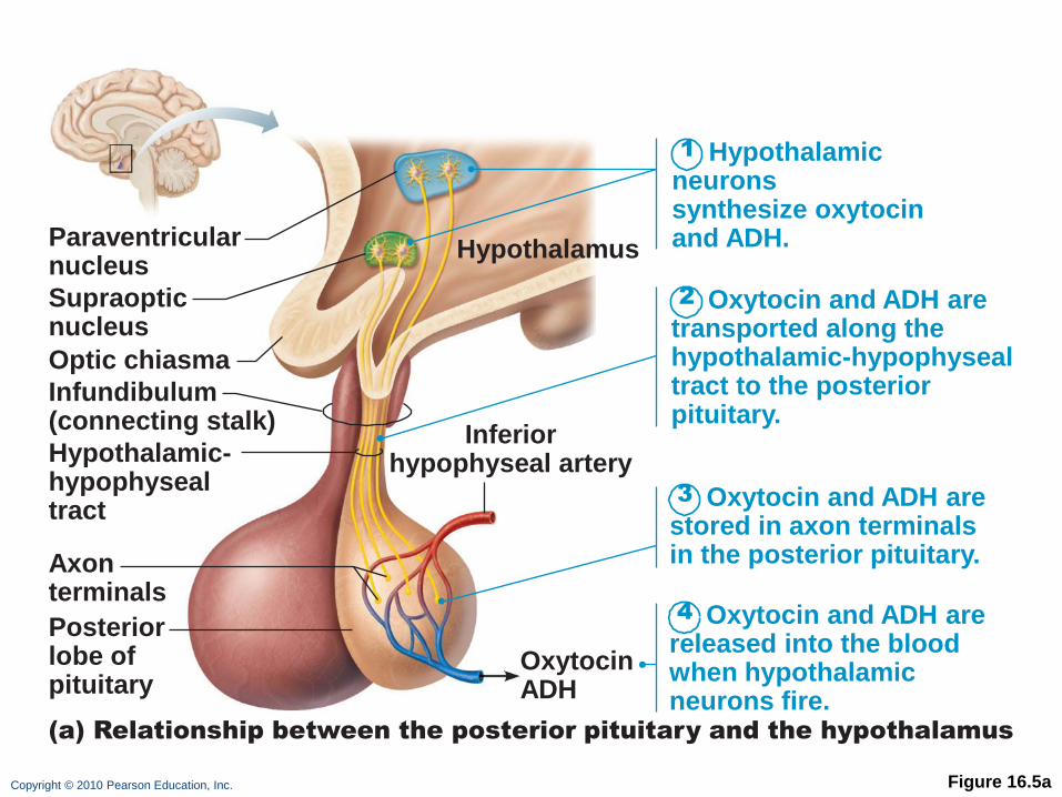
Copyright © 2010 Pearson Education, Inc. Figure 16.5a
1
2
3
4
Hypothalamic neurons
synthesize oxytocin and ADH.
Oxytocin and ADH are transported along the hypothalamic-hypophyseal tract to the posterior pituitary.
Oxytocin and ADH are stored in axon terminals in the posterior pituitary.
Oxytocin and ADH are released into the blood when hypothalamic neurons fire.
Paraventricular nucleus
Supraoptic nucleus
Optic chiasma
Hypothalamus
Inferior hypophyseal artery
Oxytocin ADH
Infundibulum (connecting stalk)
Hypothalamic- hypophyseal tract
Axon terminals
Posterior lobe of pituitary
(a) Relationship between the posterior pituitary and the hypothalamus
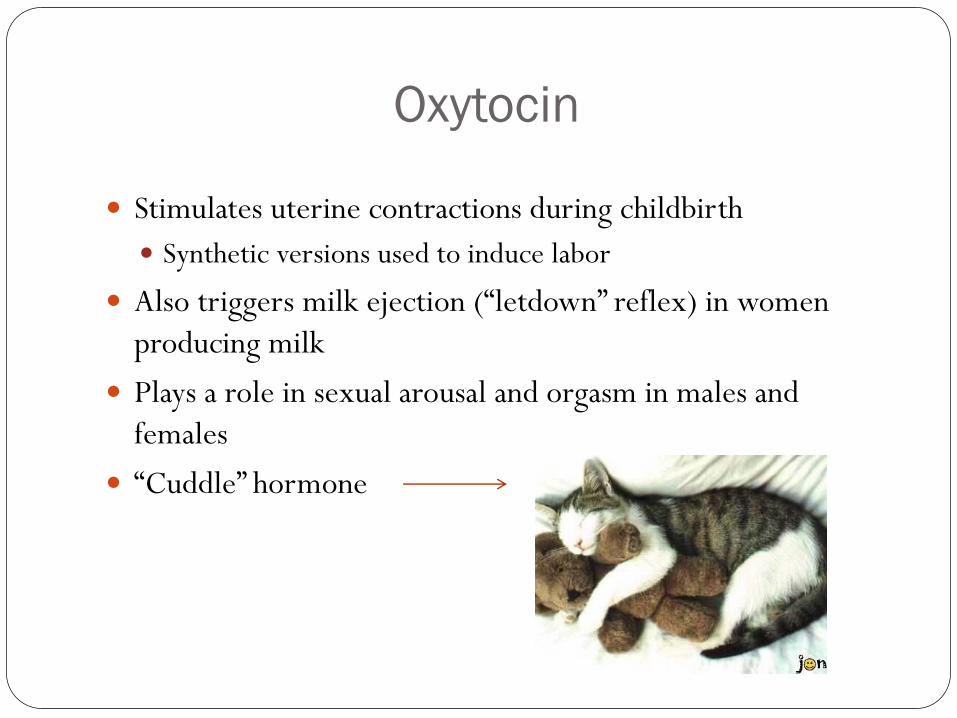
Oxytocin
Stimulates uterine contractions during childbirth
Synthetic versions used to induce labor
Also triggers milk ejection (“letdown” reflex) in women
producing milk
Plays a role in sexual arousal and orgasm in males and
females
“Cuddle” hormone
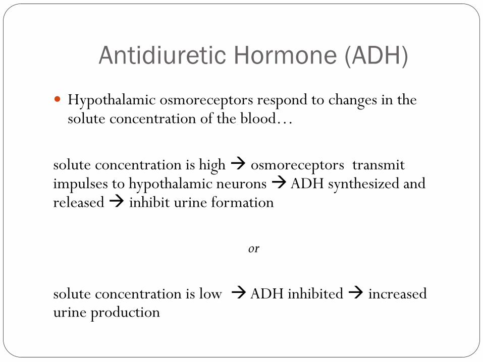
Antidiuretic Hormone (ADH)
Hypothalamic osmoreceptors respond to changes in the solute concentration of the blood…
solute concentration is high osmoreceptors transmit impulses to hypothalamic neurons ADH synthesized and released inhibit urine formation
or
solute concentration is low ADH inhibited increased urine production
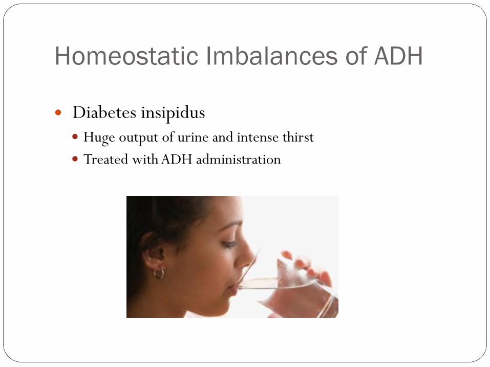
Homeostatic Imbalances of ADH
Diabetes insipidus Huge output of urine and intense thirst
Treated with ADH administration
Questions?