the epidemiology of rabbit haemorrhagic disease virus in new zealand
TRANSCRIPT

Factors influencing the epidemiology
of Rabbit haemorrhagic disease virus
in New Zealand
A thesis presented
in partial fulfilment of the requirements
for the degree of Doctor of Philosophy
at Massey University, Palmerston North, New Zealand
Jörg Henning
2003

ii

iii
Abstract
The European rabbit (Oryctolagus cuniculus) is a major pest species in New Zealand.
The illegal introduction of Rabbit haemorrhagic disease virus (RHDV) in 1997, for
purposes of biological control of pest rabbit populations, was a controversial event due
to uncertainties about the impact of the disease on both rabbit populations and other
potential host species. This thesis presents a series of studies conducted to investigate
several aspects of the epidemiology and biology of RHDV in New Zealand, and to
assess the opinions of farmers about the usefulness of RHDV for rabbit control.
A longitudinal study was conducted in an area of low rabbit density near Himatangi
in the lower North Island of New Zealand. Rabbits were trapped at weekly intervals
over 37 months using a capture-mark-recapture approach. The study was initiated
shortly after the first rabbit haemorrhagic disease (RHD) epidemic occurred in the area,
and focused on evaluating the relationships between the occurrence of RHD and the
dynamics of the rabbit population. Over the course of the study, predation, particularly
by cats, was the principal cause of rabbit mortality. RHDV was present every year in the
late summer-autumn period, but caused discernible outbreaks with high mortality only
in the first and third years. Fluctuations in population immunity due to population
turnover and influx of susceptible immigrants appeared to be key factors contributing to
the intermittent occurrence of the disease. Infected migrant rabbits may also be a source
of reintroduction of virus and new disease outbreaks. Rabbit deaths due to RHD were
clustered in time and space, and RHD affected animals died closer to their home range
centroid than rabbits dying of other causes.
In cats, ferrets, stoats and hedgehogs that were trapped at the site, seropositivity to
RHDV was detected up to several months before and after RHDV infections in wild
rabbits. These predatory and scavenging species may act as vectors causing localized
spread of the disease. During the course of the study, the abundance of six fly species
identified as potential RHD vectors was also determined. The influence of climatic
factors on fly abundance varied between species, and peaks in fly abundance in late
summer and autumn coincided with RHD outbreaks.
iv
Two aspects of the survival of RHDV in the environment were investigated using
experimental exposure of laboratory rabbits to determine viral infectivity. The survival
of RHDV on two matrices (liver and cotton) exposed to environmental conditions on
open pasture was evaluated. RHDV in bovine liver tissue, used to emulate a rabbit
carcass, remained infective for up to three months under field environmental conditions.
RHDV on cotton, which was used to emulate excreted RHDV on an inanimate
substrate, remained infective for less than half that time. These observations suggest
that RHDV in decomposing rabbit carcasses could be a relatively persistent reservoir of
the virus. RHDV that was inactivated with UV-light failed to induce protective
immunity in rabbits following oral or parenteral injection, indicating that inactivated
virus on baits is unlikely to induce protective immunity in wild rabbits and thereby
jeopardise the effectiveness of RHDV use.
Using a multistage sampling frame, the attitudes and practices of farmers regarding
rabbit control, and particularly RHDV, were evaluated using a mail questionnaire.
Shooting remains the predominant method that farmers use to control rabbits, although
10% of farmers used RHDV baiting. The use of poisoning and trapping for rabbit
control has declined since the introduction of RHDV. Most farmers considered that the
introduction of RHDV has been beneficial.
The impact of RHDV on rabbit populations appears to be highly variable. These
studies have provided detailed documentation on the occurrence of RHDV and its
relationship to rabbit population dynamics in an area of low rabbit density. Overall, the
findings suggest that both the expected benefits and the potential ecological risks from
introducing RHD to New Zealand, were overstated. While the disease certainly had a
marked impact on the population at Himatangi at certain times, outbreaks were
intermittent and other 'natural' causes of mortality may exert greater constraints on
rabbit populations. Better understanding of the factors that contribute to the variability
in frequency and severity of RHD outbreaks may enable more efficient use of this
method in the future. RHDV is likely to remain a useful option for rabbit control,
particularly in areas of severe rabbit proneness, and will likely prove most effective
when used in conjunction with other methods.

v
Acknowledgements
If I had not started this PhD, I might have never visited Aotearoa, New Zealand. But
I came to this country and spent more then five years not only researching, but also
falling in love with this ‘Land of the Long White Cloud’. This was a wonderful
opportunity to learn and to live in this country, from which I will move on, like most of
my supervisors did.
Firstly I want to thank Dirk Pfeiffer, who sent me an e-mail from Malawi and
described this project and changed my plans for the future. I never started the
anticipated PhD in the Rift valley and went to Palmerston North instead where I met
one of the most passionate data-analysts. I thank Dirk for his consistent exposure to all
the new statistical and epidemiological tools, which make life both easy and
complicated, even after his departure to England. I am so grateful to Joanne Meers, for
her enthusiasm and friendship during all these years. I acknowledge gratefully her help
and guidance in all aspects of virology. Joanne’s tremendous support still remained
vibrant after her move to Australia. Peter Davies was another exceptional supervisor,
with great wisdom and an ability to refine my many pages to the heart of the matter. I
acknowledge Peter’s help in reviewing my work and I was lucky to finish most of it
before he left for America. I also thank John Parkes, whose immediate reply from the
South Island and help in all technical issues of the project was outstanding. I finally
want to thank, my fifth supervisor, Roger Morris, who guided with his good judgment
the overall progress of the work.
The project involved intensive fieldwork and I thank the farmers, who allowed me to
‘chase rabbits’ on their properties. I thank the local Regional Council field staff for their
encouragement from the beginning (‘…there is no way to catch a rabbit alive, the only
way to get them is to shoot or poison them’) and their assistance in all ‘rabbit issues’
from night counts to night shooting.
I also would like to acknowledge all the help I received from so many people from
the EpiCentre, Nigel Perkins’ logical explanations, Mark Stevenson’s Aussie spirit,
Cord Heuer’s unlimited experience and Ron Jackson’s philosophical approach, Deb’s
and Julie’s budgeting, Fiona’s and Colleen’s secretarial assistance, and Solis Norton’s

vi
and Daniel Russell’s technical support. I thank all students from so many countries, who
crossed my path during my time in the EpiCentre, but I am especially grateful to the
German contingent, who always gave some native language support; thank you Dirk,
Carola, Klim and Cord.
I would like to thank all those friends I met over the years in Palmerston North and
New Zealand, and the list would be too long to name all of them, but I would like to
especially thank Sylvia, Delwyn, Peter, Petra, Steffen, Bruce and Joanna.
I thank my partner Sonja, who came to live with me in New Zealand, and I
appreciate her help with radio-tracking rabbits through thorny gorse bushes and her
patience for waiting until everything was written up.
Finally I want to thank my family in Germany. Thank you Kerstin, my sister, for the
parcels with items from our former ‘Heimat’. I thank my parents for their endless
support. They visited me and I saw them a few times back home, but I can only imagine
how difficult it must have been for them to see me always flying back to the other side
of the planet.
‘Writing in the speed of thoughts remains the dream of an author’ (Heiner Müller,
East-German play writer in ‘Krieg ohne Schlacht’, 1999, Kiepenheuer and Witsch,
Köln, Germany), and thoughts need nutrients to flourish. I also found strength and joy
for my research in the hours I spent away from it, on the top of some mountains or in
remote rugged patches of native bush, and must therefore somehow acknowledge the
New Zealand wilderness.
Jörg Henning
EpiCentre, Institute of Veterinary, Animal and Biomedical Sciences
Massey University, Palmerston North, New Zealand
26th June 2003
vii
Table of Contents
ABSTRACT.................................................................................................................iii
ACKNOWLEDGEMENTS.......................................................................................... v
TABLE OF CONTENTS............................................................................................vii
LIST OF FIGURES ...................................................................................................xiii
LIST OF TABLES..................................................................................................... xvi
LIST OF APPENDICES............................................................................................ xix
INTRODUCTION ............................................................................................. 1
REFERENCES ............................................................................................................. 5
CHAPTER 1 RABBIT HAEMORRHAGIC DISEASE: A LITERATURE REVIEW ......................................................................................................... 9
THE EMERGENCE OF RABBIT HAEMORRHAGIC DISEASE .......................... 11
RHDV TAXONOMY AND MOLECULAR BIOLOGY .......................................... 14
NON-PATHOGENIC RABBIT CALICIVIRUS....................................................... 16
PROPERTIES OF RABBIT HAEMORRHAGIC DISEASE VIRUS............................ 19
HOST RANGE OF RABBIT HAEMORRHAGIC DISEASE VIRUS .......................... 21
THE TRANSMISSION OF RHD............................................................................... 25
Transmission between rabbits................................................................................. 25 Transmission via insects ......................................................................................... 27
Transmission routes via insects .......................................................................... 27 Potential arthropod vectors of RHDV in New Zealand...................................... 28 Arthropod vectors of RHDV in Australia........................................................... 29 Factors influencing transmission via insects ...................................................... 30
Transmission via predators and scavengers............................................................ 31 Transmission by humans ........................................................................................ 31
THE CLINICAL SIGNS OF RHD............................................................................. 32
THE PATHOLOGY OF RHD.................................................................................... 34
LABORATORY DIAGNOSIS OF RHD ................................................................... 36
Detection of RHDV ................................................................................................ 37 Detection of RHDV antibodies............................................................................... 38
EPIDEMIOLOGY OF RHD IN WILD RABBITS, WITH EMPHASIS ON NEW ZEALAND.................................................................................................................. 41
Europe..................................................................................................................... 41 Australia.................................................................................................................. 42 New Zealand ........................................................................................................... 42 General patterns of disease occurrence................................................................... 43
OMISSIONS IN THE LITERATURE ....................................................................... 46

viii
THE PRESENT RESEARCH PROJECT: OUTLINE OF RESEARCH AIMS AND INTENDED OUTCOMES..........................................................................................48
REFERENCES............................................................................................................49
CHAPTER 2 A THREE-YEAR STUDY OF RABBIT HAEMORRHAGIC DISEASE VIRUS INFECTION IN A WILD RABBIT POPULATION IN NEW ZEALAND ....77
ABSTRACT ................................................................................................................79
INTRODUCTION.......................................................................................................79
MATERIALS AND METHODS ................................................................................80
Study site .................................................................................................................80 Data collection.........................................................................................................80
Serology ..............................................................................................................81 Age classification ................................................................................................82 Detection of RHDV infection in dead rabbits .....................................................83 Rabbit abundance ................................................................................................83 Monthly (or seasonal) serological and mortality data.........................................84
RESULTS....................................................................................................................84
Rabbit population dynamics....................................................................................84 Serology of adult (>11 weeks) rabbits ....................................................................87 Serology of young (≤11 weeks) rabbits ..................................................................92
DISCUSSION .............................................................................................................93
Rabbit population dynamics....................................................................................93 Population size and migration .............................................................................93 Temporal patterns in reproduction ......................................................................95 Mortalities ...........................................................................................................96
Seroepidemiology....................................................................................................96 Test characteristics and cut-off selection ............................................................96 Evidence of an RCV-like virus ...........................................................................98 Maternally derived antibodies.............................................................................98 Seroconversion in adult rabbits surviving epidemics..........................................99
ACKNOWLEDGEMENTS ......................................................................................101
REFERENCES..........................................................................................................102
CHAPTER 3 SPATIAL AND TEMPORAL DYNAMICS OF MORTALITY AND HOME RANGE USE IN WILD RABBITS (ORYCTOLAGUS CUNICULUS) WITH AN EMPHASIS ON RABBIT HAEMORRHAGIC DISEASE (RHD) .................111
ABSTRACT ..............................................................................................................113
INTRODUCTION.....................................................................................................113
MATERIALS AND METHODS ..............................................................................114
Study area..............................................................................................................114 Radio-tracking of rabbit locations.........................................................................114 Statistical analyses.................................................................................................116
Cause-specific mortality....................................................................................116 Temporal patterns of mortality..........................................................................116

ix
Core and home range estimation ...................................................................... 117 Spatio-temporal clustering of RHD deaths....................................................... 118
RESULTS ................................................................................................................. 119
Cause-specific mortality ....................................................................................... 119 Temporal patterns of mortality ............................................................................. 121
Mortality as a function of calendar time........................................................... 121 Mortality as a function of animal age ............................................................... 122
Core and home ranges........................................................................................... 123 Core range size.................................................................................................. 124 Home range size................................................................................................ 125 Core range habitat ............................................................................................. 127 Death location in relation to home range centroid............................................ 128
Spatio-temporal clustering of RHD deaths........................................................... 128
DISCUSSION........................................................................................................... 129
Cause-specific mortality ....................................................................................... 129 Temporal patterns of mortality ............................................................................. 131 Core and home ranges........................................................................................... 132
Core range size.................................................................................................. 132 Home range size................................................................................................ 133 Core range habitat ............................................................................................. 134 Death location in relation to home range centroid............................................ 136
Spatio-temporal clustering of RHD deaths........................................................... 136
ACKNOWLEDGEMENTS...................................................................................... 138
REFERENCES ......................................................................................................... 138
CHAPTER 4 TEMPORAL RELATIONSHIPS BETWEEN SEROPOSITIVITY TO RABBIT HAEMORRHAGIC DISEASE VIRUS IN RABBITS AND OTHER MAMMALS IN NEW ZEALAND................................................................... 147
ABSTRACT.............................................................................................................. 149
INTRODUCTION .................................................................................................... 149
MATERIALS AND METHODS.............................................................................. 150
RESULTS ................................................................................................................. 151
DISCUSSION........................................................................................................... 155
REFERENCES ......................................................................................................... 160
CHAPTER 5 INFLUENCE OF WEATHER CONDITIONS ON FLY ABUNDANCE AND ITS IMPLICATIONS FOR TRANSMISSION OF RABBIT HAEMORRHAGIC DISEASE VIRUS IN THE NORTH ISLAND OF NEW ZEALAND..................... 169
ABSTRACT.............................................................................................................. 171
INTRODUCTION .................................................................................................... 171
MATERIALS AND METHODS.............................................................................. 172
Study site............................................................................................................... 172 Sampling and identification of flies...................................................................... 173

x
Climate data...........................................................................................................173 Data analysis .........................................................................................................174
RESULTS..................................................................................................................175
DISCUSSION ...........................................................................................................181
REFERENCES..........................................................................................................188
CHAPTER 6 SURVIVAL OF RABBIT HAEMORRHAGIC DISEASE VIRUS (RHDV) IN THE ENVIRONMENT...............................................................197
SUMMARY ..............................................................................................................199
INTRODUCTION.....................................................................................................199
MATERIALS AND METHODS ..............................................................................200
Virus ......................................................................................................................200 Preparation of viral suspension and its exposure to the environment ...................200 Rabbits...................................................................................................................201 RHDV antibody testing.........................................................................................202 Study design ..........................................................................................................203
Pilot experiment 1 .............................................................................................203 Pilot experiment 2 .............................................................................................203 Long-term exposure study.................................................................................204
Weather data recording .........................................................................................204 Data analysis .........................................................................................................204
RESULTS..................................................................................................................205
Pilot Experiment 1.................................................................................................205 Pilot Experiment 2.................................................................................................206 Long-term exposure study.....................................................................................207 Serology ................................................................................................................209 Survival analysis ...................................................................................................209 Environmental conditions .....................................................................................211
DISCUSSION ...........................................................................................................214
ACKNOWLEDGEMENTS ......................................................................................218
REFERENCES..........................................................................................................219
CHAPTER 7 EXPOSURE OF RABBITS TO ULTRAVIOLET LIGHT-INACTIVATED RABBIT HAEMORRHAGIC DISEASE VIRUS (RHDV) AND SUBSEQUENT CHALLENGE WITH VIRULENT VIRUS.................................225
SUMMARY ..............................................................................................................227
INTRODUCTION.....................................................................................................227
MATERIALS AND METHODS ..............................................................................228
Animals .................................................................................................................228 RHDV inactivation................................................................................................228 Experimental Design .............................................................................................229 Assessment of outcomes .......................................................................................229
RESULTS..................................................................................................................230

xi
DISCUSSION........................................................................................................... 231
ACKNOWLEDGEMENTS...................................................................................... 233
REFERENCES ......................................................................................................... 233
CHAPTER 8 ATTITUDES OF NEW ZEALAND FARMERS TO METHODS USED TO CONTROL WILD RABBITS ......................................................... 237
ABSTRACT.............................................................................................................. 239
INTRODUCTION .................................................................................................... 239
MATERIALS AND METHODS.............................................................................. 240
Selection of regions .............................................................................................. 240 Classification of farms .......................................................................................... 242 Selection of farms within regions ......................................................................... 243 Statistical analysis................................................................................................. 245
RESULTS ................................................................................................................. 246
Response rate ........................................................................................................ 246 Perceptions of problems caused by rabbits........................................................... 246 Methods used for rabbit control............................................................................ 249
Shooting ............................................................................................................ 249 RHD baiting...................................................................................................... 250 Use of poisoning and trapping .......................................................................... 251
Farmers’ perceptions about rabbit control methods ............................................ 252 Factors associated with the use of rabbit control methods ................................... 254
Shooting ............................................................................................................ 254 Use of other control methods............................................................................ 256
Farmers’ concerns about Rabbit Haemorrhagic Disease...................................... 257
DISCUSSION........................................................................................................... 258
ACKNOWLEDGEMENTS...................................................................................... 262
REFERENCES ......................................................................................................... 262
CHAPTER 9 GENERAL DISCUSSION ....................................................... 273
RHDV IN WILD RABBIT POPULATIONS .......................................................... 275
FLY VECTORS........................................................................................................ 278
METHODS OF RABBIT CONTROL IN RELATION TO NATURAL MORTALITY........................................................................................................... 278
THE LONG-TERM EFFECT OF RHDV ................................................................ 280
CONCLUSION......................................................................................................... 281
REFERENCES ......................................................................................................... 281

xii

xiii
List of Figures
Figure 1: Histogram for age at first capture for animals up to 155 days of age. ............ 83
Figure 2: Number of rabbits alive and mortality incidence density per 1000 rabbit days for a rabbit population in Himatangi, New Zealand between May 1998 and June 2001....................................................................................... 85
Figure 3: Proportion of female rabbits pregnant and numbers of rabbits under 11 weeks in Himatangi, New Zealand between September 1998 and June 2001............................................................................................................... 85
Figure 4: Number of rabbits older than 11 weeks sampled monthly and temporal pattern of seroprevalence of RHDV for this age group in Himatangi, New Zealand between May 1998 and June 2001. .............................. 87
Figure 5: Number of adult animals (> 11 weeks) sampled by season, stratified for residents and immigrants in Himatangi, New Zealand between winter 1998 and winter 2001................................................................................. 89
Figure 6: Serological status to RHDV of adult rabbits by season from winter 1998 to winter 2001 in Himatangi, New Zealand. ................................................ 91
Figure 7: Number of seropositive rabbits categorised by immune status, based on isotype ELISA results in Himatangi, New Zealand between May 1998 and June 2001............................................................................................... 92
Figure 8: Number of immature rabbits (<11 weeks) sampled monthly and their temporal pattern of seroprevalence in Himatangi, New Zealand between May 1998 and June 2001. ....................................................................... 93
Figure 9: Cause-specific incidence density of mortalities (expressed as deaths per 1,000 rabbit-days at risk) as a function of calendar time for the three major natural mortality causes recorded in this study: RHD, predation and other natural causes. ..................................................................................... 121
Figure 10: Number of animals dying from RHD in seven day intervals during the 2001 RHD epidemic. ..................................................................................... 122
Figure 11: Kaplan-Meier survival curves showing the estimated survival as a function of age stratified by the three natural causes of death (RHD, predation, and other natural causes). ................................................................... 123
Figure 12: Box-and-whisker plots of the distributions of observed home range size (ha) based on 95% kernel density estimation............................................... 124
Figure 13: Death locations of rabbits dying between 1/03/1999 and 31/07/2001........................................................................................................... 129
Figure 14: Number of RHDV seropositive and seronegative cats trapped between August 1999 and June 2001. ................................................................. 153
Figure 15: Number of RHDV seropositive and seronegative stoats trapped between August 1999 and June 2001. ................................................................. 153
Figure 16: Number of RHDV seropositive and seronegative ferrets trapped between August 1999 and June 2001. ................................................................. 154

xiv
Figure 17: Number of RHDV seropositive and seronegative hedgehogs trapped between August 1999 and June 2001 .....................................................154
Figure 18: Positions of flytraps located under shrubs and on open pasture in relation to vegetation. ..........................................................................................172
Figure 19: Number of flies trapped per species over the entire study period. ..............175
Figure 20: Number of flies trapped per species on open pasture and gorse edges. ...................................................................................................................176
Figure 21: Abundance of blowfly and flesh fly species from October 1999 until June 2001 at Himatangi. ..............................................................................176
Figure 22 A-E: Climate pattern and abundance of O. varia from October 1999 until June 2001 at Himatangi. ..............................................................................179
Figure 23: Kaplan Meier survival plot for hours to death of rabbits inoculated with a 10 RHDV dilution prepared from liver after exposure to environmental conditions.
0
....................................................................................210
Figure 24: Kaplan Meier survival plot for hours to death of rabbits inoculated with a 10 RHDV dilution prepared from liver after exposure to environmental conditions.
-2
....................................................................................210
Figure 25: Kaplan Meier survival plot for hours to death of rabbits inoculated with a 10 RHDV dilution derived from cotton after exposure to environmental conditions.
0
....................................................................................210
Figure 26: Kaplan Meier survival plot for hours to death of rabbits inoculated with a 10 RHDV dilution derived from cotton after exposure to environmental conditions.
-2
....................................................................................210
Figure 27: Daily average and daily range of the Temperature-Humidity Index for the periods of RHDV exposure to the environment.......................................213
Figure 28: Day-time and night-time range of the Temperature-Humidity Index for the periods of RHDV exposure to environmental conditions ........................213
Figure 29: Percentage of farms categorised by rabbit damage severity in 1995 and 2001...............................................................................................................248
Figure 30: Use of RHD baiting by 58 farms for different farm types (BEF beef, DAR dairy, NLS non-livestock, SHP sheep, SNB mixed sheep-and-beef, OLF other livestock) in five different rabbit-prone areas in 2001 (as percentages)...........................................................................................251
Figure 31: Frequency distributions of farmer opinion about the humaneness of rabbit control methods .........................................................................................252
Figure 32: Frequency distributions of farmer opinion about the environmental safety of rabbit control methods ..........................................................................253
Figure 33: Frequency distributions of farmer opinion about the effectiveness of rabbit control methods.....................................................................................254
Figure 34: Odds ratio (95% CI) describing the strength of the association between shooting and farmers’ perceptions on the extent that rabbits compete with livestock for grazing......................................................................255

xv
Figure 35: Odds ratio (95% CI) describing the strength of association between shooting and farmers’ perceptions on the effectiveness of shooting................... 255

xvi

xvii
List of Tables
Table 1: Gross findings and microscopic lesions found in RHD infected rabbits .................................................................................................................... 34
Table 2: Categories of rabbit immune status to RHDV based on results of the competition and isotype ELISAs........................................................................... 82
Table 3: Age distribution of rabbits alive (based on trapping and radio-tracking events) by season and year ...................................................................... 86
Table 4: Number of resident and immigrant seropositive and seronegative adult rabbits for RHDV by season ........................................................................ 90
Table 5: Criteria used to assign causes of rabbit death................................................. 116
Table 6: Classification of body condition at death for 107 rabbit deaths examined in a 38 month observational study of rabbit mortality in a 30 ha rural area in Himatangi, New Zealand............................................................ 119
Table 7: Summary of mortality causes and descriptive statistics of estimated age at the time of death for 93 rabbit carcasses................................................... 120
Table 8: Geometric least squares mean of estimated core range area (ha) stratified by sex and season for rabbits radio-tracked during daylight hours between autumn 1999 and winter 2001. Core ranges were estimated using kernel density and mean convex polygon methods................... 125
Table 9: Geometric least squares mean of estimated home range area (ha) stratified by sex and season for rabbits radio-tracked during daylight hours between autumn 1999 and winter 2001. Home ranges have been estimated using kernel density and mean convex polygon methods................... 126
Table 10: Observed and geometric least squares means of core and home range areas (ha) for rabbits radio-tracked between autumn 1999 and winter 2001. Core and home ranges have been described using kernel density and mean convex polygon methods........................................................ 127
Table 11: Vegetation types used by rabbits as denning sites located during daytime ................................................................................................................ 127
Table 12: Distance between home range centroid and death location for different causes of death in wild rabbits, stratified for sex ................................. 128
Table 13: RHDV sero-prevalence in non-target species stratified by sex.................... 152
Table 14: Variables obtained from principal factor analysis and their number of correlations (correlation coefficient > 0.7) with other climate variables............................................................................................................... 177
Table 15: Summary of General Linear Models for climate factors influencing the abundance of six different fly species (log numbers +1). ............................. 180
Table 16: Separate General Linear Model for the climate factors influencing the abundance of O. varia on pasture and gorse (log numbers +1)..................... 181

xviii
Table 17: Number of rabbits showing disease or sero-conversion to RHDV following intramuscular injection with virus preparations from inoculated cotton tape and bovine liver held at 4°C for 24 hours........................205
Table 18: Number of rabbits showing disease or sero-conversion to RHDV following oral dosing with virus preparations from inoculated cotton tape and bovine liver held in the environment for 1 or 5 days ............................206
Table 19: Number of rabbits showing disease or sero-conversion to RHDV following oral dosing with virus preparations from inoculated cotton tape and bovine liver held in the environment for 1, 10, 44 and 91 days............208
Table 20: Antibody titres and time of death (in hours) of RHDV- inoculated rabbits...................................................................................................................209
Table 21: Mean daily temperature and humidity recordings for different periods of RHDV exposure to the environment ..................................................212
Table 22: Mean day-time and night-time temperature and humidity recordings for different periods of RHDV exposure to the environment..............................212
Table 23: Serology and mortality results after inoculation of rabbits with inactivated RHDV followed by challenge with virulent RHDV .........................230
Table 24: UV energy dosages (microwatt-seconds per square centimetre) required to inactivate selected organisms ............................................................232
Table 25: General characteristics of regions selected for surveying.............................241
Table 26: Distribution of New Zealand farms by selected farm types..........................243
Table 27: Matrix showing proportional weights (farm type by rabbit-proneness) and number of questionnaires targeted, mailed and returned............244
Table 28: Percentage of farmers who perceived rabbits to be a problem, classified by rabbit-proneness, region and farm type ..........................................247
Table 29: Farmers perceptions of mechanisms by which rabbits impact on their enterprises for each problem caused by rabbits...........................................248
Table 30: Reported percentage usage of rabbit control methods in 1995 and in 2001 amongst 574 farmers...................................................................................249
Table 31: Percentage of farmers using RHDV in 2001 stratified by rabbit-proneness .............................................................................................................250
Table 32: Odds ratios from binary logistic regression of the use of RHDV to control rabbits ......................................................................................................256
Table 33: Farmers’ opinions on aspects of RHDV as a tool for rabbit control.............258

xix
List of Appendices
Appendix 1: Climate variables created from data collected at Himatangi study site and obtained from regional weather stations ................................................ 194
Appendix 2: Life cycle data obtained from the literature for two different fly species. ................................................................................................................ 194
Appendix 3: Rabbit-proneness map of New Zealand used as a primary spatial stratum for selection of farms.............................................................................. 266
Appendix 4: Questionnaire used to survey New Zealand farmers on their attitudes to methods used to control wild rabbits. ............................................... 267


1
Introduction

2
3
Man has successfully domesticated only a few mammalian species as sources of food
and fibre, for transport and labour, and for recreation and companionship. Among these
species, the European rabbit (Oryctolagus cuniculus) has arguably the most complex
relationship with our species, being either revered or maligned according to the
circumstances. Originating from the Iberian Peninsula, the territories occupied by
rabbits expanded throughout most of Europe, and subsequently to all continents other
than Antarctica, facilitated by traders and early settlers, sailors and hunters. The rabbit
is farmed for meat and fur production and is an important game species, especially in
Europe. It also plays vital roles in some ecosystems, either as stable prey for endangered
predator species or by contributing to seed dispersal (Staniforth and Cavers 1977;
Palomares 2001). In contrast, the introduction of rabbits into favourable environments,
particularly in New Zealand and Australia, has had undesirable outcomes for local
ecology and agriculture. Rabbits compete with livestock for grazing, damage the soil
surface, destroy crops and seedlings and pose a threat to the ecology through heavy
grazing on native plants (Gibb and Williams 1994). In New Zealand, the direct costs of
rabbit control in 1995 were estimated to be NZ$ 22 million (Parkes 1995). The principal
means of control are shooting and poisoning, with efforts at biological control in the
early 1950s using myxomatosis being unsuccessful (Filmer 1953).
An apparently novel infectious disease of rabbits, now known as rabbit haemorrhagic
disease (RHD), was first recognised in China in 1984. A number of general reviews on
the biology of rabbit haemorrhagic disease (RHD) have been published in recent years
(Ohlinger et al. 1993; Thiel and König 1999; Anon. 1996; Cooke and Fenner 2002).
The high morbidity and mortality of RHD observed in domesticated and wild rabbits
heralded the significance of this disease as both a threat to desirable rabbit populations,
and a potential tool for biological control of pest rabbit populations. In contrast with the
low public acceptance of myxomatosis in New Zealand in the 1950s (Gibb and
Williams 1994), in the mid 1990s public recognition and acceptance of the potential of
RHD as a means for rabbit control was considerably stronger (Fitzgerald et al. 1996;
Wilkinson and Fitzgerald 1998). However, after thorough investigation into the
anticipated benefits and risks of introducing Rabbit haemorrhagic disease virus
(RHDV), the New Zealand government denied an application to import the virus. The
virus was subsequently released illegally in 1997 (O'Hara 1997), and is now widely
established in this country. However, many aspects of RHD epidemiology in New

4
Zealand, including temporal and spatial patterns of occurrence and its impact on rabbit
populations, remain uncertain.
This thesis reports several studies conducted to investigate aspects of the
epidemiology of RHD in an area of low rabbit density in New Zealand, and to evaluate
farmers’ perceptions on the role of RHD in rabbit control. The reports on these studies
are preceded by a literature review on RHDV infection in rabbits. The literature relevant
to each of the specific research areas is reviewed in the introduction of each chapter.
In detail, the thesis presented is set out in the following chapters:
Chapter 1: Literature review on the existing knowledge about the agent causing
RHD, including characteristics of the disease and its epidemiology in wild rabbit
populations.
Chapter 2: Longitudinal study of RHDV infection and rabbit population dynamics in
a wild rabbit population.
Chapter 3: Longitudinal study of spatial and temporal patterns of mortality due to
RHDV infection and other causes of death in wild rabbits.
Chapter 4: Longitudinal study on temporal association between RHDV infection in
wild rabbits and seropositivity in rabbit predators and scavengers.
Chapter 5: Longitudinal study of the influence of weather conditions on abundance
of blowflies and flesh flies most likely to be involved in the transmission of RHDV.
Chapter 6: Experimental study of the persistence of RHDV in the environment.
Chapter 7: Experimental study of immune responses of rabbits inoculated orally and
intramuscularly with RHDV inactivated with UV light.
Chapter 8: Survey of farmers’ attitudes to the use of RHDV and other rabbit control
methods.
Chapter 9: General discussion of the results, implications for further research and the
use of RHDV as a biological control agent in the future.
Data collection for all longitudinal studies (Chapter 2-Chapter 5) was conducted on a
site near Himatangi in the North Island of New Zealand. Experimental studies on
rabbits were conducted at Massey University, Palmerston North. Animal ethics
approval was obtained for all research involving live animals.
Chapters 2-8 are manuscripts of papers submitted to peer reviewed journals and are
presented in the style required by the journal. All papers were submitted before the

5
submission of the thesis and the journal title is given on the title page of the relevant
chapter. Tables, figures and appendices are numbered sequentially throughout the
thesis. References and appendices are given at the end of each chapter. The taxonomy
for the names and abbreviations of viruses described in the research results of this thesis
are according to the Seventh Report of the International Committee on Taxonomy of
Viruses (van Regenmortel et al. 2000).
References
Anon. (1996). 'Import impact assessment and application to the Director General of
Agriculture to approve importation of rabbit calicivirus'. (RCD Applicant Group
(final document): Dunedin, New Zealand.)
Cooke, B. D., and Fenner, F. (2002). Rabbit haemorrhagic disease and the biological
control of wild rabbits, Oryctolagus cuniculus, in Australia and New Zealand.
Wildlife Research 29, 689-706.
Filmer, J. F. (1953). Disappointing tests of myxomatosis as rabbit control. New Zealand
Journal of Agriculture 87, 402-404.
Fitzgerald, G. P., Saunders, L. S., and Wilkinson, R. L. (Eds.) (1996). 'Public attitudes
to the biological control of rabbits in New Zealand'. (Ministry of Agriculture: New
Zealand.)
Gibb, J. A., and Williams, J. M. (1994). The rabbit in New Zealand. In 'The European
rabbit: the history and biology of a successful colonizer'. (Eds. G. B. Corbet, J. E.
C. Flux, P. M. Rogers, C. P. Arthur, R. C. Soriguer, K. Myers, I. Parer, D. Wood,
B. D. Cooke, J. A. Gibb, J. M. Williams, F. Fenner, J. Ross, H. V. Thompson, and
C. M. King.) pp. 158-200. (Oxford University Press: Oxford, UK.)
O'Hara, P. J. (1997). 'Decision on the application to approve the importation of rabbit
calicivirus as a biological control agent for feral rabbits. 2 July 1997'. (Ministry of
Agriculture: New Zealand.)

6
Ohlinger, V. F., Haas, B., and Thiel, H. J. (1993). Rabbit haemorrhagic disease (RHD):
characterisation of the causative calicivirus. Veterinary Research 24, 103-16.
Palomares, F. (2001). Vegetation structure and prey abundance requirements of the
Iberian lynx: implications for the design of reserves and corridors. Journal of
Applied Ecology 38, 9-18.
Parkes, J. (1995). 'Rabbits as pests in New Zealand: A summary of the issues and
critical information, Landcare Research Contract Report LC9495/141'. (Landcare
Research: Lincoln, New Zealand.)
Staniforth, R. J., and Cavers, P. B. (1977). The importance of cottontail rabbits in the
dispersal of Polygonum Spp. Journal of Applied Ecology 14, 261-268.
Thiel, H. J., and König, M. (1999). Caliciviruses: an Overview. Veterinary
Microbiology 69, 55-62.
van Regenmortel, M. H. V., Fauquet, C. M., Bishop, D. H. L., Carstens, E. B., Estes, M.
K., Lemon, S. M., Maniloff, J., Mayo, M. A., McGeoch, D. J., Pringle, C. R., and
Wickner, R. B. (2000). Family Caliviridae. In 'Virus Taxonomy. Classification and
Nomenclature of Viruses. Seventh Report of the International Committee on
Taxonomy of Viruses'. pp. 725-739. (Academic Press: San Diego, USA.)
Wilkinson, R. and Fitzgerald, G. (Eds.) (1998). 'Public attitudes to rabbit calicivirus
disease in New Zealand'. (Manaaki Whenua Press: Lincoln, Canterbury, New
Zealand.)

7

8
9
Chapter 1
Rabbit haemorrhagic disease:
A literature review

10

11
The emergence of rabbit haemorrhagic disease
The first report of an apparently novel infectious disease, now generally known as
rabbit haemorrhagic disease (RHD), came from the Jiangsu Province, in the People’s
Republic of China in 1984 (Liu et al. 1984). In a relatively short period of time, this
peracute infectious disease of rabbits was recognised in several countries and given
numerous names prior to the identification of the causative agent. These included ‘X-
disease’ or ‘Mallatia X’ (Cancellotti and Renzi 1991), ‘Rabbit viral haemorrhagic
disease’ (Liu et al. 1984), ‘Viral haemorrhagic disease of rabbits’ (Xu et al. 1985),
‘Rabbit viral sudden death’ (Lee and Park 1987), ‘Picornavirus haemorrhagic fever in
rabbits’ (An et al. 1988), ‘Infectious Necrotising Hepatitis of Rabbits’ (Boujon et al.
1989), ‘Viral haemorrhagic pneumonia in rabbits’ (Cao et al. 1986) and ‘Infectious
haemorrhagic disease of rabbits’ (Löliger et al. 1989).
In common with all apparently novel infectious diseases in man and animals, the
origin of the causative agent has been the subject of considerable interest and
speculation. The location of the index outbreak in China clearly suggests a Chinese
origin of the disease, and the subsequent spread to Europe has been attributed to exports
of rabbits and rabbit meat from China to European states (Haas and Thiel 1993).
However, other authors have questioned this explanation, as RHD outbreaks occurred
simultaneously in 1988 in several European countries spanning a long distance from
eastern to western and southern Europe (Löliger and Eskens 1991).
Although first recognised in China, these initial cases of RHD occurred in Angora
rabbits that had been imported from Germany a few days before the outbreak, and a
European origin of the agent has been postulated (Xu et al. 1988; Löliger and Eskens
1991; Xu 1991). It was later suggested, with some support from serological data (Rodak
et al. 1990a), that a non-pathogenic calicivirus possibly present in Europe for years,
developed into a pathogenic strain in the different environment of China (Haas and
Thiel 1993).
Some authors saw a possible connection between RHD and a clinically and
pathologically similar disease in the European brown hare population (termed European
Brown Hare Syndrome - EBHS). EBHS was recognised prior to the emergence of RHD
and occurred in hare populations in geographical areas of Europe similar to those

12
affected by RHD. In addition, experimental studies indicated that cross-infection of
these viruses in the heterologous species was possible (di Modugno and Nasti 1990).
Genetic studies found a 52% to 60% nucleotide homology between the viruses causing
RHD and EBHS, indicating that they are distinct caliciviruses, albeit more closely
related to each other than to other known caliciviruses (Nowotny et al. 1997).
A feature of RHD has been its rapid dissemination after its initial occurrence in a
region. In China, RHD swept over an area of 50,000 km² in less than nine months (Xu
1991). In response to this an eradication program was implemented that included the
development of vaccine from inactivated virus, and by the end of 1986 the disease was
gradually coming under control (Huang 1991). In 1986, the first cases of RHD were
reported from the Republic of Korea (An et al. 1988; Park et al. 1991). The disease
spread in a westerly direction towards Europe, affecting many countries on the way.
Outbreaks occurred in the former USSR in 1986 and 1987 and the first clinical cases in
Europe were reported in Italy, also in 1986 (Morisse et al. 1991). Rabbit meat is an
important food in Europe, therefore most of the early reports focused on farmed rabbits.
Trade in contaminated rabbit products is suspected to have been the source of many
outbreaks (Morisse et al. 1991).
In former Czechoslovakia, morbidity and mortality from RHD outbreaks were
estimated to be between 80-100%. The spread of the disease was very fast and
approximately 30 million animals died in the summer and autumn of 1987 in this
country alone (Rodak et al. 1991). The first outbreaks of RHD in Germany were
recorded in the second half of 1988 (Soike et al. 1989; Löliger et al. 1989), with
mortality varying from 5 to 100% (Maess and Matthes 1990; Schlüter et al. 1990a). In
common with observations in Austria (Nowotny et al. 1992) and in Czechoslovakia
(Rodak et al. 1991), the affected farms were typically small (Löliger and Eskens 1991).
It was suggested that small rabbitries may use more fresh grass or other green feed,
which was potentially contaminated by virus from wild rabbits, while large farms prefer
industrial meshed feed (Schlüter and Schirrmeier 1991; Löliger and Eskens 1991).
RHD outbreaks were reported from most continental European countries between
1987 and 1990 (Morisse et al. 1991). In 1992, the first RHD outbreak in the United
Kingdom was reported in Ascot. One of the outbreaks in the United Kingdom was
traced to a shipment of frozen rabbit meat from China, but later transmission

13
experiments from the same Chinese meat source failed (Chasey 1994). All other
outbreaks occurred over a wide area of southeast England, which suggested RHDV had
crossed the British channel via ferry traffic, aerosols or birds.
The source for the first outbreak in Mexico City was 18 tonnes of Chinese rabbit
meat imported through the United States (Gregg et al. 1991). The outbreak was traced
to a colony of rabbits owned by a commercial rabbit breeder, who had contact with the
frozen meat six days before the first case was reported. These circumstantial links were
later supported by phylogenetic studies indicating a close relationship between the
Chinese and the Mexican viruses (Nowotny et al. 1997). Successful eradication of RHD
from Mexico was achieved without vaccination by destroying 110,000 domestic rabbits
(Gregg et al. 1991), and was undoubtedly aided by the absence of wild European rabbits
in this country.
Reunion Island also regularly imports frozen rabbit meat from China. The
breakdown of refrigerating units following a cyclone in 1989 resulted in large numbers
of spoilt rabbit carcasses having to be disposed. It had been suggested that farmed rabbit
colonies on the island became contaminated after dogs had contact with the Chinese
rabbit carcasses (Morisse et al. 1991). Other non-European countries reporting RHD
outbreaks were Egypt (1988), Lebanon (1989), Tunisia (1989), Israel (1990), India
(1990) and Cuba (1993). More recent RHD epidemics occurred in 1995 in Benin
(Kpodekon and Alogninouwa 1998), in 1996 in Saudi Arabia (Elzein and Al-Afaleq
1999) and in 2000 in the mid-west of the USA (Anon. 2000).
In contrast to many countries, where rabbit meat is considered a valuable protein
source, in New Zealand and Australia wild rabbits are viewed as major pests for
agriculture and an ecological threat to native fauna and flora. The high morbidity and
mortality observed in European outbreaks of RHD pointed to the potential application
of the Rabbit haemorrhagic disease virus (RHDV) as a biological control agent for pest
rabbit populations. Studies to examine the potential of RHDV as a biocontrol agent
were underway in Australia when, in 1995, the agent escaped from Wardang Island, a
small island 4 km offshore from mainland Australia, and spread rapidly across the
continent (Cooke and Fenner 2002). RHDV was illegally introduced in August 1997 to
New Zealand, following a rejection by the Ministry of Agriculture to import RHDV as a

14
means of wild rabbit control (Thompson and Clark 1997). Subsequently, the release of
RHDV to control rabbits was legalised and a commercial RHDV product became
available.
RHDV taxonomy and molecular biology
Although Liu et al. (1984) identified the causative agent of RHD to be viral, its
taxonomic status remained uncertain for several years. Early reports classified the agent
variously as a picornavirus (Pu et al. 1985; Cao et al. 1986; Lee and Park 1987) a
parvovirus (Xu et al. 1988; Gregg and House 1989) or, because of the larger size of
RHDV, a ‘parvo-like’ virus (Du 1990). Ohlinger et al. (1989) were the first to classify
the RHD virus as a calicivirus. After other research teams confirmed this observation
(Soike et al. 1989; Smid et al. 1989; Granzow et al. 1989; Valicek et al. 1990; Nowotny
et al. 1990; Rodak et al. 1990b; Erber et al. 1991; Park et al. 1991) Rabbit
haemorrhagic disease virus (RHDV) was placed in the family Caliciviridae (Moussa et
al. 1992).
Caliciviruses are grouped in four genera. Two of the genera include viruses causing
human gastroenteritis (Norwalk-like and Sapporo-like viruses). The genus Vesivirus,
includes Feline calicivirus (FCV), San Miguel sea lion virus (SMSV) and Vesicular
exanthema of swine virus (VESV). The genus Lagovirus includes Rabbit Haemorrhagic
disease virus (RHDV) and European brown hare syndrome virus (EBHSV) (Green et
al. 2000).
Estimates of the mean diameter of the virions range from 27 nm (Erber et al. 1991)
to 40 nm (Ohlinger et al. 1989). The virus surface consists of regularly arranged, cup-
shaped depressions (Smid et al. 1989; Granzow et al. 1989; Ohlinger et al. 1989, 1990a;
Rodak et al. 1991; Marcato et al. 1991). A single stranded, non-segmented, positive
sense RNA genome of 7,437 nucleotides is enclosed within a protein capsid (Meyers et
al. 1991a, 1991b). The genomic RNA encodes, in its first long open reading frame
(ORF1), a large polyprotein of 257 kDa. In addition to the genomic RNA of 7.5 kb, a
subgenomic RNA of 2.2 kb, which is colinear with the 3' region of the genome, has
been found (Ohlinger et al. 1990a). RHDV, in common with other caliciviruses,
synthesises the capsid protein (VP60) from translation of both genomic and subgenomic
15
RNA (Parra et al. 1993; Boniotti et al. 1994; Wirblich et al. 1996; Capucci et al.
1996a). This large structural capsid protein has a molecular weight of 60 kDa (Parra and
Prieto 1990; Ohlinger et al. 1990a). Small amounts of a minor structural protein VP10
are encoded by the second ORF (ORF2) located at the 3' end of the RNA (Meyers et al.
1991a; Wirblich et al. 1996). Furthermore a virion protein, VPg, is covalently attached
to the 5' end of both the genomic and subgenomic RNA (Meyers et al. 1991a, 2000;
Thiel and König 1999).
A low level of genetic variation between RHD viruses has been described in several
studies (Meyers et al. 1991a; Milton et al. 1992; Boga et al. 1994; Guittre et al. 1995;
Nowotny et al. 1997; Asgari et al. 1999; Moss et al. 2002; Le Gall-Recule et al. 2003).
Guittre et al. (1995) found the degree of nucleotide sequence homology for four
geographically distinct strains to be 96%, while Nowotny et al. (1997) found the
sequence homology to be between 89.4-100% for samples from 17 different countries.
These results strongly suggested that all RHDV strains are closely related to each other.
In another study, eight different phylogenetic groups of RHDV have been identified
based on capsid sequences of British RHDV samples and published sequences from 9
other countries (Moss et al. 2002). European isolates had a maximum difference of 14%
from the British samples, but little geographical correlation between individual viruses
was found, indicating an efficient dispersion of viruses by vectors and human activities.
Likewise no clear clustering of sequences according to their geographical origin was
found for Australian samples (Asgari et al. 1999).
Little genomic variation among RHD viruses was seen over time between Australian
samples obtained over a two-year period (Asgari et al. 1999), nor between French
isolates collected over 11 years (Le Gall-Recule et al. 2003). The Australian study
indicated only minor genetic differences (98.2% to 100% identity) in the RHDV
sequences from different field samples compared to the original Czechoslovakian strain
of the virus (Asgari et al. 1999).
In contrast, some studies assessed antigenic variation in RHDV, based on the virus’s
ability to agglutinate human erythrocytes (Chasey et al. 1995; Capucci et al. 1996b),
while others demonstrated greater variation in the VP60 gene between RHDV strains
obtained from vaccinated rabbit flocks in Germany (Schirrmeier et al. 1999). A

16
particularly interesting development in the epidemiology of the disease, is the isolation
of a highly pathogenic form of RHDV, which was found in Italy (Capucci et al. 1998)
and more recently in France (Le Gall-Recule et al. 2003).
Non-pathogenic rabbit calicivirus
A non-pathogenic calicivirus, which was named rabbit calicivirus (RCV) by Capucci
et al. (1996a), was found in domestic rabbits in Europe (Capucci et al. 1997). This virus
is closely related to RHDV, but has notable differences with regards to pathogenicity,
tissue tropism and the primary sequence of the structural protein (Capucci et al. 1996a).
While various RHDV isolates have shown 98% homology with each other, the non-
pathogenic calicivirus has an average of 91.5% amino acid identity with RHDV
(Capucci et al. 1996a; 1996c). RCV infected rabbits appeared healthy; there was no
evidence of histopathological lesions after necroscopy and the highest concentrations of
the virus were detected in the intestines (Capucci et al. 1996a). Moreover, animals,
which were experimentally infected with this virus, seroconverted to RHDV and were
protected against challenge with virulent RHDV. In Italy, natural infection with non-
pathogenic calicivirus was further demonstrated in a commercial rabbitry, where
animals became infected shortly after weaning leading to seroconversion in the absence
of clinical disease (Capucci et al. 1997). There was up to 19% variation in the
nucleotide sequence of the Italian avirulent RCV compared to British viruses (Moss et
al. 2002).
Serological evidence from Europe points to the existence of non-pathogenic viruses
related to RHD preceding the recognition of the disease. Sera from Czechoslovakian
laboratory rabbits collected up to 12 years before the detection of RHD in this country,
contained antibodies that cross-reacted with RHDV (Rodak et al. 1990a, 1991). Similar
observations were made in experimental and domestic rabbit populations, where no
clinical signs of RHD had ever occurred (Capucci et al. 1991; Smid et al. 1991;
Nowotny et al. 1992; Chasey et al. 1995). Moss et al. (2002) also showed that RNA in
sera from healthy domestic rabbits stored for nearly 50 years, and in rabbit sera from
healthy wild rabbits collected during the 1990s in Britain, is genetically highly
homologous to virulent RHDV circulating in rabbit populations today. These authors
concluded that viruses closely related to RHDV had almost certainly circulated

17
harmlessly in Britain and Europe for centuries prior to the emergence of the disease.
Additional evidence consistent with this possibility comes from serological surveys that
found antibodies against RHDV in areas where the disease was not fully established.
RHDV antibodies were found in wild rabbit sera from areas in both the United
Kingdom and Ireland when the disease was not widespread, supporting the view that a
non-pathogenic RHD was circulating in these wild rabbit populations (Chasey et al.
1997a; Trout et al. 1997a).
A low degree of seroreactivity, that did not provide protection against disease, was
detected in wild rabbits in Australia prior to the escape of the virus onto the continent
(Lenghaus et al. 1994). Later, serological investigations confirmed the presence of
‘pre-existing’ (i.e. prior to the introduction of virulent RHDV) cross-reactive antibodies
to RHDV in Australian rabbits (Nagesha et al. 2000; Robinson et al. 2002a). Similarly,
in New Zealand approximately 6% of rabbit sera, collected before the introduction of
RHDV, tested positive on a competition Enzyme Linked Immunosorbent Assay
(ELISA) (dilution 1:40), which was consistent with the existence of a non-pathogenic
calicivirus in this country (O'Keefe et al. 1999). However, as most seroreactivity occurs
at low dilutions in the competition ELISA, for RHD affected areas differentiation
between titres to RHDV and to the putative RHDV-like virus is difficult. Cooke et al.
(2000; 2002) used a combination of competitition ELISA and isotype ELISA titres in an
attempt to classify the immunological status of rabbits. The combination of strong
reactivity (dilution unspecified) in the IgG ELISA for a rabbit serum, with low titres
(≤1:10) in the competition ELISA, was interpreted as evidence of the rabbit’s exposure
to an RHDV-like agent. This approach not only allows identification of seroreactivity
in ‘pre-RHD’ sera, but provides a means of discriminating between exposure to non-
pathogenic virus and pathogenic RHDV in areas where RHDV is present. However,
despite serological evidence, the presence of such non-pathogenic viruses has not been
confirmed by virus isolation anywhere other than in Italy.
The ability of seropositivity to RHDV-like viruses to confer protective immunity to
RHD appears to be inconsistent. Experimental challenge with RHDV of seropositive
domestic and wild rabbits, that had not experienced RHD outbreaks, gave the rabbits
effective protection from the disease (Rodak et al. 1991; Smid et al. 1991; Chasey et al.
1995, 1997b). Cross-immunity induced by a benign RHDV-like virus is presumed to be
widespread in Britain, and may explain the relatively low impact of RHDV in wild

18
rabbits in many areas (Trout et al. 1997a; Trout 1999). In Australia, 20 wild rabbits with
low antibody titres trapped before RHDV spread throughout the country, all succumbed
to experimental infection with RHDV (Lenghaus et al. 1994), while in a second
Australian study only 11 of 23 seropositive rabbits caught in an RHD free area were
infected following experimental infection (Nagesha et al. 2000). Similarly, only 12 of
60 rabbits obtained from Northland, New Zealand, before the arrival of RHDV,
survived experimental challenge with the virus (Parkes et al. 2002). Furthermore,
correlations between antibody titres prior to challenge and survival following challenge
have been inconsistent between studies (Nagesha et al. 2000; Cooke et al. 2002; Parkes
et al. 2002). Cooke and Fenner (2002) concluded that Australian rabbits with antibodies
to the putative RHDV-like viruses are not fully protected against RHD, and suggested
that these agents may be more remotely related to RHDV than non-pathogenic viruses
in Europe. The moderate impact of RHD in some high annual rainfall areas in Australia
(Henzell et al. 2001, 2002) has been noted. More specifically Cooke et al. (2002)
reported a higher prevalence of antibodies against RHDV-like virus in areas that receive
an annual rainfall in excess of 400-500 mm.
Using modelling, White et al. (2001) investigated the variable impact of RHDV
between areas in Britain, which was explained by the dominance of either pathogenic or
the non-pathogenic virus. The authors assumed that the pathogenic virus is highly
infectious, but only for a short time, while infection with the non-pathogenic virus
results in a life-long persistence associated with population immunity to the pathogenic
agent at some sites. However, in further modelling studies, the same authors
downplayed the importance of different viruses and emphasised the likely role of acute
versus chronic infection with RHDV. Acute infection would result from large viral
doses and manifest itself as a short duration, highly infectious disease with high
mortality. In contrast, chronic infection would be of longer duration, but with
diminished viral shedding and no mortality (White et al. 2002). The authors suggested
that chronic infection could exclude acute infections in a population, but not the reverse,
although the possibility of disease emergence due to immunosuppression of chronically
infected animals was raised. However, many of the assumptions in this theoretical work,
including the existence of long-term carrier rabbits and dose-response effects associated
with chronic or acute states of infection, are yet to be confirmed in the field.

19
Properties of Rabbit haemorrhagic disease virus
The ability of a virus to survive in the environment is a key factor in its
epidemiology and control. In common with other caliciviruses, RHDV is a relatively
resistant virus. It is resistant to ether and chloroform and can survive in solutions with
pH values as low as 3.0 (Xu and Chen 1989; Du 1990; Xu 1991). However, solutions of
1% formalin, 0.5% sodium hydrochloride (10% household bleach), 2% vanadine, 4%
calcium hydroxide (whitewash) and 2% ‘One Stroke Environ’ are all effective
disinfectants for RHDV (Gregg et al. 1991). Xu (1991) was able to destroy RHDV after
incubation of infected liver emulsions at 37ºC for 1 hour with both 2% NaOH and 2%
formalin solutions. Ohlinger et al. (1990a) used a concentration of 0.4% formalin for 24
hours at 37ºC to produce inactivated virus for immunisation. In contrast, Smid et al.
(1989) reported RHDV to be highly resistant to formaldehyde. A recommended
disinfection procedure for rabbit cages or sheds should involve a treatment with 10%
bleaching powder (or 3% formalin), followed by 2% NaOH with housing then left
empty for a minimum of two weeks in summer to up to two months in winter (Xu
1991).
RHDV is also tolerant of broad ranges in temperature. Infected liver samples did not
show any loss of infectivity after storage at –5°C for 413 days, at –20°C for 560 days,
or at –70°C for 4.5 years (Xu 1991). RHDV also resists heating to 50°C for 60 minutes
(Du 1990; Xu 1991). Smid et al. (1991) showed that RHDV dried on cloth survived at
room temperature (approximately 20°C) for 105 days, although there was some
evidence of reduced infectivity. Furthermore, RHDV survived in an organ suspension at
4°C for at least 225 days (maximum time tested) and also retained its infectivity at 60°C
for 2 days (maximum time tested) in an organ suspension and when dried on cloth.
Environmental persistence of RHDV was also indicated by RHD mortalities in
rabbits introduced into temperature-controlled rooms (22°C) in which RHDV
inoculation experiments had been conducted 4 weeks previously (Lenghaus et al. 1994).
McColl et al. (2002a) examined the persistence of RHDV in infected and decomposing
rabbit carcasses, also at 22°C (McColl et al. 2002a). The death of three susceptible
rabbits occurred following their exposure to samples collected after 20 days. However,
no deaths resulted from using samples collected after 26 or 30 days of storage, although

20
seroconversion occurred in some rabbits. Bone marrow from known RHDV-positive
rabbits was shown, using the reverse transcriptase polymerase chain reaction (RT-PCR),
to contain RHDV RNA after 7 weeks of exposure to the environment (Moss et al.
2002). However, the authors did not confirm the viability or infectivity of the virus.
Overall, there is a body of evidence that indicates that the ability of RHDV to survive in
the environment, and particularly in carcasses, may be an important attribute of the
virus. However, the importance of virus survival has not been directly evaluated under
field conditions.
Existing knowledge about the survival of the virus in the natural environment is
derived from field observations of rabbit populations. Based on the presence of ongoing
infection and mortality of rabbits, Marchandeau et al. (1998) in France, and Simon et al.
(1998) in Spain, suggested that RHDV persisted in the environment over periods of
several months. Cooke (1999a) combined data on the presence of virus in rabbits and in
flies on the Gum Creek Station study site in Australia during 1996 and 1997, and
calculated that the longest period that the virus could have been absent from the study
site was three months. However, the apparent persistence of RHDV from year to year in
the same areas could be attributable to virus shedding by carrier rabbits, or its
mechanical spread by vectors or rabbits, rather than the long-term survival of RHDV in
the environment (Cooke 2002; Calvete et al. 2002).
The potential importance of ambient temperature (Cooke and Fenner 2002) can be
inferred from observations that RHDV releases and natural outbreaks are promoted by
cooler temperatures in Australia (Kovaliski 1998; Lugton 1999). This evidence was
supported by more detailed analysis (Neave 1999; Henzell et al. 2002). Rabbit mortality
was in general higher in drier areas of inland Australia than in the cooler, wetter areas in
eastern Australia (Neave 1999). The same dataset was analysed by comparing rabbit
spotlight count data as a measure of rabbit survival with principal components of 35
climate variables (Henzell et al. 2001, 2002). This model indicates that a combination
of high humidity and high temperatures results in reduced RHD effectiveness in
summer. The authors suggested that RHDV inside the rabbit warrens would be
inactivated under these conditions in summer. In contrast, preparation of warrens for
breeding in autumn would expose RHDV deep in the warren system, which may persist
longer in this environment due to lower temperatures and lower humidity, thereby

21
resulting in RHD outbreaks. The differential persistence of RHDV inside burrows might
therefore influence the effectiveness of the disease (Calvete et al. 2002). Infected
burrows would be a reservoir for new infections, resulting in endemic outbreaks in the
breeding season, whereas low RHDV activity in areas where burrows are sparse would
lead to an irregular pattern of outbreaks influenced by reintroduction of virus by
infected animals. Some authors have raised the possibility that inactivated RHDV on
contaminated baits might result in protective immunity of rabbits (O'Keefe et al. 1999);
the high prevalence and titres of RHDV antibodies associated with high survival rates
has occurred in areas where the deliberate release of RHDV baits (‘biociding’) was used
in New Zealand. Although the survival of RHDV in the environment in baits, carcasses
and on other matrices may greatly influence the epidemiology of RHD (Cooke 1999a),
there is a paucity of data on virus survival under natural conditions (Henzell et al.
2002).
Host range of Rabbit haemorrhagic disease virus
The potential host range of an infectious agent is another key determinant of the
predominant epidemiological patterns of its associated disease. Caliciviruses are
widespread in nature and have been isolated from numerous mammalian species
including humans, chimpanzees, calves, pigs, cats, mink, skunks, rabbits, hares, sea
lions and dolphins. SMSV, VESV and FCV can be grown in tissue culture, while the
human caliciviruses, EBHSV and RHDV, cannot be cultured in vitro (Lenghaus et al.
2000). It has been suggested that culturable caliciviruses in particular could be capable
of infecting diverse mammalian hosts leading to the horizontal spread and emergence of
new disease syndromes in the secondary host species (Smith and Boyt 1990) and that
caliciviruses of marine origin have considerable potential to infect other hosts (Smith et
al. 1998a).
SMSV, which is able to infect several marine mammals and fish species (Smith et al.
1980), can cause vesicular disease in swine (Barlough et al. 1986a, 1986b; Berry et al.
1990) and humans (Smith et al. 1998b). However, the host range of some other
caliciviruses appears to be restricted only to the primary host and closely related species
(Lenghaus et al. 2000). For example, Norwalk virus appears to infect only humans and
chimpanzees (Wyatt et al. 1978), and FCV disease has only been described in Felidae
22
(Smith et al. 1998a; Lenghaus et al. 2000). Although mutations of FCV have been
reported (Seal et al. 1993), there is no evidence that this was associated with an
expanded host range.
The evaluation of host range is of interest with respect to the possible origin of an
apparently novel agent, and to the potential ecological impact that could ensue were the
agent introduced into a new environment. Logically, the most likely hosts to share
susceptibility to a given virus are the most closely related host species. Epidemics of
clinical EBHS in hares preceded RHD epidemics in rabbits by 2-3 years in France
(Morisse et al. 1991), Italy (Cancellotti and Renzi 1991), Denmark (Henriksen et al.
1989), Great Britain (Chasey and Duff 1990) and Germany (Goldhorn 1987; Eskens et
al. 1987; Eskens and Vollmer 1989; Löliger and Eskens 1991). On the Swedish island
of Gotland, the lag between clinical cases of EBHS and occurrence of RHD was
approximately 10 years (Gavier-Widen and Morner 1991). These temporal patterns,
together with antigenic and clinical similarities, led to suggestions that RHDV may have
arisen from the virus causing European Brown Hare Syndrome (Cancellotti and Renzi
1991).
Experimental infection of hares with RHDV gave variable results (Mocsari 1990;
Löliger 1990; Kölbl et al. 1990; Du 1990; di Modugno and Nasti 1990; Ohlinger et al.
1990b; Morisse et al. 1991; Chasey et al. 1992). Cross protection experiments
(Nauwynck et al. 1993; Lavazza et al. 1996; Laurent et al. 1997) indicated that EBHS
virus does not protect rabbits against RHDV challenge, and vice versa. These results
were supported by the findings of Capucci et al. (1991), Chasey et al. (1992) and
Nowotny et al. (1997), who, by using a range of procedures including nucleotide
sequencing, found that there were distinct differences between RHDV and EBHS, and
concluded that is was unlikely that RHDV originated from EBHS. However, cross-
reactions with monoclonal antibodies to the heterologous virus were reported by
Capucci et al. (1991). Type specific antibodies appear to react with exposed proteins
responsible for haemagglutination and viral infectivity, while monoclonal antibodies
react with epitopes on core protein structures. Along with hares, several other
lagomorph species including cottontail rabbits (Sylvagus sp.), jack rabbits (Lepus sp.),
and volcano rabbits (Romerolagus diazzi sp.) appear not to be susceptible to RHDV
(Gregg et al. 1991). Intranasal inoculation of RHDV of hares, guinea pigs, mice, golden

23
hamsters, chinchillas and piglets did not result in clinical signs of RHDV one month
post inoculation, but no further serological and virological diagnostic procedures were
performed in this experiment (Smid et al. 1991). Shien and Lee (2000) were able to
detect RHDV RNA in lung and liver tissue up to 14 days post inoculation (p.i.) in
subcutaneously infected piglets. However, viral detection was transient and antibody
titres were low (peaked at 6 days p.i. at 1:32 dilution) and of short duration, despite the
high doses (at least 106.5 rabbit LD50) used in the inoculum. The authors inferred from
these results that RHDV could be transported from the inoculation site to different
organs if a high viral dose was used. Overall, current evidence suggests that the host
range of RHDV may be relatively narrow.
In Australia and New Zealand, ecological concerns about introducing RHDV as a
biological control agent led to intensive transmission studies in non-target species.
Laboratory testing of 28 different domestic, native and feral Australian animals was
carried out by the Commonwealth Scientific and Industrial Research Organisation
(CSIRO) in a high security Australian Animal Health Laboratory (AAHL) (Lenghaus et
al. 1994; Gould et al. 1997). Four animals of each species were inoculated
intramuscularly with 1000 rabbit LD50 and monitored for clinical disease and
seroconversion (Lenghaus et al. 1994). Pathological examinations were conducted after
two weeks and tissue samples were tested for the presence of RHDV (Gould et al.
1997). No clinical signs or gross-pathological or histopathological changes were
observed and no evidence of viral infection was detected using RT-PCR. The only
concerning result was found in mice where three mice developed low antibody titres,
but these titres were suggested to be just an immune response to injected RHDV antigen
rather than a direct result of viral replication.
In New Zealand the Department of Conservation (DOC) decided to evaluate RHDV
susceptibility in two selected native species, which were considered likely to come in
contact with forages and burrows used by rabbits. These were the North Island brown
kiwi (Apteryx australis mantelli) and the lesser short-tailed bat (Mystacina tuberculate).
Bats are the only native land mammal of New Zealand. Inoculation of the bats did not
induce any serological response, while all Kiwis tested seropositive by 14 days post
inoculation, with rising titres up to 33 days. No gross or histological lesions of RHDV
were observed in two kiwis killed after 48 days and no RHDV RNA was detected. In
24
the other two birds, antibodies persisted until 136 days post exposure when the study
terminated. Again, the serological response was attributed to a response to the viral
antigen in the inoculum, which was 300-fold greater than in the CSIRO-AAHL study
(Buddle et al. 1997).
Smith (1999) questioned the conclusions drawn from the CSIRO-AAHL tests and the
inoculation of New Zealand native species. He criticised the standard of laboratory
practices and the sampling procedures used and concluded that 11 of these species
showed evidence of seroconversion following inoculation.
Some predating and scavenging species will have intimate contact with rabbit
carcasses. The possibility that serological responses may develop following
consumption of RHDV infected rabbit was confirmed experimentally in six red foxes
(Vulpus vulpus) (Leighton et al. 1995). The foxes were dosed orally with approximately
half of an adult rabbit liver (40 g) from rabbits that had succumbed to RHD. Liver
homogenate was also painted on all surfaces of the mouth and pharynx. Antibodies
were detected 7 days post infection and declined after 14 days, but three foxes retained
titres of 1:20 after 176 days. No effort was made to detect RHDV in the exposed foxes,
and the occurrence of RHDV replication in this species remains uncertain.
Seroconversion in domestic dogs, which were inoculated by intraocular and parenteral
routes with RHDV, was also reported, but replication of virus was not assessed (Simon
et al. 1994). Infective RHDV that caused disease in susceptible rabbits was also found
in faeces of dogs which had been fed infected rabbit livers (Simon et al. 1994).
A serological survey of 352 red foxes shot in Germany revealed 18 samples (5%) to
be seropositive using a blocking ELISA, and eight samples to be positive in the
haemagglutination inhibition test (Dedek and Frölich 1997; Frölich et al. 1998). In
contrast, no antibodies were found in 75 free-ranging foxes in France when the less
sensitive haemagglutination inhibition test was used (Simon et al. 1994).
Serological tests of 259 humans, who were potentially exposed to RHDV in
Australia, did not detect antibodies to RHDV (Carman et al. 1998; Carman 1999). In
addition a survey of disease symptoms did not find significant differences between
exposed and unexposed people, or with subsequent episodes of illness. Again Smith

25
(1999) and Matson (1999) re-analysed the data and claimed evidence for increased
disease incidence, and the outcome remains in dispute (Carman et al. 1998; Carman
1999). Before the introduction of RHDV into New Zealand, 495 samples from blood
donors living in areas of high rabbit infestation were tested, but no specific antibodies to
RHDV were found (Jennings 1999). In two rural communities, no serological evidence
of RHD infection in humans was found in 104 participants, including many heavily
exposed individuals, tested 4 months after the formal identification of the virus in New
Zealand (Greenslade et al. 2001). The only anecdotal case of a human developing
RHDV antibodies was reported during an RHD eradication program in a high security
laboratory in Mexico. A staff member, who had a high antigen exposure, developed
antibodies against RHD at a ‘low level’ over a short time (Bureau of Resource Studies
1996).
The transmission of RHD
Transmission of RHDV can occur via oral (contaminated food, biting), nasal
(aerosols), ocular (flies, aerosols) or parenteral (blood drawing insects) routes and may
be either direct or indirect. The virus is commonly stated to be present in all secretions
and excretions of diseased rabbits (Ohlinger et al. 1993), but supportive research about
the duration of excretion or secretion is scarce.
Transmission between rabbits
Passage of RHDV from infected liver cells, the major site of viral replication, via
bile to the intestines leads to viral excretion in faeces (Morisse et al. 1991), and the
faecal-oral route is assumed to be the most important transmission method (Gregg et al.
1991; Morisse et al. 1991; Nowotny et al. 1993). Faeces and urine from rabbits that
recovered from RHD was infectious for up to one month, but not after 8 weeks
following recovery (Gregg et al. 1991). Although Collins et al. (1996) were unable to
detect RHDV antigen in urine and faeces from infected rabbits, Nowotny et al. (1993)
found the highest concentration of antigen in the bile, followed by faeces (up to 7 days
post inoculation) and then urine. In contrast, conjunctival secretions had low and
irregular titres. Based on the titres, Nowotny et al. (1993) inferred that urine and
conjunctival secretions play only a minor role in RHDV transmission. Shien et al.

26
(2000) indicated that RHDV can be gradually cleared from young infected rabbits (4-5
weeks old). Viral RNA in urine and faeces was detected after 36 hours post inoculation,
but was not detectable 4 days after inoculation, while virus persisted in bile and spleen
for at least 47 days post-inoculation. Some wild rabbits from the South Island of New
Zealand, which tested negative for antibodies in the competition ELISA, but had
elevated IgG titres, had detectable amounts of RHDV in their livers or intestine (Zheng
et al. 2002). It was hypothesised that these animals were persistently infected following
exposure to sub-lethal doses of RHDV and act as carriers for disease transmission and
maintenance.
Direct transmission routes would include oral or conjunctival transmission during
grooming or transmission through skin laceration (e.g. from fighting) by subcutaneous,
intradermal or intramuscular routes. In addition, oral transmission would occur during
the process of coprophagy or indirectly via consumption of contaminated food.
Experimental transmission of RHDV has been achieved via the intramuscular
(Schirrmeier et al. 1990; Cooke and Berman 2000; Robinson et al. 2002b), intradermal
(Cooke and Berman 2000), subcutaneous (Frescura et al. 1989a), conjunctival
(Gehrmann and Kretzschmar 1991), intranasal (Löliger et al. 1989) and oral (Cooke and
Berman 2000; Parkes et al. 2002) routes. Experimental studies testing the effects of
inoculation routes have shown that rabbits exposed by either intradermal or
intramuscular routes died approximately 21 hours earlier than animals dosed orally
(Cooke and Berman 2000).
Aerosol transmission is unlikely to be an important transmission route, unless rabbit
faeces or blood are aerosolised, as may occur during cleaning procedures in rabbitries
for example (Gregg et al. 1991). This was confirmed in experimental studies, where no
infection occurred in control rabbits kept in a fly-free room at 50 cm distant from
RHDV inoculated animals (Gehrmann and Kretzschmar 1991). No studies have
investigated whether venereal transmission can occur.

27
Transmission via insects
Transmission routes via insects
Transmission of RHDV by insects has been shown in several studies. A
commissioned Ministry of Agriculture and Fisheries policy report on potential vectors
of RHD from Crosby and McLennan (1996) classified flies as direct-contact (virus
transmission between infected and uninfected rabbit) and indirect contact (virus
transmission via rabbit foraging) vectors.
Asgari et al. (1998) demonstrated that blowflies (Calliphora and Chrysomya spp.)
retained viable virus for up to nine days after feeding on infected rabbit liver and
estimated that infectious flyspots contained 2 to 3 lethal doses50% (LD50) of RHDV.
Gehrmann and Kretzschmar (1991) observed that iridescent flies (Phormia spp.)
transmitted RHDV to susceptible rabbits seven hours after being contaminated with
RHDV-infected liver material. These authors proposed that conjunctival contact is the
principal route for fly-borne transmission of RHDV, and achieved conjunctival
infection of rabbits with approximately 102 RHD virus particles.
Studies of RHDV in bush flies and blowflies in Australia and New Zealand
(Westbury 1996; Heath et al. 1999; McColl et al. 2002b) indicate that RHDV can be
ingested by flies, and that sufficient virus is present in the environment to contaminate
flies externally. Using PCR techniques, RHDV was detected in the bodies of blowflies
for several days after feeding on infected liver, but for only 7 hours on fly legs (Asgari
et al. 1998). The researchers concluded that contamination of pasture with viable virus
through fly faeces has to be expected and that this route may be the most likely
mechanism of virus transmission from blowflies (Asgari et al. 1998).
Transmission of RHDV to rabbits by non-biting flies might occur by direct contact of
flies with mucous membranes (eyes, nose, anus), ingestion of herbage contaminated
with fly faeces, or through contact with burrow entrances contaminated by flies (Asgari
et al. 1998; Heath et al. 1999). If RHDV is deposited by flies on a rabbit body,
grooming of the contaminated area by rabbits will probably be necessary to result in
infection (Westbury 1996).
28
RHDV most likely does not replicate in insects (Westbury 1996). No RHDV virus
was detected in adult flies that developed from maggots on RHDV-infected rabbit
carcasses; neither infection of internal tissues of flies occurred, and nor was any virus
eliminated from fly tissues during metamorphosis (Asgari et al. 1998).
Variability in the severity of RHD may in part be due to the route of infection. Parkes
et al. (2001; 2002) found that an oral inoculation of virus did not result in serological
response in young rabbits, while other studies described seroconversion subsequent to
intramuscular inoculation (Robinson et al. 2002b). Although direct comparisons were
not made in the same experiments, Parkes et al. (2001; 2002) postulated that
intramuscular infection (and by extension intradermal infection, which is most likely
with stinging insects) is more likely to induce immunity than infection per os. The
authors claimed theoretical support for this hypothesis from reports that ABH blood-
group antigens of the epithelial cells of the upper respiratory and digestive tract are
necessary to allow RHDV to attach to and penetrate these epithelial cells (Ruvoen-
Clouet et al. 2000).
Potential arthropod vectors of RHDV in New Zealand
The suspected importance of insects as vectors of RHDV in other countries has
drawn attention to the role that arthropods may play in the disease in New Zealand.
Crosby and McLennan (1996) classified three species of blowflies, Lucilia cuprina
(Wiedemann), Lucilia sericata (Meigen) and Calliphora stygia (Fabricus), as likely
indirect or direct vectors of RHDV. In total, eight species of Dipteria were identified,
including Hybopygia varia (Walker) (alternative nomenclature Oxysarcodexia varia).
This species was demonstrated to be the most likely vector in a study in Otago, New
Zealand (Barratt et al. 1998). In this field experiment it was shown that caged rabbits
exposed to insects in an area where RHD was present, became infected with the disease,
and positive RT PCR results were obtained on H. varia trapped inside the rabbit cages.
RHDV has been also detected on New Zealand’s four most common blowflies: C.
vicina, C. stygia, C. quadrimaculata (Svederus) and L. sericata, with the last most
frequently testing positive for RHDV in Central Otago (Heath et al. 1999). RHDV was
detectable in higher proportions (71%) in areas of natural introduction of the virus, with

29
C. quadrimaculata (Svederus) having the highest proportion of positive pools. In
contrast, only 36% of pooled fly samples from biocided areas were positive for RHDV,
with L. sericata being the species with the highest number of positive pools. L. sericata
and H. varia were also the most abundant species trapped in the rabbit prone areas of
the South Island. Although no RHDV was detected on these species in another South
Island study (Barratt et al. 2001), based on previous studies they are still considered as
the fly species most likely to be involved in the spread of RHDV.
Although a mite (Listrophus gibbus) and a louse (Haemodipsus venticosus) are
widespread among wild rabbits (Bull 1953), it is considered unlikely that biting insects
are vectors of RHDV in New Zealand (Parkes et al. 2002).
Arthropod vectors of RHDV in Australia
Experimental studies on haematophagus insects found that European rabbit fleas
(Spilopsyllus cuniculi), Spanish rabbit fleas (Xenopsylla cunicularis) and mosquitos
(Culex annulirostris), that fed on infected rabbits contained RHDV and were able to
infect susceptible rabbits (Lenghaus et al. 1994). Cooke (1999a) proposed that fleas
may be important for local disease transmission within warrens, while mosquitos are
able to carry RHDV over longer distances. He further reported that the mosquito
Ocherotatus (Aedes) postspiraculosus was able to carry viable RHDV (Cooke 2001).
These results are consistent with the analysis of questionnaire data on Australian RHDV
releases, which found a strong statistical association (Odds Ratio = 2.7) between high
flea infestation and rabbit population decline (Lugton 1999).
The role of other insects, including non-haematophagus species, has been also
investigated in Australia, and RHDV was detected in 13 different pools of insect
species, including blowflies and bush flies (Westbury 1996). The blowfly, Calliphora
stygia, was shown to be RHDV positive during the escape of RHDV from Wardang
Island (Westbury 1996; Mutze et al. 1998), and bushflies (Musca vetustissima) were
shown experimentally to be capable of transmitting RHDV to susceptible rabbits
(Westbury 1996; McColl et al. 2002a, 2002b). In another field study, eight different fly
species were found to be contaminated with RHDV (Asgari et al. 1998). A total of 13 of
16 insect samples (5 fly and 3 mosquito species) collected from several sites in

30
Australia tested positive for RHDV using PCR, but only one sample contained enough
virus to cause infection and death in a susceptible rabbit (McColl et al. 2002b).
Factors influencing transmission via insects
Given the strong evidence for the involvement of flies as RHDV vectors, it is
implicit that factors that affect fly abundance and activity will influence the
transmission of RHDV. In Australia, geographical and seasonal variations in the
distribution of fly species led to a great variability in vector populations (Cooke 1999a).
Seasonal abundance of vectors, such as Caenopsylla laptevi iberica in autumn in Spain,
could potentially contribute to seasonal occurrence of RHD outbreaks (Cooke 1999b).
Cooke (1999a) commented that RHDV activity is more likely to be linked to the
seasonal abundance of fly species collectively, rather than to a specific vector.
However, some species have been implicated in certain outbreaks. Fly abundance peaks
might coincide with the occurrence of RHD outbreaks, and was probably the
mechanism of escape of RHDV from Wardang Island (Wardhaugh and Rochester 1995;
Cooke and Fenner 2002; McColl et al. 2002b).
Parkes (2000) noted that no published information is available on the seasonal
abundance of fly species identified as potential RHDV vectors in New Zealand.
However, Cottam et al. (1998) has described some annual fly variation in the southern
North Island. More recently, Barratt et al. (2001) identified the predominant calliphorid
and sarcophid species in the most rabbit prone areas of the South Island.
Climatic factors influence fly abundance (Barratt et al. 2001; Henzell et al. 2002),
but the role of particular climate variables on activity peaks of possible RHDV vectors
are not fully quantified. Furthermore, the RHDV titre in flies declines over time and
successful transmission to rabbits is a function of the distance between rabbits, the
distance flies are able to cover, the frequency of feeding by either flies or rabbits, and
the decay of the virus. All of these factors are highly likely to be influenced by climatic
conditions (Heath et al. 1999).

31
To gain a better understanding of the importance of insect transmission, further
knowledge of the probability of viral contamination of insects, and of the period over
which RHDV-contaminated insects can infect rabbits is needed (McColl et al. 2002b).
Transmission via predators and scavengers
Vertebrate vectors may also play an important role in the spread of the disease.
Predators are able to open rabbit carcasses and expose virus to insect vectors and
scavengers. Some species that eat rabbits that died from, or were infected with, RHDV
can produce antibodies in response to the virus (Simon et al. 1994; Leighton et al. 1995;
Dedek and Frölich 1997; Frölich et al. 1998), and could mechanically transmit RHDV
to other areas (on their feet or claws). Faeces of experimentally infected dogs were
shown also to be a source of virulent virus (Simon et al. 1994), suggesting that virus
shedding possibly also occurs in some non-target species.
Transport of RHDV on the claws, beaks or in the alimentary tract of rabbit-
scavenging birds (e.g. Herring gulls) was described as a possible source of RHDV
outbreaks in the UK (Chasey 1994). Likewise, virus transmission by birds was also
considered to be responsible for the introduction of RHD to the island of Gotland,
100 km away from the Swedish mainland (Gavier-Widen and Morner 1993).
Transmission by humans
Gregg et al. (1991) stated that humans are probably the most important vector for the
spread of RHDV between rabbitries and most likely through contaminated hands and
footwear. Passive virus transmission through human traffic has been suggested as a
possible origin for natural RHD outbreaks in the UK (Chasey 1994) and for
dissemination of the disease in Spain (Villafuerte et al. 1994). Trade and exhibitions of
domestic rabbits (Löliger et al. 1989; Schlüter et al. 1990a; Nowotny et al. 1992) and
exports of rabbit meat (Gregg et al. 1991; Morisse et al. 1991) have all been attributed
as sources of disease outbreaks.
32
The clinical signs of RHD
Following infection, the incubation period of RHDV is typically 20 to 48 hours, with
a maximum of 3 days (Liu et al. 1984; Xu and Chen 1989; Patton 1989; Schirrmeier et
al. 1990; Marcato et al. 1991). In peracute cases, no characteristic clinical signs occur
and death happens suddenly after 6-12 hours, with haematuria, vaginal haemorrhage
and foamy discharge from the nostrils being observed occasionally (Ohlinger et al.
1989; Marcato et al. 1991). Several authors describe the peracute form as the most
common clinical manifestation of the disease (Liu et al. 1984; Xu and Chen 1989;
Boujon et al. 1989; Schlüter et al. 1990b).
In the acute form, affected animals appear quiet with an increased respiratory rate.
Clinical signs include anorexia, pyrexia, apathy, dullness, prostration, side recumbency,
lacrimation, epistaxis and mucohaemorrhagic nasal discharge, ocular haemorrhaging
and cyanoses of mucous membranes, ears and eyelids (Löliger et al. 1989; Xu and Chen
1989; Patton 1989; Nowotny et al. 1990; Du 1990; Marcato et al. 1991). Death may
ensue 12-48 hours after the onset of these clinical signs (Xu and Chen 1989), but
sometimes animals die as late as 8 days after infection (Ohlinger et al. 1993). Some
animals develop severe nervous signs such as convulsions, contractions, ataxia,
posterior paralysis, and opisthotonos (Du 1990; Nowotny et al. 1990; Schlüter et al.
1990b). Body temperature increases in later phases of the infection, with fever up to
41°C (Du 1990), while shortly before death a temperature decrease may occur (Xu and
Chen 1989). Relaxation of the anus with constipation or diarrhoea can occur shortly
before death, with mucous material or faeces covering the perianal area (Marcato et al.
1991). Groans and cries before death are reported (Schlüter et al. 1990b; Marcato et al.
1991), which are interpreted as pain by some authors (Patton 1989). Cases in pregnant
does can lead to abortion of dead foetuses (Marcato et al. 1991). If animals recover
from the acute initial phase of the disease, severe icteric discolouration of the mucus
membranes can persist, with death occuring some weeks later (Marcato et al. 1991).
Subacute or mild forms of clinical RHD have been described in the later stages of
epidemics (Patton 1989; Xu and Chen 1989; Marcato et al. 1991). Animals show more
subtle signs and survive (Löliger 1990; Marcato et al. 1991). They also develop
antibodies against RHDV that are protective against reinfection (Patton 1989); these

33
antibody titres gradually decline over time, but can be boosted after re-exposure to
RHDV (Cooke et al. 2000). Clinical symptoms are present for two to three days (Xu
and Chen 1989). Marcato et al. (1991) reported a chronic form of RHDV infection that
was asymptomatic, but considered to be extremely rare. No more details are given about
this clinical form in the literature. A New Zealand study revealed an interesting clinical
sign in rabbits shot on several farms (Clark et al. 1999). An estimated 1-5% of rabbits
showed firm nodular or ridge-like lesions or necrosis in the auricular cartilage resulting
in folding of the ear or complete loss of the outer pinna. These findings were
significantly associated with higher RHDV antibody titres.
Infection occurs in rabbits of all age groups, but clinical disease is only present in
animals older than 5-7 weeks (Goodly 2001). Young rabbits born from antibody-free
dams are resistant for up to four weeks while susceptibility increases rapidly there after
(Mocsari 1990; Smid et al. 1991; Morisse et al. 1991; Nowotny et al. 1993). Using PCR
techniques, RHDV RNA was detectable between 1 and 2 days post infection in most
organs (and also in faeces) of 4-5 week old rabbits, but persisted only in spleen and bile
by the end of the full observation period of 47 days (Shien et al. 2000). In addition,
antibody titres also persisted for the full 47 days (Shien et al. 2000), and it has been
suggested that immunity following infection may by lifelong (Cooke and Fenner 2002;
Robinson et al. 2002b).
Several researchers investigated the pathogenesis of this age-related resistance. The
activity of liver enzymes (transaminases) increases with age, and such age-related
changes in liver metabolism may be essential for supporting the replication of the virus
(Morisse et al. 1991). Presence of the VP60 antigen could not be detected in liver
tissues of rabbits infected experimentally at younger than four weeks, but was present in
very low proportions (0.01-0.2%) in the hepatocytes of six-week old rabbits. It has been
inferred that changes in liver structure as rabbits mature in this age range (from six
weeks and older) may support viral replication (Prieto et al. 2000). Greater
susceptibility to RHDV infection of hepatocytes from four week old rabbits compared
with those from two-week-old rabbits was also shown histologically (Mikami et al.
1999). The authors suggested that dietary changes from predominantly milk to grass
diets induce changes in liver function. Another possible mechanism of natural resistance
of immature rabbits was demonstrated by Ruvoen-Clouet et al. (2000). Binding of

34
RHDV to antigens of tracheal epithelial cells was weak in 6-week-old rabbits compared
to adult rabbits. Furthermore, compared to adult rabbits, the epithelial tissues of young
rabbits express very little of the A and H major histo-blood group antigens that are
considered important for viral attachment and replication (Ruvoen-Clouet et al. 2000).
Apart from this apparently innate RHDV resistance of young rabbits, passive
immunity can be passed transplacentally to foetuses from dams that survived RHDV
infection. In a field study in Australia, Cooke et al. (2000) found that maternal
antibodies persisted for up to 11 weeks from birth, with a mean persistence of detectable
antibody of about 8 weeks. Experimental studies have shown that the susceptibility to
RHDV of kittens born to seropositive does depends on the does' titres rather than the
antibody titres of the offspring (Robinson et al. 2002b). Kittens can be protected by
maternal antibodies up to 13 weeks of age, well beyond the age (5-6 weeks) at which
innate resistance has been ascribed to.
The pathology of RHD
Sudden death from RHD results from multiple organ failure including lung oedema,
adrenocortical necrosis, circulatory disorders of the kidneys and liver necrosis (Marcato
et al. 1991). Severe disseminated intravascular coagulation (DIC) leads to acute
thrombosis in major blood vessels and capillaries, which in turn results in infarction in
many organs (Xu and Chen 1989; Gregg et al. 1991). Poor blood coagulation and
multifocal haemorrhages occur due to exhaustion of procoagulant factors (Marcato et
al. 1991). In addition, massive hepatic necrosis, resulting from direct virus replication,
is central to RHD pathogenesis, as it initiates DIC which in turn exacerbates the hepatic
injury (Plassiart et al. 1992). The pathological aspects of RHD, modified from Danner
(1995), are summarised in Table 1.
Table 1: Gross findings and microscopic lesions found in RHD infected rabbits
Organ Gross findings Microscopic lesions
Liver • Reduced consistency, in some cases
moderately swollen (Boujon et al.
1989; Albert and Wenzel 1990)
• Pale yellow or dark reddish colour
(Ohlinger et al. 1989; Marcato et al.
• Multifocal necrosis, particularly in the
peripheral areas of the lobules, considered to
be pathognomonic for RHD (Marcato et al.
1991; Fuchs and Weissenbock 1992)
• Necrotic hepatocytes with strong
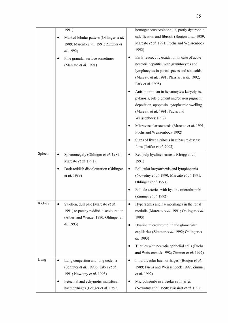
35
1991)
• Marked lobular pattern (Ohlinger et al.
1989; Marcato et al. 1991; Zimmer et
al. 1992)
• Fine granular surface sometimes
(Marcato et al. 1991)
homogeneous eosinophilia, partly dystrophic
calcification and fibrosis (Boujon et al. 1989;
Marcato et al. 1991; Fuchs and Weissenbock
1992)
• Early leucocytic exudation in case of acute
necrotic hepatitis, with granulocytes and
lymphocytes in portal spaces and sinusoids
(Marcato et al. 1991; Plassiart et al. 1992;
Park et al. 1995)
• Anisomorphism in hepatocytes: karyolysis,
pyknosis, bile pigment and/or iron pigment
deposition, apoptosis, cytoplasmic swelling
(Marcato et al. 1991; Fuchs and
Weissenbock 1992)
• Microvascular steatosis (Marcato et al. 1991;
Fuchs and Weissenbock 1992)
• Signs of liver cirrhosis in subacute disease
form (Teifke et al. 2002)
Spleen • Splenomegaly (Ohlinger et al. 1989;
Marcato et al. 1991)
• Dark reddish discolouration (Ohlinger
et al. 1989)
• Red pulp hyaline necrosis (Gregg et al.
1991)
• Follicular karyorrhexis and lymphopenia
(Nowotny et al. 1990; Marcato et al. 1991;
Ohlinger et al. 1993)
• Follicle arteries with hyaline microthrombi
(Zimmer et al. 1992)
Kidney • Swollen, dull pale (Marcato et al.
1991) to patchy reddish discolouration
(Albert and Wenzel 1990; Ohlinger et
al. 1993)
• Hyperaemia and haemorrhages in the renal
medulla (Marcato et al. 1991; Ohlinger et al.
1993)
• Hyaline microthrombi in the glomerular
capillaries (Zimmer et al. 1992; Ohlinger et
al. 1993)
• Tubules with necrotic epithelial cells (Fuchs
and Weissenbock 1992; Zimmer et al. 1992)
Lung • Lung congestion and lung oedema
(Schlüter et al. 1990b; Erber et al.
1991; Nowotny et al. 1993)
• Petechial and echymotic multifocal
haemorrhages (Löliger et al. 1989;
• Intra-alveolar haemorrhages (Boujon et al.
1989; Fuchs and Weissenbock 1992; Zimmer
et al. 1992)
• Microthrombi in alveolar capillaries
(Nowotny et al. 1990; Plassiart et al. 1992;

36
Boujon et al. 1989; Zimmer et al.
1992)
Zimmer et al. 1992)
• Alveolar oedema (Zimmer et al. 1992;
Ohlinger et al. 1993)
Trachea • Hyperaemic, dark reddening of the
tracheal mucosa, containing frothy
fluid (Ohlinger et al. 1989; Albert and
Wenzel 1990; Marcato et al. 1991)
• Hyperaemia of the tracheal mucosa (Marcato
et al. 1991; Zimmer et al. 1992)
• Leukocyte infiltration (Marcato et al. 1991;
Ohlinger et al. 1993)
• Calcification of tracheal cartilage (Marcato
et al. 1991)
Heart • Often haemorrhages in the
myocardium (Marcato et al. 1991;
Zimmer et al. 1992)
• Subepicardial and myocardial haemorrhages
and degeneration of some heart muscle cells
(Zimmer et al. 1992)
Gastro-
intestinal
tract
• Catarrhal gastritis with mucosal
erosions (Marcato et al. 1991)
• Stomach and caecum obstipation
(Boujon et al. 1989)
• Sometimes subserous bleedings
(Albert and Wenzel 1990)
• Necrosis of the small intestinal crypts and
subsequent villous atrophy (Gregg et al.,
1991)
Thymus • Often haemorrhages (Marcato et al.
1991; Zimmer et al. 1992)
• Necrosis of lymphocytes (Plassiart et al.
1992)
Lymph
nodes • Enlargement of the mesenteric lymph
nodes (Marcato et al. 1991)
• Occasionally hyperaemia (Boujon et
al. 1989)
• Karyorrhexis and depletion of lymphocytes
(Xu and Chen 1989)
Central
nervous
system
• Occasionally bleeding in spinal cord
and brain (Nowotny et al. 1990)
• Intramyelinic oedema and microthrombi
(Marcato et al. 1991)
• Non-purulent leptomeningitis (Nowotny et
al. 1990; Fuchs and Weissenbock 1992)
Blood • Poor blood coagulation (Ohlinger et
al. 1989; Marcato et al. 1991)
Laboratory diagnosis of RHD
Although some caliciviruses can be readily cultured in vitro, other members of this
family cannot be cultured by conventional virological methods (Xu and Chen 1989;
Erber et al. 1991; Gregg et al. 1991; Park et al. 1991). Therefore, while conventional

37
methods of antibody or antigen detection can be used to diagnose RHDV, bioassays
using susceptible rabbits is the only method available to confirm the presence of viable
and infective RHDV. Laboratory diagnostic methods can be categorised into methods
for detection of virus or viral components, and methods for detection of immune
responses of rabbits (Meers 1998).
Detection of RHDV
Electron microscopy (EM) of homogenised rabbit tissues can be used to demonstrate
the presence of virus particles that are morphologically consistent with RHDV. Rodak
et al. (1991) used EM as a screening technique for large numbers of diagnostic samples,
and it also has had an important role in characterising RHDV morphology (Smid et al.
1989; Valicek et al. 1990; Park and Itakura 1992; Alexandrov et al. 1993; Zheng et al.
2001).
The first test developed for detecting RHDV was the haemagglutination test (Pu et
al. 1985), which is based on the ability of RHDV to agglutinate human erythrocytes
(Liu et al. 1984). This test was widely used by Chinese and European scientists as a
screening method (Xu and Chen 1989; Nowotny et al. 1990; Villafuerte et al. 1994;
Marchandeau et al. 1998; Calvete et al. 2002). However, the sensitivity and specificity
of this method appear to be unsatisfactory. Investigations estimated the specificity of the
test to be 92%, and its sensitivity to be in the order of 80 to 90% (Capucci et al. 1991;
Zimmer et al. 1992; Haas and Thiel 1993).
Immunohistochemical diagnosis using immunoperoxidase (Rodak et al. 1990b;
Carrasco et al. 1991; Alexandrov et al. 1992; Stoerckle-Berger et al. 1992; von Elling
1992) and immunofluorescence techniques (Nowotny et al. 1990; Valicek et al. 1992;
Collins et al. 1995; Nawwar et al. 1996) have been also applied to detect RHDV
antigens. These methods offer the advantage of better sensitivity to detect low levels of
virus particles in tissue sections. However, Haas and Thiel (1993) counselled against
using immunoflurescence in routine diagnosis, due to the problem of non-specific
binding of fluorescent conjugates in uninfected samples. An alternative approach for
antigen detection is the use of antigen-capture ELISAs employing monoclonal (Rodak
et al. 1990b; Capucci et al. 1991, 1995; Collins et al. 1996) or polyclonal antibodies

38
(Nardelli et al. 1996). Although the latter have been shown to cross-react with liver
components, antigen ELISAs employing monoclonal antibodies to RHDV antigens are
often used for confirmation of RHDV infection in rabbit tissues.
The most sensitive and specific method for detection of RHDV is the use of the
reverse transcriptase polymerase chain reaction (RT-PCR) to detect viral RNA.
Lenghaus et al. (1994) found liver samples of rabbits tested positive with RT-PCR at
dilutions up to 10-10. RT-PCR also has been used successfully to detect viral RNA in
rabbit serum (Moss et al. 2002) and in tissues of non-target species, including insects
(Asgari et al. 1998). RT-PCR has the advantage that it can applied to detect the viral
genome in very young rabbits, in which other tests have limitations (Gould et al. 1997).
By sequencing RT-PCR products, phylogenetic relationships between strains can be
made (Nowotny et al. 1997; Moss et al. 2002; Le Gall-Recule et al. 2003) and the
evolution of the virus and the genetic drifts determined (Asgari et al. 1999). However,
Gould et al. (1997) recommended the use of RT-PCR in conjunction with other tests, as
false positive results due to contamination can occur.
Detection of RHDV antibodies
Immune responses to RHDV comprise both cell-mediated and humoral responses
(Meers 1998). Deng et al. (1991), cited in Huang (1991), used a monolayer liquid-phase
haemagglutination technique to identify antibody-forming and antibody-secreting cells
following immunisation against RHD. While this method can provide important
information about anti-viral immunity, it is complex and not often used in RHD
diagnostics. No other studies on cell-mediated immunity to RHDV have been reported,
and available diagnostic techniques are limited to detection of humoral antibody
responses.
The haemagglutination inhibition test (HI) was the first test reported to detect RHDV
antibodies (Pu et al. 1985). HI-tests are in common use and are an inexpensive and
simple method for RHDV antibody detection (Berninger and House 1995; Trout et al.
1997a; Frölich et al. 1998; Elghaffar et al. 2000; Mizoguchi et al. 2003). However, to
avoid false positive results it is necessary to remove natural agglutinins in the serum,
and a range of procedures for serum pre-treatment have been used (Maess et al. 1989;

39
Kiepker 1990; Park et al. 1991; Collins et al. 1995). False positive test results for
RHDV were also observed in animals infected with Pasteurella spp. (Rodak et al.
1991). Another disadavantage of the test is its inability to detect low concentrations of
antibodies, which has been observed by some researchers (Danner 1995; Dedek and
Frölich 1997).
Indirect Enzyme Linked Immunosorbent Assays (ELISA), for the detection of
RHDV antibodies have been developed in Italy (Frescura et al. 1989b), Germany
(Schirrmeier et al. 1990), Czechoslovakia (Rodak et al. 1990a), Australia (Collins et al.
1995) and England (Chasey et al. 1995). Indirect ELISAs, in common with all
serological assays, are often confronted with the problem, that in addition to antigen-
specific antibodies, non-specific antibodies may bind resulting in false positive
reactions (Capucci et al. 1991).
Competition ELISAs for RHDV have been developed by several researchers (Haas
and Ohlinger 1990; Capucci et al. 1995; Collins et al. 1995; Fitzner and Niedbalski
1996). The Capucci competition ELISA (Lavazza and Capucci 1996) is an Office
International des Epizooties accepted method for RHDV antibody detection. The
competition step is initiated when diluted test serum and RHDV antigen are added to
the monoclonal or polyclonal antiserum (Capucci et al. 1996a) which coats the solid
phase. Virus bound to the solid phase is then labelled with a dilution of enzyme-
conjugated monoclonal antibody. After the enzymatic reaction has taken place, the
presence of antibodies in the sera is detected photometrically. The titre of a positive test
serum is taken as the dilution, which generates an optical density (OD) value of less
than 50% of the negative reference serum (at the 1:10 dilution) (Capucci et al. 1997;
Cooke et al. 2000; Zheng et al. 2002).
Researchers have used various cut-offs for the competition ELISA to identify a
sample as seropositive. Zheng et al. (2002) listed serum dilutions as positive, if the
serum dilution exhibited 50% inhibition compared with the OD of the negative control
serum at a 1:10 dilution, while Cooke et al. (2000) only considered samples to be
seropositive, if the OD value of the first dilution of the sample was lower than 0.75
times the OD value of the first dilution of the negative reference serum. Similarly
Robinson et al. (2002a) recommended the use of 75% inhibition as a cut-off for the

40
detection of truly RHDV positive sera in the presence of cross-reactive antibodies.
McPhee et al. (2002) demonstrated that test specificity increased to a maximum when a
cut-off of 50% at a 1:10 dilution was selected. However the test sensitivity decreased to
39%, and this incorrectly classified a large proportion of samples as seronegative. In
addition, the interpretation of the competition ELISA may differ between regions,
which makes it difficult to choose a universal cut-off value. Furthermore, age (based on
eye lens weight) also has been shown to affect the competition ELISA results (McPhee
et al. 2002). To avoid any false positive results due to ‘background noise’ of the ELISA
procedure, Parkes et al. (2002) only considered a sample to be positive at the higher
dilution of 1:40 (when the OD of the sample inhibits the value of the negative control at
1:10 dilution by more than 50%). In general, Australian studies on RHDV in wild rabbit
populations consider serum samples positive at a dilution of 1:10 (Cooke et al. 2000;
Mutze et al. 2002), while New Zealand researchers primarily use the 1:40 dilution
(Reddiex et al. 2002; Parkes et al. 2002). Therefore, serological results from different
sources should be interpreted with caution.
It has become generally accepted that the competition ELISA is more specific than
the indirect ELISA (Capucci et al. 1991). This was well demonstrated in a study of
Robinson et al. (2002a) where a significant proportion of positive cross-reactors were
detected in Australian rabbit sera only with the indirect ELISA, but not with the
competition ELISA. Because the Capucci competition ELISA fails to distinguish
between pathogenic RHDV and cross-reactive RHDV-like virus it is only adequate for
the detection of RHDV in a rabbit population, and should be used with caution for
estimating prevalence of RHDV immunity (McPhee et al. 2002).
RHDV specific antibodies (IgG, IgA and IgM) can be determined by using isotype
ELISA (Capucci et al. 1997) to distinguish between different classes of immunity
(maternal antibodies, recent infection and past infection, re-exposure to RHDV and an
indication for exposure to RHDV-like agent) (Cooke et al. 2000), to compare
vaccinated and naturally exposed rabbits (Capucci et al. 1997) and to specify the
infection process in single animals in more detail (Cooke et al. 2002). These tests are an
important tool to clarify test results based on the competition ELISA. Procedures differ
between isotype classes. In IgA and IgM ELISAs, diluted rabbit serum is added to wells
coated with anti-IgM or anti-IgA monoclonal antibodies, followed by the addition of

41
RHDV antigen and enzyme-conjugated anti-RHDV monoclonal antibodies. In IgG
ELISAs, plates are coated with anti-RHDV monoclonal antibodies, followed by the
addition of RHDV, test serum and enzyme-conjugated anti-IgG monoclonal antibodies.
After washing the plates, substrate is added and the absorbance measured. The titre of a
serum is expressed relative to the positive reference serum (Cooke et al. 2000) or to the
serum control at 1:40 dilution without RHDV (Zheng et al. 2002).
Epidemiology of RHD in wild rabbits, with emphasis on New
Zealand
Europe
Most European reports on the initial impact of RHD focus on domestic rabbit
populations, and detailed studies on wild rabbits have been limited to Spain and France.
In these countries, rabbits are significant game species and also play an important role
in ecological systems. In general, following the emergence of RHD, wild rabbit
numbers declined in all European countries, but the decreases were relatively small in
Britain and Scandinavia compared to other countries (Cooke and Fenner 2002).
From 1988 to 1990, the rabbit mortality in initial RHD outbreaks in Spain was
estimated to be between 55% to 75% (Villafuerte et al. 1994, 1995). Interviews with
hunters indicated that mortality risks declined to around 30% in outbreaks in subsequent
years, with annual outbreaks concentrated in the spring and winter seasons (53% and
34% of outbreaks respectively). Recovery of rabbit numbers was slow in many sites
with low population densities, but more rapid in dense populations. It has been
suggested that high population densities facilitate the transmission of RHD and increase
mortality, resulting in a reduction in the median age of the population to the point where
rabbits are no longer susceptible when challenged (Calvete and Estrada 2000, cited in
Cooke and Fenner 2002). These surviving animals would be recruited into the new
breeding population, leading to reduction in the overall RHD mortality.
Similar to the observations in Spain, RHD spread slowly throughout France and did
not reach some areas until several years after its first occurrence in 1998 (Marchandeau
et al. 1998). Crude mortality estimates of up to 88% were recorded in the first year of

42
RHD occurrence, with mortality directly attributed to RHD estimated to be
approximately 45%. A wild rabbit population studied near Paris did not recover within
three years from its initial epidemic, after which time only one more outbreak occurred
despite serological evidence of ongoing presence of the virus (Marchandeau et al.
2000).
Australia
The initial spread of RHD through Australia appeared to be influenced by climatic
and geographical patterns. In arid and semi-arid regions population declines between 60
to 97% were observed (Bowen and Read 1998; Mutze et al. 1998; Cooke 1999a), while
in more moist areas in New South Wales the initial impact of the disease was variable.
Reduction in rabbit numbers of between 68-91% occurred in populations in some moist
areas, while substantial increases of up to 87% above pre-RHD density were observed
in others (Saunders et al. 1999). In South Australia, rabbit numbers were reduced in the
longer term by 85% in arid areas, but only by 12% in coastal areas (Mutze et al. 2002).
In addition, seasonal peaks in rabbit abundance shifted from spring to late summer
(Mutze et al. 2002).
New Zealand
Rabbit numbers are generally lower in the North Island of New Zealand than in the
South Island. On 23 farms where RHDV was released between late 1997 and early
1998, rabbit density was estimated to be between 2 and 37 rabbits per spotlight
kilometre (median 13) (Sanson et al. 2000). The estimated reduction of rabbit numbers
was in the order of 50%, but highly variable (0% to 77%). Although 80% of surveyed
farmers reported that the disease spread from the release locations, only 27% of these
farmers indicated that dead rabbits were found more than 300 m away from the release
sites.
A serological survey from several sites in Central Otago in the South Island of New
Zealand was conducted on sites where RHDV had been deliberately released and
subsequently spread naturally (O'Keefe et al. 1999). Higher survival rates and levels of
antibodies were observed at sites where RHDV was spread on carrots or oats than on

43
properties to which RHD had spread naturally. It has been suggested that large-scale
baiting could result in exposure of rabbits to inactivated RHDV, which in turn could
induce protective immunity. Further investigations of RHDV in the South Island
indicated population reduction of more than 90% in high rabbit density areas (39-69
rabbits per spotlight kilometre pre-RHD). Epidemics recurred annually and apparent
seroprevalence in survivors was approximately 35% at both biocided sites and sites
where the virus spread naturally (Parkes et al. 2002). In areas of the Mackenzie Basin
with lower rabbit densities, population reductions were in the order of 50%, epidemics
recurred biannually and seroprevalence in survivors was approximately 20%. In
contrast, some areas in North Canterbury experienced minimal population reductions
and 50-80% of surviving rabbits were seropositive. The existence of another RHD-like
calicivirus that might protect against RHD in New Zealand has been postulated, but
remains unproven (Zheng et al. 2002). Some ecological concerns exist that a rapid
reduction in rabbit numbers could lead to increased short-term predation on other non-
pest species in New Zealand, and in particular native birds and their eggs (Haselmayer
and Jamieson 2001; Norbury et al. 2002). However, it has been put forward that
decreases in rabbit predator numbers (feral cats and ferrets) following declines in rabbit
populations may yet yield long-term benefits for native fauna (Norbury et al. 2002).
General patterns of disease occurrence
The rate of local spread of RHD during the initial epidemic in Central Otago was
estimated to be about 200 m per day (or 6 km per month) with the prevailing wind
(Parkes et al. 1999). This was considerably less than estimates made in Spain (2 to
10 km per month) (Villafuerte et al. 1995) and Australia (9 to 414 km per month)
(Kovaliski 1998), but greater than a figure of less then one kilometre per month reported
for England (Trout et al. 1997b). Vectors are one factor that could affect the rate of
spread of RHDV. The likely variability in vector abundance between seasons will be
influenced by climatic factors that may also affect the survival of the virus and the
timing and duration of rabbit breeding. Seasonal peaks of RHD epidemics are expected
to occur in association with increased rabbit production, especially after young animals
have lost their maternal immunity (Cooke and Fenner 2002).

44
In Spain, RHD outbreaks typically lasted four to five weeks and occurred during the
main breeding season from late autumn to early spring, but predominantly during the
winter months (Cooke 2002; Calvete et al. 2002). Deaths of seronegative does most
likely contributed to the death of suckling offspring. In addition, outbreaks late in the
breeding season resulted in a 50% mortality of juvenile animals, probably as a direct
result of them no longer being protected by innate age-related resistance or maternally-
derived passive immunity (Cooke 2002). Maximum mortality in adults was also
observed in the second half of the breeding season, when the highest proportion of
females were found (Calvete et al. 2002).
In semi-arid inland areas of Australia, RHD epidemics were also observed in the
beginning of the breeding season in late autumn-early winter (May-July). These
epidemics killed up to 87% of young adults and was probably directly attributed to a
loss of maternal immunity over the summer months (Mutze et al. 2002; Cooke and
Fenner 2002). The observation that animals born late in the same season were usually
not affected was attributed to the possibility that RHDV did not persist (outbreaks rarely
occur in summer) until the time when these late-born animals became susceptible.
These animals were then incorporated into the new breeding population, and are
susceptible to infection when cooler temperatures predispose to RHD outbreaks the
following autumn.
RHD outbreaks in dry inland areas of Central Otago in the South Island also were
reported in the autumn months. Affected carcasses were found up to 40 days after the
first case was reported on sites with naturally introduced outbreaks, and up to 80 days
on biocided sites (Parkes et al. 2002). In contrast, RHD outbreaks occurred in late
spring to early summer in cooler regions of North Canterbury, and post-emergent
mortality was highest in early-born rabbits (August-October) as they became
susceptible to RHD in the late breeding season (Reddiex et al. 2002). Prior to the
introduction of RHDV to New Zealand, modelling of the likely spread of RHD
predicted that outbreaks would occur in summer or autumn. In these relatively simple
models, all seasonal factors other than rabbit production (such as virus-half life) were
assumed constant, although some of these factors are probably important determinants
for the timing of outbreaks (Barlow and Kean 1998). Subsequent modelling work
predicted yearly epidemics alternating between spring and winter (Barlow et al. 2002).
However, despite juvenile recruitment that supported post-epidemic population

45
recovery over summer, no second outbreak was observed on field sites in Otago. The
authors suggested that a low recruitment in the spring-summer period following the
RHD release, for instance due to predation or poor environmental conditions, could
result in rabbit density failing to reach a threshold level required to sustain a new
epidemic in the following winter (Barlow et al. 2002).
Several authors have observed a relationship between RHD and other causes of
rabbit mortality. The interaction between mortality due to RHD and patterns of
predation in juvenile rabbits was investigated in a study in North Canterbury, New
Zealand (Reddiex et al. 2002). In this study, it was observed that population declines
were significantly lower at sites where predators had been removed compared to sites
where predator populations were not removed. In a Spanish study, predation continued
to be the predominant cause of rabbit mortality, accounting for 60% of rabbit deaths
(Calvete et al. 2002). Increased rabbit numbers due to a decrease in predation was
thought to increase the basic reproduction rate of RHDV and hence RHD deaths, which
partly compensated for the decrease in predation. In France, predation by red foxes may
have delayed population recovery after an initial RHD epidemic, and then restricted
rabbit numbers to levels at which further RHD was undetected (Marchandeau et al.
2000).
Furthermore, interaction between myxomatosis and RHD has been described.
Immunosuppression by the Myxoma virus could contribute to higher mortalities in
rabbits exposed to RHD, despite these animals having high RHDV antibody titres
(Marchandeau et al. 1998). The timing of myxomatosis epidemics in Australia may be
affected by RHD outbreaks, and be delayed from late spring to early summer and
autumn (Mutze et al. 2002).
A frequent impression, that has not been thoroughly investigated in field studies, is
the apparent patchiness of the disease. Several attempts were made to model the disease,
which appears to exhibit considerable spatial heterogeneity. Barlow and Kean (1998)
and Barlow (1999) postulated a hide and seek phenomenon for RHD in which recurrent
epidemics at a location could result in reintroduction of the agent. In this scenario, the
agent would not persist at an outbreak site following an epidemic, but would be
dispersed to surrounding sites by dispersal or vectors. Reintroduction from other
46
infected sites could then be repeated when the population had recovered. This
phenomenon depends on the existence of spatial heterogeneity, the scale of which
would be affected by the mobility of vectors and the rapidity with which the disease
occurred. However, this cannot explain the epidemic pattern in which many, small,
partially synchronised, epidemics occur.
Variability in rabbit population demography as described by Trout et al. (2000) could
result in different levels of risk for neighbouring populations (White et al. 2001). One
modelling study explored the scenario in which pathogenic and non-pathogenic strains
occurred, and concluded that the strain with the higher basic reproduction rate would
dominate (White et al. 2001). The authors suggested a spatial effect on population
immunity, where neighbouring populations that are protected by a non-pathogenic strain
of the virus could shield an area of high RHDV mortality risk (White et al. 2001). This
model was later modified by using chronic infection and acute infection states to
explain spatial heterogeneity, rather than the presence of a non-pathogenic virus (White
et al. 2002). An individual-based spatial model to describe RHD in European wild
rabbit populations was developed recently, but was mainly used to describe the current
situation, rather than to provide new insights into the disease process (Fa et al. 2001).
Unfortunately, quality ecological models of RHD epidemiology are restrained by a
marked lack of longitudinal seroprevalence data before and after epidemics, and a lack
of accurate data recording the relationships of epidemics with population dynamics
(White et al. 2001).
Omissions in the literature
This review of the literature on RHD has brought several shortcomings to light.
Although the aetiological, clinical and pathological aspects of RHD are well
documented, there is a clear deficit of longitudinal studies on the disease in wild rabbit
populations. Modellers of RHD epidemiology consistently point out the lack of
sufficient information about RHD to validate their models. In addition, RHD is only one
source of mortality in wild rabbits, and its likely interrelationships with other causes of
mortality require more intensive investigation.
Secondly, although the temporal patterns of outbreaks of RHD often have been
described, there has been little attention given to spatial aspects of the disease, although

47
the variable severity and impact of the disease on rabbit populations (‘patchiness’) is
commented on in several studies. There has been no study that has investigated the
spatial occurrence of RHD in relation to rabbit activity, in particular on the home ranges
of the animals.
Thirdly, rabbits play an important part in the ecology of other species, most notably
rabbit predators and scavengers. Seroconversions in these species have been reported,
but were never related to temporal aspects of RHD outbreaks. To understand RHDV
behaviour in non-target species, predators and scavengers have to be monitored in a
longitudinal study.
Although potential arthropod vectors for RHDV have been described, there is lack of
detailed information on fly abundance in relation to RHD outbreaks. The likely
importance of climate on the occurrence of RHD outbreaks has been noted, but detailed
analyses of climatic factors that influence the abundance of potential vector species are
not available in the literature.
Furthermore, there is a paucity of data on the properties of RHDV with respect to its
persistence in the environment, most probably because the virus is not culturable.
Knowledge of the survival of RHDV in the environment is important for understanding
the epidemiology of the disease, and this question warrants further investigation. In
New Zealand, the use of RHDV contaminated baits has become a common practice for
rabbit control. However, there are concerns that the efficiency of this approach will be
undermined if inactivated RHDV on baits in the field can induce protective immunity in
rabbits. This hypothesis has yet to be directly tested.
Finally, initial research on RHDV use in New Zealand has mainly focused on RHDV
outbreaks, and neglected the use of other control methods that are currently employed.
Rabbit control has been practised for more than a century in New Zealand, yet RHDV is
a very new disease. Its patterns of use by farmers and their perceptions about its
performance have not been formally assessed. Nevertheless, New Zealand farmers
introduced RHDV and their awareness of its use and its benefits has to be investigated.
48
The present research project: outline of research aims and
intended outcomes
In this chapter, I have discussed the epidemiology of RHD, which is a relatively new
disease in New Zealand. To understand any infectious disease at a population level, it is
essential to determine the biological characteristics of the infectious agent and its
behaviour in the host, methods for the identification of the agent and of host responses
to infection, and the nature of disease transmission. The purpose of my research project
was to advance understanding of the epidemiology of RHDV in the first four years after
its introduction to New Zealand. The natural history of RHD in the lower North Island
of New Zealand was described in a longitudinal observational study that included
intensive observations on a well characterised population of individually identified
rabbits. Multiple aspects of rabbit ecology had to be considered, as RHD is only one of
numerous causes of death in rabbits. The temporal and spatial dynamics of RHD in the
population were determined, together with the likely involvement of non-target
vetebrate species and arthropods as potential disease vectors. Experimental studies were
also conducted to obtain a better understanding of the persistence of the virus in the
environment. Finally, farmers were surveyed to understand the perceptions of the users
and beneficiaries of RHD with respect to the utility of RHD as a tool in rabbit control.
The overall body of work was founded on the belief that better understanding of the
natural occurrence of RHD is essential for appropriate and optimal employment of
RHDV as a bio-control agent for rabbits in New Zealand.

49
References
Albert, G., and Wenzel, U. (1990). Calicivirusinfection der Kaninchen im Bezirk
Leipzig [in German]. In '7. Arbeitstagung über Haltung und Krankheiten der
Kaninchen, Pelztiere und Heimtiere, Celle.' pp. 253-256. (Deutsche
Veterinärmedizinische Gesellschaft: Giessen, Germany.)
Alexandrov, M., Peshev, R., Yanchev, I., Bozhkov, S., Doumanova, L., Dimitrov, T.,
and Zacharieva, S. (1992). Immunohistochemical localisation of the rabbit
haemorrhagic disease viral antigen. Archives of Virology 127, 355-363.
Alexandrov, M., Peshev, R., Bozhkov, S., Yanchev, I., and Doumanova, L. (1993).
Electron- and immunoelectron-microscopic investigation of the Rabbit
haemorrhagic disease virus. Comparative Immunology, Microbiology and
Infectious Diseases 16, 21-27.
An, S. H., Kim, B., Lee, J. B., Song, J. K., Park, B. K., Kwon, Y. B., Jung, J. S., and
Lee, Y. S. (1988). Studies on picornavirus haemorrhagic fever (tentative name) in
rabbits. Physico-chemical properties of the causative virus [in Korean]. Research
Reports of the Rural Development Veterinary Administration 30, 55-61, Abstract in
Veterinary Bulletin (1989) 59, p. 387, Abstr. Nr. 2789.
Anon. (2000). Rabbit calicivirus infection confirmed in Iowa rabbitry. Journal of the
American Veterinary Medical Association 216, 1537.
Asgari, S., Hardy, J. R., Sinclair, R. G., and Cooke, B. D. (1998). Field evidence for
mechanical transmission of Rabbit haemorrhagic disease virus (RHDV) by flies
(Diptera: Calliphoridae) among wild rabbits in Australia. Virus Research 54, 123-
32.
Asgari, S., Hardy, J. R., and Cooke, B. D. (1999). Sequence analysis of Rabbit
haemorrhagic disease virus (RHDV) in Australia: alterations after its release.
Archives of Virology 144, 135-45.
Barlough, J. E., Berry, E. S., Skilling, D. E., and Smith, A. W. (1986a). The marine

50
calicivirus story. part I. Compendium on Continuing Education for the Practicing
Veterinarian 8, F5-F14.
Barlough, J. E., Berry, E. S., Skilling, D. E., and Smith, A. W. (1986b). The marine
calicivirus story. part II. Compendium on Continuing Education for the Practicing
Veterinarian 8, F75-F83.
Barlow, N. D., and Kean, J. M. (1998). Simple models for the impact of rabbit
calicivirus disease (RCD) on Australasian rabbits. Ecological Modelling 109, 225-
241.
Barlow, N. D. (1999). Modelling rabbit haemorrhagic disease. In 'Rabbit Control, RCD:
Dilemmas and Implications. Proceedings of the Conference, 30-31 March 1998'.
pp. 76-79. (Royal Society of New Zealand Miscellaneous Series: Wellington, New
Zealand.)
Barlow, N. D., Barron, M. C., and Parkes, J. (2002). Rabbit haemorrhagic disease in
New Zealand: field test of a disease-host model. Wildlife Research 29, 649-653.
Barratt, B. I. P., Ferguson, C. M., Heath, A. C. G., Evans, A. A., and Logan, R. A. S.
(1998). Can insects transmit Rabbit haemorrhagic disease virus? In 'Proceedings of
the Fifty First New Zealand Plant Protection Conference, Hamilton, New Zealand.'
pp. 245-250. (New Zealand Plant Protection Society: Rotorua, New Zealand.)
Barratt, B. I. P., Ferguson, C. M., Heath, A. C. G., and Logan, R. A. S. (2001). Relative
abundance and seasonality of Calliphoridae and Sarcophagidae (Diptera), potential
vectors of rabbit haemorrhagic disease virus (RHDV) in the South Island of New
Zealand. New Zealand Journal of Zoology 28, 417-428.
Berninger, M. L., and House, C. (1995). Serologic comparison of four isolates of Rabbit
hemorrhagic disease virus. Veterinary Microbiology 47, 157-165.
Berry, E. S., Skilling, D. E., Barlough, J. E., Vedros, N. A., Gage, L. J., and Smith, A.
W. (1990). New marine calicivirus serotype infective for swine. American Journal
of Veterinary Research 51, 1184-1187.

51
Boga, J. A., Casais, R., Marin, M. S., Martin-Alonso, J. M., Carmenes, R. S., Prieto, M.,
and Parra, F. (1994). Molecular cloning, sequencing and expression in Escherichia
coli of capsid protein gene from Rabbit haemorrhagic disease virus (Spanish isolate
AST/89). Journal of General Virology 75, 2409-2413.
Boniotti, B., Wirblich, C., Sibilia, M., Meyers, G., Thiel, H. J., and Rossi, C. (1994).
Identification and characterisation of a 3C-like protease from Rabbit haemorrhagic
disease virus, a calicivirus. Journal of Virology 68, 6487-95.
Boujon, C. E., Gafner, F. R., and Bestetti, G. E. (1989). The infectious necrotizing
hepatitis of rabbits - First cases in Switzerland [in German]. Schweizer Archiv für
Tierheilkunde 131, 71-76.
Bowen, Z., and Read, J. (1998). Population and demographic patterns of rabbits
(Oryctolagus cuniculus) at Roxby Downs in arid South Australia and the influence
of rabbit haemorrhagic disease. Wildlife Research 25, 655-662.
Buddle, B. M., de. Lisle, G. W., McColl, K., Collins, B. J., Morrissy, C., and Westbury,
H. A. (1997). Response of the North Island brown kiwi, Apteryx australis mantellli
and the lesser short-tailed bat, Mystacina tuberculata to a measured dose of Rabbit
haemorrhagic disease virus. New Zealand Veterinary Journal 45, 109-113.
Bull, P. C. (1953). Parasites of the wild rabbit, Oryctolagus cuniculus (L.). New
Zealand Journal of Science and Technology (B) 34, 341-72.
Bureau of Resource Studies (1996). 'Rabbit calicivirus disease'. (Australian Government
Printing Office: Canberra, Australia.)
Calvete, C., and Estrada, R. (2002). Epidemiologia de enfermedad hemorragica (VHD)
y mixomatosis en el conejo sylvestre en el valle medio del Ebro [in Spanish].
Herramientas de gestion. Consejo de la Proteccion de la Naturaleza de Aragon.
ARPIrelieve, SA. 175 pp.
Calvete, C., Estrada, R., Villafuerte, R., Osacar, J. J., and Lucientes, J. (2002).
Epidemiology of viral haemorrhagic disease and myxomatosis in a free-living

52
population of wild rabbits. Veterinary Record 150, 776-782.
Cancellotti, F. M., and Renzi, M. (1991). Epidemiology and current situation of viral
haemorrhagic disease of rabbits and the European brown hare syndrom in Italy.
Revue scientifique et technique de l'Office International des Epizooties 10, 409-
422.
Cao, S. Z., Liu, S. G., Gan, M. H., Liu, R. P., Cai, S. W., and Liu, S. F. (1986). A
preliminary report on viral haemorrhagic pneumonia (tentative name) in rabbits [in
Chinese]. Chinese Journal of Veterinary Medicine 12, 9-11, Abstract in Veterinary
Bulletin (1987) 57, p. 191, Abstr. 1348.
Capucci, L., Scicluna, M. T., and Lavazza, A. (1991). Diagnosis of viral haemorrhagic
disease of rabbits and the European brown hare syndrome. Revue scientifique et
technique de l'Office International des Epizooties 10, 347-370.
Capucci, L., Frigoli, G., Ronshold, L., Lavazza, A., Brocchi, E., and Rossi, C. (1995).
Antigenicity of the Rabbit haemorrhagic disease virus studied by its reactivity with
monoclonal antibodies. Virus Research 37, 221-238.
Capucci, L., Fusi, P., Lavazza, A., Pacciarini, M. L., and Rossi, C. (1996a). Detection
and preliminary characterisation of a new rabbit calicivirus related to Rabbit
haemorrhagic disease virus but nonpathogenic. Journal of Virology 70, 8614-8623.
Capucci, L., Chasey, D., Lavazza, A., and Westcott, D. (1996b). Preliminary
characterization of a non-haemagglutinating strain of rabbit haemorrhagic disease
virus from the United Kingdom. Journal of Veterinary Medicine Series B 43, 245-
250.
Capucci, L., Fusi, P., Nardin, A., Pacciarini, M. L., Rossi, C., and Lavazza, A. (1996c).
Rabbit haemorrhagic disease virus has a 'good brother'. [in Italian]. Rivista di
Coniglicoltura 33, 21-28.
Capucci, L., Nardin, A., and Lavazza, A. (1997). Seroconversion in an industrial unit of
rabbits infected with a non-pathogenic rabbit haemorrhagic disease-like virus.

53
Veterinary Record 140, 647-650.
Capucci, L., Fallacara, F., Grazioli, S., Lavazza, A., Pacciarini, M. L., and Brocchi, E.
(1998). A further step in the evolution of Rabbit haemorrhagic disease virus: the
appearance of the first consistent antigenic variant. Virus Research 58, 115-26.
Carman, J. A., Garner, M. G., Catton, M. G., Thomas, S., Westbury, H. A., Cannon, R.
M., Collins, B. J., and Tribe, I. G. (1998). Viral haemorrhagic disease of rabbits and
human health. Epidemiology and Infection 121, 408-418.
Carman, J. A. (1999). Rabbit calicivirus disease and human health: results from the
Australian study. In 'Rabbit Control, RCD: Dilemmas and Implications.
Proceedings of the Conference, 30-31 March 1998'. pp. 55-61. (Royal Society of
New Zealand Miscellaneous Series: Wellington, New Zealand.)
Carrasco, L., Rodriguez, F., de las. Mulas, J. M., Wohlsein, P., and Fernandez, A.
(1991). Immunohistological diagnosis of rabbit haemorrhagic disease in
experimentally infected rabbits in Spain. Journal of Veterinary Medicine Series B
38, 552-555.
Chasey, D., and Duff, P. (1990). European brown hare syndrome and associated virus-
particles in the UK. Veterinary Record 126, 623-624.
Chasey, D., Lucas, M., Westcott, D., and Williams, M. (1992). European brown hare
syndrome in the UK; a calicivirus related to but distinct from that of viral
haemorrhagic disease in rabbits. Archives of Virology 124, 363-370.
Chasey, D. (1994). Possible origin of rabbit haemorrhagic disease in the United
Kingdom. Veterinary Record 135, 496-499.
Chasey, D., Lucas, M. H., Westcott, D. G., Sharp, G., Kitching, A., and Hughes, S. K.
(1995). Development of a diagnostic approach to the identification of rabbit
haemorrhagic disease. Veterinary Record 137, 158-160.
Chasey, D., Trout, R. C., Sharp, G., and Edwards, S. (1997a). Seroepidemiology of

54
rabbit haemorrhagic disease in wild rabbits in the UK and susceptibility to
infection. In 'First International Symposium Caliciviruses ESVV'. pp. 156-162.
(European Society for Veterinary Virology: Addelstone, UK.)
Chasey, D., Trout, R. C., and Edwards, S. (1997b). Susceptibility of wild rabbits
(Oryctolagus cuniculus) in the United Kingdom to Rabbit haemorrhagic disease
(RHD). Veterinary Research 28, 271-276.
Clark, R. G., Sanson, R. L., Donaldson, J. W., Motha, M. X. J., and Knowles, G. J. E.
(1999). A chondropathy of the pinna in rabbits associated with rabbit haemorrhagic
disease. New Zealand Veterinary Journal 47, 8-12.
Collins, B. J., White, J. R., Lenghaus, C., Boyd, V., and Westbury, H. A. (1995). A
competition ELISA for the detection of antibodies to rabbit haemorrhagic disease
virus. Veterinary Microbiology 43, 85-96.
Collins, B. J., White, J. R., Lenghaus, C., Morrissy, C. J., and Westbury, H. A. (1996).
Presence of Rabbit haemorrhagic disease virus antigen in rabbit tissues as revealed
by a monoclonal antibody dependent capture ELISA. Journal of Virological
Methods 58, 145-54.
Cooke, B. D. (Ed.) (1999a). 'Rabbit Calicivirus Disease Program Report 2:
Epidemiology, Spread and Release in Wild Rabbit Populations in Australia'.
(Bureau of Rural Sciences: Canberra, Australia.)
Cooke, B. D. (1999b). Notes on the life-history of the rabbit flea Caenopsylla laptevi
ibera Beaucournu & Marquez, 1987 (Siphonaptera: Ceratophyllidae) in eastern
Spain. Parasite 6, 347-354.
Cooke, B. D., and Berman, D. (2000). Effect of inoculation route and ambient
temperature on the survival time of rabbits, Oryctolagus cuniculus (L.), infected
with Rabbit haemorrhagic disease virus. Wildlife Research 27, 137-142.
Cooke, B. D., Robinson, A. J., Merchant, J. C., Nardin, A., and Capucci, L. (2000). Use
of ELISAs in field studies of rabbit haemorrhagic disease (RHD) in Australia.

55
Epidemiology & Infection 124, 563-576.
Cooke, B. D. (2001). Rabbit haemorrhagic disease. In 'The Encyclopedia of Arthropod-
transmitted Infections'. (Ed. M. W. Service.) pp. 420-422. (CABI Publishing:
Wallingford, UK.)
Cooke, B. D., and Fenner, F. (2002). Rabbit haemorrhagic disease and the biological
control of wild rabbits, Oryctolagus cuniculus, in Australia and New Zealand.
Wildlife Research 29, 689-706.
Cooke, B. D., McPhee, S., Robinson, A. J., and Capucci, L. (2002). Rabbit
haemorrhagic disease: Does a pre-existing RHDV-like virus reduce the
effectiveness of RHD as a biological control in Australia? Wildlife Research 29,
673-682.
Cooke, B. D. (2002). Rabbit haemorrhagic disease: Field epidemiology and the
management of wild rabbit populations. Revue scientifique et technique de l'Office
International des Epizooties 21, 347-358.
Cottam, Y. H., Blair, H. T., Potter, M. A., and Elvidge, D. G. (1998). Monitoring some
Muscoid fly populations on Massey University sheep farms in the Manawatu. In
'Proceedings of the New Zealand Society of Animal Production 58th Annual
Conference'. pp. 220-223. (Wellington, New Zealand.)
Crosby, T. K. and McLennan, J. (Eds.) (1996). 'Potential Vectors of Rabbit Calicivirus
Disease (RCD) in New Zealand: A Review'. (MAF policy: Wellington, New
Zealand.)
Danner, K. J. (1995). Diagnosis and epidemiology of rabbit haemorrhagic disease [in
German]. (Unpublished thesis. Fachbereich Veterinärmedizin, Justus-Liebig-
Universität : Giessen, Germany.)
Dedek, J., and Frölich, K. (1997). Serological survey for calicivirus antibody among
free-living red foxes in Mecklenburg-Vorpommern, Germany [in German].
Tierärztliche Umschau 52, 149-152.

56
Deng, R. T., Xu, Y. F., and Yu, N. K. (1991). A new method to determine antibody-
secreting cells - momolayer liquid-phase haemagglutination technique [in Chinese].
Shanghai Journal of Immunology in press, cited in Huang, H. B. Revue scientifique
et technique de l'Office International des Epizooties (1991) 10, 481-498.
di Modugno, G., and Nasti, R. (1990). La malattia emorrhagica virale in Puglia.
Contributio sperimentale [in Italian]. Rivista di Coniglicoltura 27, 25-32.
Du, N. (1990). Rabbit haemorrhagic Disease (RHD) - a new disease and its viral
etiology. Deutsche Tierärztliche Wochenschrift 97, 114-116.
Elghaffar, S. K. A., Aly, M., Fatma, A., Moustafa, A., and Mahmoud, A. Z. (2000).
Pathological studies on the rabbit viral hemorrhagic disease (RVHD) with special
reference to the use of vitamins A, E and C as prophylaxis. Assiut Veterinary
Medical Journal 43, 251-274.
Elzein, E. M. E. A., and Al-Afaleq, A. I. (1999). Rabbit haemorrhagic disease in Saudi
Arabia. Veterinary Record 144, 480-481.
Erber, M., Gerbermann, H., Meiler, H., and Schindlmayer, R. (1991). Rabbit
haemorrhagic disease in domestic rabbits in Bavaria - epidemiologic, pathological
and virological aspects [in German]. Tierärztliche Umschau 46, 146-155.
Eskens, U., Klima, H., Nilz, J., and Wiegand, D. (1987). Leberdystrophie bei Hasen,
Ätiologie und epidemiologische Untersuchungen eines Feldhasensterbens in
Mittelhessen [in German]. Tierärztliche Praxis 15, 229-235.
Eskens, U., and Vollmer, K. (1989). Untersuchungen zur Ätiologie der Leberdystrophie
des Feldhasen (Lepus europaeus pallas) [in German]. Deutsche Tierärztliche
Wochenschrift 96, 464-466.
Fa, J. E., Sharples, C. M., Bell, D. J., and de Angelis, D. (2001). An individual-based
model of rabbit viral haemorrhagic disease in European wild rabbits (Oryctolagus
cuniculus). Ecological Modelling 144, 121-138.

57
Fitzner, A., and Niedbalski, W. (1996). Application of the ELISA for the virologic and
serologic diagnosis of viral haemorrhagic disease of rabbits (VHD). Bulletin of the
Veterinary Institute of Pulawy 40, 69-76.
Frescura, T., Gialletti, L., Rutilti, D., Fioroni, A., Marini, C., and de Mia, G. M.
(1989a). Malatti emorrhagica virale del coniglio (Viral haemorrhagic diseae of
rabbits). Obiettivi Documenti Veterinaria 10, 37-40, Abstract in: Veterinary
Bulletin (1989) 59, p.585, Abstr. 4042.
Frescura, T., Rutilti, D., Rondini, C., de Mia, G. M., Marini, C., and Scuota, S. (1989b).
Applicazione della prova IHA ed ELISA per la ricera degli anticorpi della malatti
emorrhagica virale del coniglio [in Italian]. Selezione Veterinaria 30, 1357-1365.
Frölich, K., Klima, F., and Dedek, J. (1998). Antibodies against Rabbit haemorrhagic
disease virus in free-ranging red foxes from Germany. Journal of Wildlife Diseases
34, 436-442.
Fuchs, A., and Weissenbock, H. (1992). Comparative histopathological study of rabbit
haemorrhagic disease (RHD) and European brown hare syndrome (EBHS). Journal
of Comparative Pathology 107, 103-113.
Gavier-Widen, D., and Morner, T. (1991). Epidemiology and diagnosis of the European
Brown Hare Syndrome in Scandinavian countries: a review. Revue scientifique et
technique de l'Office International des Epizooties 10, 453-458.
Gavier-Widen, D., and Morner, T. (1993). Descriptive epizootiological study of
European brown hare syndrom in Sweden. Journal of Wildlife Disease 29, 15-20.
Gehrmann, B., and Kretzschmar, C. (1991). Experimental investigations on rabbit
haemorrhagic disease (RHD) - transmission by flies [in German]. Berliner und
Münchener Tierärztliche Wochenschrift 104, 192-194.
Goldhorn, W. (1987). Das 'Hasensterben 1986' [in German]. Der praktische Tierarzt 68,
42-43.

58
Goodly, L. (2001). Rabbit haemorrhagic disease. Compendium on Continuing
Education for the Practicing Veterinarian 23, 249-254.
Gould, A. R., Kattenbelt, J. A., Lenghaus, C., Morrissy, C., Chamberlain, T., Collins, B.
J., and Westbury, H. A. (1997). The complete nucleotide sequence of Rabbit
haemorrhagic disease virus (Czech strain V351): use of the polymerase chain
reaction to detect replication in Australian vertebrates and analysis of viral
population sequence variation. Virus Research 47, 7-17.
Granzow, H., Schirrmeier, H., and Tews, G. (1989). Hämorrhagische Septikämie der
Kaninchen-Erregernachweis und erste elektronenmikroskopische Charakterisierung
[in German]. Monatshafte Veterinärmedizin 44, 379-380.
Green, K. Y., Ando, T., Balayan, M. S., Berke, T., Clarke, I. N., Estes, M. K., Matson,
D. O., Nakata, S., Neill, J. D., Studdert, M. J., and Thiel, H. J. (2000). Taxonomy of
the caliciviruses. Journal of Infectious Diseases 181, 322-330.
Greenslade, E., Weinstein, P., Woodward, A., Capucci, L., Salmond, C., and Beasley,
R. (2001). A serological survey of antibodies to Rabbit haemorrhagic disease virus
(rabbit calicivirus disease) in two rural Central Otago communities. New Zealand
Medical Journal 114, 55-58.
Gregg, D. A., and House, C. (1989). Necrotic hepatitis of rabbits in Mexico: a
parvovirus. Veterinary Record 125, 603-604.
Gregg, D. A., House, C., Meyer, R., and Berninger, M. (1991). Viral haemorrhagic
disease of rabbit in Mexico: epidemiology and viral characterisation. Revue
scientifique et technique de l'Office International des Epizooties 10, 435-451.
Guittre, C., Baginski, I., le Gall, G., Prave, M., Trepo, C., and Cova, L. (1995).
Detection of Rabbit haemorrhagic disease virus isolates and sequence comparison
of the N-terminus of the capsid protein gene by the polymerase chain reaction.
Research in Veterinary Science 58, 128-132.
Haas, B., and Ohlinger, V. F. (1990). Diagnostik der Hämorrhagischen

59
Kaninchenseuche [in German]. In '7. Arbeitstagung über Haltung und Krankheiten
der Kaninchen, Pelztiere und Heimtiere, Celle.' pp. 266-270. (Deutsche
Veterinärmedizinische Gesellschaft: Giessen, Germany.)
Haas, B., and Thiel, H. J. (1993). Rabbit haemorrhagic disease (RHD). [in German].
Deutsche Tierärztliche Wochenschrift 100, 131-137.
Haselmayer, J., and Jamieson, I. G. (2001). Increased predation on pukeko eggs after
the application of rabbit control measures. New Zealand Journal of Ecology 25, 89-
93.
Heath, A. C. G., Heyward, R., and Norbury, G. (1999). Blowflies (Calliphoridae) as
potential vectors of Rabbit haemorrhagic disease virus in New Zealand. In 'Rabbit
Control, RCD: Dilemmas and Implications. Proceedings of the Conference, 30-31
March 1998'. pp. 85-88. (Royal Society of New Zealand Miscellaneous Series:
Wellington, New Zealand.)
Henriksen, P, Gavier, D., and Elling, F. (1989). Acute necrotising hepatitis in Danish
farmed hares. Veterinary Record 125, 486-487.
Henzell, R., Cunningham, R. B., and Neave, H. (2001). Influence of climate and other
factors on the survival of Australian rabbits exposed to rabbit haemorrhagic
disease. In 'Proceedings of the 12th Australasian Vertebrate Pest Conference,
Melbourne, Australia'. pp. 36-40. (Department of Natural Resources and
Environment: Melbourne, Australia.)
Henzell, R. P., Cunningham, R. B., and Neave, H. M. (2002). Factors affecting the
survival of Australian wild rabbits exposed to rabbit haemorrhagic disease. Wildlife
Research 29, 523-542.
Huang, H. B. (1991). Vaccination against the immune response to viral haemorrhagic
disease of rabbits: a review of researach in the People's Republic of China. Revue
scientifique et technique de l'Office International des Epizooties 10, 481-498.
Jennings, L. C. (1999). Human health monitoring in New Zealand. In 'Rabbit Control,

60
RCD: Dilemmas and Implications. Proceedings of the Conference, 30-31 March
1998'. pp. 52-54. (Royal Society of New Zealand Miscellaneous Series:
Wellington, New Zealand.)
Kiepker, C. (1990). Untersuchungen über das Vorkommen der viralen hämorrhagischen
Erkrankung (RVHD) in Hauskaninchenbeständen (Schwerpunkt Niedersachsen) [in
German]. (Unpublished thesis. Tierärztliche Hochschule Hannover: Hannover,
Germany.)
Kovaliski, J. (1998). Monitoring the spread of Rabbit hemorrhagic disease virus as a
new biological agent for control of wild European rabbits in Australia. Journal of
Wildlife Diseases 34, 421-428.
Kpodekon, M., and Alogninouwa, T. (1998). Control of rabbit viral haemorrhagic
disease in Benin by vaccination. Veterinary Record 143, 693-694.
Kölbl, S., Settele, J., and Schönbauer, M. (1990). Erstes Auftreten der infektiösen
hämorrhagischen Krankheit der Kaninchen in Österreich [in German]. Berliner
Münchner Tierärztliche Wochenschrift 103, 261-266.
Laurent, S., Vautherot, J. F., le Gall, G., Madelaine, M. F., and Rasschaert, D. (1997).
Structural, antigenic and immunogenic relationships between European brown hare
syndrome virus and Rabbit haemorrhagic disease virus. Journal of General
Virology 78 , 2803-11.
Lavazza, A., Scicluna, M. T., and Capucci, L. (1996). Susceptibility of hares and rabbits
to the European brown hare syndrome virus (EBHSV) and Rabbit haemorrhagic
disease virus (RHDV) under experimental conditions. Journal of Veterinary
Medicine Series B 43, 401-410.
Lavazza, A. and Capucci, L. (1996). Viral haemorrhagic disease of rabbits. In 'Office
International des Epizooties. Manual of Standards for Diagnostic Tests and
Vaccines'. pp. 589-97. (OIE: Paris, France)
Le Gall-Recule, G., Zwingelstein, F., Laurent, S., de Boisseson, C., Portejoie, Y., and

61
Rasschaert, D. (2003). Phylogenetic analysis of Rabbit haemorrhagic disease virus
in France between 1993 and 2000, and the characterisation RHDV antigenic
variants. Archives of Virology 148, 65-81.
Lee, C. S., and Park, C. K. (1987). Etiological studies on the acute fatal disease of
Angora rabbits: The so-called Rabbit Viral Sudden Death. Korean Journal of
Veterinary Research 27, 277-290.
Leighton, F. A., Artois, M., Capucci, L., Gavier-Widen, D., and Morisse, J. P. (1995).
Antibody response to rabbit viral hemorrhagic disease virus in red foxes (Vulpes
vulpes) consuming livers of infected rabbits (Oryctolagus cuniculus). Journal of
Wildlife Diseases 31, 541-544.
Lenghaus, C., Westbury, H., Collins, B., Ratnamohan, N., and Morrissy, C. (1994).
Overview of the RHD project in Australia. In 'Rabbit Haemorrhagic Disease: Issues
in Assessment for Biological Control'. (Eds. R. K. Munro and R. T. Williams.) pp.
104-129. (Bureau of Resource Sciences, Australian Goverment Publishing Service:
Canberra.)
Lenghaus, C., Studdert, M. J., and Gavier-Widen, D. (2000). Calicivirus Infections. In
'Infectious Diseases of Wild Mammals'. (Eds. E. S. Williams and I. K. Barker.) pp.
280-291. (Iowa State University Press, Ames, USA.)
Liu, S. J., Xue, H. P., Pu, B. Q., and Qian, N. H. (1984). A new viral disease in rabbits -
Rabbit Viral Hemorrhagic Disease [in Chinese]. Animal Husbandry & Veterinary
Medicine 16, 253-255, Abstract in Veterinary Bulletin (1985), 55, p. 688, Abstr.
5600.
Löliger, H.-Ch., Matthes, S., and Liess, B. (1989). Über das Auftrereten einer
infektiösen Hämorrhagischen Erkrankung bei Hauskaninchen in der BR
Deutschland [German]. Tierärztliche Umschau 44, 22-25.
Löliger, H.-Ch. (1990). Aktuelle Erkrankungen der Hauskaninchen-Diagnose und
Bekämpfung [in German]. Der Praktischer Tierarzt 5, 42-54.

62
Löliger, H.-Ch., and Eskens, U. (1991). Incidence, epizootiology and control of viral of
viral haemorrhagic disease of rabbits and the European brown hare syndrome in
Germany. Revue scientifique et technique de l'Office International des Epizooties
10, 423-434.
Lugton, I. W. (1999). A cross-sectional study of risk factors affecting the outcome of
Rabbit haemorrhagic disease virus releases in New South Wales. Australian
Veterinary Journal 77, 322-328.
Maess, J., Matthes, S., and Flauss, G. (1989). Serologische Untersuchungen über das
Vorkommen der infektiösen hämorrhagischen Erkrankung der Hauskaninchen
(Rabbit Viral Haemorrhagic Disease) in Norddeutschland [in German].
Tierärztliche Umschau 44, 423-425.
Maess, J., and Matthes, S. (1990). Die infetiöse Hämorrhagische Erkrankung der
Hauskaninchen. Virologische and epidemiologische Untersuchungen. [in German].
In '7. Arbeitstagung über Haltung und Krankheiten der Kaninchen, Pelztiere und
Heimtiere, Celle.' pp. 249-252. (Deutsche Veterinämedizinische Gesellschaft:
Giessen, Germany.)
Marcato, P. S., Benazzi, C., Vecchi, G., Galeotti, M., Salda, L. D., della Salda, L., Sarli,
G., and Lucidi, P. (1991). Clinical and pathological features of viral haemorrhagic
disease of rabbits and the European brown hare syndrome. Revue scientifique et
technique de l'Office International des Epizooties 10, 371-392.
Marchandeau, S., Chantal, J., Portejoie, Y., Barraud, S., and Chaval, Y. (1998). Impact
of viral hemorrhagic disease on a wild population of European rabbits in France.
Journal of Wildlife Diseases 34, 429-435.
Marchandeau, S., Chaval, Y., and le Goff, E. (2000). Prolonged decline in the
abundance of wild European rabbits (Oryctolagus cuniculus) and high immunity
level over three years following the arrival of rabbit haemorrhagic disease. Wildlife
Biology 6, 141-147.

63
Matson, A. W. (1999). Re-analysis of serologic data from the Australian study of
human health risks of infection by Rabbit haemorrhagic disease virus. In 'Rabbit
Control, RCD: Dilemmas and Implications. Proceedings of the Conference, 30-31
March 1998'. pp. 62-66. (Royal Society of New Zealand Miscellaneous Series:
Wellington, New Zealand.)
McColl, K. A., Morrissy, C. J., Collins, B. J., and Westbury, H. A. (2002a). Persistence
of Rabbit haemorrhagic disease virus in decomposing rabbit carcasses. Australian
Veterinary Journal 80, 298-299.
McColl, K. A., Merchant, J. C., Hardy, J., Cooke, B. D., Robinson, A., and Westbury,
H. A. (2002b). Evidence for insect transmission of Rabbit haemorrhagic disease
virus. Epidemiology and Infection 129, 655-663.
McPhee, S. R., Berman, D., Gonzales, A., Butler, K. L., Humphrey, J., Muller, J.,
Waddington, J. N., Daniels, P., Koch, S., and Marks, C. A. (2002). Efficacy of a
competitive enzyme-linked immunosorbent assay (cELISA) for estimating
prevalence of immunity to Rabbit haemorrhagic disease virus (RHDV) in
populations of Australian wild rabbits (Oryctolagus cuniculus). Wildlife Research
29, 635-647.
Meers, J. (1998). 'Review of the diagnosis of rabbit haemorrhagic disease virus
infection.'. (Institut of Veterinary, Animal and Biomedical Sciences, Massey
University: Palmerston North, New Zealand.)
Meyers, G., Wirblich, C., and Thiel, H. J. (1991a). Genomic and subgenomic RNAs of
Rabbit haemorrhagic disease virus are both protein-linked and packaged into
particles. Virology (New York) 184, 677-686.
Meyers, G., Wirblich, C., and Thiel, H. J. (1991b). Rabbit haemorrhagic disease virus -
molecular cloning and nucleotide sequencing of a calicivirus genome. Virology
(New York) 184, 664-676.
Meyers, G., Wirblich, C., Thiel, H. J., and Thumfart, J. O. (2000). Rabbit haemorrhagic
64
disease virus: Genome organization and polyprotein processing of a calicivirus
studied after transient expression of cDNA constructs. Virology 276, 349-363.
Mikami, O., Park, J. H., Kimura, T., Ochiai, K., and Itakura, C. (1999). Hepatic lesions
in young rabbits experimentally infected with Rabbit haemorrhagic disease virus.
Research in Veterinary Science 66, 237-42.
Milton, I. D., Vlasak, R., Nowotny, N., Rodak, L., and Carter, M. J. (1992). Genomic 3'
terminal sequence comparison of three isolates of Rabbit haemorrhagic disease
virus. FEMS Microbiology Letters 93, 37-42.
Mizoguchi, T., Itou, K., and Sakurai, M. (2003). Haemagglutination and antigenic
comparison of Rabbit haemorrhagic disease virus. Journal of Veterinary Medical
Science 65, 95-97.
Mocsari, E. (1990). Epidemiological studies on the rabbit haemorrhagic disease and its
control by vaccination in Hungary. In '7. Arbeitstagung über Haltung und
Krankheiten der Kaninchen, Pelztiere und Heimtiere, Celle.' pp. 257-265.
(Deutsche Veterinärmedizinische Gesellschaft: Giessen, Germany.)
Morisse, J.-P., le Gall, G., and Boilletot, E. (1991). Hepatitis of viral origin in
Leporidae: introduction and aetiological hypotheses. Revue scientifique et
technique de l'Office International des Epizooties 10, 283-295.
Moss, S. R., Turner, S. L., Trout, R. C., White, P. J., Hudson, P. J., Desai, A., Armesto,
M., Forrester, N. L., and Gould, E. A. (2002). Molecular epidemiology of Rabbit
haemorrhagic disease virus. Journal of General Virology 83, 2461-2467.
Moussa, A., Chasey, D., Lavazza, A., Capucci, L., Smid, B., Meyers, G., Rossi, C.,
Thiel, H. J., Vlasak, R., Ronsholt, L., Nowotny, N., McCullough, K., and Gavier-
Widen, D. (1992). Haemorrhagic disease of lagomorphs - Evidence for a
Calicivirus. Veterinary Microbiology 33, 375-381.
Mutze, G., Cooke, B., and Alexander, P. (1998). The initial impact of rabbit
haemorrhagic disease on European rabbit populations in South Australia. Journal
65
of Wildlife Diseases 34, 221-227.
Mutze, G., Bird, P., Kovaliski, J., Peacock, D., Jennings, S., and Cooke, B. (2002).
Emerging epidemiological patterns in rabbit haemorrhagic disease, its interaction
with myxomatosis, and their effects on rabbit populations in South Australia.
Wildlife Research 29, 577-590.
Nagesha, H. S., McColl, K. A., Collins, B. J., Morrissy, C. J., Wang, L. F., and
Westbury, H. A. (2000). The presence of cross-reactive antibodies to Rabbit
haemorrhagic disease virus in Australian wild rabbits prior to the escape of virus
from quarantine. Archives of Virology 145, 749-757.
Nardelli, S., Agnoletti, F., Costantini, F., and Parpajola, R. (1996). Diagnosis of rabbit
haemorrhagic disease (RHD) and European brown hare syndrome (EBHS) by
indirect sandwich polyclonal ELISA. Journal of Veterinary Medicine Series B 43,
393-400.
Nauwynck, H., Callebaut, P., Peeters, J., Ducatelle, R., and Uyttebroek, E. (1993).
Susceptibility of hares and rabbits to a Belgian isolate of European brown hare
syndrome virus. Journal of Wildlife Diseases 29, 203-208.
Nawwar, A. A. M., Mousa, H. A. A., Amin, D. S., and El-Aziz, T. A. (1996). Studies on
rapid diagnostic methods for rabbit haemorrhagic viral disease (RHVD). Veterinary
Medical Journal Giza 44, 601-611.
Neave, H. S. (1999). 'Rabbit Calicivirus Disease Program Report 1: Overview of effects
on Australian wild rabbit populations and implications for agriculture and
biodiversity. A report of research conducted by participants of the Rabbit
Calicivirus Disease Monitoring and Surveilance Program and Epidemiology
Research Program. Prepared for the RCD Management Group.' (Bureau of Rural
Sciences: Canberra, Australia.)
Norbury, G., Heyward, R., and Parkes, J. (2002). Short-term ecological effects of rabbit
haemorrhagic disease in the short-tussock grasslands of the South Island, New

66
Zealand. Wildlife Research 29, 599-604.
Nowotny, N., Fuchs, A., Schilcher, F., and Loupal, G. (1990). Occurrence of viral
haemorrhagic disease of rabbits in Austria. I. Pathological and virological
investigations. [in German]. Wiener Tierärztliche Monatsschrift 77, 19-23.
Nowotny, N., Schilcher, F., Fuchs, A., and Loupal, G. (1992). Occurrence of rabbit
haemorrhagic disease (RHD) in Austria. 2. Epizootiological investigations [in
German]. Wiener Tierärztliche Monatsschrift 79 , 134-140.
Nowotny, N., Leidinger, J., Fuchs, A., Vlasak, R., Schwendenwein, I. Schilcher F., and
Loupal, G. (1993). Rabbit haemorrhagic disease (RDH): experimental infection of
domestic rabbits with regard to clinical, haematological, chemical, virological,
serological and pathomorphological features [in German]. Wiener Tierärztliche
Monatsschrift 80, 65-74.
Nowotny, N., Bascunana, C. R., Ballagi-Pordany, A., Gavier-Widen, D., Uhlen, M., and
Belak, S. (1997). Phylogenetic analysis of rabbit haemorrhagic disease and
European brown hare syndrome viruses by comparison of sequences from the
capsid protein gene. Archives of Virology 142, 657-73.
O'Keefe, J. S., Tempero, J. E., Motha, M. X., Hansen, M. F., and Atkinson, P. H.
(1999). Serology of rabbit haemorrhagic disease virus in wild rabbits before and
after release of the virus in New Zealand. Veterinary Microbiology 66, 29-40.
Ohlinger, V. F., Haas, B., Ahl, R., and Weiland, F. (1989). Infectious haemorrhagic
disease of rabbits. Contagious disease caused by calicivirus. [in German].
Tierärztliche Umschau 44, 284-94.
Ohlinger, V. F., Haas, B., Meyers, G., Weiland, F., and Thiel, H. J. (1990a).
Identification and characterisation of the virus causing rabbit haemorrhagic disease.
Journal of Virology 64, 3331-3336.
Ohlinger, V. F., Haas, B., and Thiel, H. J. (1990b). Report on a meeting of European
Society for Veterinary Virology (ESVV) on viral haemorrhagic disease of rabbits.

67
7.-8.12.1989. Lyon. [in German].
Ohlinger, V. F., Haas, B., and Thiel, H. J. (1993). Rabbit haemorrhagic disease (RHD):
characterisation of the causative calicivirus. Veterinary Research 24, 103-16.
Park, J. H., Kida, H., Ueda, K., Ochiai, K., Goryo, M., and Itakura, C. (1991). Etiology
of rabbit haemorrhagic disease spontaneously occurring in Korea. Journal of
Veterinary Medicine Series B 38, 749-754.
Park, J. H., and Itakura, C. (1992). Detection of Rabbit haemorrhagic disease virus
antigen in tissues by immunohistochemistry. Research in Veterinary Science 52,
299-306.
Park, J. H., Lee, Y. S., and Itakura, C. (1995). Pathogenesis of acute necrotic hepatitis in
rabbit hemorrhagic disease. Laboratory Animal Science 45, 445-449.
Parkes, J. P., Norbury, G., and Heyward, R. (1999). Initial epidemiology of rabbit
haemorrhagic disease in New Zealand. In 'Rabbit Control, RCD: Dilemmas and
Implications. Proceedings of the Conference, 30-31 March 1998'. pp. 80-83. (Royal
Society of New Zealand Miscellaneous Series: Wellington, New Zealand.)
Parkes, J. P. (2000). 'Rabbit Haemorrhagic Disease in New Zealand: Results of
Research 1997-2000'. (Landcare Research Report LC 0001/002: New Zealand.)
Parkes, J. P., Norbury, G. L., Heyward, R. P., and Heath, A. C. G. (2001). Potential
transmission routes of rabbit haemorrhagic disease: consequences for epidemiology
of RHD in wild rabbits. In 'Proceedings of the New Zealand Society of Animal
Production' pp. 68-70. (AgResearch: Ruakura, New Zealand)
Parkes, J. P., Norbury, G. L., Heyward, R. P., and Sullivan, G. (2002). Epidemiology of
rabbit haemorrhagic disease (RHD) in the South Island, New Zealand, 1997-2001.
Wildlife Research 29, 543-555.
Parra, F., and Prieto, M. (1990). Purification and characterization of a calicivirus as the
causative agent of a lethal hemorrhagic disease in rabbits. Journal of Virology 64,

68
4013-4015.
Parra, F., Boga, J. A., Marin, M. S., and Casais, R (1993). The amino terminal sequence
of VP60 from rabbit hemorrhagic disease virus supports its putative subgenomic
origin. Virus Research 27, 219-228.
Patton, N. W. (1989). Viral hemorrhagic disease of rabbits. The Journal of Applied
Rabbit Research 12, 64-66.
Plassiart, G., Guelfi, J. F., Ganiere, J. P., Wang, B., Andre-Fontaine, G., and Wyers, M.
(1992). Haematological parameters and visceral lesion relationships in rabbit viral
hemorrhagic disease. Journal of Veterinary Medicine Series B 39, 443-453.
Prieto, J. M., Fernandez, F., Alvarez, V., Espi, A., Marin, J. F. G., Alvarez, M., Martin,
J. M., and Parra, F. (2000). Immunohistochemical localisation of Rabbit
haemorrhagic disease virus VP-60 antigen in early infection of young and adult
rabbits. Research in Veterinary Science 68, 181-187.
Pu, B. Q., Quian, N. H., and Cui, S. (1985). Micro HA and HI tests for the detection of
antibody titres to so-called 'haemorrhagic pneumonia' in rabbits [in Chinese].
Chinese Journal of Veterinary Medicine 11, 16-17, Abstract in Veterinary Bulletin
(1987), 57, p. 110, Abstr. 809.
Reddiex, B., Hickling, G. J., Norbury, G. L., and Frampton, C. M. (2002). Effects of
predation and rabbit haemorrhagic disease on population dynamics of rabbits
(Oryctolagus cuniculus) in North Canterbury, New Zealand. Wildlife Research 29,
627-633.
Robinson, A. J., Kirkland, P. D., Forrester, R. I., Capucci, L., Cooke, B. D., and
Philbey, A. W. (2002a). Serological evidence for the presence of a calicivirus in
Australian wild rabbits, Oryctolagus cuniculus, before the introduction of Rabbit
haemorrhagic disease virus (RHDV): Its potential influence on the specificity of a
competitive ELISA for RHDV. Wildlife Research 29, 655-662.
Robinson, A. J., So, P. T. M., Muller, W. J., Cooke, B. D., and Capucci, L. (2002b).

69
Statistical models for the effect of age and maternal antibodies on the development
of rabbit haemorrhagic disease in Australian wild rabbits. Wildlife Research 29,
663-671.
Rodak, L., Smid, B., Valicek, L., Vesely, T., Stephanek, J., Hampl, J., and Jurak, E.
(1990a). Enzyme-linked immunosorbent assay of antibodies to Rabbit
haemorrhagic disease virus and determination of its major structural proteins.
Journal of General Virology 71, 1075-1080.
Rodak, L., Granatova, M., Valicek, L., Smid, B., Vesely, T., and Nevorankova, Z.
(1990b). Monoclonal antibodies to Rabbit haemorrhagic disease virus and their use
in the diagnosis of infection. Journal of General Virology 71, 2593-2598.
Rodak, L., Smid, B., and Valicek, L. (1991). Application of control measures against
viral haemorrhagic disease of rabbits in the Czech and Slovak Federal Republic.
Revue scientifique et technique de l'Office International des Epizooties 10, 513-
524.
Ruvoen-Clouet, N., Ganiere, J. P., Andre-Fontaine, G., Blanchard, D., and le Pendu, J.
(2000). Binding of Rabbit haemorrhagic disease virus to antigens of the ABH histo-
blood group family. Journal of Virology 74, 11950-11954.
Sanson, R. L., Brooks, H. V., and Horner, G. W. (2000). An epidemiological study of
the spread of Rabbit haemorrhagic disease virus amongst previously non-exposed
rabbit populations in the North Island of New Zealand. New Zealand Veterinary
Journal 48, 105-110.
Saunders, G., Choquenot, D., McIlroy, J., and Packwood, R. (1999). Initial effects of
rabbit haemorrhagic disease on free-living rabbit (Oryctolagus cuniculus)
populations in central-western New South Wales. Wildlife Research 26, 69-74.
Schirrmeier, H., Granzow, H., Bergmann, H., and Schlüter, H. (1990). Experimentelle
Untersuchungen zur Hämorrhagischen Septikämie der Kaninchen [in German].
Monatshefte Veterinärmedizin 45, 193-197.

70
Schirrmeier, H., Reimann, I., Kollner, B., and Granzow, H. (1999). Pathogenic,
antigenic and molecular properties of Rabbit haemorrhagic disease virus (RHDV)
isolated from vaccinated rabbits: detection and characterisation of antigenic
variants. Archives of Virology 144, 719-35.
Schlüter, H., Schirrmeiser, H., and Böhme, R. (1990a). Epizootologische Erhebungen
zur hämorrhagischen Septikämie der Kaninchen in der DDR von 1988-1990 [in
German]. In '7. Arbeitstagung über Haltung und Krankheiten der Kaninchen,
Pelztiere und Heimtiere, Celle.' pp. 310-316. (Deutsche Veterinämedizinische
Gesellschaft: Giessen, Germany.)
Schlüter, H., Schirrmeier, H., and Bohme, R. (1990b). Stand der Diagnostik und
Bekämpfung der Hämorrhagischen Septikäemie der Kaninchen in der DDR [in
German] . Monatshefte Veterinärmedizin 45, 286-289.
Schlüter, H., and Schirrmeier, H. (1991). The course of rabbit haemorrhagic disease in
the region of the former German Democratic Republic [in German]. Tierärztliche
Umschau 46, 466-471.
Seal, B., Ridpath, J., and Mengeling, W. (1993). Analysis of feline calicivirus capsid
protein genes: identification of variable antigenetic determinant regions of the
protein. Journal of General Virology 79, 2519-2524.
Shien, J. H., Shieh, H. K., and Lee, L. H. (2000). Experimental infections of rabbits
with Rabbit haemorrhagic disease virus monitored by polymerase chain reaction.
Research in Veterinary Science 68, 255-259.
Shien, J. H., and Lee, L. H. (2000). Susceptibility of piglets to Rabbit haemorrhagic
disease virus following experimental infection. Canadian Journal of Veterinary
Research 64, 134-137.
Simon, M. C., Maguruza, R., Alonso, J. L., Muzquiz, J. L., Girones, O., and Haffar, A.
(1994). The search for the virus which causes rabbit haemorrhagic disease (RHD)
in domestic dogs and the transmission of the disease [in French]. Recueil De
71
Medecine Veterinaire 170, 841-845.
Simon, M. C., Ortega, C., Maynar, P., Muzquiz, J. L., de Blas, I., Girones, O., Alonso,
J. L., and Sanchez, J (1998). Studies in wild rabbit (Oryctolagus cuniculus)
populations in Navarra (Spain) I. Epidemiology of rabbit viral haemorrhagic
disease. Gibier Faune Sauvage 15, 47-64.
Smid, B., Valicek, J., Stephanek, J., Jurak, E., and Rodak, L. (1989). Experimental
transmission and electron microscopic demonstration of the virus of haemorrhagic
disease of rabbits in Czechoslovakia. Journal of Veterinary Medicine 36, 237-240.
Smid, B., Valicek, L., Rodak, L., Stephanek, J., and Jurak, E. (1991). Rabbit
haemorrhagic disease: an investigation of some properties of the virus and
evaluation of an inactivated vaccine. Veterinary Microbiology 26, 77-85.
Smith, A. W., Skilling, D. E., and Brown, R. J. (1980). Preliminary investigation of a
possible lung worm (Parafilaroides decorus), fish (Girella nigricans), and marine
mammal (Callorhinus ursinus) cycle for San-Miguel Sea Lion Virus Type-5.
American Journal of Veterinary Research 41, 1846-1850.
Smith, A. W., and Boyt, P. M. (1990). Caliciviruses of ocean origin - a review. Journal
of Zoo and Wildlife Medicine 21, 3-23.
Smith, A. W., Skilling, D. E., Cherry, N., Mead, J. H., and Matson, D. O. (1998a).
Calicivirus emergence from ocean reservoirs: Zoonotic and interspecies
movements. Emerging Infectious Diseases 4, 13-20.
Smith, A. W., Berry, E. S., Skilling, D. E., Barlough, J. E., Poet, S. E., Berke, T., Mead,
J., and Matson, D. O. (1998b). In vitro isolation and characterization of a
Calicivirus causing a vesicular disease of the hands and feet. Clinical Infectious
Diseases 26, 434-439.
Smith, A. W. (1999). Calicivirus models of emerging and zoonotic diseases. In 'Rabbit
Control, RCD: Dilemmas and Implications. Proceedings of the Conference, 30-31
March 1998'. pp. 37-42. (Royal Society of New Zealand Miscellaneous Series:

72
Wellington, New Zealand.)
Soike, D. I., Wilke, B., Tutte, M., Stropp, M., Roesler, D., Rummler, H. J., Richter, W.,
Werdier, H., Schlüter, H., and Böhme, R. (1989). Erste Erfahrungen bei der
Diagnostik und Bekämpfung der hämorrhagischen Septikämie der Kaninchen im
Bezirk Potsdam [in German]. Monatshefte Veterinärmedizin 44, 376-378.
Stoerckle-Berger, N., Keller-Berger, B., Ackermann, M., and Ehrensperger, F. (1992).
Immunohistological diagnosis of rabbit haemorrhagic disease (RHD). Journal of
Veterinary Medicine Series B 39, 237-245.
Teifke, J. P., Reimann, I., and Schirrmeier, H. (2002). Subacute liver necrosis after
experimental infection with Rabbit haemorrhagic disease virus (RHDV). Journal of
Comparative Pathology 126, 231-234.
Thiel, H. J., and König, M. (1999). Caliciviruses: an Overview. Veterinary
Microbiology 69, 55-62.
Thompson, J., and Clark, G. (1997). Rabbit calicivirus now established in New Zealand.
Surveillance 24, 5-6.
Trout, R. C., Chasey, D., and Sharp, G. (1997a). Seroepidemiology of rabbit
haemorrhagic disease (RHD) in wild rabbits (Oryctolagus cuniculus) in the United
Kingdom. Journal of Zoology 243, 846-853.
Trout, R. C., Chasey, D., Holloway, S., and Bullock, I. (1997b). The National and local
rate of spread of rabbit haemorrhagic disease in Britain. In 'First International
Symposium Caliciviruses ESVV'. pp. 163-171. (European Society for Veterinary
Virology: Addelstone, UK.)
Trout, R. C. (1999). Results from field studies of rabbit haemorrhagic disease in Britain.
In 'Rabbit Control, RCD: Dilemmas and Implications. Proceedings of the
Conference, 30-31 March 1998'. pp. 67-71. (Royal Society of New Zealand
Miscellaneous Series: Wellington, New Zealand.)

73
Trout, R. C., Langton, S., Smith, G. C., and Haines-Young, R. H. (2000). Factors
affecting the abundance of rabbits (Oryctolagus cuniculus) in England and Wales.
Journal of Zoology (London) 252, 227-238.
Valicek, L., Smid, B., Rodak, L., and Kudrna, J. (1990). Electron and immunoelectron
microscopy of Rabbit haemorrhagic disease virus (RHDV). Archives of Virology
112, 271-275.
Valicek, L., Smid, B., and Rodak, L. (1992). Immunoelectron microscopy of Rabbit
haemorrhagic disease virus using monoclonal antibodies. Acta Virologica 36, 589-
591.
Villafuerte, R., Calvete, C., Gortazar, C., and Moreno, S. (1994). First epizootic of
rabbit haemorrhagic disease in free living populations of Oryctolagus cuniculus at
Donana National Park, Spain. Journal of Wildlife Diseases 30, 176-179.
Villafuerte, R., Calvete, C., Blanco, J. C., and Lucientes, J. (1995). Incidence of viral
haemorrhagic disease in wild rabbit populations in Spain. Mammalia 59, 651-659.
von Elling, U. (1992). Immunohistochemical demonstration (by the indirect peroxidase
test) of Rabbit haemorrhagic disease virus in the organs of naturally and
experimentally infected domestic rabbits [in German]. (Unpublished thesis.
Tierärztliche Hochschule: Hannover, Germany.)
Wardhaugh, K. and Rochester, W. (1995). 'Wardang Island - A retrospective analyses of
weather conditions in relation to insect activity and displacement'. (CSIRO
Division of Entomology: Canberra, Australia.)
Westbury, H. (Ed.) (1996). 'Field Evaluations of RCD under Quarantine. Final Report
of Project CS. 236'. (Meat Research Corporation: Sydney, Australia.)
White, P. J., Norman, R. A., Trout, R. C., Gould, E. A., and Hudson, P. J. (2001). The
emergence of Rabbit haemorrhagic disease virus: Will a non-pathogenic strain
protect the UK? Philosophical Transactions of the Royal Society of London Series
B-Biological Sciences 356, 1087-1095.

74
White, P. J., Norman, R. A., and Hudson, P. J. (2002). Epidemiological consequences of
a pathogen having both virulent and avirulent modes of transmission: the case of
Rabbit haemorrhagic disease virus. Epidemiology and Infection 129, 665-677.
Wirblich, C., Thiel, H. J., and Meyers, G. (1996). Genetic map of the calicivirus Rabbit
hemorrhagic disease virus as deduced from in vitro translation studies. Journal of
Virology 70, 7974-7983.
Wyatt, R. G., Greenberg, H. G., Dalgard, D. W., Allwood, W. P., Sly, D. L., Thorhill,
T. S., Chanock, R. M., and Kapikian, A. Z. (1978). Experimental infection of
chimpanzees with the Norwalk agent of epidemic viral gastroenteritis. Journal of
Medical Virology 2, 89-96.
Xu, F. N., Shen, W. P., and Liu, S. J. (1985). Study of the pathology of viral
haemorrhagic disease in rabbits [in Chinese]. Animal Husbandry and Veterinary
Medicine 17, 153-157, Abstract in Veterinary Bulletin (1986) 56, p. 694, Abstr.
5261.
Xu, W. Y., Du, N. X., and Liu, S. J. (1988). A new virus isolated from haemorrhagic
disease in rabbits. In 'Proceedings of the 4th World Rabbit Science Association
Congress'. pp. 456-461. (Budapest, Hungary.)
Xu, W. Y. (1991). Viral haemorrhagic disease of rabbit in the People's Republic of
China: epidemiology and viral characterisation. Revue scientifique et technique de
l'Office International des Epizooties 10, 393-408.
Xu, Z. J., and Chen, W. X. (1989). Viral haemorrhagic disease in rabbits: a review.
Veterinary Research Communications 13, 205-212.
Zheng, D., Xue, T., Chen, D. H., Zheng, M., Zhou, Z. H., and Xu, W. (2001). Three-
dimensional structure of the wild-type RHDV. Chinese Science Bulletin 46, 1005-
1009.
Zheng, T., Napier, A. M., Parkes, J. P., O'Keefe, J. S., and Atkinson, P. H. (2002).
Detection of RNA of Rabbit haemorrhagic disease virus from New Zealand wild

75
rabbits. Wildlife Research 29, 683-688.
Zimmer, K., Hurter, K. P., Haas, B., Haberkorn, B., and Jonas, D. (1992). Contribution
to the diagnosis of rabbit haemorrhagic disease and the European brown hare
syndrome. [in German]. Tierärztliche Umschau 47, 908-920

76
Rabbit in modified single capture cage trap

77
Chapter 2
A three-year study of Rabbit haemorrhagic disease
virus infection in a wild rabbit population in New
Zealand*
*Submitted by J. Henning, D. U. Pfeiffer, P. R. Davies, J. Meers and R. S. Morris to Wildlife Research

78

79
Abstract
A longitudinal capture-mark-recapture study was conducted to determine the
temporal dynamics of rabbit haemorrhagic disease (RHD) in a low to moderate rabbit
density population on sand hill country in the lower North Island of New Zealand. A
combination of sampling (trapping and radio-tracking) and diagnostic (cELISA, PCR
and isotype ELISA) methods was employed to obtain data weekly from May 1998 until
June 2001. Although Rabbit haemorrhagic disease virus (RHDV) infection was
detected in the study population in all three years, disease epidemics were evident only
in the late summer or autumn months in 1999 and 2001. Overall, 20% of 385 samples
obtained from adult animals older then 11 weeks were seropositive. An RHD outbreak
in 1999 contributed to an estimated population decline of 26%. A second RHD
epidemic in February 2001 was associated with a population decline of 52% over the
subsequent month. Following the outbreaks, the seroprevalence in adult survivors was
between 40 and 50%. During 2000, no deaths due to RHDV were confirmed and
mortalities were predominantly attributed to predation. Influx of seronegative
immigrants was greatest in the 1999 and 2001 breeding seasons, and preceded the RHD
epidemics in those years. RHD epidemics require the population immunity level to fall
below a threshold where propagation of infection can be maintained through the
population, and then the epidemic is initiated and maintained by both direct and indirect
transmission mechanisms. Inter-epidemic virus persistence is likely to be in carrier
animals.
Introduction
Feral rabbits reached pest proportions in New Zealand in the mid 1940s and pose a
major threat to the agricultural productivity of the country (Gibb and Williams 1994).
While conventional methods for rabbit control have been employed for decades, both
farmers and the general public expressed optimism about the potential for Rabbit
haemorrhagic disease virus (RHDV) to combat the rabbit problem (Wilkinson and
Fitzgerald 1998; Henning et al. 2003a). However, legal approval to introduce the virus
was denied due to concerns about ecological and epidemiological consequences (O'Hara
1997). When RHDV was illegally released in the South Island of New Zealand in 1997
(Thompson and Clark 1997), the long-term benefits and risks of its establishment were

80
uncertain. Subsequent to the initial outbreak of rabbit haemorrhagic disease (RHD) near
Himatangi in the Manawatu region of the North Island, we conducted a three-year
longitudinal study to evaluate RHDV infection and immunity in a low-density wild
rabbit population. This publication is the first in a series of three papers describing the
spatial and temporal aspects of RHDV in wild rabbits and non-target species.
Materials and Methods
Study site
The study site was located in rabbit-prone sand country near Himatangi (longitude:
175.300, latitude: -40.425) in the southern part of the North Island of New Zealand,
Aotearoa. The site was comprised of approximately 10 ha of dense vegetation,
consisting of gorse (Ulex europaeus), broom (Cytisus scoparius) and toi toi grass
(Cortaderia sp.) that was surrounded by 20 ha of pasture grazed periodically by beef
cattle or sheep. In this region most rabbits dwell above the surface as opposed to living
in burrows (Henning et al. 2003b). Based on spotlight night counts, rabbit density in the
area prior to the study was estimated to be 3 to 4 rabbits per square kilometre. The first
confirmed RHD case on the study site occurred by natural spread (i.e. in the absence of
baiting by the farmer) in March 1998.
Data collection
A capture-mark-recapture and radio-telemetry study was initiated in May 1998 and
continued until June 2001. Trapping and sampling was suspended in the months of
October to December 2000, but radio-tracking was conducted throughout this period.
Trapping was carried out on two nights per week. A trapping technique was devised in
which traps were integrated in a rabbit-proof fence that was constructed between rabbit
denning areas of dense vegetation and feeding areas of pasture. The fence consisted of
wire-mesh netting extending from 20 cm beneath to 120 cm above the ground, and
enclosed an area of approximately 2.5 ha. A total of 86 traps of 7 different designs were
employed. No bait was used in the traps and rabbits were caught as they passed through
the traps.

81
Trapped animals were anaesthetised by intramuscular injection of a combination of
ketamine hydrochloride (100 mg/ml; Phoenix Pharm Distributors Limited, Auckland,
New Zealand) and xylazine (20 mg/ml; Phoenix Pharm Distributors Limited, Auckland,
New Zealand) to facilitate examination. All rabbits were identified with metal ear tags
(893 JIFFY, Size 3, National Band & Tag Co., Newport, Kentucky, USA). At each
capture, rabbits were weighed, measured (body, ear length and hind leg length), and
scored for body condition (five scores increasing from emaciated to fat). The pregnancy
and lactation status of does was determined by palpation (Dunsmore 1974; Gibb et al.
1978) and testicles of bucks were measured. A blood sample (0.5 to 1 ml) was collected
for serology from V. auricularis caudalis, V. auricularis rostralis, A. auricularis
caudalis r. intermedii or V. saphena medialis. In order to locate and retrieve cadavers
for necropsy, radio transmitters (Titley Collars,Waipara, New Zealand or Sirtrack
collars, Sirtrack Limited, Havelock North, New Zealand) were attached to rabbits for
tracking with a portable radio telemetry receiver (Model CE 12, Custom Electronics of
Urbana Inc., Urbana, Illinois, USA). Post mortem examinations were performed on all
carcasses recovered, and samples collected for histology and virus detection.
Serology
Serum samples were tested for antibody to RHDV using a competition ELISA
(Capucci et al. 1991) at four dilutions (1:10, 1:40, 1:160 and 1:640). A sample was
considered positive when the corresponding optical density (OD) value of the sample at
the 1:40 dilution reduced the OD value of the negative control at 1:10 dilution by more
than 50% (Parkes et al. 2002). The titre of a positive sample was determined as the
highest dilution reducing the OD value of the negative reference serum at 1:10 by at
least 50% (Zheng et al. 2002).
All samples that were positive on competition ELISA were subsequently tested at
three dilutions (1:40, 1:160 and 1:640) with isotype ELISAs (Capucci et al. 1997) to
determine the presence of IgG, IgA and IgM antibodies to RHDV. A 1:40 dilution of
each serum sample, in the absence of RHDV antigen, was used as a serum control. A
sample was deemed positive if the OD value at 1:40 in the presence of RHDV was more
than 0.2 units above its serum control (Zheng et al. 2002). The titre for each serum was
the highest dilution at which the OD exceeded that of its serum control by at least 0.2
units.

82
Our classification of rabbits by immune status (Table 2) was adapted from Cooke et
al. (2000; 2002), Robinson et al. (2002a; 2002b), and Parkes et al. (2002).
Table 2: Categories of rabbit immune status to RHDV based on results of the competition and isotype ELISAs [adapted from Cooke et al. (2000; 2002), Robinson et al. (2002a; 2002b), Parkes et al. (2002)]
c-ELISA a IgG IgA IgM Comment
Pre-existing antibodies b ≤ 1:10 ≥ 1:640 none none
Maternal immunity ≥ 1:40 ≥ 1:40 none none ≤ 11weeks
Recent infection ≥ 1:40 ≥ 1:40 ≥ 1:40 ≥ 1:640
Past infection ≥ 1:40 ≥ 1:40 ≥ 1:40 or
none
≥ 1:40 or
none
Re-infection ≥ 1:40 ≥ 1:40 ≥ 1:160 none
a The titre of each serum was taken as the dilution reducing optical density by 50% relative to the negative reference serum. b Antibodies to the RHDV-like virus.
Age classification
Age estimates were derived from body weight changes over time using the Gompertz
growth model (Cowan 1983; Webb 1993; Henning, unpublished data). Rabbits were
categorised into two age groups of less than/equal to, or older than, 11 weeks (77 days),
representing immature and adult rabbits respectively. The selection of 11 weeks for
dichotomising age groups was based on two considerations. Firstly, young rabbits lose
maternal antibodies between 5 and 11 weeks (Cooke et al., 2000), hence seropositive
reactions in rabbits older than 11 weeks should indicate RHDV infection after loss of
maternal immunity. Secondly, when we examined the frequency distribution for age at
first trapping (Figure 1) it was bimodal, suggesting the existence of two sub-populations
of rabbits, with the minimum between modes occurring at approximately 80 days.

83
Age of rabbits at first trapping (age range up to 155 days)
140 - 155
125 - 140
110 - 125
95 - 110
80 - 95
65 - 80
50 - 65
35 - 50
20 - 35
5 - 20
Num
ber o
f rab
bits
trap
ped
50
40
30
20
10
0
Figure 1: Histogram for age at first capture for animals up to 155 days of age.
Detection of RHDV infection in dead rabbits
Rabbit haemorrhagic disease was confirmed by detection of RHDV by reverse
transcriptase polymerase chain reaction (RT-PCR) on liver tissues of rabbit carcasses
using methods and primers described by Nowotny et al. (1997).
Rabbit abundance
Abundance of rabbits is often measured as number of animals alive, based on
repeated trapping data (Henderson 1979). We estimated rabbit abundance from both
trapping and radio-tracking data to calculate the number of animals alive. If an animal
was trapped on a subsequent trapping occasion or if it was radio-tracked, but not
trapped in the same month, it was deemed to be alive. Abundance data were calculated
by month and season. The breeding activity of the population was assessed as the
proportion of female rabbits older than 4 months that were pregnant, this being the
youngest age at which females are found pregnant at autopsy (Fraser 1988).

84
Monthly (or seasonal) serological and mortality data
For rabbits sampled once in a month (or season), seropositivity was defined by that
single test result. Rabbits sampled more than once in a month (or season) were deemed
seropositive for that month if any test result was positive. Animals were deemed
seronegative for any month in which all test results were negative. Seasons were
defined as following: Spring - September, October and November; Summer -
December, January and February; Autumn - March, April and May; and Winter - June,
July and August.
Prevalence was calculated as the proportion of seropositive animals among all
animals examined. Chi-squared and Fisher’s Exact tests were used to test for differences
between proportions (Dawson-Saunders and Trapp 1994). Monthly mortality incidence
density (deaths per 1,000 rabbit-days) was calculated to quantify the rapidity with which
new cases (mortality) occurred (Thrusfield 1997) using exact denominators of the sum
of days at risk for all animals in the population.
Results
Over a total of 503 days of trapping and radio-tracking, 847 rabbit captures and
1,021 radio-tracking events were recorded, and 817 serum samples were collected.
Rabbit population dynamics
The estimated rabbit population fluctuated over time with peaks observed in the
summer months due to recruitment of young rabbits (Figure 2). Peaks in mortality
incidence coincided with confirmed cases of RHD in March/April 1999 and February
2001.

85
0
10
20
30
40
50
60
1998 1999 2000 2001
Month/Year
0
5
10
15
20
25
30
35
40
45
Figure 2: Number of rabbits alive (dashed line) and mortality incidence density per 1000 rabbit days
(solid line) for a rabbit population in Himatangi, New Zealand between May 1998 and June 2001.
Pregnant females were present during most months of the study (Figure 3).
0
0.1
0.2
0.3
0.4
0.5
0.6
0.7
0.8
0.9
1
1999 2000 2001
Month/Year
0
5
10
15
20
25
Figure 3: Proportion of female rabbits pregnant (solid line) and numbers of rabbits under 11 weeks
(dashed line) in Himatangi, New Zealand between September 1998 and June 2001.
However, peaks and troughs in the prevalence of pregnancy were evident, and no
females in palpable stages of gestation were observed during April of any year.

86
Pregnancy prevalence was highest in the spring months of 1999, when up to 100% of
females were detected pregnant. Naturally, the highest peak of young rabbits followed
this peak in pregnancy prevalence.
The proportion of immature (χ2 = 88.9, df = 12, P < 0.0001) and adult rabbits varied
between seasons and year, with rabbits younger than 11 weeks of age being least
common during the autumn months (Table 3). The gender distributions of immature
rabbits (χ2 = 15.29, df = 11, P = 0.158) and adult rabbits (χ2 =14.36, df = 12, P = 0.279)
did not vary between seasons - year periods (data not shown).
Table 3: Age distribution [numbers (%)] of rabbits alive (based on trapping and radio-tracking events) by season and year
Season - year ≤11 weeks >11 weeks Total
Winter 1998 6 (46%) 7 (54%) 13
Spring 1998 11 (41%) 16 (59%) 27
Summer 1998/1999 20 (43%) 27 (57%) 47
Autumn 1999 4 (9%) 39 (91%) 43
Winter 1999 5 (15%) 28 (85%) 33
Spring 1999 30 (57%) 23 (43%) 53
Summer 1999/2000 37 (59%) 26 (41%) 63
Autumn 2000 4 (11%) 33 (89%) 37
Winter 2000 3 (10%) 27 (90%) 30
Spring 2000 a 5 (22%) 18 (78%) 23
Summer 2000/2001 b 6 (16%) 32 (84%) 38
Autumn 2001 2 (11%) 16 (89%) 18
Winter 2001 c 0 (0%) 18 (100%) 18
Total 133 (30%) 310 (70%) 443
a No trapping in October and November 2000, b no trapping in December 2000, c only trapping in June 2001.
Peaks in monthly mortality rate (Figure 2) occurred in July 1998 (9 deaths per 1,000
rabbit-days), in April 1999 (12 per 1,000 rabbit-days) and in February 2001 (42 deaths
per 1,000 rabbit-days). The last two peaks were associated with outbreaks of RHD
confirmed by detection of viral RNA in carcasses. The outbreak in March/April 1999

87
contributed to an estimated 26% decline in the study population. The second RHD
outbreak in February 2001 was associated with the highest monthly mortality recorded
during the study, and the estimated number of rabbits alive decreased by 52% in the
following month. Although substantial mortality was observed over several months
(particularly February to April) in 2000, there were no confirmed cases of RHD, and
deaths were predominantly attributed to predation (Henning et al. 2003b). The mortality
peak in the winter of 1998 was entirely associated with predation (Henning et al.
2003b).
Serology of adult (>11 weeks) rabbits
636 serum samples were collected from animals older than 11 weeks, with up to 35
samples collected from individual animals. After exclusion of multiple samples from
individuals within months as described above, 385 results from 128 animals were used
for analysis. Seroprevalence increased to 44% after the RHD outbreak in March/April
1999, before declining to 7% by January 2000 with detectable titres persisting in
survivors for several months following the epidemic (Figure 4).
0 .00
0 .10
0 .20
0 .30
0 .40
0 .50
0 .60
0 .70
0 .80
0 .90
1.00
5 6 7 8 9 10 11 12 1 2 3 4 5 6 7 8 9 10 11 12 1 2 3 4 5 6 7 8 9 10 11 12 1 2 3 4 5 6
19 98 199 9 2 00 0 20 01
Month/Year
Prev
alen
ce
0
5
10
15
20
25
30
Num
ber
of a
nim
als o
ver
11 w
eeks
sa
mpl
ed
Figure 4: Number of rabbits older than 11 weeks sampled monthly (line) and temporal pattern of
seroprevalence of RHDV for this age group (bars) in Himatangi, New Zealand between May 1998 and
June 2001.
88
Similarly, seroprevalence in adult rabbits increased to 50% among survivors of the
RHD epidemic in 2001 with individual adult rabbits that were sampled multiple times
having antibody titres that remained stable for several weeks. Declining titres were
observed in only four (17%) of 23 seropositive animals sampled on several occasions
over at least 5 weeks. Although no mortalities attributable to RHDV were observed in
the autumn of 2000, during this period there was an apparent increase in seroprevalence
to 17%, which persisted for several months. Overall, 77 (20%) of 385 monthly samples
obtained from animals older than 11 weeks were seropositive.
The population experienced considerable influx of immigrant animals (newly-
trapped animals older than 11 weeks of age), which constituted 36.2% of trapped rabbits
across all seasons (Figure 5). To analyse seasonal patterns of immigrant activity, the
first trapping season of winter 1998 was excluded and rabbits trapped in this period
were deemed to constitute the initial resident population. Trapping was discontinued for
the last two months in spring 2000. To prevent immigrants entering the site during this
period from being misclassified as immigrating during the summer of 2000/2001, 30%
of immigrants amongst rabbits first trapped in summer 2000/2001 were reallocated to
the spring months (approximating the expected proportion had spring trapping
continued). Adopting this conservative approach, the proportion of immigrant animals
varied between seasons (χ2 = 22.3, df = 11, P = 0.023), and was highest in spring-
summer of 1998/1999 and summer 2000/2001. Males (64%) predominated (χ2 = 6.9, df
= 1, P = 0.012) among the 84 rabbits classified as immigrants from spring 1998 until
winter 2001.
The serological status of newly-trapped animals older than 11 weeks (probable
immigrants) and re-trapped animals older than 11 weeks (resident adult population) was
compared.

89
Season and year
winter 2001
autumn 2001
summer 2000/2001
spring 2000
winter 2000
autumn 2000
summer 1999/2000
spring 1999
winter 1999
autumn 1999
summer 1998/1999
spring 1998
winter 1998
Num
ber o
f ani
mal
s ove
r 11
wee
ks sa
mpl
ed
40
30
20
10
0
residents
immigrants
Figure 5: Number of adult animals (> 11 weeks) sampled by season, stratified for residents and
immigrants in Himatangi, New Zealand between winter 1998 and winter 2001.
Again excluding data from the initial trapping season in winter 1998, no significant
differences in seroprevalence between seasons (Table 4) were observed for adult
resident rabbits (χ2 = 13.1, df = 11, P = 0.259). Seroprevalence was similar between
sexes with 29% of male adult resident rabbits and 25% of female adult resident rabbits
being seropositive across all seasons, and the overall prevalence of residents was 26.9%.
Overall the seroprevalence of RHDV in immigrant animals was 16.7%, and
fluctuated (χ2 = 14.4, df = 11, P = 0.099) between seasons with more immigrants
trapped in the autumn-winter period tending to be seropositive, and more immigrants
trapped in the spring-summer period tending to be seronegative (Table 4).
Seroprevalence was similar between male (19%) and female immigrants (13%).

90
Table 4: Number of resident and immigrant seropositive and seronegative adult rabbits for RHDV by season
Immigrant adult rabbits Residental adult rabbits
Negative Positive Total Negative Positive Total
Winter 1998 * - - - 3 (50%) 3 (50%) 6 (100%)
Spring 1998 9 (60%) 1 (7%) 10 (67%) 2 (13%) 3 (20%) 5 (33%)
Summer 1998/1999 10 (42%) 0 (0%) 10 (42%) 10 (42%) 4 (16%) 14 (58%)
Autumn 1999 8 (25%) 2 (6%) 10 (31%) 17 (53%) 5 (16%) 22 (69%)
Winter 1999 5 (23%) 4 (18%) 9 (41%) 9 (41%) 4 (18%) 13 (59%)
Spring 1999 1 (6%) 0 (0%) 1 (6%) 9 (56%) 6 (38%) 15 (94%)
Summer 1999/2000 3 (16%) 0 (0%) 3 (16%) 11 (58%) 5 (26%) 16 (84%)
Autumn 2000 6 (23%) 1 (4%) 7 (27%) 17 (65%) 2 (8%) 19 (73%)
Winter 2000 3 (17%) 0 (0%) 3 (17%) 13 (72%) 2 (11%) 15 (83%)
Spring 2000 a 6 (43%) 0 (0%) 6 (43%) 7 (50%) 1 (7%) 8 (57%)
Summer 2000/2001 b 12 (50%) 1 (4%) 13 (54%) 9 (38%) 2 (8%) 11 (46%)
Autumn 2001 6 (43%) 3 (21%) 9 (64%) 2 (14%) 3 (21%) 5 (36%)
Winter 2001 c 1 (6%) 2 (13%) 3 (19%) 8 (50%) 5 (31%) 13 (81%)
* Winter 1998 was excluded from statistical analysis.
a No trapping in October and November 2000, b no trapping in December 2000, c only trapping in June 2001.
Overall, higher proportions of seropositive females were observed in the peak
breeding (spring) seasons of 1999 (31%), and 2000 (7%), but no females were
seropositive in the spring of 1998 (Figure 6). Highest proportions of seropositive males
tended to occur during the winter months, especially following the RHD outbreaks of
1999 and 2001 (Figure 6).

91
0%
20%
40%
60%
80%
100%
winter
1998
spring
1998
summer
1998
/1999
autum
n 199
9
winter
1999
spring
1999
summer
1999
/2000
autum
n 200
0
winter
2000
spring
2000
summer
2000
/2001
autum
n 200
1
winter
2001
Season and year
Perc
enta
ge o
f adu
lt an
imal
s sam
pled
male negative female negative male positive female positive
Figure 6: Serological status to RHDV of adult rabbits by season from winter 1998 to winter 2001 in
Himatangi, New Zealand (no trapping in October to December 2000).
Based upon isotype ELISA results, the majority of seropositive rabbits on the site
were animals that had survived outbreaks of disease (Figure 7). Evidence of very recent
infection, based on high IgM titres, was observed in rabbits during the observed
epidemics in March/April 1999 and February 2001. IgM was also detected in one rabbit
in April 2000 when no confirmed RHDV mortalities were found. Antibody profiles
consistent with seropositivity to an RHDV-like agent were found in one adult animal in
December 1998 and two animals in April 1999. All three of these rabbits subsequently
became RHDV seropositive based on the competition ELISA.

92
0
1
2
3
4
5
6
7
8
5 6 7 8 9 10 11 12 1 2 3 4 5 6 7 8 9 10 11 12 1 2 3 4 5 6 7 8 9 10 11 12 1 2 3 4 5 6
1998 1999 2000 2001
Month/Year
recently infected re-and past infected pre-existing AB maternal AB
Figure 7: Number of seropositive rabbits categorised by immune status, based on isotype ELISA results
in Himatangi, New Zealand between May 1998 and June 2001 (no sampling between October and
December 2000, see Table 2 for definition of serological categories).
Serology of young (≤11 weeks) rabbits
During the entire study, 181 samples were obtained from rabbits less than 11 weeks
of age. After exclusion of multiple samples from individuals within the same month, a
total of 128 samples from 108 rabbits were included in the analysis. Of these rabbits, 12
animals (11%) were seropositive on the competition ELISA. In these rabbits, isotype
ELISAs detected IgG, but not IgA or IgM, a profile consistent with the presence of
maternally-derived antibody. The young rabbits with maternal antibody were
temporally clustered, with 10 born during the breeding season following the 1999
outbreak (Figure 8).

93
0.000.100.200.300.400.500.600.700.800.901.00
5 6 7 8 9 10 11 12 1 2 3 4 5 6 7 8 9 10 11 12 1 2 3 4 5 6 7 8 9 10 11 12 1 2 3 4 5 6
1998 1999 2000 2001
Month/Year
0
5
10
15
20
25
Figure 8: Number of immature rabbits (<11 weeks) sampled monthly (line) and their temporal pattern of
seroprevalence (bars) in Himatangi, New Zealand between May 1998 and June 2001.
Discussion
Rabbit population dynamics
Population size and migration
Owing to inherent difficulties in obtaining adequate numbers and representative
samples, epidemiological studies in wildlife populations face problems of sampling bias
beyond those of studies on domestic animal populations (Courchamp et al. 2000). With
respect to determining the size of a study population, some authors have shown that
estimation of rabbit population size is particularly problematic (Daly 1980; Smith et al.
1995). Traditional methods of capture-mark-recapture analysis (Begon 1979) for
estimating population size in rabbits are inherently flawed because probability of
capture is not homogeneous among adult rabbits but is influenced by factors such as
age, sex, and time of the year (Daly 1980). Despite these limitations some researchers
have employed capture-recapture methods (Dunnet 1957; Parer and Fullager 1986). We
elected to combine intensive trapping with radio-tracking procedures in an effort to
obtain more accurate estimates of population abundance and composition.
A large proportion (36.2%) of individuals were trapped for the first time as adults.
These animals were either rabbits born at the study site that avoided trapping earlier in

94
life, or adult rabbits that immigrated from outside the study site. Most studies of rabbit
populations have been conducted in enclosures which preclude the possibility of
migration (Mykytowycz 1958; Myers 1958; Myers and Poole 1959; Gibb et al. 1978;
Daly 1980; Webb 1993). The few studies conducted on open populations have provided
minimal information about migration patterns (Dunsmore 1974; Gibb 1993; Gibb and
Fitzgerald 1998). Daly (1980) considered adult rabbits to be sedentary and that the
variation in the number of animals caught reflected variation in the trap response. He
concluded that seasonal variation in trap response of adults could be due to mortality,
trap saturation and sexual behavioural changes in the breeding season, but ignored the
possibility of migration. Gibb (1993) reported the appearance of full-grown rabbits, and
particularly males, of unknown provenance throughout the trapping period. Wood
(1980) also reported this phenomenon, which he attributed wholly to rabbits born in the
area avoiding capture as kittens, disregarding the possibility of immigration. Migratory
behaviour in rabbits has obvious implications for transmission of infectious agents such
as RHDV and also could influence temporal fluctuations in population immunity if
migrants have different exposure histories to resident animals.
Consistent with previous studies, and owing to the intensity of trapping conducted,
we consider that migration into the study site is a more probable explanation than
avoidance of trapping for most of the rabbits first trapped as adults. This hypothesis is
supported by the bimodal distribution of age at first trapping (Figure 1), the higher ratio
of males among rabbits first trapped as adults, but not at less than 11 weeks, and the
apparent difference in serological patterns of 'immigrant' compared with resident adults.
All these observations point to the existence of two populations among animals at first
trapping.
It is highly improbable that any rabbits trapped at less than 11 weeks of age would
not have been born on the study site, and the distribution of age at first capture among
these rabbits had a mode at 30 to 40 days, then declined noticeably. This distribution
indicates that the majority of native-born rabbits would be trapped well before 11 weeks
of age, and few rabbits would avoid capture beyond the minimum cut-off of 11 weeks
used to categorise animals as immigrants. The predominance of males among rabbits
first trapped as adults in this study and others (Dunsmore 1974) may be attributable to
the larger home range of bucks (Henning et al. 2003b). This is consistent with the

95
findings of Myers and Poole (1961), who observed that movements of adult males were
more numerous and widespread than those of females, although different mortality rates
between sexes could also be involved. We interpret the collected information to reflect
that immigration was not a trivial factor in the dynamics of this population and may
have occurred to an extent that could contribute significantly to fluctuations in
population immunity over time.
Temporal patterns in reproduction
Previous research in the North Island of New Zealand found breeding activity to be
highest between June and November (Watson 1957). In the current study, pregnant
rabbits were observed in all months of the year other than April, confirming that the
rabbits bred for a large majority of the year. Gibb and Fitzgerald (1998), in a study of a
sparse population on river flats, reported that breeding occurred throughout the year,
except for autumn-winter, but that breeding could occur at all times if rabbits had access
to improved pasture (as in the current study) (Gibb et al. 1969; 1985). The periods of
maximum rabbit abundance (December to March) observed in this study are consistent
with the findings of Gibb et al. (1978). Under the more extreme environmental
conditions in the Central Otago region of New Zealand relative to the Manawatu,
seasonality of breeding is more evident, and over 99% of pregnancies occurred within
the period from September to February (Fraser 1988). Clearly, conditions of climate and
feed availability will be major determinants of breeding activity and rabbit abundance
from year to year.
A conspicuous feature of the data was the relatively high prevalence of pregnant
females during several months in late 1999, and the consequent increase in numbers of
young rabbits born on the site. However, this peak in pregnancies was not associated
with any evident shift in the ratio of males to females. These events may best be
explained by increased fertility in the breeding pairs. In females, marked seasonal
variation (Watson 1957) and annual variation (McIlwaine 1962) in the number of ova
ovulated have been described, and male fertility based on testicular position (abdominal
or scrotal) fluctuates between consecutive months (Fraser 1988). Although climatic
factors are considered to be important determinants of breeding activity and fertility

96
(Myers and Poole 1962), it is not possible to attribute the observed patterns to specific
climatic events during this study (Gibb et al. 1978; Fraser 1988).
Mortalities
RHD outbreaks in 1999 and 2001 immediately preceded rapid and dramatic declines
in rabbit abundance. A decline in rabbit abundance during mid-2000 was comparable in
magnitude to the declines observed following RHD epidemics, albeit the reduction in
population was more gradual. Mortalities at this time were attributed primarily to
predation and there were no confirmed RHDV deaths (Henning et al. 2003b). The high
risk of predation or scavenging of carcasses at this time may have restricted the
availability of samples for PCR testing, and hence the ability to detect RHD mortalities.
Hence, some contribution of RHD deaths at this time cannot be completely excluded.
Reddiex et al. (2002) described an interaction between predation and RHD, where
populations declined more rapidly after RHD outbreaks on sites were predators where
present than on sites where predators had been removed.
Seroepidemiology
Test characteristics and cut-off selection
Reliable estimates of the sensitivity and specificity of serological tests for RHDV
have proved difficult to obtain (McPhee et al. 2002), and different authors have selected
different cut-off values for the Capucci competition ELISA employed in this study
(Cooke et al. 2000; Zheng et al. 2002; Robinson et al. 2002a). All published authors
(Capucci et al. 1997; Cooke et al. 2000; 2002; Zheng et al. 2002) agree that the titre of
a sample is defined as the serum dilution at which the OD value of the negative control
is reduced by 50%. McPhee et al. (2002) estimated that 100% specificity was achieved
with the competition ELISA at a cut-off of 50% in 1:10 serum dilution, but sensitivity
was only 39%. The same authors observed that the performance characteristics might
vary with host factors such as age, and between regions. Serological reactions at low
titres are difficult to interpret and have been attributed to exposure to cross-reacting
viruses, including an avirulent variant of RHDV (Capucci et al. 1997). Using the
competition ELISA, Capucci et al. (1997) found low antibody titres in a commercial
rabbitry which he attributed to cross-reaction following infection with a non-pathogenic

97
RHDV-like rabbit calicivirus (RCV). Experimental infections with RCV resulted in
antibody titres in the competition ELISA that were 10-20 fold lower than those of
RHDV-convalescent rabbits (Capucci et al. 1996). Low titres of antibody in the
competition ELISA in samples from wild rabbits collected before the introduction of
RHDV in New Zealand (O'Keefe et al. 1999) and Australia (Cooke et al. 2000;
Robinson et al. 2002a), in association with high IgG titres (Cooke et al. 2000), were
also interpreted as evidence for infection with a non-pathogenic calicivirus. These titres
appear to have some protective effect against RHDV and are clustered in wetter regions
of Australia, providing a potential explanation for the lower impact of RHDV in these
areas (Cooke et al. 2002).
Although O’Keefe et al. (1999) detected antibodies against RHDV in New Zealand
rabbits before the virus was introduced, based on the high mortality rates reported after
the release of the virus it appears that these antibodies did not protect against RHD
epidemics. The origin of these reactions is not known. Because false positive results
have been documented in wild rabbits (Cooke et al. 2002; Robinson et al. 2002a), in
line with Parkes et al. (2002) we adopted a conservative approach, and only titres
greater or equal to a 1:40 dilution in the competition ELISA were deemed positive for
RHDV. With respect to the possibility of an RCV-like virus occurring at the site, we
used the criteria of Cooke et al. (2002); if the serum profile of the competition ELISA
had a titre of ≤ 1:10, there was no IgA and IgM, and the IgG titre was high (≥ 1:640),
then this was considered indicative of an RCV-like infection.
Approaches to interpretation of the isotype ELISAs have also varied. Cooke et al.
(2000; 2002) referred to OD values to the positive and negative control reference
samples rather than the OD value of the serum control (serum sample at 1:40 without
adding RHDV antigen to the well) used in this study and previously by Zheng et al.
(2002). We assigned rabbits to serological categories based on the estimated titres,
which retains more information than dichotomous (positive or negative) results (B.
Cooke pers. comm.).
High titres (1:640) of IgM occur within 5 days after inoculation of rabbits with
RHDV (Henning, unpublished data), peak within 2 weeks and decline quickly (Cooke
et al. 2000). IgA titres decline more slowly than IgM, and IgG titres can persist for
98
several months (Cooke et al. 2000). Therefore, the ability to identify recent, past and re-
infection events based on loss of IgM and IgA depends upon frequent capture and
sampling. If trapping is infrequent it is difficult to discriminate between past infections
and re-infection. For this reason, the suggested categories of past infection and re-
infection, based on Cooke et al. (2000) and shown in Table 2 were combined to form a
single category (Figure 7).
Evidence of an RCV-like virus
Throughout our study, only three rabbits sampled in late 1998 and early 1999 had
serological profiles consistent with an RCV-like infection. However, not all samples
that were seronegative in the competition ELISA were tested using the isotype ELISAs.
These three rabbits were all born in mid-1998, and showed serological profiles
indicating a protective immune response that was similar to profiles of rabbits infected
experimentally with RCV (Capucci et al. 1996). All three had IgG titres of 1:640 prior
to seroconverting in the competition ELISA. Rabbits with high titres of cross-reactive
antibodies have a higher probability of surviving RHD outbreaks (Nagesha et al. 2000).
Although we consider the influence of an RCV-like infection on RHD epidemiology in
our study was probably negligible, the origin and importance of serological reactions to
RCV-like viruses in New Zealand warrants further investigation.
Maternally derived antibodies
Resistance to RHDV infection in young rabbits is attributable to either innate
resistance or maternally derived antibodies. Robinson et al. (2002b) modelled survival
of kittens born to seronegative does, and predicted 90% survival for rabbits exposed to
RHDV at 4 weeks of age compared with 10% survival for rabbits exposed at about 9
weeks of age. Cooke et al. (2000) claimed that maternal antibodies should be
exclusively IgG (i.e. no IgA and IgM), and only persist in rabbits up to 11 weeks of age
(mean 8 weeks). At the cut-off value at the 1:40 dilution that we adopted, the prevalence
of maternal immunity in our study was very low (11%) among rabbits sampled at less
than 11 weeks of age and assumed born on the study site. Thus, most young rabbits
were probably susceptible to new RHD epidemics throughout most of the study period.
Most rabbits with maternal antibodies were observed in late 1999, corresponding with

99
the peak of offspring born after the 1999 epidemic. The 2000/2001 breeding season did
not produce any kittens with maternal immunity, probably as no RHD epidemic
occurred in 2000. Had animals in this age range, beyond the age of innate resistance to
infection and without maternally derived antibody been exposed to RHDV, then it
should have resulted in disease (Smid et al. 1991; Morisse et al. 1991).
Parkes et al. (2002) speculated that offspring of does surviving RHD may become
infected and immune without clinical disease provided that ‘the right dose at the right
age’ was transmitted from the does to their offspring. Our longitudinal data revealed
that high antibody titres (and possibly protection) following infection in this age group
of rabbits may be transient. However, low maternal antibodies in kittens may be
undetectable in the competition ELISA, but still protective (Robinson et al. 2002b). In
an experimental study, Parkes et al. (2002) showed that kittens born to seropositive
does, but seronegative at the time of first challenge, survived oral inoculation at the age
of 9 weeks, but mostly died at 17 weeks after another challenge. Robinson et al.
(2002b) also implied that viral replication may be suppressed if rabbits are exposed
when the maternal antibody titre is higher than 1:40, and such animals may become
susceptible to later challenge after antibody loss. It has been also suggested that
seroconversion and persistent protection is induced if maternal antibody titres are lower
than 1:40 at the time of challenge. The absence of detectable maternal antibody in
young rabbits at most times, other than in the immediate wake of epidemics of RHDV
induced mortality, is likely to be one factor contributing to the apparent cyclical patterns
of mortality, that we observed.
Seroconversion in adult rabbits surviving epidemics
Seroprevalence in adult animals following outbreaks was between 40 and 50%,
which is comparable to observations by Parkes et al. (2002) in Otago (30-45%). The
overall proportion of adult seropositive rabbits over the study period was 20% (77
seropositive adult animals of 385 monthly samples), which was again similar to New
Zealand observations in cross-sectional studies in Central Otago (Earnscleugh: 28%)
and the Mackenzie Basin (19%), but lower than a prevalence of 55% in North
Canterbury (Parkes et al. 2002). Direct comparisons to Australian sites are difficult to

100
conduct, as most researchers use the 1:10 dilution of sera, to consider samples to be
positive (Mutze et al. 2002).
High post-epidemic seroprevalence was observed for seven months in 1999 and
preceded the peak in maternal antibody prevalence described in young rabbits. Despite
the fact that most adult animals were seronegative in autumn 2000, and the presence of
one rabbit at that time with IgM (indicating recent infection), no mortalities were
attributable to RHDV nor was there evidence of significant seroconversion. Better
understanding of the relationship between ELISA titres and protective immunity would
aid explanation of the prolonged inter-epidemic intervals and apparent cyclicity of the
disease.
Declining titres were observed in only 17% of adults and loss of detectable antibody
in seropositive adult animals appears to be a relatively minor factor influencing shifts in
population immunity. The decrease in seroprevalence in 2000 was mainly attributable to
i) a steady influx of seronegative adult animals, which may not have had contact with
RHDV and ii) the loss of maternal antibody in young adults. The influx of seronegative
animals was greater in the 1999 and 2001 breeding seasons preceding the RHD
outbreaks than in 2000. This supports the hypothesis that fluctuation in population
immunity is a key determinant of the temporal occurrence of RHDV outbreaks in a
sparse population, and that both immigration and population turnover are key factors.
Overall, we conclude that RHDV persisted in this population in the absence of
deliberate re-release of the agent. Outbreaks were intermittent and associated with sharp
declines in rabbit abundance. However, comparable declines in the population may
occur due to competing risks of mortality, particularly predation, at different times.
Similar observations were made in France (Marchandeau et al. 2000), where following
initial outbreaks of RHD in 1995 and 1996, seroconversion in the absence of mortalities
occurred in subsequent years, suggesting the establishment of effective population
immunity in the face of persistent challenge. Marchandeau et al. (2000) further
hypothesised that predation (by red foxes) may have constrained population recovery
following outbreaks as rabbit numbers remained low for a period of three years.

101
The probability of propagated epidemics of RHD mortality will be determined by
fluctuations in population dynamics and the proportion of susceptible individuals, as
well as factors influencing the probability of effective exposure and transmission.
Contributing factors may include climate, which influences the abundance of flying
vectors (Henning et al. 2003c), and breeding patterns (Saunders et al. 1999; Mutze et al.
2002). RHDV is generally less effective in high-rainfall, cooler, wetter areas in
Australia (Saunders et al. 1999; Henzell et al. 2002). Based on comparison of our data
collected in the Manawatu with observations in the drier region of Otago (Parkes et al.
2002) this may be also true in New Zealand. This could be attributable to cross
protection from related viruses (Cooke et al. 2002) or other factors such as viral
persistence in the population. The persistence of virus in the environment appears to be
limited to a few months (Henning et al. 2003d), but the reservoirs of viral persistence in
the inter-epidemic periods are not well defined and the potential role of carrier rabbits
needs further investigation (Shien et al. 2000; Zheng et al. 2002).
Acknowledgements
We gratefully acknowledge Robin Chrystall and his family, who allowed us to
conduct this study on their property. We also thank the neighbouring farmer, George
Robinson, who allowed us access to his land during radio-tracking work and for his
bulldozing work in preparation of the study site. We thank Rex Munn, George Robinson
and Phillip Thompson from the Manawatu-Wanganui Regional Council (Horizons
Manawatu-Wanganui) for their comprehensive help in relation to all ‘rabbit issues’,
including night counts, rabbit shooting and building a rabbit fence. We also thank Ross
Naylor for advice on designing the rabbit fence, Jim Hargreaves for building many of
the rabbit traps and all the friends who helped in building and maintaining the study
site. We are indebted to Dugald Hall, who conducted many of the PCRs and to John
Parkes for weighing the eye lenses. This research was supported financially by the
Foundation for Research, Science and Technology, New Zealand, the Institute of
Veterinary, Animal and Biomedical Sciences, Massey University and by the German
Academic Exchange Service.

102
References
Begon, M. (Ed.) (1979). 'Investigating Animal Abundance: capture-recapture for
biologists'. (Edward Arnold Limited: London, Great Britain.)
Capucci, L., Scicluna, M. T., and Lavazza, A. (1991). Diagnosis of viral haemorrhagic
disease of rabbits and the European brown hare syndrome. Revue scientifique et
technique de l'Office International des Epizooties 10, 347-370.
Capucci, L., Fusi, P., Lavazza, A., Pacciarini, M. L., and Rossi, C. (1996). Detection
and preliminary characterisation of a new rabbit calicivirus related to Rabbit
haemorrhagic disease virus but nonpathogenic. Journal of Virology 70, 8614-8623.
Capucci, L., Nardin, A., and Lavazza, A. (1997). Seroconversion in an industrial unit of
rabbits infected with a non-pathogenic rabbit haemorrhagic disease-like virus.
Veterinary Record 140, 647-650.
Cooke, B. D., Robinson, A. J., Merchant, J. C., Nardin, A., and Capucci, L. (2000). Use
of ELISAs in field studies of rabbit haemorrhagic disease (RHD) in Australia.
Epidemiology & Infection 124, 563-576.
Cooke, B. D., McPhee, S., Robinson, A. J., and Capucci, L. (2002). Rabbit
haemorrhagic disease: Does a pre-existing RHDV-like virus reduce the
effectiveness of RHD as a biological control in Australia? Wildlife Research 29,
673-682.
Courchamp, F., Say, L., and Pontier, D. (2000). Detection, identification, and correction
of a bias in a epidemiological study. Journal of Wildlife Diseases 36, 71-78.
Cowan, D. P. (1983). Aspects of the behavioural ecology of a free-living population of
the European wild rabbit, Oryctolagus cuniculus L. in southern England. (PhD
thesis. University of London: London, Great Britain.)
Daly, J. C. (1980). Age, sex and season: factors which determine the trap response of
the European wild rabbit, Oryctolagus cuniculus. Australian Wildlife Research 7,

103
421-432.
Dawson-Saunders, B., and Trapp, R. G. (Eds.) (1994). 'Basic and Clinical
Biostatistics'. (Appleton and Lange: Norwalk, Connecticut, USA.)
Dunnet, G. M. (1957). A test of the recapture method of estimating the number of
rabbits, Oryctolagus cuniculus (L.) in a warren. C.S.I.R.O. Wildlife Research 2, 90-
100.
Dunsmore, J. D. (1974). The rabbit in subalpine south-eastern Australia. I. Population
structure and productivity. Australian Wildlife Research 1, 1-16.
Fraser, K. W. (1988). Reproductive biology of rabbits, Oryctolagus cuniculus (L.), in
Central Otago, New Zealand. New Zealand Journal of Ecology 11, 79-88.
Gibb, J. A., Ward, G. D., and Ward, C. P. (1969). An experiment in the control of a
sparse population of wild rabbits (Oryctolagus cuniculus L.) in New Zealand. New
Zealand Journal of Science 12, 509-534.
Gibb, J. A., Ward, C. P., and Ward, G. D. (1978). Natural control of a population of
rabbits, Oryctolagus cuniculus (L.), for ten years in the Kourarau enclosure. New
Zealand Department of Scientific and Industrial Research Bulletin 223, 1-89.
Gibb, J. A., White, A. J., and Ward, C. P. (1985). Population ecology of rabbits in the
Wairarapa, New Zealand. New Zealand Journal of Ecology 8, 55-82.
Gibb, J. A. (1993). Sociality, time and space in a sparse population of rabbits
(Oryctolagus cuniculus). Journal of Zoology 229, 581-607.
Gibb, J. A., and Williams, J. M. (1994). The rabbit in New Zealand. In 'The European
rabbit: the history and biology of a successful coloniser'. (Eds. G. B. Corbet, J. E.
C. Flux, P. M. Rogers, C. P. Arthur, R. C. Soriguer, K. Myers, I. Parer, D. Wood,
B. D. Cooke, J. A. Gibb, J. M. Williams, F. Fenner, J. Ross, H. V. Thompson, and
C. M. King.) pp. 158-200. (Oxford University Press: Oxford, UK.)

104
Gibb, J. A., and Fitzgerald, B. M. (1998). Dynamics of sparse rabbits (Oryctolagus
cuniculus), Orongorongo Valley, New Zealand. New Zealand Journal of Zoology
25, 231-243.
Henderson, B. A. (1979). Regulation of the size of the breeding population of the
European rabbit, Oryctolagus cuniculus, by social behaviour. Journal of Applied
Ecology 16, 383-392.
Henning, J., Heuer, C., and Davies, P. R. (2003a). Attitudes of New Zealand farmers to
methods used to control wild rabbits. Preventive Veterinary Medicine submitted.
Henning, J., Pfeiffer, D. U., Davies, P. R., and Stevenson, M. A. (2003b). Spatial and
temporal dynamics of mortality and home range use in wild rabbits (Oryctolagus
cuniculus) with an emphasis on rabbit haemorrhagic disease (RHD). Wildlife
Research submitted.
Henning, J., Schnitzler, F. R., Pfeiffer, D. U., and Davies, P. R. (2003c). Influence of
weather conditions on fly abundance and its implications for transmission of Rabbit
haemorrhagic disease virus in the North Island of New Zealand. Medical and
Veterinary Entomology submitted.
Henning, J., Meers, J., Davies, P. R., and Morris, R. S. (2003d). Survival of Rabbit
haemorrhagic disease virus (RHDV) in the environment. Epidemiology and
Infection submitted.
Henzell, R. P., Cunningham, R. B., and Neave, H. M. (2002). Factors affecting the
survival of Australian wild rabbits exposed to rabbit haemorrhagic disease. Wildlife
Research 29, 523-542.
Marchandeau, S., Chaval, Y., and le Goff, E. (2000). Prolonged decline in the
abundance of wild European rabbits (Oryctolagus cuniculus) and high immunity
level over three years following the arrival of rabbit haemorrhagic disease. Wildlife
Biology 6, 141-147.
McIlwaine, C. P. (1962). Reproduction and body weights of the wild rabbit,

105
Oryctolagus cuniculus (L.) in Hawke's Bay, New Zealand. New Zealand Journal of
Science 5, 325-341.
McPhee, S. R., Berman, D., Gonzales, A., Butler, K. L., Humphrey, J., Muller, J.,
Waddington, J. N., Daniels, P., Koch, S., and Marks, C. A. (2002). Efficacy of a
competitive enzyme-linked immunosorbent assay (cELISA) for estimating
prevalence of immunity to Rabbit haemorrhagic disease virus (RHDV) in
populations of Australian wild rabbits (Oryctolagus cuniculus). Wildlife Research
29, 635-647.
Morisse, J.-P., le Gall, G., and Boilletot, E. (1991). Hepatitis of viral origin in
Leporidae: introduction and aetiological hypotheses. Revue scientifique et
technique de l'Office International des Epizooties 10, 283-295.
Mutze, G., Bird, P., Kovaliski, J., Peacock, D., Jennings, S., and Cooke, B. (2002).
Emerging epidemiological patterns in rabbit haemorrhagic disease, its interaction
with myxomatosis, and their effects on rabbit populations in South Australia.
Wildlife Research 29, 577-590.
Myers, K. (1958). Further observations on the use of field enclosures for the study of
the wild rabbit, Oryctolagus cuniculus (L.). C.S.I.R.O. Wildlife Research 3, 40-49.
Myers, K., and Poole, W. E. (1959). A study of the biology of the wild rabbit,
Oryctolagus cuniculus (L.), in confined populations. I. The effects of density on
home range and the formation of breeding groups. C.S.I.R.O. Wildlife Resaerch 4,
14-27.
Myers, K., and Poole, W. E. (1961). A study of the biology of the wild rabbit,
Oryctolagus cuniculus (L.), in confined populations. II. The effects of season and
population increase on behaviour. C.S.I.R.O. Wildlife Research 6, 1-41.
Myers, K., and Poole, W. E. (1962). A study of the biology of the wild rabbit,
Oryctolagus cuniculus (L.), in confined populations. III. Reproduction. Australian
Journal of Zoology 10, 225-267.

106
Mykytowycz, R. (1958). Social behaviour of an experimental colony of wild rabbits,
Oryctolagus cuniculus (L.), Establishment of the colony. C.S.I.R.O. Wildlife
Research 3, 7-25.
Nagesha, H. S., McColl, K. A., Collins, B. J., Morrissy, C. J., Wang, L. F., and
Westbury, H. A. (2000). The presence of cross-reactive antibodies to Rabbit
haemorrhagic disease virus in Australian wild rabbits prior to the escape of virus
from quarantine. Archives of Virology 145, 749-757.
Nowotny, N., Bascunana, C. R., Ballagi-Pordany, A., Gavier-Widen, D., Uhlen, M., and
Belak, S. (1997). Phylogenetic analysis of rabbit haemorrhagic disease and
European brown hare syndrome viruses by comparison of sequences from the
capsid protein gene. Archives of Virology 142, 657-73.
O'Hara, P. J. (1997). 'Decision on the application to approve the importation of rabbit
calicivirus as a biological control agent for feral rabbits. 2 July 1997'. (Ministry of
Agriculture: New Zealand.)
O'Keefe, J. S., Tempero, J. E., Motha, M. X., Hansen, M. F., and Atkinson, P. H.
(1999). Serology of rabbit haemorrhagic disease virus in wild rabbits before and
after release of the virus in New Zealand. Veterinary Microbiology 66, 29-40.
Parer, I., and Fullager, P. J. (1986). Biology of rabbits, Oryctolagus cuniculus, in
subtropical Queensland. Australian Wildlife Research 13, 545-557.
Parkes, J. P., Norbury, G. L., Heyward, R. P., and Sullivan, G. (2002). Epidemiology of
rabbit haemorrhagic disease (RHD) in the South Island, New Zealand, 1997-2001.
Wildlife Research 29, 543-555.
Reddiex, B., Hickling, G. J., Norbury, G. L., and Frampton, C. M. (2002). Effects of
predation and rabbit haemorrhagic disease on population dynamics of rabbits
(Oryctolagus cuniculus) in North Canterbury, New Zealand. Wildlife Research 29,
627-633.
Robinson, A. J., Kirkland, P. D., Forrester, R. I., Capucci, L., Cooke, B. D., and

107
Philbey, A. W. (2002a). Serological evidence for the presence of a calicivirus in
Australian wild rabbits, Oryctolagus cuniculus, before the introduction of Rabbit
haemorrhagic disease virus (RHDV): Its potential influence on the specificity of a
competitive ELISA for RHDV. Wildlife Research 29, 655-662.
Robinson, A. J., So, P. T. M., Muller, W. J., Cooke, B. D., and Capucci, L. (2002b).
Statistical models for the effect of age and maternal antibodies on the development
of rabbit haemorrhagic disease in Australian wild rabbits. Wildlife Research 29,
663-671.
Saunders, G., Choquenot, D., McIlroy, J., and Packwood, R. (1999). Initial effects of
rabbit haemorrhagic disease on free-living rabbit (Oryctolagus cuniculus)
populations in central-western New South Wales. Wildlife Research 26, 69-74.
Shien, J. H., Shieh, H. K., and Lee, L. H. (2000). Experimental infections of rabbits
with Rabbit haemorrhagic disease virus monitored by polymerase chain reaction.
Research in Veterinary Science 68, 255-259.
Smid, B., Valicek, L., Rodak, L., Stephanek, J., and Jurak, E. (1991). Rabbit
haemorrhagic disease: an investigation of some properties of the virus and
evaluation of an inactivated vaccine. Veterinary Microbiology 26, 77-85.
Smith, G. C., Pugh, B., and Trout, R. C. (1995). Age and sex bias in samples of wild
rabbits, Oryctolagus cuniculus, from wild populations in southern England. New
Zealand Journal of Zoology 22, 115-121.
Thompson, J., and Clark, G. (1997). Rabbit calicivirus now established in New Zealand.
Surveillance 24, 5-6.
Thrusfield, M. (Ed.) (1997). 'Veterinary Epidemiology'. (Blackwell Science Inc.:
London, Great Britain.)
Watson, J. S. (1957). Reproduction of the wild rabbit, Oryctolagus cuniculus (L.) in
Hawke's Bay, New Zealand. New Zealand Journal of Science and Technology B
38, 451-482.

108
Webb, N. J. (1993). Growth and mortality in juvenile European wild rabbits
(Oryctolagus cuniculus). Journal of Zoology, London 230, 665-677.
Wilkinson, R., and Fitzgerald, G. (Eds.) (1998). 'Public attitudes to rabbit calicivirus
disease in New Zealand'. (Manaaki Whenua Press: Lincoln, Canterbury, New
Zealand.)
Wood, D. H. (1980). The demography of a rabbit population in an arid region of New
South Wales, Australia. Journal of Animal Ecology 49, 55-79.
Zheng, T., Napier, A. M., Parkes, J. P., O'Keefe, J. S., and Atkinson, P. H. (2002).
Detection of RNA of Rabbit haemorrhagic disease virus from New Zealand wild
rabbits. Wildlife Research 29, 683-688.

109
Typical position of a rabbit succumbing to rabbit haemorrhagic disease
Rabbit carcass found after cat predation

110
Ulex europaeus
Cytisus scoparius
Cortaderia sp.

111
Chapter 3
Spatial and temporal dynamics of mortality and
home range use in wild rabbits (Oryctolagus
cuniculus) with an emphasis on rabbit
haemorrhagic disease (RHD)*
*Submitted by J. Henning, D. U. Pfeiffer, P. R. Davies and M. A. Stevenson to Wildlife Research

112

113
Abstract
A sparse rabbit population in New Zealand was intensively monitored over three
years to assess the temporal and spatial dynamics of rabbit mortality. A total of 107
deaths were recorded, of which 93 could be classified by cause. The predominant cause
of mortality was predation (49% of deaths), followed by rabbit haemorrhagic disease
(20%). Deaths due to rabbit haemorrhagic disease (RHD) were clustered in time (within
three weeks) and in space, predation occurred most actively from late autumn to spring
while other causes of death did not show pronounced seasonal peaks. No differences in
cause-specific death risk were observed between sexes. Deaths due to predation were
most common for younger animals, while RHD mortality occurred mainly in older
rabbits. Kernel density estimators and minimum convex polygons were used to
calculate core and home ranges of rabbits, and estimated least squares geometrical
means of core range and home range were adjusted for sex and season. Core ranges
were larger for males than for females in winter and the home ranges of male rabbits
were significant larger in the winter and spring season compared to female home
ranges. Rabbits with RHD died closer to their home range centroid than animals dying
from other causes. We conclude that rabbit control by spread of Rabbit haemorrhagic
disease virus (RHDV) contaminated baits might be more successful when distributed
close to burrow entrances or to surface cover used by rabbits as denning sites.
Introduction
Studies of mortality in animal populations usually define a population of individuals-
at-risk, and determine the reasons and timing of death events occurring during a defined
follow-up period. Such studies define both reasons for mortality and periods of time
when risk of death is greatest (McLennan et al. 1996; McLellan et al. 1999; Robertson
and Colbourne 2001; Taylor et al. 2002; Brown and Sleeman 2002). Whereas in most
animal populations the goal of this type of research is to identify risk factors for
mortality and to decrease losses, for pest populations the goal is the opposite: to identify
risk factors for death that can enable control activities to be optimised, thereby
minimising the destructive capacity of these pest populations at an acceptable cost.

114
The use of radio telemetry has provided a major advance in the study of mortality in
wild rabbit (Oryctolagus cuniculus) populations, allowing carcasses to be retrieved for a
more precise diagnosis of death (Richardson and Wood 1982; Robson 1993; Moriarty et
al. 2000; Reddiex et al. 2002). Integrated studies of cause-specific mortality and area
use by rabbits are non-existent in the literature. However, this approach has obvious
applications with regards to infectious diseases such as rabbit haemorrhagic disease
(RHD), since home range usage is likely to influence spatial patterns of RHD outbreaks.
This is the second paper in a series examining the epidemiology of RHD in an open
population of wild rabbits. Here we used radio-tracking techniques to monitor a
population of rabbits over a 38-month period to firstly describe cause-specific mortality
risks, and secondly describe the spatial and temporal patterns of mortality.
Materials and Methods
Study area
The study area was located near Himatangi (longitude: 175.300, latitude: -40.425),
30 kilometres west of Palmerston North in the lower North Island of New Zealand. The
study area was comprised of 30 hectares of dense vegetation and pasture farmland, as
described previously (Henning et al. 2003a). The period of study was from 1 May 1998
to 30 June 2001. Throughout this period the study area was visited three times per week
during day-time and at each visit, trapped rabbits were sampled and the site traversed
for 2 to 12 hours using a radio-tracking device to identify individual rabbit locations.
Radio-tracking of rabbit locations
A series of 40 marker points was established throughout the study area, consisting of
either 3.5 metre wooden pillars (n = 12) or prominent features of the landscape (trees,
tree stumps and fence corners, n = 28). At the start of the study the exact location of
each marker location was determined using a Global Positioning System (GPS) device
(Trimble Navigation Ltd., Sunnyvale, CA, USA). Cage traps, incorporated in a fence
line between known densely vegetated denning areas and pastoral feeding areas
(Henning et al. 2003a), were used to trap rabbits as they joined the resident population
(immigrants) and to monitor those animals already present in the study area. At each

115
visit to the study site trapped animals were anaesthetised, sexed and weighed and a
blood sample was collected for testing for antibodies to RHDV (Henning et al. 2003a).
Animals trapped for the first time were ear tagged and fitted with a radio collar using a
plastic cable tie collar to attach the radio to the animal’s neck. Two types of radio
collars were used: Titley collars (Titley Collars, Waipara, New Zealand) and Sirtrack
collars (Sirtrack Limited, Havelock North, New Zealand). Both types of collars
incorporated a motion link device, which generated a higher pulse rate after a set period
of inactivity.
Animals were tracked using a portable radio telemetry receiver (Model CE 12,
Custom Electronics of Urbana Inc., Urbana, Illinois, USA) and a three-element yagi
antenna. Radio-tracking was conducted to localise an animal within one to two metres,
and if possible to sight it. For animals identified at each tracking session the distance (in
metres) to the nearest marker point was estimated visually and the direction to the
marker point estimated using a Recta DP 6 Swiss army compass. Animals were located
a minimum of once a week and typically several times a week. Time of the day was
classified as day or dusk depending on the light conditions (Gibb 1993), and the
vegetation characteristics of each location were recorded.
Rabbits that had died since the previous visit were located (on the basis of a
characteristic inactivity tracking signal) and retrieved for post mortem examination.
Reasons for death were based on post mortem examination and histology of organ
samples. Rabbit haemorrhagic disease virus (RHDV) was detected using reverse
transcriptase polymerase chain reaction (RT-PCR) of liver tissues (Nowotny et al.
1997). Criteria for assigning cause of death are shown in Table 5. At the end of the
study, all members of the resident population were trapped and euthanised. Liver
samples were retrieved from this group and submitted for RHDV PCR.

116
Table 5: Criteria used to assign causes of rabbit death
Reason Criteria
Rabbit haemorrhagic disease Characteristic gross pathological signs, confirmed by histology of tissue samples and positive
RHDV PCR in liver tissue.
Predation Typical predation patterns recorded, as described below. Predation by certain species usually
confirmed by trapping the same mammalian predator species within 2-3 days of each rabbit death
(Henning et al. 2003b). No RHDV detected by PCR of liver tissue.
Criteria for determining predator species as follows:
Cat: body opened and one or more long bones broken; internal organs often removed (frequently
only stomach remained); muscle flesh eaten; head removed or ears eaten; often only remains of
skin, which is regularly ‘pulled over the legs’; bite marks typically 3-4 cm apart.
Stoat or ferret: large long bones usually not broken; internal organs completely or partly eaten;
neck broken between cervical vertebrae III and VI; muscle flesh never completely eaten; bite
marks less than 2 cm apart.
Hawk: carcass surrounded by fur; internal organs eaten or spread in a circle 20-50 cm in diameter;
eyeballs usually removed.
Other natural causes Death without signs of predation or accident. PCR on liver tissue was negative for RHDV.
Accidents Death occurring as a result of conduct of the study.
Statistical analyses
Cause-specific mortality
The Chi-squared test was used to test for differences in the gender composition of
each cause of death.
Temporal patterns of mortality
Cause-specific rates of death were described in terms of incidence density
(Thrusfield 1997). Here, the numerator was the number of animals succumbing to a
given cause of death (RHD, predation, and other natural causes). Estimates of rabbit
abundance were used to determine the denominator, the total number of rabbit days at
risk (Henning et al. 2003a). Incidence density was described as the number of deaths
per 1,000 rabbit-days at risk.
On the basis of weight changes (recorded at each trapping event), a Gompertz growth
model was derived and individual animal birth dates were estimated (Henning
unpublished data; Cowan 1983; Webb 1993). Age at death was described using a

117
survival analysis approach where the outcome of interest was age (in days) at death for
each of the three assigned natural death reasons. Animals still alive at the end of the
study period or which lost their radio collar were treated as censored observations. A
competing risks approach was used to characterise cause-specific mortality patterns as a
function of rabbit age (Marubini and Valsecchi 1996). Under the assumption that there
was independence among the different reasons of death, a log rank test was used to test
for differences in ages at death for each of the three natural death reasons.
Core and home range estimation
The Geographic Information System (GIS) software ArcView for Windows Version
3.1 (Environmental Systems Research Institute, Inc. Redlands, CA, USA) was used to
plot trap and animal sighting locations. Marker locations were plotted in the GIS and the
recorded distance and direction of each rabbit sighting from individual reference
markers were used to estimate the precise location of each sighting event. Animal
sighting locations were used to describe core ranges and home ranges. Core ranges,
representing the main denning areas used during the day, were estimated based on day-
time radio-tracking locations. Home ranges, comprised of the core range plus an
additional area representing the area of grazing and social interaction were estimated
from data obtained from trapping locations in addition to all radio-tracking events
(including those obtained at dusk).
Core and home ranges were estimated using two methods: i) minimum convex
polygon (MCP) techniques (Mohr 1947), and ii) kernel density estimation techniques
(Worton 1989). The MCP method combines all peripheral points with every internal
angle not exceeding 180 degrees. Kernel density methods use accumulated point
locations to determine the area of highest individual rabbit sighting density. Fixed
smoothing parameters were used for the kernel density estimation method, selected by
least squares cross-validation. Core and home ranges were described in terms of contour
maps with contour lines identifying the probability of finding an animal at a given
location. Probabilities of animal locations (50% and 95% isopleths) were used to divide
the area into use intensity, with the 95% isopleth representing the area within which an
animal could be located on 95% of occasions (Ford and Krumme 1979).

118
Analyses were conducted to determine the minimum number of radio-tracking events
necessary to estimate core and home range sizes. This was determined graphically by
plotting estimated home range size as a function of the number of radio-tracked events
(Harris et al. 1990). The number of tracking fixes required to describe core range was
that at which the core range size reached an asymptote. The cumulative percentage of
core range size did not reach complete stabilisation in the asymptote, but increases in
range sizes were minimal after 7 radio-fixes. As ranges were anticipated to vary
between seasons, only animals with more than 7 radio-fixes per season were used for
the analyses presented.
A general mixed marginal model (PROC MIXED macro in SAS, The SAS System
for Windows, Release 8.2, SAS Institute Inc., Cary, NC, USA) was used to determine
the least squares geometric mean estimate of core range and home range area (termed
the geometric least squares mean in the discussion that follows). This measure uses an
exchangeable correlation structure and provides an estimate of core range and home
range area adjusted for the influence of sex, season, and repeated observations made on
the same animal over time (Diggle et al. 2002).
A generalised linear model approach was used to compare the natural logarithm of
distances between death location and home range centroid for each of the three death
categories. Multiple comparisons were conducted using Fisher’s least significant
difference test (LSD).
Spatio-temporal clustering of RHD deaths
The presence of spatio-temporal clustering of RHD deaths during the study period
was assessed using the space-time scan statistic (Kulldorff and Nagarwalla 1995).
Adopting a case-control approach, RHD deaths were assigned as cases, and deaths for
all other reasons were assigned as controls. The time and place of RHD deaths was
assumed to follow a Bernoulli distribution and the null hypothesis was that the risk of a
rabbit death being due to RHD was the same throughout the study area. The space-time
scan statistic imposes a ‘cylinder’ centred on each of the identified death locations. For
each death location, the radius of the window is set to vary continuously in size from
zero to an upper limit of not greater than 50% of the total area under investigation. The

119
height of the cylinder is set to vary according to time. A large number of distinct
cylinders are created, each with a different set of rabbit death events within it, and each
cylinder is a possible candidate for delineating a spatio-temporal cluster of RHD deaths.
The method tests the null hypothesis that there is the same risk of RHD deaths occurring
within the current cylinder when compared with those deaths outside of the cylinder
(Kulldorff 1997).
Results
Cause-specific mortality
Throughout the 38-month study period, 107 rabbit deaths were recorded. A total of
86 carcasses were found on the basis of a radio-signal frequency indicative of mortality;
seven rabbits were found dead when trapped. At the end of the study, radio signals were
used to locate and humanely euthanase 14 radio-collared rabbits. Liver samples were
retrieved from this group and all were negative for RHDV PCR. A summary of data on
body condition at death is shown in Table 6.
Table 6: Classification of body condition at death for 107 rabbit deaths examined in a 38 month
observational study of rabbit mortality in a 30 ha rural area in Himatangi, New Zealand
Carcass condition Number (%)
Carcass in good condition. Body cavity intact and all internal organs present. 48 (45%)
Carcass partially eaten. Abdomen or chest cavity opened and internal organs removed. 17 (16%)
Carcass mostly eaten. Internal organs removed from the abdomen or chest cavity.
Skin, bones and only small amounts of internal organs remaining. 36 (33%)
Carcass showing no signs of being scavenged although autolysed. 3 (3%)
Carcass showing no signs of being scavenged. Body cavity intact, although dehydrated. 3 (3%)
Counts of rabbit deaths and descriptive statistics of age at death, stratified by cause
of death are shown in Table 7.

120
Table 7: Summary of mortality causes and descriptive statistics of estimated age at the time of
death for 93 rabbit carcasses
Cause of death Number (%) Estimated age at death (days)
Mean (SD) Median Q1, Q3
Rabbit haemorrhagic disease: 18 (20%) 280 (211) 202 152, 358
Predation:
Cat 37 (40%)
Stoat or ferret 5 (5%)
Hawk 2 (2%)
Total deaths from predation 44 (47%) 252 (178) 207 127, 335
Other natural causes:
Pneumonia 5 (5%)
Kidney failure 2 (2%)
Intestinal bleeding 1 (1%)
Coccidiosis 1 (1%)
Undiagnosed 9 (10%)
Total deaths from other natural causes 18 (19%) 327 (300) 282 71, 410
Accidental deaths:
Anaesthesia a 3 (3%)
Trapping b 5 (6%)
Radio collar c 2 (2%)
Other interventions 3 (3%)
Total accidental deaths 13 (14%) 245 (262) 214 70, 245
a Deaths that had occurred under anaesthesia, representing 0.35% of all anaesthetic procedures conducted.
b Deaths associated with trapping, representing 0.5% of all trapping events.
c Deaths associated with application of radio collars.
A total of 34 females and 46 males died from RHD, predation or other natural causes
of death. The proportion of deaths attributed to RHD did not differ between sexes
(females: 7 of 34; males: 11 of 46; χ2 = 0.0003; df = 1; P = 0.99). Similarly, there were
no differences between sexes for deaths attributed to predation (females: 18 of 34;
males: 26 of 46; χ2 = 0.0003; df = 1; P = 0.99) or to other natural causes (females: 9 of
34; males: 9 of 46; χ2 = 0.1; df =1; P = 0.75).

121
Temporal patterns of mortality
Mortality as a function of calendar time
Line plots showing the incidence density of death as a function of calendar time,
stratified by each of the natural causes of death (RHD, predation and other natural
causes) are shown in Figure 9.
0
5
10
15
20
25
30
35
40
5 6 7 8 9 10 11 12 1 2 3 4 5 6 7 8 9 10 11 12 1 2 3 4 5 6 7 8 9 10 11 12 1 2 3 4 5
1998 1999 2000 2001
Month/Year
Mor
talit
y in
cide
nce
dens
ity p
er 1
,000
rab
bit d
ays
RHD predation other deaths
Figure 9: Cause-specific incidence density of mortalities (expressed as deaths per 1,000 rabbit-days at
risk) as a function of calendar time for the three major natural mortality causes recorded in this study:
RHD, predation and other natural causes.
The first RHD outbreak on the study site, confirmed by detection of RHD in the liver
tissue from a carcass retrieved in March 1998, occurred before the official start of the
study. The first mortality peak of the study was a high predation peak observed in July
1998, followed by a peak in deaths from other natural causes in August and October
1998. In 1999 there was a relatively small increase in the incidence density of RHD
during March and April. In 2000, no carcasses were found to be positive for RHDV and
predation was the major reason for death during the late summer and autumn months
(February through to April 2000). Predation declined over the winter months, and then
increased again in the spring and early summer (September through to December 2000).
In general, the incidence density of deaths due to predation peaked over the summer and

122
autumn period. In February 2001, a second RHD outbreak occurred, resulting in the
highest mortality rate observed over the entire study period. RHD deaths, when they
occurred, were temporally clustered within time frames of three to four weeks
(Figure 10).
0
1
2
3
4
5
6
7
1 7 14 21 28
Days since first RHD case
Figure 10: Number of animals dying from RHD in seven day intervals during the 2001 RHD epidemic.
Mortality as a function of animal age
Kaplan-Meier survival curves showing the cumulative proportion of rabbits that
remained alive, as a function of age and stratified by the three natural death reasons are
shown in Figure 11. Predation resulted in a high rate of mortality among rabbits of
young age and accounted for the greatest number of deaths. Differences in the age of
death were significant among death categories (log rank test statistic = 16.9; df = 2;
P < 0.0001).

123
0 200 400 600 800 1000 1200
Survival time in days from birth date
0.0
0.2
0.4
0.6
0.8
1.0
Est
imat
ed su
rviv
al fu
nctio
n
RHDPredationOther natural causes
Figure 11: Kaplan-Meier survival curves showing the estimated survival as a function of age stratified by
the three natural causes of death (RHD, predation, and other natural causes).
Core and home ranges
A total of 1,021 radio-tracking records were obtained between 15 August 1998 and
27 June 2001. Of this total, 945 records were used in the analyses presented. The
remaining records were excluded on account of data entry errors, recording errors or
insufficient information about individual animals tracked. Of the records used for
analysis, 825 were obtained during daylight hours and 120 were obtained at dusk.
A total of 105 animals (67 males and 38 females) were radio-tracked throughout the
study period and the median number of tracking events per animal was 4 (95% CI
1-36). Individual animals were radio-tracked for a maximum of 942 days in core areas
(median 207 days, 95% CI 12-788 days) and the median interval between radio-tracking
events for calculation of core areas was 7 days (95% CI 1-34 days).

124
For home range estimations, rabbits were monitored for a maximum of 955 days
(95% CI 0–724 days) and the median interval between consecutive home range
localisations of the same animal was 7 days (95% CI 0-34 days).
Box-and-whisker plots showing the distribution of observed home range areas (based
on the 95% isopleth of the kernel density estimation), stratified by sex and season are
shown in Figure 12.
912 1119 1013 89N =
Sex
femalesmales
Obs
erve
d ho
me
rang
e ar
ea in
ha
(Ker
nel 9
5%)
20
15
10
5
0
Season
spring
summer
autumn
winter
Figure 12: Box-and-whisker plots of the distributions of observed home range size (ha) based on 95%
kernel density estimation. Home range areas were estimated for rabbits with greater than 7 tracking
events per season. Outlier (◦) and extreme values (*) are displayed. The dotted bar indicates the median.
Observed home range areas were highly skewed, and for this reason the geometric
least squares means have been used to describe home range and core range areas.
Core range size
Geometric least squares mean core range estimates are shown in Table 8. Core range
areas were larger for males than females, but the difference between sexes was not
significant for any of the three calculation methods used when analysed independently
from season (results not shown). Males had the smallest core range area during the

125
autumn and females had the smallest core range area during winter. Winter core ranges
for males were significantly larger than those for females for all three methods of
calculation (Table 8).
Table 8: Geometric least squares mean (Geo-LSM) of estimated core range area (ha) stratified by
sex and season for rabbits radio-tracked during daylight hours between autumn 1999 and winter
2001. Core ranges were estimated using kernel density and mean convex polygon (MCP) methods
Sex Season n Geo-LSM (95% CI)
kernel 50% kernel 95% MCP
Male Spring 6 0.23 (0.08 - 0.73) 1.20 (0.36 - 4.01) 0.38 (0.11 - 1.34)
Summer 3 0.17 (0.04 - 0.78) 0.83 (0.16 - 4.33) 0.29 (0.05 - 1.63)
Autumn 5 0.11 (0.03 - 0.41) 0.78 (0.20 - 3.00) 0.23 (0.06 - 0.94)
Winter 10 0.30 (0.12 - 0.77) a 1.65 (0.62 - 4.39) b 0.52 (0.19 - 1.41) c
Female Spring 4 0.07 (0.02 - 0.28) 0.41 (0.09 - 1.77) 0.17 (0.04 - 0.77)
Autumn 8 0.18 (0.04 - 0.87) 1.26 (0.23 - 6.84) 0.36 (0.06 - 2.10)
Winter 9 0.05 (0.02 - 0.16) a 0.31 (0.10 - 0.93) b 0.12 (0.04 - 0.36) c
CI: confidence interval.
a Test of fixed-effects least squares mean significantly different (t statistic = 2.62, df = 11, P = 0.024).
b Test of fixed-effects least squares mean significantly different (t statistic = 2.51, df = 11, P = 0.029).
c Test of fixed-effects least squares mean significantly different (t statistic = 2.20, df = 11, P = 0.050).
Home range size
Geometric least squares mean home range estimates are shown in Table 9. Home
range areas were larger for males than females, with the difference between sexes being
significant (P < 0.05) for both kernel 50% and kernel 95% estimation methods (results
not shown). Males had the smallest core range area during the summer and females had
the smallest core range area during winter and spring. Males had significantly larger
(P < 0.05) home range areas than females in winter and spring for kernel 50% and only
in winter for kernel 95%, but not for MCP (Table 9).

126
Table 9: Geometric least squares mean (Geo-LSM) of estimated home range area (ha) stratified by
sex and season for rabbits radio-tracked during daylight hours between autumn 1999 and winter
2001. Home ranges have been estimated using kernel density and mean convex polygon (MCP)
methods
Sex Season n Geo-LSM (95% CI)
kernel 50% kernel 95% MCP
Male Spring 9 0.30 (0.13 - 0.69) a 1.37 (0.61 - 3.07) 0.43 (0.17 - 1.14)
Summer 13 0.16 (0.08 - 0.31) 1.05 (0.53 - 2.07) 0.39 (0.17 - 0.88)
Autumn 19 0.26 (0.14 - 0.46) 1.58 (0.90 - 2.78) 0.61 (0.31 - 1.19)
Winter 12 0.35 (0.17 - 0.72) b 2.15 (1.06 - 4.34) c 0.55 (0.24 - 1.27)
Female Spring 8 0.08 (0.03 - 0.18) a 0.47 (0.20 - 1.11) 0.19 (0.07 - 0.52)
Summer 10 0.13 (0.06 - 0.29) 0.91 (0.41 - 1.99) 0.24 (0.10 - 0.62)
Autumn 11 0.20 (0.09 - 0.43) 1.09 (0.51 - 2.30) 0.48 (0.20 - 1.16)
Winter 9 0.06 (0.03 - 0.15) b 0.42 (0.19 - 0.95) c 0.22 (0.08 - 0.57)
CI: confidence interval.
a Test of fixed-effects least squares mean significantly different (t statistic = 2.27, df = 34, P = 0.030).
b Test of fixed-effects least squares mean significantly different (t statistic = 3.09, df = 34, P = 0.004).
c Test of fixed-effects least squares mean significantly different (t statistic = 3.08, df = 34, P = 0.004).
A comparison between observed and expected core and home range areas is shown in
Table 10.

127
Table 10: Observed and geometric least squares means (Geo-LSM) of core and home range areas
(ha) for rabbits radio-tracked between autumn 1999 and winter 2001. Core and home ranges have
been described using kernel density and mean convex polygon (MCP) methods
Area Sex n kernel 50% kernel 95% MCP
Mean Geo-LSM Mean Geo-LSM Mean Geo-LSM
Core range:
Male 24 0.51 0.20 3.07 1.11 1.02 0.36
Female 21 0.21 0.16 1.19 0.92 0.43 0.30
Total 45 0.39 0.16 2.35 0.92 0.79 0.30
Home range:
Male 53 0.71 0.26 3.48 1.54 1.25 0.50
Female 38 0.20 0.12 1.18 0.72 0.55 0.28
Total 91 0.50 0.19 2.52 1.13 0.95 0.39
Core range habitat
A breakdown of the habitat composition, summarised across all core areas is shown
in Table 11. In 392 of 632 (61%) tracking locations with recorded habitat, natural living
vegetation was used as a denning site and dead vegetation (referred to as scrub) was the
habitat for 118 of 632 (19%) tracking locations. In 15% of tracking locations, rabbits
were found grazing during daytime and in 5% of cases, burrows were used.
Table 11: Vegetation types used by rabbits as denning sites located during daytime
Vegetation type Number (%)
Gorse (Ulex europaeus) 276 (44%)
Broom (Cytisus scoparius L.) 86 (13%)
Toi toi (Cortaderia sp.) 25 (4%)
Scrub a 118 (19%)
Pasture 97 (15%)
Burrow 30 (5%)
a Mainly dead vegetative material.
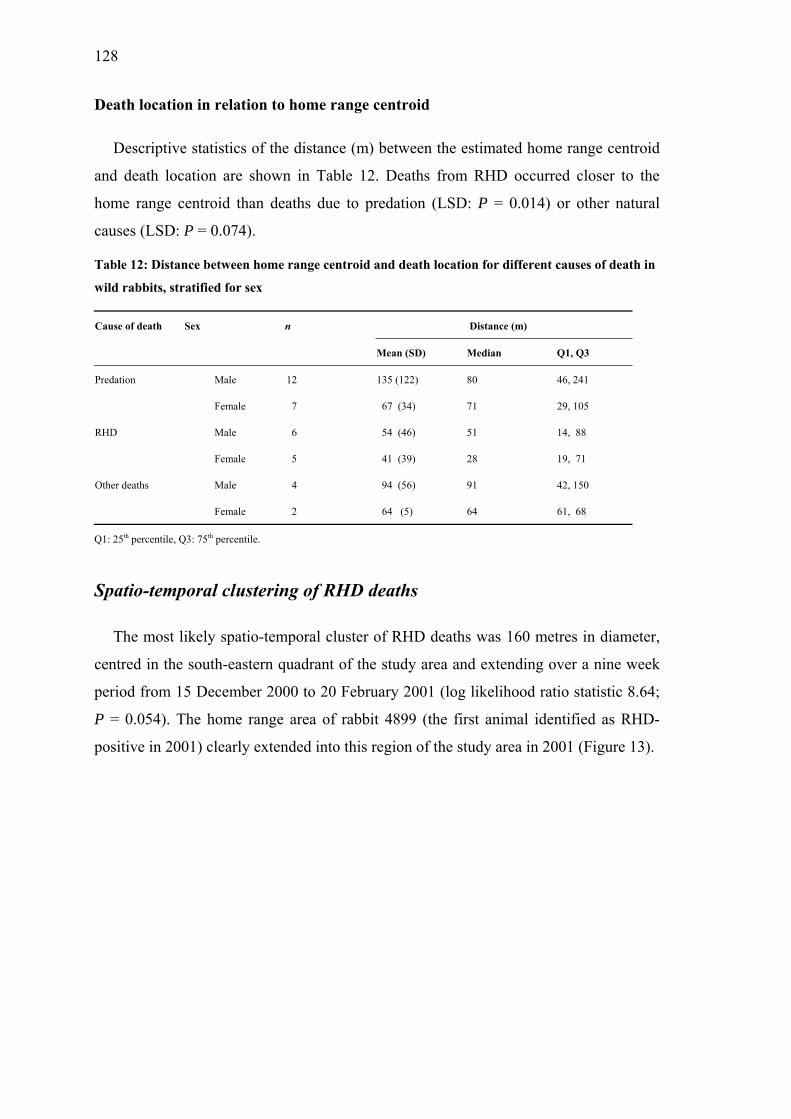
128
Death location in relation to home range centroid
Descriptive statistics of the distance (m) between the estimated home range centroid
and death location are shown in Table 12. Deaths from RHD occurred closer to the
home range centroid than deaths due to predation (LSD: P = 0.014) or other natural
causes (LSD: P = 0.074).
Table 12: Distance between home range centroid and death location for different causes of death in
wild rabbits, stratified for sex
Cause of death Sex n Distance (m)
Mean (SD) Median Q1, Q3
Predation Male 12 135 (122) 80 46, 241
Female 7 67 (34) 71 29, 105
RHD Male 6 54 (46) 51 14, 88
Female 5 41 (39) 28 19, 71
Other deaths Male 4 94 (56) 91 42, 150
Female 2 64 (5) 64 61, 68
Q1: 25th percentile, Q3: 75th percentile.
Spatio-temporal clustering of RHD deaths
The most likely spatio-temporal cluster of RHD deaths was 160 metres in diameter,
centred in the south-eastern quadrant of the study area and extending over a nine week
period from 15 December 2000 to 20 February 2001 (log likelihood ratio statistic 8.64;
P = 0.054). The home range area of rabbit 4899 (the first animal identified as RHD-
positive in 2001) clearly extended into this region of the study area in 2001 (Figure 13).

129
Figure 13: Death locations of rabbits dying between 1/03/1999 and 31/07/2001. Single solid lines
represent fence lines; are locations of deaths due to causes other than RHD in 2001; are RHD death
locations in 2001. The double solid line circle represents a space-temporal cluster of rabbit deaths
(Relative Risk = 8.64, P = 0.054); represents the first trapping location of rabbit 4899 and the dotted
contour represents the home range area of rabbit 4899 in summer 2001 (kernel density estimate, 95%
isopleth).
Discussion
Cause-specific mortality
Consistent with previous studies, predation was the predominant cause of rabbit
mortality in this population. Studies on rabbit predation are difficult to conduct because
rabbits are a preferred prey for many predator species and are usually consumed within
minutes with any remains being subsequently scavenged by a variety of species.

130
Therefore uncertainty about the fate of some animals is inevitable. An experimental
mortality study on post-emerged kittens in the Australian Capital Territory (south
eastern Australia) by Richardson and Wood (1982) classified 56% of rabbits with
unknown fate as victims of predation while a study of adult rabbit mortality in New
South Wales, Australia was only able to definitively record the fate of 98 of 274 (36%)
radio-tagged animals (Moriarty et al. 2000). Intensive monitoring in our study yielded a
relatively high rate of carcass retrieval and diagnosis of death – especially during the
2001 RHD outbreak when all cadavers were retrieved fully intact. Only three confirmed
RHD deaths were detected in 1999, but this is probably underestimated as some
carcasses may have been completely devoured by predators. Misclassification as
predation could have occurred if animals were weakened due to disease and became
easy prey, or animals that had died from disease were subsequently scavenged.
Juvenile rabbits have a particularly high rate of mortality. Reddiex et al. (2002)
attributed all deaths within a group of post-emergent, radio-collared rabbits to predation
or RHD, but were not able to measure the importance of the respective causes, because
rabbit carcasses had been scavenged heavily. Robson (1993), in a study of rabbit
mortality in North Canterbury, New Zealand, showed that at least 38% of juvenile litters
in breeding stops had been preyed upon, 29% of deaths were attributed to drowning,
20% of deaths were due to a combination of predation and drowning, and only 13% of
litters were weaned succesfully. Ground predators were responsible for more than half
of the mortality of post-emergent kittens observed in the Australian Capital Territory
study (Richardson and Wood 1982). Predation is also one of the major causes of death
in adult rabbits. Of the 98 adult rabbit deaths recorded by Moriarty et al. (2000), 53
(44%) were thought to be due to predation.
In common with previous New Zealand studies, cats were the main predators at our
site (Gibb et al. 1978). In comparison cats ranked third in importance as predators of
adult rabbits (6% of all deaths) in a study in New South Wales, Australia, behind foxes
(28%) and birds of prey (10%) (Moriarty et al. 2000). Cats are usually seen hunting
rabbits by day and night (Gibb et al. 1978) and predation by cats occurred throughout
the year at our site (Henning et al. 2003b). In contrast mustelids prey upon smaller
rabbits than cats, and therefore their predatory activity is more dependent on the rabbit
breeding season (Robson 1993). Unlike cats, ferrets and stoats dig into breeding stops to

131
kill where they mainly attack juveniles and nestlings (Gibb et al. 1978). Consistent with
these observations, in our study area stoats and ferrets were only trapped in summer and
autumn when abundance of younger rabbits was high (Henning et al. 2003a, 2003b).
With regards to birds of prey Richardson and Wood (1982) reported a daily mortality
risk of 0.7%, while rates of 4% were reported in an earlier Australian study (Calaby
1951). In New Zealand, the harrier is the only bird of prey, but its success in catching
rabbits, especially full-grown animals, is poor (Gibb et al. 1978).
Deaths due to other natural causes (Table 7) and deaths due to RHD accounted for
equivalent rates of mortality (20% and 19% respectively) but only RHD deaths were
strongly clustered in time. Deaths during the 2001 RHD outbreak peaked within one
week, similar to observations made at a study site in Central Otago, New Zealand,
where RHD spread naturally (Parkes et al. 2002). Although no appreciable RHD
epidemic was detected in 2000, RHDV was active on the site, possibly interacting or
being replaced by other causes of mortality, in particular predation (Henning et al.
2003a). The overall mortality attributed to RHD in our study was 20%, which is similar
to the proportion of RHD deaths (16%) observed in a Australian study by Moriarty et
al. (2000). Pneumonia was the predominant cause of death we observed under the
category of other natural causes and affected primarily young animals during the wet
winter months, while Eimeria infection, often regarded as an important cause of death
(Bull 1953; 1958), had only a minor influence on mortality. Similar observations were
made in Australia, where parasitism by nematodes and coccidia were not important
causes of death, implying that other mortality factors were responsible for density-
dependent regulation in those populations (Hobbs et al. 1999).
In common with our study, most studies on adult mortality in males and females do
not reveal gender specific differences (Parer 1977; Wood 1980; Gilbert et al. 1987;
Webb 1993; Moriarty et al. 2000), although a higher mortality in males compared with
females has been recorded (Cowan 1983).
Temporal patterns of mortality
Crude and cause-specific mortality rates are a function of age, which must be
considered in the analysis. Kaplan-Meier estimates treat age as a continuous variable

132
with no grouping by age categories, and therefore no age-specific mortality rates were
calculated. Predation resulted in death of many rabbits at a young age during the study
period and young rabbits were also more vulnerable to other natural causes of death
(e.g. pneumonia), while death from RHD occurred in older animals (after they had lost
maternal antibodies and natural resilience).
Core and home ranges
The decision to only use rabbits with at least seven radio fixes per season resulted in
many animals being excluded from core and home range analyses. Although this limited
the power of the analysis, it did increase confidence in the accuracy of the core and
home range estimates. The probability of the utilisation distribution was chosen to be
50% (the minimum area which contains 50% of the observations of an animal’s core or
home range) to represent the smallest size range estimate and 95% to include most of
the area covered by an animal (Bond et al. 2002). Several authors have noted that an
increase in the minimum area of probability (MAP) results in increased bias for non-
parametric methods of range estimation (Worton 1989; Harris et al. 1990). Anderson
(1982) recommended using a MAP of 50%, in particular for the core areas as they
reflect areas of particulary high usage. It could be argued that the 95% isopleths may
overestimate range areas, as they tend to be larger than minimum convex polygon
(MCP) estimates, which are strongly influenced by outliers. Worton (1995) used brush
rabbit (Sylvilagus bachmani) telemetry data to calculate 95% probability contours of
non-parametric density estimators and regarded them as less biased then other
estimation methods. Disadvantages of minimum convex polygons are that they can
include areas never visited by the animal (Worton 1989). We used different analytical
methods and were able to compare the estimates they delivered. Correlation between
home range estimates from different methods was poor if a low number of radio-fixes
was used per animal, but values of Pearson’s correlation coefficient exceeded 0.9 if
seven fixes or more were used as in our study.
Core range size
Core ranges are not often reported in the literature for rabbits, however recent
research has tried to distinguish between core and home range areas (Bond et al. 2001;

133
White et al. 2003). Cowan (1983) described mean range sizes of 0.71 hectares for males
and 0.43 hectares for females using maximum convex polygon and boundary strip
estimation methods (Southwood 1966). Ranges of less than one hectare were also
observed in Australian studies (Myers and Poole 1961). Daytime ranges in New
Zealand of 0.25 hectares for males and 0.18 hectares for females have also been
calculated (Gibb et al. 1978). Records in all these population studies were obtained
during daytime or several hours before sunset and can be therefore regarded as core
range estimates. The described core range estimates for the European rabbit were based
on rabbit observation from lookouts, enabling only monitoring of animals within a
distance of a few hundred metres and rabbits that were concealed by ground cover
would not have been recorded. However, the use of radio-tracking allows recording of
any animal movements and therefore larger estimates are to be expected. Using radio-
tracking core ranges of 0.54 hectares for males and 0.57 hectares for female surface-
dwelling rabbits were calculated, based on 60% isopleths using the harmonic mean
method (White et al. 2003). Our geometric least squares mean core range sizes based on
MCP (0.30 hectares) and 50% kernel (0.16 hectares) were similar to those reported
studies, although our 95% kernel estimate was considerably larger (0.92 hectares).
Although differences in core range areas across seasons were similar for each sex,
estimated core range areas were larger for males than females in winter, when mating
activity begins.
Home range size
Using a distance measure from the centre of activity for comparison (Hayne 1949),
Gibb (1993) found a larger dispersion of males during daytime in summer and winter
and reported that females were less variable in their seasonality. Based on kernel
density estimation methods (95% isopleth), we detected significant differences in home
range size between sexes during the winter and spring months, when males increase
their movements towards does. This is consistent with observations that female rabbits
tend to be more sedentary (Dunsmore 1974; Kunkele and von Holst 1996). Although
gender-season differences were not readily observed using kernel 50% and MCP
methods, home ranges showed clear differences between sexes. This indicates that
feeding, territorial and mating behaviours have a different impact for the sexes. Parer
and Fullager (1986) found that immature males disperse more frequently than other

134
classes of rabbits. Young adults are forced to move during the period of the greatest
social pressure, when breeding groups are under formation, which accounts for the
larger home range of male rabbits in autumn and the early winter months (Myers and
Poole 1961).
Myers and Poole (1961) described a marked seasonal decrease in home range size
during the breeding season, when activities related to territorial behaviour are at their
maximum, and a marked expansion in the home range when breeding ceased. Breeding
peaks on our sites were observed in spring and early summer (Henning et al. 2003a) and
the smallest home ranges were also found in summer (males) and spring (females).
Home range size is influenced by habitat and distribution of available food (Gibb 1993;
Zollner et al. 2000). However, variance component analysis revealed up to 50%
variation between individual animals. The geometric least squares mean estimates were
smaller than the observed values (but similar to the observed median values) and this
discrepancy between estimated and observed values is possibly due to the large
variation between animals. Similarly, observed means reported in wildlife studies may
present a distorted picture of the actual area used by animals, as they are strongly
influenced by excursions of single animals. High individual variability has been also
reported from other telemetry studies (Gibb 1993; Hulbert et al. 1996). Rabbit home
ranges recorded by radio-telemetry in the Orongorongo valley, New Zealand, had a
minimum size of 2.6 hectares using the minimum convex polygon method, with the
largest ranges for bucks being up to 6.5 hectares and for does about 4.5 hectares (Gibb
1993). These results suggest that geometric least squares means could be more
appropriate than range estimates. In order to reveal differences in core and home range
estimates for variables of interest, further range studies will require a larger number of
animals to be monitored.
Core range habitat
Vegetation types were only compared for core areas since most tracking events were
recorded during day-time. Spatial patterns of home ranges are dependent on the habitat.
Dry conditions and loose ground allow burrowing and the development of large warren
systems in Australia (Myers et al. 1994). However in different habitats the situation is

135
reversed, with one Australian study showing that up to 93% of radio locations were
recorded in dense native vegetation, and 76% above the ground, when pastureland was
surrounded by large areas of uncleared undergrowth (Wheeler et al. 1981). There are
several reasons why rabbits den above ground. Australian studies suggested that surface
dwelling could be the result of social regulation mechanisms, with socially sub-
dominant animals being unable to establish themselves in warren systems (Mykytowycz
and Gambale 1965; White et al. 2003). However, this could not have applied to our site,
as warren systems were not present. Rabbits may be forced to den above ground if the
ground material is inappropriate for burrowing (Gibb 1993), or choose to den above the
surface in areas where sufficient and suitable ground cover is available, despite the soil
being appropriate for burrowing (King et al. 1984). Ample ground cover was probably
the reason why burrows were rarely used in our study. Rabbits prefer medium-height
(50-100 cm) ground layer vegetation (Fa et al. 1999). On our site, vegetation grows up
to 300 cm and very dense vegetation types such as gorse, broom and toi-toi grass were
used as preferred denning areas. Dense vegetation provides shelter from weather
conditions for many species (Williams and Karl 2002) and protects rabbits from
predators and in particular birds of prey (Gibb 1993). The vegetation type might also
have implications in the epidemiology of RHD. In areas with dense vegetation (and less
burrowing) the lack of close contact and lower incidence of urinary and faecal
contaminated burrows could limit the spread of RHDV infection. This would explain
the lower incidence of RHD in higher rainfall areas where there is usually more
vegetation available as ground cover. However, a positive correlation between RHD
prevalence and vegetation cover has been described in an early report of the disease in
Europe (Mitro and Krauss 1993). Moriarty et al. (2000) described that more surface-
than warren-dwelling rabbits were killed by RHD. Contacts between surface- and
warren-dwelling rabbits tend to be more aggressive than encounters between
neighbouring warren dwellers and could therefore play a role in the long distance
transmission of RHDV (White et al. 2003). In addition, if deaths occur under cover,
carcasses may not be easily accessible for scavengers, which would facilitate access to
carcasses by insects that can transmit RHDV mechanically (Henning et al. 2003c). The
fly species Oxysarcodexia varia (Walker), considered as a main vector of RHDV
(Barratt et al. 1998), was also the most abundant fly species on open pasture in this
study (Henning et al. 2003c).

136
Death location in relation to home range centroid
There was evidence that RHD-infected rabbits died closer to their den site than
animals dying from other causes. Short distances between an animal’s home range
centroid and death location are typical for acute diseases like RHD, where animals die
suddenly. Carcasses close to den sites are more likely to be a source of new infection
when infested by flies, than animals dying further away which are more likely to be
rapidly scavenged. Deaths due to predation usually occur during feeding excursions on
pasture, when rabbits are observed and killed by cats, ferrets or hawks.
Spatio-temporal clustering of RHD deaths
The spatial pattern of cause-specific mortality (Figure 13) illustrates that in this study
RHD deaths were concentrated in small areas, whereas deaths due to predation or other
natural causes were more widely dispersed. Environmental issues such as vegetation
probably define these pockets of RHD deaths. The space-time cluster detected in this
study was an area of small gorse bushes between grass patches, while most of the other
parts of the study area contained extensive ground cover over many acres of open
pasture. This half-open vegetation type with a scattered ground cover edging
pastureland may reflect the best habitat for fly species transmitting the disease and
could explain some of the observed patchiness of RHD within a small area.
The importance of carrier rabbits has been described previously by other authors
(Cooke et al. 2000; Henning et al. 2003a). Circumstantial evidence indicates that a
newly sero-converted animal (4899) in the population was the most likely index case for
the 2001 RHD epidemic. We base this assertion on the following observations:
i) There was no rabbit, which was sero-positive at a high diagnostic test
dilution, trapped during the three weeks prior to the outbreak. High titres
(>1:640) of antibodies are usually found in survivors of RHD outbreaks
(Cooke et al. 2000). This individual probably had recovered from a previous
infection as indicated by high IgG (1:640) and IgA titres (1:160) (Cooke et al.
2000; Henning et al. 2003a).

137
ii) This rabbit was an adult male (6.4 months old), which migrated into the
population, as he had not been trapped in the area earlier.
iii) On the day of the first trapping of this animal, the first RHD death was
observed. Eleven animals died during this outbreak (40% of the monitored
population), but rabbit 4899 survived until predated on by a cat three months
later.
iv) The home range area of rabbit 4899 (Figure 13) clearly extended into the area
of the RHD cluster in 2001.
RHDV can be transmitted by direct contact or through urine and faeces from infected
animals (Ohlinger et al. 1993), but it has been shown that excreted RHDV can only
survive for a limited time in the environment (Gregg et al. 1991; Henning et al. 2003d).
Carrier rabbits can be the source of localised spread of the disease, aided by migration
due to mating, feeding and territorial behaviour. Henning et al. (2003a) showed that
immigration is a major factor influencing population immunity by increasing the
number of susceptibles, but here it was also shown that immigration of infectious
animals could induce new outbreaks.
There are always limitations when sparse rabbit populations are monitored. Gibb
(1993) mentioned the importance of studies of low-density populations, as they may
have different characteristics than higher density populations. He monitored only 60
rabbits for about 10 years, but drew important conclusions about social behaviour and
space use of rabbits over time. Radio-collaring of every animal trapped and intensive
monitoring in our study enabled us to describe a sparse population in detail in terms of
temporal-spatial aspects of different causes of death.
This study has i) identified cause-specific mortality rates with predation being the
major factor of rabbit mortality, ii) produced precise core and home range estimates by
adjusting for seasonal and gender influences and repeated measurements of the same
animal and iii) confirmed that RHD deaths appear clustered in time and space and close
to rabbit denning sites. These findings have important implications for rabbit control
with RHDV contaminated baits. The impact of the disease could be potentially
optimised by distributing baits near burrow entrances or, where rabbits are mainly

138
surface dwelling, spreading baits in close proximity to ground cover used as denning
sites.
Acknowledgements
We thank Robin Chrystall and George Robinson who allowed us to conduct this
study on their properties and are grateful to Solis Norton and Sonja Kay for their help
with radio-tracking and Dr. Cord Heuer’s help with the statistical analysis.
References
Anderson, D. J. (1982). The home range - a new nonparametric-estimation technique.
Ecology 63, 103-112.
Barratt, B. I. P., Ferguson, C. M., Heath, A. C. G., Evans, A. A., and Logan, R. A. S.
(1998). Can insects transmit Rabbit haemorrhagic disease virus? In 'Proceedings of
the Fifty First New Zealand Plant Protection Conference, Hamilton, New Zealand.'
pp. 245-250. (New Zealand Plant Protection Society: Rotorua, New Zealand.)
Bond, B. T., Leopold, B. D., Burger, L. W., and Godwin, K. D. (2001). Movements
and home range dynamics of cottontail rabbits in Mississippi. Journal of Wildlife
Management 65, 1004-1013.
Bond, B. T., Burger, L. W., Leopold, B. D., Jones, J. C., and Godwin, K. D. (2002).
Habitat use by cottontail rabbits across multiple spatial scales in Mississippi.
Journal of Wildlife Management 66, 1171-1178.
Brown, J. D., and Sleeman, J. M. (2002). Morbidity and mortality of reptiles admitted to
the Wildlife Center of Virginia, 1991 to 2000. Journal of Wildlife Diseases 38, 699-
705.
Bull, P. C. (1953). Parasites of the wild rabbit, Oryctolagus cuniculus (L.). New
Zealand Journal of Science and Technology (B) 34, 341-72.
Bull, P. C. (1958). Incidence of coccidia (Sporozoa) in wild rabbits, Oryctolagus

139
cuniculus (L.), in Hawke's Bay, New Zealand. New Zealand Journal of Science 1,
289-328.
Calaby, J. H. (1951). Notes on the little eagle, with particular reference to rabbit
predation. Emu 51, 33-56.
Cooke, B. D., Robinson, A. J., Merchant, J. C., Nardin, A., and Capucci, L. (2000). Use
of ELISAs in field studies of rabbit haemorrhagic disease (RHD) in Australia.
Epidemiology and Infection 124, 563-576.
Cowan, D. P. (1983). Aspects of the behavioural ecology of a free-living population of
the European wild rabbit, Oryctolagus cuniculus L. in southern England. (PhD
thesis. University of London: London, UK.)
Diggle, P. J., Heagerty, P. J., Liang, K.-Y., and Zeger, S. L. (Eds.) (2002). 'Analysis of
longitudinal data'. (Oxford University Press: Oxford, UK.)
Dunsmore, J. D. (1974). The rabbit in subalpine south-eastern Australia. I. Population
structure and productivity. Australian Wildlife Research 1, 1-16.
Fa, J. E., Sharples, C. M., and Bell, D. J. (1999). Habitat correlates of European rabbit
(Oryctolagus cuniculus) distribution after the spread of RVHD in Cadiz Province,
Spain. Journal of Zoology 249, 83-96.
Ford, R. G., and Krumme, D. W. (1979). The analysis of space use patterns. Journal of
Theoretical Biology 76, 125-155.
Gibb, J. A. (1993). Sociality, time and space in a sparse population of rabbits
(Oryctolagus cuniculus). Journal of Zoology 229, 581-607.
Gibb, J. A., Ward, C. P., and Ward, G. D. (1978). Natural control of a population of
rabbits, Oryctolagus cuniculus (L.), for ten years in the Kourarau enclosure. New
Zealand Department of Scientific and Industrial Research Bulletin 223, 1-89.
Gilbert, N., Myers, K., Cooke, B. D., Dunsmore, J. D., Fullagar, P. J., Gibb, J. A., King,

140
D. R., Parer, I., Wheeler, S. H., and Wood, D. H. (1987). Comparative dynamics of
Australasian rabbit populations. Australian Wildlife Research 14, 491-503.
Gregg, D. A., House, C., Meyer, R., and Berninger, M. (1991). Viral haemorrhagic
disease of rabbit in Mexico: epidemiology and viral characterisation. Revue
scientifique et technique de l'Office International des Epizooties 10, 435-451.
Harris, S., Cresswell, W. J., Forde, P. G., Trewhella, T., Wollard, T., and Wray, S.
(1990). Home-range analysis using radio-tracking data - a review of problems and
techniques particularly applied to the study of animals. Mammal Review 20, 97-
123.
Hayne, D. W. (1949). Calculation of size of home range. Journal of Mammalogy 30, 1-
8.
Henning, J., Pfeiffer, D. U., Davies, P. R., Meers, J. and Morris, R. S. (2003a). A three-
year study of Rabbit haemorrhagic disease virus infection in a wild rabbit
population in New Zealand. Wildlife Research submitted.
Henning, J., Davies, P. R., and Meers, J. (2003b). Temporal relationships between
seropositivity to Rabbit haemorrhagic disease virus in rabbits and other mammals
in New Zealand. Wildlife Research submitted.
Henning, J., Schnitzler, F. R., Pfeiffer, D. U., and Davies, P. R. (2003c). Influence of
weather conditions on fly abundance and its implications for transmission of Rabbit
haemorrhagic disease virus in the North Island of New Zealand. Medical and
Veterinary Entomology submitted.
Henning, J., Meers, J., Davies, P. R., and Morris, R. S. (2003d). Survival of Rabbit
haemorrhagic disease virus (RHDV) in the environment. Epidemiology and
Infection submitted.
Hobbs, R. P., Twigg, L. E., Elliot, A. D., and Wheeler, A. G. (1999). Evaluation of the
association of parasitism with mortality of wild European rabbits Oryctolagus
cuniculus (L.) in southwestern Australia. Journal of Parasitology 85, 803-808.
141
Hulbert, I. A. R., Iason, G. R., Elston, D. A., and Racey, P. A. (1996). Home-range sizes
in a stratified upland landscape of two Lagomorphs with different feeding
strategies. Journal of Applied Ecology 33, 1479-1488.
King, D. R., Wheeler, S. H., and Robinson, M. H. (1984). Daytime locations of
European rabbits at three localities in south-western Australia. Australian Wildlife
Research 11, 89-92.
Kulldorff, M. (1997). A spatial scan statistic. Communications in Statistics - Theory and
Methods 26, 1481-1496.
Kulldorff, M., and Nagarwalla, N. (1995). Spatial disease clusters - detection and
inference. Statistics in Medicine 14, 799-810.
Kunkele, J., and von Holst, D. (1996). Natal dispersal in the European wild rabbit.
Animal Behaviour 51, 1047-1059.
Marubini, E. and Valsecchi, M. G. (1996). Analysing survival data from clinical trials
and observational studies. In 'Competing risks'. pp. 331-361. (John Wiley & Sons
Ltd: Chichester, UK.)
McLellan, B. N., Hovey, F. W., Mace, R. D., Woods, J. G., Carney, D. W., Gibeau, M.
L., Wakkinen, W. L., and Kasworm, W. F. (1999). Rates and causes of grizzly bear
mortality in the interior mountains of British Columbia, Alberta, Montana,
Washington, and Idaho. Journal of Wildlife Management 63, 911-920.
McLennan, J. A., Potter, M. A., Robertson, H. A., Wake, G. C., Colbourne, R., Dew, L.,
Joyce, L., McCann, A. J., Miles, J., Miller, P. J., and Reid, J. (1996). Role of
predation in the decline of kiwi, Apteryx Spp, in New Zealand. New Zealand
Journal of Ecology 20, 27-35.
Mitro, S., and Krauss, H. (1993). Rabbit hemorrhagic disease - a review with special
reference to its epizootiology. European Journal of Epidemiology 9, 70-78.
Mohr, C. O. (1947). Table of equivalent populations of North American small

142
mammals. American Midland Naturalist 27, 223-249.
Moriarty, A., Saunders, G., and Richardson, B. J. (2000). Mortality factors acting on
adult rabbits in central-western New South Wales. Wildlife Research 27, 613-619.
Myers, K., Parer, D., Wood, D., and Cooke, B. (Eds.) (1994). 'The rabbit in Australia'.
(Oxford University Press: Oxford, UK.)
Myers, K., and Poole, W. E. (1961). A study of the biology of the wild rabbit,
Oryctolagus cuniculus (L.), in confined populations. II. The effects of season and
population increase on behaviour. C.S.I.R.O. Wildlife Research 6, 1-41.
Mykytowycz, R., and Gambale, S. (1965). A study on the inter-warren activities and
dispersal of wild rabbits Oryctolagus cuniculus (L.) in a 45-ac. paddock. C.S.I.R.O.
Wildlife Research 10, 111-123.
Nowotny, N., Bascunana, C. R., Ballagi-Pordany, A., Gavier-Widen, D., Uhlen, M., and
Belak, S. (1997). Phylogenetic analysis of rabbit haemorrhagic disease and
European brown hare syndrome viruses by comparison of sequences from the
capsid protein gene. Archives of Virology 142, 657-73.
Ohlinger, V. F., Haas, B., and Thiel, H. J. (1993). Rabbit haemorrhagic disease (RHD):
characterisation of the causative calicivirus. Veterinary Research 24, 103-16.
Parer, I. (1977). The population ecology of the wild rabbit (Oryctolagus cuniculus L.),
in a Mediterranean-type climate in New South Wales. Australian Wildlife Research
4, 171-205.
Parer, I., and Fullager, P. J. (1986). Biology of rabbits, Oryctolagus cuniculus, in
subtropical Queensland. Australian Wildlife Research 13, 545-557.
Parkes, J. P., Norbury, G. L., Heyward, R. P., and Sullivan, G. (2002). Epidemiology of
rabbit haemorrhagic disease (RHD) in the South Island, New Zealand, 1997-2001.
Wildlife Research 29, 543-555.

143
Reddiex, B., Hickling, G. J., Norbury, G. L., and Frampton, C. M. (2002). Effects of
predation and rabbit haemorrhagic disease on population dynamics of rabbits
(Oryctolagus cuniculus) in North Canterbury, New Zealand. Wildlife Research 29,
627-633.
Richardson, B. J., and Wood, D. H. (1982). Experimental studies on a subalpine rabbit
population. I. Mortality factors acting on emergent kittens. Australian Wildlife
Research 9, 443-50.
Robertson, H. A., and Colbourne, R. M. (2001). Survival of Little Spotted Kiwi exposed
to the rodenticide Brodifacoum. Journal of Wildlife Management 65, 29-34.
Robson, D. L. (1993). Natural mortality of juvenile rabbits (Oryctolagus cuniculus) in
North-Canterbury, New Zealand. Wildlife Research 20, 815-831.
Southwood, T. R. E. (Ed.) (1966). 'Ecological methods: with particular reference to the
study of insect populations'. (Chapman & Hall: London, UK.)
Taylor, S. K., Buergelt, C. D., Roelke-Parker, M. E., Homer, B. L., and Rotstein, D. S.
(2002). Causes of mortality of free-ranging Florida panthers. Journal of Wildlife
Diseases 38, 107-114.
Thrusfield, M. (Ed.) (1997). 'Veterinary Epidemiology'. (Blackwell Science Inc.:
London, UK.)
Webb, N. J. (1993). Growth and mortality in juvenile European wild rabbits
(Oryctolagus cuniculus). Journal of Zoology, London 230, 665-677.
Wheeler, S. H., King, D. R., and Robinson, M. H. (1981). Habitat and warren utilisation
by the European Rabbit, Oryctolagus cuniculs (L.), as determined by radio-
tracking. Australian Wildlife Research 8, 581-88.
White, P. C. L., Newton-Cross, G. A., Gray, M., Ashford, R., White, C., and Saunders,
G. (2003). Spatial interactions and habitat use of rabbits on pasture and
implications for the spread of rabbit haemorrhagic disease in New South Wales.

144
Wildlife Research 30, 49-58.
Williams, P. A., and Karl, B. J. (2002). Birds and small mammals in kanuka (Kunzea
ericoides) and gorse (Ulex europaeus) scrub and the resulting seed rain and
seedling dynamics. New Zealand Journal of Ecology 26, 31-41.
Wood, D. H. (1980). The demography of a rabbit population in an arid region of New
South Wales Australia. Journal of Animal Ecology 49, 55-79.
Worton, B. J. (1989). Kernel methods for estimating the utilisation distribution in home-
range studies. Ecology 70, 164-168.
Worton, B. J. (1995). Using Monte-Carlo simulation to evaluate kernel-based home-
range estimators. Journal of Wildlife Management 59, 794-800.
Zollner, P. A., Smith, W. P., and Brennan, L. A. (2000). Home range use by Swamp
Rabbits (Sylvilagus Aquaticus) in a frequently inundated bottomland forest.
American Midland Naturalist 143, 64-69.

145

146
Stoat trapped on the study site
Hedgehogs captured within one day in spring

147
Chapter 4
Temporal relationships between seropositivity to
Rabbit haemorrhagic disease virus in rabbits and
other mammals in New Zealand*
*Submitted by J. Henning, P. R. Davies and J. Meers to Wildlife Research

148

149
Abstract
As part of a longitudinal study of the epidemiology of Rabbit haemorrhagic disease
virus (RHDV) in New Zealand, serum samples were obtained from trapped feral
animals that may have consumed rabbit carcasses (non-target species). During a 21
month period when RHDV infection was monitored in a defined wild rabbit population,
16 feral cats (Felis catus), 11 stoats (Mustela erminea L.), 4 ferrets (Mustela furo) and
126 hedgehogs (Erinaceus europaeus) were incidentally captured in the rabbit traps.
The proportions of samples that were seropositive to RHDV were 6 of 16 (38%) cats, 2
of 11 stoats (18%), 1 of 4 ferrets (25%) and 5 of 126 hedgehogs (4%). Seropositive non-
target species were trapped in April 2000, in the absence of an overt epidemic of rabbit
haemorrhagic disease (RHD) in the rabbit population, but evidence of recent infection
in rabbits was shown. Seropositive non-target species were found up to two and a half
months before and one month after this RHDV activity in wild rabbits was detected.
Seropositive predators were also trapped on the site between one and four and a half
months after a dramatic RHD epidemic in February 2001. This study has shown that
high antibody titres can be found in non-target species when there are no longer rabbits
succumbing to RHD, although a temporal relationship was not detected statistically due
to the small sample size. Predators and scavengers contribute to localised spread of
RHDV through their movements.
Introduction
Rabbit haemorrhagic disease virus (RHDV) is a calicivirus that causes a peracute
and often fatal disease in the European rabbit (Oryctolagus cuniculus). Although
disease caused by RHDV, rabbit haemorrhagic disease (RHD), has not been reported in
other species, the host range of the virus is not well defined and experimental infections
of hares (Lepus europaeus) have yielded conflicting results (Ohlinger et al. 1990;
Mocsari 1990; Löliger 1990; Kölbl et al. 1990; di Modugno and Nasti 1990; Du 1990;
Chasey et al. 1992). In countries where introduction of RHDV to control pest rabbit
populations has been considered, a major concern has been the risk that non-target
species, particularly native mammals, could be susceptible to the disease. Serological
evidence for cross-infection of non-target species was evaluated in relation to the
proposed introduction of RHDV to control wild rabbits in both Australia and New

150
Zealand (Lenghaus et al. 1994; Buddle et al. 1997; Gould et al. 1997). Although
seroconversion occurred in mice (Mus musculus) and kiwis (Apteryx australis), no
evidence of a productive viral infection was detected in either species (Lenghaus et al.
1994; Buddle et al. 1997). Infection of humans, despite high levels of RHDV exposure,
has not been detected serologically in Australia or New Zealand (Carman et al. 1998;
Carman 1999; Greenslade et al. 2001). However some researchers have alluded to the
potential for mutation of RHDV that could enable it to cross species barriers and cause
disease (Smith et al. 1998a; Smith 1999). Both predators and scavengers of rabbits are
likely to be exposed to RHDV when the disease is prevalent in wild rabbit populations,
and seroconversion to the agent has been demonstrated in foxes (Vulpes vulpes) fed
RHDV infected liver (Leighton et al. 1995). Field studies have demonstrated RHDV
antibodies in free-ranging foxes in Germany (Dedek and Frölich 1997; Frölich et al.
1998) and recently in several predatory and scavenging species in New Zealand (Parkes
et al. 2003). However seropositivity in these studies was not investigated in relation to
the occurrence of RHD in associated rabbit populations.
An intensive longitudinal study of RHD in a wild rabbit population in New Zealand
provided an opportunity to compare temporal patterns of RHD infection of rabbits with
the serological status of other feral mammals trapped at the study location. This paper is
the last of three papers reporting the findings of these investigations (Henning et al.
2003a; 2003b).
Materials and methods
This study was conducted on a 30 hectare site in the Manawatu region of New
Zealand (Henning et al. 2003a; 2003b). A rabbit-proof fence enclosed 2.5 hectares of
the site, and traps were incorporated into this fence to capture animals moving from one
side of the fence to the other. The live-capture traps were set two nights per week, and
trapped rabbits were identified, examined and released. Animals that wanted to cross
the fence-line had to pass via the traps, although traps could be bypassed by climbing
animals such as cats. In the period from September 1999 until June 2001, any trapped
non-target species were anaesthetised by intramuscular injection of ketamine
hydrochloride (100 mg/ml; Phoenix Pharm Distributors Limited, Auckland, New
Zealand) to enable examination and sample collection. For each animal, the body
151
length, sex, weight, maturity and body condition were recorded and a 1 ml blood sample
collected from the heart. Serum samples were tested for RHDV antibodies with a
competitive ELISA (Capucci et al. 1991) at dilutions 1:10, 1:40, 1:160 and 1:640.
Samples were classified as positive if the percentage inhibition was larger than 50% at a
1:40 dilution (Parkes et al. 2002). Hedgehogs were released away from the study site,
and predator species were euthanased by an intracardiac injection of sodium
pentobarbitone (Pentobarb 300, 300 mg/ml; National Veterinary Supplies Limited,
Auckland, New Zealand).
Rabbits trapped on the site were tested for RHDV antibodies with the competitive
ELISA and also with isotype ELISAs for IgG, IgM and IgA as described previously
(Henning et al. 2003a). Rabbit carcasses retrieved by radio-tracking (Henning et al.
2003b) were autopsied and tested for RHDV on liver tissue using the reverse
transcriptase polymerase chain reaction (RT-PCR) (Nowotny et al. 1997). A small
sample (n = 8) of non-target species was also screened for RHDV using RT-PCR.
Chi squared and Fisher’s exact tests were used to compare count data, and odds
ratios were calculated. Logistic regression was used to test for the influence of sex,
reproductive status, body condition and weight on seropositivity to RHDV (Hosmer and
Lemeshow 2000).
Results
Results for rabbits have been reported in detail elsewhere (Henning et al. 2003b).
Briefly, high levels of IgM indicating recent RHDV infection were detected in rabbits in
April 2000 and February 2001. RHDV was detected in liver tissue from dead rabbits in
February 2001, but not in April 2000. Therefore, there was evidence of RHDV
transmission in the rabbit population in the months of April 2000 and February 2001,
although confirmed mortalities were limited to February 2001.
A total of 158 non-target animals was trapped and sampled over the study period,
including 16 feral cats, Felis catus (10%), 11 stoats, Mustela erminea L. (7%), 4 ferrets,
Mustela furo (3%) and 126 hedgehogs, Erinaceus europaeus (80%). Significantly more
male hedgehogs (P = 0.04) were trapped than females. The apparent prevalence of

152
animals with antibodies to RHD varied between species, being highest in cats (38%),
and lowest in hedgehogs (4%) (Table 13).
Table 13: RHDV sero-prevalence in non-target species stratified by sex
males females
seropositive (%) n seropositive (%) n seropositive overall
Cat 50 6 30 10 38
Stoat 17 6 20 5 18
Ferret 0 0 25 4 25
Hedgehog 3 75 6 52 4
All 4 ferrets, including the single seropositive ferret, were trapped in January 2001.
Trapping of cats, stoats and hedgehogs occurred more evenly during the study period.
The temporal patterns of detection of seropositive samples from these species is shown
in Figures 14-17. Positive samples were collected from cats in October and December
1999, April 2000, and in March, April and June of 2001, from stoats in April and May
2000, and from hedgehogs in February 2000 and April 2001. Antibody titres higher than
1 in 40 were found in one cat trapped in December 1999 (1:160), one stoat trapped in
May 2000 (1:160), one hedgehog trapped in April 2001 (1:640), and two cats trapped in
March and April 2001 (1:640).
No statistically significant association between potential physiological risk factors
and the odds of seropositivity was detected using multivariable logistic regression.
Seropositive non-target species were found up to 2 weeks before and up to 15 weeks
after the recovery of an RHDV infected rabbit carcass in 2001. In 2000, seropositive
non-target species were found from 10 weeks before to 4 weeks after the detection of a
recent infection in a rabbit. Although there was a trend that predators were more likely
to become seropositive in the two months before or after the detection of RHDV in
rabbits (Figures 14-17), this was not supported statistically. The odds ratio for predators
being seropositive between two months prior and two months after RHDV evidence in
rabbits was 1.67 (95% CI: 0.33-8.36) compared to all other times of the year. No RHDV
was detected by RT-PCR on liver tissues of three seronegative animals (2 stoats, 1
ferret) and 5 seropositive animals, comprised of two cats (seropositive at 1:40 and
1:640), two stoats (seropositive at 1:40 and 1:160) and one ferret (seropositive at 1:40).

153
M onth/Y ear
A ug/99 O ct/99 D ec/99 Feb/00 A pr/00 Jun/00 A ug/00 O ct/00 D ec/00 Feb/01 A pr/01 Jun/01
Num
ber o
f ani
mal
s
1
2
3
Figure 14: Number of RHDV seropositive (black) and seronegative (grey) cats trapped between August
1999 and June 2001. Arrows indicate months in which RHD transmission was observed in rabbits.
M o n th /Y ea r
A u g /9 9 O c t/9 9 D ec /9 9 F eb /0 0 A p r/0 0 Ju n /0 0 A u g /0 0 O c t/0 0 D ec /0 0 F eb /0 1 A p r/0 1 Ju n /0 1
Num
ber o
f ani
mal
s
1
2
3
Figure 15: Number of RHDV seropositive (black) and seronegative (grey) stoats trapped between August
1999 and June 2001. Arrows indicate months during which RHDV transmission was observed in rabbits.

154
M o n th /Y ea r
A u g /9 9 O c t/9 9 D ec /9 9 F eb /0 0 A p r/0 0 Ju n /0 0 A u g /0 0 O c t/0 0 D ec /0 0 F eb /0 1 A p r/0 1 Ju n /0 1
Num
ber o
f ani
mal
s
0
1
2
3
4
Figure 16: Number of RHDV seropositive (black) and seronegative (grey) ferrets trapped between
August 1999 and June 2001. Arrows indicate months during which RHDV transmission was observed in
rabbits.
M onth/Year
Aug/99 Oct/99 Dec/99 Feb/00 Apr/00 Jun/00 Aug/00 Oct/00 Dec/00 Feb/01 Apr/01 Jun/01
Num
ber o
f ani
mal
s
0
5
10
15
20
25
Figure 17: Number of RHDV seropositive (black) and seronegative (grey) hedgehogs trapped between
August 1999 and June 2001. Arrows indicate months during which RHDV transmission was observed in
rabbits.

155
Discussion
Interest in RHD infection of non-target species relates to concerns about
i) detrimental ecological outcomes on non-target species that infection, viral replication
and potential mutations of RHDV could have, and ii) the role that predatory and
scavenging species may have in dissemination of the disease. Our observations
complement a concurrent New Zealand report (Parkes et al. 2003) on antibody
responses to RHDV in predators, scavengers, and hares. In the current study we were
able to compare the time of RHD seropositivity detection in non-target species to events
of RHD transmission in rabbits.
Rabbit haemorrhagic disease virus is generally considered to be highly species-
specific (Ohlinger et al. 1993; Lenghaus et al. 2000; Cooke and Fenner 2002).
However, this opinion is not universally accepted (Smith 1999), as there is evidence that
some caliciviruses can infect and spread among multiple hosts (Smith and Boyt 1990).
The best documented example of this is the San Miguel sea lion virus (SMSV). Several
serotypes of SMSV having been isolated from a range of species of marine mammals
and fish (Smith et al. 1980) and this virus can also cause vesicular disease in humans
(Smith et al. 1998b) and in experimentally infected swine (Barlough et al. 1986a; Berry
et al. 1990). Moreover, the agent of vesicular exanthema of swine, which causes clinical
signs indistinguishable from foot and mouth disease, is thought to have emerged from
SMSV (Barlough et al. 1986b; Smith et al. 1998a), and a Vesicular exanthema of
swine-like calicivirus has recently been isolated from an aborted bovine foetus (Smith et
al. 2002). In contrast, other calicivirues, such as Feline calicivirus (FCV) and Norwalk
calicivirus, appear to have a very limited host range (Wyatt et al. 1978; Seal et al. 1993;
Lenghaus et al. 2000).
Similar clinical findings, morphological similarities, and geographic distributions
between RHDV and the European brown hare syndrome virus (EBHSV) in European
hares suggested that RHDV may have developed from EBHSV (Capucci et al. 1991;
Cancellotti and Renzi 1991). However it now known, that RHDV and EBHSV are two
distinct caliciviruses (Capucci et al. 1991; Nowotny et al. 1997). The apparent diversity
of biological behaviour (i.e. host specificity) among viruses classified as caliciviridae

156
means that it is questionable to base arguments about the risk of inter-species
transmission of RHDV solely on analogy.
Due to resource limitations, in this study we routinely conducted clinical and
serological evaluations of non-target species without the more definitive diagnostic
procedures (i.e. histology and PCR). However, there was no evidence of clinical disease
in any of the non-target species captured, and the limited PCR testing conducted was
negative for RHDV. To date, there has been only one published report on recovery of
RHDV from animals other than lagomorphs. Shien and Lee (2000) injected piglets
subcutaneously with 106.5 rabbit LD50 of RHDV and found that virus was detectable by
PCR in lung and liver up to 14 days post inoculation, and that the exposure led to low
antibody titres of short duration.
New Zealand is ecologically unique in that, with the exception of two species of
bats, it has no native terrestrial mammals, and almost all feral mammals are classified as
pests. The most serious ecological concern in relation to RHD in New Zealand is the
arguably unlikely potential for infection of non-mammalian hosts, most notably native
bird species. Such events require both a mechanism of exposure to virus under field
conditions and capability of the virus to infect the host. Probably a more realistic,
although indirect risk of RHD to the New Zealand ecosystem is the exposure of native
species to altered predator behaviour that could result from a rapid reduction of rabbit
populations (Haselmayer and Jamieson 2001).
Four out of four North Island brown kiwis, Apteryx australis matelli, developed
RHDV antibodies 14 days following intramuscular inoculation (106.5 rabbit LD50). The
birds had a rising titre over a period of 5 weeks and two birds remained seropositive
until the end of the study at 136 days, by which time virus could not be demonstrated in
tissues (Buddle et al. 1997). Similarly, seroconversion in the absence of detectable virus
was observed following experimental infection of mice, and was considered most likely
to be a response to injected antigen rather than an indication of viral replication
(Lenghaus et al. 1994). With respect to species for which rabbits are an immediate food
source, antibodies were detected up to 14 days after oral exposure of foxes to infected
rabbit livers (Leighton et al. 1995). However, the study did not include tests for the
presence of RHDV in fox tissues. Seroconversions have been also reported in domestic

157
dogs inoculated intraocularly and parenterally with RHDV (Simon et al. 1994). The
seroconversions following experimental exposures to RHDV in mice, dogs and foxes do
not emulate natural conditions (particularly parenteral routes) and thus does not
constitute evidence for viral infection and replication. The latter is also the case with
seropositivity in free-ranging animals, as described in this study. Seropositivity in free
ranging animals allows identification of species that warrant experimental investigation
as potential non-target hosts and reservoirs of the virus; these animals could possibly
also serve as sentinels for RHDV.
We have previously shown that RHDV inactivated with ultraviolet light did not
induce seroconversion in susceptible rabbits (Henning et al. 2003c). Therefore it is
probable that consumption of inactivated RHDV by non-target species would also not
induce a serological response. The possibility that viable RHDV consumed by non-
target species may replicate in their tissues causing the observed seroconversion cannot
be excluded.
Assuming rabbits are the sole significant reservoir of RHDV in an ecosystem,
observed seroprevalence in non-target species will be determined by the performance
characteristics (sensitivity and specificity) of the test, the prevalence of infected rabbits,
and the nature and rates of interspecies contacts. RHDV epidemics are typically
intermittent, occurring in annual or biannual cycles (Marchandeau et al. 2000; Calvete
et al. 2002; Parkes et al. 2002) and at our study site only persisted for about 4 weeks
(Henning et al. 2003a). The observed seroprevalence may be influenced by the
population dynamics and incidence of capture of non-target species in relation to the
temporal and spatial occurrence of these intermittent and transient rabbit epidemics.
Despite a trend of seropositivity in non-target species being apparently associated with
events of RHDV transmission in rabbits, our data were inadequate to support a temporal
relationship statistically. Key limitations of the data were the small sample size, and
heterogeneity over time in the populations of non-target species that were trapped (for
example all 4 ferrets were trapped in a single month). The likely importance of the
persistence of detectable titres to RHDV following exposure also needs to be
acknowledged (Leighton et al. 1995). Unfortunately, the performance characteristics of
serological tests for RHD in non-target species have not been systematically evaluated.
A major concern in this respect is the potential for cross-reactions, particularly in

158
species such as cats, which are known to be susceptible to other calicivirus infections
(Pedersen et al. 2000).
Given the existence of RHDV infected rabbits, the probability of seroconversion of
predatory and scavenging species will depend on their feeding behaviours and the
concentrations of virus in the consumed tissues. Feeding behaviours are likely to be
influenced by season and the availability of other food sources. Seasonal scat
examination of feral cats captured in a 10 year study of rabbits in the nearby Wairarapa
region in New Zealand revealed that, at most times of the year, at least half the volume
of faeces was composed of rabbit remains, and in some years this increased to 90 to
100% (Gibb et al. 1978). Other important food sources for cats, in decreasing order of
importance, were birds, insects and mice (Gibb et al. 1978). Cats favour young rabbits
as prey (Jones 1977; Borkenhagen 1978; Catling 1988) and rabbit consumption by cats
is particularly predominant in spring, when many young rabbits are present (Gibb et al.
1969). Gibb and Fitzgerald (1998) observed an increase of young rabbits in the diet of
cats in a period from 1984-1988 from 10 up to 50% in the spring and summer months,
which is the breeding season of rabbits. Recent studies in New Zealand and Australia
conducted after RHD became endemic and rabbit numbers declined, showed that rabbits
remain the preferred prey of feral cats (Molsher et al. 1999; Gillies 2001).
Lagomorph remains were detected in 16% of stoat guts in an open forest valley in
Fiordland, New Zealand, and were more prevalent (up to 26%) in years of low stoat
density (Murphy and Dowding 1995). The fact that when small rodents are more
abundant, they become a major prey item for stoats (Murphy and Dowding 1995),
suggests stoats prefer to prey on smaller animals, including young rabbits. Ferret
droppings examined from 1961 to 1964 showed variation with respect to content of
remains from young and old rabbits (Gibb et al. 1978), with faeces containing twice as
much remains of young rabbits compared with full grown rabbits in the breeding
season. The overall proportion containing rabbit remains varied between 24% and 95%,
depending on the abundance of rabbits, while other studies have found up to 65% of
lagomorph remains in ferret stomachs (Smith et al. 1995).
Although birds were not captured in our study, the diet of harriers is similarly
influenced by rabbit abundance and in particular the abundance of smaller rabbits (Gibb

159
et al. 1978). Gurr (1968) found 90% of harrier stomachs contained rabbit remains
during periods of high rabbit abundance in Otago, while only 20% had traces of rabbit
remains when abundance was low (Carroll 1968). In another study in the Mackenzie
Basin, Pierce and Maloney (1989) found that rabbits constitute 80-99% of the harrier
diet. It has also been reported that that harriers had poor success rates in killing rabbits
and mainly feed on rabbit carrion killed by other predators (Gibb et al. 1978). By
analogy this would include rabbits infected with RHDV. With respect to environmental
concerns about interspecies transmission of RHDV to native birds in New Zealand, a
case could be made for the evaluation of RHDV infectivity in those avian species, such
as harriers, that are most likely to be exposed to RHDV under natural conditions.
Mammalian predators also prey on other scavengers and predators. Cat faeces
contained hedgehog and stoat remains, while hedgehog remains have been found in
ferret faeces (Roser and Lavers 1976; Gibb et al. 1978). Secondary infection following
consumption of infected non-target species is theoretically possible but unlikely. In cats
the proportion of hedgehog and stoat remains did not exceed 6% or 1% respectively
(Gibb et al. 1978) and in ferrets the proportion of hedgehog remains was only 8%
(Roser and Lavers 1976). A study on pastoral farmland in New Zealand revealed
scavenging of ferrets on ferret carcasses and hedgehogs scavenging on possum
carcasses, but not on rabbit carcasses (Ragg et al. 2000). Although it was mentioned
that this needs more exploration, these findings are consistent with our observations of a
relatively low seroprevalence of RHDV in hedgehogs.
In our study, seropositive non-target species were trapped several weeks before and
after RHDV emergence in rabbits, suggesting that they either moved in from
neighbouring RHDV infected areas into the study region, or that they had contact with
infected rabbit carcasses not detected on our site (Henning et al. 2003b). Prolonged high
antibody titres in non-target species may be the consequence of their consumption of
large amounts of rabbits that died from RHD or through them consistently preying on
seropositive rabbits, which survived previous RHD outbreaks and contained RHDV in
their tissues. The latter seems less likely, as RHDV could not be detected in liver tissue
of rabbits shot in a cross-sectional survey, despite the rabbits having high RHDV
antibody serum titres (Henning and Davies 2003).

160
Scavengers and predators may play a role in the localised spread of RHDV via
environmental contamination by saliva, fur or feet. In addition, studies on domestic
dogs fed infected rabbit livers showed that dogs shed virus infective for rabbits (Simon
et al. 1994) and it appears likely that the transient faecal excretion of viable virus
following consumption of large doses of RHDV is another probable mechanism for the
localised spread of RHDV. Interaction between predation and RHD have been reported
previously (Reddiex et al. 2002) and this could be due to the localised spread of RHDV
through non-target species.
References
Barlough, J. E., Berry, E. S., Skilling, D. E., and Smith, A. W. (1986a). The marine
calicivirus story. part I. Compendium on Continuing Education for the Practicing
Veterinarian 8, F5-F14.
Barlough, J. E., Berry, E. S., Skilling, D. E., and Smith, A. W. (1986b). The marine
calicivirus story. part II. Compendium on Continuing Education for the Practicing
Veterinarian 8, F75-F83.
Berry, E. S., Skilling, D. E., Barlough, J. E., Vedros, N. A., Gage, L. J., and Smith, A.
W. (1990). New marine calicivirus serotype infective for swine. American Journal
of Veterinary Research 51, 1184-1187.
Borkenhagen, P. (1978). Von Hauskatzen (Felis sylvestris f. catus L., 1758)
eingetragene Beute [in German]. Zeitschrift für Jagdwissenschaft 24, 27-33.
Buddle, B. M., de Lisle, G. W., McColl, K., Collins, B. J., Morrissy, C., and Westbury,
H. A. (1997). Response of the North Island brown kiwi, Apteryx australis mantellli
and the lesser short-tailed bat, Mystacina tuberculata to a measured dose of Rabbit
haemorrhagic disease virus. New Zealand Veterinary Journal 45, 109-113.
Calvete, C., Estrada, R., Villafuerte, R., Osacar, J. J., and Lucientes, J. (2002).
Epidemiology of viral haemorrhagic disease and myxomatosis in a free-living
population of wild rabbits. Veterinary Record 150, 776-782.

161
Cancellotti, F. M., and Renzi, M. (1991). Epidemiology and current situation of viral
haemorrhagic disease of rabbits and the European brown hare syndrome in Italy.
Revue scientifique et technique de l'Office International des Epizooties 10, 409-
422.
Capucci, L., Scicluna, M. T., and Lavazza, A. (1991). Diagnosis of viral haemorrhagic
disease of rabbits and the European brown hare syndrome. Revue scientifique et
technique de l'Office International des Epizooties 10, 347-370.
Carman, J. A., Garner, M. G., Catton, M. G., Thomas, S., Westbury, H. A., Cannon, R.
M., Collins, B. J., and Tribe, I. G. (1998). Viral haemorrhagic disease of rabbits and
human health. Epidemiology and Infection 121, 408-418.
Carman, J. (1999). Rabbit calicivirus disease and human health: results from the
Australian study. In 'Rabbit Control, RCD: Dilemmas and Implications.
Proceedings of the Conference, 30-31 March 1998'. pp. 55-61. (Royal Society of
New Zealand Miscellaneous Series: Wellington, New Zealand.)
Carroll, A. L. K. (1968). Foods of the harrier. Notornis 15, 23-28.
Catling, P. C. (1988). Similarities and contrasts in the diets of foxes, Vulpus vulpus, and
cats, Felis catus, relative to fluctuating prey populations and drought. Australian
Wildlife Research 15, 307-317.
Chasey, D., Lucas, M., Westcott, D., and Williams, M. (1992). European brown hare
syndrome in the UK; a calicivirus related to, but distinct from that of viral
haemorrhagic disease in rabbits. Archives of Virology 124, 363-370.
Cooke, B. D., and Fenner, F. (2002). Rabbit haemorrhagic disease and the biological
control of wild rabbits, Oryctolagus cuniculus, in Australia and New Zealand.
Wildlife Research 29, 689-706.
Dedek, J., and Frölich, K. (1997). Serological survey for calicivirus antibody among
free-living red foxes in Mecklenburg-Vorpommern, Germany. [in German].
Tierärztliche Umschau 52, 149-152.

162
di Modugno, G., and Nasti, R. (1990). La malattia emorrhagica virale in Puglia.
Contributio sperimentale [in Italian]. Rivista di Coniglicoltura 27, 25-32.
Du, N. (1990). Rabbit haemorrhagic disease (RHD) - a new disease and its viral
etiology. Deutsche Tierärztliche Wochenschrift 97, 114-116.
Frölich, K., Klima, F., and Dedek, J. (1998). Antibodies against Rabbit haemorrhagic
disease virus in free-ranging red foxes from Germany. Journal of Wildlife Diseases
34, 436-442.
Gibb, J. A., Ward, G. D., and Ward, C. P. (1969). An experiment in the control of a
sparse population of wild rabbits (Oryctolagus cuniculus L.) in New Zealand. New
Zealand Journal of Science 12, 509-534.
Gibb, J. A., Ward, C. P., and Ward, G. D. (1978). Natural control of a population of
rabbits, Oryctolagus cuniculus (L.), for ten years in the Kourarau enclosure. New
Zealand Department of Scientific and Industrial Research Bulletin 223, 1-89.
Gibb, J. A., and Fitzgerald, B. M. (1998). Dynamics of sparse rabbits (Oryctolagus
cuniculus), Orongorongo Valley, New Zealand. New Zealand Journal of Zoology
25, 231-243.
Gillies, C. (2001). Advances in New Zealand Mammalogy 1990-2000: House Cat.
Journal of the Royal Society of New Zealand 31, 205-218.
Gould, A. R., Kattenbelt, J. A., Lenghaus, C., Morrissy, C., Chamberlain, T., Collins, B.
J., and Westbury, H. A. (1997). The complete nucleotide sequence of Rabbit
haemorrhagic disease virus (Czech strain V351): use of the polymerase chain
reaction to detect replication in Australian vertebrates and analysis of viral
population sequence variation. Virus Research 47, 7-17.
Greenslade, E., Weinstein, P., Woodward, A., Capucci, L., Salmond, C., and Beasley,
R. (2001). A serological survey of antibodies to Rabbit haemorrhagic disease virus
(rabbit calicivirus disease) in two rural Central Otago communities. New Zealand
Medical Journal 114, 55-58.

163
Gurr, L. (1968). Communal roosting behaviour of the Australasian harrier, Circus
approximans in New Zealand. Ibis 110, 332-337.
Haselmayer, J., and Jamieson, I. G. (2001). Increased predation on pukeko eggs after
the application of rabbit control measures. New Zealand Journal of Ecology 25, 89-
93.
Henning, J., and Davies, P. R. (2003). A serial cross-sectional study of the prevalence of
rabbit haemorrhagic disease (RHD) on three farms in the lower North Island of
New Zealand. New Zealand Veterinary Journal submitted.
Henning, J., Pfeiffer, D. U., Davies, P. R., Meers, J. and Morris, R. S. (2003a). A three-
year study of Rabbit haemorrhagic disease virus infection in a wild rabbit
population in New Zealand. Wildlife Research submitted.
Henning, J., Pfeiffer, D. U., Davies, P. R., and Stevenson, M. A. (2003b). Spatial and
temporal dynamics of mortality and home range use in wild rabbits (Oryctolagus
cuniculus) with emphasis on rabbit haemorrhagic disease (RHD). Wildlife Research
submitted.
Henning, J., Meers, J., and Davies, P. R. (2003c). Exposure of rabbits to ultraviolet
light-inactivated Rabbit haemorrhagic disease virus (RHDV) and subsequent
challenge with virulent virus. Epidemiology and Infection submitted.
Hosmer, D. W., and Lemeshow, S. (Eds.) (2000). 'Applied Logistic Regression'. (John
Wiley & Sons, Inc: New York, USA.)
Jones, E. (1977). Ecology of the feral cat, Felis catus (L.), (Carnivora: Felidae) on
MacQuarie Island. Australian Wildlife Research 4, 249-262.
Kölbl, S., Settele, J., and Schönbauer, M. (1990). Erstes Auftreten der infektiösen
hämorrhagischen Krankheit der Kaninchen in Österreich [in German]. Berliner
Münchner Tierärztliche Wochenschrift 103, 261-266.
Leighton, F. A., Artois, M., Capucci, L., Gavier-Widen, D., and Morisse, J. P. (1995).

164
Antibody response to rabbit viral hemorrhagic disease virus in red foxes (Vulpes
vulpes) consuming livers of infected rabbits (Oryctolagus cuniculus). Journal of
Wildlife Diseases 31, 541-544.
Lenghaus, C., Westbury, H., Collins, B., Ratnamohan, N., and Morrissy, C. (1994).
Overview of the RHD project in Australia. In 'Rabbit Haemorrhagic Disease: Issues
in Assessment for Biological Control'. (Eds. R. K. Munro and R. T. Williams.) pp.
104-129. (Bureau of Resource Sciences, Australian Goverment Publishing Service:
Canberra.)
Lenghaus, C., Studdert, M. J., and Gavier-Widen, D. (2000). Calicivirus infections. In
'Infectious Diseases of Wild Mammals'. (Eds. E. S. Williams and I. K. Barker.) pp.
280-291. (Iowa State University Press, Ames, USA.)
Löliger, H. Ch. (1990). Aktuelle Erkrankungen der Hauskaninchen-Diagnose und
Bekämpfung [in German]. Der Praktischer Tierarzt 5, 42-54.
Marchandeau, S., Chaval, Y., and le Goff, E. (2000). Prolonged decline in the
abundance of wild European rabbits (Oryctolagus cuniculus) and high immunity
level over three years following the arrival of rabbit haemorrhagic disease. Wildlife
Biology 6, 141-147.
Mocsari, E. (1990). Epidemiological studies on the rabbit haemorrhagic disease and its
control by vaccination in Hungary. In '7. Arbeitstagung über Haltung und
Krankheiten der Kaninchen, Pelztiere und Heimtiere, Celle.' pp. 257-265.
(Deutsche Veterinärmedizinische Gesellschaft: Giessen, Germany.)
Molsher, R., Newsome, A., and Dickman, C. (1999). Feeding ecology and population
dynamics of the feral cat (Felis catus) in relation to the availability of prey in
central-eastern New South Wales. Wildlife Research 26, 593-607.
Murphy, E. C., and Dowding, J. E. (1995). Ecology of the stoat in Nothofagus forest:
home range, habitat use and diet at different stages of the beech mast cycle. New
Zealand Journal of Ecology 19, 97-109.

165
Nowotny, N., Bascunana, C. R., Ballagi-Pordany, A., Gavier-Widen, D., Uhlen, M., and
Belak, S. (1997). Phylogenetic analysis of rabbit haemorrhagic disease and
European brown hare syndrome viruses by comparison of sequences from the
capsid protein gene. Archives of Virology 142, 657-73.
Ohlinger, V. F., Haas, B., and Thiel, H.-J. (1990). Report on a meeting of European
Society for Veterinary Virology (ESVV) on viral haemorrhagic disease of rabbits.
7-8.12.1989. Lyon. [in German].
Ohlinger, V. F., Haas, B., and Thiel, H. J. (1993). Rabbit haemorrhagic disease (RHD):
characterisation of the causative calicivirus. Veterinary Research 24, 103-16.
Parkes, J. P., Norbury, G. L., Heyward, R. P., and Sullivan, G. (2002). Epidemiology of
rabbit haemorrhagic disease (RHD) in the South Island, New Zealand, 1997-2001.
Wildlife Research 29, 543-555.
Parkes, J. P., Heyward, R. P., Henning, J., and Motha, M. X. J. (2003). Antibody
responses to Rabbit haemorrhagic disease virus in predators, scavangers, and hares
in New Zealand during epidemics in sympatric rabbit populations. New Zealand
Veterinary Journal in press.
Pedersen, N. C., Elliott, J. B., Glasgow, A., Poland, A., and Keel, K. (2000). An isolated
epizootic of haemorrhagic-like fever in cats caused by a novel and highly virulent
strain of Feline calicivirus. Veterinary Microbiology 73, 281-300.
Pierce, R. J., and Maloney, R. F. (1989). Responses of harriers in the Mackenzie Basin
to the abundance of rabbits. Notornis 36, 1-12.
Ragg, J. R., Mackintosh, C. G., and Moller, H. (2000). The scavanging behaviour of
ferrets (Mustelo furo), feral cats (Felis domesticus), possums (Trichosurus
vulpecula), hedgehogs (Erinaceus europaeus) and harrier hawks (Circus
approximans) on pastoral farmland in New Zealand: Implications for bovine
tuberculosis transmission. New Zealand Veterinary Journal 48, 166-175.

166
Reddiex, B., Hickling, G. J., Norbury, G. L., and Frampton, C. M. (2002). Effects of
predation and rabbit haemorrhagic disease on population dynamics of rabbits
(Oryctolagus cuniculus) in North Canterbury, New Zealand. Wildlife Research 29,
627-633.
Roser, R. J., and Lavers, R. B. (1976). Food habits of the ferret (Mustela putorius L.) at
Pukepuke Lagoon, New Zealand. New Zealand Journal of Zoology 3, 269-275.
Seal, B., Ridpath, J., and Mengeling, W. (1993). Analysis of feline calicivirus capsid
protein genes: identification of variable antigenetic determinant regions of the
protein. Journal of General Virology 79, 2519-2524.
Shien, J. H., and Lee, L. H. (2000). Susceptibility of piglets to Rabbit haemorrhagic
disease virus following experimental infection. Canadian Journal of Veterinary
Research 64, 134-137.
Simon, M. C., Muguruza, R., Alonso, J. L., Muzquiz, J. L., Girones, O., and Haffar, A.
(1994). Rechereche du virus de la maladie hemorrhagic viral du lapin (RHD) et role
des canides dometiques dans la transmission de la maladie [in French]. Recueil de
Medecine Veterinaire 170, 841-845.
Smith, A. W., Skilling, D. E., and Brown, R. J. (1980). Preliminary investigation of a
possible lung worm (Parafilaroides decorus), fish (Girella nigricans), and marine
mammal (Callorhinus ursinus) cycle for San-Miguel sea lion virus type-5.
American Journal of Veterinary Research 41, 1846-1850.
Smith, A. W., and Boyt, P. M. (1990). Caliciviruses of ocean origin - a review. Journal
of Zoo and Wildlife Medicine 21, 3-23.
Smith, A. W., Skilling, D. E., Cherry, N., Mead, J. H., and Matson, D. O. (1998a).
Calicivirus emergence from ocean reservoirs: Zoonotic and interspecies
movements. Emerging Infectious Diseases 4, 13-20.
Smith, A. W., Berry, E. S., Skilling, D. E., Barlough, J. E., Poet, S. E., Berke, T., Mead,
J., and Matson, D. O. (1998b). In vitro isolation and characterisation of a calicivirus

167
causing a vesicular disease of the hands and feet. Clinical Infectious Diseases 26,
434-439.
Smith, A. W. (1999). Calicivirus models of emerging and zoonotic diseases. In 'Rabbit
Control, RCD: Dilemmas and Implications. Proceedings of the Conference, 30-31
March 1998'. pp. 37-42. (Royal Society of New Zealand Miscellaneous Series:
Wellington, New Zealand.)
Smith, A. W., Skilling, D. E., Matson, D. O., Kroeker, A. D., Stein, D. A., Berke, T.,
and Iversen, P. L. (2002). Detection of Vesicular exanthema of swine-like
calicivirus in tissues from a naturally infected spontaneously aborted bovine fetus.
Journal of the American Veterinary Medical Association 220, 455-458.
Smith, G. P., Ragg, J. R., Moller, H., and Waldrup, K. A. (1995). Diet of feral ferrets
(Mustela furo) from pastoral habitats in Otago and Southland, New Zealand. New
Zealand Journal of Zoology 22, 363-369.
Wyatt, R. G., Greenberg, H. G., Dalgard, D. W., Allwood, W. P., Sly, D. L., Thorhill,
T. S., Chanock, R. M., and Kapikian, A. Z. (1978). Experimental infection of
chimpanzees with the Norwalk agent of epidemic viral gastroenteritis. Journal of
Medical Virology 2, 89-96.

168
Fly bottle trap (modified Western Australian style) after one week of trapping in summer
Oxysarcodexia varia Walker

169
Chapter 5
Influence of weather conditions on fly abundance
and its implications for transmission of
Rabbit haemorrhagic disease virus
in the North Island of New Zealand*
*Submitted by J. Henning, F. R. Schnitzler, D. U. Pfeiffer and P. R. Davies, to Medical and Veterinary
Entomology

170

171
Abstract
Blowflies (Diptera: Calliphoridae) and flesh flies (Diptera: Sarcophagidae) are
potential vectors of Rabbit haemorrhagic disease virus (RHDV) in New Zealand. The
associations between habitat and weather factors on the abundance of these flies were
investigated. Between October 1999 and June 2001, flies were trapped on open pasture
and in dense vegetation patches on farmland in the Himatangi area of the North Island.
Five calliphorid species were trapped commonly at scrub edges and the most abundant
sarcophagid, Oxysarcodexia varia Walker, was trapped mainly on open pasture.
Overall abundance of flies varied according to habitat and species, and species numbers
differed between seasons and years. The all-day minimum temperature three weeks
before trapping was a significant variable in all models of fly abundance, while average
rainfall did not affect fly abundance. The all-day temperature range was significant only
for O. varia. The influence of other climatic factors varied between fly species.
Climate dependent variations in fly abundance may contribute to the risk of
transmission of rabbit haemorrhagic disease, which occurred intermittently on the site
during the study period.
Introduction
Flies can affect animal health and production through irritating animals and
interfering with grazing or eating behaviour, or by acting as biological or mechanical
vectors of pathogens (Greenberg, 1973; Wardhaugh & Morton, 1990; Hall & Wall,
1995; Heath & Bishop, 1995; Kettle, 1995; Fischer et al., 2001; Heath, 2002).
Rabbit haemorrhagic disease virus (RHDV) was introduced illegally into New
Zealand in 1997 (Thompson & Clark, 1997). The virus causes peracute disease and high
rates of mortality in rabbits, and is used as a biological control tool for wild rabbit
populations (Sanson et al., 2000; Parkes et al., 2002). The effectiveness of the disease in
reducing pest rabbit populations appears to be highly variable, and epidemics generally
occur intermittently at annual or bi-annual intervals (Parkes et al., 2002; Henning et al.,
2003a). The abundance and activity of insect vectors may be one factor that contributes
to the observed variability in the impact of RHD (Barratt et al., 2001). Climatic factors
are major determinants of fly abundance and population dynamics (Davies & Hobson,
1935; Evans, 1936; Davidson, 1944; Hughes & Sands, 1979; Vogt et al., 1983, 1985;

172
Vogt, 1986, 1988; Wall et al., 2001) with different fly species prefering different
habitats (Dymock & Forgie, 1993; Gleeson & Heath, 1997; Heath & Appleton, 2000).
In New Zealand, eight fly species have been identified as potential vectors of RHDV
(Crosby & McLennan, 1996) and their role was investigated in field studies (Barratt et
al., 1998; Heath et al., 1999).
The aims of this paper are: (1) to describe the seasonal abundance of the most
common blowfly and flesh fly species likely to be involved in RHDV transmission in
the North Island of New Zealand, (2) to identify the influence of habitat and season on
the abundance of fly species, and (3) to investigate their association with climatic
variables.
Materials and Methods
Study site
The study site was located near Himatangi (longitude 175.317, latitude -40.400), in
the North Island of New Zealand. The vegetation on the study site consisted
predominantly of grass patches, dense gorse bush (Ulex europaeus) and scrub. Twelve
traps were placed in two lines 50 m apart with 50 m intervals between traps on the same
line (Figure 18). Six traps were placed on open pasture, while the other six traps were
located within or on the edge of dense gorse patches.
Figure 18: Positions of flytraps located under shrubs ( ) and on open pasture ( ) in relation to vegetation.
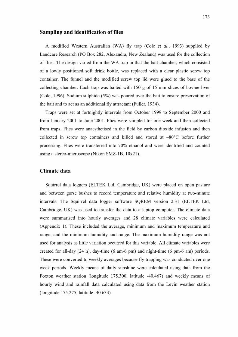
173
Sampling and identification of flies
A modified Western Australian (WA) fly trap (Cole et al., 1993) supplied by
Landcare Research (PO Box 282, Alexandra, New Zealand) was used for the collection
of flies. The design varied from the WA trap in that the bait chamber, which consisted
of a lowly positioned soft drink bottle, was replaced with a clear plastic screw top
container. The funnel and the modified screw top lid were glued to the base of the
collecting chamber. Each trap was baited with 150 g of 15 mm slices of bovine liver
(Cole, 1996). Sodium sulphide (5%) was poured over the bait to ensure preservation of
the bait and to act as an additional fly attractant (Fuller, 1934).
Traps were set at fortnightly intervals from October 1999 to September 2000 and
from January 2001 to June 2001. Flies were sampled for one week and then collected
from traps. Flies were anaesthetised in the field by carbon dioxide infusion and then
collected in screw top containers and killed and stored at –80°C before further
processing. Flies were transferred into 70% ethanol and were identified and counted
using a stereo-microscope (Nikon SMZ-1B, 10x21).
Climate data
Squirrel data loggers (ELTEK Ltd, Cambridge, UK) were placed on open pasture
and between gorse bushes to record temperature and relative humidity at two-minute
intervals. The Squirrel data logger software SQREM version 2.31 (ELTEK Ltd,
Cambridge, UK) was used to transfer the data to a laptop computer. The climate data
were summarised into hourly averages and 28 climate variables were calculated
(Appendix 1). These included the average, minimum and maximum temperature and
range, and the minimum humidity and range. The maximum humidity range was not
used for analysis as little variation occurred for this variable. All climate variables were
created for all-day (24 h), day-time (6 am-6 pm) and night-time (6 pm-6 am) periods.
These were converted to weekly averages because fly trapping was conducted over one
week periods. Weekly means of daily sunshine were calculated using data from the
Foxton weather station (longitude 175.300, latitude -40.467) and weekly means of
hourly wind and rainfall data calculated using data from the Levin weather station
(longitude 175.275, latitude -40.633).

174
Data analysis
Analyses were conducted using the statistical software packages SPSS 11.0 (SPSS
Inc, Chicago, IL, USA) and STATA 7.0 (STATA Corporation, College Station, TX,
USA).
The total number of flies per species and the number of flies per species across
different trap vegetation environments were compared using Chi squared tests. A
random effect Poisson regression model was used to compare fly abundance in different
trap vegetation environments (pasture and gorse) per month (Cameron & Trivedi,
1998). The calendar month for each year and the trap vegetation environments were
used as fixed effects and the fly species was incorporated as a random effect in the
model.
Two approaches were used to reduce the number of climate variables in the analysis.
(1) A factor analysis approach was used to identify relationships present in the
original variables. Factors created reflected unique variance separated from the common
variance of these climate variables. Eigenvectors were selected using the latent root
criterion and scree plots (McGarigal et al., 2000), and were rotated (varimax rotation) to
simplify their interpretation (Hair et al., 1998). Rotated eigenvectors were used to
identify single climate representative variables, which were entered into the regression
models.
(2) Testing for multicollinearity was based on inspection of correlation matrices
between all climate variables. A Pearson correlation greater than 0.7 was used to
identify a high correlation between two variables and suggested the presence of
multicollinearity.
Fly count data were transformed using ln+1 to normalise their distribution. A
univariate GLM model was developed with linear and exponential (where appropriate)
climate variables as predictors. Lagged values of climate variables for up to three weeks
before a particular week of trapping were also included as predictors to account for the
climate dependent pre-adult phase in fly life cycles. The trap vegetation environment
(pasture and gorse) was used as a categorical fixed effect variable and a stepwise
forward selection procedure was used to select influential climate variables and
interaction terms (P < 0.05). Comparisons between the full model and the best
parsimonious model were conducted using the adjusted R squared statistic (Hair et al.,
1998) and regression diagnostics were used to assess the goodness-of-fit of the model.

175
All models were computed with and without trap number as a random effect to account
for the repeated measure characteristics of the dataset, and the statistical significance of
the random effect was tested using the Hausman specification test for random effects
(Hausman, 1978). Separate regression analyses were conducted for each fly species. In
addition, the abundance of Oxysarcodexia varia Walker was assessed in relation to
habitat.
Results
A total of 29,415 flies were counted. Fly abundance was not distributed normally for
each sampling occasion and abundance was different among individual species
(χ2 = 5885.75, P < 0.001, df = 5) (Figure 19). For each species, the number of flies
differed significantly between the two trap vegetation environments (pasture and gorse
bushes) (χ2 = 3245.92, P < 0.001, df = 5) (Figure 20).
Species
Oxysarcodexia varia
Lucilia sericata
Chrysomya rufifacies
Calliphora vicina
Calliphora stygia
Calliphora hilli
Num
ber o
f flie
s
10000
8000
6000
4000
2000
0
Figure 19: Number of flies trapped per species over the entire study period.
O. varia was the most abundant fly (n = 8,436), and was trapped mainly on open
pasture, while all other blowflies were present in higher numbers on the gorse edges. Of
the six species studied, the least abundant species was Calliphora vicina Robineau-
Desvoidy (n = 1,668), followed by C. hilli Patton (n = 2,825), Lucilia sericata Meigen
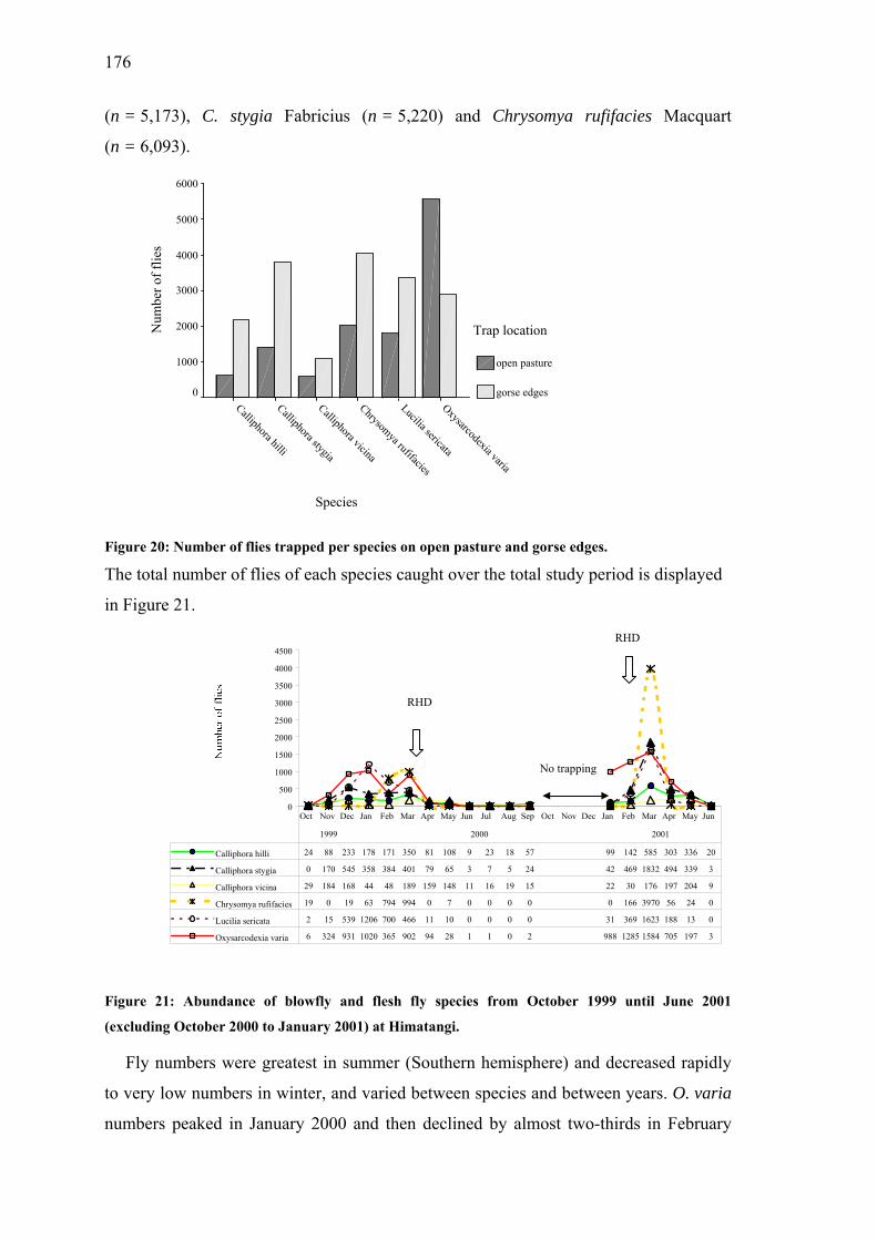
176
(n = 5,173), C. stygia Fabricius (n = 5,220) and Chrysomya rufifacies Macquart
(n = 6,093).
Species
Oxysarcodexia varia
Lucilia sericata
Chrysomya rufifacies
Calliphora vicina
Calliphora stygia
Calliphora hilli
Num
ber o
f flie
s
6000
5000
4000
3000
2000
1000
0
Trap location
open pasture
gorse edges
Figure 20: Number of flies trapped per species on open pasture and gorse edges.
The total number of flies of each species caught over the total study period is displayed
in Figure 21.
0
500
1000
1500
2000
2500
3000
3500
4000
4500
Calliphora hilli 24 88 233 178 171 350 81 108 9 23 18 57 99 142 585 303 336 20
Calliphora stygia 0 170 545 358 384 401 79 65 3 7 5 24 42 469 1832 494 339 3
Calliphora vicina 29 184 168 44 48 189 159 148 11 16 19 15 22 30 176 197 204 9
Chrysomya rufifacies 19 0 19 63 794 994 0 7 0 0 0 0 0 166 3970 56 24 0
Lucilia sericata 2 15 539 1206 700 466 11 10 0 0 0 0 31 369 1623 188 13 0
Oxysarcodexia varia 6 324 931 1020 365 902 94 28 1 1 0 2 988 1285 1584 705 197 3
Oct Nov Dec Jan Feb Mar Apr May Jun Jul Aug Sep Oct Nov Dec Jan Feb Mar Apr May Jun
1999 2000 2001
RHD
RHD
No trapping
Figure 21: Abundance of blowfly and flesh fly species from October 1999 until June 2001
(excluding October 2000 to January 2001) at Himatangi.
Fly numbers were greatest in summer (Southern hemisphere) and decreased rapidly
to very low numbers in winter, and varied between species and between years. O. varia
numbers peaked in January 2000 and then declined by almost two-thirds in February
177
2000. They then returned to similar numbers as in January by March 2000 before
decreasing to 10% of the peak value by April 2000. In the following year, abundance of
O. varia was about 40% higher in summer (January-March) 2001, and peaked in March.
In both years, C. rufifacies numbers peaked in March, but were approximately four fold
higher in 2001 compared to 2000. C. stygia numbers were highest in December 1999
and remained at similar levels until March 2000. C. stygia numbers were still very low
in January 2001, reached a peak in March 2001 and then remained elevated until May
2001, which was later than the same season in the previous year. L. sericata also had its
highest abundance later in 2001 (March) than in the preceding year (January), but
dropped dramatically in April for both years. No difference in the abundance peaks was
observed for C. hilli. Abundance of this fly species peaked in March in both 2000 and
2001, but persisted at higher numbers for an additional two months in 2001 compared to
2000. C. vicina was highly abundant in the late summer and autumn period (March-
May) of both years, but the additional spring peak in November-December 1999 was
not observable in 2000 as no trapping occurred during this period. C. hilli and C. vicina
were the predominant fly species over the winter months. A comparison of fly
abundance in the two vegetation environments over the 18 months of trapping using the
Poisson regression model with fly species as a random effect, showed highly significant
differences (P < 0.01) for all months, except for June 2000
(P = 0.057), July 2000 (P = 0.614), June 2001 (P = 0.013) and July 2001 (P = 0.064).
The factor analysis of all 28 climate variables revealed 5 factors with eigenvectors
greater than 1, which together accounted for 94% of the total variation. After rotation,
the 5 climate variables with the highest rotated factor loadings were selected (Table 14).
Table 14: Variables obtained from principal factor analysis and their number of correlations
(correlation coefficient > 0.7) with other climate variables (n = 28)
Climate variable selected by factor analysis Number of correlations with other climate variables
All day (24 h) temperature range 11
All day (24 h) temperature minimum 1
Average rain Levin 1
Night time (6 pm-6 am) humidity range 3
Average wind Levin 1

178
The correlation matrix revealed up to 13 correlations > 0.7 for a single climate
variable. A list of variables selected by factor analysis and the number of correlations
for each of these variables is also shown in Table 14. Four climate variables selected by
factor analysis had fewer than 4 correlations higher than 0.7 with other climate
variables, and one variable had a relatively high number of such correlations. Therefore
we were confident, that the selected variables explained most of the variability in the
climate variables, and that most of the multicollinearity was avoided by selecting these
variables.
Figure 22 shows the climate pattern of these 5 variables selected by factor analysis
for the most abundant fly, O. varia. Climate data are displayed by 3 weekly moving
averages. The graphical pattern suggests that the maximum daily (24 h) temperature
range, the minimum daily (24 h) temperature and the humidity range at night (6 pm-
6 am) may influence the abundance of O. varia, while average daily rainfall and average
daily wind speed do not show clear correlations with abundance for this species.
no trapping
no trapping
no trapping0
200
400
600
800
1000
1200
A S O N D J F M A M J J A S O N J M A M J
1999 2000 2001
D F
Month / Year
Num
ber o
f flie
s
0
5
10
15
20
25
30
35
Dai
ly te
mpe
ratu
re ra
nge
(°C)
0
200
400
600
800
1000
1200
A S O N D J F M A M J J A S O N D J F M A M J
1999 2000 2001
Month / Year
Num
ber o
f flie
s
-4
-2
0
2
4
6
8
10
12
Min
imum
dai
ly te
mpe
ratu
re (°
C)
No trapping
No trapping
(B)
(A)

179
0
200
400
600
800
1000
1200
A S
1
Num
ber o
f flie
s
7
0
200
400
600
800
1000
1200
A S
Num
ber o
f flie
s
0
200
400
600
800
1000
1200
A S
Num
ber o
f flie
s
Figure 22 A-E: Clim
(excluding O. varia
(C)
O N D J F M A M J J A S O N D J F M A M J
999 2000 2001
Month / Year
0
1
2
3
4
5
6
Dai
ly a
vera
ge ra
infa
ll (m
m)
No trapping
O N D J F M A M J J A S O N D J F M A M J
1999 2000 2001
Month / Year
0
5
10
15
20
25
30
35
40
Ave
rage
hum
idid
ty ra
nge
at n
ight
(%)(D)
No trapping
O N D J F M A M J J A S O N D J F M A M J
1999 2000 2001
0
1
2
3
4
5
6
Ave
rage
dai
ly w
ind
spee
d (m
s-1)
Month / Year
(E)
No trapping
ate pattern and abundance of O. varia from October 1999 until June 2001
abundance from October 2000 to January 2001) at Himatangi.

180
The GLM models for different fly species are shown in Table 15. The all day (24 h)
minimum temperature three weeks before trapping was a significant variable in all
models of fly abundance estimation. All day (24 h) minimum temperature for the week
of trapping was significant for four species and the all day (24 h) temperature range was
only significant for O. varia. The night-time (6 pm-6 am) humidity range three weeks
before the trapping period was significant for three fly species. The abundance of two
species was associated with the night-time (6 pm-6 am) humidity range in the week of
trapping. Average wind speed was an important variable for three fly species. The
categorical variable trap vegetation environment was significant for C. hilli, C. stygia
and C. vicina. Average rainfall had no effect on any of the models.
Table 15: Summary of General Linear Models for climate factors influencing the abundance of six
different fly species (log numbers +1).
Callíphora
hilli
Callíphora
stygia
Callíphora
vicina
Chrysomya
rufifacies
Lucilla
sericata
Oxysarc.
varia
Variables Coeff (s.e.) Coeff (s.e.) Coeff (s.e.) Coeff (s.e.) Coeff (s.e.) Coeff (s.e.)
Intercept 1.51(0.24) -0.16 (0.13) 1.53 (0.23) 0.12 (0.29) -0.63 (0.10) -1.87 (0.13)
AD TRng - - - - - 0.15 (0.01)
AD TMin 0.08 (0.02) 0.13 (0.02) - - 0.21 (0.02) 0.22 (0.02)
AD TMin 3w lag 0.11 (0.02) 0.15 (0.02) 0.05 (0.01) 0.20 (0.02) 0.11 (0.02) 0.13 (0.02)
NT HRng - - - 0.04 (0.01) 0.04 (0.01) -
NT HRng 3w lag - 0.04 (0.01) 0.03 (0.01) 0.03 (0.01) - -
Average wind -0.28 (0.07) - -0.34 (0.07) -0.43 (0.09) - -
Trap vegetation -0.46(0.09) -0.32 (0.13) -0.34 (0.10) - - -
Adjusted R2 0.40 0.45 0.19 0.50 0.55 0.68
AD All day (24 h) NT Night time (6 pm-6 am)
TRng Temperature range TMin Minimum temperature
HRng Humidity range 3w lag 3 weeks lag
Coeff Coefficient s.e. Standard error
A comparison of GLM models for the two trap vegetation environments (pasture and
gorse) for the most abundant fly, O. varia, is shown in Table 16. The all day (24 h)
minimum temperature was significant in both models, while the all day (24 h) minimum

181
temperature three weeks before trapping was only significant for gorse trap catches. The
variable all day (24 h) temperature range influenced only the fly abundance on pasture.
Table 16: Separate General Linear Model for the climate factors influencing the abundance of
O. varia on pasture and gorse (log numbers +1).
Pasture Gorse Overall
Variables Coeff (s.e.) Coeff (s.e.) Coeff (s.e.)
Intercept -2.39 (0.24) -0.58 (0.15) -1.87 (0.13)
AD TRng 0.20 (0.01) 0.15 (0.01)
AD TMin 0.22 (0.02) 0.18 (0.03) 0.22 (0.02)
AD TMin 3w lag 0.20 (0.03) 0.13 (0.02)
Adjusted R2 0.77 0.51 0.68
AD All day (24h) TRng Temperature range
TMin Minimum temperature 3w lag 3 weeks lag
Coeff Coefficient s.e. Standard error
Discussion
The data collected in this study allowed the abundance patterns of five Calliphoridae
species and one Sarcophagidae species over a period of nearly two years to be
determined. Peaks in abundance persisted for up to two months longer in the 2000/2001
summer/autumn season compared to the 1999/2000 season. Fly numbers for all species
were generally higher in 2001 compared to the previous year. Abundance peaks of all
species, except for C. hilli and C. rufifacies, appeared two months later in the
2000/2001 summer than in the previous season.
Cottam et al. (1998) observed several peaks of weekly-trapped blowflies per year in
a similar geographical region and suggested that these peaks reflected emergence of
successive fly generations. Our study also revealed multiple peaks for C. hilli
(December 1999 and March 2000) and as well for O. varia (January and March 2000),
but multiple peaks were not observed in the second season in 2001. The first peaks
occurred earlier in the year (December 1999 for C. hilli and January 2001 for O. varia)
compared to Cottam et al. (1998) who observed first abundance peaks in February/early
March 1994, 1996 (trapping in this season started in February 1996) and 1997. This

182
suggests that the conditions influencing the biological activities of the flies differed
between years. However, direct comparisons of weather patterns between only a few
consecutive years are difficult because of the limited number of seasons.
Weather conditions affect all periods of the life cycle of a fly. Thus, fly population
dynamics are always influenced by climatic conditions in the early part of the fly life
cycle. Egg, larval and pupal survival depend on dung characteristics and soil conditions,
which are strongly influenced by temperature and humidity (Hughes & Sands, 1979).
Adult flies free-living in open air are also vulnerable to changes in weather conditions.
Prolonged exposure to low temperatures increases adult fly mortality (Hughes & Sands,
1979) while rainfall (Cottam et al., 1998), wind speed (Vogt et al., 1983) and wind
direction (Barratt et al., 2001) all influence fly numbers.
Consecutive days with markedly different temperatures are more likely to reveal
differences in response to temperature (Vogt et al., 1985) than temperature ranges
within a day. The use of weekly averaged data, as applied in our analysis, would have
taken this into account. Based on life cycle data cited in the literature (Appendix 2), a
three-week lag for climate variables was chosen to cover specific climatic conditions of
the pre-adult phases of the trapped flies. Hence, the number of flies trapped in a specific
week was defined as a function of the climate conditions in that week as well as the
climate conditions three weeks earlier. Cottam et al. (1998) used two- and four-week
lags in separate models to relate fly abundance to weather variables and concluded that
weather recordings offset by two weeks did not account for variations in catch numbers.
We decided that time lags of three weeks would be sufficient to explain climate
variation of the pre-adult generation as the average fly life expectancy is between three
to four weeks. By adding weather time lags to the coincident weather records we
observed model improvements between 5 and 10%.
The GLM models revealed differences in the type of climate variables, which
influenced the abundance of certain fly species. The improvement of all models by the
inclusion of the all day (24 h) minimum temperature three weeks before trapping
suggests that pre-adult climatic conditions are an important determinant for abundance
of adult flies. The all day (24 h) minimum temperature during the week of trapping was
also an important variable for fly numbers for the Sarcophagidae species and three of
the five Calliphoridae species. O. varia numbers are influenced mainly by temperature

183
with O. varia being the only fly species, which has its abundance affected significantly
by the all day (24 h) temperature range. This is consistent with the observation that 70%
of the trap catch variation in Musca vetustissima Walker (an Australian bushfly with
similar behaviour to O. varia (Barratt et al., 2001)) was accounted for by within day
deviance of temperature (Vogt, 1986).
The GLM models explained a total of 77% of the variation in observed O. varia log
numbers on pasture compared to only 51% under gorse. The all-day temperature range
only had an effect on open pasture, which probably reflects the larger fluctuations in
temperature that would be experienced in the open compared to the sheltered
environment present under gorse bushes. The 20% discrepancy between the two
environments for O. varia may be due to unmeasured gorse associated factors. Nearly
twice as many individuals of O. varia were trapped on open pasture compared to under
gorse bushes. As larvae of ovoviviparous fly species, such as O. varia, are known to
reach full size ahead of oviparous species (Levot et al., 1979), this would account for
the earlier peak in O. varia abundance.
All other fly species were present in higher numbers under gorse bushes or on gorse
edges and this probably reflects their slightly different behaviour. Calliphoridae are
attracted by concentrated odours (Heath & Appleton, 2000) and feed from several
sources (Oldroyd, 1964). They are fond of sweet and fermenting liquids (Zumpt, 1965)
and nectar from flowering vegetation (Barratt et al., 2001). However, females still need
a protein-containing meal, such as a carcass, for maturing eggs (Zumpt, 1965).
Successful colonisation of carcasses depends on the position of a fly in space relative to
the place where a carcass is found (Kneidel, 1984), and carcass size and carcass position
also influence productivity (Heath & Appleton, 2000). Depending upon where the
carcasses are located, this may obscure the true fly abundance for the different
vegetation types. A slower dehydration of carrion under gorse may also be of
importance for larval development (Heath & Appleton, 2000).
The three species from the genera Calliphora, which were also abundant in winter
months, had very similar climate variable patterns. The minimum all-day (24 h)
temperature and the habitat were important determinants of fly abundance for two and
three Calliphora species respectively. Wind speed resulted in a decrease of fly numbers

184
with very high coefficients for C. hilli and C. vicina while the night time (6 pm-6 am)
humidity range three weeks prior to trapping also influenced C. stygia and C. vicina
abundance, although its effect was relatively small. The model of C. vicina was able to
predict only 19% of the observed variation and other localised factors not measured in
this study (e.g. soil moisture and amount of dung produced by farm animals) may
improve the prediction in these models.
The numbers of C. rufifacies were strongly dependent on wind speed and on the all-
day (24 h) temperature minimum three weeks prior to trapping. The night-time (6 pm-
6 am) humidity range in the week of trapping and three weeks prior to trapping were
less important. The much higher abundance peak of C. rufifacies in March 2001
compared to the peak in the previous year was probably due to optimal conditions of
high temperature and humidity combined with low wind speed. In common with O.
varia, L. sericata abundance was strongly associated with the minimum temperature
and somewhat less so by the night time humidity range. Barratt et al. (2001) found these
two species to be the most abundant species trapped in the rabbit prone areas of the
South Island of New Zealand. The mainly temperature dependent abundance of these
species shown in our study is consistent with the high sightings observed by Barratt et
al. (2001) under the dry, warm summer conditions of the Central Otago region.
Weekly average rainfall and fly catches showed high variation and no obvious
relationship. Although rainfall did not appear to be a significant factor in any of the fly
abundance models, it may have been partly accounted for by the humidity variable.
Wind speed and rainfall were not measured directly on the study site, but 30 km away in
Levin. Therefore, different values for pasture and gorse bushes could not be used in the
modelling procedure. Regressors also were examined graphically for patterns of non-
linear relationships, and squared transformed terms for the temperature and humidity
variables were included in the models. However, no improvement of the models was
achieved using these terms, despite improvements having been reported by other
researchers (Vogt, 1986). Minimum and range values of the climate variables used in
this study may not follow the same exponential pattern as mean temperatures used in
previous works (Vogt, 1986). By using minimum and range values for temperature, the
more extreme weather conditions, which would have a large impact on fly survival
rates, are taken into account. Although recent studies with climate variables note the

185
inherent multicollinearity between climate variables (Baylis et al., 1999) that can
influence the accuracy of the data analysis, strong correlations between predictor
variables are often neglected and analysis is conducted regardless. The factor analysis
and correlation matrices used here for climate variable selection allowed the potential
impact of multicollinearity to be reduced. The incorporation of time lags of the same
variables also will increase the likelihood of autocorrelation. However, based on the
assessment of the variance inflation factors, no evidence of this was found.
All models were re-analysed using GLM models with the variable trap included as a
random effect. The random effects were not significant for different fly species and this
indicated that the trap effects were not correlated with the regressors and a particular
trap did not influence the number of flies caught.
The survival of RHDV is limited on open pasture, but it is extended for at least three
months in animal tissue (Henning et al., 2003b). Blowflies and flesh flies rely on
protein sources such as decaying carcasses for their development (Zumpt, 1965) and can
carry RHDV for up to eleven days after feeding on RHDV infected rabbit liver (Asgari
et al., 1998). Survival of RHDV in rabbit carcasses could therefore provide a persistent
reservoir of virus, which could initiate new infections or even epidemics, when
transmitted by flies to susceptible rabbits. Relative fly abundance may influence virus
transmission, and seasonal peaks in fly abundance could correspond to the occurrence
of RHD outbreaks. A pronounced RHD outbreak in the same study area occurred in
February 2001 (Henning et al., 2003a), when fly abundance started to rise exponentially
(Figure 21). In contrast, in 2000 there was no distinct epidemic (Henning et al., 2003a)
and observed fly numbers were lower compared to 2001 (Figure 21). Likewise, the
escape of RHDV from Wardang Island to the mainland of Australia was associated with
the first warm spring days and high abundance of bush and blowflies (McColl et al.,
2002; Cooke & Fenner, 2002).
Although certain fly species have been linked to outbreaks (Cooke, 1999), RHDV
activity is unlikely to be dependent on a specific vector. In a study in Central Otago,
New Zealand, RHDV was detected on C. vicina, C. stygia, C. quadrimaculata and
L. sericata, with the latter most frequently testing positive for RHDV (Heath et al.,
1999). Barratt et al. (1998) conducted a field experiment in New Zealand investigating

186
whether healthy rabbits exposed to insects in an area in which RHD was present could
be infected with the disease. RHDV was detected by reverse transcriptase polymerase
chain reaction in Hybopygia varia Walker (known as Oxysarcodexia varia) and
infection in susceptible rabbits occurred. Our study revealed that during the February
2001 RHD outbreak, O. varia had the highest abundance, and that abundance of L.
sericata and C. stygia had increased ten-fold from the previous month (Figure 21).
These abundance peaks of particular species may have supported or triggered RHDV
transmission and contributed to the 2001 RHD epidemic. A PCR-method for detection
of RHDV in different fly species is currently being developed.
Climatic conditions could directly affect the necrophilous insect succession on
carcasses, as shown in temporal patterns of rabbit carcass colonisation in northern
France (Bourel et al., 1999). In this study, fly species attracted to carcasses after 40 days
of exposure during the spring of 1996, appeared after only 10 days exposure during the
spring of 1997. The temperatures in 1997 were stable compared with the fluctuations
observed in 1996. This corresponds with our observation that fly abundance was greater
in late summer/early autumn 2001, when the daily temperature range was smaller and
the minimum daily temperature higher, compared with the same period in 2000. When
temperatures are low insects on carcasses develop more slowly thereby delaying the
appearance of adult flies (Bourel et al., 1999). In addition, favourable high temperatures
increase the rate of carcass decomposition (Bourel et al., 1999) resulting in suitable
conditions for fly breeding. The presence of scavengers may also support fly breeding
through the opening up of rabbit carcasses, and exposing them more thoroughly to
climatic conditions (Henning et al., 2003c; Henning et al., 2003d).
Replication of RHDV in arthropods is unlikely (Westbury, 1996) and RHDV
transmission by insects is considered to be mechanical (Cooke & Fenner, 2002). In the
blowfly life cycle, eggs are usually laid around the eyes, nostrils and ears of a cadaver,
followed by development of the larval form, a pupal stage away from the cadaver and
adult fly emergence (Smeeton et al., 1984). The conjunctivae are the preferred location
for flies on live animals, and Gehrmann and Kretzschmar (1991) found that exposure to
100 RHD virus particles resulted in conjunctival infection and death of rabbits. In
addition, Asgari et al. (1998) showed that ingestion of RHDV infected fly faeces leads
to infection and death.

187
Dynamics of fly populations are complex as access to cadavers and prevalence of a
particular insect species will influence patterns of oviposition and larviposition. The
development of insects on carrion is location-specific, depending on the species present
and the local climate. The only study in this context performed in New Zealand was
conducted on human corpses found in the Auckland region (Smeeton et al., 1984). It
showed that C. vicinia, C. stygia and L. sericata ovoposit within a few hours of death
and continue to do so for at least another two weeks. In comparison, the flesh fly
Sarcophaga crassipalpis Macquart does not appear to larviposit on bodies less than a
week old. This indicates that different fly species may contribute to several stages of
RHDV transmission. Furthermore decomposition and arthropod succession is probably
faster in rabbit carcasses compared to human corpses because of their smaller size
(Bourel et al., 1999).
The role of insect transmission in the epidemiology of RHDV is uncertain, but is
likely to vary due to geographic and climatic factors. In this longitudinal study, we
described fluctuations in abundance of several potential vectors of RHDV in relation to
observed natural epidemics of the disease in a wild rabbit population. Our findings are
consistent with the hypothesis that increases in abundance of some fly species, and in
particular O. varia, may contribute to RHDV transmission in this region, and that
climatic factors affect the risk of transmission from year to year.

188
References
Asgari, S., Hardy, J.R., Sinclair, R.G. & Cooke, B.D. (1998) Field evidence for
mechanical transmission of Rabbit haemorrhagic disease virus (RHDV) by flies
(Diptera: Calliphoridae) among wild rabbits in Australia. Virus Research, 54, 123-
32.
Barratt, B.I.P., Ferguson, C.M., Heath, A.C.G., Evans, A.A. & Logan, R.A.S. (1998)
Can insects transmit Rabbit haemorrhagic disease virus? In: Proceedings of the Fifty
First New Zealand Plant Protection Conference, pp. 245-50. New Zealand Plant
Protection Society, Rotorua, New Zealand.
Barratt, B.I.P., Ferguson, C.M., Heath, A.C.G. & Logan, R.A.S. (2001) Relative
abundance and seasonality of Calliphoridae and Sarcophagidae (Diptera), potential
vectors of rabbit haemorrhagic disease virus (RHDV) in the South Island of New
Zealand. New Zealand Journal of Zoology, 28, 417-428.
Baylis, M., Meiswinkel, R. & Venter, G.J. (1999) A preliminary attempt to use climate
data and satellite imagery to model the abundance and distribution of Culicoides
imicola (Diptera: Ceratopogonidae) in southern Africa. Journal of the South African
Veterinary Association, 70, 80-89.
Bourel, B., Martin-Boyer, L., Hedouin, V., Cailliez, J.-C., Derout, D. & Gosset, D.
(1999) Necrophilous insect succession on rabbit carrion in sand dune habitats in
northern France. Journal of Medical Entomology, 36, 420-425.
Cameron, A.C. & Trivedi, P.K. (1998) Regression Analysis of Count Data, Cambridge
University Press, Cambridge, U.K.
Cole, D.J., Bishop, D.M. & Heath, A.C.G. (1993) FLYtrack Resource Booklet,
AgResearch, Upper Hutt, New Zealand.
Cole, D.J.W. (1996) A further modification of the West Australian fly trap for blowfly
studies. New Zealand Entomologist, 19, 87-90.

189
Cooke, B.D. (1999) Rabbit Calicivirus Disease Program Report 2: Epidemiology,
Spread and Release in Wild Rabbit Populations in Australia, Bureau of Rural
Sciences, Canberra, Australia.
Cooke, B.D. & Fenner, F. (2002) Rabbit haemorrhagic disease and the biological
control of wild rabbits, Oryctolagus cuniculus, in Australia and New Zealand.
Wildlife Research, 29, 689-706.
Cottam, Y.H., Blair, H.T., Potter, M.A. & Elvidge, D.G. (1998) Monitoring some
Muscoid Fly Populations on Massey University Sheep Farms in the Manawatu. In:
Proceedings of the New Zealand Society of Animal Production 58th Annual
Conference, pp. 220-3. Wellinton, New Zeaand.
Crosby, T.K. & McLennan, J. (1996) Potential Vectors of Rabbit Calicivirus Disease
(RCD) in New Zealand: A Review, MAF policy, Wellington, New Zealand.
Davidson, J. (1944) On the relationship between temperature and rate of development
of insects at constant temperatures. Journal of Animal Ecology, 8, 26-38.
Davies, W.M. & Hobson, R.P. (1935) Sheep blowfly investigations. I. The relationship
of humidity to blowfly attack. Annals of Applied Biology, 22, 279-93.
Dymock, J.J. & Forgie, S.A. (1993) Habitat preferences and carcass colonisation by
sheep blowflies in the northern North Island of New Zealand. Medical and
Veterinary Entomology, 7, 155-160.
Evans, A.C. (1936) Studies on the effect of the environment on the sheep blow-fly,
Lucilia sericata Meig. IV. The indirect effect of temperature and humidity acting
through certain competing species of blowflies. Parasitology, 28, 431-39.
Fischer, O., Matlova, L., Dvorska, L., Svastova, P., Bartl, J., Melicharek, I., Weston,
R.T. & Pavlik, I. (2001) Diptera as vectors of mycobacterial infections in cattle and
pigs. Medical and Veterinary Entomology, 15, 208-211.
Fuller, M.E. (1934) Sheep blowfly investigations - some field test of baits treated with

190
sodium sulphide. Journal of the Council for Scientific and Industrial Research of
Australia, 7, 147-149.
Gleeson, D.M. & Heath, A.C.G. (1997) The population biology of the Australian sheep
blowfly, Lucilia cuprina, in New Zealand. New Zealand Journal of Agricultural
Research, 40, 529-535.
Greenberg, B. (1973) Flies and Disease, Vol 1. Ecology, Classification and Biotic
Associations, Vol 2. Biology and disease transmission, Princeton University Press,
Princeton, U.S.A.
Hair, J.F., Anderson, R.E., Tatham, R.L. & Black, W.C. (1998) Multivariate data
analysis, Prentice-Hall PTR, Upper Saddle River, New Jersey, U.S.A.
Hall, M. & Wall, R. (1995) Myiasis of humans and domestic animals. Advances in
Parasitology, 35, 257-334.
Hausman, J.A. (1978) Specification test in econometrics. Econometrica, 46, 1251-1271.
Heath, A.C.G. & Bishop, D.M. (1995) Flystrike in New Zealand. Surveillance
(Wellington), 22, 11-13.
Heath, A.C.G., Heyward, R. & Norbury, G. (1999) Blowflies (Calliphoridae) as
potential vectors of Rabbit haemorrhagic disease virus in New Zealand. In: Rabbit
Control, RCD: Dilemmas and Implications. Proceedings of the Conference, 30-31
March 1998, pp. 85-8. Royal Society of New Zealand Miscellaneous Series,
Wellington, New Zealand.
Heath, A.C.G. & Appleton, C. (2000) Small vertebrate carrion and its use by blowflies
(Calliphoridae) causing ovine myiasis (flystrike) in New Zealand. New Zealand
Entomologist, 22, 81-87.
Heath, A.C.G. (2002) Distribution, seasonality and relative abundance of Stomoxys
calcitrans (stablefly) (Diptera:Muscidae) in New Zealand. New Zealand Veterinary
Journal, 50, 93-98.

191
Henning, J., Pfeiffer, D.U., Davies, P.R., Meers, J. & Morris, R.S. (2003a) A three-year
study of Rabbit haemorrhagic disease virus infection in a wild rabbit population in
New Zealand. Wildlife Research, submitted.
Henning, J., Meers, J., Davies, P.R. & Morris, R.S. (2003b) Survival of Rabbit
haemorrhagic disease virus (RHDV) in the environment. Epidemiology and
Infection, submitted.
Henning, J., Davies, P.R. & Meers, J. (2003c) Temporal relationships between
seropositivity to Rabbit haemorrhagic disease virus in rabbits and other mammals in
New Zealand. Wildlife Research, submitted.
Henning, J., Pfeiffer, D.U., Davies, P.R. & Stevenson, M.A. (2003d) Spatial and
temporal dynamics of mortality and home range use in wild rabbits (Oryctolagus
cuniculus) with an emphasis on rabbit haemorrhagic disease (RHD). Wildlife
Research, submitted.
Hughes, R.D. & Sands, P. (1979) Modelling bushfly populations. Journal of Applied
Ecology, 16, 117-139.
Kamal, A.S. (1958) Comparative study of thirteen species of Sarcophagous,
Calliphoridae and Sarcophagidae (Diptera). I. Bionomics. Annals Entomological
Society of America, 261-271.
Kettle, D.S. (1995) Chapter 14. Calliphoridae, Sarcophagidae (Blowflies) and myasis.
Medical and Veterinary Entomology (ed. by: CAB International, Wallingford, Oxon,
U.K.
Kneidel, K.A. (1984) Competition and disturbance in communities of carrion-breeding
Diptera. Journal of Animal Ecology, 849-865.
Levot, G.W., Brown, K.R. & Shipp, E. (1979) Larval growth of some calliphorid and
sarcophagid Diptera. Bulletin of Entomological Research, 69, 469-475.
McColl, K.A., Morrissy, C.J., Collins, B.J. & Westbury, H.A. (2002) Persistence of

192
Rabbit haemorrhagic disease virus in decomposing rabbit carcasses. Australian
Veterinary Journal, 80, 298-299.
McGarigal, K., Cushman, S. & Stafford, S. (2000) Multivariate Statistics for Wildlife
and Ecology Research, Springer, New York, U.S.A.
Oldroyd, H. (1964) The Natural History of Flies, Weidenfeld and Nicolson, London,
U.K.
Parkes, J.P., Norbury, G.L., Heyward, R.P. & Sullivan, G. (2002) Epidemiology of
rabbit haemorrhagic disease (RHD) in the South Island, New Zealand, 1997-2001.
Wildlife Research, 29, 543-555.
Sanson, R.L., Brooks, H.V. & Horner, G.W. (2000) An epidemiological study of the
spread of rabbit haemorrhagic disease virus amongst previously non-exposed rabbit
populations in the North Island of New Zealand. New Zealand Veterinary Journal,
48, 105-110.
Smeeton, W.M.I., Koelmeyer, T.D., Holloway, B.A. & Singh, P. (1984) Insects
associated with exposed human corpses in Auckland, New Zealand. Medicine,
Science and the Law, 24, 167-174.
Thompson, J. & Clark, G. (1997) Rabbit calicivirus now established in New Zealand.
Surveillance, 24, 5-6.
Vogt, W.G., Woodburn, T.L., Morton, R. & Ellem, B.A. (1983) The analysis and
standardisation of trap catches of Lucilia cuprina Wiedemann (Diptera:
Calliphoridae). Bulletin of Entomological Research, 73, 609-617.
Vogt, W.G., Woodburn, T.L., Morton, R. & Ellem, B.A. (1985) The influence of
weather and time of day on trap catches of males and females of Lucilia cuprina
Wiedemann (Diptera: Calliphoridae). Bulletin of Entomological Research, 75, 315-
319.
Vogt, W.G. (1986) Influences of weather and time of day on trap catches of bush fly,

193
Musca vetustissima Walker (Diptera: Muscidae). Bulletin of Entomological
Research, 76, 359-366.
Vogt, W.G. (1988) Influence of weather on trap catches of Chrysomya rufifacies
Macquart (Diptera: Calliphoridae). Journal of the Australian Entomological Society,
27, 99-103.
Wall, R., Pitts, K.M. & Smith, K.E. (2001) Pre-adult mortality in the blowfly Lucilia
sericata. Medical and Veterinary Entomology, 15, 328-334.
Wardhaugh, K.G. & Morton, R. (1990) The incidence of flystrike in sheep in relation to
weather conditions, sheep husbandry, and the abundance of the Australian sheep
blowfly, Lucilia cuprina Wiedemann (Diptera: Calliphoridae). Australian Journal of
Agricultural Research, 41, 1155-1167.
Westbury, H. (1996) Field Evaluations of RCD under Quarantine. Final Report of
Project CS. 236, Meat Research Corporation, Sydney, Australia.
Zumpt, F. (1965) Myasis in Man and Animals in the Old World, Butterworths & Co.
Ltd., London, U.K.

194
Appendix 1: Climate variables created from data collected at Himatangi study site (1-24) and
obtained from regional weather stations (25-28)
1. Daily average temperature (24h)
2. Daily average humidity (24h)
3. Daily maximum temperature (24h)
4. Daily maximum humidity (24h)
5. Daily minimum temperature (24h)
6. Daily minimum humidity (24h)
7. Daily temperature range (24h)
8. Daily humidity range (24h)
9. Day time (6am-6pm) average temperature
10. Day time (6am-6pm) average humidity
11. Day time (6am-6pm) maximum temperature
12. Day time (6am-6pm) maximum humidity
13. Day time (6am-6pm) minimum temperature
14. Day time (6am-6pm) minimum humidity
15. Day time (6am-6pm) temperature range
16. Day time (6am-6pm) humidity range
17. Night time (6pm-6am) average temperature
18. Night time (6pm-6am) average humidity
19. Night time (6pm-6am) maximum temperature
20. Night time (6pm-6am) maximum humidity
21. Night time (6pm-6am) minimum temperature
22. Night time (6pm-6am) minimum humidity
23. Night time (6pm-6am) temperature range
24. Night time (6pm-6am) humidity range
25. Foxton daily sunshine duration in hours
26. Levin average rainfall per day
27. Levin average wind speed in m/s per day
28. Levin gust speed in m/s per day
Appendix 2: Life cycle data obtained from the literature for two different fly species (Kamal, 1958).
Species Mean (range) of total immature days Mean (range) of adult life span in days
Calliphora vicina 18 (14-25) 25 (24-35)
Lucilia sericata 12 (14-25) 46 (40-59)

195

196
Unit for exposure of samples to environmental conditions
Housing of rabbits for experimental studies

197
Chapter 6
Survival of Rabbit haemorrhagic disease virus
(RHDV) in the environment*
*Submitted by J. Henning, J. Meers, P. R. Davies and R. S. Morris to Epidemiology and Infection

198

199
SUMMARY
A study was conducted to investigate the persistence of Rabbit haemorrhagic disease
virus (RHDV) in the environment. Virus was impregnated onto two carrier materials
(cotton tape and bovine liver) and exposed to environmental conditions on pasture
during autumn in New Zealand. Samples were collected after 1, 10, 44 and 91 days and
the viability of the virus was determined by oral inoculation of susceptible 11 to 14-
week-old New Zealand White rabbits. Evidence of RHDV infection was based on
clinical and pathological signs and/or seroconversion to RHDV. Virus impregnated on
cotton cloth was viable at 10 days of exposure but not at 44 days, while in bovine liver
it was still viable at 91 days. The results of this study suggest that RHDV in animal
tissues such as rabbit carcasses can survive for at least 3 months in the field, while virus
exposed directly to environmental conditions, such as dried excreted virus, is viable for
a period of less than one month. Survival of RHDV in the tissues of dead animals could
therefore provide a persistent reservoir of virus, which could initiate new outbreaks of
disease after extended delays.
INTRODUCTION
Rabbit haemorrhagic disease virus (RHDV) emerged in China in 1984 and spread
throughout Europe in the 1980s. The virus causes a severe, systemic disease in
European rabbits (Oryctolagus cuniculus), which is characterised by hepatocellular
necrosis and disseminated intravascular coagulation. Morbidity of 100% and mortality
of 90 to 95% are observed in rabbits older than 3 months of age [1-3]. In addition to a
natural resistance of young rabbits (up to four weeks of age) to this disease [4-6],
maternal antibodies provide protection to an age of approximately 8 weeks [7].
In most countries, research into RHDV has focused on developing methods to
minimise the effects of the virus on wild, farmed and pet rabbits. However, in Australia
and New Zealand, where the introduced European rabbit is a major vertebrate pest
species, research has focused on finding methods to maximise the effect of the virus on
wild rabbit populations so that the virus can be used as a biological control agent.
The limited knowledge on the survival of RHDV in the natural environment has been
inferred from epidemiological evidence based on continuous infection and mortality

200
rates within wild rabbits populations [8], and the detection of virus in rabbits, flies and
fly spots [9]. In laboratory-based studies, Smid and colleagues [10] investigated the
survival of RHDV at various temperatures and McColl and colleagues [11] reported that
RHDV remains infective in rabbit carcasses up to 30 days post death. However, there
have been no published studies of the biological stability of RHDV under natural
environmental conditions.
The study reported in this paper, the first in a series of two, is about the influence of
environmental conditions on the survival of RHDV and on the response in rabbits to
virus exposed to the environment. The aim was to gain a better understanding of the
field epidemiology of this virus in its natural host by determining the duration of RHDV
infectivity following exposure to typical rural environmental conditions in New
Zealand. Two different carrier materials were impregnated with the virus and held in a
natural environment for up to 91 days. Daily temperature and humidity values were
recorded over this time period and the infectivity of the samples was measured at
various time intervals by inoculation into susceptible rabbits.
MATERIALS AND METHODS
Virus
The commercial product ‘RCD-ZEN’ (Zenith Technology Corporation Limited,
Dunedin, New Zealand) generated from RCD CAPM V-351 (Czechoslovakian strain)
Master Seed Virus was used. The batch purchased for this study (Z25) had a rabbit LD50
titre between 106 and 107 per ml (Max Shepherd, Zenith Technology Corporation
Limited, pers. comm.).
Preparation of viral suspension and its exposure to the environment
Two vehicles were used to study RHDV survival: cotton tape and bovine liver. The
cotton sample was prepared by absorbing viral suspension (0.5 ml per rabbit) onto a 12
cm piece of washed, sterilised 100% cotton tape (Trendy Trims, New Zealand) and
leaving to dry. The liver sample was prepared by injecting viral suspension (0.5 ml per
rabbit) into a 20 g piece of bovine liver. Untreated cotton and bovine liver samples were
used as negative controls.

201
A special unit was designed to allow exposure of samples to environmental
conditions while protecting them from damage caused by insects and animals. The units
consisted of a wooden-framed box with walls of insect-proof netting (Fibreglass
Flyscreen Mesh) and a pitched roof made of clear corrugated plastic sheets with a low
ultraviolet absorption rating (Sunlight Light Blue, Suntuf Inc.). The roof protected
samples from rainwater but allowed passage of ultraviolet light. Within the sampling
unit, samples of virus-impregnated cotton tape and bovine liver were placed in open 23
x 13 cm plastic racks and the racks placed in metal cages suspended on metal chains 15
cm above the ground. Control samples were placed in an identical sampling unit and
both units placed in an open pastural environment.
To recover virus after exposure to the environment, each cotton tape and liver sample
(diced) was placed in a 200 ml container, covered with resuspension medium (1 part
distilled water to 4 parts serum-free Eagle’s medium, 2.5 ml per rabbit to be inoculated)
and left to stand at 4oC for one hour [12]. The samples were then centrifuged at 1800 g
for 15 minutes and supernatants collected and filtered through 0.45 µm filters (Life
Technology) followed by filtration through 0.2 µm filters to remove contaminating
bacteria and fungi.
Rabbits
Eleven to fourteen week-old New Zealand White rabbits, which had not been
vaccinated against RHDV, were obtained from a single commercial laboratory animal
colony. Virus-inoculated rabbits were kept in a climate controlled room (17oC) and
negative control rabbits were housed in a separate building on the same site. One un-
inoculated sentinel rabbit per trial was housed in the same room as the virus-inoculated
rabbits to test for horizontal transmission of the virus.
Each rabbit was individually housed in a standard rabbit cage (56 x 44 x 45 cm) and
fed ad libitum with commercial pelleted rabbit feed. Rabbits were acclimatised for 5 to
7 days prior to inoculation. Following inoculation, rabbits were observed continuously
for 7 days, then at 4-hour intervals until 10 days post-inoculation (p.i.) and then at daily
intervals until 30 days p.i.. Body weight was recorded prior to inoculation, at 5, 10, 20,
and 30 days p.i. and immediately post-death.

202
As soon as severe signs of RHD were observed, rabbits were anaesthetised with an
intramuscular injection of ketamine hydrochloride (100 mg/ml; Phoenix Pharm
Distributors Limited, Auckland, New Zealand) and xylazine (20 mg/ml; Phoenix Pharm
Distributors Limited, Auckland, New Zealand), and then euthanased by intracardiac
injection of sodium pentobarbitone (Pentobarb 300, 300 mg/ml; National Veterinary
Supplies Limited, Auckland, New Zealand). Rabbits that survived 30 days p.i. were
anaesthetised and euthanased as above. Necropsies were performed on all rabbits and
gross pathological observations recorded.
A rabbit was classified as being infected with RHDV (and thus to have been
inoculated with infectious virus) if it:
• showed clinical signs typical of RHD (apathy, dullness, ocular haemorrhaging
and cyanoses of mucous membranes, ears and eyelids, anorexia, increased
respiratory rate, convulsions, ataxia, posterior paralyses) or
• showed pathological changes typical of RHD (pale yellow or greyish liver with
marked lobular pattern, petechial and echymotic, multifocal haemorrhages of the
lung, lung oedema, lung congestion, splenomegaly, poor blood coagulation,
swollen, dull, pale to patchy reddish discolouration of the kidney) or
• tested positive for RHDV antibodies with a 1:40 dilution of serum.
We concluded that if none of these criteria were met the rabbit was uninfected and the
virus in the inoculum inactivate.
RHDV antibody testing
Blood samples were collected from ear veins 3 to 5 days prior to inoculation of the
rabbit, at 5, 10, 20 and 30 days p.i., and also from euthanased and dying rabbits. Blood
samples were centrifuged at 1800 g for 15 minutes and the sera removed and stored at
-80oC until testing. Antibodies to RHDV were assayed by AgResearch (Wallaceville
Animal Research Centre, Upper Hutt, New Zealand) using a competition ELISA [13].
Samples were assayed in four-fold serial dilutions from 1:10 to 1:640. Samples were
classified as RHDV positive if inhibition was ≥ 50% in serum diluted 1:40.
203
Study design
The study was comprised of two short-term pilot experiments to develop the
methodology followed by a long-term exposure trial. The objectives of the pilot
experiments were to define the viral dosages and the route of inoculation to be used, to
determine the suitability of the carrier materials, and to minimise the number of rabbits
required in the subsequent long-term trial.
Pilot experiment 1
Experiment 1 was conducted under laboratory conditions. Virus-impregnated cotton
tape and bovine liver, were prepared as described earlier and stored at 4oC for 24 hours.
Virus was recovered from the carrier materials and two dilutions (10-2 and 10-3) were
prepared in serum-free Eagle’s medium. For each of the three treatments (undiluted,
10-2 and 10-3) two rabbits were inoculated by intramuscular inoculation. Negative
control suspensions prepared from both carrier materials were inoculated into two
rabbits each (Table 17). Rabbits were monitored for up to 30 days p.i. and assessed for
RHDV infection as above.
Pilot experiment 2
Virus was impregnated on each of the carrier materials, as described above, and
placed for up to 5 days in a sampling unit located in a rural environment near
Dannevirke (longitude 176.095, latitude -40.214) in the North Island of New Zealand.
Control samples on cotton tape and bovine liver were placed in a second unit located 10
metres away. Viral suspensions were prepared from cotton tape samples removed after
1 and 5 days and bovine liver samples removed after 5 days. Rabbits were inoculated
orally by syringe with 1 ml of either an undiluted or a 10-2 dilution of the viral
suspension or with a control preparation. Table 18 shows the number of rabbits
inoculated with each suspension.
In addition, the intramuscular and oral inoculation routes were compared by
inoculating 3 and 2 rabbits respectively with 0.5 ml of virus suspension that had been
held at 4oC for 24 hours on cotton tape.

204
Long-term exposure study
The virus-impregnated cotton tape and bovine liver were placed in a sampling unit
located in an open pastural environment near Himatangi (longitude 175.317, latitude
-40.400,) in the North Island of New Zealand. Control samples of bovine liver and
cotton tape were kept in a unit placed in the same environment 10 metres from the
sample unit. Samples were removed at 1, 10, 44 and 91 days after placement. Rabbits
were inoculated orally with 1 ml of either undiluted or a 10-2 dilution of viral suspension
prepared from the samples or with control preparation. Table 19 shows the sampling
intervals and the number of rabbits inoculated with each dilution. If virus inoculated
rabbits were clinically unaffected after exposure to a given treatment, the subsequent
exposure interval was still evaluated for that treatment. Failure to observe clinical
disease in two consecutive exposures was considered confirmation of virus inactivation
for a given treatment. For each sampling interval an un-inoculated sentinel control
rabbit was caged in the same room with the virus inoculated control rabbits.
Weather data recording
Gemini data loggers were placed in the sampling units described above and the
temperature and relative humidity recorded at 2 minute intervals. Gemini Logger
Manager Version 2.10 was used to download the data. These climate data were
summarised to hourly averages and the temperature humidity indexes [14], averages,
maximums and ranges for temperature and humidity were calculated for the different
intervals of viral exposure.
Data analysis
Counts of surviving and dying rabbits in different treatment groups were compared
using the exact estimation method in SPSS version 9.0 (SPSS Inc., Chicago, IL, USA).
When multiple pairwise comparisons were conducted, the Bonferroni correction was
applied [15]. The time interval to death was investigated with Kaplan-Meier Survival
Plots and compared statistically between groups using log rank tests in S-PLUS for
Windows version 2000 (Insightful Corp., Seattle, Washington, USA).

205
RESULTS
All rabbits used in the three experiments tested seronegative prior to the
commencement of each study.
Pilot Experiment 1
All rabbits inoculated with 100 or 10-2 virus preparations from either of the carrier
materials showed signs of viral infection or seroconverted (Table 17). Four of the eight
rabbits which received these preparations died with typical RHD signs and the
remaining four rabbits tested positive for RHDV antibodies on at least one sampling
occasion, with titres ranging from 1:160 to ≥1:640. None of the rabbits inoculated with
10-3 virus preparation or with the control preparation from either carrier material
showed signs of RHD or seroconverted. No clinical signs or seroconversion were
observed in the un-inoculated sentinel rabbit kept with the inoculated rabbits in the
same room.
Table 17: Number of rabbits showing disease or sero-conversion to RHDV following intramuscular
injection with virus preparations from inoculated cotton tape and bovine liver held at 4°C for 24 hours
Carrier material Dilution Number of rabbits infecteda
Number of rabbits inoculated
100 2/2b
10-2 2/2bCotton
10-3 0/2
100 2/2b
10-2 2/2bLiver
10-3 0/2
Control - 0/4
a Infection defined as presence of clinical signs and/or pathology typical of RHD and/or seroconversion (RHD antibody titre ≥ 1:40
on one or more sampling occasions).
b One rabbit had RHD and one rabbit seroconverted without signs of RHD.

206
Pilot Experiment 2
All rabbits that were inoculated orally with undiluted or 10-2 preparations from the
virus-impregnated cotton tape that had been held in the environment for 1 or 5 days,
died with typical signs of RHD (Table 18). Six of the 7 rabbits inoculated with
preparations from the virus-impregnated liver that had been held in the environment for
5 days, died from RHD. The remaining rabbit (inoculated with 10-2 dilution) did not
show signs of RHD but had an antibody titre of ≥1:640 at 30 days p.i.. The rabbits
inoculated with control preparations derived from either carrier material, and the in-
room sentinel rabbit, did not show signs of RHD and were not RHDV antibody positive
at any time. All rabbits inoculated intramuscularly (3) or orally (2) with undiluted virus
preparation, which had been held at 4oC for 24 hours, showed clinical signs of RHD and
died or were euthanased.
Table 18: Number of rabbits showing disease or sero-conversion to RHDV following oral dosing with
virus preparations from inoculated cotton tape and bovine liver held in the environment for 1 or 5 days
Carrier material Duration of exposure Dilution Number of rabbits infecteda
Number of rabbits inoculated
100 4/4
10-2 3/3 24 hours
Control 0/2
100 4/4
10-2 3/3
Cotton
5 days
Control 0/2
100 4/4b
10-2 3/3cLiver 5 days
Control 0/2
a Infection defined as presence of clinical signs and/or pathology typical of RHD and/or seroconversion
(RHD antibody titre ≥ 1:40 on one or more sampling occasions).
b One rabbit was seropositive at time of death (5 days p.i.).
c Two rabbits had RHD and one rabbit seroconverted without signs of RHD.

207
Long-term exposure study
Cotton tape: All rabbits that were inoculated orally with undiluted or 10–2
preparations from the virus-impregnated cotton tape that had been held in the
environment for 1 day, developed RHD (Table 19). For preparations made after 10 days
of environmental exposure, only the rabbits, which received the undiluted preparation
were infected, and none of those that received the 10-2 preparation showed signs of
RHD or seroconverted. For preparations made after 44 and 91 days of environmental
exposure, none of the rabbits, which received either undiluted or diluted preparations
showed signs of RHD or seroconverted.
Bovine liver: For the preparations eluted from bovine liver, infectious virus was still
present in samples that had been exposed to the environment for 91 days. All 4 rabbits
inoculated with the undiluted preparation at that sampling time developed RHD
(Table 19). However, none of the 4 rabbits inoculated with the diluted (10–2) preparation
from 91 days exposure were infected. At 44 days exposure, 3 of the 4 rabbits inoculated
with undiluted preparations developed RHD, but only 1 of the 4 rabbits, which received
the diluted preparation, became infected. Rabbits inoculated with control preparations
derived from either carrier material did not produce any signs of RHD or other diseases,
and were not RHDV antibody positive at any sampling occasion. None of the in-room
sentinel rabbits developed RHD symptoms or were sero-positive at any time.
For the cotton samples there was a significant association between duration of
environmental exposure and risk of death in both the diluted (P = 0.006) and the
undiluted groups (P < 0.001). Multiple pairwise comparisons for risk of death between
different exposure times of cotton samples were not significant. The risk of death was
higher (P = 0.029) for rabbits inoculated with undiluted versus diluted samples eluted
from cotton tape after exposure to the environment for 10 days. There was a significant
association between duration of environmental exposure of liver samples and risk of
death among rabbits inoculated with diluted (10-2) preparations (P = 0.055). Because
RDHV from liver samples in the high dilution group was still infective after 91 days of
environmental exposure, it was not possible to estimate the duration of infectivity in this
group. The failure to detect differences between dilutions within the liver treatment
groups at the P = 0.1 threshold for statistical significance for the shorter exposure
periods is attributed to the small sample sizes.
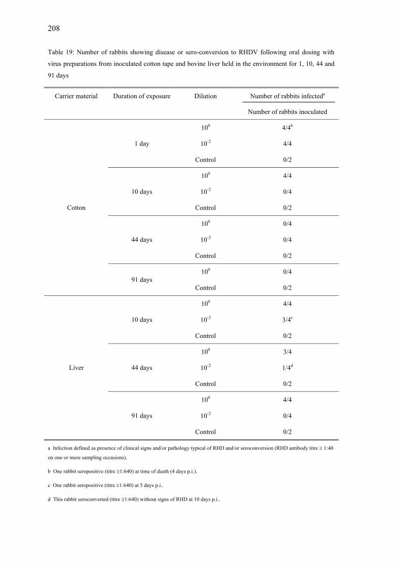
208
Table 19: Number of rabbits showing disease or sero-conversion to RHDV following oral dosing with
virus preparations from inoculated cotton tape and bovine liver held in the environment for 1, 10, 44 and
91 days
Carrier material Duration of exposure Dilution Number of rabbits infecteda
Number of rabbits inoculated
100 4/4b
10-2 4/4 1 day
Control 0/2
100 4/4
10-2 0/4 10 days
Control 0/2
100 0/4
10-2 0/4 44 days
Control 0/2
100 0/4
Cotton
91 days Control 0/2
100 4/4
10-2 3/4c10 days
Control 0/2
100 3/4
10-2 1/4d44 days
Control 0/2
100 4/4
10-2 0/4
Liver
91 days
Control 0/2
a Infection defined as presence of clinical signs and/or pathology typical of RHD and/or seroconversion (RHD antibody titre ≥ 1:40
on one or more sampling occasions).
b One rabbit seropositive (titre ≥1:640) at time of death (4 days p.i.).
c One rabbit seropositive (titre ≥1:640) at 5 days p.i..
d This rabbit seroconverted (titre ≥1:640) without signs of RHD at 10 days p.i..

209
Serology
Out of 87 virus-inoculated rabbits used in the three studies, only 9 tested positive for
antibodies to RHDV on one or more sampling occasions (Table 20). Only three of 40
animals sampled at the time of death or shortly before death had antibodies against
RHDV at that time. The six other sero-converting rabbits survived RHDV infection and
of these rabbits, three were seropositive at 5 days p.i.. The antibody titres of two of
these rabbits increased between 5 and 20 days, with the third rabbit becoming antibody
negative at 10 days p.i. and remaining negative at 20 and 30 days p.i.. The other three
surviving rabbits sero-converted at 10 or 20 days p.i.. The titres and sampling times are
shown in Table 20.
Table 20: Antibody titres and time of death (in hours) of RHDV- inoculated rabbits
Highest level of RHDV antibodies p.i.
Study Material Exposure Virus
dilution
Time to
death
RHD
death 5 d 10 d 20 d 30 d
PS1 cotton 4ºC, 1 d 10-2 n/a ≥1:640 neg neg neg
PS1 cotton 4ºC, 1 d 100 n/a neg neg ≥1:640 ≥1:640
FS liver env., 44 d 10-2 n/a neg ≥1:640 ≥1:640 ≥1:640
PS1 liver 4ºC, 1 d 100 n/a neg 1:40 ≥1:640 ≥1:640
PS1 liver 4ºC, 1 d 10-2 n/a 1:40 1:40 ≥1:640 ≥1:160
PS2 liver env., 5 d 10-2 n/a 1:40 ≥1:640 ≥1:640 ≥1:640
FS cotton env., 1 d 100 98 h ≥1:640
PS2 liver env., 5 d 100 126 h n/s 1:40
FS liver env., 10 d 10-2 165 h n/s ≥1:640
PS pilot study FS final study neg negative
env. environmental placement d day n/s not sampled
n/a not applicable
Survival analysis
Continuous monitoring of rabbits in the post inoculation period allowed the exact
hour of death to be recorded. The survival times of inoculated rabbits are compared in
Figures 23-26.
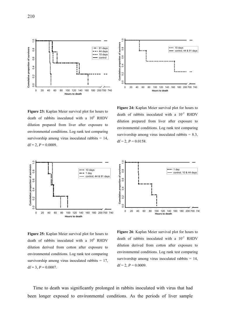
210
0 20 40 60 80 100 120 140 160 180 200 700 740Hours to death
0.0
0.2
0.4
0.6
0.8
1.0
Cum
ulat
ive
prop
ortio
n of
sur
vivo
rs 91 days44 days10 dayscontrol
Figure 23: Kaplan Meier survival plot for hours to
death of rabbits inoculated with a 100 RHDV
dilution prepared from liver after exposure to
environmental conditions. Log rank test comparing
survivorship among virus inoculated rabbits = 14,
df = 2, P = 0.0009.
0 20 40 60 80 100 120 140 160 180 200 700 740Hours to death
0.0
0.2
0.4
0.6
0.8
1.0
Cum
ulat
ive
prop
ortio
n of
sur
vivo
rs
10 dayscontrol, 44 & 91 days
Figure 24: Kaplan Meier survival plot for hours to
death of rabbits inoculated with a 10-2 RHDV
dilution prepared from liver after exposure to
environmental conditions. Log rank test comparing
survivorship among virus inoculated rabbits = 8.3,
df = 2, P = 0.0158.
0 20 40 60 80 100 120 140 160 180 200 700 740Hours to death
0.0
0.2
0.4
0.6
0.8
1.0
Cum
ulat
ive
prop
ortio
n of
sur
vivo
rs
10 days1 daycontrol; 44 & 91 days
Figure 25: Kaplan Meier survival plot for hours to
death of rabbits inoculated with a 100 RHDV
dilution derived from cotton after exposure to
environmental conditions. Log rank test comparing
survivorship among virus inoculated rabbits = 17,
df = 3, P = 0.0007.
0 20 40 60 80 100 120 140 160 180 200 700 740Hours to death
0.0
0.2
0.4
0.6
0.8
1.0
Cum
ulat
ive
prop
ortio
n of
sur
vivo
rs
1 daycontrol; 10 & 44 days
Figure 26: Kaplan Meier survival plot for hours to
death of rabbits inoculated with a 10-2 RHDV
dilution derived from cotton after exposure to
environmental conditions. Log rank test comparing
survivorship among virus inoculated rabbits = 14,
df = 2, P = 0.0009.
Time to death was significantly prolonged in rabbits inoculated with virus that had
been longer exposed to environmental conditions. As the periods of liver sample

211
exposure to the environment increased from 10 to 44 to 91 days, so did the time to death
for the rabbits treated with the respective inocula (P < 0.001) (Figure 23). Inoculation of
rabbits with diluted virus from liver samples kept from 10 to 44 days in the environment
(Figure 24) also extended the time to death (P < 0.016). Cotton samples kept for longer
intervals (10, 44 and 91 days for undiluted virus samples and 1, 10 and 44 days for
diluted virus samples) resulted in prolonged time to death for both the undiluted (Figure
25, P < 0.001) and diluted (Figure 26, P < 0.001) inocula.
Environmental conditions
The long-term exposure study was conducted from 25 March 2001 until 24 June
2001. During this period mean ambient temperatures declined (Table 21) and the
diurnal temperature range decreased by approximately 40% between days 1 and 90.
Relative humidity increased during the course of the study, although the diurnal
humidity range remained largely stable.
Temperature and humidity values for day-time (6 pm-6 am) and night-time (6 pm-
6 am) are shown in Table 22. Temperature range was considerably broader during the
day-time at the beginning of the virus exposure study, but declined to similar levels for
day- and night-time by the end of the study. Relative humidity was generally higher at
night, and the total range and average range of humidity was greater during the day.
Humidity increased as the Southern Hemisphere winter months were approached.
Figure 27 displays the daily average and the daily range of the Temperature-
Humidity Index for the period of RHDV exposure to environmental conditions. Notable
are the larger variations in Temperature-Humidity Indices from day to day in periods of
10 days and 44 days of virus exposure. However, the daily average of this index was
relatively stable over the whole period. Figure 28 compares the day-time and the night-
time range of the Temperature-Humidity. In March and April 2001 the fluctuation of
the day-time index was considerably larger than at night-time. The range of the index in
May and June 2001 followed similar patterns during the day and night with lower
values than in the previous months.

212
Table 21: Mean daily temperature (T) and humidity (H) recordings for different periods of RHDV
exposure to the environment
Interval T av (oC) T av range (oC) H av (%) H av range (%)
1 d 17.6 13.4 80.4 22.5
>1 to 10 d 15.2 12.6 74.6 24.6
>10 to 44 d 12.9 13.1 85.5 22.2
>44 to 91 d 8.8 8.8 91.6 20.4
av average d day
Table 22: Mean day-time and night-time temperature (T) and humidity (H) recordings for different
periods of RHDV exposure to the environment
T av (oC) T av range (oC) H av (%) H av range (%)
Virus exposure Day Night Day Night Day Night Day Night
1d 19.7 15.5 13.4 6.4 78.3 82.4 22.5 5.7
>1 to 10 d 17.4 12.9 9.7 7.1 72.6 76.9 17.3 17.4
>10 to 44 d 15.2 10.7 11.3 7.7 82.3 88.7 19.6 12.1
>44 to 91 d 10.1 7.5 6.8 5.7 89.9 94.1 18.0 8.5
av average d day

213
0
10
20
30
40
50
60
70
80
Time
Index average Index range
91 days44 days10 days
Figure 27: Daily average and daily range of the Temperature-Humidity Index for the periods of RHDV
exposure to the environment
0
5
10
15
20
25
30
35
40
25-M
ar
29-M
ar
2-A
pr
6-A
pr
10-A
pr
14-A
pr
18-A
pr
22-A
pr
26-A
pr
30-A
pr
4-M
ay
8-M
ay
12-M
ay
16-M
ay
20-M
ay
24-M
ay
28-M
ay
1-Ju
n
5-Ju
n
9-Ju
n
13-J
un
17-J
un
21-J
un
T ime
Ran
ge o
f the
tem
pera
ture
-hum
idid
ty In
dex
Index Range Day Index Range Night
10 days 44 days 91 days
Figure 28: Day-time and night-time range of the Temperature-Humidity Index for the periods of RHDV
exposure to environmental conditions

214
DISCUSSION
The ability of agents to survive in the environment is a key determinant of the
epidemiology of infectious diseases and of the relative importance of potential routes of
transmission. At the commencement of this study, there was little published information
available on the survival characteristics of RHDV, although data on related caliciviruses
suggested that prolonged survival under environmental conditions was likely [16-18].
The lack of methods for in vitro cultivation of RHDV presents a major obstacle to
understanding the ability of this virus to endure under field conditions. Options for
studying survival characteristics of unculturable agents include in vitro studies with a
culturable surrogate agent that is biologically related [19], or in vivo studies using
experimental challenge of susceptible hosts with the agent itself (as in this study). The
primary concern with the use of surrogate agents is the reliability with which it is
possible to extrapolate the findings across species, while the primary concern with in
vivo studies is ethical cost. The small pilot experiments in this study were designed to
verify and optimise the methods for the major study.
Our observations under natural environmental conditions indicated that virus survival
was affected by duration of field exposure and the vehicle (liver or cotton) used. Virus
kept dried on cotton cloth, which would mimic dried excreted virus in the field, was
viable for up to 10 days of exposure to environmental conditions, but not after 44 days.
Although no statistical significance could be obtained for pairwise multiple
comparisons for risk of death at prolonged exposure periods for cotton samples, a clear
trend of reduced mortality with extended environmental exposure was evident and
considered as biologically meaningful. Failure to detect statistical significance was
probably due to the small number of observations. Virus injected into bovine liver,
which was used to mimic RHDV in rabbit carcasses, was still viable after 91 days. The
buffer of surrounding tissue probably protected the virus from desiccation and UV light
compared with the dried cotton matrix. These findings indicate that the risk of
transmission of RHDV via exposure of wild rabbits to environmental reservoirs of virus
is probably influenced by the nature of the source of the infecting virus. Virus in rabbit
carcasses could remain infectious for at least three months, whereas virus secreted by
infected rabbits into the environment may retain infectivity for less than half this time.

215
Ethical constraints did not permit the accurate determination of viral titres. However,
to improve the ability to detect subtle loss of infectivity following environmental
exposure, in addition to undiluted preparations, one RHDV dilution just able to produce
infection in rabbits without environmental exposure ('borderline' dilution) was included.
The pilot experiments determined the probable endpoints and confirmed that the
proposed methodology was appropriate for conducting the larger field study.
The pilot experiments confirmed that both oral and intramuscular inoculation
resulted in infection of rabbits. Cooke and Berman [20] also showed that the route of
infection (intramuscular, intradermal and oral) with RHDV did not affect rabbit
survival. The oral route was preferred for the longitudinal study, as ingestion of virus is
considered to be the predominant direct transmission route for RHDV in the field [21],
particularly in New Zealand where distribution of RHDV-covered baits is a common
practice of rabbit control [22].
In the only detailed study published of RHDV survival, Smid and colleagues [10]
showed that RHDV in tissue suspensions can survive at 60ºC for 2 days, and at 4ºC for
225 days. Virus from tissue suspensions dried on cloth was able to survive at 60ºC for 2
days and at room temperature for 1 to 20 days, as confirmed by 100% mortality in
inoculated rabbits. However, after 50 and 150 days at room temperature, only one of
two rabbits inoculated with the same virus from dried cloth succumbed to the disease.
These observations were obtained under laboratory conditions, and conclusions for field
epidemiology of RHD are limited. McColl and colleagues [11] kept rabbit carcasses at
22ºC under laboratory conditions and collected liver samples up to 30 days post death.
Samples taken up to 20 days post death were able to infect and kill susceptible rabbits,
while samples collected after 26 and 30 days did not result in mortality and only some
rabbits seroconverted. In another study reverse transcriptase polymerase chain reaction
techniques showed that bones from RHDV-seropositive rabbits exposed to
environmental conditions contained detectable amounts of RHDV RNA for up to seven
weeks, but the viability of virus was not tested in challenge experiments [23].
The risk of epidemics of RHDV in wild rabbit populations will be influenced by the
relative proportion of susceptible individuals in the population. Field data (Henning,
unpublished observations) suggest annual RHD outbreaks occur at the end of the rabbit
breeding season. Persistence of viable virus in the environment between inter-epidemic

216
intervals is unlikely as was shown by the loss of RHDV infectivity from cotton cloth
samples and by the decline in viral infectivity from liver samples over a period of 3
months in this study. RHD outbreaks are most likely when the proportion of antibody
negative, susceptible rabbits reach a critical level and re-contamination with RHDV
occurs.
Flying insects also have been strongly implicated as mechanical vectors of RHDV
[24, 25]. Viral persistence in flies that feed on rabbit carcasses could provide an
alternative mechanism for viral persistence in an ecosystem between consecutive
epidemics. Surviving and sero-converting animals, which represented only 7% of virus-
inoculated animals in this study, may also be a virus source for infection. However, a
detailed study on viral persistence in recovered rabbits is yet to be conducted. The
outcome of infection with RHDV was influenced by the virus concentration, which was
approximated by using two dilutions of the inocula. Therefore interpretations of RHDV
infectivity and antibody responses [26] need the virus concentrations to be taken into
account. Reduction of infectivity occurred between 1 and 10 days for cotton samples,
and was first detected between 10 and 44 days for the liver samples.
Continuous observation of rabbits following infection allowed precise recording of
incubation and survival times. Cook and Berman [20] provided relatively imprecise
estimates of survival times, because animals were observed only at 8 hour intervals
following exposure, and estimated survival times were based on temperature of the
cadaver and the rabbit behaviour when last seen alive. In our study, reduction in
infectivity was reflected in the survival time of infected rabbits following challenge.
Animals exposed to lower concentrations of virus took longer to show clinical signs and
to die, suggesting that the incubation period of RHD is related to viral dose. Rabbits
inoculated with samples that had been exposed to the environment for longer durations
had longer incubation periods suggesting that the amount of infectious virus remaining
in the sample was reduced. Similar correlations between the length of the incubation
period and the virus dose have been described for Rabies virus [27] and the Measles
virus vaccine [28].
RHD has a very high mortality rate of 95-100% [29]. In the absence of sudden death
affected rabbits deteriorate and progressively lose body weight before eventually

217
succumbing to the disease. In most experimental RHDV inoculation studies, viral
antigen was detected post death using virus capture ELISAs on liver tissue [20] as death
occurs too suddenly to allow time for blood collection for antibody detection. Intensive
monitoring enabled us to bleed animals immediately before death occurred and only 8%
of rabbits (3 animals) had developed antibodies at the time of death. Gavier-Widen [30]
was not able to detect any antibodies using the haemagglutination inhibition test on
rabbit sera sampled immediately before death. She suggested that the time interval was
too short to develop antibodies as all animals in this study died or were euthanised
between 12 and 106 hours post inoculation. However, the shortest interval between
inoculation and death of a seropositive rabbit in our study was 98 hours post
inoculation. Plassiart and colleagues [31] noted biological signs of disseminated
intravascular coagulation and early necrotic changes of liver tissue as early as 30 hours
post inoculation. Therefore, it seems possible that the pathogenesis of RHD allows in
some cases antibody development after extremely short intervals post inoculation.
Antibody detection in survivors (and in the two other sero-converting and dying
animals) was observed as early as 5 days post inoculation. This is the same time period
that Shien and colleagues [32] observed after experimental infection. However, Shien’s
experiment was conducted with only two 4-5 week old rabbits, which would have
survived anyway because of their age related resilience [1, 6].
As only infectious virus will result in an antibody response [33], viral replication in
the body is necessary to obtain a constantly high antibody level. Antibody titres of the
five surviving sero-converting rabbits reached their highest levels between 10 and 20
days, while Shien and colleagues [32] detected antibody peaks three weeks post
inoculation. In our study, four animals retained high antibody titres until the conclusion
of our study at 30 days, while one animal had declining antibody titres after 5 days and
another at 20 days. It is possible that rabbits with declining antibody titres had
eliminated the virus and thus had no antigenic stimulation to maintain antibody levels.
Shien and colleagues [32] indicated that virus could gradually be cleared from infected
rabbits, resulting in a disappearance of the viraemia, but with virus persistence in bile
and the spleen. However, none of the young rabbits in Shien’s experiment (4-5 weeks
old at time of inoculation) showed any antibody decline during their study.

218
No evidence of airborne transmission of RHDV was shown in this study as none of
the un-inoculated rabbits (n = 5) kept with virus infected animals in the same room in
each trial developed RHD symptoms or antibodies against RHDV.
This study was conducted in the Southern Hemisphere’s autumn. Environmental
conditions change with the seasons, but quantification of the impact of climate factors is
difficult as they change from year to year. Climate comparisons between years and
regions are most productive over longer time periods. Exposure to the UV light and
fluctuations in temperature and humidity may have the largest effect on virus survival.
Our study was conducted over the autumn months in 2000 and revealed changing
weather conditions from a large temperature-humidity range at the beginning of the
study towards a more stable, but more humid climate. Kovaliski [34] mentioned that the
survival of the virus is probably shorter in the hot summer months in Australia. Higher
survival rates of rabbits are found in more cool, wet regions of Australia [35] while in
the lower North Island of New Zealand RHD outbreaks are occurring in the drier period
of the year (Henning, unpublished data). These observations suggest that some
environmental or other conditions trigger the event of an RHD outbreak and influence
mortality rates. In addition to affecting viral survival, weather conditions also influence
rabbit breeding, rabbit behaviour and the abundance of flying insects which are all
important mechanisms for generating RHD outbreaks.
The results of this study suggest that carcasses from rabbits dying from RHDV are
likely to be the reservoir for persistence of RHDV following an outbreak. Excreted
RHDV from infected animals or virus coated on baits may have reduced infectivity
within one to two weeks and probably do not persist longer than several weeks.
ACKNOWLEDGEMENTS
This research was funded by the Foundation for Research, Science and Technology,
New Zealand and the Institute of Veterinary, Animal and Biomedical Science, Massey
University, New Zealand and was supported by the German Academic Exchange
Service. We thank Robin Chrystall from Himatangi and Mark Redward from
Dannevirke, who gave permission for trials to be conducted on their properties. We also
thank Debbie and Karen Chesterfield for husbandry of the laboratory rabbits and Daniel
Russell for help with sample collection. Thanks to Laurie Sandall for her assistance
with virological work and Dave Pollard for help in designing the sampling unit. We also

219
gratefully acknowledge John Parkes and Peter Spradbrow for general advice and Mark
Stevenson and Nigel Perkins for statistical advice.
All experiments involving live animals were approved by the Massey University
Animal Ethics committee.
REFERENCES
1. Xu ZJ, Chen WX. Viral haemorrhagic disease in rabbits: a review. Vet Res
Commun 1989; 13: 205-12.
2. Nowotny N, Fuchs A, Schilcher F, Loupal G. Occurrence of viral haemorrhagic
disease of rabbits in Austria. I. Pathological and virological investigations. [in
German]. Wien Tierarztl Monatssch 1990; 77: 19-23.
3. Gunn M, Nowotny N. Rabbit haemorrhagic disease. Irish Vet J 1996; 49: 21-2.
4. Mocsari, E. Epidemiological studies on the rabbit haemorrhagic disease and its
control by vaccination in Hungary [in German]. In: Proceedings of the 7.
Arbeitstagung über Haltung und Krankheiten der Kaninchen, Pelztiere und
Heimtiere. Giessen, Germany: Deutsche Veterinärmedizinische Gesellschaft,
1990: 257-265.
5. Morisse J-P, Le Gall G, Boilletot E. Hepatitis of viral origin in Leporidae:
introduction and aetiological hypotheses. Rev Sci Tech Off Int Epizoot 1991; 10:
283-95.
6. Mikami O, Park JH, Kimura T, Ochiai K, Itakura C. Hepatic lesions in young
rabbits experimentally infected with Rabbit haemorrhagic disease virus. Res Vet
Sci 1999; 66: 237-42.
7. Cooke BD, Robinson AJ, Merchant JC, Nardin A, Capucci L. Use of ELISAs in
field studies of rabbit haemorrhagic disease (RHD) in Australia. Epidemiol Infect
2000; 124: 563-76.
8. Marchandeau S, Chantal J, Portejoie Y, Barraud S, Chaval Y. Impact of viral

220
hemorrhagic disease on a wild population of European rabbits in France. J Wildl
Dis 1998; 34: 429-35.
9. Asgari S, Hardy JR, Sinclair RG, Cooke BD. Field evidence for mechanical
transmission of Rabbit haemorrhagic disease virus (RHDV) by flies (Diptera:
Calliphoridae) among wild rabbits in Australia. Virus Res 1998; 54: 123-32.
10. Smid B, Valicek J, Stephanek J, Jurak E, Rodak L. Experimental transmission and
electron microscopic demonstration of the virus of haemorrhagic disease of rabbits
in Czechoslovakia. J Vet Med 1989; 36 : 237-40.
11. McColl KA, Morrissy CJ, Collins BJ, Westbury HA. Persistence of Rabbit
haemorrhagic disease virus in decomposing rabbit carcasses. Aust Vet J 2002; 80:
298-9.
12. Smid B, Valicek L, Rodak L, Stepanek J, Jurak E. Rabbit haemorrhagic disease:
an investigation of some properties of the virus and evaluation of an inactivated
vaccine. Vet Microbiol 1991; 26: 77-85.
13. Lavazza A, Capucci L. Viral haemorrhagic disease of rabbits. Office International
des Epizooties. Manual of standards for diagnostic tests and vaccines. Paris: OIE,
1996: 589-97.
14. Mayer DG, Davison TM, McGowan MR et al. Extent and economic effect of heat
loads on dairy cattle production in Australia. Aust Vet J 1999; 77: 804-8.
15. Rao PV. Statistical research methods in the life sciences. Pacific Grove, CA, USA:
Brooks/Cole Publishing Company, 1998.
16. Pedersen NC. Feline Calicivirus. In: Appel MJ, ed. Virus infections of carnivores.
Amsterdam, Netherlands: Elsevier Science, 1987.
17. Knipe DM and Howley, PM. Fields virology. Philadelphia: Lippincott Williams &
Wilkins, 2001.

221
18. Murphy FA, Gibbs, EPJ, Horzinek, MC, and Studdert, MJ. Veterinary virology.
San Diego, London: Academic Press, 1999.
19. Doultree JC, Druce JD, Birch CJ, Bowden DS, Marshall JA. Inactivation of feline
calicivirus, a Norwalk virus surrogate. J Hosp Infect 1999; 41: 51-7.
20. Cooke BD, Berman D. Effect of inoculation route and ambient temperature on the
survival time of rabbits, Oryctolagus cuniculus (L.), infected with Rabbit
haemorrhagic disease virus. Wildl Res 2000; 27: 137-42.
21. Parkes, JP, Norbury, GL, Heyward, RP, and Heath, ACG. Potential transmission
routes of rabbit haemorrhagic disease: consequences for epidemiology of RHD in
wild rabbits. In: Proceedings of the New Zealand Society of Animal Production.
Ruakura, New Zealand: AgResearch, 2001: 68-70.
22. Sanson RL, Brooks HV, Horner GW. An epidemiological study of the spread of
Rabbit haemorrhagic disease virus amongst previously non-exposed rabbit
populations in the North Island of New Zealand. NZ Vet J 2000; 48: 105-10.
23. Moss SR, Turner SL, Trout RC et al. Molecular epidemiology of Rabbit
haemorrhagic disease virus. J Gen Virol 2002; 83: 2461-7.
24. Heath ACG, Heyward R, Norbury G. Blowflies (Calliphoridae) as potential
vectors of rabbit haemorrhagic disease virus in New Zealand. In: Rabbit control,
RCD: Dilemmas and implications. Proceedings of the Conference, 30-31 March
1998. Wellington, New Zealand: Royal Society of New Zealand Miscellaneous
Series, 1999: 85-8.
25. Barratt, BIP, Ferguson, CM, Heath, ACG, Evans, AA, and Logan, RAS. Can
insects transmit Rabbit haemorrhagic disease virus? In: Proceedings of the Fifty
First New Zealand Plant Protection Conference, 11-13 August 1998. Hamilton,
New Zealand: New Zealand Plant Protection Society, 1998: 245-50.
26. Leighton FA, Artois M, Capucci L, Gavier-Widen D, Morisse JP. Antibody
response to rabbit viral hemorrhagic disease virus in red foxes (Vulpes vulpes)

222
consuming livers of infected rabbits (Oryctolagus cuniculus). J Wildl Dis 1995;
31: 541-4.
27. Fekadu M, Chandler FW, Harrison AK. Pathogenesis of rabies in dogs inoculated
with an Ethiopian rabies virus strain. Immunofluorescence, histologic and
ultrastructural studies of the central nervous system. Arch of Virol 1982; 71: 109-
26.
28. Kress S, Schluederberg AE, Hornick RH et al. Studies with live attenuated
measles-virus vaccine. Am J Dis Child 1961; 101: 701-7.
29. Ohlinger VF, Haas B, Ahl R, Weiland F. Infectious haemorrhagic disease of
rabbits. Contagious disease caused by calicivirus. [in German]. Tierarztl Umschau
1989; 44: 284-94.
30. Gavier-Widen, MD. Epidemiology, pathology and pathogenesis of two related
viral hepaties of leporides. (PhD Thesis). Davis: University of California, 1992.
31. Plassiart G, Guelfi JF, Ganiere JP, Wang B, Andre-Fontaine GWM.
Hematological parameters and visceral lesions relationships in rabbit viral
hemorrhagic disease. J Vet Med Series B 1992; 39: 443-53.
32. Shien JH, Shieh HK, Lee LH. Experimental infections of rabbits with Rabbit
haemorrhagic disease virus monitored by polymerase chain reaction. Res Vet Sci
2000; 68: 255-9.
33. Henning J, Meers J, Davies PR. Exposure of rabbits to ultraviolet light-inactivated
Rabbit haemorrhagic disease virus (RHDV) and subsequent challenge with
virulent virus. Epidemiol Infect 2003; submitted.
34. Kovaliski J. Monitoring the spread of Rabbit hemorrhagic disease virus as a new
biological agent for control of wild European rabbits in Australia. J Wildl Dis
1998; 34: 421-8.
35. Henzell RP, Cunningham RB, Neave HM. Factors affecting the survival of

223
Australian wild rabbits exposed to rabbit haemorrhagic disease. Wildl Res 2002;
29: 523-42.

224

225
Chapter 7
Exposure of rabbits to ultraviolet light-
inactivated Rabbit haemorrhagic disease virus
(RHDV) and subsequent challenge with virulent
virus*
*Submitted by J. Henning, J. Meers and P. R. Davies to Epidemiology and Infection

226
227
SUMMARY
This study investigated whether exposure to inactivated Rabbit haemorrhagic
disease virus (RHDV) can produce an antigenic response in rabbits and protect them
from a subsequent challenge with virulent virus. The aim was to determine if the
spreading of baits containing RHDV, which is a common management practice in New
Zealand to reduce rabbit numbers, could result in protective immunity in wild rabbits.
RHDV was inactivated by ultraviolet light using an electronic UV crosslinker with a
UV dose of 168.48 W-s/cm2 and a UV intensity of 0.0078 W/cm2. Two groups of four
rabbits were then inoculated with inactivated virus via oral and intramuscular routes.
Rabbits were monitored for 30 days post inoculation and then challenged orally with
virulent virus.
No rabbit exposed to inactivated RHDV developed clinical signs of RHD or had
antibodies at day 30 post infection and all animals died within 82 hours after challenge
with virulent virus. No antibodies were detected at the time of death. These findings
suggest that exposure to UV-inactivated virus in the field or on baits will not protect
rabbits against challenge with virulent virus.
INTRODUCTION
Introduced European rabbits (Ortycolagus cuniculus) are a major vertebrate pest in
New Zealand and Australia. The use of Rabbit haemorrhagic disease virus (RHDV) for
biological control of rabbits is a common practice in New Zealand. In rabbit infested
areas, a commercial RHDV product (‘RCD-ZEN’, Zenith Technology Corporation
Limited, Dunedin, New Zealand) is typically distributed on baits to initiate RHDV
epidemics. However, not all baits are taken up by rabbits, and residual RHDV baits may
undergo prolonged exposure to environmental conditions. O’Keefe and colleagues [1]
suggested that large-scale use of baits coated with RHDV, which might be inactivated
in the environment, could induce protective immunity of rabbits and reduce the
effectiveness of biological control programs.
Environmental factors such as changing temperatures, humidity and sunshine are
likely to influence the survival and infectivity of RHDV on baits. In particular, exposure
to ultraviolet radiation from sunlight is likely to be an important determinant of the
duration of virus infectivity. In a field study of RHDV survival, dried virus exposed

228
directly to sunlight on cotton cloth remained infective for susceptible rabbits for greater
than 10 days, but less than 44 days [2]. In contrast, under identical environmental
conditions, RHDV injected into organic tissue (liver) and therefore not exposed to direct
sunlight, remained infective for at least three months [2]. The possibility that
environmentally degraded RHDV virus could induce protective immunity in wild rabbit
populations has considerable implications for the efficacy of biological control using
this agent. This is the second of two studies investigating the influence of the
environment on RHDV properties. We conducted experimental investigations to
determine whether exposure to UV inactivated RHDV virus would induce
seroconversion of rabbits and protect against exposure to virulent virus.
MATERIALS AND METHODS
Animals
New Zealand White rabbits aged between 10 and 11 weeks old were purchased from
a commercial laboratory colony and housed in standard rabbit cages (56 x 44 x 45 cm)
in climate-controlled rooms at 17ºC. They had not been vaccinated against RHDV and
were tested immediately prior to the study to confirm the absence of RHDV antibodies.
Rabbits were fed ad libitum with commercial rabbit pellets and had constant access to
water.
RHDV inactivation
A commercial product ‘RCD-ZEN’ (Zenith Technology Corporation Limited,
Dunedin, New Zealand) that was produced from RCD CAPM V-351 (Czechoslovakian
strain) Master Seed Virus was used for the study. The batch purchased for this study
(Z25) had a rabbit LD50 titre of approximately 106 per ml (Max Shepherd, Zenith
Technology Corporation Limited, pers. comm.).
The RHDV product was exposed to ultraviolet light using an electronic UV
crosslinker (CEX-800, Ultralum, Inc., 6421 East Alondra Blvd, Paramount, California,
USA). UV crosslinkers are designed especially to provide uniform irradiation with short
wave UV light (254 nm) for crosslinking DNA and RNA. A high dose of UV exposure
was chosen, based on the resilient properties of RHDV [3] and published reports on the
use of UV energy to inactivate viruses and sterilise or disinfect effluents. The samples
were exposed to a UV dose of 168.48 W-s/cm2 with a UV intensity of 0.0078 W/cm2.

229
Experimental Design
Two groups of four rabbits were inoculated with 1 ml of undiluted UV-inactivated
virus; one group was inoculated orally and the other group intramuscularly. Thirty-five
days post inoculation with inactivated RHDV, rabbits were challenged orally with
approximately 104 LD50 RHDV (RCD-ZEN) in a volume of 1 ml. A positive control
group of two rabbits, not previously inoculated with inactivated virus, was also
challenged with virulent RHDV, and two negative control rabbits were dosed orally
with saline solution. Blood samples were collected prior to commencement of the study,
at 5, 10, 20 and 30 days after inoculation with inactivated virus, and at 5, 10, 20 and 30
days post challenge with virulent virus, and at the time of euthanasia. If an animal was
seronegative at 30 days following inoculation, samples from 5, 10 and 20 days post
inoculation were not tested. Blood samples were centrifuged for 15 min at 1800 g to
separate the sera. The sera were tested for antibodies to RHDV with the Capucci-
competition ELISA [4] by AgResearch (Wallaceville Animal Research Centre, Upper
Hutt, New Zealand). Four-fold serial dilutions from 1:10 to 1:640 were assayed.
Samples were classified as RHDV positive if inhibition was ≥ 50% in serum diluted
1:40.
Assessment of outcomes
Following inoculation with the inactivated virus, rabbits were observed several times
daily for clinical signs. Following challenge with virulent virus, rabbits were observed
continuously by an observer for the first 7 days and then at 4-hour intervals until 10
days post challenge (p.c.), followed by once daily until 30 days p.c.. Clinically affected
rabbits were anaesthetised with an intramuscular injection of ketamine hydrochloride
(100 mg/ml; Phoenix Pharm Distributors Limited, Auckland, New Zealand) and
xylazine (20 mg/ml; Phoenix Pharm Distributors Limited, Auckland, New Zealand) as
soon as signs of RHD [2] were observed, and then euthanased by intracardiac injection
of sodium pentobarbitone (Pentobarb 300, 300 mg/ml; National Veterinary Supplies
Limited, Auckland, New Zealand). Necropsies were performed on all rabbits and gross
pathological observations were recorded. The presence of pathological changes typical
of RHD (pale yellow or greyish liver with marked lobular pattern, petechial and
echymotic multifocal haemorrhages of the lung, lung oedema, lung congestion,
splenomegaly, poor blood coagulation and swollen, dull pale to patchy reddish
discolouration of the kidney) was interpreted as confirmation of RHD.

230
RESULTS
All animals were seronegative before inoculation. None of the rabbits inoculated
with irradiated virus developed clinical signs of RHD or had detectable antibodies to
RHDV 30 days post inoculation (Table 23). Challenge with virulent virus resulted in
clinical signs and pathology typical of RHD in all rabbits, including those previously
inoculated with irradiated virus. The mean (SD) time to death post challenge was 52.9
(11.2) hours. No antibodies were detected at the time of death in any rabbit. The two
negative control rabbits did not show any signs of disease and were negative for RHD
antibodies 30 days p.i..
Table 23: Serology and mortality results after inoculation of rabbits with inactivated RHDV followed by
challenge with virulent RHDV
Group Sex AB at 30 days p.i. Time to death p.c. AB at time of death
female neg 53 h neg
female neg 82 h neg
male neg 47 h neg
1. Inactivated RHDV:
IM route
male neg 45 h neg
female neg 42 h neg
female neg 55 h neg
male neg 55 h neg
2. Inactivated RHDV:
Oral route
male neg 54 h neg
female n/a 46 h neg 3. Positive control:
RHDV challenge only female n/a 50 h neg
female n/a n/a n/a 4. Negative control:
Saline challenge only male n/a n/a n/a
Rabbits in groups 1 and 2 were inoculated with inactivated RHDV via intramuscular (IM) and oral routes respectively. Groups 1, 2
and 3 were orally challenged with virulent RHDV 35 days later.
p.i. post inoculation with inactivated RHDV p.c. post challenge with virulent RHDV
AB antibodies neg ELISA titre < 1:10
n/a not applicable

231
DISCUSSION
Ultraviolet light at a wavelength between 100 and 280 nm is considered to be
'germicidal'. It damages the DNA and RNA of bacteria, viruses and other pathogens and
thus destroys their ability to multiply and cause disease. It does this by eliciting single
photon photochemical effects in nucleic acids through forming covalent bonds between
certain adjacent bases [5]. This modification results in incorrect codes being transmitted
from the nucleic acids and causes irreversible damage to the microorganisms.
Inoculation with UV light inactivated RHDV did not produce any antibody response in
the rabbits, and they were not protected against challenge with virulent virus.
Considering these results, it is unlikely that inactivated RHDV on baits, in rabbit
carcasses or excreted into the environment will produce an antibody reaction in naive
rabbits. Thus, rabbits are likely to be fully susceptible to further RHDV epidemics in the
field. Inactivated RHDV administered by the oral route (which would mimic ingestion
of virus) or administered intramuscularly (imitating a stinging or biting insect) will not
induce a protective antibody titre. O’Keefe and colleagues [1] proposed that inactivation
of virus after large-scale baiting with RHDV may result in seroconversion of rabbits and
protective immunity. However, the current experimental study showed no evidence to
support this hypothesis. Seroconversions in rabbits following baiting operations is more
likely to result from contact of rabbits to sub-lethal doses of virulent virus.
High titres of RHDV antibodies in surviving rabbits does not necessarily indicate
immune protection of rabbits as suggested by O’Keefe and colleagues [1]. Exposure to
RHDV in new epidemics can produce re-infection in animals that have survived
previous outbreaks [6]. Persistence of RHDV antibodies over longer time periods has
not been well documented and is worthy of further investigation in longitudinal studies.
RHDV antibody responses have been documented in experimental studies by feeding
foxes RHDV-infected rabbit carcasses [7] and by immunisation of rabbits with
recombinant virus-like particles and structural virus proteins [8, 9]. In these studies, the
ability to induce antibodies (in foxes and rabbits) may be attributable to the large,
multiple doses of antigen administered, the use of adjuvants, or the chemical
stabilization of the antigen. None of these factors are present in a true field situation.
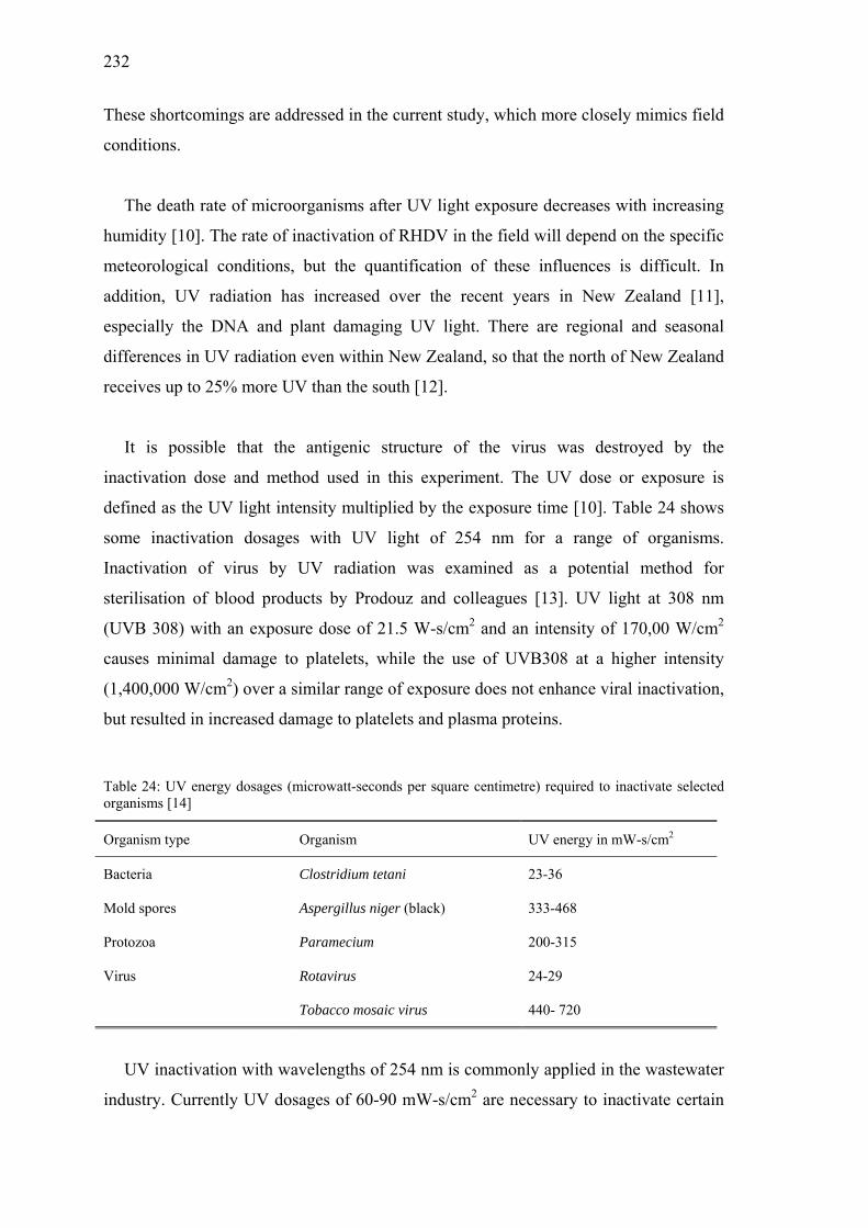
232
These shortcomings are addressed in the current study, which more closely mimics field
conditions.
The death rate of microorganisms after UV light exposure decreases with increasing
humidity [10]. The rate of inactivation of RHDV in the field will depend on the specific
meteorological conditions, but the quantification of these influences is difficult. In
addition, UV radiation has increased over the recent years in New Zealand [11],
especially the DNA and plant damaging UV light. There are regional and seasonal
differences in UV radiation even within New Zealand, so that the north of New Zealand
receives up to 25% more UV than the south [12].
It is possible that the antigenic structure of the virus was destroyed by the
inactivation dose and method used in this experiment. The UV dose or exposure is
defined as the UV light intensity multiplied by the exposure time [10]. Table 24 shows
some inactivation dosages with UV light of 254 nm for a range of organisms.
Inactivation of virus by UV radiation was examined as a potential method for
sterilisation of blood products by Prodouz and colleagues [13]. UV light at 308 nm
(UVB 308) with an exposure dose of 21.5 W-s/cm2 and an intensity of 170,00 W/cm2
causes minimal damage to platelets, while the use of UVB308 at a higher intensity
(1,400,000 W/cm2) over a similar range of exposure does not enhance viral inactivation,
but resulted in increased damage to platelets and plasma proteins.
Table 24: UV energy dosages (microwatt-seconds per square centimetre) required to inactivate selected organisms [14]
Organism type Organism UV energy in mW-s/cm2
Bacteria Clostridium tetani 23-36
Mold spores Aspergillus niger (black) 333-468
Protozoa Paramecium 200-315
Virus Rotavirus 24-29
Tobacco mosaic virus 440- 720
UV inactivation with wavelengths of 254 nm is commonly applied in the wastewater
industry. Currently UV dosages of 60-90 mW-s/cm2 are necessary to inactivate certain

233
human pathogenic viruses, such as Poliovirus, Rotavirus and Hepatitis A virus, while
MS-2 bacteriophage shows more resistance [15]. Ho Chu-Fei and colleagues [16] have
shown that a UV exposure dose of 65 mW-s/cm2 is required in a water pollution control
plant to achieve the target coliform level 95% of the time. A 95% inactivation of human
enteric Adenovirus type 40 occurs with UV doses of 103 mW-s/cm2 in treated
groundwater [17]. It has been shown also that a UV dose of 13 mW-s/cm2 is able to
reach 99% inactivation of Feline calicivirus (used as a surrogate to monitor the not-
culturable Norwalk virus) in treated drinking water [17].
This study has shown that exposure of rabbits to inactivated RHDV will not protect
them from further challenge with viable RHDV virus. The high inactivation dose and
intensity chosen in this experiment is appropriate considering the resilient properties of
RHDV. However, the influence of UV light on the antigenic structure can be
determined only with further challenge experiments using stepwise decreased UV light
exposure and intensity.
ACKNOWLEDGEMENTS
We are grateful to Debbie and Karen Chesterfield for housing the experimental
rabbits and Sonja Kay and Daniel Russell for their help with sample collection. Thanks
also to the Institute of Molecular Biosciences, Massey University, Palmerston North for
supplying the UV-crosslinker.
All experiments involving live animals were approved by the Massey University
Animal Ethics committee.
REFERENCES
1. O'Keefe JS, Tempero JE, Motha MX, Hansen MF, Atkinson PH. Serology of
Rabbit haemorrhagic disease virus in wild rabbits before and after release of the
virus in New Zealand. Vet Microbiol 1999; 66: 29-40.
2. Henning J, Meers J, Davies PR, Morris RS. Survival of Rabbit haemorrhagic
disease virus (RHDV) in the environment. Epidemiol Infect 2003; submitted.
3. Smid B, Valicek L, Rodak L, Stepanek J, Jurak E. Rabbit haemorrhagic disease:

234
an investigation of some properties of the virus and evaluation of an inactivated
vaccine. Vet Microbiol 1991; 26: 77-85.
4. Lavazza A, Capucci L. Viral haemorrhagic disease of rabbits. Office International
des Epizooties. Manual of standards for diagnostic tests and vaccines. Paris: OIE,
1996: 589-97.
5. Jori G, Spikes JD. Mapping of the three dimensional structure of proteins by
phytochemical techniques. In: Smith KC, ed. Photochemical and photobiological
reviews. New York: Plenum, 1976: 193.
6. Cooke BD, Robinson AJ, Merchant JC, Nardin A, Capucci L. Use of ELISAs in
field studies of rabbit haemorrhagic disease (RHD) in Australia. Epidemiol Infect
2000; 124: 563-76.
7. Leighton FA, Artois M, Capucci L, Gavier-Widen D, Morisse JP. Antibody
response to rabbit viral hemorrhagic disease virus in red foxes (Vulpes vulpes)
consuming livers of infected rabbits (Oryctolagus cuniculus). J Wildl Dis 1995;
31: 541-4.
8. Plana-Duran J, Bastons M, Rodriguez MJ et al. Oral immunisation of rabbits with
VP60 particles confers protection against rabbit hemorrhagic disease. Arch Virol
1996; 141: 1423-36.
9. Castanon S, Marin MS, Martin-Alonso JM et al. Immunisation with potato plants
expressing VP60 protein protects against Rabbit hemorrhagic disease virus. J
Virol 1999; 73: 4452-5.
10. Koller LR. Ultraviolet radiation. New York: Wiley, 1965: 236-245.
11. McKenzie R, Conner B. Increased summertime UV radiation in New Zealand in
response to ozone loss. Sci 09/10/99 1999; 285: 1709-12.
12. McKenzie R, Bodeker G, Conner B. Increased UV radiation in New Zealand: a
cautionary tale. Water Atmosph 1999; 7: 7-8.
13. Prodouz KN, Fratantoni JC, Boone EJ, Bonner OF. Use of laser-UV for
inactivation of virus in blood products. Blood 1987; 70: 589-92.

235
14. Trojan Technologies. Ultraviolet doses required for 99.99% inactivation of
various microbes (mW-s/cm2). Online. Available: www.trojanuvlogic.com/
UV_Applications/doses.html 3 Oct 2002.
15. Malley JP Jr, Margolin AB, Snice GA, Hogan SP. UV inactivation of viruses in
natural waters (project#180). Online. Available: www.awwarf.com/
exsums/90773.html 3 Oct 2002.
16. Ho Chu-Fei H, Pitt P, Mamais D, Chiu C, Jolis D. Evaluation of UV disinfection
systems for large-scale secondary effluent. Water Environ Res 1998; 70: 1142-50.
17. Thurston-Enriquez JA, Haas CN, Jacangelo J, Riley K, Gerba CP. Inactivation of
Feline calicivirus and Adenovirus Type 40 by UV radiation. Appl Environ
Microbiol 2003; 69: 577-82.

236

237
Chapter 8
Attitudes of New Zealand farmers to methods
used to control wild rabbits*
*Submitted by J. Henning, C. Heuer and P. R. Davies to Preventive Veterinary Medicine
Additional information is enclosed at the end of the chapter in Appendices 3 and 4

238
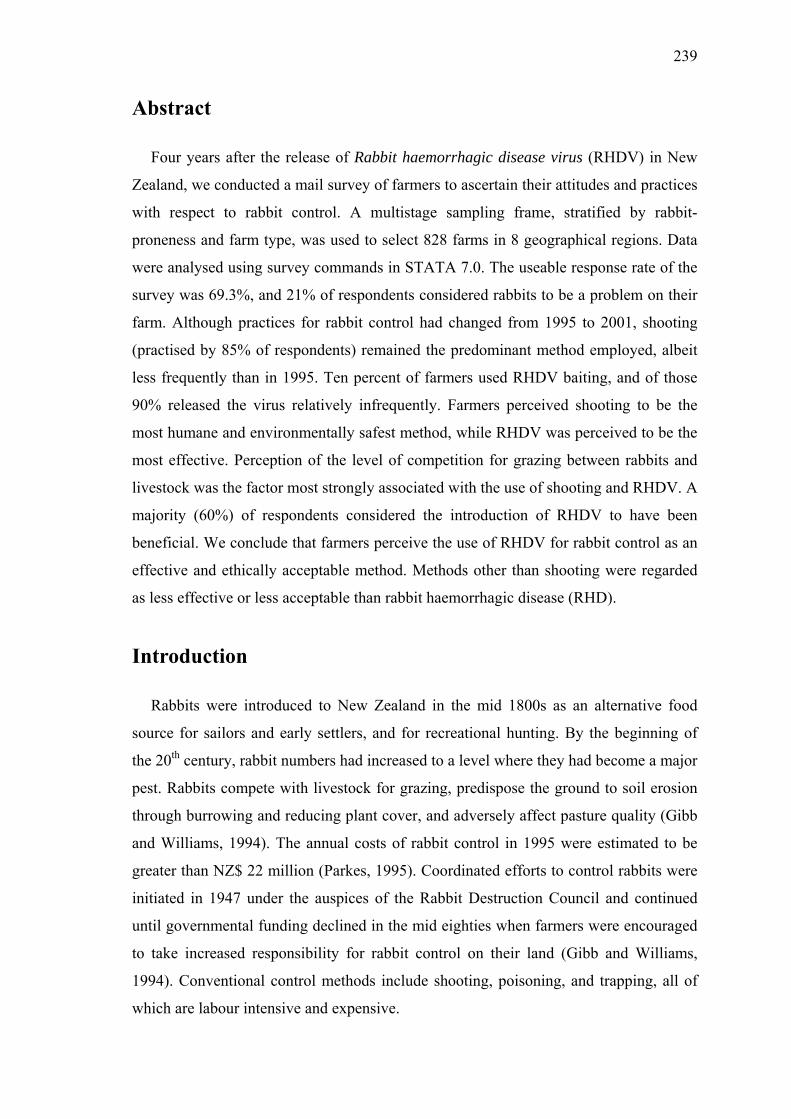
239
Abstract
Four years after the release of Rabbit haemorrhagic disease virus (RHDV) in New
Zealand, we conducted a mail survey of farmers to ascertain their attitudes and practices
with respect to rabbit control. A multistage sampling frame, stratified by rabbit-
proneness and farm type, was used to select 828 farms in 8 geographical regions. Data
were analysed using survey commands in STATA 7.0. The useable response rate of the
survey was 69.3%, and 21% of respondents considered rabbits to be a problem on their
farm. Although practices for rabbit control had changed from 1995 to 2001, shooting
(practised by 85% of respondents) remained the predominant method employed, albeit
less frequently than in 1995. Ten percent of farmers used RHDV baiting, and of those
90% released the virus relatively infrequently. Farmers perceived shooting to be the
most humane and environmentally safest method, while RHDV was perceived to be the
most effective. Perception of the level of competition for grazing between rabbits and
livestock was the factor most strongly associated with the use of shooting and RHDV. A
majority (60%) of respondents considered the introduction of RHDV to have been
beneficial. We conclude that farmers perceive the use of RHDV for rabbit control as an
effective and ethically acceptable method. Methods other than shooting were regarded
as less effective or less acceptable than rabbit haemorrhagic disease (RHD).
Introduction
Rabbits were introduced to New Zealand in the mid 1800s as an alternative food
source for sailors and early settlers, and for recreational hunting. By the beginning of
the 20th century, rabbit numbers had increased to a level where they had become a major
pest. Rabbits compete with livestock for grazing, predispose the ground to soil erosion
through burrowing and reducing plant cover, and adversely affect pasture quality (Gibb
and Williams, 1994). The annual costs of rabbit control in 1995 were estimated to be
greater than NZ$ 22 million (Parkes, 1995). Coordinated efforts to control rabbits were
initiated in 1947 under the auspices of the Rabbit Destruction Council and continued
until governmental funding declined in the mid eighties when farmers were encouraged
to take increased responsibility for rabbit control on their land (Gibb and Williams,
1994). Conventional control methods include shooting, poisoning, and trapping, all of
which are labour intensive and expensive.

240
In the 1980s, rabbit haemorrhagic disease (RHD), a novel viral disease, caused
widespread mortality of farmed and wild rabbits in China and Europe (Ohlinger et al.,
1993). The epidemiological characteristics of these outbreaks (rapid transmission, high
morbidity and mortality rates) presaged the use of the virus for biological control of pest
rabbit populations. Unintentional release of Rabbit haemorrhagic disease virus (RHDV)
in South Australia led to a dramatic reduction in wild rabbit populations (Mutze et al.,
1998). Despite an application by New Zealand farmers to introduce RHDV being
denied by the government in July 1997, the disease was deliberately released in the
South Island the following month. Subsequently, the intentional spread of the RHDV
was legalised in New Zealand, and RHDV became commercially available for rabbit
control (Sanson et al., 2000). Before the application to introduce RHDV was denied,
surveys to assess public attitudes to biological control of pests revealed widespread
recognition of RHD (Fitzgerald et al., 1996; Wilkinson and Fitzgerald, 1998). However,
opinions of farmers, the group most affected by and responsible for the control of
rabbits, have not been systematically evaluated. In 2001, four years after RHDV was
introduced into New Zealand, we conducted a mail survey of 828 farmers in eight
regions of New Zealand to determine trends and current practices of rabbit control, and
to ascertain opinions about RHDV and other control methods. We hypothesised that
both attitudes and practices related to rabbit control were likely to vary between types of
farm enterprises and with the extent of the rabbit problem that farmers had to confront.
These factors were major considerations in the design and analysis of the survey, which
focused primarily on pastoral livestock producers.
Materials and Methods
A key objective of the study was to compare the attitudes to, and practices of, rabbit
control from farmers with significant rabbit problems, with farmers who had relatively
minor rabbit problems. Particular emphasis was given to assessing opinions of farmers
engaged in the major pastoral livestock industries. A multistage spatial sampling frame
was developed stratified according to both rabbit-proneness and type of enterprise.
Selection of regions
1. Level of rabbit-proneness (the propensity of land to sustain rabbit populations): A
digital map of New Zealand identifying 5 categories of rabbit-proneness (Kerr and

241
Ross, 1990) was used as the primary spatial stratum. The New Zealand Mountain
Lands Institute, Lincoln University, New Zealand, conducted this classification for
the Ministry of Agriculture and Fisheries of New Zealand. Classification of
proneness was based on information about topography, geological formations, soil
erosion, vegetative features and capability of land for agricultural production. This
was then validated and combined with rabbit count information. Rabbit-proneness
was classified into categories 'extreme', 'high', 'medium', 'low' and 'negligible'.
2. Region: Using ArcView 3.1 (1992-1998 Environmental Systems Research Institute,
Inc., Redlands, CA, USA), the digital rabbit-proneness map was overlaid on a
regional map of New Zealand. To enable comparison of farmers living in areas with
differing rabbit-proneness and likely experience with RHD, eight regions were
selected based on the predominant agricultural activities and rabbit-proneness
(Table 25).
Table 25: General characteristics of regions selected for surveying
Region Main agricultural
activities
Rabbit-
proneness
Other
North Island Waikato Dairy Low
Hawke's Bay Horticulture, sheep Moderate Anecdotal evidence that
RHD has been ineffective
Manawatu Beef, mixed livestock Moderate Epidemiological research
on RHD
Wairarapa-
Wellington
Beef, mixed livestock Moderate
South Island Marlborough Horticulture
(viticulture)
Moderate to
high
Anecdotal evidence that
rabbits cause severe losses
in horticulture
Canterbury Sheep, mixed livestock Moderate to
high
Region with second
highest rabbit-proneness
Otago Sheep, horticulture,
mixed livestock
High Region with highest
rabbit-proneness
Southland Beef, mixed livestock,
dairy
Moderate

242
Classification of farms
The New Zealand agricultural database (‘Agribase’) is a register of all commercial
farms and their locations in New Zealand (Sanson and Pearson, 1997), with farms
classified into 33 different types. For the purpose of stratified sampling within regions,
we simplified the number of farm categories to be sampled using the following
procedures:
• All farm types with less than 100 records in Agribase (e.g. beekeeping, dogs, ratites)
were excluded (n = 279 farms).
• All farm types with ambiguous definitions or considered unimportant for the study
(e.g. lifestyle blocks, tourism, native bush) were excluded (n = 20,781 farms).
Predominant categories in this group were lifestyle blocks (8,591), 'unspecified'
(5,372) and 'other' (4,489).
• Farm types engaged in similar or related activities were aggregated to yield a
smaller number of farm categories. 'Dairy' and 'dairy dry stock' types were
aggregated into a single entity; all livestock enterprises (e.g. deer, horses, pigs and
poultry) not identified as dairy, sheep, beef, or mixed (sheep-and-beef), and farmers
grazing other people’s livestock, were aggregated into a category of 'other
livestock'. Similarly, arable cropping and seed production, flowers, forestry, fruit
growing, plant nurseries, vegetable growing and viticulture were aggregated into a
single 'non-livestock' category.
This process excluded 21,060 farms and identified a population of 82,608 farms in
New Zealand. A total of 51,716 farms among 6 categories of farm type (Table 26)
(62.6% = study population) were situated in the regions selected for the survey, and
30,892 farms (37.4%) were in other regions.

243
Table 26: Distribution of New Zealand farms by selected farm types
Description Farm code All NZ farms Farms in study regions
Beef cattle farming BEF 20,779 12,517
Dairy cattle farming DAR 15,558 10,261
Non-livestock a NLS 15,385 5,705
Other livestock farms b OLF 10,118 6,089
Sheep farming SHP 11,635 9,874
Mixed sheep-and-beef farming SNB 9,133 7,270
TOTAL 82,608 51,716
a Including arable cropping and seed production, flowers, forestry, fruit growing, plant nurseries, vegetable growing and viticulture. b Including deer, horse farming and breeding, poultry, pig production and goat farming.
To determine the rabbit-proneness classification for each farm, geographic centroids
of farms were plotted on the map of rabbit-proneness using the X-Tools extension
(Department of Forestry, Orgeon, USA) for ArcView 3.1. Thus all farms were
categorised by both farm type and rabbit-proneness. Due to logistic constraints, the total
number of farmers that could be surveyed was approximately 800 (i.e. 100 per region).
Power analysis indicated that this sample size would be adequate to detect differences in
proportions as small as 0.1 compared with a null proportion of 0.5 (power of 0.8 with
α = 0.05).
Selection of farms within regions
Rabbit-proneness was the primary criterion for the stratification of farms. However,
areas of extreme or high proneness occurred in only two of the eight regions, and the
large majority of farms were in areas of low or negligible rabbit-proneness. Similarly,
farm types were not evenly distributed among regions. Therefore, fixed proportional
sampling of all categories was not possible across regions. Consequently, in each region
100 questionnaires were allocated to the negligible, low and medium categories of
proneness at fixed ratios of 2:4:4 respectively. A further 80 questionnaires were
assigned to farms in regions of high and extreme rabbit-proneness at a 1:1 ratio so that
they would be sampled at a higher intensity. Selection of farm types was also weighted
to yield a higher representation of enterprises thought likely to be more affected by the

244
impact of rabbits. Specifically sheep, mixed sheep-and-beef, and non-livestock
enterprises were sampled twice as intensively as dairy farms and 'other livestock' farms.
The weighting matrix, the numbers of surveys allocated by farm type within each
rabbit-proneness stratum, and the actual numbers of questionnaires mailed (and
returned) are presented in Table 27. A programme was written in Visual Basic for
Applications in Access 2000 (Microsoft Corporation) to randomly select the farms
according to the specified weightings from the 51,716 eligible farms in the 8 selected
regions without replacement. However, target numbers for some categories of farms in
the medium, high and extreme rabbit-proneness areas were not achievable and all
eligible farms in these strata were surveyed (Table 27). In fact in one area there were no
eligible farms within the strata; there were no dairy farms located in areas of extreme
rabbit-proneness.
Table 27: Matrix showing proportional weights (farm type by rabbit-proneness) and number of
questionnaires targeted, mailed and returned (the last two in parentheses).
Rabbit-proneness
Negligible τ Low τ Medium τ High γ Extreme γ TOTAL
Farm type proportion 0.2 0.4 0.4 0.5 0.5
Beef cattle 0.2
32
(32/19)
64
(64/46)
64
(56/38)
8
(8/4)
8
(3/2)
176
(163/109)
Dairy cattle 0.1
16
(16/12)
32
(32/24)
32
(24/14)
4
(2/1)
4
(0/0)
88
(74/51)
Non-livestock 0.2
32
(32/21)
64
(64/44)
64
(55/37)
8
(7/6)
(4/4)
176
(162/112)
Other livestock 0.1
16
(16/10)
32
(32/25)
32
(32/22)
4
(4/2)
4
(2/0)
88
(86/59)
Sheep farming 0.2
32
(32/25)
64
(64/42)
64
(59/50)
8
(8/6)
8
(8/7)
176
(171/130)
Sheep-and-beef 0.2
32
(32/24)
64
(64/50)
64
(60/46)
8
(8/4)
8
(8/8)
176
(172/132)
TOTAL
160
(160/111)
320
(320/231)
320
(286/207)
40
(37/23)
40
(25/21)
880
(828/593)
τ Fixed ratios 4:4:2 for sampling medium, low and negligible categories across 8 regions (not achieved due to insufficient farms in
medium category in some regions) γ Target weights for extreme and high categories across 8 regions (not achieved due to insufficient farms in some regions)

245
Statistical analysis
The survey analysis commands in Stata 7.0 (Stata Statistical Software, College
Station, Stata Corporation 2001) were used to account for sampling weights (weight
matrix), sampling strata (rabbit-proneness) and clustering (farm types). The
incorporation of sampling weights allows the calculation of unbiased estimates, while
consideration of stratification and clustering produces correct standard errors (Stata
Corporation, 2001). From our multistage design of data collection we derived design-
based estimates with corrected point estimates and corrected standard errors.
Survey two-way tabulations were used to assess the effect of region, farm type and
rabbit-proneness on the outcome variable (use of a particular rabbit control method). To
account for the survey design a second order corrected F statistic with non-integer
degrees of freedom was produced from the χ2 Wald statistic, which has the same
interpretation as the Pearson χ2 statistic (Rao and Scott, 1981, 1984). Paired analysis
using the marginal homogeneity test (PROC Freq, The SAS System for Windows 8.2,
SAS Institute Inc., Cary, NC, USA) was employed to evaluate the change in the level of
rabbit damage and in the use of rabbit control methods from 1995 to 2001 (Stuart, 1955;
Maxwell, 1970).
Univariable analysis using logistic regression was used to screen the relationship
between explanatory variables and the use of specific rabbit control methods. Outcome
variables were binary ('never used' or 'used') for use of poisons (1080 and Pindone),
trapping and RHD baiting. For shooting, the outcome variable had three levels ('never',
'seldom' or 'often used'). Explanatory variables screened were the perceived damage
caused by rabbits, and the perceived humaneness, safety for the environment and
efficacy of each control method. Variables associated with the outcome variable at
P < 0.05 were used in the multivariable analysis, and logistic regression models were
used for analysis of binary response variables. A multivariable analysis of Pindone
poisoning could not be performed because the use of this control method was too
infrequent.
Ordinal logistic regression was used for the analysis of shooting. The proportional
odds assumption was tested according to Hosmer and Lemeshow (2000). Models were
developed by backward elimination using the likelihood ratio test to compare the full

246
model with a reduced model and thus assess the significance of the variables removed
(P < 0.05). Any variables not originally selected in the univariable screening were then
added back into the model and model improvements evaluated based on the likelihood
ratio test. The fit of all logistic regression models was tested and logistic regression
diagnostics were applied (Hosmer and Lemeshow, 2000). Appropriate Stata commands
for ordinal and binary logistic regression accounting for sampling weights, strata and
clustering were applied to the final models to calculate odds ratios (OR) and their
confidence intervals (Stata Corporation, 2001).
Results
Response rate
The overall response rate for the survey (Table 27) was 71.6% (593 of 828 farmers
selected), and the usable response rate was 69.3% (574 responses). Response rates did
not differ significantly by region (P = 0.28). The response rate by region was 81% for
Marlborough, 73% for Manawatu, Southland and Otago, 71% for Wairarapa-
Wellington, 70% for Canterbury, 68% for Waikato, and 64% for Hawke's Bay. The
response rate from sheep (76%) and mixed sheep-and-beef farmers (77%) tended to be
higher than from other farm types (dairy, non-livestock and 'other livestock' farmers:
69%; beef farmers: 67%). A response rate of 84% was achieved from farmers in
extreme rabbit-prone areas, compared with 62%, 72%, 70% and 70%, from farms in
high, medium, low and negligible proneness categories respectively. The response rate
in the high proneness category was relatively low with only about 50% of all farm types
responding.
Perceptions of problems caused by rabbits
The proportion of farmers who perceived rabbits to be a problem did not differ
between rabbit-proneness categories (P = 0.84). Only 21.3% of respondents perceived
that they had a rabbit problem on their farms, and 5.7% were uncertain (Table 28). The
proportion of farmers perceiving a problem with rabbits was highest in areas of high
rabbit-proneness (30.4%), while uncertainty with respect to existence of a problem was
most common in areas of extreme rabbit-proneness. Surprisingly, the percentage of
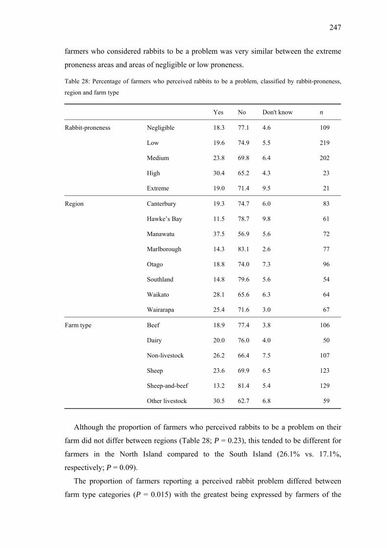
247
farmers who considered rabbits to be a problem was very similar between the extreme
proneness areas and areas of negligible or low proneness.
Table 28: Percentage of farmers who perceived rabbits to be a problem, classified by rabbit-proneness,
region and farm type
Yes No Don't know n
Rabbit-proneness Negligible 18.3 77.1 4.6 109
Low 19.6 74.9 5.5 219
Medium 23.8 69.8 6.4 202
High 30.4 65.2 4.3 23
Extreme 19.0 71.4 9.5 21
Region Canterbury 19.3 74.7 6.0 83
Hawke’s Bay 11.5 78.7 9.8 61
Manawatu 37.5 56.9 5.6 72
Marlborough 14.3 83.1 2.6 77
Otago 18.8 74.0 7.3 96
Southland 14.8 79.6 5.6 54
Waikato 28.1 65.6 6.3 64
Wairarapa 25.4 71.6 3.0 67
Farm type Beef 18.9 77.4 3.8 106
Dairy 20.0 76.0 4.0 50
Non-livestock 26.2 66.4 7.5 107
Sheep 23.6 69.9 6.5 123
Sheep-and-beef 13.2 81.4 5.4 129
Other livestock 30.5 62.7 6.8 59
Although the proportion of farmers who perceived rabbits to be a problem on their
farm did not differ between regions (Table 28; P = 0.23), this tended to be different for
farmers in the North Island compared to the South Island (26.1% vs. 17.1%,
respectively; P = 0.09).
The proportion of farmers reporting a perceived rabbit problem differed between
farm type categories (P = 0.015) with the greatest being expressed by farmers of the

248
'other livestock', non-livestock, and sheep categories, and the least concern being
expressed by mixed sheep-and-beef farmers (Table 28).
Farmers considered damage to the soil surface to be the predominant problem
attributable to rabbits (Table 29).
Table 29: Farmers perceptions (%) of mechanisms by which rabbits impact on their enterprises (n=574)
for each problem caused by rabbits
Severity of rabbit damage
Main problems caused by rabbits None Minimal Moderate Severe Heavy Don't know
Compete with livestock for grazing 31.5 38.5 8.9 4.0 1.4 15.7
Damage pasture quality 36.4 29.4 10.3 3.3 1.0 19.5
Destroy vegetables, flowers,
grapes, forest seedling etc. 44.1 23.3 10.1 4.2 2.3 16.0
Damage soil surface 23.3 42.9 16.6 5.1 2.8 9.4
On the same farms, the severity of rabbit damage was perceived to be lower in 2001
than in 1995 (P < 0.0001), with a shift in rabbit damage from higher ranks of severity to
lower ranks (Figure 29).
0
10
20
30
40
50
60
none minimal moderate severe heavy don't know
Severity of rabbit damage
damage in 1995 damage in 2001
***
ns
***
***
*** ***
Figure 29: Percentage of farms categorised by rabbit damage severity in 1995 and 2001 (***P < 0.0001;
ns non-significant P > 0.10)

249
Methods used for rabbit control
The relative frequency of the use of control methods (shooting, 1080 and Pindone
poisoning, trapping and RHDV) changed between 1995 and 2001 (Table 30). The
proportion of farmers that never used shooting for rabbit control remained constant, but
there was a drastic decline in farmers shooting rabbits frequently ('often' and 'very
often') from 1995 to 2001. Similarly, the use of 1080 and Pindone poisoning, as well as
trapping had decreased to less than half. RHDV was used by 10% of farmers to control
rabbits in 2001, but the vast majority of these farmers (90%) used the virus relatively
infrequently ('rarely' or 'occasionally').
Table 30: Reported percentage usage of rabbit control methods in 1995 and in 2001 amongst 574 farmers
(** P < 0.01; ns non-significant P > 0.10)
Shooting 1080 poisoning Pindone Trapping RHD baiting
Frequency 1995 2001 1995 2001 1995 2001 1995 2001 2001
Never 15.3 15.2 ns 82.1 91.3 ** 82.9 89.2 ** 84.0 89.0 ** 89.9
Rarely 8.4 20.0 ** 5.9 5.2 ns 5.6 5.2 ns 8.5 7.1 ns 5.1
Occasionally 38.3 45.3 ** 6.8 3.0 ** 8.2 4.9 ** 6.3 3.1 ** 4.0
Often 26.8 14.8 ** 3.8 0.4 ** 2.3 0.7 ** 1.0 0.5 ns 0.7
Very often 11.1 4.7 ** 1.6 0.2 ** 1.0 0.0 ** 0.2 0.2 ns 0.3
Shooting
The frequency of shooting was similar in all regions but differed between farm types
(P = 0.015) and rabbit-proneness categories (P = 0.046). Most respondents indicated
that they would 'occasionally' shoot rabbits. A relatively high proportion of 'other
livestock' farmers (34%) often shot rabbits while a substantial fraction (25%) of 'non-
livestock' farmers would never shoot rabbits. Sheep as well as mixed sheep-and-beef
farmers increasingly used shooting for rabbit control as rabbit-proneness increased;
almost half of all sheep farmers shot rabbits sometimes or often when rabbit-proneness
was medium to severe. In contrast beef and dairy farmers rarely controlled rabbits by
means of shooting, regardless of rabbit-proneness. However, there were very few beef
or dairy farmers in the survey that had high or extreme rabbit-prone properties.

250
RHD baiting
The percentage of farmers using RHDV baiting differed between regions (P = 0.01).
RHDV baits were most frequently used in Canterbury (18.1%) and Otago (15.6%),
while only 3-10.4% of farmers used this control method in other areas. RHD use was
positively associated with rabbit-proneness (P < 0.001), increasing from less than 10%
of farms in negligible and low rabbit-prone areas to almost half of farms in extreme
rabbit-prone areas (Table 31). However, RHDV use did not differ between farm types
(P = 0.74).
Table 31: Percentage of farmers using RHDV in 2001 stratified by rabbit-proneness
Rabbit-proneness RHD baiting
Negligible 5.5
Low 6.9
Medium 11.4
High 17.4
Extreme 47.6
The proportion of farmers using RHD baiting stratified by farm type and rabbit-
proneness varied significantly (Figure 30; P = 0.04). Mixed sheep-and-beef farmers,
non-livestock and sheep farmers in extreme rabbit-prone areas were the largest users of
RHD baiting (63%, 50% and 43% respectively). 'Other livestock' farmers were most
likely to use RHD in high rabbit-prone areas (50%) and dairy farmers in medium rabbit-
prone areas (23%).

251
0
10
20
30
40
50
60
70
negligible low medium high extreme
Farm types among rabbit proneness areas
Figure 30: Use of RHD baiting by 58 farms for different farm types (BEF beef, DAR dairy, NLS
non-livestock, SHP sheep, SNB mixed sheep-and-beef, OLF other livestock) in five different rabbit-prone
areas in 2001 (as percentages)
Use of poisoning and trapping
Prevalence of use of 1080 poisoning for rabbit control did not differ between regions
(P = 0.08) or farm type (P = 0.32). However, it did differ between rabbit-proneness
categories (P < 0.001), with the highest use of 1080 poisoning occurring in the extreme
rabbit-prone areas (33%). The interaction of farm types with rabbit-proneness categories
was not significant (P = 0.63).
In contrast, Pindone use was dissimilar between regions (P = 0.045), but not between
farm types (P = 0.24) or proneness categories (P = 0.13). Farmers in the Waikato (19%)
and Canterbury (16%) regions were most likely to use Pindone, while only 0.5% of
farmers in Hawke's Bay and 5% in Southland used Pindone.
The overall percentage of farmers who trapped rabbits was 11%, and did not differ
between regions (P = 0.61), farm type (P = 0.89) or rabbit-proneness category
(P = 0.24).

252
Farmers’ perceptions about rabbit control methods
Figure 31 shows the distribution of farmers’ perceptions about the humaneness of
these control methods among respondents who recorded an opinion other than
uncertain. Shooting was viewed to be the most humane method, followed by RHD,
Pindone and 1080 poisoning, myxomatosis infection, and trapping. While opinions of
the humaneness of the two poisoning methods were very similar, opinions differed
markedly between the two biological agents. For RHDV, 82% of respondents found the
method satisfactory, humane or completely humane compared to only 44% for
myxomatosis.
0
5
10
15
20
25
30
35
40
45
50
Shooting 1080 poisoning Pindone Trapping RHD Myxomatosis
Rabbit control methods
completely inhumane poor satisfactory preferred completely humane
Figure 31: Frequency distributions of farmer opinion about the humaneness of rabbit control methods
The percentage of farmers that were uncertain ('don’t know') about the humaneness
of rabbit control methods was only 2% for shooting, compared with 18% for trapping,
22% for 1080, 28% and 31% for RHD and myxomatosis respectively, and 55% for
Pindone poisoning.
With respect to environmental safety, ethical concerns were higher for poisoning
(particularly 1080) than for infectious agents, and lowest for shooting and trapping
(Figure 32). Many respondents were uncertain about the impact of Pindone poisoning
(54%) and the infectious agents (RHDV 35%; myxomatosis 44%) on the environment.
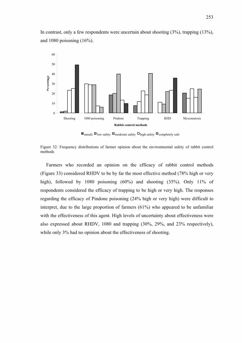
253
In contrast, only a few respondents were uncertain about shooting (3%), trapping (13%),
and 1080 poisoning (16%).
0
10
20
30
40
50
60
Shooting 1080 poisoning Pindone Trapping RHD Myxomatosis
Rabbit control methods
unsafe low safety moderate safety high safety completely safe
Figure 32: Frequency distributions of farmer opinion about the environmental safety of rabbit control methods
Farmers who recorded an opinion on the efficacy of rabbit control methods
(Figure 33) considered RHDV to be by far the most effective method (78% high or very
high), followed by 1080 poisoning (60%) and shooting (35%). Only 11% of
respondents considered the efficacy of trapping to be high or very high. The responses
regarding the efficacy of Pindone poisoning (24% high or very high) were difficult to
interpret, due to the large proportion of farmers (61%) who appeared to be unfamiliar
with the effectiveness of this agent. High levels of uncertainty about effectiveness were
also expressed about RHDV, 1080 and trapping (30%, 29%, and 23% respectively),
while only 3% had no opinion about the effectiveness of shooting.

254
0
10
20
30
40
50
60
Shooting 1080 poisoning Pindone Trapping RHD
Rabbit control methods
not effective low moderate high very high
Figure 33: Frequency distributions of farmer opinion about the effectiveness of rabbit control methods
Factors associated with the use of rabbit control methods
Shooting
The final multivariable ordinal logistic regression model included the categorical
variables 'compete with livestock for grazing' and 'effectiveness of shooting' (574 farms,
10 strata, 28 groups of rabbit proneness multiplied by farm type, P < 0.0001). The use of
shooting for rabbit control was strongly associated with the perception of the extent to
which rabbits 'competed with livestock for grazing', and were markedly elevated among
groups rating the level of competition as severe or heavy (Figure 34).

255
9.35
6.24
2.541.84 1.32
02468
101214161820222426283032
minimal moderate severe heavy don't know
Competition with livestock for grazing
Figure 34: Odds ratio (95% CI) describing the strength of the association between shooting and farmers’
perceptions on the extent that rabbits compete with livestock for grazing (reference group: 'no
competition with livestock for grazing,' dotted line illustrates Odds Ratio of 1)
The association between the perceived 'effectiveness of shooting' and the odds of
shooting was not statistically significant. However, the data indicated a 'dose response'
relationship where the odds of shooting increased with the perception that shooting was
effective (Figure 35).
3.18
0.38
2.26
1.531.12
0123456789
101112131415161718
low moderate high very high don't know
Effectiveness of shooting
Figure 35: Odds ratio (95% CI) describing the strength of association between shooting and farmers’
perceptions on the effectiveness of shooting (reference group: 'not effective,' dotted line illustrates Odds
Ratio of 1)

256
Use of other control methods
The final multivariable binary logistic regression model for the odds of RHDV use is
shown in Table 32. As with shooting, the odds for RHDV use were significantly
associated with the perception of the extent of rabbits competing for grazing. Although
the odds for the use of RHDV were not significantly related to the perception of
humaneness, there was a trend towards the more likely use of RHD as the perception of
humaneness increased.
Table 32: Odds ratios (OR) from binary logistic regression of the use of RHDV to control rabbits (574
farms, 28 groups of proneness multiplied by farm type)
Explanatory variable Categories OR OR 95% CI
Compete with livestock for grazing None 1.00
Minimal 5.09 1.35 - 19.15
Moderate 5.86 1.20 - 28.62
Severe 15.74 3.57 - 69.37
Heavy 59.48 9.63 - 367.55
Don't know 2.81 0.67 - 11.80
Humaneness of RHD Completely inhumane 1.00
Poor 0.71 0.03 - 17.87
Satisfactory 1.09 0.16 - 7.38
Preferred 1.84 0.31 - 10.91
Completely humane 1.56 0.19 - 13.08
Don't know 0.04 0.00 - 0.67
The final logistic model for the odds of farmers using trapping included the variables
of damage to pasture quality, effectiveness of trapping, and humaneness of
myxomatosis. Use of trapping was strongly associated with the perceived effectiveness
of trapping, and also with the perception of the level of pasture damage caused by
rabbits. The odds of use of trapping were also higher for farmers who perceived that
myxomatosis was inhumane or were uncertain about its humaneness.

257
The use of 1080 poisoning for rabbit control strongly increased with both the extent
to which farmers perceived rabbits to compete for grazing and with the perceived safety
of 1080.
Farmers’ concerns about Rabbit Haemorrhagic Disease
Depending on the region and level of rabbit-proneness but independent of farm type,
a variable proportion of respondents had seen rabbits dying from RHD on their
properties. The proportion of farmers observing rabbit deaths due to RHD increased
with rabbit-proneness. The highest proportions of farmers witnessing the disease were
from Otago (43%) and Canterbury (41%). Hawke’s Bay had the lowest proportion of
farmers witnessing the disease (10%), which is consistent with the fact that a high
number of respondents (28%) were uncertain whether rabbits had died from RHD in the
area.
The concerns of farmers about a range of questions regarding the success, worries
and future directions of RHDV for rabbit control are summarised in Table 33. Many
statements expressed considerable uncertainty. A majority of farmers agreed that the
use of RHD had reduced rabbit numbers (65%), considered RHD to be a cost-effective
method (57%), and thought that it was generally beneficial (64%). There was great
uncertainty among respondents about the question of ecological risks of RHDV use in
New Zealand (44%). However, 35% of respondents disagreed that the virus is an
ecological hazard, and 35% would support the introduction of other diseases to kill
rabbits. More than half of the respondents would like to see more research into RHD
(56%) and considered that RHDV is best used in conjunction with other methods
(56%).

258
Table 33: Farmers’ opinions on aspects of RHDV as a tool for rabbit control (tables shows percent of 574
respondents)
Strongly
disagree
Disagree Don’t agree
or disagree
Agree Strongly
agree
Don’t
know
RHD brought rabbit numbers down 1.6 3.3 3.7 27.9 37.1 26.5
RHD is still working very well 2.6 10.1 11.1 22.6 12.4 41.1
RHD was only a short-term solution
and is not working as well now 9.1 18.1 12.2 17.2 2.4 40.9
RHD is more cost effective than other
methods 0.5 1.6 5.1 28.7 28.0 36.1
RHD should be used with other
methods 2.4 4.2 6.6 39.4 16.9 30.5
The introduction of RHD had no
benefits 30.8 33.1 4.5 2.6 1.7 27.2
RHD is a danger for the ecology of
New Zealand 16.7 18.3 11.1 5.7 4.2 43.9
More research is needed to make RHD
fully effective 3.1 5.7 8.4 42.0 14.3 26.5
Other diseases should be introduced to
kill rabbits 15.0 19.7 14.3 11.7 5.9 33.4
Discussion
New Zealand enjoys trading advantages for its animal products due to the country’s
absence of many important livestock diseases, and allocates considerable resources to
biosecurity measures aimed at preserving the health status of its national herds. The
deliberate release of RHDV in New Zealand, subsequent to governmental refusal to
introduce the virus, was not a malicious 'bioterrorist' action, but an action motivated by
perceptions that RHDV would provide an option for rabbit control that was superior to
pre-existing methods. The results of this survey provide some insights into the attitudes
and practices of farmers in the wake of several years of experience following the release
of RHDV, and farmers’ perceptions on the impact of the disease in New Zealand.

259
In all countries, agricultural enterprises are diverse and opinions of farmers can be
expected to be highly variable within and between regions and industries. Our objective
in undertaking this study was to obtain unbiased estimates of opinions of farmers
engaged in key agricultural industries in New Zealand, with particular emphasis given
to pastoral livestock producers. The availability of a national spatially-referenced farm
database (Agribase) and geographical data on rabbit-proneness underpinned the strategy
to stratify sampling according to region, rabbit-proneness and enterprise type. Random
sampling of farms within strata should have minimised selection bias, and the useable
response rate (69%) should have been sufficient to avoid marked response bias.
However, the study population was limited to eight regions encompassing
approximately 63% of the eligible farm population, and some regional bias is possible.
The matrix of sampling weights was used to enable more precise estimates for sub-
groups of greatest interest, and the weightings were accounted for in the analysis. A
major limitation of our approach was the aggregation of all non-livestock enterprises
into a single group, and it is acknowledged that considerable variability in farmer
opinions could exist between the diverse enterprise types in this group. Similarly, the
aggregation of fundamentally different enterprises such as horse and poultry production
within the 'other livestock' category may have masked quite different perceptions
existing among sub-groups of this stratum.
With respect to the magnitude of the rabbit problem in New Zealand, only 21% of
farmers considered rabbits to be a problem on their farms in 2001, and the proportion of
respondents reporting moderate to heavy rabbit damage declined by approximately two
thirds between 1995 to 2001. The somewhat unexpected finding that the proportion of
farmers reporting that rabbits were a problem in 2001 did not vary significantly with
rabbit-proneness, may in part be attributable to the fact that the farms in the most
affected areas have been able to reduce the severity of the problem. The data strongly
support the conclusion that rabbit infestation in extreme rabbit-prone areas was reduced
after introduction of RHD (Parkes et al., 1999). Enterprise type was associated with the
farmers’ perception of having a rabbit problem, with concern most prevalent among the
diverse group of 'other livestock' farmers. Damage of the soil surface (which is probably
the most readily apparent form of damage) was perceived to be the most important
mechanism of rabbit damage. One aspect of soil damage is the occurrence of holes and
collapsing burrows, which increases the risk for lameness and foot problems in

260
livestock and horses. Lameness as a result of rabbit burrowing was one of the most
frequent additional comments made by 'other livestock' farmers.
The release of RHDV in 1997 has been associated with altered patterns of rabbit
control by farmers between 1995 and 2001. Although only 10% of farmers used RHDV
baits in 2001, some reduction was evident in the use of all pre-existing control methods.
Thus the perceived reduction in the magnitude of rabbit problems has occurred during a
period when the reported use of all traditional control methods has declined, and is
likely to be a consequence of the impact of RHDV. However it is clear that RHDV has
not been a quick fix for the rabbit problem as all pre-existing methods continued to be
employed by farmers, with shooting remaining the most prevalent method. While
respondents generally thought that RHD had resulted in reduced rabbit numbers, the
predominant opinion was that RHD was best used in conjunction with other methods.
Several patterns emerged with respect to the selection of methods used for rabbit
control. RHD baiting was most frequently used in the extreme rabbit-prone areas of the
South Island, particularly on farms associated with sheep and on non-livestock farms.
The poison 1080 was used more frequently in extreme rabbit-prone areas, and Pindone
use was highest in the Waikato region. Only trapping was conducted evenly across
regions, farm types and rabbit-proneness strata. Clearly, the choice of methods for
rabbit control will be subject to diverse influences including farmer’s personal
perceptions of efficacy and humaneness, as well as local customs, marketing and peer-
group influences. Evaluation of associations between a range of explanatory variables
and the use of respective control methods identified some likely factors. Generally use
of rabbit control methods was associated with perceived severity of rabbit damage
(competition with livestock for pasture and degree of damage to pasture quality) and
perceived attributes of the method (e.g. humaneness and efficacy of a respective
method).
Opposition to the release of RHDV was based on environmental safety and animal
welfare concerns. It is notable that respondents ranked RHD to be the second most
humane method after shooting, and preferable to poisoning. Also, while farmer
perceptions of the humaneness of the two poisons were almost identical, perceptions
regarding the two viral agents were very different. Myxomatosis was unsuccessfully

261
introduced into New Zealand (Filmer, 1953). In contrast to the peracute deaths typical
of RHD, wild rabbits infected with the Myxoma virus succumb slowly (within 10-14
days) to the disease with symptoms of exhaustion, anorexia, depression, laboured
breathing and pneumonia (Lutz, 1990). Trapping of rabbits, considered inhumane by
21% of respondents, is usually conducted with leg-hole traps and rabbits remain in the
traps until released. The level of concern about the humaneness of trapping is consistent
with a recommendation to ban trapping due to animal welfare concerns (Williams et al.,
1995).
Safety concerns of farmers were greatest for the poisoning methods, particularly
1080 (30% classified it as unsafe), that has been used for rabbit control since the 1950s
(McIntosh, 1958). It is highly toxic for all mammals with carnivores being more
susceptible than rabbits, and has no antidote (Williams et al., 1995). Despite rapid
elimination of the poison from animals that consume sub-lethal doses (Eason, 1992),
secondary poisoning has been reported in rabbit predators (Heyward and Norbury,
1999). Negative opinions from farmers about 1080 are probably driven by the
experiences of death of farm dogs and domestic livestock and as well the current public
debate on killing deer by aerial baiting (Hansford, 2003). In a 1996 survey of the New
Zealand public, Wilkinson and Fitzgerald (1998) found that 28% of respondents
considered 1080 poison the least safe method (28% high risk), while 22% of the
respondents considered RHDV to have a high safety risk. A notable point of our survey
is that 10% of farmers continue to have reservations about the ecological safety of
RHDV despite a lack of any documented safety related incidents since the release of the
agent. New Zealanders have high levels of concern about the environment (Macer,
1994), and the possibility that predation of native bird species may be an unwanted
consequence of improved rabbit control has been proposed (Haselmayer and Jamieson,
2001).
Assessment of the efficacy of rabbit control methods is not straightforward, and is
reflected in the considerable proportion (20-60%) of respondents who were uncertain
about the efficacy of all methods other than shooting (3%). Field observations indicate
that the impact of RHD on wild rabbit populations can be highly variable (Mutze et al.,
2002; Parkes et al., 2002). However, 39% of respondents ranked RHD highest for
efficacy, which was 2-3 fold higher than the next highest ranked method.

262
Based on a survey conducted before RHDV release, Wilkinson and Fitzgerald (1998)
reported that half of the respondents believed that RHD would bring more benefits to
New Zealand than problems. Similarly, the majority of farmers (approximately 60%) in
our survey stated that the introduction of RHDV has been beneficial overall. However
there were clear indications that the introduction of the agent has been far from a
complete solution to the rabbit problem, and many respondents expressed uncertainty
about most aspects of the disease and its impact.
We conclude that farmers perceive the use of RHDV for rabbit control to be an
effective and ethically acceptable method. Since its introduction, the use of shooting has
declined and other methods are regarded as less effective or less acceptable than RHD.
Acknowledgements
We thank Robert Sanson for supplying the farm locations and James Barringer for
the rabbit-proneness map and we also thank the farmers who responded to our survey
for their cooperation, honesty and time.
References
Eason, C.T., 1992. Old pesticide - new data. NZ Sc. Monthly. March, 15-16.
Filmer, J.F., 1953. Disappointing tests of myxomatosis as rabbit control. NZ J. Agric.
87, 402-404.
Fitzgerald, G.P., Saunders, L.S., Wilkinson, R.L., 1996. Public Attitudes to the
Biological Control of Rabbits in New Zealand. MAF Policy Technical Paper 96/3.
Ministry of Agriculture, New Zealand.
Gibb, J.A., Williams, J.M., 1994. The rabbit in New Zealand. In: Corbet, G.B., Flux,
J.E.C., Rogers, P.M., Arthur, C.P., Soriguer, R.C., Myers, K., Parer, I., Wood, D.,
Cooke, B.D., Gibb, J.A., Williams, J.M., Fenner, F., Ross, J., Thompson, H.V. and

263
King, C.M. (Ed.), The European Rabbit: The History and Biology of a Successful
Colonizer. Oxford University Press, Oxford, UK, pp. 158-200.
Hansford, D., 2003. Strangers in paradise. NZ Wilderness Magazine. April, 45-47.
Haselmayer, J., Jamieson, I.G., 2001. Increased predation on pukeko eggs after the
application of rabbit control measures. NZ J. Ecol. 25, 89-93.
Heyward, R.P., Norbury, G.L., 1999. Secondary poisoning of ferrets and cats after 1080
rabbit poisoning. Wildl. Res. 26, 75-80.
Hosmer, D.W., Lemeshow, S., 2000. Applied Logistic Regression. John Wiley & Sons,
Inc, New York, USA.
Kerr, I.G.C., Ross, W.D., 1990. Rabbit Management in Central Otago. Contract report
for Ministry of Agriculture and Fisheries, Wellington, New Zealand.
Lutz, W., 1990. Der Rückgang des Kaninchens unter besonderer Berücksichtigung der
Myxomatose [in German]. Niedersächsischer Jäger. 35, 248-250.
Macer, D., 1994. Bioethics for the People by the People. Eubios Ethics Institute,
Christchurch, New Zealand.
Maxwell, A.E., 1970. Comparing the classification of subjects by two independent
judges. Br. J. Psychiatry. 116, 651-655.
McIntosh, I.G., 1958. 1080 poison: outstanding animal pest destroyer. NZ J. Agric. 97,
361.
Mutze, G., Cooke, B., Alexander, P., 1998. The initial impact of rabbit hemorrhagic
disease on European rabbit populations in South Australia. J. Wildl. Dis. 34, 221-
227.
Mutze, G., Bird, P., Kovaliski, J., Peacock, D., Jennings, S., Cooke, B., 2002. Emerging
epidemiological patterns in rabbit haemorrhagic disease, its interaction with

264
myxomatosis, and their effects on rabbit populations in South Australia. Wildl. Res.
29, 577-590.
Ohlinger, V.F., Hass, B., Thiel, H.J., 1993. Rabbit haemorrhagic disease (RHD):
characterisation of the causative calicivirus. Vet. Rec. 24, 103-116
Parkes, J., 1995. Rabbits as Pests in New Zealand: A Summary of the Issues and
Critical Information, Landcare Research Contract Report LC9495/141. Landcare
Research, Lincoln, New Zealand.
Parkes, J.P., Norbury, G. L., Heyward, R. P. 1999. Has rabbit haemorrhagic disease
worked in New Zealand? In: Proceedings of the New Zealand Society of Animal
Production. Landcare Research, Lincoln, New Zealand, pp. 245-249.
Parkes, J.P., Norbury, G.L., Heyward, R.P., Sullivan, G., 2002. Epidemiology of rabbit
haemorrhagic disease (RHD) in the South Island, New Zealand, 1997-2001. Wildl.
Res. 29, 543-555.
Rao, J.N.K., Scott, A.J., 1981. The analysis of categorical data from complex sample
surveys: chi-squared test for goodness of fit and independence in two-way tables. J.
Am. Stat. Assoc. 76, 221-230.
Rao, J.N.K., Scott, A.J., 1984. On chi-squared test for multiway contingency tables with
cell proportions estimated from survey data. Anal. Stat. 12, 40-60.
Sanson, R., Pearson, A., 1997. Agribase - a National Spatial Farm Database. Epidemiol
Sante Animale. 12-13.
Sanson, R.L., Brooks, H.V., Horner, G.W., 2000. An epidemiological study of the
spread of Rabbit haemorrhagic disease virus amongst previously non-exposed rabbit
populations in the North Island of New Zealand. NZ Vet. J. 48, 105-110.
Stata Corporation, 2001. Stata Reference Manual. Release 7. Volume 4. Stata Press,
College Station, Texas, USA.

265
Stuart, A., 1955. A test of homogeneity of the marginal distributions in a two-way
classification. Biometrika. 42, 412-416.
Williams, K., Parer, I., Coman, B., Burley, J., Braysher, M., 1995. Managing Vertebrate
Pests: Rabbits. Australian Government Publishing Service, Canberra.
Wilkinson, R., Fitzgerald, G., 1998. Public Attitudes to Rabbit Calicivirus Disease in
New Zealand. Manaaki Whenua Press, Lincoln, Canterbury, New Zealand.
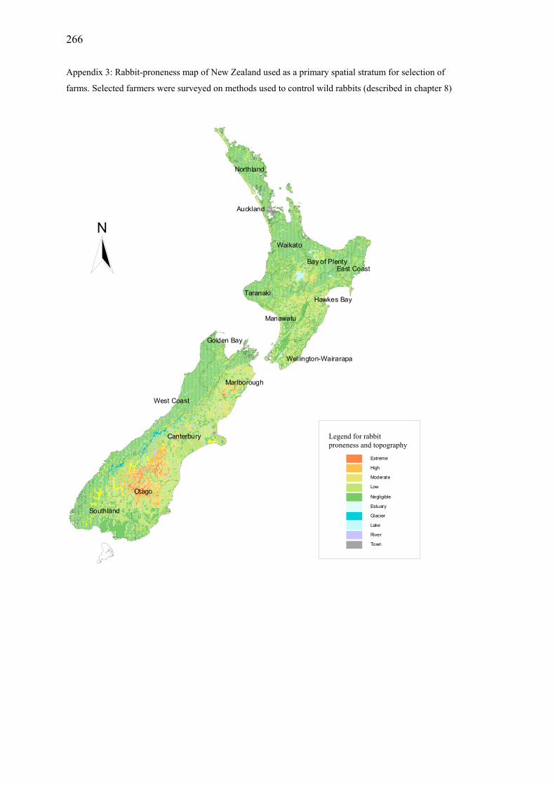
266
Appendix 3: Rabbit-proneness map of New Zealand used as a primary spatial stratum for selection of
farms. Selected farmers were surveyed on methods used to control wild rabbits (described in chapter 8)
Waikato
Manawatu
Northland
Hawkes Bay
Bay of Plenty
Taranaki
Wellington-Wairarapa
East Coast
Auckland
Otago
Canterbury
Southland
West Coast
Golden Bay
Marlborough
Extreme
High
Moderate
Low
Negligible
Estuary
Glacier
Lake
River
Town
NZ Rabbit Proneness
N
NN
Legend for rabbit proneness and topography

267
Appendix 4: Questionnaire used to survey New Zealand farmers on their attitudes to methods used to
control wild rabbits (described in chapter 8).

268

269
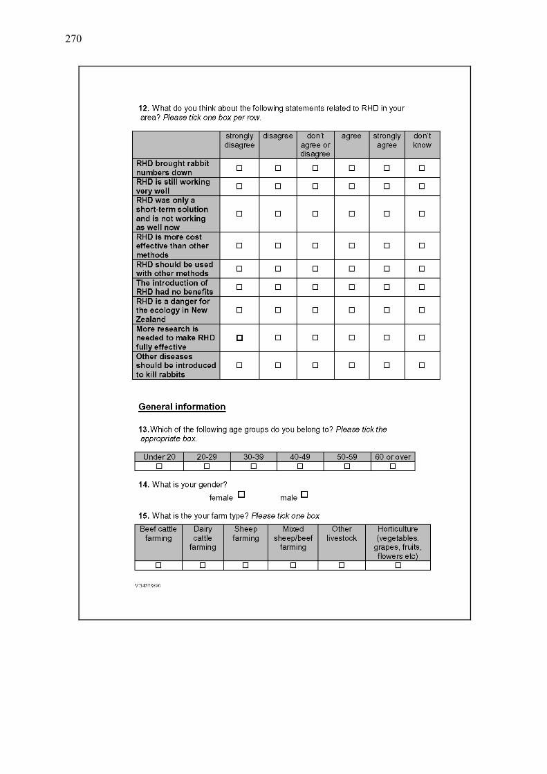
270

271

272

273
Chapter 9
General Discussion

274

275
RHDV in wild rabbit populations
There are numerous examples of negative unintended consequences from introducing
exotic species into new environments. Therefore any proposal to introduce an
apparently novel animal pathogen, such as RHDV, into New Zealand, a country with an
island ecosystem and an economy that is highly dependent on animal agriculture, will
inevitably be subject to considerable scrutiny and criticism. In 1997, the New Zealand
government rejected an application by farmer groups to introduce RHDV as a biological
control method for rabbits, because of uncertainties about the likely success of RHDV
in the New Zealand environment and the ecological risks (O'Hara 1997). These
concerns were amplified by a general paucity of knowledge about the epidemiology of
the agent and the disease. Subsequently, the illegal introduction of RHDV, and the
resulting disease outbreaks, provided a rare opportunity to study a disease soon after its
initial occurrence in a region. The intensive sampling conducted in the longitudinal
studies have provided detailed data on the temporal and spatial aspects of the disease
over a period of nearly three years. In addition experimental studies revealed properties
of RHDV that are important for understanding the disease.
The longitudinal study confirmed that RHDV had become established in the study
region and showed evidence of transmission recurring in annual cycles. However, the
cause-specific mortality rate varied considerably between years. The magnitude of
naturally occurring outbreaks of RHDV is likely to be influenced by population
demographics, individual and population immune statuses, competing risks due to other
causes of mortality, and climatic conditions that may influence virus survival, vector
abundance and rabbit behaviour. Rabbit movements, vector abundance, and activity of
predatory or scavenging species probably facilitate the spread of RHDV. Prediction of
naturally recurring RHDV outbreaks requires consideration of these factors, which are
all likely to influence the success of deliberate RHDV releases to control rabbits.
The source of RHDV infection that initiates any outbreak is usually difficult or
impossible to identify, and therefore open to speculation. The importance of predators,
scavengers and insects in mechanical transmission of RHDV will in part be determined
by the durability of the virus in carcasses. This question is difficult to address because
RHD virus is not culturable in vitro, and therefore bioassays in live rabbits are required.

276
The ethical cost of conducting such studies is considerable and the evidence base for
evaluating the importance of viral survival outside the host remains minimal.
Nevertheless, the studies undertaken clearly indicate that RHDV in a matrix of organic
tissue could remain a potential source of new infections for at least three months. In
contrast, RHDV on cotton exposed directly to environmental conditions (used to
emulate excreted virus on fomites) was less persistent. Thus excreted virus in the
environment is likely to play a major role in transmission once an epidemic is initiated,
but is less likely to contribute to reservoirs responsible for inter-epidemic survival of the
virus. In addition, the failure of inactivated virus to elicit serological responses in
rabbits suggests that unintended ‘vaccination’ of wild rabbit populations with baits is an
unlikely event.
The probable existence of carrier rabbits is now generally accepted (Parkes et al.
2002; Cooke 2002; Zheng et al. 2002), despite the lack of credible supporting data. An
important question is to define which sub-group of rabbits is the predominant source of
virus. Because the duration of viral shedding in acute and peracute cases is short, due to
the rapid mortality, most virus shedding will probably occur in rabbits that have
recently survived RHD outbreaks (White et al. 2002). However, the mean duration of
excretion of infective virus, and its variance, has yet to be quantified. Also it is not
known whether excretion of RHDV from infected rabbits is constant or intermittent, nor
if intermittent, how frequently it occurs. It is feasible that infected rabbits not excreting
virus could provide a source of infection due to predation or scavenging after death
from other causes. Mortality of RHDV in susceptible adults is very high compared to
other infectious disease in wildlife hosts, such as tuberculosis in possums (Ron Jackson,
personal communication), hence relatively few adult survivors remain to spread the
disease. Alternatively, young animals may survive infection if maternal immunity or
natural resilience is present, and could serve as long-term reservoirs of virus.
Persistent, low-level excretion by chronically infected rabbits, as modelled by White
et al. (2002), could also be a reservoir of infection. In this scenario, interactions of
extrinsic factors trigger increased excretion, leading to spread of the infection.
However, reactivation of RHDV in experimentally infected rabbits (4-5 weeks old)
which recovered from the disease was not achieved by treatment with dexamethasone or
classical swine fever virus vaccine (Shien et al. 2000). At this point in time the

277
possibility of reactivation of shedding by chronically infected rabbits remains
hypothetical.
The observation of seropositivity in several non-target species raises questions with
respect to both the host range of the virus and the role of such species in the
dissemination and transmission of the virus. It remains to be determined whether the
serological reactions observed reflect the occurrence of viral replication in non-target
species or a simple response to viral antigens. The fact that we were unable to elicit
serological responses to UV inactivated RHDV in rabbits following oral and parenteral
exposure would suggest that seroconversion in the absence of viral replication is
unlikely. However, there is still no convincing evidence confirming that non-target
species can excrete RHDV, and this question warrants further investigation. It must also
be remembered that serological tests for RHDV have not been validated for non-target
species and positive results should be interpreted with caution.
A further outstanding issue in the epidemiology of RHDV in New Zealand is the
suggested existence of a benign RHDV-like virus. A non-pathogenic calicivirus has
been isolated in Europe. To date no such virus has been identified in either New
Zealand or Australia and evidence for such a virus in New Zealand is limited to sparse
serological data. In the current studies, there were only three rabbits with serological
profiles that were consistent with infection with ‘pre-existing’ virus, and no indication
that these rabbits were protected against RHDV. Recent modelling studies in the United
Kingdom concluded that the development of acute and chronic infection states was
sufficient to explain variability in the effect of RHDV in the absence of a benign and
cross-protecting virus (White et al. 2002). In order to understand the reasons behind the
apparent temporal and spatial variability in RHD severity, detailed studies of
‘successful’ and ‘unsuccessful’ RHD outbreaks will need to be conducted (Cooke 2002;
Henning and Davies 2003). Localised patchiness may be influenced by environmental
barriers of vegetation, which in turn are correlated with the abundance of flying vectors.
In this study we found that RHDV deaths were spatially clustered within the study site,
and hot-spots of RHD recurrence can be expected.

278
Fly vectors
We identified several climate parameters associated with abundance of the fly
species considered most likely to play a role in the transmission of RHDV in New
Zealand. Climate influences fly activity, ovi- and vivipositions and fly development. It
also affects the decomposition of carcasses, which are a source of contamination of flies
with RHDV. Disease transmission by flies can either occur directly to healthy rabbits or
indirectly through contamination of the environment. Observed differences in the
climate pattern and associated fly abundance, between corresponding periods in
different years could be one factor contributing to variability in the months in which
RHD outbreaks were observed in 2000 and 2001. Previously, climate changes have also
been linked with virus survival and rabbit breeding (Henzell et al. 2002). However, it is
unlikely that occurrence of an outbreak will be dictated by a single factor that is climate
dependent. The seasonal pattern of outbreaks in late summer and autumn is most
probably a result of the interaction of multiple factors related to the host population and
environment, many of which may be influenced by climatic factors.
Given the apparent temporal association between fly abundance and the occurrence
of RHD outbreaks, high fly abundance may serve as a marker for environmental
conditions that favour RHDV transmission. The release of virus-contaminated baits is
probably best suited to warm dry periods of optimal fly breeding and activity. Rabbits
succumbing to RHD should be left on pasture close to ground cover, where they are
easily accessed by fly species. As previously reported by Barratt et al. (1998), the role
of Oxysarcodexia varia in particular could be enhanced by stock movements to provide
dung-breeding opportunities for this species.
Methods of rabbit control in relation to natural mortality
Rabbit numbers and rabbit damage were reduced following RHDV introduction and
the overall effect of RHDV is generally considered satisfactory by farmers. Some of the
more conventional control methods have limitations, in particular poisoning. Use of
1080 over several decades resulted in bait avoidance (neophobia). Neophobia, an
avoidance of novel objects, is particularly prevalent in areas of long-term poisoning
(Rowley 1963; Oliver et al. 1982). A second type of aversion is bait shyness, an

279
avoidance of toxic baits through negative conditioning to bait related effects (Poole
1963; Devine and Cook 1998). Furthermore there is a risk of poisoning non-target
species (Booth and Wickstrom 1999; Powlesland et al. 2000; Lloyd and McQueen
2002). In addition to improving the overall effectiveness of rabbit control, the
introduction of RHDV has reduced the environmental and administrative problems
associated with 1080 poisoning, as well as the animal welfare concerns related to
trapping. Consistent with an analysis conducted in Australia by Saunders et al. (2002),
this should have reduced the overall cost of rabbit management, and thus increased the
economic viability of properties previously afflicted by high rabbit numbers.
RHDV will not replace shooting as the most popular rabbit control method. Barlow
(1999) described a rabbit density-dependent model in which a supplementary control
method like shooting may have adverse consequences for RHD effectiveness if rabbit
density is reduced below the threshold level for disease persistence. However, the
number of remaining rabbits depends on initial rabbit density, and we have shown that
RHD can also recur annually in sparse rabbit populations under influence of shooting
(Henning and Davies 2003). The impact of natural RHD outbreaks can differ between
years, and shooting may be a preferred option in years with low RHD mortality. Parkes
et al. (2001) also suggested a change from the expensive active employment of
conventional control methods (aimed to maintain constantly low rabbit densities) to a
cheaper, more reactive strategy of applying conventional control methods only when
RHD was not successful. Active RHDV ‘biociding’ is mainly practised in the most
rabbit prone areas. Investigation of the causes of success and failure of RHDV releases
was not an objective of the research presented in this thesis, but remains an important
issue for optimisation of biological control of rabbits with RHD (Henning, unpublished
data). Likely factors include rabbit population biology and immunity status, virus
survival and vector activity (Bowen and Read 1998; Parkes et al. 1999).
The interactions between natural RHD outbreaks, biociding and shooting occur in a
broader context that includes other competing causes of rabbit death, and in particular
predation. There is debate over whether predators can regulate rabbit populations
(Newsome et al. 1989; Krebs 1995; Sinclair and Pech 1996). Some authors consider
predation as a limiting, but not regulating, factor (Banks 2000) while others have shown
predator regulation of rabbits especially in low rabbit density areas (Pech et al. 1992).

280
Reddiex et al. (2002) demonstrated that RHD has a higher impact when predators are
not controlled. Predation was the principal cause of mortality in our study and resulted
in more consistent rabbit mortality compared to the more dramatic, but transient short
term impact of RHD epidemics.
Active rabbit control can be viewed as the deliberate augmentation of natural causes
of death, which are widespread and prominent, with additional risks of mortality.
Natural mortality will fluctuate over time, as will rabbit reproductive efficiency, and
human intervention is an appropriate complementary tool for population regulation in
times of high rabbit numbers.
The long-term effect of RHDV
Cooke and Fenner (2002) suggest that the future of RHDV will depend on two
characteristics: firstly, on the probability that rabbits develop genetic resistance, and
secondly that RHDV retains its high virulence. Furthermore, the authors suggest that
retention of high virulence depends on the mode of transmission. Selection for sustained
virulence is most probable in scenarios in which airborne vectors contaminated by
RHDV are the predominant mode of viral transmission (Cooke and Fenner 2002).
Genetic resistance of rabbits to RHDV has not yet been identified. A recent French
study indicated that despite high mortality rates during past epizootics and in particular
myxomatosis, no loss of genetic variability in wild rabbits has been observed (Queney
et al. 2000). RHDV is a relatively young disease in New Zealand and genetic changes in
rabbits would not be expected in this short period. A genetically-modified, attenuated
strain of Myxoma virus, which can express the RHDV major capsid protein and thus
protect against both RHD and myxomatosis, was recently developed in Spain (Barcena
et al. 2000). The recombinant viruses can be safely transmitted by direct contact
between rabbits and will be used by conservationists to protect wild rabbits in Europe
(Torres et al. 2000; 2001). However, the deliberate release of such an agent, as an act of
bioterrorism, could have a devastating effect in countries where RHDV plays a useful
role in controlling rabbits. Rabbit management has different goals and strategies in
different countries and international regulations alone may not fully protect countries
from unwanted organisms. Thus it is the scientist who must take responsibility for

281
reducing the risk of the accidental or illegal spread of any genetically modified rabbit
viruses (Angulo and Cooke 2002).
Conclusion
The work undertaken in this thesis illustrates the complexity of RHDV epidemiology
in a low density wild rabbit population in New Zealand. The improved understanding of
patterns of natural outbreaks in this environment should enhance the application of
RHDV as a biocontrol agent in this country. The work is unusual as epidemiological
knowledge is typically applied to minimise the impact of animal diseases rather than to
promote its spread. The results of this study have indicated areas requiring further
research and in particular the importance of carrier rabbits. Successful rabbit control in
the future will depend on how knowledge of RHDV epidemiology is integrated into
more complex control strategies.
References
Angulo, E., and Cooke, B. (2002). First synthesise new viruses then regulate their
release? The case of the wild rabbit. Molecular Ecology 11, 2703-2709.
Banks, P. B. (2000). Can foxes regulate rabbit populations? Journal of Wildlife
Management 64, 401-406.
Barcena, J., Morales, M., Vazquez, B., Boga, J. A., Parra, F., Lucientes, J., Pages-
Mante, A., Sanchez-Vizcaino, J. M., Blasco, R., and Torres, J. M. (2000).
Horizontal transmissible protection against myxomatosis and rabbit hemorrhagic
disease by using a recombinant Myxoma virus. Journal of Virology 74, 1114-1123.
Barlow, N. (1999). Modelling rabbit haemorrhagic disease. In 'Rabbit Control, RCD:
Dilemmas and Implications. Proceedings of the Conference, 30-31 March 1998.'
pp. 76-79. (Royal Society of New Zealand Miscellaneous Series: Wellington, New
Zealand.)
Barratt, B. I. P., Ferguson, C. M., Heath, A. C. G., Evans, A. A., and Logan, R. A. S.

282
(1998). Can insects transmit Rabbit haemorrhagic disease virus? In 'Proceedings of
the Fifty First New Zealand Plant Protection Conference, Hamilton, New Zealand.'
pp. 245-250. (New Zealand Plant Protection Society: Rotorua, New Zealand.)
Booth, L. H., and Wickstrom, M. L. (1999). The toxicity of sodium monofluoroacetate
(1080) to Huberia striata, a New Zealand native ant. New Zealand Journal of
Ecology 23, 161-165.
Bowen, Z., and Read, J. (1998). Population and demographic patterns of rabbits
(Oryctolagus cuniculus) at Roxby Downs in arid South Australia and the influence
of rabbit haemorrhagic disease. Wildlife Research 25, 655-662.
Cooke, B. D. (2002). Rabbit haemorrhagic disease: Field epidemiology and the
management of wild rabbit populations. Revue scientifique et technique de l'Office
International des Epizooties 21, 347-358.
Cooke, B. D., and Fenner, F. (2002). Rabbit haemorrhagic disease and the biological
control of wild rabbits, Oryctolagus cuniculus, in Australia and New Zealand.
Wildlife Research 29, 689-706.
Devine, C. D., and Cook, C. J. (1998). Bait shyness and its prevention in the rabbit
Oryctolagus cuniculus L. New Zealand Journal of Zoology 25, 223-229.
Henning, J., and Davies, P. R. (2003). A serial cross-sectional study of the prevalence of
rabbit haemorrhagic disease (RHD) on three farms in the lower North Island of
New Zealand. New Zealand Veterinary Journal submitted.
Henzell, R. P., Cunningham, R. B., and Neave, H. M. (2002). Factors affecting the
survival of Australian wild rabbits exposed to rabbit haemorrhagic disease. Wildlife
Research 29, 523-542.
Krebs, C. J. (1995). Two paradigms of population regulation. Wildlife Research 22, 1-
10.
Lloyd, B. D., and McQueen, S. M. (2002). Measuring mortality in short-tailed bats

283
(Mystacina tuberculata ) as they return from foraging after an aerial 1080 possum
control operation. New Zealand Journal of Ecology 26, 53-59.
Newsome, A. E., Parer, I., and Catling, P. C. (1989). Prolonged prey suppression by
carnivores - predator-removal experiments. Oecologia 78, 458-467.
O'Hara, P. J. (1997). 'Decision on the application to approve the importation of rabbit
calicivirus as a biological control agent for feral rabbits. 2 July 1997'. (Ministry of
Agriculture: New Zealand.)
Oliver, A. J., Wheeler, S. H., and Gooding, C. D. (1982). Field-evaluation of 1080 and
Pindone oat bait, and the possible decline in effectiveness of poison baiting for the
control of the rabbit, Oryctolagus cuniculus. Australian Wildlife Research 9, 125-
134.
Parkes, J. P., Norbury, G. L., and Heyward, R. P. (1999). Has rabbit haemorrhagic
disease worked in New Zealand? In 'Proceedings of the New Zealand Society of
Animal Production.' pp. 245-249. (Landcare Research: Lincoln, New Zealand.)
Parkes, J. P., Norbury, G. L., Heyward, R. P., and Heath, A. C. G. (2001). Potential
transmission routes of rabbit haemorrhagic disease: consequences for epidemiology
of RHD in wild rabbits. In 'Proceedings of the New Zealand Society of Animal
Production.' pp. 68-70. (AgResearch: Ruakura, New Zealand)
Parkes, J. P., Norbury, G. L., Heyward, R. P., and Sullivan, G. (2002). Epidemiology of
rabbit haemorrhagic disease (RHD) in the South Island, New Zealand, 1997-2001.
Wildlife Research 29, 543-555.
Pech, R. P., Sinclair, A. R. E., Newsome, A. E., and Catling, P. C. (1992). Limits to
predator regulation of rabbits in Australia - evidence from predator-removal
experiments. Oecologia 89, 102-112.
Poole, W. E. (1963). Field enclosure experiments on the techniques of poisoning the
rabbit Oryctolagus cuniculus (L.) IV. A study of territorial behaviour and furrow
poisoning. CSIRO Wildlife Research 8, 36-51.

284
Powlesland, R. G., Knegtmans, J. W., and Styche, A. (2000). Mortality of North Island
tomtits (Petroica macrocephala toitoi) caused by aerial 1080 possum control
operations, 1997-1998, Pureora Forest Park. New Zealand Journal of Ecology 24,
161-168.
Queney, G., Ferrand, N., Marchandeau, S., Azevedo, M., Mougel, F., Branco, M., and
Monnerot, M. (2000). Absence of a genetic bottleneck in a wild rabbit
(Oryctolagus cuniculus) population exposed to a severe viral epizootic. Molecular
Ecology 9, 1253-1264.
Reddiex, B., Hickling, G. J., Norbury, G. L., and Frampton, C. M. (2002). Effects of
predation and rabbit haemorrhagic disease on population dynamics of rabbits
(Oryctolagus cuniculus) in North Canterbury, New Zealand. Wildlife Research 29,
627-633.
Rowley, I. (1963). Field enclosure experiments on the techniques of poisoning the
rabbit Oryctolagus cuniculus (L.) IV. A study of shyness in wild rabbits subjected
to 1080 poisoning. CSIRO Wildlife Research 8, 142-153.
Saunders, G., Kay, B., Mutze, G., and Choquenot, D. (2002). Observations on the
impacts of rabbit haemorrhagic disease on agricultural production values in
Australia. Wildlife Research 29, 605-613.
Shien, J. H., Shieh, H. K., and Lee, L. H. (2000). Experimental infections of rabbits
with Rabbit haemorrhagic disease virus monitored by polymerase chain reaction.
Research in Veterinary Science 68, 255-259.
Sinclair, A. R. E., and Pech, R. P. (1996). Density dependence, stochasticity,
compensation and predator regulation. Oikos 75, 164-173.
Torres, J. M., Ramirez, M. A., Morales, M., Barcena, J., Vazquez, B., Espuna, E.,
Pages-Mante, A., and Sanchez-Vizcaino, J. M. (2000). Safety evaluation of a
recombinant Myxoma-RHDV virus inducing horizontal transmissible protection
against myxomatosis and rabbit haemorrhagic disease. Vaccine 19, 174-182.

285
Torres, J. M., Sanchez, C., Ramirez, M. A., Morales, M., Barcena, J., Ferrer, J., Espuna,
E., Pages-Mante, A., and Sanchez-Vizcaino, J. M. (2001). First field trial of a
transmissible recombinant vaccine against myxomatosis and rabbit hemorrhagic
disease. Vaccine 19, 4536-4543.
White, P. J., Norman, R. A., and Hudson, P. J. (2002). Epidemiological consequences of
a pathogen having both virulent and avirulent modes of transmission: the case of
Rabbit haemorrhagic disease virus. Epidemiology and Infection 129, 665-677.
Zheng, T., Napier, A. M., Parkes, J. P., O'Keefe, J. S., and Atkinson, P. H. (2002).
Detection of RNA of Rabbit haemorrhagic disease virus from New Zealand wild
rabbits. Wildlife Research 29, 683-688.