the essential guide to brain tumours
TRANSCRIPT
Essential Guideto Brain Tumors
The
symptoms
complementary medicine
understanding the brainseeking helpCAUSES
clinical trialssupport
TREATMENT TEAM
treatment options
survivorship DIAGNOSING
caregiving

The Essential Guide to Brain Tumors
www.braintumor.org
National Brain Tumor Society (NBTS) is fiercely committed to
finding a cure for brain tumors. NBTS funds research aimed at
moving industry forward and is making new strategic alliances
with industry, government, and academic institutions to drive
research to therapies as fast as possible.
Patients and caregivers need to have the necessary information
to deal with their diagnosis and advocate for themselves as well
as play a vital role in advocating for public policies that meet the
critical needs of the brain tumor community.
Visit www.braintumor.org to get more information about
research, public policy advocacy efforts, and how you can get
involved and make a difference in the fight against brain tumors.
Visit www.braintumor.org/help to find:
•alistofsomeofthemostfrequentlyaskedquestions
thatmaybeofassistancetoyou
•onlineinformationandresourcesforbraintumorpatients,
families,andcaregivers
•informationontumortypes,treatmentoptions,and
wheretofindatreatmentcenter
•howtoconnectwithothersinthebraintumorcommunity

National Brain Tumor Society | www.braintumor.org National Brain Tumor Society | www.braintumor.org
Acknowledgments ................................................................................................................................................................................ 1
Introduction ............................................................................................................................................................................................ 3
Chapter 1: Understanding the Brain ............................................................................................................................................ 5
The Central Nervous System ........................................................................................................................................................................ 5
The Sections of the Brain ............................................................................................................................................................................... 6
The Internal Nerve Structures of the Brain ............................................................................................................................................ 7
Chapter 2: Diagnosing a Brain Tumor ......................................................................................................................................... 8
Brain Tumor Symptoms .................................................................................................................................................................................. 8
Neurological Examination .............................................................................................................................................................................. 8
Scans and Imaging Techniques ................................................................................................................................................................... 9
Biopsy ...................................................................................................................................................................................................................... 12
Chapter 3: Known and Possible Causes ..................................................................................................................................... 14
Incidence Rates ................................................................................................................................................................................................... 14
Brain Tumor Trends .......................................................................................................................................................................................... 14
Known and Possible Causes .......................................................................................................................................................................... 16
Direction for Future Studies .......................................................................................................................................................................... 18
Chapter 4: Brain Tumor Types ....................................................................................................................................................... 20
Classifying Brain Tumors ................................................................................................................................................................................ 20
Primary Brain Tumors ...................................................................................................................................................................................... 21
Glial Tumors
Astrocytoma ..................................................................................................................................................................................................... 21
Pilocytic Astrocytoma .................................................................................................................................................................................. 22
Low-Grade Astrocytoma ............................................................................................................................................................................. 22
Anaplastic Astrocytoma .............................................................................................................................................................................. 22
Glioblastoma .................................................................................................................................................................................................... 22
Brain Stem Glioma ........................................................................................................................................................................................ 23
Ependymoma .................................................................................................................................................................................................. 23
Mixed Glioma ................................................................................................................................................................................................... 24
Oligodendroglioma ........................................................................................................................................................................................ 24
Optic Nerve Glioma ....................................................................................................................................................................................... 25
Subependymoma .......................................................................................................................................................................................... 25
Non-Glial Tumors
Acoustic Neuroma ......................................................................................................................................................................................... 26
Chordoma ......................................................................................................................................................................................................... 26
CNS Lymphoma ............................................................................................................................................................................................. 26
Craniopharyngioma ...................................................................................................................................................................................... 27
Hemangioblastoma ....................................................................................................................................................................................... 27
As a member of the Board of Directors of the National Brain Tumor Foundation, one of the legacy organizations
of today’s National Brain Tumor Society, Karen’s tireless work—always laden with great wit and enthusiasm—was
centered on the creation of the Guide. Karen—a teacher and author— survived her brain tumor but was stricken
with a lung disease that caused her untimely passing in 1989. Her spirit and her vision continue to thrive in this new
updated version of the Guide.
Elizabeth “Libby” Stevenson served as the first Executive Director of the National Brain Tumor Foundation and later
on its Board of Directors. Libby was diagnosed with a brain tumor in 1980 and survived her disease for over 20 years
until she passed away in 2003. Her gracious manner and infectious smile served as an inspiration to all who met her
and provided hope and compassion to other brain tumor survivors. She lived by her motto “always, there is hope”
and was an ever-vigilant advocate for brain tumor patients. The Essential Guide to Brain Tumors is proudly dedicated to
these two pioneers in the brain tumor community—Karen Osney Brownstein and Libby Stevenson.
Karen Osney Brownstein and Elizabeth “Libby” Stevenson conceived of and
authored the original National Brain Tumor Guide in 1986 for brain tumor
patients and their families. They created the Guide to offer a supportive and
jargon-free resource to the brain tumor community. The Guide continues
to speak in very human terms to those confronting the trauma of their own
medical crisis, letting them know that they are not alone, and that there are
vast resources available to them during a time of confusion and need.
Table of ContentsDedication
Karen O. Brownstein Elizabeth “Libby“ Stevenson
National Brain Tumor Society | www.braintumor.org
• Terri S. Armstrong, MS, APRN, BC
University of Texas M.D. Anderson Cancer Center,
Houston, Texas
• Mitchel S. Berger, MD
University of California, San Francisco
Medical Center, San Francisco, California
• Melissa Bondy, PhD
University of Texas M.D. Anderson Cancer Center,
Houston, Texas
• Susan Chang, MD
University of California, San Francisco
Medical Center, San Francisco, California
• Terri Chew, MPH
University of California, San Francisco
Medical Center, San Francisco, California
• Tim Cloughesy, MD
University of California, Los Angeles
Medical Center, Los Angeles, California
• Charles Cobbs, MD
California Pacific Medical Center, San Francisco,
California
• Nancy Conn-Levin, MA
Monmouth and Ocean County Brain Tumor Support
Group, Inc., Oakhurst, New Jersey
• Mark R. Gilbert, MD
University of Texas M.D. Anderson Cancer Center,
Houston, Texas
• Peter Gruen, MD
University of Southern California,
Los Angeles, California
• Philip Gutin, MD
Memorial Sloan-Kettering Cancer Center,
New York, New York
• Yuriko Minn, MS
Stanford University, Stanford, California
• Kathleen Mogensen, MS, ANP-C
Roswell Park Cancer Institute, Buffalo, New York
• Margaretta Page, RN, MS
University of California, San Francisco
Medical Center, San Francisco, California
• Kendra Peterson, MD
Stanford University, Stanford, California
• Michael Prados, MD
University of California, San Francisco
Medical Center, San Francisco, California
• Raul Rodas, MD
Central Florida Regional Hospital, Neurohealth
Sciences Center, Sanford, Florida
• Evan Ross, LAc, DOM
Cedars-Sinai Medical Center, Los Angeles, California
• Edward Roy, PhD
University of Illinois at Urbana-Champaign, Urbana,
Illinois
Medulloblastoma............................................................................................................................................................................................ 27
Meningioma ...................................................................................................................................................................................................... 28
Pineal Tumor .................................................................................................................................................................................................... 28
Pituitary Tumor ............................................................................................................................................................................................... 29
Primitive Neuroectodermal Tumors (PNET) ...................................................................................................................................... 29
Rhabdoid Tumor............................................................................................................................................................................................. 30
Schwannoma ................................................................................................................................................................................................... 30
Recurrent Tumors .............................................................................................................................................................................................. 30
Metastatic Brain Tumors ................................................................................................................................................................................. 31
Other Tumor-Related Conditions ................................................................................................................................................................. 31
Chapter 5: Brain Tumor Treatments ............................................................................................................................................ 32
Tumor Board ........................................................................................................................................................................................................ 32
Medical Management ....................................................................................................................................................................................... 32
The Treatment Team ......................................................................................................................................................................................... 33
Surgery ................................................................................................................................................................................................................... 34
Radiation Therapy .............................................................................................................................................................................................. 37
Chemotherapy ..................................................................................................................................................................................................... 39
Clinical Trials ........................................................................................................................................................................................................ 40
Biologic/Targeted Therapies ......................................................................................................................................................................... 42
Complementary and Alternative Medicine (CAM) ............................................................................................................................... 44
Chapter 6: Symptom Management .............................................................................................................................................. 47
Physical Symptoms ........................................................................................................................................................................................... 47
Cognitive and Behavioral Symptoms ........................................................................................................................................................ 48
Headache ............................................................................................................................................................................................................... 49
Seizures .................................................................................................................................................................................................................. 49
Fatigue ..................................................................................................................................................................................................................... 51
Blood Clots (Thrombosis) ............................................................................................................................................................................... 51
Other Symptoms ................................................................................................................................................................................................ 52
Chapter 7: Survivorship ..................................................................................................................................................................... 53
“Survivor” Defined ............................................................................................................................................................................................. 53
Dealing with Emotions and Grief ................................................................................................................................................................. 54
Managing Follow-up Care ............................................................................................................................................................................... 56
Returning to Work .............................................................................................................................................................................................. 58
Chapter 8: Suggestions for Caregivers ....................................................................................................................................... 60
Taking Care of the Caregiver ......................................................................................................................................................................... 60
Getting and Managing Information ............................................................................................................................................................ 60
Seeking Help ........................................................................................................................................................................................................ 61
Impact on the Family ........................................................................................................................................................................................ 62
Emotional Issues ................................................................................................................................................................................................. 63
Glossary .................................................................................................................................................................................................... 65
Index .......................................................................................................................................................................................................... 71
National Brain Tumor Society (NBTS) wishes to thank all the people whose
generous contributions of funds, expertise, and good will made possible
the publication of The Essential Guide to Brain Tumors.
Acknowledgments
We are grateful to the following health professionals who volunteered their assistance in the writing of The Essential
Guide to Brain Tumors.
National Brain Tumor Society | www.braintumor.orgNational Brain Tumor Society | www.braintumor.org
No one can prepare to be diagnosed with a brain
tumor. It shatters your sense of well-being and personal
security. It is common to experience a flurry of emotions
and feelings when you receive the news that you have
a brain tumor. One moment you may feel angry and
overwhelmed, then dazed and numb the next. Many
personal and practical questions come to mind: why
me? Where will I turn for help? How will my family cope
emotionally and economically? In a short time, you and
your family are expected to make important decisions
about your treatment and future, many of which are
confusing and frightening.
Every question you ask and decision you make is im-
portant in determining what’s best for you. That’s why
The Essential Guide to Brain Tumors has been designed
to educate you, answer your questions, guide you
through the treatment process and life after treatment,
and encourage the participation and support of your
friends and family. This Essential Guide is an informative
• Raymond Sawaya, MD
University of Texas M.D. Anderson Cancer Center,
Houston, Texas
• Edward Shaw, MD
Wake Forest University School of Medicine,
Winston-Salem, North Carolina
• Karen Smith, RN, CNRN
University of California Davis Health System,
Sacramento, California
• Paul Sperduto, MD
Methodist Hospital HealthSystem Minnesota,
Minneapolis, Minnesota
• Geline Tamayo, MSN, CS
University of Texas M.D. Anderson Cancer Center,
Houston, Texas
• Jeannine Walston
Writer, Healing Focus
• Susan M. Weisberg, LCSW
Stanford University Medical Center, Stanford,
California
• Margaret Wrensch, PhD
University of California, San Francisco
San Francisco, California
Acknowledgements
Professional Contributor • Mary Lovely, RN, PhD
Writer • Radha McLean
Editor • Edythe Vassall
Design • e.g. communications
Cover Design • National Brain Tumor Society
Copyright © 2004, 2005, 2007, 2009, 2010, 2012
National Brain Tumor Society
www.braintumor.org
All rights reserved.
resource to help you navigate new medical terminology,
as well as offer emotional and practical advice for the
challenges you may face. Because the Essential Guide is
comprehensive, rather than focused on one particular
type of tumor or treatment, you may find that some
of the information does not pertain to your needs. The
Essential Guide to Brain Tumors is not meant to replace
medical advice, but to inform you and assist you in your
quest for answers, information and support.
The Essential Guide to Brain Tumors begins with a look
at how the brain functions. It then discusses a brain
tumor diagnosis, tumor types, treatment options, and
survival tips. You will also learn about conventional and
integrative treatments, symptom management and the
latest research about potential causes. The Essential
Guide offers information for caregivers and references
to organizations that can provide additional information.
Throughout the Essential Guide you will find personal
experiences and helpful recommendations from brain
tumor survivors.
Facts About Brain Tumors
• Each year over 190,000 people in the United States
and 10,000 people in Canada are diagnosed with a
primary or metastatic brain tumor.
• Brain tumors are the second leading cause of
cancer-related deaths among children ages 0-19. An
estimated 4,030 children under the age of 20 will be
diagnosed with primary brain tumor in 2010. 1
“Why me? That’s a natural question but I doubt if you’ll ever get an answer.
Why not me? The answer is because no one is immune. It is Me! This is
where you are today. Take it one day at a time, face what you’re up against,
assemble your plan to combat it, and most of all, believe in yourself and
your plan.”— Linda Kendall, Hemangioblastoma survivor, diagnosed in 1986
Introduction
What is a brain tumor?
A brain tumor is a mass of cells that have
grown and multiplied uncontrollably. Primary
brain tumors originate in the brain and rarely
spread to other parts of the body. Metastatic
(or secondary) brain tumors come from cancer
cells in another part of the body. The diseased
cells spread to the brain by moving through the
bloodstream. This process is called metastasis.
Many thanks to the following NBTS volunteers, brain
tumor survivors, family and friends who donated their
time and expertise reviewing The Essential Guide to Brain
Tumors.
• Mike Coda
• Marcelo Ho
• Rachel Kimball
• Scott and Cheryl Norris
• Richard Pittman
• William and Janet Thoma
• Ximena Vergara
• Becky Withers
The information in this publication is subject to change. The
reader is advised that information obtained from a physician
should be considered more up to date and accurate than the
information in the publication and that this publication does
not and cannot purport to address facts and circumstances
particular to any patient. This is something that can only be
done by the patient's physician. Sponsorship of this publication
does not imply the National Brain Tumor Society's endorsement
or recommendation of any particular form or forms of therapy,
regimen, or behavior. The information in this publication is not
meant to be legal advice or replace the advice of an attorney.
2 THE ESSENTIAL GUIDE TO BRAIN TUMORS
National Brain Tumor Society | www.braintumor.org National Brain Tumor Society | www.braintumor.org
4 THE ESSENTIAL GUIDE TO BRAIN TUMORS
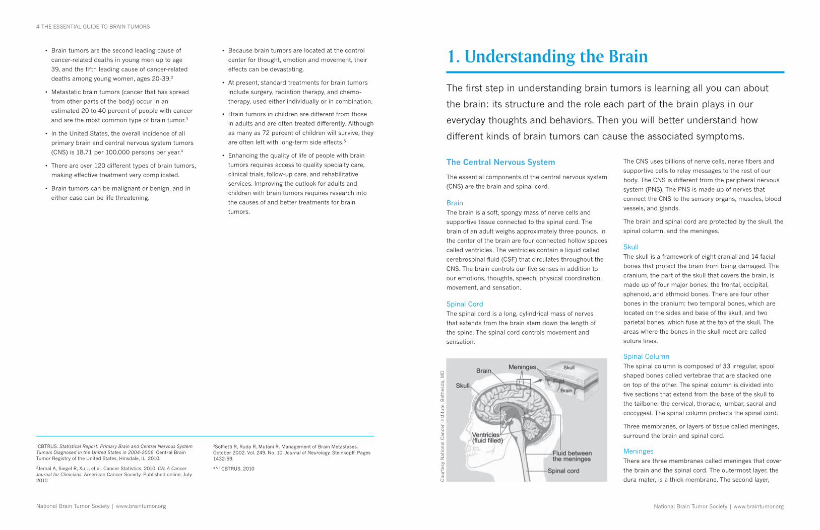
The Central Nervous System
The essential components of the central nervous system
(CNS) are the brain and spinal cord.
BrainThe brain is a soft, spongy mass of nerve cells and
supportive tissue connected to the spinal cord. The
brain of an adult weighs approximately three pounds. In
the center of the brain are four connected hollow spaces
called ventricles. The ventricles contain a liquid called
cerebrospinal fluid (CSF) that circulates throughout the
CNS. The brain controls our five senses in addition to
our emotions, thoughts, speech, physical coordination,
movement, and sensation.
Spinal CordThe spinal cord is a long, cylin drical mass of nerves
that extends from the brain stem down the length of
the spine. The spinal cord controls movement and
sensation.
• Brain tumors are the second leading cause of
cancer-related deaths in young men up to age
39, and the fifth leading cause of cancer-related
deaths among young women, ages 20-39.2
• Metastatic brain tumors (cancer that has spread
from other parts of the body) occur in an
estimated 20 to 40 percent of people with cancer
and are the most common type of brain tumor.3
• In the United States, the overall incidence of all
primary brain and central nervous system tumors
(CNS) is 18.71 per 100,000 persons per year.4
• There are over 120 different types of brain tumors,
making effective treatment very complicated.
• Brain tumors can be malignant or benign, and in
either case can be life threatening.
The first step in understanding brain tumors is learning all you can about
the brain: its structure and the role each part of the brain plays in our
everyday thoughts and behaviors. Then you will better understand how
different kinds of brain tumors can cause the associated symptoms.
Cou
rtes
y N
atio
nal
Can
cer
Inst
itute
, Bet
hes
da,
MD
1. Understanding the Brain
The CNS uses billions of nerve cells, nerve fibers and
supportive cells to relay messages to the rest of our
body. The CNS is different from the peripheral nervous
system (PNS). The PNS is made up of nerves that
connect the CNS to the sensory organs, muscles, blood
vessels, and glands.
The brain and spinal cord are protected by the skull, the
spinal column, and the meninges.
Skull The skull is a framework of eight cranial and 14 facial
bones that protect the brain from being damaged. The
cranium, the part of the skull that covers the brain, is
made up of four major bones: the frontal, occipital,
sphenoid, and ethmoid bones. There are four other
bones in the cranium: two temporal bones, which are
located on the sides and base of the skull, and two
parietal bones, which fuse at the top of the skull. The
areas where the bones in the skull meet are called
suture lines.
Spinal Column The spinal column is composed of 33 irregular, spool
shaped bones called vertebrae that are stacked one
on top of the other. The spinal column is divided into
five sections that extend from the base of the skull to
the tailbone: the cervical, thoracic, lumbar, sacral and
coccygeal. The spinal column protects the spinal cord.
Three membranes, or layers of tissue called meninges,
surround the brain and spinal cord.
Meninges There are three membranes called meninges that cover
the brain and the spinal cord. The outermost layer, the
dura mater, is a thick membrane. The second layer,
1CBTRUS. Statistical Report: Primary Brain and Central Nervous System Tumors Diagnosed in the United States in 2004-2006. Central Brain Tumor Registry of the United States, Hinsdale, IL, 2010.
2Jemal A, Siegel R, Xu J, et al. Cancer Statistics, 2010. CA: A Cancer Journal for Clinicians. American Cancer Society. Published online, July 2010.
• Because brain tumors are located at the control
center for thought, emotion and movement, their
effects can be devastating.
• At present, standard treatments for brain tumors
include surgery, radiation therapy, and chemo-
therapy, used either individually or in combination.
• Brain tumors in children are different from those
in adults and are often treated differently. Although
as many as 72 percent of children will survive, they
are often left with long-term side effects.5
• Enhancing the quality of life of people with brain
tumors requires access to quality specialty care,
clinical trials, follow-up care, and rehabilitative
services. Improving the outlook for adults and
children with brain tumors requires research into
the causes of and better treatments for brain
tumors.
3Soffietti R, Ruda R, Mutani R. Management of Brain Metastases. October 2002. Vol. 249, No. 10. Journal of Neurology. Steinkopff. Pages 1432-59.
4 & 5 CBTRUS, 2010

National Brain Tumor Society | www.braintumor.org National Brain Tumor Society | www.braintumor.org
6 THE ESSENTIAL GUIDE TO BRAIN TUMORS UNDERSTANDING THE BRAIN 7
called the arachnoid, and the third layer, called the pia
mater, are thin membranes.
There are three spaces between the layers of the
meninges. The space between the skull and dura mater
is called the epidural space. The space between the
dura mater and the arachnoid is called the subdural
space. The space between the arachnoid and the pia
mater is called the subarachnoid space.
VentriclesThe ventricles are four connected, fluid-filled cavities
located in the center of the brain. The ventricles contain
the choroid plexus, structures that produce cerebrospi-
nal fluid.
Cerebrospinal Fluid (CSF) Cerebrospinal fluid is a clear liquid that surrounds the
brain and spinal cord. It cushions and protects them
against injury. CSF circulates through the four ventricles
and the subarachnoid space. The CNS has a closed
cir culatory system that drains into the bloodstream.
The Sections of the Brain
The brain is divided into sections, each of which
controls a distinct aspect of human movement and
be havior. A brain tumor can affect function (movement
and/or behavior) depending on where in the brain the
tumor is located (see Brain Structures and Their Func-
tions, page 9).
CerebrumThe cerebrum is the largest area of the brain. It has
two sections called the right and left hemispheres. The
right cerebral hemisphere typically controls the left
side of the body, whereas the left cerebral hemisphere
controls the right side of the body. Each hemisphere
is further divided into four sections called lobes: the
frontal, parietal, temporal and occipital lobes. Each lobe
controls different behaviors and sections of the body.
The outer layer of the brain is called the cortex. It is
made up of bodies of nerve cells known as gray matter.
Much of the brain’s activities occur in the gray matter.
The internal layers of the cerebrum are made up of
nerve fibers called axons or white matter. The white
matter contains nerve fibers that allow communication
The Internal Nerve Structures of the Brain
Thalamus
The thalamus is a pair of egg-shaped masses of gray
matter located in the center of the two hemispheres,
above the hypothal amus. The thalamus acts as a path-
way for most of the messages that pass to and from the
brain. It also is involved in our conscious awareness of
pain, focusing of attention, certain aspects of speech/
language, memory, motor and sensory functions, and
the sleep/wake cycle.
HypothalamusThe hypothalamus, located in the center of the brain,
regulates automatic body activity such as heart rate,
temperature, thirst, appetite, sleeping patterns, growth
hormone, and physical expressions of emotions such as
blushing, dry mouth, and sweating.
Pituitary GlandThe pituitary gland, also called the hypophysis, is found
at the part of the brain between and behind the eyes. It
is connected to the hypothalamus. The hypothalamus
transmits messages to the pituitary gland, telling it
to secrete the hormones that regulate growth, blood
pressure, the thyroid, and gender-related functions (i.e.
testosterone secretion, menstruation, and lactation). The
pituitary gland also produces a hormone that controls
Illustration: Laurel V. Schaubert Courtesy of: Charles B. Wilson, MD
between the brain and various parts of the body.
The cerebrum also houses many internal nerve struc-
tures, such as the thalamus, hypothalamus and pituitary
gland. These structures are responsible for processing
different messages being sent to the brain and for send-
ing messages from the brain to other parts of the body.
Frontal Lobes The frontal lobes make up the front portion of the
cerebral hemisphere. The frontal lobes control many
of the brain’s activities including attention, abstract
thought, problem solving, reasoning, judgment, initiative,
inhibition, memory, parts of speech, moods, major body
movements, and bowel and bladder control.
Parietal Lobes The parietal lobes are in the upper central portion of
the cerebral hemispheres. The parietal lobes process all
messages being sent to and from the brain regarding
physical sensations. The parietal lobes are responsible
for interpreting the meaning of physical sensations to
determine such factors as size, shape, weight, texture
and consistency. They interpret spatial orientation and
how we are aware of the parts of our own body. The
parietal lobes also help us to make calculations, read
and write.
Temporal Lobes The temporal lobes form the lower portion of the
cerebral hemispheres. The temporal lobes manage
most auditory activities in the brain by translating words
into meaning. There is also a small, important section
of the temporal lobe that controls the brain’s ability to
form long-term memory patterns. The left temporal
lobe controls language comprehension in most people.
For this reason, the left temporal lobe is considered the
dominant lobe.
Occipital Lobes The occipital lobes are in the back portion of the cere-
bral hemispheres. The occipital lobes control vision. The
right occipital lobe processes what is seen out of the left
field of vision, and the left occipital lobe processes what
is seen out of the right field of vision.
the rate that water is secreted into the urine. This, in
effect, controls the amount of water in the body.
Brain Stem The brain stem, located at the base of the brain,
includes three parts: midbrain, pons, and medulla
oblongata. The brain stem contains the 12 cranial
nerves, which control hearing, vision, sense of smell,
and balance. The brain stem also contains pathways
going from the spinal cord to the brain for messages
related to movement and the senses. In addition, the
brain stem controls involuntary functions, including
breathing and heartbeat and our sleep/wake cycle. All
functions controlled by the cerebrum pass through the
brain stem.
CerebellumThe cerebellum, located behind the brain stem, has
many connections to the brain and the spinal cord.
The cerebellum is responsible for coordinating muscle
groups and controlling small movements and balance.
Corpus CallosumThe corpus callosum connects the left side of the brain
to the right side of the brain. It is located in the center of
the brain, surrounded by the cerebrum.
Cross-Section of Brain

National Brain Tumor Society | www.braintumor.org National Brain Tumor Society | www.braintumor.org
DIAGNOSING A BRAIN TUMOR 9
Brain Tumor Symptoms
A brain tumor can block the flow of cerebrospinal fluid
(CSF) between the ventricles, causing a buildup of CSF
and swelling, called brain edema. Edema can lead to
symptoms including headaches, seizures, or focal
deficits. Focal deficits include damage to sensory or
movement abilities, problems in the ability to process
information, person ality changes, and speech disorders.
A tumor of the spinal cord can block the communication
between the brain and nerves through out the body.
This can lead to problems with movement or physical
sensation.
The most common symptoms include:
• Headaches, which can be most severe in the morning
• Seizures or convulsions
• Difficulty thinking, speaking, or finding words
• Personality changes
• Weakness or paralysis in one part or one side of the body
• Loss of balance
• Vision changes
• Nausea or vomiting
• Confusion and disorientation
The doctor will perform a physical exam to detect the
signs and symptoms associated with a brain tumor.
This exam is called a neurological examination.
• Reflexes
• Balance and coordination
• Long- and short-term memory
• Judgment and reasoning
• Muscle strength
• Motor skills
• Gait
• Sensation
• Language
• Calculation
If responses to the exam are not normal, the doctor may
order a brain scan or refer the patient to a neurologist
or neurosurgeon, who will then order a scan.
Scans and Imaging Techniques
A scan is a picture of the internal structures in the
brain. A specialized machine takes a scan in much the
same way a digital camera takes a photograph. Using
computer technology, a scan compiles an image of the
brain by photographing it from various angles.
Some types of scans use a contrast agent (or contrast
dye), or a ferromagnetic substance such as gadolinium.
This is injected into a vein and flows into brain tissue.
Abnormal or diseased brain tissue absorbs more dye
2. Diagnosing a Brain Tumor
A brain tumor takes up space within the skull and interferes with normal
brain activity. A tumor can cause damage by increasing pressure in the
brain, shifting the brain or pushing against the skull, and invading and
damaging nerves and healthy brain tissue. The location of a brain tumor
influences the type of symptoms that occur. This is because different
functions are controlled by different parts of the brain.
Neurological Examination
A neurological examination tests a person’s physical
and mental functions. The doctor will begin by taking a
medical history and asking questions about:
• How the patient feels at the present time
• When the patient first noticed symptoms
• How often the symptoms occur
• Whether the symptoms are worsening
• The intensity and location of headaches
• Personality changes
• Changes in sense of touch, taste, smell, vision, or
hearing
• What procedures or tests have already been
performed
• What treatments or interventions have been
prescribed
After asking questions to de termine these conditions,
the doctor will conduct a series of tests to measure the
function of the patient’s nervous system, and physical
and mental alertness. This part of the exam includes
checking the following functions:
• Eye movement and pupil reaction
• Hearing
• Smell
• Tongue movement and sensation
than normal, healthy tissue. The contrast agent allows
the doctor to see the difference between normal and
abnormal brain tissue.
Types of Imaging TechniquesThanks to modern technology, several types of imaging
scans are available to help diagnose brain tumors. Each
type of scan is effective in capturing a different part and
function of the brain. The patient may have more than
one scan or different kinds of scans depending on the
type and location of the tumor.
The most commonly used scan for detecting brain
tumors is magnetic resonance imaging (MRI). There are
two types of MRI scanning equipment: standard and
open.
Magnetic Resonance Imaging (MRI) An MRI is the standard imaging technique for suspected
brain tumors. An MRI is a scanning device that uses
magnetic fields and computers to capture images of the
brain on film. It does not use x-rays. It provides pictures
from various planes, which permits doctors to create a
three-dimensional image of the tumor. The MRI detects
signals emitted from normal and abnormal tissue,
providing clear images of most tumors.
To prepare for a standard MRI scan, the patient will be
asked to lie down on a long table, which slides through
a long, cylindrical tube with a narrow opening. Although
there is enough room for the patient’s body inside the
Frontal lobe
•movement •initiative•reasoning •inhibition•behavior •mood•memory•personality•planning•decisionmaking•judgment
Temporal lobe
•languagecomprehension•behavior•memory•hearing•emotions
Pituitary gland
•hormones•growth•fertility
Brain stem
•breathing•bloodpressure•heartbeat•swallowing
Cerebellum
•balance•coordination•finemusclecontrol
Occipital lobe
•vision
Parietal lobe
•tellingrightfromleft•calculations•sensations•reading•writing
Brain Structures and Their Functions

National Brain Tumor Society | www.braintumor.org National Brain Tumor Society | www.braintumor.org
10 THE ESSENTIAL GUIDE TO BRAIN TUMORS DIAGNOSING A BRAIN TUMOR 11
speech, movement, and sensation. This information is
important in planning surgery, radi ation therapy, or other
treatments.
Magnetic Resonance Spectroscopy (MRI Spect or MRS)Magnetic Resonance Spectroscopy is a type of MRI that
measures the levels of metabolites in the body. Metabo-
lites are chemicals produced by living cells. The activity
of metabolites in tumors is different from that of normal
tissue. An MRS can detect patterns of activity that may
help diagnose the type of tumor and evaluate tumor
response to therapies. The types of metabolites com-
monly measured with an MRS are choline, lactate, and
N-Acetyl-Aspartate (NAA). An MRS may also be used
to determine the grade of a tumor and to distinguish
active recurrent tumor cells from dead cells destroyed
by radiation treatment, called radiation necrosis.
Other types of imaging techniques may also be used to
help diagnose and treat brain tumors.
Perfusion MRIPerfusion MRI uses contrast agents to examine the flow
of blood into the tissues. In some institutions, perfusion
MRI is used to grade certain types of tumors.
Diffusion and Diffusion-Tensor MRIThese forms of MRI measure the diffusion (random mo-
tion) of water in the tissues. Since the diffusion of water
along the nerve can be measured with this technique, it
can be used in surgical planning to avoid injuring nerve
bundles (ganglia) that have been shifted or displaced.
cylinder, the patient will not be able to move around.
The scan takes approximately 15–45 minutes. During
the scan, the patient will hear loud banging sounds,
caused by the electronics within the machine. Patients
may request earplugs to reduce noise. Some people
feel claustrophobic during MRIs and ask for a sedative
beforehand to relax. Other people request an open MRI.
An open MRI machine does not have a cylinder, so the
patient is not enclosed. The procedure lasts approxi-
mately 45 minutes. There is some discussion among
doctors concerning the quality of the images of an open
MRI compared to the standard or closed MRI.
Brain tumor patients will usually have more than one
MRI procedure during the course of treatment. Here
are some tips to help make the procedure more
comfortable:
• Bring your favorite music CD
• Wear pants that have a comfortable, loose waist
• Wear short sleeves if you will be receiving a
contrast agent injection
• Cover your eyes with a towel
There are several types of MRI scans. These techniques
aid doctors before and during surgery in various ways.
Magnetic Resonance Angiography (MRA)Magnetic Resonance Angiography generates an image
of blood vessels and blood flow without the use of a
contrast agent. It is less invasive than an arteriogram,
a traditional blood-vessel exam that requires injecting
a contrast agent. MRA is used to find the presence
and position of the blood vessels leading to the tumor
and determine whether or not these vessels have been
displaced due to the tumor mass. This method is
normally used before surgery to remove a tumor with
a large supply of blood, or one that is in an area of the
brain that has a large number of blood vessels.
Contrast-Enhanced MRA (CE-MRA)Contrast-Enhanced MRA is a form of MRA that uses
contrast agents to look at the movement of blood
through the region of interest.
Flow Sensitive MRI (FS MRI)Flow Sensitive MRI shows the flow of CSF through the
ventricles and spinal cord. It is used when examining
tumors in the spinal cord and at the base of the skull. It
can also be helpful when removing tumors that cause
hy drocephalus. Hydrocephalus is a condition marked
by blockage of CSF pathways, leading to a buildup of
CSF in the skull.
Functional MRI (fMRI) Functional MRI provides instant images of brain activity
by tracking the use of oxygen in the brain as it occurs.
fMRI identifies the motor, sensory, and language
centers of the brain, called the eloquent cortex. fMRI
can help radiologists determine precisely which part of
the brain is handling critical functions such as thought,
Computed Tomography (CT or CAT Scan)Computed Tomography combines sophisticated x-ray
and computer technology. Unlike other medical imaging
techniques, CT has the ability to show a combination of
soft tissue, bone, and blood vessels. CT is effective at ex-
amining bone and tissue calcification and hemorrhage.
CT images can determine some types of tumors, as well
as help detect swelling and bleeding. Usually, iodine is
the contrast agent used during a CT scan. Patients who
are allergic to iodine should inform their doctor before
having a CT scan.
In preparation for a CT scan, the patient lies on a long
table that slides into a round opening above his or her
head. The CT circles around the patient’s head, taking
pictures of the brain from different directions. The
scan makes a slight clicking sound as it moves. The
information gathered is then sent to a computer, which
translates it into pictures.
CT is a valuable diagnostic tool and its use has been
increasing rapidly. However, CT scans involve exposure
to ionizing radiation. This is a concern for people who
have multiple CT scans and for children, because they
are more sensitive to radiation than adults. It is wise
for patients who have had frequent x-ray exams and
parents of children who have brain tumors to keep a
record of their x-ray history. If a patient changes health
care providers, this information can help doctors make
informed decisions. Hospitals typically reduce the
dose of radiation given to children from adult levels to
levels based on the weight of the child. This minimizes
radiation over-exposure.
What is the difference between a neurologist
and a neurosurgeon?
A neurologist is a medical doctor who specializes
in diagnosing and treating illnesses in the brain
and spine. A neurosurgeon is a medical doctor
who performs surgery on the brain and spine.
What is a contrast agent?
A contrast agent or contrast dye is a substance
used to help detect a tumor by highlighting
abnormal tissue in a scan.
What is the difference between invasive and
non-invasive procedures?
An invasive procedure involves a puncture or
incision of the skin, or insertion of an instrument
or foreign material, such as a contrast dye, into
the body. A non-invasive pro cedure means that
there is no puncture of the skin or insertion of
foreign material into the body.
“ I concentrate on my family and all the wonderful
things we have been blessed with. I have come
to know the technicians in the MRI area quite
well, and when I enter the room, they hand me
a plastic rosary. They know that’s all I need to
make it through.”
–Glioblastoma survivor
An MRI may not be an option for
certain patients because the intense
magnetic fields can damage some
types of implanted medical devices.
Patients should advise the doctor
if they have a pacemaker, cardiac
monitor, surgical clip, or facial tattoos.
National Brain Tumor Society | www.braintumor.org National Brain Tumor Society | www.braintumor.org
12 THE ESSENTIAL GUIDE TO BRAIN TUMORS DIAGNOSING A BRAIN TUMOR 13
Positron Emission Tomography (PET)Positron Emission Tomography is a supplementary test
that can be used to gain additional information after an
MRI. PET scans provide a picture of the brain’s activity,
rather than its structure, by measuring the rate at which
a tumor absorbs glucose (a sugar). To do this, the pa-
tient is injected with glucose that has been labeled with
radioactive markers. Then the PET scan measures the
brain’s activity and sends this information to a computer,
which creates a live image. Doctors use PET scans to
see the difference between scar tissue, recurring tumor
cells, and cells destroyed by radiation treatment. PET
is also used during brain mapping procedures (see
chapter 5). PET requires using highly complex, expensive
equipment and can produce results that are difficult to
interpret. At this time, PET is not widely used to evaluate
recurrence of a brain tumor.
Angiography Angiography is a common technique used to outline
the position of blood vessels in the brain. A catheter
is placed in the groin and fed up to carotid or vertebral
arteries in the brain. A contrast dye is injected into the
catheter, and x-rays follow its path through the blood
vessels of the brain.
Biopsy
Often used in combination with a scan of the brain,
biopsies are the most accurate test for identifying
the type and grade of a tumor. A biopsy is a surgical
procedure in which a small sample of tissue is taken
from the tumor site and examined under a microscope.
The biopsy provides information on types of abnormal
cells present in the tumor. The results help the doctor
diagnose the type of tumor.
There are two kinds of biopsy procedures: an open and
a closed biopsy.
Open BiopsyAn open biopsy is done during a craniotomy. A crani-
otomy is a surgical procedure that involves removing
a piece of the skull in order to get access to the brain.
After the tumor is debulked or resected, the bone
is usually put back into place. The patient is under
anesthesia during this procedure.
Closed BiopsyA closed biopsy (also called stereo tactic or needle
biopsy) is performed when the surgeon wants to avoid
removing healthy tissue from the area surrounding
the tumor, or when the tumor is in an area of the brain
that is difficult to reach. The patient may be given a local
anesthetic to numb the skin and a sedative medication
and remain awake during the procedure. Or, the patient
may undergo a general anesthesia, where he or she will
be unconscious during the procedure. Guided by a CT
or MRI that is performed prior to the procedure, the
surgeon drills a small hole into the skull and passes a
narrow, hol low needle through the hole into the tumor
to remove a sample of tissue.
Once a sample is obtained, a pathologist will examine
the tissue under a microscope. Further tests or analysis
may be performed on the tumor tissue. Then the
pathologist will write a pathology report.
Pathology Report A pathology report contains the analysis of brain tissue
taken at the time of a craniotomy or needle biopsy. It
provides the information to make a diagnosis of the
tumor type. Sometimes the pathologist may not be
able to make an exact diagnosis. This may be because
more than one grade of tumor cells exists within the
same tumor. If cells of only one grade are removed and
classified during a biopsy, it is possible that the tumor
grade will be misdiagnosed. This is called a sampling
error. In some cases, the tissue may be sent to another
institution for additional input.
Should a person consider getting a second opinion after receiving an initial diagnosis?
Yes. A patient can get a second opinion to confirm the initial diagnosis and to compare the suggested courses
of treatment. Some insurance companies require that a patient get a second opinion before undergoing major
therapy. A second opinion should be obtained as soon as possible to avoid delaying treatment. Patients can ask
their family physician to recommend other doctors.
Important Points About Diagnostic Tests
• Some people may have an allergic reaction to
iodine, the contrast agent most commonly used
for CT scanning. Allergic reactions can
include rashes, a warm sensation, or in rare
cases, difficulty breathing. If you know you are
allergic to iodine, tell your doctor.
• Gadolinium, the contrast agent used with an MRI,
may cause temporary headaches but has no
other known side effects.
• Because an MRI uses magnetic fields, people
who have metal implanted in their body in any
form (including tattoos) should let the doctor
know about it before scheduling the procedure.
• Medication can be given to help relieve the
symptoms of anxiety or claustrophobia when
having an MRI. Patients should let the doctor
know their concerns before scheduling an MRI.
Brain tumors grow because their cells divide to make
more cells. The process of cell division is called
proliferation. The per centage of cells that are actively
dividing within the tumor are measured by a test called
the MIB-1 labeling index. If no cells are actively dividing,
the MIB-1 labeling index (LI) would be expected to be 0
percent. An MIB-1 LI of greater than 30 percent indi-
cates that tumor cells are growing very rapidly. Several
studies have shown that gliomas with an MIB-1 LI of 10
percent or less have a more favorable prognosis than
those higher than 10 percent.
National Brain Tumor Society | www.braintumor.org
KNOWN AND POSSIBLE CAUSES 15
Many brain tumors can be removed. Others can be kept
under control for many years with treatment. Some
brain tumor survivors don’t experience recurrence
while others do. For this reason, most brain tumors
need to be monitored by periodic scans throughout the
survivor’s lifetime. The frequency of scans decreases as
time goes by.
Survival is strongly related to a person’s age and tumor
type. On average, about 35 percent of people diagnosed
with a malignant brain tumor in the U.S. will survive
for five years. However, the five-year survival rate in
children under age 20 with brain and other nervous
system cancers is about 72 percent, much higher than
average. People with glioblastoma consistently have the
poorest survival in all age groups. For all people with
meningiomas (including benign, atypical, and malig-
nant), the five-year survival rate is 69 percent, but it is
slightly lower for people with malignant meningiomas
(55 percent).
Survival rates are strongly related to several factors.
These factors include:
• Age. Among all brain tumor patients, children from
five to 20 years of age and young adults from 20
to 44 years of age have higher survival rates than
children under five and adults aged 45 or older.
• Tumor type. Brain tumors that grow slowly have a
better prognosis than fast-growing tumors. Tumor
progression from benign to malignant negatively
affects survival.
mental hazards are suspected, although no major risk
factors have been clearly identified.
Another cause may have been a change in brain tumor
classification in the mid 1980s. Some gliomas previ-
ously considered benign were changed to the status
of malignant. This may have led to an increase in the
number of brain tumors being registered. (Previous to
the year 2002, benign brain tumors were not counted
in cancer registries. The Benign Brain Tumor Cancer
Registries Amendment, passed in 2002, now mandates
that cancer registries record all brain tumors regard-
less of their type.)
Geography and EthnicityThere is a lot of variation in the trends of brain tumor
patients along geographic and ethnic lines. Access to
health care is one influential factor. Reported rates for
primary malignant brain tumors tend to be higher in
countries with more accessible and highly developed
medical care, such as Northern Europe and the United
States. Countries such as India and the Philippines have
the lowest reported rates. This would seem to indicate
that the difference is due to better diagnosis and report-
ing in more developed countries.
However, there is some evidence that cultural, ethnic, or
geographic differences do play a role in the disease.
The incidence rate for malignant brain tumors in
Japan is less than half that in Northern Europe. In the
US, gliomas affect more whites than blacks, but the
incidence of meningioma is nearly equal among blacks
and whites. These differences cannot be attributed only
to differences in access to health care or in diagnostic
practices. These and other findings suggest that further
research about genetic differences needs to be done to
explain variations in brain tumor incidence rates among
different ethnic groups.
Survival and PrognosisWhen diagnosed with a brain tumor, one of the first
things a patient usually wants to know is “How long
will I live?” The answer is never certain, and we encour-
age patients and families not to focus on statistics.
No individual is a statistic; each person’s prognosis is
different.
These trends include age, gender, geography, environ-
mental influences, and behavioral patterns. Trends may
provide some insight into the possible causes of brain
tumors. This chapter will provide an overview of trends
found in studies of brain tumor patients.
Incidence Rates
According to the Central Brain Tumor Registry of the
United States (CBTRUS), 62,930 primary, benign and
malignant tumors of the brain and central nervous
system (CNS) are expected to be diagnosed in 2010.
About 18.71 per 100,000 persons per year in the U.S.
are diagnosed with a primary brain tumor each year,
and of those, about 7 per 100,000 are diagnosed with
a primary malignant brain tumor. The incidence rate of
primary tumors of children between ages 0 to 19 years
is approximately 5 cases per 100,000. Meningiomas
and gliomas are the most frequent types of primary
and other nervous system tumors, making up 34% and
32%, respectively.
Brain Tumor Trends
Age and Gender The average age that an adult is diagnosed with a
brain tumor is 57 years. The age and number of
children diagnosed with brain tumors is distributed
evenly between the ages of 0 and 19. Trends in age
vary depending on the type and location of the tumor.
This suggests that many different factors influence the
3. Known and Possible Causes
Each year over 190,000 people in the United States and 10,000 people
in Canada are diagnosed with a primary or metastatic (secondary) brain
tumor. Many studies are looking into the causes of brain tumors, but the
results have not been conclusive. Although the causes of brain tumors are
not certain, there are many trends among people who get them.
* These figures do not include metastatic brain tumors, which are the most common type of brain tumor.
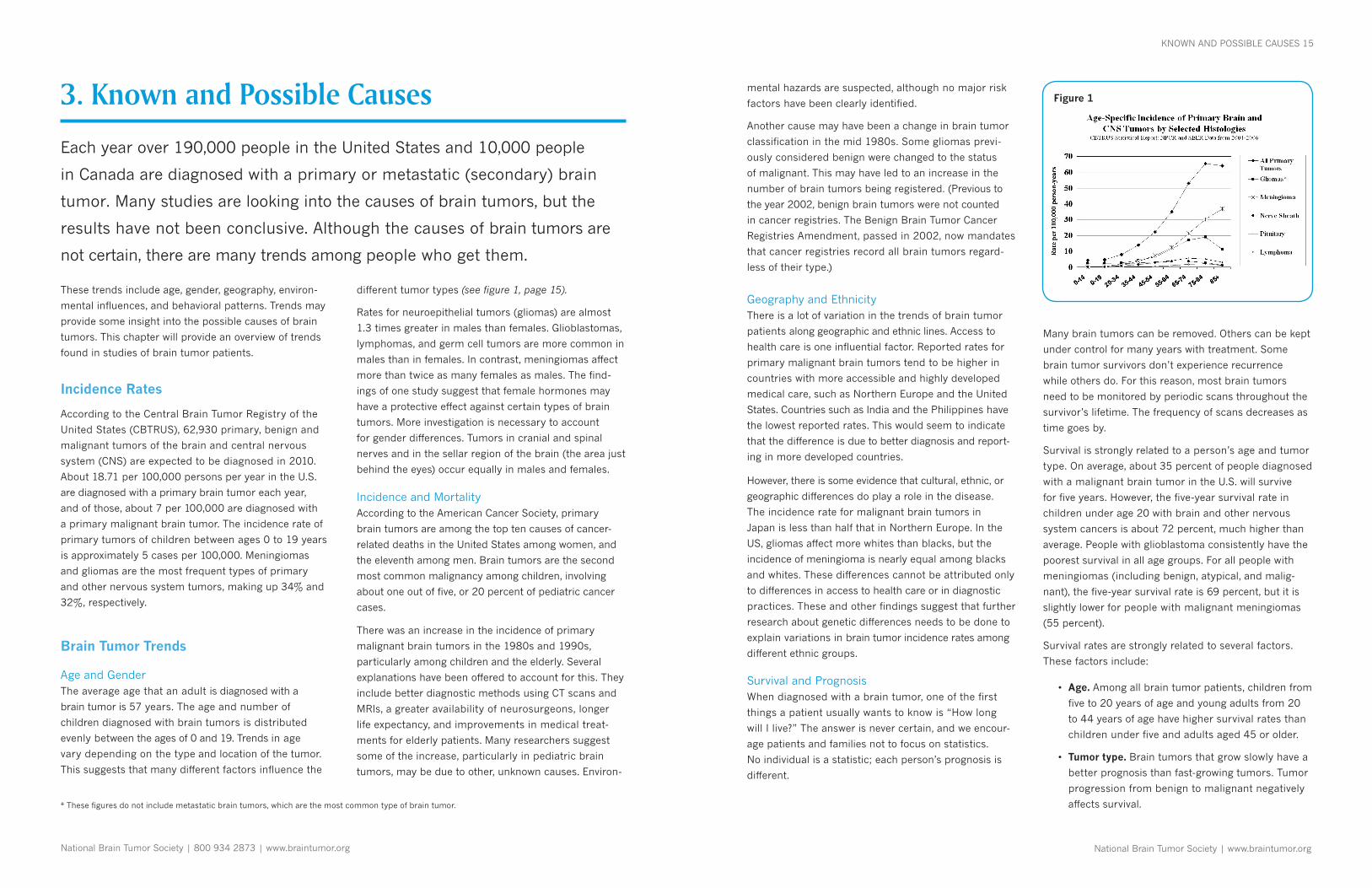
different tumor types (see figure 1, page 15).
Rates for neuroepi thelial tumors (gliomas) are almost
1.3 times greater in males than females. Glioblastomas,
lymphomas, and germ cell tumors are more common in
males than in females. In contrast, meningiomas affect
more than twice as many females as males. The find-
ings of one study suggest that female hormones may
have a protective effect against certain types of brain
tumors. More investigation is nec es sary to account
for gender differences. Tumors in cranial and spinal
nerves and in the sellar region of the brain (the area just
behind the eyes) occur equally in males and females.
Incidence and MortalityAccording to the American Cancer Society, primary
brain tumors are among the top ten causes of cancer-
related deaths in the United States among women, and
the eleventh among men. Brain tumors are the second
most common malignancy among children, involving
about one out of five, or 20 percent of pediatric cancer
cases.
There was an increase in the incidence of primary
malignant brain tumors in the 1980s and 1990s,
particularly among children and the elderly. Several
explanations have been offered to account for this. They
include better diagnostic methods using CT scans and
MRIs, a greater availability of neurosurgeons, longer
life expectancy, and improvements in medical treat-
ments for elderly patients. Many researchers suggest
some of the increase, particularly in pediatric brain
tumors, may be due to other, unknown causes. Environ-
National Brain Tumor Society | 800 934 2873 | www.braintumor.org
Figure 1

National Brain Tumor Society | www.braintumor.org National Brain Tumor Society | www.braintumor.org
16 THE ESSENTIAL GUIDE TO BRAIN TUMORS KNOWN AND POSSIBLE CAUSES 17
shingles) may play a role in preventing brain tumors.
More study is needed.
Head Injuries and SeizuresSerious head trauma has long been suspected as a
cause of brain tumors. In fact, studies show a positive
correlation between head trauma and meningioma, but
a negative link to glioma. A history of seizures has been
consistently associated with brain tumors, but since
brain tumors are known to cause seizures, it is unclear if
seizures and/or seizure medication can increase tumor
risk. As for drugs and med i ca tions, there have been few
studies of any links to adult brain tumors.
DietIn animal studies, certain chemical substances known
as N-nitroso compounds have been clearly identified
as carcinogenic (causing cancer) to the nervous system.
N-nitroso compounds are present in cured meats
(nitrites), cigarette smoke, cosmetics, and many other
sources. These compounds are also produced inside
the human body as the digestive process breaks down
food (including vegetables) and drugs. Given the great
amount of exposure to these compounds and the variety
of sources, it is extremely difficult to determine any
individual’s lifetime exposure.
Some studies of diet and vitamin supplementation seem
to indicate that dietary N-nitroso compounds might
influence the risk of both pediatric and adult brain
tumors. Researchers have observed in some studies
that brain tumor patients (or their mothers) have
generally consumed more cured foods than control
groups. Avoiding cured food and eating more fruits and
vegetables that are high in anti oxidant vitamins may
lessen the risk of developing cancer.
Chemicals in the Workplace and the HomeSome workers are exposed to carcinogenic or toxic
substances in the workplace. Researchers have at-
tempted to pinpoint links to brain tumors, but gather-
ing evidence is difficult. Workers are rarely exposed to
one single chemical, and certain chemicals probably
interact with others to increase or decrease risk. There-
fore, researchers have been unable to make any definite
links between brain tumors and specific chemicals, even
those known to be carcinogenic.
• Location. This determines the type of symptoms
a person may have, whether or not the tumor can
be surgically removed, and how much of it can be
removed.
• Treatment. Differences in the type of treatment
and the patient’s response to it also affect survival.
At present, brain tumors are treated by surgery,
radiation therapy and chemotherapy, used either
individually or in combination.
• Functional status. People who are more functional
at time of diagnosis do better than those who are
more disabled. The Karnofsky Performance Scale
(KPS) is a means of rating the patient’s overall
functioning level. KPS scores range from 100 to
0, where 100 represents normal functioning and 0
indicates the end of life.
Known and Possible Causes
To date, the only proven causes of brain tumors are
rare hereditary syndromes, therapeutic ra diation, and
immunosuppression that gives rise to brain lymphomas.
Yet these causes account for only a small percentage
of cases. Although a lot of research has been done on
the potential risk factors for primary brain tumors, most
of the findings are uncertain. There is little agreement
about the nature and extent of the risk factors. It is dif-
ficult to measure amounts of exposure to suspected risk
factors and to define latency periods (the amount of
time it takes for a brain tumor to develop after exposure
to a risk factor). The large number of different tumor
types (more than 120) is another obstacle. However,
studies have examined and continue to examine many
factors that may cause brain tumors. Here are some of
the findings:
Hereditary and Genetic InfluencesSome hereditary syndromes, such as tuberous sclerosis,
von Hippel-Lindau syndrome and neuro fibroma tosis
types 1 and 2, are associated with a higher risk of de-
veloping brain tumors. Yet “genetic predisposition,” as it
is called, probably accounts for less than five percent of
brain tumors. Other people may have what researchers
call a “genetic suscep tibility” for developing cancer.
Genetic susceptibility means their bodies may not be
as efficient at processing certain substances, removing
carcinogens, or repairing damaged DNA. When exposed
to toxic agents in the environment, they may more easily
develop cancer. It seems likely that the majority of brain
tumors are linked to interactions between genes and tox-
ins in the environment, because such a small percentage
of brain tumors are linked to heredity.
Molecular studies have found deletions (missing parts)
or mu tations (defects) of crucial genes that control the
cell cycle. These are suspected to play a role in forming
brain tumors. Many patterns of deletions and mutations
have been identified in some tumor types. There is still
much work to be done to systematically identify the
molecular alterations in primary brain tumors and to
develop methods to treat them.
Ionizing RadiationTreatment of disease with therapeutic ionizing radiation
(including x-rays) is a strong risk factor for brain tumors.
Relatively low doses of radiation used to treat tinea
capitis (ringworm) and skin hemangioma in children
or infants have been associated with relative risks for
nerve sheath tumors, meningioma, and glioma. One
study showed a high rate of prior therapeutic irradiation
among patients with glioblastoma. Another reported
an increased risk of glioma or other brain tumors
in patients who had undergone irradiation for acute
lymphoblastic leukemia as children. Second primary
brain tumors also occur more frequently than expected
especially among patients treated with radiation therapy.
Exposure to Infections, Viruses, and AllergensSeveral types of viruses have been shown to cause
brain tumors in experimental animal studies. Since it
is so difficult to design meaningful studies on humans,
the topic has received little attention. There have been
findings which raise the possibility that certain allergies
and common infections (including chicken pox and
There has been compelling evidence that workers in the
production of synthetic rubber and polyvinyl chloride,
and workers in certain parts of the petrochemical,
petroleum, and oil industries are at greater risk for
developing brain tumors. However, studies are contra-
dictory and inconclusive. Increased risk was not found
in adults who work in manufacturing of pesticides or
fertilizers. However, four out of five studies of pesticide
applicators have shown there is an increased risk for
these professionals.
It is possible that parents exposed to carcinogens in the
workplace might possibly increase the risk of cancer
in their children. A mother’s exposures might have a
direct impact on the developing fetus, and a father’s
exposures before conception might damage his DNA.
Higher risks of pediatric brain tumors were reported for
fathers working with, or working in industries involving:
paper and pulp, solvents, painting, printing and graphic
arts, oil or chemical refining, farming, metallurgy, and
air and space. One theory is that chemical carcinogens
from the workplace might remain on a parent’s skin
or clothing. When the parent goes home, his or her
children might then be exposed to the carcinogens.
However, there is no conclusive proof of this.
Studies of chemical exposures in the home have
focused on the role of pre- and postnatal pesticide ex-
posures in pediatric brain tumors. A recent large study
found increased risk in children exposed before birth
to flea and tick pesticides. The authors of the study
have urged further investigation of pesticide exposures
during pregnancy.
Cellular Telephones and Radio Frequency (RF) Electromagnetic FieldsWith the expansion of wireless communication technolo-
gies, radio frequency (RF) exposure is an important
concern. It is important not to confuse RF fields with
ionizing radiation, such as x-rays or gamma rays. Unlike
ionizing ra diation, RF fields cannot cause ionization or
radioactivity in the body. Because of this, RF fields are
called non-ionizing.
Concern over possible health effects of using cellular
telephones has prompted studies looking at the relation
between cell phone usage and an increased risk of
brain tumors. The results of several studies, including
“ Doctors may prescribe a regimen that may be
effective, but they don’t prescribe hope. I repeat:
it’s not science that has made a quantum leap
in brain tumor survival, it’s the brain tumor
patients themselves. They have become
empowered.”
–Neuro-oncologist

National Brain Tumor Society | www.braintumor.org National Brain Tumor Society | www.braintumor.org
18 THE ESSENTIAL GUIDE TO BRAIN TUMORS KNOWN AND POSSIBLE CAUSES 19
a recent large multinational study, suggest that there
is no association. However, it may be important to
continue research in this area because cell phone usage
is becoming increasingly common. Many studies were
conducted during a time when analog phones were the
main type of cell phone, as compared to digital phones
today. Total amount of phone use was lower, and the
number of cell phone users was fewer then. Moreover,
long-term studies are probably needed because some
brain tumors may take a long time to develop.
The World Health Organization (WHO) suggests that
individuals who are concerned about potential dangers
of cell phone use may choose to do the following: limit
their own or their children’s RF exposure by limiting the
length of calls, or use hands-free devices (headsets) to
keep mobile phones away from the head and body.
Air PollutionCertain toxic air pollutants are known to cause cancer
in humans. Ultra fine particles, including diesel soot and
other combustion products, are able to lodge deep
in human lungs and even enter the bloodstream due
to their microscopic size. One study is investigating a
possible link between brain tumors and air pollution.
Direction for Future Studies
There is a growing interest in understanding the causes
of brain tumors. Progress in molecular research may
lead to identifying new types of tumors. Advances
in genetic research may shed light on what makes a
person suscep tible or resistant to developing a brain
tumor. Developing new technologies, improving tech-
niques for classification, using molecular markers more
often, and keeping better records of the diagnoses and
prognoses of primary brain tumors are all factors that
will help us come to a better understanding of brain
tumors and their causes. This knowledge could lead to
strategies for preventing brain tumors, determining who
might be sensitive to radiation therapy or certain drug
treatments, and improving the survival rate of people di-
agnosed with malignant tumors. Clinical trials, research
studies that involve people, are important means to
search for answers to these types of questions.
Further studies are needed to explain gender and
ethnic diff erences found among people diagnosed
with gliomas and menin giomas. Studies are needed to
find out why environmental factors and changes to DNA
might increase vulnerability to brain tumors, and to
help us understand the way that brain tumors develop.
There is hope that further research can lead to preven-
tion and a cure.
What causes cells to become tumors?
Scientists believe that primary brain tumors
develop when changes to the genetic makeup of
cells allow abnormal cells to escape destruction
by the immune system. Researchers also suspect
that carcinogens, or poisons, may damage DNA
in brain cells, leading to brain tumors. Studying
how alterations in cell development occur will
help to understand the causes of brain tumors
and to identify similarities between different
types of brain tumors.
Information for this chapter was adapted from the
article “Epidemi ology of Primary Brain Tumors: Current
Concepts and Review of the Literature,” published in
Neuro- Oncology, Volume 4, Issue 4, October 2002. The
article was written by the following researchers, and we
are very grateful for their assistance:
Mitchel S. Berger, MD, Department of Neurological
Surgery and Brain Tumor Research Center, University of
California, San Francisco, San Francisco, California
Melissa Bondy, PhD, Department of Epidemiology,
The University of Texas M. D. Anderson Cancer Center,
University of Texas, Houston, Texas
Terri Chew, MPH, Department of Epidemiology and Bio-
statistics and Brain Tumor Research Center, University
of California, San Francisco, San Francisco, California
Yuriko Minn, MS, Department of Neurology, Stanford
University, Stanford, California
Margaret Wrensch, PhD, Department of Epidemiology
and Biostatistics and Brain Tumor Research Center,
University of California, San Francisco, San Francisco,
California
This current edition incorporates updates from the
following sources:
Altekruse SF, Kosary CL, Krapcho M, et al., eds.
SEER Cancer Statistics Review, 1975-2007, National
Cancer Institute. Bethesda, MD, http://seer.cancer.gov/
csr/1975_2007/, based on November 2009 SEER data
submission, posted to the SEER web site, 2010.
Bondy ML, Scheurer ME, Malmer B, et al. Brain Tumor
Epidemiology: Consensus from the Brain Tumor
Epidemiology Consortium. October 2008. Vol. 113,
No. 7 Suppl. Cancer. American Cancer Society. Pages
1953-68.
CBTRUS. Statistical Report: Primary Brain and Central
Nervous System Tumors Diagnosed in the United States
in 2004-2006. Central Brain Tumor Registry of the
United States, Hinsdale, IL, 2010.
The INTERPHONE Study Group. Brain tumour risk in
relation to mobile telephone use: results of the INTER-
PHONE international case-control study. May 2010. Vol.
39, No. 3. International Journal of Epidemiology. Pages
675 - 694
Jemal A, Siegel R, Xu J, et al. Cancer Statistics, 2010.
CA: A Cancer Journal for Clinicians. American Cancer
Society. Published online, July 2010.
National Brain Tumor Society | www.braintumor.org National Brain Tumor Society | www.braintumor.org
BRAIN TUMOR TYPES 21
4. Brain Tumor Types
To determine the course of treatment, the type of brain tumor must first
be identified. This chapter discusses brain tumor types and how they are
classified. Under each tumor type, you will find a list of the symptoms
and treatment options that are typically prescribed. Many tumors share the
same symptoms and treatments. Treatment is based on the tumor type and
location. The three main treatment methods are surgery, radiation, and
chemotherapy.
Classifying Brain Tumors
There are over 120 different types of brain tumors. In
most cases, a brain tumor is named for the cell type
of origin. Some brain tumors are named according to
their location. Today, most medical institutions use the
World Health Organization (WHO) classification system
to identify brain tumors. The WHO classification, which
is used throughout this guide, classifies brain tumors
by cell origin and how the cells behave, from the least
aggressive (benign) to the most aggressive (malignant).
Some tumor types are assigned a grade, which signi-
fies the rate of growth. There are variations in grading
systems, depending on the tumor type. The classifica-
tion and grade of an individual tumor help predict its
likely behavior.
Although they may fall into a specific classification or
category, brain tumors are specific to each individual.
Brain tumors have vastly different characteristics and
patterns of growth due to the molecular makeup of
the individual tumor.
A series of criteria are used to make a diagnosis. One
important criterion is anaplasia, the manner in which
tumor cells grow with the loss of normal form or struc-
ture. The degree of anaplasia helps to forecast a tumor’s
growth potential. The most rapidly growing tumors have
the highest degree of anaplasia. Tumor cells that show a
lower degree of anaplasia are generally slow growing.
In addition to other criteria, some tumors are now
examined for their genetic traits to evaluate the nature
of the tumor. For example, certain patterns of mutations
or chromosomal defects have been identified in certain
tumor types.
For more information, visit www.braintumor.org.
Primary Brain Tumors
GLIAL TUMORS
There are two types of cells that make up the nervous system: neurons and neuroglia. Neurons send and receive
nerve messages. Neuroglia, otherwise known as glial cells, often surround the neurons. Glial cells play a supportive
role by nourishing, protecting and supporting neurons. There are six kinds of glial cells: oligodendrocytes, astrocytes,
ependymal cells, Schwann cells, microglia, and satellite cells.
A brain tumor that develops from glial cells is called a glioma. About one third of all primary brain and other nervous
system tumors form from glial cells. Gliomas tend to grow in the cerebral hemispheres, but may also occur in the
brain stem, optic nerves, spinal cord, and cerebellum.
Gliomas are divided into subgroups depending on the origin of the glial cells. The most common type of glioma is an
astrocytoma.
AstrocytomaAn astrocytoma develops from star-shaped glial cells
(astrocytes) that support nerve cells. These tumors can
be located anywhere in the brain, but the most common
location is in the frontal lobe. Astrocytomas are the most
common primary CNS tumor.
The physician, usually the neurosurgeon or neuro-
oncologist, will discuss the type and location of an
astrocytoma. The pathologist will assign it a grade.
Astrocytomas are generally classified as low or high
grade. Low-grade astrocytomas (grades I and II) are
slow growing. High-grade astrocytomas (grades III and
IV) grow more quickly. The main tumor type is listed for
each grade. There are additional tumor types in each of
these grades.
The WHO classification divides astrocytomas into four
grades (see following page for more detail):
• Grade I Pilocytic Astrocytoma
• Grade II Low-Grade Astrocytoma
• Grade III Anaplastic Astrocytoma
• Grade IV Glioblastoma (or GBM)
CharacteristicsThe characteristics of an astrocytoma vary depending
on the tumor’s grade and location. Most people are
functioning normally when diagnosed with a low-grade
astrocytoma. Symptoms tend to be subtle and may
take one to two years to diagnose. This is because the
brain can often adapt to a slow-growing tumor for a
period of time. High-grade tumors may present with
changes that are sudden and dramatic.
Symptoms• Headaches
• Seizures or convulsions
• Difficulty thinking or speaking
• Behavioral or cognitive changes (related to thinking,
reasoning, and memory)
• Weakness or paralysis in one part or one side of the
body
• Loss of balance
• Vision changes
• Nausea or vomiting
Primary brain tumors originate in the brain itself. Primary brain tumors usually do not spread from the brain to other
parts of the body. However, there are some exceptions to this rule.
Primary brain tumors are classified into two groups: glial tumors, which are called gliomas, and non-glial tumors.
Pediatric Brain Tumors
Some tumor types are more common in children
than in adults. The most common types of pedi-
atric tumors are medulloblastomas, low-grade as-
trocytomas, ependymomas, craniopharyngiomas,
and brain stem gliomas. Treatment for pediatric
brain tumors often involves surgery, radiation
and/or chemotherapy. Age is an important factor
in determining the course of treatment since
children’s brains are still developing. Survivors
of childhood tumors may experience late effects
of treatment, such as developmental delays and
learning or physical disability, and require regular
medical follow-up. Some children may require
accommodations at school; the medical team
and education professionals can help to address
school-related concerns.
National Brain Tumor Society | www.braintumor.org National Brain Tumor Society | www.braintumor.org
22 THE ESSENTIAL GUIDE TO BRAIN TUMORS BRAIN TUMOR TYPES 23
BRAIN STEM GLIOMA
Characteristics• Named for its location at the base of the brain
• Can range from low grade to high grade
• Occurs most often in children between three and ten
years of age, but can occur in adults
Symptoms • Headaches
• Nausea
• Speech or balance abnormalities
• Difficulty swallowing
• Weakness or numbness of the arms and/or legs
• Facial weakness
• Double vision
Symptoms can develop slowly and subtly and may go
unnoticed for months. In other cases, the symptoms
may arise abruptly. A sudden onset of symptoms tends
to occur with rapidly growing, high-grade tumors.
Treatment Surgery may not be an option because the brain stem
controls vital life functions and can easily be damaged.
Radiation therapy can reduce symptoms and help slow
the tumor’s growth. Low-grade brain stem gliomas can
have very long periods of remission.
EPENDyMOMA
Ependymal tumors begin in the ependyma, cells that line the passageways in the brain where CSF is produced and
stored. Ependymomas are classified as either supratentorial (in the cerebral hemispheres) or infratentorial (in the
back of the brain). Variations of this tumor type include subependymoma, subependymal giant-cell astrocytoma, and
malignant ependymoma. Ependymoblastoma, which occurs in infants and children under three years, is no longer
considered a subtype of ependymoma. For ependymoblastoma, see primitive neuroectodermal tumor (PNET).
Characteristics• Usually localized to one area of the brain
• Develops from cells that line the hollow cavities at
the bottom of the brain and the canal containing the
spinal cord
• Can be slow growing or fast growing
• May be located in the ventricles
• May block the ventricles, causing hydrocephalus
(water on the brain)
• Sometimes extends to the spinal cord
• Common among men and women in their 40s and
50s, and in children
• Occurrence peaks at age five and again at age 34
• Accounts for two percent of all brain tumors
Symptoms • Headaches
• Nausea
• Speech or balance abnormalities
• Difficulty swallowing
• Weakness or numbness of the arms and/or legs
• Facial weakness
• Double vision
TreatmentThe doctor will perform tests to determine if it has
spread to the spinal cord. Surgery followed by radia-
tion therapy is the usual course of treatment. A shunt
may be needed to treat hydrocephalus caused by
blockage of the ventricles.
ASTROCyTOMA GRADES
Pilocytic Astrocytoma (Grade I)This tumor is also known as a juvenile pilocytic astrocytoma, or by the initials JPA.
Characteristics• Slow growing, with relatively well-defined borders
• Grows in the cerebrum, optic nerve pathways, brain
stem and cerebellum
• Occurs most often in children and teens
• Accounts for two percent of all brain tumors
Treatment Surgery is the standard treatment. If the tumor
cannot be com pletely resected, radiation or chemo-
therapy may be given. Chemotherapy may be given
to very young children instead of radiation therapy
to avoid damage to the developing brain. Some of
these tumors can progress to a higher grade, so it is
important to be diligent about following up with the
medical team after treatment.
Low-Grade Astrocytoma (Grade II)
Characteristics• Slow growing
• Rarely spreads to other parts of the CNS
• Borders not well defined
• Common among men and women in their 20s–50s
Treatment Treatment depends on the size and location of the
tumor. The doctor will most likely perform a biopsy
or surgery to remove the tumor. Partial resections
or inoperable tumors may be treated with radiation.
Recurring tumors may require additional surgery,
radiation and/or chemotherapy.
Anaplastic Astrocytoma (Grade III)
Characteristics• Grows faster and more aggressively than grade II
astrocytomas
• Tumor cells are not uniform in appearance
• Invades neighboring tissue
• Common among men and women in their 30s–50s
• More common in men than women
• Accounts for two percent of all primary brain tumors
Treatment Treatment depends on the location of the tumor
and how far it has progressed. Surgery and radia-
tion therapy, with chemotherapy during or following
radiation, are the standard treatments. If surgery
is not an option, then the doctor may recommend
radiation and/or chemotherapy. Many clinical trials
using radiation, chemotherapy, or a combination
are available for initial and recurrent anaplastic
astrocytomas.
Glioblastoma (GBM) (Grade IV Astrocytoma)
Characteristics• Most invasive type of glial tumor
• Grows rapidly and commonly spreads to nearby tissue
• May be composed of several different kinds of cells
(i.e., astrocytes, oligodendrocytes)
• Includes distinct genetic subtypes
• May have evolved from a low-grade astrocytoma or an
oligodendroglioma
• Common among men and women in their 50s–70s
• More common in men than women
• Accounts for 17 percent of all primary brain tumors
Treatment Standard treatment is surgery followed by radiation
therapy. If surgery is not an option, the doctor may
administer ra diation therapy. Chemotherapy is
usually given during and after ra diation therapy or if
the tumor recurs. Many clinical trials (ex perimental
treatments) using radiation, chemotherapy, or a
combination are available for initial and recurrent
GBM.
What is the difference between benign and malignant tumors?
Benign tumors are slow-growing tumors that can be removed or destroyed if in an accessible location.
Malignant tumors (brain cancer) are rapidly growing tumors that invade or infiltrate and destroy normal brain
tissue. Tumors are graded to indicate how quickly they are growing. Low-grade tumors grow more slowly than
high-grade tumors. Benign or malignant tumors can recur, which means they grow back after treatment for the
initial tumor.
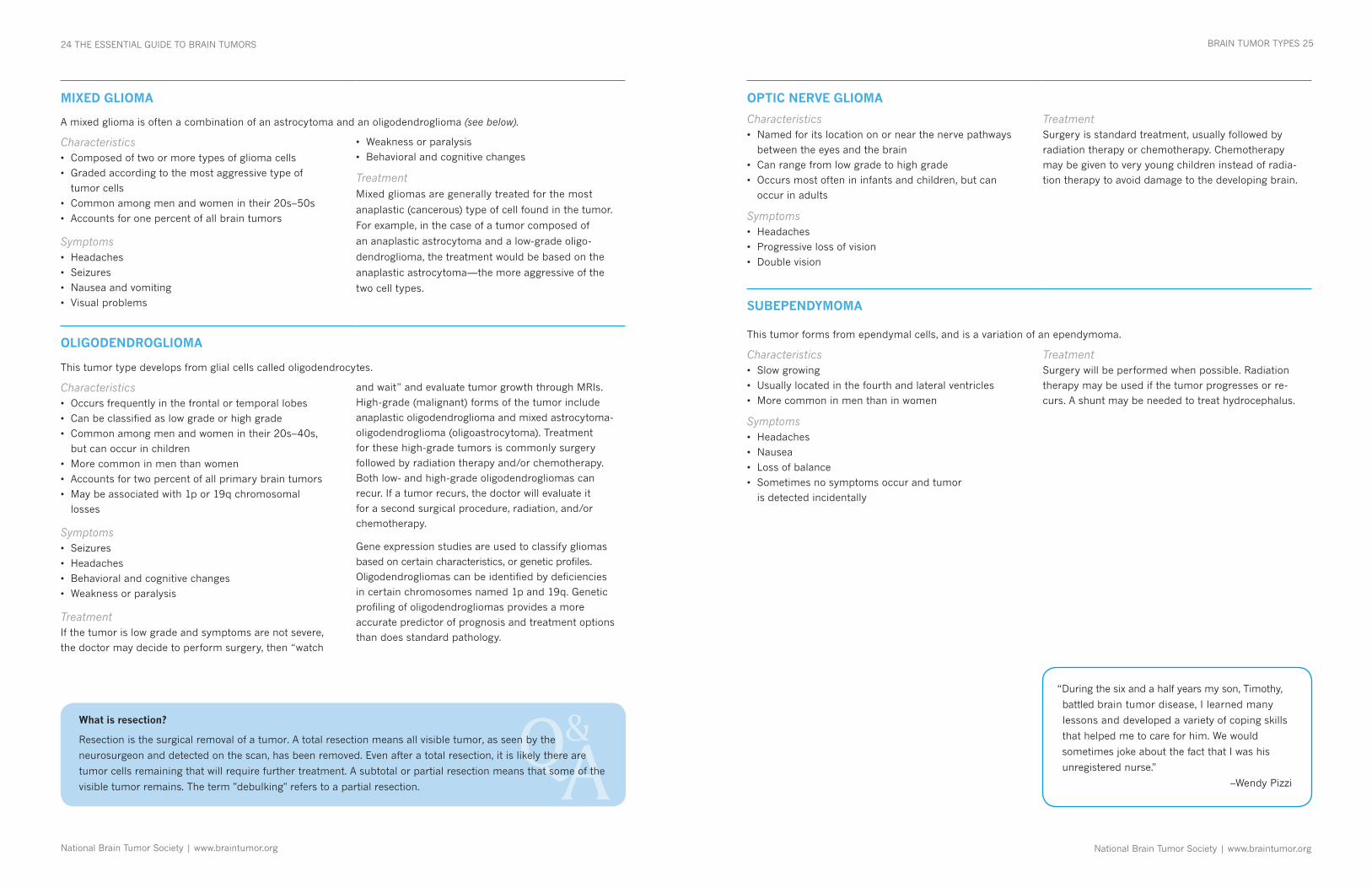
National Brain Tumor Society | www.braintumor.org National Brain Tumor Society | www.braintumor.org
24 THE ESSENTIAL GUIDE TO BRAIN TUMORS BRAIN TUMOR TYPES 25
“ During the six and a half years my son, Timothy,
battled brain tumor disease, I learned many
lessons and developed a variety of coping skills
that helped me to care for him. We would
sometimes joke about the fact that I was his
unregistered nurse.”
–Wendy Pizzi
MIxED GLIOMA
A mixed glioma is often a combination of an astrocytoma and an oligodendroglioma (see below).
Characteristics • Composed of two or more types of glioma cells
• Graded according to the most aggressive type of
tumor cells
• Common among men and women in their 20s–50s
• Accounts for one percent of all brain tumors
Symptoms• Headaches
• Seizures
• Nausea and vomiting
• Visual problems
• Weakness or paralysis
• Behavioral and cognitive changes
TreatmentMixed gliomas are generally treated for the most
ana plastic (cancerous) type of cell found in the tumor.
For example, in the case of a tumor composed of
an ana plastic astrocytoma and a low-grade oligo-
dendroglioma, the treatment would be based on the
anaplastic astro cytoma—the more aggressive of the
two cell types.
OLIGODENDROGLIOMA
This tumor type develops from glial cells called oligodendrocytes.
Characteristics• Occurs frequently in the frontal or temporal lobes
• Can be classified as low grade or high grade
• Common among men and women in their 20s–40s,
but can occur in children
• More common in men than women
• Accounts for two percent of all primary brain tumors
• May be associated with 1p or 19q chromosomal
losses
Symptoms• Seizures
• Headaches
• Behavioral and cognitive changes
• Weakness or paralysis
Treatment If the tumor is low grade and symptoms are not severe,
the doctor may decide to perform surgery, then “watch
and wait” and evaluate tumor growth through MRIs.
High-grade (malignant) forms of the tumor include
anaplastic oligodendroglioma and mixed astrocytoma-
oligodendroglioma (oligoastrocytoma). Treatment
for these high-grade tumors is commonly surgery
followed by radiation therapy and/or chemotherapy.
Both low- and high-grade oligodendrogliomas can
recur. If a tumor recurs, the doctor will evaluate it
for a second surgical procedure, radiation, and/or
chemotherapy.
Gene expression studies are used to classify gliomas
based on certain characteristics, or genetic profiles.
Oligo dendrogliomas can be identified by deficiencies
in certain chromosomes named 1p and 19q. Genetic
profiling of oligodendrogliomas provides a more
accurate pre dictor of prognosis and treatment options
than does standard pathology.
OPTIC NERVE GLIOMA
Characteristics • Named for its location on or near the nerve pathways
between the eyes and the brain
• Can range from low grade to high grade
• Occurs most often in infants and children, but can
occur in adults
Symptoms • Headaches
• Progressive loss of vision
• Double vision
TreatmentSurgery is standard treatment, usually followed by
radiation therapy or chemotherapy. Chemotherapy
may be given to very young children instead of radia-
tion therapy to avoid damage to the developing brain.
SUBEPENDyMOMA
This tumor forms from ependymal cells, and is a variation of an ependymoma.
Characteristics• Slow growing
• Usually located in the fourth and lateral ventricles
• More common in men than in women
Symptoms• Headaches
• Nausea
• Loss of balance
• Sometimes no symptoms occur and tumor
is detected incidentally
TreatmentSurgery will be performed when possible. Radiation
therapy may be used if the tumor progresses or re-
curs. A shunt may be needed to treat hydrocephalus.
What is resection?
Resection is the surgical removal of a tumor. A total resection means all visible tumor, as seen by the
neurosurgeon and detected on the scan, has been removed. Even after a total resection, it is likely there are
tumor cells remaining that will require further treatment. A subtotal or partial resection means that some of the
visible tumor remains. The term "debulking" refers to a partial resection.

National Brain Tumor Society | www.braintumor.org National Brain Tumor Society | www.braintumor.org
26 THE ESSENTIAL GUIDE TO BRAIN TUMORS BRAIN TUMOR TYPES 27
NON-GLIAL TUMORS
The following tumor types develop on or in structures within the brain, such as nerves, blood vessels, and glands.
Acoustic Neuroma An acoustic neuroma is also known as a vestibular schwannoma or neurilemmoma.
Characteristics• Arises from cells that form a protective sheath around
nerve fibers
• Typically grows around the eighth cranial nerve, but
can be found around other cranial or spinal nerves
Symptoms • Hearing loss in one ear • Tingling or numbness
• Dizziness or vertigo in the face
• Tinnitus (ringing in the ear) • Balance problems
• Coordination problems
TreatmentThe tumor may be observed to monitor its growth, or
surgery may be performed. The goal of surgery is the
complete removal of the tumor without harming the
seventh cranial nerve (which controls facial movement)
or causing hearing loss. Radiosurgery may be an
option. This focused, high-energy radiation prevents
the growth of acoustic neuromas, but actual shrinkage
of the tumor may never occur or may take several
months.
Chordoma
Characteristics• Rare and low grade
• Occurs at the sacrum, near the lower tip of the spine,
or at the base of the skull
• Originates from cells left over from early fetal
development
• Invades the bone and soft tissues but rarely the brain
tissue
• Can block the ventricles, causing hydrocephalus
• Can metastasize (spread) or recur
Symptoms• Double vision
• Headaches
Treatment Surgery and radiation therapy are the common forms
of treatment. Chordomas at the base of the skull
can be difficult to remove. Surgical resection may be
possible if the tumor is located in the spine.
CNS LymphomaCNS Lymphoma is a type of cancer that develops in the lymphatic system. The lymphatic system is a network of
small organs called lymph nodes and vessels (similar to blood vessels) that carry a clear, watery fluid called lymph
throughout the body. This fluid supplies cells called lymphocytes that fight disease and infection. To correctly
diagnose primary CNS Lymphoma, staging must be done. Staging is the process of using CT scanning to examine
many parts of the body. Staging helps confirm where the cancer originated and how far it has spread.
Characteristics• Very aggressive
• Usually involves multiple tumors throughout the CNS
• More common in people whose immune systems are
compromised
• Often develops in the brain, commonly in the areas
adjacent to the ventricles
• Can be primary (originating in the brain) or secondary
• Most common among men and women in their
60s–80s, but incidence is increasing in young adults
• More common in men than women
• Accounts for about 2 percent of all brain tumors
Symptoms• Headaches
• Partial paralysis on one side of the body
• Seizures
• Cognitive or speech disorders
• Vision problems
Treatment Radiation therapy, chemotherapy, and steroids are the
most common forms of treatment. Surgery is rarely
an option because there are usually multiple lesions.
However, a biopsy at the start of steroid treatment can
be critical to ensure the correct diagnosis.
Craniopharyngioma
Characteristics• Most common in the parasellar region, an area at the
base of the brain and near the optic nerves
• Also grows in the regions of the optic nerves and the
hypothalamus, near the pituitary gland
• Tends to be low grade
• Often accompanied by a cyst
• Originates in cells left over from early fetal development
• Occurs in children and men and women in their 50s
and 60s
Symptoms• Headaches
• Visual changes
• Weight gain
• Delayed development in children
Treatment Surgery is the most common treatment. Radiation
therapy may be used.
Hemangioblastoma
Characteristics• Commonly located in the cerebellum
• Slow growing
• Originates from blood vessels
• Can be large in size
• Often includes a cyst
• Common among men and women in their 40s–60s
• More common in men than women
• Accounts for approximately one percent of all
brain tumors
• Sometimes found in conjunction with von Hippel-
Lindau syndrome, an inherited condition that may
cause a tendency toward hemangioblastomas or
kidney cancers
Symptoms• Headaches
• Nausea and vomiting
• Walking and balance problems
TreatmentSurgery is the standard treatment. Radiosurgery may
be given to destroy multiple inoperable lesions.
Medulloblastoma
Characteristics• A type of primitive neuroectodermal tumor (PNET)
• Often located in the cerebellum or near the brain stem
• Can spread to the spinal cord through the CSF
• May obstruct the fourth ventricle, causing
hydrocephalus
• Occurs most often in children under the age of ten,
but may occur in adults
• Slightly more common in males than females
Symptoms• Headaches
• Early morning vomiting
• Lethargy or sleepiness
• Lack of coordination
• Double vision
• Behavioral or personality changes
• Signs of pressure seen behind the eye when
examined with an ophthalmoscope
TreatmentSurgery is the standard treatment when possible.
Chemo therapy is usually part of the treatment plan.
Radiation of the brain and spine is often recom-
mended in adults and children over three years of age.
A shunt may be needed to treat hydrocephalus. This
tumor may recur years later if not totally resected.

National Brain Tumor Society | www.braintumor.org National Brain Tumor Society | www.braintumor.org
28 THE ESSENTIAL GUIDE TO BRAIN TUMORS BRAIN TUMOR TYPES 29
Meningioma These tumors grow from the meninges, the layers of tissue covering the brain and spinal cord. Meningiomas are
graded from low to high. The lower the grade, the lower the risk of recurrence and aggressive growth. The WHO
classification divides meningiomas into three grades:
• Grade I, Benign Meningioma
• Grade II, Atypical Meningioma
• Grade III, Malignant (Anaplastic) Meningioma
Characteristics• May arise after previous treatment from ionizing
radiation or excessive x-ray exposure
• Common among women and men in their 40s–50s,
but can occur at any age
• Twice as common in women as in men
• Accounts for 34 percent of all primary brain tumors
• In very rare cases, can invade the skull or metastasize
to the skin or lungs
• Women with meningiomas can experience tumor
growth during pregnancy
• In rare cases, multiple meningiomas can develop at
the same time
Symptoms• Seizures
• Headaches
• Nausea and vomiting
• Vision changes
• Behavioral and cognitive changes
TreatmentIf there are no symptoms, the doctor may monitor the
tumor with MRIs. Otherwise, surgery is the standard
treatment. If the tumor cannot be completely resected
or if it recurs, radiation therapy may be given as well.
Chemo therapy for unresectable, aggressive, atypical,
or recurrent meningiomas is being tested through
clinical trials. Follow-up scans are needed indefi-
nitely, because meningiomas can recur years or even
decades after treatment.
Pineal TumorA malignant form of pineal tumor is called pineoblastoma.
Characteristics• Named for its location in or around the pineal gland
(near the center of the brain)
• Can range from low grade to high grade
• Can produce an excess of melatonin, a hormone that
controls the sleep/wake cycle
• Can block the ventricles, causing hydrocephalus
• High-grade pineal tumors can spread to the spinal
cord through the CSF
• Common types include germ cell tumors, pineal
parenchymal tumors, and gliomas
• Occurs most often in children and young adults
Symptoms• Headaches
• Nausea and vomiting
• Fatigue
• Visual problems
• Memory problems
TreatmentSurgery is standard treatment when possible. Radia-
tion therapy may be used as primary treatment in
adults and children over three. Chemotherapy may
be given to delay the use of radiation therapy in very
young patients. Clinical trials using chemotherapy
drugs are available for pineal tumors. A shunt may be
needed to treat hydrocephalus caused by blockage of
the ventricles. Treatment for high-grade (malignant)
pineal tumors such as a pineoblastoma may involve
radiation to the brain and spine to control spread
through the CSF. Clinical trials using chemotherapy
or biological therapy following radiation therapy are
being investigated.
Pituitary TumorThe pituitary gland produces hormones that affect growth and the functions of other glands in the body. Certain
pituitary tumors secrete abnormally high amounts of their respective hormones and cause related symptoms.
Other pituitary tumors do not secrete hormones, but grow and compress brain tissue, causing other symptoms.
Characteristics• Named for its location on or near the pituitary gland,
located at the center of the brain behind and above
the nose
• Can range from low grade to high grade
• May cause excessive secretion of hormones
• Common among men and women in their 50s–80s
• Accounts for about 13 percent of all brain tumors
Symptoms • Headache
• Depression
• Vision loss
• Nausea or vomiting
• Behavioral and cognitive changes
• Cessation of menstrual periods (amenorrhea)
• Leaking of fluid from the breasts (galactorrhea)
• Hair growth in women
• Impotence in men
• Abnormal growth of hands and feet
• Abnormal weight gain
TreatmentIf the tumor is large or compressing the optic nerve,
standard treatment is surgery. This can be transphe-
noidal surgery, which gains access to the tumor by
entering through the nasal passage (see chapter 5).
Radiation therapy may also be used. Some pituitary
tumors may be treated with medication, and/or
observed with MRI scans. Certain drugs can block
the pituitary gland from making too many hormones.
Follow up with an endo crinologist may be necessary to
manage hormonal changes.
Primitive Neuroectodermal Tumors (PNET) There are several tumor types in this category. Names of specific PNETs may be based on the tumor location.
Examples include pineoblastoma (located in the pineal region), medulloblastoma (located in the cerebellum), and
cerebral cortex PNET (located in the cerebral cortex).
Characteristics• Very aggressive and tend to spread throughout the
CNS
• Grow from undeveloped brain cells
• Commonly include cysts and calcification (calcium
deposits)
• Tend to be large
• Occur most often in young children
Symptoms• Can vary depending on location of tumor
• Weakness or change in sensation on one side of the
body
• Morning headache or headache that goes away after
vomiting
• Nausea and vomiting
• Seizures
• Unusual sleepiness or lethargy
• Behavioral or personality changes
• Unexplained weight loss or weight gain
TreatmentSurgery is the standard treatment when possible. In
adults and children over three years of age, surgery
may be followed by radiation therapy to the whole
brain and spinal cord, and chemotherapy. In children
under three years of age, surgery may be followed by
chemotherapy or a clinical trial of chemotherapy to
delay or reduce the need for radiation therapy.

National Brain Tumor Society | www.braintumor.org National Brain Tumor Society | www.braintumor.org
30 THE ESSENTIAL GUIDE TO BRAIN TUMORS BRAIN TUMOR TYPES 31
Rhabdoid Tumor
Characteristics• Rare
• Highly aggressive and tends to spread throughout the
CNS
• Often appears in multiple sites in the body, especially
the kidneys
• Difficult to classify; may be confused with
medulloblastoma or PNETs
• Occurs most often in young children but can also
occur in adults
Symptoms• Vary depending on location of tumor in the brain or
body
• An orbital tumor may cause the eye to protrude
• External tumors cause noticeable lumps; internal tumor
symptoms vary based on location
• Balance problems may occur
• External tumors cause noticeable lumps; internal tumor
symptoms vary based on location
TreatmentWhenever possible, surgery is performed to remove as
much of the tumor as possible. This is usually followed
by chemotherapy and radiation therapy. In children
under three years of age, surgery may be followed by
chemo therapy alone. Clinical trials are being studied using
autologous bone marrow transplantation (see glossary)
after high-dose chemotherapy for recurrent or multiple
rhabdoid tumors.
Schwannoma Also known as vestibular schwannoma, neurilemmoma and acoustic neuroma (see acoustic neuroma).
Characteristics• Arises from cells that form a protective sheath around
nerve fibers
• Typically grows around the eighth cranial nerve, but
can be found around other cranial or spinal nerves
Symptoms• Reduced hearing in the ear on the side of the tumor
when eighth cranial nerve is involved
• Tinnitus (ringing in the ear)
• Balance problems
• Deficits depend on the nerve that is affected
TreatmentSurgery and radiotherapy are the most common forms of
treatment. If the tumor is not completely removed, recur-
rence is likely.
Characteristics• The primary cancer is usually in the lung, breast,
colon, kidney, or skin (melanoma), but can originate in
any part of the body
• Most are located in the cerebrum, but can also
develop in the cere bellum or brain stem
• More than half of people with metastatic tumors have
multiple lesions (tumors)
• Common among middle-aged and elderly men and
women
Metastatic Brain Tumors
A metastatic, or secondary, brain tumor is one that begins as cancer in another part of the body. Some of the cancer
cells may be carried to the brain by the blood, or may spread from adjacent tissue. The site where the cancerous cells
originated is referred to as the primary cancer. Metastatic brain tumors are often referred to as lesions. Metastatic
brain tumors are the most common brain tumors. There has been an increase in metastatic lesions as people are
surviving primary cancers for longer periods of time.
Other Tumor-Related Conditions
CystsA cyst is a fluid-filled sac that may encapsulate (sur-
round) or be located next to a tumor. Based on its
location in the brain, a cyst can cause symptoms such as
headache, pain, seizures, or a neurological deficit. Cysts
can be surgically removed or drained. If a cyst is not
causing neurological difficulties, the doctor will conduct
ongoing MRI and CT scans to observe its growth
pattern.
Neurofibromatosis Neurofibromatosis is a genetic disorder that can cause
tumors in various parts of the nervous system. There
are two types of neurofibromatosis. Type 1, the more
common kind, usually occurs outside of the CNS. Type 2
occurs within the CNS. Type 2 neurofibromatosis causes
multiple CNS tumors, including neurofibromas, multiple
meningiomas, bilateral vestibular schwannomas, optic
nerve gliomas, and spinal cord tumors. Symptoms
include loss of balance, tinnitus, total hearing loss,
facial pain or numbness, and headache. Surgery is the
standard treatment.
Pseudotumor Cerebri This condition is not a brain tumor, but its symptoms
mimic a brain tumor. Pseudotumor cerebri most com-
monly afflicts obese adolescent girls and young women.
Symptoms include head aches, blurred vision, dizziness,
and a slight numbness of the face. The symptoms are
caused by a buildup of CSF. Treatment is given to relieve
the symptoms, par tic ularly visual impairment. Treat ment
may include repeated lumbar punctures or medications to
decrease CSF. In severe cases, a shunt may be needed.
Tuberous Sclerosis Tuberous Sclerosis is a genetic disorder that causes
numerous neurological and physical symptoms, including
tumors of the CNS, eyes, and kidneys. Most cases occur
in children under 20 years of age. About 50 percent of
tuberous sclerosis patients develop brain tumors. Subep-
endymal giant-cell astrocytomas are the most common
type, but other tumor types are also associated with this
condition. Most patients suffer from seizures. Treatment
may involve inserting a shunt to prevent hydrocephalus.
Surgery is another treatment option.
Symptoms • Seizures
• Headaches
• Behavioral and cognitive changes
• Problems with coordination
Treatment Surgery and/or radiosurgery are used if lesions are
accessible and limited in number. Both of these treat-
ments may be followed by whole brain radiation therapy
(WBRT). In cases of multiple lesions, WBRT and/or
radiosurgery may be given. Chemotherapy specific to
the brain-located metastatic tumor may be used.
A recurrent tumor is a tumor that has grown back
after being removed, stabilized, or shrunken. Recur-
rence may occur in the same area as the original
tumor or elsewhere in the brain or spinal cord.
Usually, symptoms that may indicate a tumor recur-
rence are those the patient had previously, such as
leg or arm weakness or problems with coordination.
The symptoms may return or become more pro-
nounced. Headaches or seizures may occur if there
is pressure on the brain.
Treatment options for a recurrence may be surgery,
radiation, chemotherapy or a combination depending
on the size and location of the tumor(s), and the
patient’s functional status. Patients who have already
had a standard course of radiation therapy may not
be able to receive additional treatments because of
the risk of severe cognitive damage. However, radio-
surgery, an intense beam of radiation that focuses
only on a specific area, may be an option. Also, many
patients whose tumors have recurred participate in
clinical trials.
A tumor recurrence can be traumatic for patients and
families. It brings back feelings of having a life-threat-
ening disease. While some people do well, recovery
after treatment may be prolonged for others. Some
feel more tired and find that it takes longer to return
to their normal routine. Fatigue may occur due to
either an emotional reaction or physical aspects of
the tumor. Patients and families may also experience
depression. Doctors, social workers and counselors
can help you cope with difficult emotions.
Recurrent Tumors
National Brain Tumor Society | www.braintumor.org National Brain Tumor Society | www.braintumor.org
BRAIN TUMOR TREATMENTS 33
Before receiving treatment, the patient’s condition must
be evaluated through clinical examinations and imaging
tests (see chapter 2). When possible, the doctor, patient,
and family will discuss a treatment plan based on the
type and location of the tumor, the patient’s general
health, medical history, and preferences.
In diagnosing and treating brain tumors, a multi-disci-
plinary treatment team, made up of various specialists,
is generally considered the preferred approach.
Tumor Board
Some medical centers offer the services of a tumor
board. A tumor board is a multidisciplinary group of
health care providers involved in the treatment and
diagnosis of CNS tumors. Neurosurgeons, neurologists,
radiation oncologists, pathologists, and other medical
professionals may be part of a tumor board. This group
meets and reviews the patient’s MRI films and clinical
and pathological information. The tumor board then
discusses treatment options and makes a group recom-
mendation regarding treatment.
Before receiving treatment, patients commonly receive
medication(s) to manage the symptoms of the tumor.
Medical Management
Brain EdemaBrain tumors add mass within the skull. Tumors can
interfere with the circulation of the brain’s blood supply,
causing swelling of the normal tissue surrounding
them. This swelling is called brain edema. Brain edema
causes increased intracranial pressure (pressure inside
the skull) because the skull is a rigid container with no
5. Brain Tumor Treatments
This chapter examines the standard treatments for brain tumors: surgery,
radiation therapy, and chemotherapy. This chapter also describes biologic
(targeted) therapy, new treatments currently being tested in clinical trials,
and complementary and alternative medicine (CAM).
room for expansion. Increased intra cranial pressure can
cause general symptoms including headaches, seizures,
nausea, vomiting, and blurred vision.
Brain edema can also cause focal symptoms by put-
ting pressure on specific structures within the brain.
Examples of focal symptoms include problems with
language comprehension, speech problems, and
weakness on the opposite side of the body from where
the tumor is located.
Steroids (glucocortico-steroids) are drugs that reduce
brain swelling. The most common steroid is dexametha-
sone (Decadron). Other steroids are prednisone and
methylprednisolone.
Steroids and Their Side EffectsSteroids are prescribed to reduce inflammation and
control swelling of the brain. Steroids can help relieve
pre-surgery symptoms, which may increase the time to
make treatment decisions. Steroids may be prescribed
at diagnosis, or before or after surgery. These drugs do
not kill tumor cells, but can improve a patient’s condi-
tion. Steroids may be taken alone or combined with
other forms of treatment. When the swelling is under
control, then the dosage is gradually tapered off.
Steroids have a range of short- and long-term side
effects. Short-term side effects include insomnia, facial
swelling and flushing, increased sweating, increased
appetite, moderate abdominal pain, nervousness,
personality and mood changes, hoarseness, throat
irritation, weight gain, water retention, and thirst. Some
long-term side effects include muscle wasting (especially
thigh muscles), peptic ulcers, glaucoma, fatigue, weak-
ness, osteoporosis, impairment of the hip joint, diabetes,
hypertension, and loss of bone calcification.
The Treatment Team
Neurologist A doctor specializing in the diagnosis
and treatment of disorders and diseases affecting
the CNS.
Neuroradiologist A radiologist (an expert in imaging
techniques) specializing in the interpretation of
diagnostic images (scans) of the brain, spinal cord,
and nervous system.
Neurosurgeon A surgeon specializing in the
di agnosis, treatment, and surgical management
of disorders and diseases of the CNS.
Neuropathologist A doctor specializing in the
di agnosis of neurological disorders and diseases
through microscopic examination of biopsied tissues
(tumor cells).
Neuro-oncologist An oncologist (cancer doctor)
specializing in the treatment of cancers and tumors
affecting the CNS.
Radiation Oncologist A doctor specializing in the
delivery of radiation therapy.
Nurse Practitioner A registered nurse who indepen-
dently performs physical exams, common diagnostic
and laboratory tests and (in most states) can
prescribe certain medications.
Physician Assistant A certified health care worker
who may perform physical exams, identify health
problems, and plan treatment under the direction
and supervision of a medical doctor.
Neuro-oncology Nurse Specialist A registered nurse
specializing in patient education (including symptom
management) and support services for brain tumor
patients.
Neuropsychologist A licensed psychologist special-
izing in how the brain functions and the impact
that brain damage has on one’s abilities. (Please
note: although neuropsychologists are frequently
not consulted until after treatment, it may be
preferable to do so at diagnosis. That way, a more
accurate evaluation can be made before treatment
for comparison of changes that may occur after
treatment.)
In addition, the services of the following
professionals may be beneficial:
Medical Social Worker (MSW) A licensed social
worker who assists patients and families with
health-related problems and concerns. MSWs help
locate appropriate health care, legal resources, and
financial aid.
Nutritionist A person who specializes in information
about dietary needs.
Physical Therapist A trained individual who helps
patients restore function, improve mobility, relieve
pain, and prevent or limit permanent physical dis-
abilities.
Speech Therapist A professional who helps people
overcome problems understanding and producing
language. Speech therapists also help with eating
and swallowing difficulties caused by oral motor
problems.
Rehabilitation Counselor A specialist who helps
people deal with the personal, social, and vocational
effects of disabilities, and works toward increasing
the client’s capacity to live independently.
Occupational Therapist An individual who helps
clients to compensate for permanent loss of
function by improving basic motor functions and
reasoning abilities.
Clinical Psychologist A licensed professional who
can help patients and their families adjust to the
effects of illness on their lives.
Physiatrist or Physical Medicine and Rehabilita-
tion Specialist A physician specializing in physical
medicine and reha bilitation. Physiatrists specialize
in restoring optimal function to people with injuries
to the muscles, bones, tissues, and nervous system
(such as stroke survivors).
National Brain Tumor Society | www.braintumor.org National Brain Tumor Society | www.braintumor.org
34 THE ESSENTIAL GUIDE TO BRAIN TUMORS BRAIN TUMOR TREATMENTS 35
Some patients who take steroids for a few days or weeks
do not experience side effects; others do. Steroids can
cause both short-term and long-term side effects if taken
over a long period of time, or stopped suddenly without
being monitored by a doctor. Patients should never stop
taking steroids without the doctor’s knowledge. Patients
should discuss monitoring and all possible side effects
with the doctor.
Surgery
Surgery or resection is the primary form of treatment
for brain tumors. The goal of surgery is to remove the
tumor without causing damage to critical neurological
functions. When only part of a tumor can be removed,
it is possible that the tumor will recur. Therefore, the
entire tumor is removed whenever feasible.
Debulking is another term used for a partial resection.
Debulking attempts to remove as much of a tumor
as is possible. It can alleviate symptoms and improve
the effectiveness of other therapies, such as radiation
therapy and chemotherapy. In general, radiation and
chemotherapy treatments are used as secondary or
adjuvant treatments for tumors that cannot be managed
using only surgery. However, they may be used without
surgery if the tumor is inoperable.
Prior to surgery, the neurosurgeon needs to determine
the location of the tumor and plan the safest method
of performing the operation. To do this, the patient will
receive one or more of the following: a scan (MRI, CT or
PET), and hearing, neuropsychological or speech tests.
The patient will meet with the neurosurgeon to discuss
the surgery, length of hospital stay, any pre-existing
medical conditions, and possible risks of surgery. Pa-
tients should inform the neurosurgeon of all medicines
being taken, including over-the-counter medications,
herbs, and supplements. Patients may also meet with
a neuro-oncologist or other members of the treatment
team. It is helpful to prepare a list of questions and
plan to keep a journal to record important information.
Sample questions might include:
• Is my tumor benign or malignant?
• What is the name, type, and grade of my tumor?
• What are the treatment options?
• What are the side effects of each treatment option?
• Which treatment would you take if you had my
tumor?
• If I am more interested in quality of life than how
long I live, which treatment would you recommend?
• Are there any clinical trials for which I am eligible?
• What questions are those clinical trials asking?
• Do you have the equipment necessary to deliver
stereotactic radiosurgery and fractionated
stereotactic radiation therapy?
• If I wanted a second opinion from another
neurosurgeon or radiation oncologist, whom would
you recommend?
• What factors do you look at to predict how I am
going to do?
In some cases, surgery is urgent because of the tumor’s
location. The surgeon will obtain the patient’s medical
history and perform surgery as soon as possible.
Just before surgery, all or part of the patient’s head will
be shaved. If possible, the patient will discuss the type
of anesthesia to be used with the anesthesiologist. An
intravenous (IV) line will be placed in the arm to adminis-
ter fluids and medications.
Surgical Procedures
CraniotomyCraniotomy is the surgical opening of the skull (cranium).
During this procedure, the patient is usually under gen-
eral anesthesia. The surgeon makes an incision through
a section of the scalp. Then, using sophisticated surgical
tools, a part of the skull is removed and the covering of
the brain (dura) is opened to reach the tumor. As much
of the tumor as possible is removed. The dura is sutured
and the part of the skull is put back into place. Then the
scalp is closed with staples or stitches. These are usually
removed after about seven to 10 days.
Awake Craniotomy Awake craniotomy, or surgery under local anesthesia, is
a pro cedure in which the patient is allowed to return to
consciousness after the brain has been exposed. With
the patient awake, the neurosurgeon can perform brain
mapping. Brain mapping is a procedure that allows the
neurosurgeon to identify the eloquent cortex, the areas
of the brain that control important functions such as
speech, sensation, and movement. Once these delicate
areas have been identified, the patient is put back under
general anesthesia, and the surgery is completed.
BiopsyA biopsy is a surgical procedure in which a small sample
of tissue is taken from the tumor and examined under
a microscope. The purpose of a biopsy is to diagnose a
tumor in order to find out its type and grade.
Stereotactic or Needle BiopsyA stereotactic or needle biopsy is used to take tissue
samples from tumors in hard-to-reach areas of the
brain. The patient is usually awake during this proce-
dure, but it can also be performed with the patient
under anesthesia. A special frame called a static head
frame holds the patient’s head in place. Another method,
called “frameless” or computer-assisted image-guided
brain needle biopsy, does not use a static head frame.
After giving a local anesthesia, the neurosurgeon makes
a small hole in the skull. A narrow, hollow needle is
inserted through the hole to extract tumor tissue. After a
needle biopsy, the patient is monitored for several hours
in the recovery room. The patient may spend a few
additional days in the hospital.
Risks and Complications of SurgeryA craniotomy is a major operation. Risks and complica-
tions of surgery can include bleeding, infection, brain
edema, seizures, paralysis, and behavioral or cognitive
changes. Some normal brain tissue may be damaged
during surgery. This can lead to permanent impairment
of the functions related to the injured area of the brain.
For a short time after surgery, symptoms may appear
to be worse than before surgery. However, most side
effects of surgery usually decrease or disappear over
time.
Post-Operative Care After waking up in a recovery area, the craniotomy
patient will be moved to the intensive care unit (ICU).
Heart rate, intracranial pressure, and other functions
will be closely monitored. The patient may wear special
stockings to prevent blood clots. He or she may be
temporarily put on a machine called a ventilator that
helps with breathing. Generally after spending anywhere
from several hours to two days in the ICU, the patient
will be moved to a regular room.
During recovery, breathing exercises are encouraged to
keep the lungs clear. The health care team will work with
the patient to get him or her up and around as soon as
possible. This will help get the body’s systems function-
ing again.
The doctor may give a referral to a rehabilitation
program if necessary. Therapists can help with balance,
strength, walking, talking, and other daily activities.
Social workers, case managers, and discharge planners
can help coordinate continued care before leaving the
hospital.
Other Surgical Procedures and Devices
Embolization Embolization is a treatment that cuts off a tumor’s blood
supply by clogging the small blood vessels that feed the
tumor. A radiologist or neurosurgeon inserts a catheter
through an incision in the skin of the groin. Guided by
an x-ray, the catheter is advanced to the tumor site. An
embolic agent is injected into the tumor. This may be a
liquid or powdered substance, or more commonly, tiny
metal coils. Embolization can reduce blood loss during
brain surgery, shorten the length of time in surgery,
increase the chances of a complete resection, and
reduce the risk of damage to adjacent normal tissue.
Lumbar Puncture (Spinal Tap)A lumbar puncture is a procedure that extracts CSF
surrounding the spinal cord. A local anesthetic is given
to numb the spine in the area where the needle is
inserted. The CSF is examined to detect abnormal cells,
an infection, or the presence of protein or blood.
Shunt A shunt, or catheter, is a device consisting of a thin tube
and a valve that controls the flow of fluid. A shunt is
used to divert CSF from the brain into the abdominal
cavity, where it is absorbed into the bloodstream. During
a cerebral shunt procedure, the surgeon drills a small
hole in the skull. One end of the shunt is inserted into a
ventricle; the other end is tunneled under the skin to the
abdomen. Then, excess CSF drains into the abdomen.

National Brain Tumor Society | www.braintumor.org National Brain Tumor Society | www.braintumor.org
36 THE ESSENTIAL GUIDE TO BRAIN TUMORS BRAIN TUMOR TREATMENTS 37
Ommaya Reservoir An Ommaya reservoir is a surgically implanted catheter
(tube) used to inject medicine or extract CSF from
the ventricles. The catheter is attached to a small
mushroom-shaped chamber (reservoir) beneath the
scalp. A needle is inserted through the skin and into the
reservoir to administer medication and/or to obtain CSF
samples.
Brain MappingBrain mapping is used during an awake craniotomy. It
helps the neurosurgeon to avoid injuring delicate areas
of the brain that may not be visible in a scan. In this
procedure, small electrodes are used to identify the
areas called the eloquent cortex that control speech,
sensation, and movement. The patient is required to
talk, count, and perform other basic tasks during brain
mapping. The patient feels no pain, as there are no
pain receptors in the brain. This part of the procedure
typically lasts from 10 to 40 minutes. Brain mapping
improves the accuracy of a cran i otomy. It can also
clarify the borders of a tumor and distinguish tumor
tissue from necrosis (dead tumor cells), a cyst, CSF, and
normal brain tissue.
Another technique to identify eloquent areas of the
brain that control speech, sensation, and movement
is a Functional MRI. A Functional MRI is done before
surgery. (See chapter 2 for a detailed description of
Functional MRI.)
Intraoperative MRI Intraoperative MRI is an imaging system that works in
real-time to capture and display images throughout
surgery. This technology allows the surgeon to pinpoint
the tumor before the incision is made, and navigate it
more precisely throughout the surgery to make sure
that a more complete resection can be made. Some of
the newer intraoperative MRIs have magnets that are
as strong as 3 Tesla, which are optimal in obtaining a
high-resolution image. Because of its location system,
intraoperative MRI generally helps surgeons to make
smaller incisions, which leads to less invasive proce-
dures and as a result, a shorter recovery time for the
patient. An MRI scan is often performed before patients
leave the operating room so that the surgeon can verify
the tumor resection, and it eliminates the need for the
patient to have an immediate follow-up scan a few days
after surgery.
Intraoperative Ultrasound ImagingIntraoperative ultrasound imaging is a method used
during surgery to gauge the depth, diameter or center,
and edges of the tumor. It also helps to distinguish
between the tumor, necrosis, a cyst, fluid buildup, and
normal brain tissue. The device sends high-frequency
sound waves to the brain. The waves bounce off the
brain and are sent to a computer that transfers the
information into a two-dimensional picture displayed on
a television screen.
MicrosurgeryMicrosurgery involves the use of a high-powered
microscope to magnify the operating area, thereby
increasing precision. Microsurgery is a standard pro-
cedure in craniotomies.
Transphenoidal SurgeryTransphenoidal surgery is a method of surgery used
to reach tumors in the sellar area, just behind the eyes.
It accesses brain tumors through an incision made
through the nostrils. Transphenoidal surgery is com-
monly used for pituitary tumors.
Stereotactic SurgeryStereotactic surgery uses a scanning device to find the
exact loca tion of the tumor and the structures surround-
ing it. It sends the information to a computer, which
produces a three-dimensional image of the brain. This
technique helps the surgeon both plan the surgery with
greater precision and arrive at difficult to reach areas
during surgery. Stereo tactic surgery is used in several
capacities: to perform a biopsy, remove a tumor, implant
radiation pellets or for other treatments. It helps the
neurosurgeon navigate his or her way around during
surgery, and minimize the length of surgery.
Photodynamic Therapy (PDT)Photodynamic therapy is a cancer treatment in which
patients are injected with a light-sensitive drug. This drug
stays in malignant cells longer than in normal cells. The
neurosurgeon performs laser surgery, shining a laser
light directly on the tumor. A chemical reaction with the
photosensitive drug destroys the cancer cells without
harming surrounding normal tissue. PDT can be used
Conformal RadiationConformal radiation is a type of conventional radiation
treatment. A linear accelerator sends a high dose of
radiation that has been modified by computer to match
or conform to the shape of the tumor. The objective is
to apply a uniform level of radiation to the tumor while
reducing the amount of radiation that reaches other
parts of the brain. Conformal radiation may be used to
minimize injury to delicate structures such as the salivary
glands, optic pathways, and brain stem.
Intensity Modulated Radiation Therapy (IMRT)Intensity Modulated Radiation Therapy (IMRT), is a
type of conformal radiation therapy. IMRT is designed
to restrict the treatment beam to the tumor, regardless
of its shape. The intensity of radiation is made to vary
across the beam, so that a higher intensity reaches the
thickest parts of the tumor. Multiple beams are used
simultaneously and meet at the target point.
Stereotactic Radiosurgery (SRS)Stereotactic radiosurgery (SRS) is different than
conventional radiation therapy. SRS delivers a single,
high dose of radiation in a one-day session. The dose
can range from 2–30 Gy. Radiosurgery is not surgery,
but like surgery, it is meant to be ablative: to destroy
all the tumor cells in its path. Size and location of the
tumor are important eligibility criteria for SRS. In an SRS
procedure, a head frame is attached to the skull. Then CT
or MRI scans are taken. With the aid of computer imag-
ing, the location of the tumor is accurately calculated. The
radiation is delivered directly to the tumor, often from
in conjunction with other treatments, including surgery,
radiation therapy and chemotherapy. The only common
side effect is significant sensitivity to sunlight for about
one month after treatment.
Radiation Therapy
Radiation therapy (RT) uses high-energy x-rays or other
types of ionizing radiation to stop cancer cells from
dividing. Radiation therapy may be used when surgery
is not advised, or for tumors that cannot be completely
resected. It may be used after surgery to destroy
residual tumor cells and prevent or delay tumor recur-
rence. Radiation therapy can stop or slow the growth of
inoperable tumors. Use of radiation therapy is avoided
in children below the age of three because it damages
the developing brain.
Ionizing radiation damages the basic building material in
cells (DNA). Normal healthy cells can repair the damage
better than tu mor cells. Over time, irradiated tumor cells
die. Thus, the results of radiation therapy may not be
apparent until several months after treatment.
External beam radiation is the most frequently used
form of radiation therapy. It involves machines called
linear accelerators and cobalt machines. They direct
radiation at the tumor from outside the patient's body.
Conventional Radiation TherapyConventional radiation therapy delivers an external
beam of radiation focused at an entire region of the
brain containing the tumor. After resection, the area
where the tumor was located and the surrounding
margin is irradiated. The radiation is fractionated into
many small doses and given over a period of time (usu-
ally five to seven weeks, excluding weekends). A typical
daily dose of radiation therapy is 1.8–2.0 Gy (Gray). The
total dose of a radiation treatment will vary depending
on the tumor type, but ranges between 50–60 Gy. The
doses may be hyperfractionated, or divided into more
frequent, less intense doses.
Conventional radiation therapy can be either focused
or whole brain radiation therapy (WBRT), depending on
the location and size of the tumor(s). Focused radiation
therapy aims x-rays at the tumor and area surrounding
it. WBRT aims radiation at the entire brain. WBRT is used
to treat multiple tumors and metastatic brain tumors.

National Brain Tumor Society | www.braintumor.org National Brain Tumor Society | www.braintumor.org
38 THE ESSENTIAL GUIDE TO BRAIN TUMORS BRAIN TUMOR TREATMENTS 39
several different directions. Newer methods of SRS do
not use a head frame.
There are several types of machines that are used to
administer radiosurgery: Gamma Knife®, LINAC, XKnife®,
Synergy® S, Trilogy®, CyberKnife®, Novalis Tx™ and cyclo-
tron. The prin ciples are the same, but they use different
sources of energy and different methods to target the
tumor (see example images of radiosurgery on the right).
Gamma Knife RadiosurgeryThe Gamma Knife machine contains 201 sources of ra-
dioactive cobalt. It focuses a high dose of radiation to a
small target area. The Gamma Knife is most effective for
small tumors that are round or oval shaped. Gamma
Knife can be used as a primary form of treatment, or as
a secondary treatment after surgery.
LINAC RadiosurgeryLinear accelerator (LINAC) radiosurgery sends a single,
high-energy x-ray that is designed by a computer to
match the shape of the tumor and avoid healthy tissue.
A linear accelerator is also used for Intensity Modulated
Radiation Therapy (IMRT).
Proton Beam RadiosurgeryProton beam radiosurgery is also called heavy particle
radiation therapy. This method uses beams of charged
protons (atomic par ticles) produced by a machine called
a cyclotron. Similar to LINAC, the cyclotron sends a
single beam that is designed by a computer to match
the shape of the tumor. Proton beam radiosurgery is
used to treat tumors at the base of the skull and tumors
of unusual shapes.
Stereotactic Radiotherapy (SRT)Stereotactic radiotherapy (SRT) combines the precision
of radiosurgery with fractionated radiation. SRT utilizes
computer imaging to precisely locate the tumor. Then
a series of low doses of radiation are given over several
treatment sessions. Treatment may be given once or
twice a day for several weeks. These fractionated doses
add up to a larger total dose of radiation than would be
given in a single SRS treatment.
SRT has some advantages over other types of radiation
therapy. The precision of delivery and lower individual
doses of radiation cause less damage to normal tissue.
This allows SRT to be used in situations where single
dose SRS cannot be applied safely. Some applications
include: for tumors near the optic nerves, brain stem, or
eyes; for some large tumors; and for pediatric patients.
Side Effects of Radiation TherapyShort-term side effects occur during or shortly after
radiation treatment. Most can be treated or in some
cases will decrease or dis appear after treatment has
been completed. The most common short-term side
effects of radiation therapy are fatigue, loss of ap-
petite, nausea, and short-term memory loss (loss of
recently-learned information). Short-term memory loss
usually subsides within two months after RT treatment is
completed. Brain edema may occur and may be treated
with steroids. Skin reactions (rash, redness, or irritation)
and hair loss may occur in the area where the radiation
is focused.
Some patients may experience long-term, or delayed,
side effects. These may occur months or years after
treatment. Delayed side effects can include varying
degrees of memory loss, coordination problems,
incontinence, and problems with reasoning and think-
ing. Loss of pituitary gland function can also occur. In
the case of children, RT can result in delayed physical
growth and/or delayed cognitive development and can
cause learning disabilities.
Sometimes dead tumor cells form a mass in the brain.
This is called radiation necrosis. Necrosis may look like
a tumor on a scan and can cause similar symptoms
(headache, memory loss, seizures, personality changes,
and cognitive problems). A biopsy may be required to
make a correct diagnosis. Surgery and/or steroids may
be necessary to treat necrosis.
If a patient begins to have symptoms after radiation
therapy and the tumor has not recurred, he or she may
be experiencing delayed side effects. The patient should
talk to the doctor about these symptoms and how to
manage them. Currently, there is no definitive treatment
for the long-term effects of radiation therapy, although
in some instances surgery, steroids, anti-coagulants, or
CNS stimulants may help alleviate symptoms.
Chemotherapy
Chemotherapy is the treatment of disease by means of
chemicals (drugs) that have a toxic effect on tumor cells
as they divide. Chemo therapy is given in cycles, which
consist of “on” and “off” phases—days of treatment
followed by periods of time between treatments. Cycles
vary depending on the drug or drugs used. Chemo-
therapy is usually taken orally or by injection. It may be
given alone or in combination with other treatments.
Chemotherapy is usually a secondary therapy. It is also
used to delay or replace radiation treatment in young
children. Chemo therapy may be an important com-
ponent of therapy for some “chemo-sensitive” tumors
(e.g., CNS lymphoma, medulloblastoma).
One approved technique to deliver chemotherapy
directly to brain tumors is the polymer wafer implant.
Biodegradable wafers are saturated with the chemo-
therapy drug, BCNU. Several wafers are inserted into
the tumor cavity after the tumor has been resected. The
wafers are left there to dissolve over time. In this way, a
concentrated dose of BCNU (approximately 100 times
higher than that tolerated through IV) is able to bypass
the blood brain barrier.
The brain has a defense mech anism called the blood
brain barrier, which keeps out harmful substances such
as bacteria and chem icals. The blood brain barrier can
also prevent some chemo therapy drugs from entering
the brain. However, a technique called blood brain bar-
rier disruption can be used to temporarily interrupt the
barrier and allow chemotherapy drugs to pass through
the blood vessels into the brain. In this procedure, most
commonly used in patients with lymphoma, a catheter
is fed into the cerebral artery. A drug called mannitol is
injected into the catheter. Mannitol disrupts the blood
brain barrier. While the blood brain barrier is open,
chemo therapy is delivered through the catheter. The
effects of mannitol wear off after a period of time.
There are some cases where chemotherapy treatment
is not used. Certain types of tumors do not respond to
chemotherapy. Also, chemotherapy can be phy sically
taxing. Patients should have overall good health in
order to tolerate the side effects of these treatments.
How is radiation measured?
Radiation is measured by an international unit
called Gray (Gy). Each dose of Gy represents a
specific amount of energy that is absorbed by
the body.
How does radiation destroy a tumor?
Radiation works by damaging DNA. This damage
stops cells from being able to divide, or reproduce.
Over a period of time, tumor cells that received
radiation will die when trying to divide.
Radiosurgery images from top to bottom:
CyberKnife®, courtesy of Accuray, Inc.
Leksell Gamma Knife®, courtesy of Elekta, Inc.
Novalis Tx™, courtesy of Brainlab AG
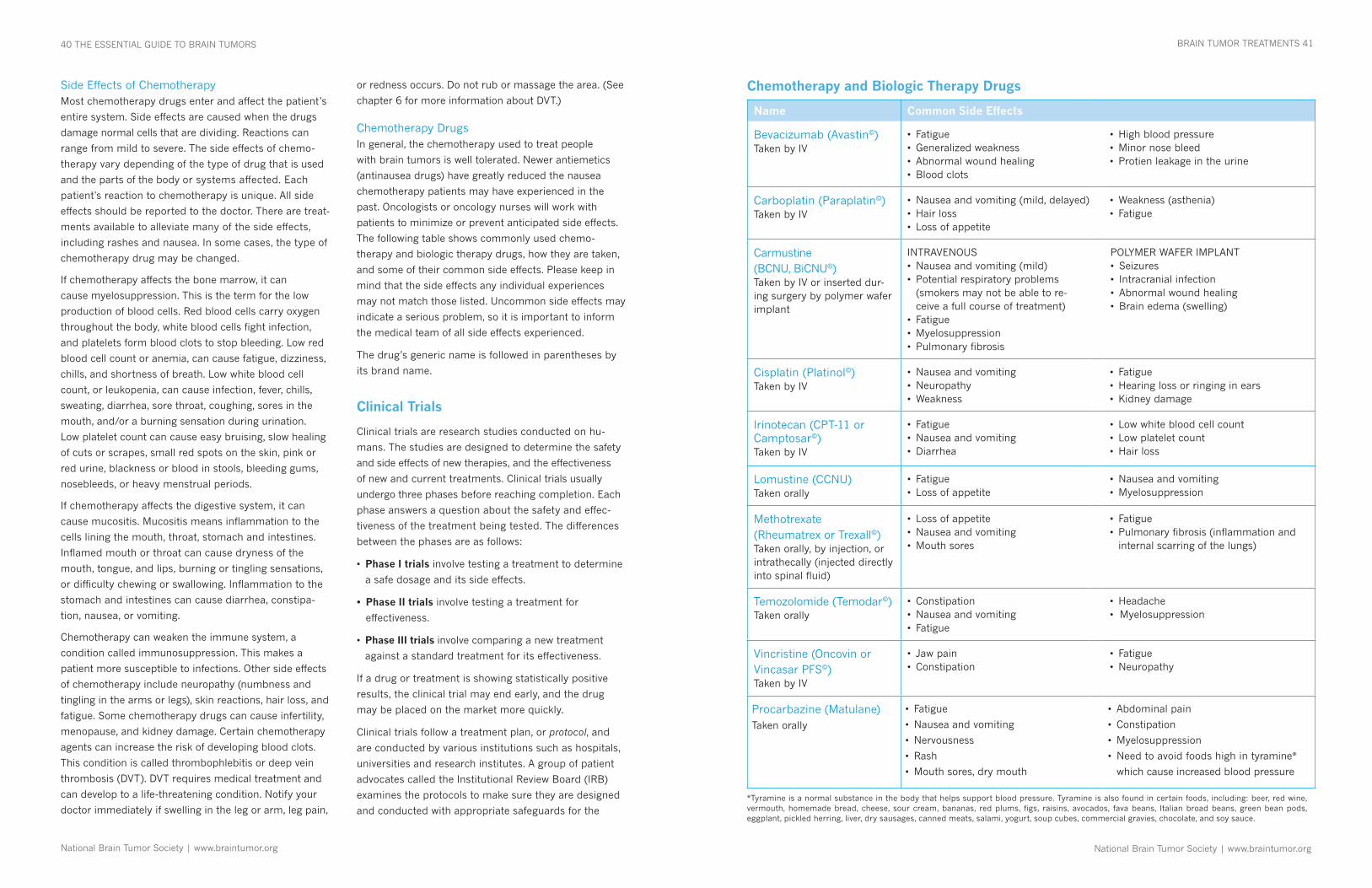
National Brain Tumor Society | www.braintumor.org National Brain Tumor Society | www.braintumor.org
40 THE ESSENTIAL GUIDE TO BRAIN TUMORS BRAIN TUMOR TREATMENTS 41
Side Effects of ChemotherapyMost chemotherapy drugs enter and affect the patient’s
entire system. Side effects are caused when the drugs
damage normal cells that are dividing. Reactions can
range from mild to severe. The side effects of chemo-
therapy vary depending of the type of drug that is used
and the parts of the body or systems affected. Each
patient's reaction to chemotherapy is unique. All side
effects should be reported to the doctor. There are treat-
ments available to alle viate many of the side effects,
including rashes and nausea. In some cases, the type of
chemo therapy drug may be changed.
If chemotherapy affects the bone marrow, it can
cause myelosuppression. This is the term for the low
production of blood cells. Red blood cells carry oxygen
throughout the body, white blood cells fight infection,
and platelets form blood clots to stop bleeding. Low red
blood cell count or anemia, can cause fatigue, dizziness,
chills, and shortness of breath. Low white blood cell
count, or leukopenia, can cause infection, fever, chills,
sweating, diarrhea, sore throat, coughing, sores in the
mouth, and/or a burning sensa tion during urination.
Low platelet count can cause easy bruising, slow healing
of cuts or scrapes, small red spots on the skin, pink or
red urine, blackness or blood in stools, bleeding gums,
nosebleeds, or heavy menstrual periods.
If chemotherapy affects the di gestive system, it can
cause mucositis. Mucositis means in flammation to the
cells lining the mouth, throat, stomach and intestines.
Inflamed mouth or throat can cause dryness of the
mouth, tongue, and lips, burning or tingling sensations,
or difficulty chewing or swallowing. Inflamma tion to the
stomach and intestines can cause diarrhea, constipa-
tion, nausea, or vomiting.
Chemotherapy can weaken the immune system, a
condition called immunosuppression. This makes a
patient more susceptible to infections. Other side effects
of chemotherapy include neuropathy (numbness and
tingling in the arms or legs), skin reactions, hair loss, and
fatigue. Some chemo ther apy drugs can cause infertility,
menopause, and kidney damage. Certain chemotherapy
agents can increase the risk of developing blood clots.
This condition is called thrombophlebitis or deep vein
thrombosis (DVT). DVT requires medical treatment and
can develop to a life-threatening condition. Notify your
doctor immediately if swell ing in the leg or arm, leg pain,
or redness occurs. Do not rub or massage the area. (See
chapter 6 for more information about DVT.)
Chemotherapy Drugs In general, the chemotherapy used to treat people
with brain tumors is well tolerated. Newer antiemetics
(antinausea drugs) have greatly reduced the nausea
chemotherapy patients may have experienced in the
past. Oncologists or oncology nurses will work with
patients to minimize or prevent anticipated side effects.
The following table shows commonly used chemo-
therapy and biologic therapy drugs, how they are taken,
and some of their common side effects. Please keep in
mind that the side effects any individual experiences
may not match those listed. Uncommon side effects may
indicate a serious problem, so it is important to inform
the medical team of all side effects experienced.
The drug’s generic name is followed in parentheses by
its brand name.
Clinical Trials
Clinical trials are research studies conducted on hu-
mans. The studies are designed to determine the safety
and side effects of new therapies, and the effectiveness
of new and current treatments. Clinical trials usually
undergo three phases before reaching completion. Each
phase answers a question about the safety and effec-
tiveness of the treatment being tested. The differences
between the phases are as follows:
• Phase I trials involve testing a treatment to determine
a safe dosage and its side effects.
• Phase II trials involve testing a treatment for
effectiveness.
• Phase III trials involve comparing a new treatment
against a standard treatment for its effectiveness.
If a drug or treatment is showing statistically positive
results, the clinical trial may end early, and the drug
may be placed on the market more quickly.
Clinical trials follow a treatment plan, or protocol, and
are conducted by various institutions such as hospitals,
universities and research institutes. A group of patient
advocates called the In stitutional Review Board (IRB)
examines the protocols to make sure they are designed
and conducted with appropriate safeguards for the
Chemotherapy and Biologic Therapy Drugs
Name Common Side Effects
Bevacizumab (Avastin©)Taken by IV
• Fatigue• Generalizedweakness• Abnormalwoundhealing• Bloodclots
• Highbloodpressure• Minornosebleed• Protienleakageintheurine
Carboplatin (Paraplatin©)Taken by IV
• Nauseaandvomiting(mild,delayed)• Hairloss• Lossofappetite
• Weakness(asthenia)• Fatigue
Carmustine (BCNU, BiCNU©) Taken by IV or inserted dur-ing surgery by polymer wafer implant
INTRAVENOUS• Nauseaandvomiting(mild)• Potentialrespiratoryproblems
(smokers may not be able to re-ceive a full course of treatment)
• Fatigue• Myelosuppression• Pulmonaryfibrosis
POLYMER WAFER IMPLANT• Seizures• Intracranialinfection• Abnormalwoundhealing• Brainedema(swelling)
Cisplatin (Platinol©)Taken by IV
• Nauseaandvomiting• Neuropathy• Weakness
• Fatigue• Hearinglossorringinginears• Kidneydamage
Irinotecan (CPT-11 or Camptosar©)Taken by IV
• Fatigue• Nauseaandvomiting• Diarrhea
• Lowwhitebloodcellcount• Lowplateletcount• Hairloss
Lomustine (CCNU)Taken orally
• Fatigue• Lossofappetite
• Nauseaandvomiting• Myelosuppression
Methotrexate (Rheumatrex or Trexall©)Taken orally, by injection, or intrathecally (injected directly into spinal fluid)
• Lossofappetite• Nauseaandvomiting• Mouthsores
• Fatigue• Pulmonaryfibrosis(inflammationand
internal scarring of the lungs)
Temozolomide (Temodar©)Taken orally
• Constipation• Nauseaandvomiting• Fatigue
• Headache•Myelosuppression
Vincristine (Oncovin or Vincasar PFS©)Taken by IV
• Jawpain• Constipation
• Fatigue• Neuropathy
Procarbazine (Matulane)Taken orally
• Fatigue
• Nauseaandvomiting
• Nervousness
• Rash
• Mouthsores,drymouth
• Abdominalpain
• Constipation
• Myelosuppression
• Needtoavoidfoodshighintyramine*
which cause increased blood pressure
*Tyramine is a normal substance in the body that helps support blood pressure. Tyramine is also found in certain foods, including: beer, red wine, vermouth, homemade bread, cheese, sour cream, bananas, red plums, figs, raisins, avocados, fava beans, Italian broad beans, green bean pods, eggplant, pickled herring, liver, dry sausages, canned meats, salami, yogurt, soup cubes, commercial gravies, chocolate, and soy sauce.

National Brain Tumor Society | www.braintumor.org National Brain Tumor Society | www.braintumor.org
42 THE ESSENTIAL GUIDE TO BRAIN TUMORS BRAIN TUMOR TREATMENTS 43
patients. There is an IRB at every institution that is
conducting research.
People participate in clinical trials for a number of
reasons: to try a new treatment method, to contribute
to developing improved treatments, or to help find a
cure. If a patient is considering participating in a trial,
the trial he or she chooses will depend on the patient’s
tumor type, when it was diagnosed, treatments that
have already been received, and the patient’s state of
health. Most trials have a series of criteria a patient
needs to meet in order to participate.
There are both benefits and drawbacks to consider.
Participants in clinical trials receive high-quality medi-
cal care. Par ticipants are also among the first to benefit
if a new approach is proven effective. However, a new
treatment may not be better than or even as good as
the standard treatment. There may also be unexpected
side effects.
A patient should ask the following questions when
deciding on a clinical trial:
• Who is conducting the study? Is it conducted by an
individual institution, or part of a national study?
• Where is the study being done? Are there multiple
locations? Is there a participating institution closer
to home?
• Does the center have a designated neuro-oncology
(brain tumor) program?
• What is the phase of the study? Has it been tested
on people with brain tumors before?
• What tests, treatments, and commitment of time
does the study involve? Does it involve a hospital
stay? If so, for how long?
• What other choices are available? How does the
study treatment compare to the alternatives?
• What potential side effects may occur and how can
they affect day-to-day life?
• What does the treatment cost? Is any part of it
provided for free?
Obtaining the answers to these questions can help a
patient decide which treatment is right for him/her.
When a decision is made to participate in a clinical
trial, the patient or his/her representative will undergo
a process called informed consent. Informed consent
will explain the study’s treatments and tests as well as
possible benefits and risks. If a patient agrees, he/she
or the representative will sign a consent form. A patient
can decide to leave the trial at any time. During the trial,
patients will be monitored closely and asked to report
all symptoms of the treatment.
For more clinical trial information visit www.
braintumor.org or contact the National Cancer
In stitute (NCI) at 1 800 4CANCER (1 800 422
6237) or visit www.cancer.gov.
Biologic/Targeted Therapies
Biologic (also known as targeted) therapies offer an
option for treatment for people who have tumor recur-
rence after surgery, radiation therapy or chemotherapy.
Biologic therapies work through a variety of mechanisms
including stimulating a person’s own immune system
to turn against the tumor or targeting specific pathways
in the tumor cell that give the tumor its characteristics.
These characteristics include increased rate of growth
(proliferation), the ability to invade brain tissue, and
resistance to normal signals that induce cell death
(apoptosis). Biological response modifiers (BRM) may
be naturally occurring substances in the body that fight
infections or diseases or agents specifically designed
to target specific cellular pathways. The body normally
produces small amounts of some BRMs. Some biologic
therapies can cause the body to produce larger amounts
of BRMs than normal.
Some of the new biologic therapies are: angiogenesis
“ Be aware that the patient, family, friends, and
loved ones will go through a range of emotions
and reactions to a brain tumor diagnosis and
treatment. These feelings will include denial,
anger, acceptance, and even guilt. I feel strongly
that it is part of your doctor’s responsibility, not
just to deliver the treatment, but also to help pa-
tients and their families cope with the disease.”
–Radiation oncologist
inhibitors, which interfere with the growth of blood ves-
sels that feed the tu mor; antisense therapies (proteins
designed to block the function of another protein); and
differentiators, which make cells less likely to divide. In
addition, there are new chemotherapy treatments, gene
therapy, growth inhibitors, and immunotoxin therapy.
Biologic Therapy DrugsBevacizumab (Avastin©) is an angiogenesis inhibitor
that has been granted accelerated FDA approval to
treat glioblastoma in patients who have progressive
disease following prior therapy. A number of clinical
trials are underway to investigate the use of beva-
cizumab alone and in different drug combinations
to treat brain tumors as an initial treatment or for
recurrence.
Isotretinoin (Accutane©) and thalidomide (Thalamid©)
are other biologic therapies being tested in clinical
trials for use in brain tumor patients. These drugs
are also FDA approved for treatment of other medical
conditions. Both isotretinoin and thalidomide cause
severe birth defects. Therefore, men and women
receiving these drugs are required to use at least two
forms of contraception, and women are required to
take monthly pregnancy tests.
Other biologic therapy drugs continue to be developed
and many are currently being tested in clinical trials,
some in combination with previously approved agents
(i.e. irinotecan, BCNU). The trials include drugs such
as cediranib (Recentin©), sorafenib (Nexavar©) and
erlotinib (Tarceva©).
Other Biologic Therapies
Gene TherapyClinical studies have identified certain genes as tumor
suppressors. Tumor suppressors tell cells to die when
their DNA becomes damaged. Cancer cells have
damaged, mutated DNA. Instead of dying, they multiply
uncontrollably. Gene therapy is a method to introduce a
non-mutated gene into a tumor. This may be achieved
by inserting the gene into a virus, and the virus acts as
a carrier, or vector. The virus attaches to the tumor cells
and transfers the gene into them. The transferred gene
then acts as a tu mor suppressor, causing the tumor
cells to die. Surgery is often required to directly inject
the viral vector carrying the non-mutated gene into the
tumor.
Vaccine TherapyThis method attempts to use the patient’s immune
system to attack the tumor. Typically, a portion of the
brain tumor is removed from a patient and processed
in a number of ways to produce a vaccine. This vaccine
of processed tumor tissue is then injected into the
patient’s skin. The patient’s immune system recognizes
the injected substance as foreign or abnormal, and
specialized immune cells in the body learn to recognize
the abnormal tissue. Those immune cells circulate
around the body and if they come in contact with brain
tumor cells, they direct other immune cells to try and
kill the tumor cells. Alternatively, investigations are
ongoing to develop vaccines against proteins that are
unique to tumor cells in general, which would eliminate
the need to undergo the expensive and time consuming
process of developing a patient-specific vaccine. Vaccine
therapy is still experimental and has not yet been
proven to work in all patients, but much research is
being conducted.
Immunotoxin TherapyAn immunotoxin is a poison linked to a part of the
body’s immune system, such as an antibody or protein.
Immunotoxin therapy is a process of fighting cancer
cells by introducing a toxin directly into tumor cells.
The toxins are usually bacterial. Because of the risk of
introducing toxins into normal brain tissue, the toxins
are attached to other proteins that will selectively target
only tumor cells. The goal of immu no toxin therapy is to
target tumor cells and kill them without harming normal
brain cells. Immunotoxin therapy is experimental.
Research is ongoing and several different im mu notoxins
are being developed.
What happens after treatment is over?
After treatment is over, most patients are usually
monitored for tumor recurrence on a regular
basis through MRI or CT scans. The doctor may
decide to take scans every two to six months
depending on the tumor type and patient’s
status.

National Brain Tumor Society | www.braintumor.org National Brain Tumor Society | www.braintumor.org
44 THE ESSENTIAL GUIDE TO BRAIN TUMORS BRAIN TUMOR TREATMENTS 45
pain, and balance the central nervous system.
Acupuncture is primarily used to treat body pain,
headaches, fatigue, nausea, vomiting, constipation,
diarrhea, and insomnia. However, it is also quite useful
in treating anxiety, depression, mood swings, weight
loss, edema, neuropathy, muscle atrophy, and muscle
weakness.
Acupuncture should not be used when the following
conditions are present: a low platelet count (<15,000),
a low white blood cell count (<1.0), throm bosis or
embolism (blood clots), hemophilia, and/or the use of
medications that increase bleeding (i.e., blood thinners
such as Coumadin).
AyurvedaAyurveda, which was developed in India, consists mainly
of specific dietary recommendations and custom-
ized herbal preparations. It is used to treat the same
disorders as acupuncture. Like acupuncture, diagnosis
is based on complex pulse analysis and a physical
examination; however, needles are seldom used. It is
difficult to find qualified practitioners because ayurvedic
physicians are not yet licensed in the United States.
ChiropracticChiropractic involves the adjustment or manipulation
of spinal vertebrae. This treatment may return balance
to the CNS to enhance immune function and decrease
inflammation. Chiropractic is primarily used to treat
body pain, headaches, anxiety, insomnia, constipation,
diarrhea, neuropathy, and muscle weakness.
Diet and NutritionThe purpose of altering the diet is to provide additional
energy for immune function. Basic alterations frequently
include an emphasis on organic “whole” (unprocessed)
foods, and avoidance of refined sugar, dairy, and/
or acidic and spicy foods. Meat intake is minimized;
however, fish may be substituted. There is a need to
maintain adequate protein intake.
Several types of diets exist. They range from macro-
biotics, which is based on Asian medical principles,
to ayurvedic diet, which is based on Indian medical
principles, to blood type diets, which are founded on
Western medical principles. It is recommended that
Convection-enhanced delivery is an experimental method
to introduce immunotoxins into tumors. This method
inserts immunotoxins into the brain by a small, pres-
surized pump. Con vection-enhanced delivery is able to
bypass the blood brain barrier. The treatment is usually
admin istered over a period of several hours and is often
completed during one sitting. Convection-enhanced
delivery is currently being tested in clinical trials as a
promising tool in the treatment of brain tumors.
Complementary and Alternative Medicine (CAM)
Standard or conventional therapy for brain tumor
patients usually means surgery, radiation, and/or
chemotherapy. Complementary medicine is used in
addition to standard therapy. Alternative medicine is
used instead of standard therapy. A simple definition
of CAM is anything people do other than conventional
therapies to maintain or achieve good health. Integrative
medicine is a combination of conventional medicine
with complementary and/or alternative therapies.
The goal of CAM is to enable patients to tolerate higher
doses of standard therapy at closer intervals with fewer
side effects. CAM also aims to increase the quality of
life both during and after treatment. It is important to
integrate CAM into conventional medicine safely, under
the supervision of a CAM health professional.
Major Systems of CAMThere are countless CAM therapies available today. The
major systems of CAM include: acu puncture, ayurveda,
chiropractic, diet and nutrition, exercise, guided imagery,
healing touch, herbal medicine, homeopathy, massage
therapy, meditation, and nutritional supplements.
AcupunctureAcupuncture, which was developed in China, involves
the painless insertion of disposable, hair-fine needles
into various points on the surface of the body. It may
increase circulation, activate the immune system, relieve
“ Every patient is a unique person in a unique
environment...”
–Neuropsychologist
Massage TherapyMassage therapy involves manipulation of muscles and
connective tissues of the body. Massage therapists ap-
ply pressure to the surface of the body primarily with
their hands. Massage techniques affect various systems
of the body, including the musculoskeletal, circulatory,
lymphatic, and nervous systems.
Benefits of massage therapy can include stress
reduction, increase in blood circulation and lymph
flow, relaxation of muscles to relieve chronic pain, and
improvement of range of motion in the joints.
Training requirements for massage therapists vary from
state to state, but approximately 37 states in the United
States presently license massage therapists.
Herbal MedicineHerbal medicine utilizes plant and animal substances
brewed into a tea or manufactured into powder or
capsule form and taken orally. The goal is to enhance
immune function of specific organs, for example, to
decrease liver toxicity after chemotherapy. In order to
avoid unsafe herb-drug interactions, commu nication
between the CAM provider and physician is strongly
recommended. It is also important for the patient to
be monitored by a board certified herbalist and to use
only quality-controlled herbs. The patient should always
make sure that herbs prescribed conform to Good
Manufacturing Practice (GMP) standards. GMP is the
FDA’s current requirement to regulate quality of herbal
substances. Patients should tell their medical providers
about any herbal medicines or supplements they are
taking.
HomeopathyHomeopathy involves the ingestion of modified or
watered down viruses and bacteria believed to carry
an energetic imprint of the original substance. This is
similar to vaccination. The goal is to gently inform the
body of a foreign pres ence so that the immune system
will attack it. Homeopathy is used to treat most major
symptoms and disorders. While homeopathic remedies
are readily available, one should not attempt to self-
treat. Licensed doctors of homeopathy are common
throughout the United States.
people read about the many approaches available and
become educated, then find a licensed CAM profes-
sional to help make the right choice for their particular
situation.
It is not recommended to alter the diet during chemo-
therapy or radiation treatments, when appetite is poor.
It is more important for the patient to eat whatever food
he or she can tolerate. Ask the doctor if a particular
diet has any specific contraindications (things not
recommended). For example, a diet high in antioxidants
may have an adverse effect on radiation treatment and
chemotherapy.
ExerciseThe primary purpose of exercise is to enhance—rather
than deplete—energy, strength, and vitality. The goal
is to breathe properly and increase lung capacity, which
in turn benefits the immune system. Exercise has some
secondary psychological benefits as well.
Exercise can include walking, light weight lifting, or
oriental practices such as Tai Chi, yoga, or qi gong.
Tai Chi and yoga postures may enhance the immune
system. Qi gong postures may improve specific physi-
ological weaknesses. It is important to consult with a
doctor before beginning any exercise regimen.
Guided ImageryThis involves using visualization, or mind over matter,
and effectively enhances immune function. There are
two types of guided imagery: suggestive, where the
patient externally or internally guides the immune
system to perform specific tasks, and interactive, where
the patient is externally guided to communicate with
the disease to gather information from the unconscious.
The patient tries to understand why the disease is pres-
ent, what it wants, and what it needs to make it go away.
Healing TouchHealing touch involves the laying on of hands by a
qualified practitioner. The goal is to detect and correct
imbalances in body temperature and energy fields,
known as chakras, through the practitioner’s palms.
There are several types of healing touch, including
therapeutic touch and reiki.

National Brain Tumor Society | www.braintumor.org National Brain Tumor Society | www.braintumor.org
46 THE ESSENTIAL GUIDE TO BRAIN TUMORS
might try one of the various types of meditation.
An intellectual who wants to be more physical
could try reading exercise books, then exercising
in a way consistent with what he or she learned
about appropriate exercise. A spiritual person who
needs to be more physical might try yoga or Tai
Chi, since such exercises are essentially moving
meditation.
Choosing a PractitionerWhen a person is choosing a CAM practitioner, it is
important to determine his or her validity. Look for
licensed and/or board certified nutritionists and
prac titioners of acupuncture, homeopathy, and Oriental
medicine. People can also ask their doctors for personal
recommendations.
One way to test the knowledge and honesty of a
practitioner is to ask a complex medical question to
which the patient already knows the answer. Observe
the practitioner’s response. Patients should follow their
instincts on how they feel about the practitioner.
MeditationThis is a process of clearing the mind so that one can
achieve relaxation. Meditation generally involves spend-
ing time sitting or lying comfortably and quietly
and breathing deeply. It is extraordinarily effective
for reducing stress. Meditation has been used for
thousands of years with no adverse effects. There are
various types of meditation including transcendental,
shamanic, guided, and others.
Nutritional SupplementsThere are a variety of substances (vitamins, minerals,
etc.) that may strengthen the immune system and/
or organ function. Two major types relevant to brain
tumors are antioxidants (organic substances that
counteract the damaging effects of oxidation in living
tissue) and COX-2 inhibitors (drugs that block an
enzyme which causes inflammation). However, some
nutritional supplements have negative in teractions with
certain Western medicines and therapies. For example,
antioxidants can reduce the effectiveness of radiation
and/or chemotherapy treatments. Use of nutritional
supplements requires supervision by a board certified
herbalist or nutritionist. The doctor should be aware of
any nutritional supplements a patient is consuming.
Ways to Approach CAMThere are three aspects to our being: body, mind, and
spirit. A full program of healing must incorporate all
three aspects, with a slant toward the patient’s person-
ality. To figure out the best approach, people should
ask themselves three questions:
• What kind of person am I?
Three personality types are kinesthetic (or
physical), in tellectual, and spiritual.
• What did I like to do prior to diagnosis (i.e.,
occupation and hobbies)?
A kinesthetic or physical person might like cooking
or exercise. An intellectual might prefer reading
or psycho therapy. A spiritual person may enjoy
meditation or practicing yoga.
• How can I combine my talents to achieve my
health goals?
A physical person who wants to be more spiritual
This chapter also discusses some of the various medica-
tions a doctor may prescribe to manage brain tumor
symptoms and treatment side effects. Doctors and
nurses can provide patients with guidance for coping
with symptoms and side effects, and can also refer
patients to other specialists within the health care
system, including pharmacists, psychologists, and social
workers.
Physical Symptoms
Brain and spinal cord tumors can affect parts of the
central nervous system that control movement, physi-
cal sensation, the five senses, and motor skills. Some
physical symptoms may include:
• Hemiparesis (numbness, weakness, or paralysis on
one side of the body)
• Difficulty with balance
• Visual and spatial disorders
• Bowel and bladder dysfunction
People with any of these symptoms may benefit from
one of several forms of treatment available at rehabilita-
tion centers. Physical and occupational therapists are
experts in rehabilitation. Occupational therapists teach
patients how to manage their side effects so that they
can go about their lives and perform daily activities.
Physical therapists help patients improve their walk-
ing, balance and strength. Every person with a brain
tumor deserves to function optimally, so patients need
to be evaluated for the appropriate rehabilitation and
treatment.
If permanent mobility problems occur there are several
options to continue with a good quality of life. Regular
range of motion exercises to the affected limb and
proper positioning help to decrease pain and freezing of
6. Symptom Management
The following chapter includes explanations of different symptoms and side
effects that can be caused by a brain tumor or its treatments. Learning to
manage symptoms will contribute to an overall better quality of life.
the limb(s). The exercises and positioning are commonly
shown to the patient by a physical or occupational
therapist.
In addition to exercises, there are many assistive devices
to help with mobility, low vision, and functioning
around the house. Handrails and grab rails, and bath or
shower chairs are some of these assistive devices. There
are several innovative assistive devices to help with
eating and dressing, as well as aids for poor vision.
There are also assistive technology and accommoda-
tion products that can help maintain computer access
and independence. Federal and state programs exist
that link people with disabilities to services, technol-
ogy, and funding to allow them access to assistive
technology.
For more information about federal and state
programs and assistive technology resources, see
the U.S. government benefits website at www.
govbenefits.gov, or www.abledata.com.
Medication Tips
• To prevent harmful drug interactions, tell your
doctor about any medications you may be
taking, including over-the-counter drugs and
herbal supplements.
• Medications should always be taken as
directed.
• If you experience side effects with any
medication, report them to your doctor
right away.
• Never stop taking medications without first
talking to your doctor.
National Brain Tumor Society | www.braintumor.org National Brain Tumor Society | www.braintumor.org
48 THE ESSENTIAL GUIDE TO BRAIN TUMORS SYMPTOM MANAGEMENT 49
mental and behavioral abilities. This evaluation helps to
identify the areas of the brain that have been affected
by the tumor. Although neuro psychologists are often not
consulted until after treatment, it may be preferable to
have a neuropsychological evaluation before treatment.
That way, a more accurate comparison can be made of
changes that may occur following treatment.
After analyzing the results of the tests, the neuropsy-
chologist can make recommendations for rehabilitation,
therapy, or medi cations to help patients regain as
much function as possible. For example, stimulants
such as methylphenidate (Ritalin) may improve
cognitive function. A neuropsychologist may offer
training in compensation techniques for the mental
and/or physical abilities that cannot be fully regained.
Neuro-oncologists or neurologists can give a referral
to a neuropsychologist. Re ferrals can also be found in
rehabilitation settings.
Cognitive RehabilitationCognitive rehabilitation is designed to help people regain
as much of their mental, physical and emotional abilities
as pos sible. When full recovery is not possible, treatment
includes compensation techniques. These are methods
to develop other skills to make up for those that have
been lost. Therapy can include tasks and exercises to
strengthen sight, speech and movement. Compensa-
tion techniques include learning to live with memory
loss by keeping calendars and organizers. If a patient
experiences major behavioral and personality changes
including impulsiveness, frustration, and frequent mood
changes, he or she may benefit from counseling, anger
management training, or medication. For people who
experience serious loss of mental or physical abilities,
cognitive rehabilitation therapy will be provided at home,
work or school. Caregivers and/or family members will
Cognitive and Behavioral Symptoms
A brain tumor or its treatment(s) can cause changes to
a person’s cognitive (thinking) abilities, behavior and/
or emotions. Patients may experience difficulties with
language use, attention and concentration, learning
and memory, general intellectual abilities, executive
functioning, and emotions.
• Language: difficulty speaking, writing, and/or
reading
• Attention and concentration: being easily
distracted, confused, and disoriented; difficulty
doing more than one task at a time
• Learning and memory: short-term memory loss,
slowed thinking
• Executive functioning: difficulty with problem
solving, judgment, and multi-tasking (doing more
than one thing at a time)
• Emotion and personality: depression, irritability,
anxiety, mood swings, obsessive-compulsive
tendencies, dis inhibition, and withdrawal
The symptoms will vary depending on where in the
brain the tumor is located and what method of treat-
ment is received. These difficulties may affect a patient’s
ability to work or go about his/her daily life. This can
cause a strain on both the patient and his or her family
members.
Neuropsychological TestingNeuropsychological testing is the method used to
identify changes in the cognitive functioning of a patient.
A series of tests conducted by a licensed neuropsy-
chologist can assess the patient’s emotional state, and
What is a neuropsychologist?
A neuropsychologist is a licensed psychologist who has specialized training and experience studying how the
brain functions. A neuropsychologist does testing and research to attempt to explain the relationship between
brain activity and behavior, and to understand how functions within the brain influence thinking, learning
and emotions. He or she studies the impact that brain damage has on a person’s functioning and abilities. A
neuropsychologist can help brain tumor patients and survivors evaluate changes resulting from their brain
tumor or treatment, and can help develop a plan for rehabilitation.
Seizures
A seizure is a sudden attack or con vulsion caused by
an abnormal, uncontrollable burst of electrical activity
in the brain. It can cause a range of reactions, from
muscle contractions, staring, and tongue biting, to loss
of consciousness.
Seizures are a common brain tumor symptom, occur-
ring in 60 percent of all brain tumor patients. Seizures
may be the first indi cation of an abnormal growth.
Some people only experience one seizure while others
suffer from reoccurring seizures, or epilepsy. Seizures
are common with slow-growing gliomas, meningiomas,
and metastatic brain tumors.
Seizures can be dangerous to patients, and certain
precautions should be taken to avoid injury. For the sake
of safety, people who experience uncontrolled seizures
are restricted from driving a car or motor vehicle. There
are many differences in the laws regarding driving
restrictions in the United States. Two basic approaches
are used to determine if a person is allowed to drive: 1)
the person must be seizure-free for a specific period of
time (usually 3, 6, or 12 months), or 2) a physician or
medical advisory board evaluates the person and makes
a recommendation. The evaluation methods are not
standardized, so they vary from one doctor to another.
Some states require doctors to report patients who
experience seizures to motor vehicle agencies. Some
patients may not tell their doctors about their seizures
because they are afraid that they will lose their driving
privileges. This is dangerous because the patients’
seizures will go untreated. Whether or when driving
privileges are restored depends on whether the sei-
zures can be controlled. However, patients should report
seizures in all cases. Check with your state department
of public safety for more information regarding local
laws.
also be taught how to modify the patient’s lifestyle to
help compensate for difficulties.
Headache
Headache is a common initial brain tumor symptom.
The types and qualities of headaches patients experi-
ence depends on the type and location of the tumor.
The typical brain tumor headache is described as
severe, worse in the morning, and associated with
other neurological symptoms such as nausea or
vomiting. Many patients have tension headaches; a
smaller number have migraine headaches.
Headaches are often due to edema, swelling of the
brain caused by the tumor. Steroids such as dexa-
methesone may be prescribed to reduce edema. This
decreases the headaches by relieving the pressure.
Surgical removal of the tumor will often relieve head-
aches as well. However, after surgery some patients
may experience post-operative headache. This may
be a tension headache with lo calized pain around the
surgical site. Pain of this source is often relieved by
analgesics (pain kill ers). Once the initial post-operative
headache is relieved, most patients do not experience
more headaches. The return of head aches can be a sign
of recurrent edema or tumor growth. This may indicate
the need to be evaluated for tumor recurrence.
Some patients will begin to experience headaches near
the end of life. In these instances, palliative steps are
taken to relieve symptoms and increase the patient’s
comfort.
Headaches are rarely persistent in brain tumor pa-
tients. When patients continue to have head aches, they
need to be carefully evaluated and treated specifically
for the pain in order to maintain a good quality of life.
“ Knowing that you aren’t alone has been, in my
expe rience in talking with many other patients,
the single most significant factor in improving
a person’s attitude, and taking away some of
the fear.”
–Libby Stevenson, brain tumor advocate
“ I attribute my survival to being willing to take
risks on unconventional therapies, being willing
to think outside the box and being willing to say
I want to try something different.”
–Glioblastoma survivor

National Brain Tumor Society | www.braintumor.org National Brain Tumor Society | www.braintumor.org
50 THE ESSENTIAL GUIDE TO BRAIN TUMORS SYMPTOM MANAGEMENT 51
and legs, loss of consciousness, then twitching and
relaxing of muscles. Other symptoms include tongue
biting, incontinence, and shallow breathing. The seizure
usually lasts two to three minutes at the most, and will
cease by itself. Afterward, the patient may experience
limpness, sleepiness, headache, confusion, or sore
muscles. Generalized seizures may or may not begin as
partial seizures.
Antiepileptic Drugs (AED) Medications that are used to control seizures have
several names: antiepileptic drugs (AEDs), anticonvul-
sants, antiseizure drugs, and epilepsy drugs. A patient
may be put on AEDs if he or she expe riences a seizure
or as a precautionary measure. If the patient has not
experienced seizures or if the patient is seizure-free for
an extended period of time, the AED may be tapered
off. AEDs should never be stopped abruptly. The type
and amount of medication is based on the level of
seizure control needed and the side effects from the
medication.
Some of the more common AEDs are lamotrigine (Lamic-
tal®), levetiracetam (Keppra®), phenytoin (Dilantin)® and
toiramate (Topomax®).
Some possible side effects of AEDs include loss of bal-
ance, fatigue, jerking eye movements, itching, and skin
rashes. A rash is an allergic reaction, and the patient
must be seen by a doctor immediately. It is important
to remember that side effects of these AEDs vary greatly
from person to person. If side effects are a serious
problem, a doctor may change a patient’s medication.
There are two common types of seizures associated with
brain tumors; partial (focal) and generalized seizures.
Partial Seizures Partial or focal seizures result from a tumor affecting one
part of the cerebral hemisphere. There are two types
of partial seizures: simple partial seizures and complex
partial seizures.
Simple Partial SeizuresSimple partial seizures do not cause altered conscious-
ness. They can cause involuntary jerking, twitching,
tingling or numbness in one part of the body, buzzing in
the ears, chewing or lip smacking, sweating, and dilated
pupils. The seizure can be short or may last for an
extended length of time. An example of a focal seizure
could be jerking of one arm, or a strange sensation in
one hand.
Complex Partial Seizures Complex partial seizures, asso ciated with dysfunction
of the temporal lobe, altered loss of consciousness. A
patient may be aware of his or her surroundings but
unable to speak before, during, or after the seizure. Ad-
ditional symptoms that may follow this type of seizure
are confusion and hallucinations (imagining sights,
odors, and sounds). These seizures may last for several
minutes before the patient returns to full awareness.
Generalized SeizuresGeneralized seizures are also called tonic-clonic or
grand mal seizures. They begin with a sudden attack or
loss of physical control that can cause flailing of arms
Helping Someone Who is Having a Seizure
If someone is experiencing a seizure, stay with the person and allow the seizure to pass. Make sure the person
is breathing. Remove harmful objects around him or her to prevent injury. Do not put anything in the person’s
mouth. If the person is having a grand mal seizure, try to protect the head from banging or getting bumped.
Help the person lie on his or her side if possible. Call for emergency help if the person is having trouble breath-
ing or is injured. Time the seizure. If the seizure lasts longer than five minutes or if a second seizure follows the
first one, call for emergency help.
Patients who suffer from multiple seizures should keep a journal of when and for how long the seizures occur
so that the doctor can identify a pattern. Seizures may be triggered by alcohol use, physical or emotional stress,
other illnesses that put a strain on the body (such as colds or flu), or a change in medications that can alter
levels of antiepileptic drugs.
becoming aware of personal energy level patterns,
patients can perform their daily activities at these times.
They can also learn to conserve energy and develop
ways to use their limited amount of energy efficiently.
Organizing their home and time, using devices such
as grab rails and extension handles to assist with basic
tasks, asking for assistance from others, and resting
when necessary are some ways to manage fatigue.
Medical treatment for fatigue has shown varying
results, depending on the cause. A low red blood cell
count (anemia) is one cause of fatigue. For this, a
patient may be put on medication to help produce more
red blood cells. When a patient experiences severe
fatigue, the doctor may prescribe a drug to stimulate
alertness. Studies using methy phen idate or modifinal
show varying results, but may be worth considering.
Blood Clots (Thrombosis)
Patients with glial tumors expe rience an increased
production of clotting factor in their blood. This can
cause deep vein thrombosis (DVT). DVT is a condition
in which blood clots form, often in the legs. The clots
block one or more veins, disrupting the blood flow.
Common signs of DVT are tenderness or pain in the calf,
behind the knee, or in the thigh. There may be swelling
in one leg, particularly the weaker leg, and/or pain in the
leg where it is pressed or when the foot is flexed upwards
towards the face. Another symptom is hemiparesis
(partial paralysis), particularly in the affected leg. DVT
can cause loss of mobility, resulting in being confined to
bed for a short period of time.
On rare occasions, blood clots in the leg may break
loose and travel to the lungs, where they can block
one or more blood vessels. This is called pulmonary
embolus (PE). PE can happen suddenly and can be life
threatening.
Other anti-epileptic drugs are available, including
carbamezepine (Tegretol®), gabapentin (Neurontin®),
tigabine (Gabitril®), and valproate (Depakote®). A patient
should ask his or her doctor for more information about
these medications.
One concern when taking AEDs is the possibility of inter-
actions with other drugs. Many medications will either
increase or reduce the effect of AEDs. Specifically, some
chemo therapy drugs prescribed to brain tumor patients
may interact with AEDs. The most commonly used AEDs
are metabolized (absorbed) through a specific path way
in the bloodstream. Many other drugs are metabolized
through the same pathway. The various drugs compete
with each other. This changes the effectiveness of either
or both drugs. Blood tests and other methods are used
to monitor possible drug interactions.
Fatigue
Fatigue can vary from a feeling of tiredness to total
exhaustion. It can limit a person to accomplishing no
more than one activity a day. Fatigue is not relieved
by a good night’s sleep. It can last for a short period of
time or for many years. It can be the initial symptom
of the brain tumor, and is a common side effect of
surgery, radiation therapy and chemotherapy. Fatigue is
considered one of the most debilitating symptoms and
side effects of a brain tumor because it limits a person’s
degree of physical activity.
The most common way to manage fatigue is to
understand how and when fatigue occurs and to plan
daily activities according to energy levels. Most people
experience high energy at certain times of the day. By
“ Finding little solutions that increase my stamina
is satisfying and gives me some sense of
control.”
–Anaplastic Oligodendroma survivor
Keep a list of all medications, including
over-the-counter products. Keep track
of symptoms that are possible side
effects. These are important ways to
help your doctors recognize possible
drug interactions.

National Brain Tumor Society | www.braintumor.org National Brain Tumor Society | www.braintumor.org
52 THE ESSENTIAL GUIDE TO BRAIN TUMORS
doctor may prescribe laxatives. When constipation
arises, ask the doctor or nurse to develop a plan so it will
not alter quality of life.
Hormonal ChangesChanges in hormones and endocrine function may
occur due to tumors in the hypothalamus, pituitary
or pineal glands, or as a delayed effect of radiation
therapy. Patients may experience changes in their
estrogen, pro gesterone, testosterone, growth, thyroid,
or steroid levels. This can be a disturbing, chronic prob-
lem that causes mood and personality changes and
sexual dysfunction. These conditions require specialized
treatment and monitoring by an endocrinologist.
DepressionDepression is common in people with brain injury,
including tumor, stroke, or head injury. Symptoms
can include decreased pleasure in usual activities,
decreased concentration, apathy, withdrawal, and mood
swings. While many of these symptoms can also be
attributed to a tumor, depression can and should be
treated. Treatments include anti-depressant medica-
tion and counseling with a psychologist, social worker,
or clergy experienced in working with patients with
life-threatening illnesses.
Symptoms of PE are shortness of breath, chest or back
pain, an onset of fatigue, persistent cough, sweating,
rapid heart rate, heart palpitations, anxiety or restless-
ness, and change of color in the lips or nails (they may
become pale, blue, or purple).
Several methods are used to prevent DVT from
occurring. After surgery, brain tumor patients wear
pneumatic compression stockings, which stimulate the
muscular action of the legs to keep the blood flowing.
Post-surgery patients are encouraged to get out of
bed and walk as soon as possible. Physicians may
have nurses or physical therapists teach patients leg
exercises while in the hospital. In addition, patients may
receive a small dose of heparin or a heparin derivative to
thin the blood and prevent the formation of blood clots.
DVT and PE may occur after hospitalization, espe-
cially due to decreased physical activity. Patients and
caregivers need to be aware of the symptoms. Call the
doctor immediately if any of the symptoms mentioned
above are present.
Other Symptoms
Nausea and VomitingNausea and vomiting may occur in patients with cer-
ebellar or brain stem tumors and in patients undergoing
chemotherapy. Anti nausea medications called anti-
emetics are available for people who experience nausea
and/or vomiting. A doctor can explain the options.
ConstipationConstipation is a common side effect of chemotherapy.
The doctor or nurse will work with patients to address
this problem. Adding fiber to the diet and increasing
physical activity are common methods used to alleviate
constipation. If there is no relief with this regimen, the
“ I eventually came to the realization that the
whole process was not just about avoiding pain
and making my tumor go away, but was about
enjoying and savoring life. When I started living
my life that way, amazing things happened.”
–Meningioma survivor
ments may fail the patient, but the patient never fails the
treatment. This is why the National Coalition of Cancer
Survivorship (NCCS) introduced the following definition
of survivor, “From the moment of its discovery and for
the balance of life, an individual diagnosed with cancer
is a survivor.”
You are a survivor—from the moment of your brain
tumor diagnosis until long after treatment ends. How
does that feel to you? It is important to know that not
everyone takes on this definition. “Survivorship” is a
dynamic process, and everyone deals with his or her
brain tumor differently. You may feel uncomfortable
calling yourself a survivor after your treatment ends, or
you may wait until you are disease free. You may use the
term when you feel some normalcy in your life, or avoid
the term altogether.
Stages of SurvivorshipBrain tumor survivorship involves much more than
the label “survivor.” Survivorship is an ever-changing
process, an experience of living with, through, or beyond
brain tumor disease. The concept of survivorship
includes everything in life that changes as a result of the
diagnosis and its aftermath. Many patients struggle to
make sense of the changes and to feel a sense of order
in life. Fitzhugh Mullan, MD, a physician and cancer
survivor, wrote an essay entitled “Seasons of Survival:
Reflections of a Physician with Cancer.” In it, he inserts
some structure into his cancer experience by proposing
the following three survival stages.
The brain is the essence of who we are. It controls our
thoughts, emotions, communication, movements, and
organ functions.
A brain tumor diagnosis usually turns the patient’s world
upside down. For everyone involved, the brain tumor
experience is a journey into an unknown land filled with
uncertainty. Through the diagnosis, treatments and
follow-up visits, there’s much to learn and cope with
physically, emotionally, and spiritually.
Patients may want to know when treatments will end,
symptoms will stop, and when or if they will be healthy
again. At some point, patients will usually ask the
question, “Will I survive this disease?” and “When will I
know that I’m a survivor?”
“Survivor” Defined
What is the definition of a brain tumor survivor? Before
the 1960s, the term “survivor” was primarily used for
family members whose loved one died of cancer.* The
definition changed when patients began living longer
due to new radiation and chemotherapy treatments. The
medical community began to use “survivor” to describe
those who lived for at least five years after their
diagnosis or the end of their treatment.
This definition is not accurate for patients who may live
more than five years, but are not cured. Some patients
may go into remission, requiring treatments many years
later. It is wrong to suggest that some patients triumph,
and others lose the battle against brain tumors. Treat-
7. Survivorship
* Editor’s Note: This chapter makes several references to cancer. That is because sources for this article included the National Coalition of Cancer Survivorship and the National Cancer Institute, as well as cancer patients and survivors. Not all brain tumors are malignant. However, because of the unique nature of brain tumor disease and its consequences, we feel that the information presented here is relevant to all brain tumor patients and survivors, including those with a benign diagnosis. Please keep this in mind as you read through the chapter. It may be helpful in places to substitute the words “brain tumor” for the word “cancer.”
Patients diagnosed with brain tumors go through a very difficult and life-
threatening experience. This chapter examines what it means to be a brain
tumor survivor.

National Brain Tumor Society | www.braintumor.org National Brain Tumor Society | www.braintumor.org
54 THE ESSENTIAL GUIDE TO BRAIN TUMORS SURVIVORSHIP 55
Acute StageAcute stage includes the time from diagnosis through the
beginning of treatment, when the focus is on the physi-
cal disease. Patients and caregivers struggle to navigate
their situations and may rely heavily on their oncologists
(or neurologists) for information. Supportive services like
health care professionals and family support systems
are widely available to help patients through this process.
Extended StageExtended stage begins when and if the patient
responds to treatment. Patients and caregivers may
feel positive yet uncertain. Fear of recurrence is often
present. Recovery focuses on the physical, emotional,
and psychological effects of treatment. Mixed emotional
extremes are common. Medical services are no longer
available on a regular basis. Patients and their families
usually rely on community and peer networks for
support.
Permanent StagePermanent stage refers to the long-term stage of
survival, when a level of trust for health and life returns
to the patient. The outside world praises the patient for
his or her recovery. However, the patient must manage
the long-term physical and psychological effects of
the disease. Survivors may require continued care by
specialists with knowledge about long-term and late
effects of their disease and its treatments.
The process of brain tumor survivorship involves many
feelings: anger, sadness, cognitive fatigue, the fear of
seizures, and the hope that treatment will destroy the
brain tumor and enable the patient to reclaim his or
her life.
Survivorship can teach a new appreciation of the world,
oneself, and others. The awareness of the imperma-
nence of life can make it much more precious. And
even when a cure is not possible, there may be an
opportunity for healing. Treatments focus on the elimi-
nation of disease, while healing focuses on psychologi-
cal and spiritual wellness.
Influences on SurvivorshipThere are several key factors that may be related to
brain tumor survivorship:
• Physical factors including age, gender, type and
stage of brain tumor, treatments, and progression
of disease. Children older than age three tend to
have a higher survival rate than infants, and adults
below the age of 45 do better than older adults.
Tumors that are slow growing generally have a
better prognosis than aggressive tumors.
• Side effects such as cognitive disorientation,
physical limitation, disfigurement, and disability. In
general, less severe side effects allow for a better
quality of survivorship.
• Psychological factors influenced by age, previous
experience with illness, psychological strengths
and weaknesses, coping mechanisms, prior history
of depression, anxiety and other mental health
issues, self-esteem, and personality traits like
independence and motivation. These factors affect
a person’s outlook on life.
• Social factors including race, gender, ethnicity,
religious orientation, education level, employment
history, financial stability, available social support,
and the patient’s role in his or her family, such
as being a spouse, parent, or child. Social factors
affect a person’s ability to get health care as well as
the quality of care.
Dealing with Emotions and Grief
Brain tumor patients experience different types of
loss throughout their journey: from the loss of hair, to
the loss of various levels of cognitive functioning, the
ability to take part in certain activities, relationships,
self-esteem, and innocence. People affected by brain
tumors often endure emotional pain and suffering from
these losses. Relaxation activities like deep breathing,
imagery, meditation, and light exercise can help ease
stress. Survivors with severe emotional stress should
consider seeking assistance from a mental health
professional.
Intense emotional suffering caused by loss is called
grief. Processing grief can be a long journey for patients
and caregivers. Hidden grief can manifest itself
in emotions like anger, guilt, anxiety, helplessness,
and sadness. Everyone grieves in his or her own way.
However, psychologists believe that once grief is recog-
nized, those mourning go through the following steps:
accepting what has been lost, experiencing the pain
associated with the loss, adjusting to the new environ-
ment, and moving on.
Several studies and surveys show that survivors need
emotional support, especially reassurance. Many brain
tumor patients welcome the opportunity to express
themselves and share their experiences. Caregivers and
friends can play an important role by educating their
loved ones about the grieving process and by encourag-
ing them to verbalize their feelings. Many patients and
their families benefit from attending support groups.
Post-traumatic Stress Disorder and Post-traumatic GrowthSome brain tumor patients may perceive their diagnosis
and its accompanying treatments as a traumatic event.
Post-traumatic Stress Disorder (PTSD) is a term for an
anxiety disorder that may result from a severe trauma.
The diagnosis of a life-threatening illness or learning of
one’s child’s diagnosis can be traumatic. Symptoms
of PTSD include fear, helplessness, and horror, re-
experiencing the event, avoiding reminders associated
with the event, and increased stressful responses for at
least one month.
Studies suggest that some pediatric brain tumor survi-
vors, their parents, and adult survivors experience PTSD.
Brain tumors are unique from other traumatic stresses
because of the neuro log ical effects and the uncertainty
of recurrence. If you think that you might have PTSD, you
should consider seeing a psychologist who can provide
you with a clinical diagnosis and professional assistance
through your trauma.
Post-traumatic Growth (PTG) is a term for the positive
life changes that can come from trauma. PTG is consid-
ered to be the opposite of PTSD. For some patients and
caregivers, the brain tumor experience inspires them to
question their beliefs, attitudes, life goals, and relation-
ships. This period of re-evaluation, which can at first
be traumatic, may ultimately create positive changes.
Studies suggest that brain tumor survivors who endure
trauma grow from their ordeal. Specific areas of PTG
include appreciation of life, personal strength, and
improved ability in relating to others. A long-term
glioblastoma survivor describes positive changes com-
ing from his journey:
“It was five years ago when I was diagnosed with a brain
tumor and, based on statistics, given a rather dire progno-
sis. The irony is that I, like others in a similar situation, have
come to refer to my disease as a gift because it was a wake-
up call. And it was not, ‘Hey, wake up, you’re going to die.’
It was, ‘Hey, wake up, you are alive.’ Wake up and recognize
the preciousness of this moment and all the wonderful
moments life so generously and graciously gives you.”
Remaining HopefulBrain tumor patients have a right to remain hopeful in
the fight against their illness and its aftermath. Hope is
a powerful concept and coping strategy that empowers
patients to look beyond the moment and into the future.
One brain tumor patient expresses the importance he
places on a positive outlook:
“I have no question that the mind-body connection is
very real and can affect the outcome of our illness. I do
not intend my outcome to be translated as a cure. Some
people will get better while others will have periods of
remission. Whatever time remains—and I base this on
many, many conversations with patients as well as my own
experience—it will be higher quality if we engage our
mind’s ability to affect our attitude.”
However, hope does not necessarily translate into being
cured. Kendra Peterson, MD, a neurologist with much
experience working with brain tumor patients, explains:
“It is important to explore what other things a patient
hopes for, for example: freedom from pain or other
physical symptoms, maintaining maximum independence
for as long as possible, participation in a par ticular family
event or gathering, making peace in a troubled relationship,
dying with dignity or dying at home. What people hope for
frequently changes throughout their lives and at the end of
life, but rarely do they stop hoping.”
Support GroupsA support group is a meeting of patients, family mem-
bers, and others, who are dealing with the same issue
or illness. Support groups are usually led or facilitated
by a health professional, such as a social worker,
psy chologist, or nurse. Support groups can be in person
(often held at a hospital) or online, where members
communicate by email.
Support groups serve several functions. They give
patients and families opportunities to talk with knowl-

National Brain Tumor Society | www.braintumor.org National Brain Tumor Society | www.braintumor.org
56 THE ESSENTIAL GUIDE TO BRAIN TUMORS SURVIVORSHIP 57
edgeable people, including health care professionals,
who can educate them and provide information about
their disease. Support groups let individuals know that
they are not alone. They provide a chance to talk with
people who are in or have survived similar situations.
Support groups can offer emotional support and practi-
cal experience to help cope with the crisis of a brain
tumor diagnosis. And support groups can smooth the
transitions that patients and families must make as they
deal with unfamiliar environments, such as hospitals
and outpatient clinics.
Support groups are free of charge. Most of them have
no requirements about regular attendance or participa-
tion. People can choose to come or not, stay as long as
they like, and feel com fortable about participating or
just listening to others in the group. Support groups
are not all the same. Some can be intense and emo-
tional, others fun and energizing.
Support groups are not for everyone—at least not
consistently for everyone. It depends on the person
and his or her particular feelings at any particular time.
However, when a person and family have to face the
diagnosis of a brain tumor, it is important to realize that
everyone involved may benefit from having someone
to talk with. It is often important to have a person who
is not emotionally involved who can offer an outside
perspective. National Brain Tumor Society (NBTS)
always recommends counseling, whether one-on-one or
for the family.
Managing Follow-up Care
Brain tumor patients need to receive the best quality
of care available. Quality care may be found at local
hospitals or may require travel to a well-known brain
tu mor treatment center, depending upon the diagnosis.
Some patients do not want to receive treatments far
from home. A second opinion from a major oncology
center can confirm the best local treatment plan.
Tips for Doctors’ AppointmentsDoctors’ appointments can be very stressful for patients
and caregivers. The exchange of information can be
more difficult than most people realize. Here are some Members of the Sacramento area Brain Tumor Support Group staff an information table at a resource fair.
tips to help you get the most out of these important
meetings:
• Take someone to medical appointments that can
ask the doctor questions, understand what was
talked about during the appointment, and discuss
it with you afterwards.
• Keep a notebook with questions and concerns, and
write down notes from the meeting.
• Take a tape recorder and ask the doctor if you may
tape the conversation.
• Request to meet with the doctor in a private room
with the door closed. This is especially important
for brain tumor patients, who may be more easily
distracted than other people.
• Tell your doctor at the beginning of the meeting
if you have several questions, and ask the most
important questions first. Ask the doctor to
schedule a longer appointment for next time if
necessary. Ask when you can talk on the telephone
about your immediate questions if there is not
enough time during the meeting.
• Ask if your doctor works with a health care
provider, such as a nurse practitioner or
physician’s assistant, whom you can contact if
your doctor is unavailable.
• Express yourself clearly and directly, and ask
for what you want or need. Let the doctor know
when you have heard enough or want more
information. Ask for additional resources on
specific topics.
• Repeat what you heard the doctor say, and ask
him or her to explain things in simpler terms if
you don’t understand.
• As a last resort, consider finding a new doctor if
your needs are not being met.
Specific Questions about Follow-up CareAfter treatment, patients need monitoring or follow up
with a member of their medical team who knows the
original diagnosis and treatment history. This person
can determine how often and for how long follow-up
care will be necessary.
Important questions to ask your neurosurgeon, neuro-
oncologist, or neurologist about follow-up care:
• How often should I see you or other members of
my health care team for follow-up visits?
• What follow-up tests should be done and how
often?
• What symptoms should I be concerned about?
• Who should I contact if I develop those
symptoms?
Medical History and RecordsPatients and their primary caregivers need to have basic
knowledge about tumor type and treatment history. It
empowers patients and their caregivers to make better
medical decisions. This information can be organized
in a notebook, portable file holder, or even a binder
dedicated to medical history and records. It should be
shared with new doctors.
Patients have the right to see and to get access to their
medical records. The process of viewing medical records
involves signing a release form. Fees may be charged
for copies of records and scans. Get copies of films,
disks, and reports as soon as possible. The best time
to request these copies is when the tests are first given.
Take your medical records with you to appointments.
Keep du pli cates for second opinions and in the event
that your copies should get lost in the mail or elsewhere.
Health Insurance, Costs, and Financial AssistanceBrain tumor treatments, tests, and doctors’ appoint-
ments can be a financial burden for patients and their
families. It is extremely important for patients to have
health insurance that will offer some medical coverage.
Patients may want to ask a caregiver, family member, or
close friend to help them manage their health insur-
ance. Having a designated health care advocate can
make a big difference in the type and quality of care
a patient receives. Because there are so many different
types of health insurance plans, it is important for
patients and their caregivers to understand the scope
of their insurance coverage. Your insurance company
can provide answers to questions about your policy
and available coverage. Your state insurance office has
information about how and where to get new insurance.
Sometimes coverage is denied by health maintenance
organizations (HMOs) or employer-sponsored plans.
There is usually an opportunity to appeal the decision
by making claims such as medical necessity. Even
simple claims that were processed incorrectly may
require several phone calls and letters to resolve, which
takes time and energy. At times like these it is especially
worthwhile to have a caregiver, family member, or close
friend who can assist with insurance issues. Patients
may need to reserve their energy for taking care of
themselves and getting stronger.
Medical records patients should keep
• The type of brain tumor you were diagnosed
with and copies of pathology reports
• Date of diagnosis and treatment history,
including sur gical reports, sites and levels of
radiation, types and amounts of chemotherapy
and any other drugs, lab reports, and doctor
and hospital names with contact information
• MRI reports, and copies of scans if possible

National Brain Tumor Society | www.braintumor.org National Brain Tumor Society | www.braintumor.org
58 THE ESSENTIAL GUIDE TO BRAIN TUMORS SURVIVORSHIP 59
Returning to Work
The period after diagnosis and during treatment can
involve changes when patients and their caregivers
redefine expectations and family roles. Brain tumor
patients may leave their jobs or reduce their work
hours during this period. Later, many patients can and
do return to work. Other brain tumor patients, however,
cannot work at all, or can only handle some of their
former responsibilities.
Making the DecisionYour capabilities, needs, and the advice from your
physician and neuropsychologist should all be factors
considered when deciding whether or not to return
to work. Your physical and mental condition are also
important. Your condition will be affected by the size,
location, and type of tumor, as well as the treatments
received. A neuropsychologist can anticipate problems
you might have at work, and provide strategies to
help you compensate. The biggest challenge you may
experience when returning to work is that some tasks
may be more difficult.
Strategies to help the patient prepare for returning to
work and adjusting after starting:
• If necessary, seek cognitive training and
assistance from a neuropsychologist.
• Consider going back to work part-time to start.
Many patients make the mistake of going back to
full-time work too quickly.
• When accepting a new job or going back to
an old one, be honest about your needs and
expectations.
• Organize your environment so that it helps you
do your job well. You might want to arrange an
enclosed workspace and use ear plugs to avoid
distractions. It might help to create checklists, keep
a calendar of appointments, and employ auditory
and visual cues such as appointment reminders
from your computer or color-coded files.
• Relearn tasks through repetition and modeling.
• Prioritize work assignments.
• Understand your limitations and adapt.
• Work with your employer to make reasonable
accommodations in the workplace.
Americans with Disabilities ActThe Americans with Disabilities Act of 1990 (ADA) pre-
vents job discrimination for individuals with a qualifying
disability. If a person with a disa bility can perform the
essential duties of a job, the employer cannot discrimi-
nate in the hiring process. By law, the employer must
also make reasonable accommodations for people
with disabilities.
Federal and State Disability ProgramsTemporary and permanent benefit programs offer
financial assistance for those who cannot return to
work. A hospital social worker or local social services
agency can provide more detailed information and
possibly assist with the application process.
MedicaidMedicaid is a state-administered health insurance
program for eligible people below a designated income
level. It is jointly funded by federal and state govern-
ments. Each state has different eligibility requirements.
For information about Medicaid coverage, visit
www.cms.gov or call your local Department of
Social Services.
MedicareMedicare is a federal health insurance program for
eligible individuals aged 65 or older, people with per-
manent kidney failure, and disabled people under the
age of 65. Disabled applicants must have been receiving
Social Security Disability Insurance (SSDI) benefits for 24
months to qualify. The Medicare hotline has information
about state counseling and assistance programs and
about the Health Insurance Portability and Accountability
Act (HIPAA) of 1996. HIPAA is a law that offers pro-
tections against employers who exclude employees from
group coverage or who increase in surance costs because
of an employee’s medical history.
Call 1 800 MEDICARE (1 800 633 4227), or visit
www.medicare.gov or www.cms.gov.
Social Security AdministrationThe Social Security Administration offers two programs
for people with disabilities: Social Security Disability
Insurance (SSDI) and Supplemental Security Income
(SSI). Both programs define a person with a disability
as someone who is unable to perform any “substantial
gainful activity” (work) because of a mental or physical
impairment. The impairment must be 1) medically
documented and, 2) considered terminal, or expected to
keep the person from working for at least 12 months.
Social Security Disability Insurance (SSDI) is not
based on need. SSDI is based on money deducted
from a worker’s paychecks. In contrast, Supplemental
Security Income (SSI) is a need-based program. Past
contributions to the Social Security system do not
affect eligibility.
For more information about SSI and SSDI, call
1 800 772 1213, or visit www.ssa.gov.
Department of Veterans AffairsVeterans Health Administration offers eligible veterans
and their dependents medical treatment based on
financial need. Call 1 877 222 VETS (1 877 222 8387),
or visit www.va.gov.
This chapter was written with the assistance of Jeannine
Walston, and we are very grateful for her help.
National Brain Tumor Society | www.braintumor.org National Brain Tumor Society | www.braintumor.org
SUGGESTIONS FOR CAREGIVERS 61
Caregivers can select the suggestions that are useful to
them. Remember, there is no right or wrong approach
to caregiving. It is important to always remain flexible
and creative because the needs of a patient can vary
depending on the situation.
What is a caregiver? A caregiver is a person who is
responsible for attending to the needs of a dependent
child or a frail, elderly, or disabled adult. Changes in
our health care system have resulted in a shift of care
from the inpatient context to the outpatient clinic. This
means that caregiving requires more involvement and
responsibilities from the care-receiver’s family.
Everyone is an advocate for patient determination,
believing that an illness need not make a patient any
less competent. However, brain tumor disease often
impacts both the physical and mental functioning of
the person. Treatments can be phys ically debilitating,
and a patient can become exhausted and vulnerable.
Brain tumor patients can experience changes in mental
functioning or behavior. When behavioral changes occur,
a loved one may act in ways that are completely out of
character and sometimes difficult to manage. Therefore,
it is important to determine when to allow the patient to
assert his or her independence and when to intervene.
Taking Care of the Caregiver
Quite often, loved ones are thrust into the caregiver
role without preparation. Caregivers need to take care
of themselves emotionally and physically so that they
are able to provide the strenuous support and care
demanded by the patient. As a caregiver, remember the
basics: eat well, get enough sleep, and exercise.
8. Suggestions for Caregivers
The caregiver plays an important and complex role. This chapter offers
suggestions and insights in the hope that they may help the participants
re-establish a sense of inner control, develop a new routine, and perhaps
help create a new “norm” for the life of the brain tumor survivor.
Mitch Golant, PhD, a Los Angeles-based psychologist,
defines the caregiver as a strengthened ally who
provides comfort and strength through self-care and
knowledge. He explains that caregivers should nurture
themselves so that they can nurture the patient. In ad-
dition to the basics—eating well, getting enough sleep,
and regular exercise—Dr. Golant reminds caregivers of
the importance of educating themselves and of setting
limits.
Consider joining a support group. Support groups can
provide an encouraging setting where caregivers are
able to come together and share information, vent their
frustrations, ask questions, and get new ideas. Support
groups are helpful during all phases of the brain tumor
journey—at diagnosis, during treatment and recovery,
into survivorship, and beyond.
Seek help when you need a break. Help can be sought
in friends, family members, therapists, spiritual advi-
sors, or counselors. Having a support system is critical
to prevent burn out.
Getting and Managing Information
Brain tumor patients often experience changes in
mental functioning and/or behavior. For this reason, a
caregiver may need to become his or her loved one’s
advocate and manage the patient’s care. Here are some
suggestions for information management.
DiagnosisIt is essential to know the patient’s tumor type and to
get a written description of the treatment plan. This
way, the caregiver or patient can clearly describe the
diagnosis to other medical professionals.
AdvocacyWhen dealing with insurance companies, repeated
follow up may be necessary to make sure claims get
processed and paid. Insurance companies sometimes
deny claims. When this happens, seek assistance
from the medical team. The doc tor can prove medical
necessity. The medical center’s billing department can
also advocate for the patient.
Seeking Help
Caregivers have to figure out when something is beyond
their expertise and when to call upon someone else for
help. If the patient is seriously depressed, for instance,
it may be best to seek treatment from a mental health
professional.
For the patient, a neuropsy chological evaluation can
help un der stand the causes of neuro psychiatric symp-
toms such as depression, anger, and mood swings. The
neuropsychologist can offer suggestions for possible
treatments or other methods to manage the symptoms.
For example, some rehabilitation services for head
injury survivors may be applicable to brain tumor
survivors as well.
Behavioral changes can be temporary, or the loved one
may never be the same again. Such changes create
feelings of loss and are a source of grief for the family.
However, there are places to turn to for guidance on
dealing with these changes. There is help available
through professionals and peers, community resources,
books and publications, and on the Internet. A doctor or
medical social worker can offer referrals.
It is beneficial to develop a good working relationship
with the patient’s health care team. Find a nurse who
can help your loved one get the best care possible.
As the patient’s advocate, a caregiver may ask the
treatment team what to expect throughout the course
of treatment. That is, what is typical in terms of the
patient’s response to treatment and what possible side
effects may occur. Remember that it is important to
respect and be considerate of the members of the
health care team. They can become invaluable as provid-
ers and friends.
Spiritual questions may arise in times of crisis and
suffering, as we seek meaning and comfort. Empathic
MedicationsKeep a list of all medications used, including over-the-
counter drugs. Ask the doctor about possible drug
interactions. Write down all side effects and note when
you informed the physician of any problems.
CommunicationGood com mu nication with health professionals is es-
sential. If something is not clear, ask for an explanation.
Find out which staff member(s) can answer questions
when the doctor is busy. Take notes at doctors’
visits. Document the reason for the visit, the doctor’s
responses to questions, any procedures performed, and
other vital information. Using a tape recorder can be
very efficient. As a courtesy, ask your doctor first if it’s
okay to record the conversation.
TreatmentInquire about other treatment options. Find out how
much time you can take to make a treatment decision.
Get the names of specialists the doctor would seek
advice from if he or she were the patient. Get a second
opinion right away.
Medical RecordsKeep a chart for your loved one at home. Keep a copy
of all scans, lab tests, and medical records. These will
be helpful in getting a second opinion, if a complaint
should arise, or if there are questions about the pa-
tient’s medical history.
Resources and ReferralsIf you need financial help or assistance getting access
to other resources, contact a hospital social worker.
Hospital social workers are aware of local resources
that might benefit you. Get the name of a helpful social
worker and always talk with that person. He or she will
become familiar with your case and can help things go
more smoothly.
InsuranceChallenge your insurance company and your doctors
if you think your loved one is not getting the care he or
she deserves. If this is difficult for you, find an advocate
in a family member, a hospital social worker, or through
a patient advocate organization.

National Brain Tumor Society | www.braintumor.org National Brain Tumor Society | www.braintumor.org
62 THE ESSENTIAL GUIDE TO BRAIN TUMORS SUGGESTIONS FOR CAREGIVERS 63
the discussions. This can be a friend, social worker, or
religious advisor.
It often falls on one family member to be the primary
caregiver. However, no one person should have to take
on all the responsibility of caregiving alone. When
caregivers need a break, they can seek assistance from
other family members or friends. Caregivers should
recognize that some people are better able than others
to handle certain tasks or levels of responsibility. If one
person does not respond, ask someone else. When
family or friends are not available, respite care or an
adult day care center (if appropriate) may provide a
break for the caregiver. If the patient’s condition should
decline drastically, or near the end of life, the caregiver
and family will have to decide if the loved one can be
best cared for at home, or if hospice or nursing home
care is needed.
Dealing with legal and financial matters is a must. In the
words of one caregiver, “Check your financial situation
including wills and life insurance immediately upon
diagnosis! In a tight family, the loss of the person does
not need to be a loss or diminishment in their legacy or
the personal, business, and ethical principles that he or
she lived by.” Keep records of all financial transactions,
including investments, resources, creditors, debtors,
and business transactions. Keep a record of where
important documents are kept so they can be easily
located when needed.
The end of life is not something we like to think about.
However, planning ahead can help the patient and
family avoid the burden of uncertainty and disagree-
ments at this emotionally and physically stressful time.
By filling out a legal document called a living will (or
advance health care directive), patients can ensure that
their wishes will be carried out if they should become
listening and attention to these matters by the caregiver
can be a great gift to the patient. However, these issues
can be complex, and the caregiver does not necessar-
ily have to be the only source of spiritual comfort. In
the book, Share the Care: How to Organize a Group to
Care for Someone Who Is Seriously Ill, authors Cappy
Capossela and Sheila Warnock promote the wisdom
of psychol ogist Stephen Levine, PhD. Dr. Levine, who
blends existential philosophy with Buddhist tra dition,
explains that spiritual growth can lead to enormous
comfort for the patient. A person's spirit may be healed
even if his or her body is not. Turning to professional
and lay clergy for advice can be a great source of
comfort, but only if your loved one welcomes this.
Caregivers need to tend to their own spiritual needs as
well. Caregivers can talk to a spiritual advisor or mental
health professional concerning their own spiritual
concerns or the desire to examine their faith or beliefs.
Impact on the Family
Illness is not a personal problem; it’s a family affair.
Caring for a loved one with a brain tumor can be a
family's greatest challenge. It is essential that the family
meets and talks about the situation and all the possible
outcomes. Children need age-appropriate information
about the illness and treatment. Young family members
should be included in discussions to prevent them from
feeling isolated and confused. Encourage input from all
family members. If there are communication problems,
it may be helpful to get a neutral person to facilitate
Caregiver Checklist
I eat three balanced meals a day.
I get at least seven restful hours of
sleep a night.
I talk with or visit three friends or
relatives weekly.
I get out or exercise at least once a week.
I keep my annual medical and dental
appointments.
I take only the medications prescribed
to maintain my health.
“We all had to find a new normal, make sacri-
fices, and face some realities that were difficult.
The real challenge is understanding that some
of these changes have enriched our lives.”
—Mother of a 15-year-old
pilocytic astro cytoma survivor
the illness sooner. Some of us may have learned to feel
guilty when something goes wrong, regardless of the
cause. Guilt is quite subjective.
Grief is a normal reaction to loss. Most people associate
grief with the loss of a life. However, any change result-
ing in a loss will cause grief, including illness. Especially
when there are changes in personality or mental func-
tioning, loved ones must deal with the loss of the person
that used to be. Grief is expressed in ways that are
emotional, physical, and behavioral. Emotionally, grief
can cause sadness, anxiety, frustration, and despair.
Physically, it can cause exhaustion, insomnia, restless-
ness, and a change or loss of appetite. Human grief is
a process that often follows a healing pattern. However,
there is no timeline or smooth passage from one phase
of grieving to another.
All feelings are normal. It is what we do with these
feelings that counts. Being mindful of the ebb and flow
of one’s emotions is part of the process of adjusting to
life after a brain tumor diagnosis. Recognize difficult
feelings early on, accept them, and find a constructive
way to deal with them. It is important to talk or write
about these feelings. A Vietnamese proverb says, “A grief
requires a hundred tellings.” Caregivers and patients
may need to examine their feelings again and again.
Figuring out a way to express, manage, and accept
difficult feelings requires constant attention and focus.
While caregiving is difficult, being a patient is not easy
either. Patients want to be heard and to have choices.
Honoring the patient’s emotional needs takes skill.
unable to make decisions or to express themselves. A
living will can give specific instructions and/or authorize
a chosen person (called an agent) to make health care
decisions for the patient. This legal document expresses
a person’s wishes for health care treatment at the end
of life. A living will can be cancelled or replaced at
any time. Everyone—regardless of his or her state of
health—should have a living will.
Emotional Issues
Caregivers and brain tumor patients alike may feel
overwhelmed and confused when they first receive
the diagnosis. They may experience a flood of feelings
followed by an attempt to regain a sense of equilibrium.
This process can be challenging. The book, The Human
Side of Cancer: Living with Hope, Coping with Uncertainty,
speaks to this. Authors Jimmie Holland, MD of Memorial
Sloan-Kettering Hospital in New York, and Sheldon
Lewis, editor of the International Journal of Integrative
Medicine, emphasize finding a balance between the
psychological and the logistical. Caregivers may have to
deal with being empathic and supportive on the outside
while feeling heart-broken and despairing on the inside.
Caregivers and brain tumor patients alike may experi-
ence a range of emotions upon diag nosis, including
anger, fear, guilt, and grief.
Anger may arise for several reasons following news of
a diagnosis. It may be anger about the diagnosis itself.
It is natural to be angry and upset when one’s life has
been turned upside-down. Dealing with the hospital or
treatment team may cause anger and frustration. The
health care professionals encountered during and after
treatment may not be as helpful and understanding as
one would like.
Caregivers may be afraid of what the future may bring
after learning about their loved one’s brain tumor
diagnosis. It is frightening to witness a loved one suffer-
ing from a serious illness.
If a child has been diagnosed, parents may feel guilty.
They may think they might have done something to
cause the brain tumor of that they could have protected
their child. If an adult loved one has been diagnosed,
caregivers may think that they should have recognized
Changes in Behavior and Personality
Depression, anger, confusion, and mood swings
are neuro psychiatric symptoms. Dealing with
the neuropsychiatric symptoms that brain tumor
patients often experience is one of the most
difficult and stressful things for caregivers and
families. Behavioral and personality changes in
a loved one can be subtle or drastic. Speak with
your doctor if you notice these types of changes.
The symptoms may be treatable.
3̈
3̈
3̈
3
3̈
3̈
¨

National Brain Tumor Society | www.braintumor.org National Brain Tumor Society | www.braintumor.org
64 THE ESSENTIAL GUIDE TO BRAIN TUMORS
Caregivers may find it difficult to balance the patient’s
sense of independence with his or her need for help.
However, whatever his or her level of functioning, there
are almost always possibilities for patients to have
choices. An experienced caregiver offered these words
of wisdom: “Let your loved ones have a voice in their care.
This is their body and their experience. It can be as simple
as the color band-aid they wear or what time they go to
physical therapy. Feeling important and like she is still a
person, not just a patient, is one of the most important
things to my daughter.”
Caregiving is not an easy job, but a sense of humor can
help you deal with the emotional ups and downs. Don’t
overlook the benefits of having fun. “It is really important
that we change what we are doing once in a while,” one
caregiver suggested. “Take a road trip, a humor break, or
eat breakfast at midnight. Play with the routine; get a new
perspective.” Humor is important to our psychological
and physical health, and laughter is good medicine. It is
a universal coping mechanism for dealing with stress.
This chapter was written by Susan Weisberg, LCSW, and
we are very grateful for her assistance.
accessible Located in an area that can be approached by surgery. Inaccessible tumors are located deep in the brain or beneath critical brain structures.
acuity Sharpness or clarity of hearing or sight.
adenoma A benign tumor that grows from or in a gland (ex. pituitary adenoma).
adjuvant A method, drug, or treatment used in addition to a primary treatment to increase its effectiveness.
AED See antiepileptic drug.
agnosia Impairment or lack of the ability to recognize objects or people. This symptom may occur with tumors located in the brain’s parietal lobes.
agraphia Impairment or lack of the ability to write. This symptom may occur with tumors located in the parietal lobe of the dominant cerebral hemisphere.
alopecia Loss of hair. This is a common side effect of radiation therapy and some chemotherapy drugs.
alternative medicine Treatment used in place of conventional medicine.
analgesic A substance that reduces or relieves pain without causing loss of consciousness.
anaplasia The growth of tumor cells without form, structure, or orientation to one another, a character istic of cancer cells. The degree, or grade, of anaplasia reflects the tumor’s potential for growth. The fastest growing tumors have the greatest degree of anaplasia, and are the most malignant.
anesthesiologist A doctor specializing in the study and administration of drugs to decrease sensation and pain during surgery.
angiogenesis The formation of new blood vessels to support growing tissue.
angiogenesis inhibitor A substance that limits the growth of new blood vessels. This is one of the biologic therapies currently being studied. Also called anti-angiogenesis therapy.
angiogram A procedure used to diagnose and monitor blood vessels and brain tumors. The patient is injected with a contrast agent, and x-rays chart the flow of blood to the brain, revealing any abnormalities along the blood vessel pathways. Also called arteriogram.
anorexia Uncontrolled lack of appetite.
anosmia Loss of the sense of smell.
anterior Located toward the front (of the brain).
Glossary
antibody A protein produced by specialized white blood cells (lymphocytes) in response to a foreign substance in the body (antigen). Antibodies are part of the immune system and fight infection and disease.
antiemetic Medication to help control nausea and vomiting.
antiepileptic drug (AED) Medication used to control seizures. Also called anticonvulsant, antiseizure drug, and epilepsy drug.
antigen A foreign substance that causes the immune system to respond and produce antibodies.
aphagia Loss of the ability to swallow.
aphasia Loss of the ability to speak, write, or understand spoken or written language. This symptom may occur with tumors located in the dominant cerebral hemisphere.
ataxia Loss of the ability to coordinate muscles in voluntary movement.
ataxic gait Loss of motor control of the legs, causing a clumsy walk.
BBB See blood brain barrier.
BCNU (carmustine) A chemotherapy drug used for treating malignant glioma.
benign Slow growing, not malignant or cancerous.
bilateral Occurring on both sides of the body or brain.
biologic response modifier (BRM) A substance that helps the body’s immune system to resist or stop tumor growth.
biopsy A surgical procedure that involves removing a small amount of tissue and examining it under a microscope in order to determine the tumor type.
blood brain barrier (BBB) A natural defense the brain develops that prevents foreign substances from entering the brain through the blood.
bone marrow transplantation (BMT) A procedure in which bone marrow is injected into a patient after intensive chemo-therapy treatment. Autologous transplants involve cells that have been taken from the patient, stored, and then reinfused following high-dose therapy. Allogenic transplants use marrow donated by another person. Syngenic transplants use marrow donated by an identical twin.
brachytherapy A form of radiation therapy in which small radioactive pellets or seeds are implanted directly into the tumor. Also called interstitial radiation.
BRM See biologic response modifier.

National Brain Tumor Society | www.braintumor.org National Brain Tumor Society | www.braintumor.org
66 THE ESSENTIAL GUIDE TO BRAIN TUMORS GLOSSARY 67
calcification A deposit of calcium.
CAM Complementary and alternative medicine.
cancer Malignant tissue made of abnormal cells that divide uncontrolled. Cancer tends to invade and destroy normal cells, and spread to other sites.
carcinogen A cancer-causing substance or agent.
carmustine See BCNU.
carotid artery One of two major blood vessels that carry blood to the head.
case A person or group of people being treated in a study or clinical trial. The case group is compared to the control group.
CAT scan Computerized Axial Tomography scan. Also called CT scan. See computerized tomography.
catheter A hollow, flexible tube that is inserted into the body (or brain) to remove or introduce fluids.
cell The smallest structure of living tissue that can function independently. A cell contains a nucleus, cytoplasm and a membrane.
central nervous system (CNS) The brain, spinal cord, and cranial nerves.
cerebellum The part of the brain responsible for voluntary muscle movement. This structure is connected to the brain stem.
cerebral hemisphere See cerebrum.
cerebrospinal fluid (CSF) The clear liquid that surrounds the brain and spinal cord. CSF is formed in the four ventricles (cavities) of the brain. CSF circulates through the ventricles, the subarachnoid space, and the central canal of the spinal cord. Also called cerebral spinal fluid and spinal fluid.
cerebrum The large, rounded part of the brain in the top portion of the skull. The cerebrum contains two halves, or hemispheres. Each hemisphere is made up of four lobes: fron-tal, temporal, parietal, and occipital.
chemotherapy A treatment using chemical drugs to kill tumor cells. Chemotherapy is usually given by mouth (orally) or injected into a vein (intravenously). Chemotherapy can also be placed in the tumor cavity during surgery (polymer wafers), or infused in liquid form directly into the tumor using a small pump (convection-enhanced drug delivery).
choked disk See papilledema.
chromosome Structures in the nucleus of a cell that contain genes.
chromosomal loss A portion of a chromosome that is missing one or more genes.
clinical trial A research study conducted with patients to determine the safety and effectiveness of new treatments, or to improve existing ones.
CNS See central nervous system.
complementary medicine Treatment used together with conventional medicine (ex: using aromatherapy to reduce a patient’s discomfort after surgery).
computerized tomography (CT scan) A method to diagnose and monitor brain tumors using x-ray and computer technol-ogy to produce an image, or scan, of the brain. Also called CAT scan.
contraindication Something that makes a particular treatment or procedure inadvisable.
contrast agent A material, or dye, that is injected into the bloodstream of a patient and accumulates in abnormal tissue. Contrast agents can highlight areas of MRI and CT scans, and angiograms. Common contrast agents include iodine and gadolinium.
control A group of people against whom a case study is being compared. The control group may be from the general popula-tion, unaffected by disease, or may be receiving standard treatment.
craniotomy Surgery involving the removal of a portion of the skull to get access to the brain. After surgery, the removed portion of skull is put back in place. When the piece of the skull removed during surgery is not replaced, the operation is called a craniectomy.
cranium The skull.
CSF See cerebrospinal fluid.
CT scan See computerized tomography.
cyst A membrane or sac filled with fluid.
debulk To remove part of the bulk of (a tumor).
deep vein thrombosis (DVT) See thrombosis.
deoxyribonucleic acid (DNA) The material in the nucleus of a cell that is the source of a person’s inherited char acteristics.
differentiation The process by which immature cells mature into normal cells. Differentiated cells perform specific func-tions and are not likely to divide. Differentiation also applies to the similarity between normal cells and cancer cells. Cancer cells that are well differentiated are similar to the original cell and are usually less aggressive.
diplopia The visual perception of two objects where there is only one; double vision. This symptom may occur with tumors located in the brain stem.
DNA See deoxyribonucleic acid.
dura mater The outermost layer of the three membranes in the meninges, which cover the brain and spinal cord.
DVT Deep vein thrombosis. See thrombosis.
dysarthria Inability to express or articulate words. This symptom may occur with tumors located in the medulla or the cerebellum.
dysphagia Difficulty swallowing. This symptom may occur with tumors located in the medulla.
dysphasia Inability to use language correctly or understand written or spoken words. This symptom may occur with tumors located in the dominant cerebral hemisphere of the brain, particularly in the frontal, temporal, and parietal lobes.
dysplasia Abnormal development of cells or tissue that can lead to cancer.
edema Swelling caused by an excessive accumulation of fluid in the cells or tissue.
electroencephalogram (EEG) A recording of the electrical activity in the brain. An EEG can be useful for monitoring seizures.
embolism The sudden blocking of an artery by a blood clot or foreign material in the bloodstream.
encapsulated Enclosed in or surrounded by a gelatinous covering or membrane. An encapsulated tumor is confined to a specific area and may be surrounded by a cyst.
endocrine dysfunction Disorders which involve the overpro-duction or underproduction of hormones by the pituitary gland. These hormones affect growth and the functions of other glands in the body.
endocrinologist A medical professional specializing in disorders of the hormone-secreting glands, including the pituitary gland.
epidemiologist A specialist in the population-based trends and patterns of diseases.
epithelium A type of membrane that covers the surface of an internal organ or lines a cavity inside the body.
etiology The study of the causes of diseases.
fatigue A condition of extreme lack of energy.
focal Limited to a single area.
Foley catheter A tube that is placed in the bladder to drain and measure the amount of excreted urine.
foramen An opening in a bone or membrane.
fractionated Divided into several small doses. A term used in radiation therapy.
gadolinium A contrast agent used in MRI scans.
gait Pattern of walking.
Gamma Knife A machine that focuses high-intensity radiation on a small target area. See stereotactic radiosurgery.
ganglia A mass of nerve tissue (gray matter) or a group of nerve cell bodies. Also called nerve bundle. Ganglia can refer to specific groups of nerves in the brain and spinal cord, such as basal ganglia.
gene A part of a cell formed from DNA that controls
hereditary characteristics or the information to perform a specific function. Each gene exists at a specific location on a chromosome.
gene therapy Treatment that replaces or repairs abnormal genes that cause disease.
generic A drug not protected by a trademark; the scientific name of a drug.
genetic Related to heredity; having to do with the transfer of characteristics through the genes.
germ cell A reproductive cell (egg or sperm). Germ cell tumors can develop in the pineal region of the brain.
gland An organ that produces hormones.
glial cells Supporting tissue of the central and peri pheral nervous system that exists between nerve cells and blood vessels. Glial cells in the central nervous system include oligodendroglial cells, astrocytes and ependymal cells. Also called neuroglia.
glioma A tumor formed from glial tissue.
gray matter The outer surface (cortex) of the brain that is made up of nerve cells and blood vessels.
hemianopia Loss of vision or blindness affecting one-half of the visual field in one or both eyes. Also called hemianopsia.
hemiparesis Muscle weakness or partial paralysis affecting only one side of the body.
heterogeneous Made up of more than one cell type.
histology The science of the microscopic structure of cells, tissues and organs in relation to their functions. Histopathol-ogy refers to the structure of abnormal or diseased tissue.
homogeneous Made up of one cell type.
hormone A substance produced by glands that is released into the bloodstream. Hormones affect the behavior and metabolism (physical and chemical processes) of other cells. Hormones also affect many body functions, including growth and maturation.
hydrocephalus An abnormal buildup of fluid inside the ventricles of the brain. Also called water on the brain.
hyperfractionated radiation A delivery method of conven-tional radiation therapy in which the radiation is divided into many doses of low intensity.
hypertension High blood pressure.
hyperthermia A treatment using heat produced by microwave sources to kill tumor cells.
hypothalamus A structure near the pituitary gland that has a role in the functions of the nervous system, hormonal pro-cesses, regulating body temperature, and sexual maturation.
immune system The body’s natural defense system, consist-ing of certain organs and cells that protect against infection, disease, and foreign substances.
National Brain Tumor Society | www.braintumor.org National Brain Tumor Society | www.braintumor.org
68 THE ESSENTIAL GUIDE TO BRAIN TUMORS GLOSSARY 69
immunosupression Weakening or prevention of the natural biological defenses which protect the body against disease and illness.
ingest To take into the stomach for digestion; to eat.
inoperable Located in an area that is not accessible by surgery.
integrative medicine Treatment that combines main stream medical therapies with CAM therapies for which there is scientific proof of safety and effectiveness.
interstitial irradiation A form of radiation therapy in which small radioactive pellets or seeds are implanted directly into the tumor. Also called brachytherapy.
intracranial pressure (ICP) Pressure inside the cranium, or skull. Increased ICP can be caused by a tumor or by excess fluid (edema).
intravenous (IV) Into a vein. Often refers to a method of delivering medication using a needle to inject the substance into the bloodstream.
invasive Any procedure that involves puncturing or cutting the skin, or inserting an instrument or foreign material into the body. Also refers to the ability of cancer to spread into normal tissue.
ionizing radiation A type of radioactive energy that can break chemical bonds or strip electrons from atoms, causing damage to the DNA in cells. Examples of ionizing radiation are gamma rays and x-rays.
irradiation The use of radioactive rays, including x-rays or gamma rays, to destroy tumor cells. Also called radiation therapy.
Karnofsky scale A performance measurement for rating a person’s level of physical activity. It is used to evaluate a patient’s condition.
laser A medical instrument that produces a powerful beam of light and can produce intense heat at close range. Used to vaporize tissue during surgery.
lateral Refers to location on the side or sides of the body or brain.
lesion Diseased or abnormal tissue; tumor.
leukopenia Low white blood cell count.
lumbar puncture A procedure in which a needle is inserted into the lower spine to withdraw cerebro spinal fluid. Also called spinal tap.
lymph A clear, watery fluid that contains white blood cells. Lymph circulates through the body via the lymphatic system and removes bacteria and certain proteins from the tissues.
magnetic resonance imaging (MRI) A scanning technique used to create high-quality images of soft tissue structures inside the human body. MRI is used to diagnose and monitor the growth of brain tumors.
malignant Rapidly growing, aggressive, invasive, or cancerous.
medulla The center area or inner portion of the brain.
membrane A thin layer of tissue that can function as a protective covering over a surface, the lining of a cavity, or a division between a space or an organ.
meninges Any of the three membranes that cover the brain and spinal cord. The meninges include the arachnoid, dura mater, and pia mater.
metastasis The spreading of a disease from an original site to another or other locations in the body.
metastatic brain tumor A type of brain tumor that comes from diseased cells in another part of the body. The tumor cells spread to the brain by moving through the bloodstream or the lymph system (metastasis). Also called secondary brain tumor.
MIB-1 labeling index A method of measurement used to estimate how slowly or quickly a tumor is growing. Also called MIB-1 proliferation index.
molecular marker A substance in the blood or body that helps to indicate the presence of a tumor or cancer. Also called tumor marker.
monoclonal antibodies (MAB) Antibodies made in the laboratory from a single type of immune system cell. These antibodies can identify substances on cancer cells or normal substances that may help cancer cells grow. The antibodies attach to the substances and kill the cancer cells or block their growth.
morbidity A diseased state, or complications resulting from treatment.
motor A muscle or nerve that produces or controls movement.
MRI See magnetic resonance imaging.
multidisciplinary Made up of individuals from different fields or specialties.
myelosuppression Weakening or prevention of the bone marrow’s production of blood cells and platelets.
nasogastric tube A tube inserted through the nose into the stomach, to drain the contents of the stomach or to provide nourishment to a patient who is unable to swallow. Also called feeding tube.
necrosis Death of cells or tissue through injury or disease. Necrosis can be a side effect of radiation treatment.
neoplasm A tumor.
nervous system The entire grouping or functional unit of nerve tissue in the body made up of the brain, brain stem, spinal cord, nerves, and ganglia.
neuroepithelium The part of an embryo that de velops into the
nervous system. Neuroepithelium has qualities of both neural and epithelial cells.
neuroglia See glial cells.
neurologist A doctor specializing in the diagnosis and treat-ment of disorders and diseases affecting the brain, spinal cord and nervous system.
neuron A nerve cell that receives electrical signals (impulses) from other neurons and transmits impulses to muscles or other neurons.
neuro-oncologist A doctor specializing in the diagnosis and treatment of cancers affecting the brain, spinal cord and nervous system.
neuro-ophthalmologist A doctor specializing in the treatment of diseases of the eyes affected by the nervous system.
neuro-otologist A specialist in the diagnosis and treatment of disorders and diseases affecting the acoustic nerve, the ears, or hearing.
neuropathy Numbness or tingling in the hands or feet.
neuropsychologist A licensed psychologist specializing in the study of how the brain functions and the impact of brain damage on a person’s behavior and abilities.
neuroradiologist See radiologist.
neurosurgeon A surgeon specializing in the diagnosis, treat-ment and surgical management of disorders and diseases of the brain, spine and nervous system.
non per os (NPO) Nothing to eat or drink. (Nothing through the mouth, in Latin.)
nuclear medicine The branch of medicine specializing in the use of radioactive chemical elements for diagnosis and treatment of disease.
nucleus The center of a cell, which contains chromosomes and DNA and is essential to cell functioning.
oncogene A mutated gene that can transform a normal cell into a tumor cell. Also called transforming gene.
oncologist A doctor specializing in the treatment of cancer.
ophthalmologist A doctor specializing in the diagnosis and treatment of eye disorders and diseases.
otolaryngologist A doctor specializing in the diagnosis and treatment of disorders and diseases of the ear, nose and throat.
palliative Treatment intended to reduce pain and increase comfort, rather than to cure a disease.
papilledema Swelling of the part of the optic nerve called the optic disk. This can be caused by increased intracranial pressure. Also called choked disk.
paralysis The loss of motor function (the ability to move) in part or all of the body.
paresis Muscular weakness involving partial or incomplete paralysis.
pathologist A doctor who specializes in diagnosing diseases by studying tissue under a microscope.
per os (PO) Orally. (Through the mouth, in Latin.)
pituitary gland A small, oval structure located at the base of the brain in the center of the head, behind the eyes. The pituitary gland secretes hormones which help control the body’s other glands and regulate growth and metabolism.
placebo An inactive material that has no effect. A placebo may be used in an experiment to test the effectiveness of another substance or drug.
pleocytosis The presence of a greater number of cells than normal in the cerebrospinal fluid.
PNET Primitive Neuroectodermal Tumor.
positron emission tomography (PET) A medical imaging tool used to detect abnormal tissue. The patient is injected with a glucose-based contrast agent, which collects in diseased areas. The PET scan creates computer images of those areas.
posterior Located toward the back of the body (or brain).
primary brain tumor A tumor that originates within the brain.
procarbazine A chemotherapy drug used to treat malignant gliomas.
prognosis A prediction or projection about the probable outcome of a disease, or the prospects of recovery.
proliferation The reproduction or multiplication of cells.
protocol The treatment plan in a clinical study.
pulmonary embolus A condition in which blood clots in one part of the body break loose and travel to the lungs, where they can block one or more blood vessels.
pulmonary fibrosis A condition in which the air sacs of the lungs are gradually replaced by scar tissue.
quality of life (QOL) The level of comfort, enjoyment, and ability of a patient to pursue daily activities. QOL is often used in reference to treatment options.
radiation necrosis See necrosis.
radiation oncologist A doctor specializing in the administra-tion of radiation therapy.
radiation therapy Treatment using high-energy, ionizing radia-tion to stop cancer cells from dividing. Also called radiotherapy.
radiologist A doctor specializing in the interpretation of x-ray films, scans, and images, including CT and MRI scans.
radiosurgery See stereotactic radiosurgery.
National Brain Tumor Society | www.braintumor.org National Brain Tumor Society | www.braintumor.org
70 THE ESSENTIAL GUIDE TO BRAIN TUMORS
tissue A group of similar cells united to perform a specific function.
tumor An abnormal mass of tissue that results from uncon-trolled cell division. Also called neoplasm or lesion.
tumor marker A substance in the blood or body that helps to indicate the presence of a tumor or cancer. Also called molecular marker.
ultrasound A type of imaging technique which uses high-frequency sound waves to create a two-dimensional picture of soft tissue in the body.
vascular Referring to blood vessels.
ventricle A space in an organ through which fluid circulates. Ventricles of the brain contain CSF; ven tricles of the heart contain blood.
vertebra One of 23 bones that make up the spine.
vertigo Dizziness, or an illusion of movement. This symptom may occur with tumors located in or near the acoustic nerve.
white matter Brain tissue made up of nerve cell fibers coated with myelin, a substance that conducts nerve impulses. White matter transmits information between the nerve cells in the brain and spinal cord.
whole brain radiation therapy (WBRT) A type of conventional radiation therapy that aims radiation at the entire brain. WBRT is used to treat multiple tumors and metastatic brain tumors.
radiotherapy See radiation therapy.
recurrence The regrowth of a tumor after treatment. This may be indicated by a return of symptoms.
remediation Specialized instruction for children to maximize their development, learning abilities, and quality of life, and to help make up for impairments caused by a brain tumor.
remission The reduction or disappearance of symptoms or of a tumor in response to treatment. Remission can be tempo-rary or permanent.
resection The removal of a tumor by surgery.
secondary brain tumor See metastatic brain tumor.
seizure A sudden attack or convulsion due to an uncontrolled burst of electrical activity in the brain.
shunt A hollow tube or catheter surgically placed in the body to drain fluids. A shunt may be inserted in the brain to relieve increased intracranial pressure caused by blocked cerebrospinal fluid.
spinal fluid See cerebrospinal spinal fluid.
spinal tap See lumbar puncture.
standard treatment A proven, effective procedure to coun-teract disease. Standard brain tumor treatments are surgery, radiation therapy, and chemotherapy.
stem cell A primitive cell with the ability to differentiate (mature) into a specialized adult cell.
stereotactic radiosurgery (SRS) A type of radiation therapy that uses a large number of precisely aimed beams of ionizing radiation. The beams are aimed from many directions and meet at a specific point to deliver a single, high dose of radia-tion directly to the tumor. SRS avoids exposing normal tissue to radiation.
steroid A type of drug used to reduce swelling (edema) caused by tumors or treatment.
subcutaneous Under the skin.
suction The process of sucking or drawing out fluid by using negative pressure. A device such as a pump or valve can cause suction.
supratentorium The upper part of the brain.
systemic Affecting the body as a whole.
tentorium A fold of the dura mater that separates the cerebral hemispheres from the cerebellum in the back of the brain.
thrombosis A condition in which blood clots form, often in the legs. The clots block one or more veins, disrupting the blood flow.
tinnitus Buzzing or ringing in the ear. This symptom may occur with tumors located in or near the acoustic nerve.
A
Accutane (isotretinoin), 43
acoustic neuroma, 26. See also schwannoma, vestibular schwannoma
acupuncture, 44, 46
acute stage, survivorship, 54
age of patient
average at diagnosis, 14
survival rates, 15–16
tumor type, 15
air pollution, 18
Americans with Disabilities Act (ADA), 58
anaplasia, 20
anemia, 40, 51
angiography, 10, 12
antiepileptic drugs (AED), 50-51
arachnoid, 6
astrocytoma, 21, 22, 23, 24, 31
Avastin (bevacizumab), 41, 43
awake craniotomy, 34, 36
ayurveda, 44
B
behavioral symptoms, 48, 60, 61
benign vs. malignant, 4, 14, 16, 20, 23, 28
bevacizumab (Avastin), 41, 43
biologic therapies, 41–44
biopsy, 12, 35
blood brain barrier, 39, 44
blood brain barrier disruption, 39
blood clots, 35, 40, 44, 51–52
brain
functions of, 9
structure of, 5-7, 9
tumor, defined, 3
brain edema, 32, 49
brain mapping, 34, 36
brain scans, 9–12. See also specific types
brain stem, 7, 9
brain stem glioma, 23
C
care and treatment, 35, 56–57
caregivers
emotional impact, 60–64
management and help, 60–61
CAT scan, 11
causes of tumors, possible
cellular phones and radio frequency (RF), 17–18
chemicals and environment, 17–18
diet, 17
electromagnetic fields, 17–18
head injuries and seizures, 17
hereditary and genetic influences, 16
ionizing radiation, 16
cediranib (Recentin), 43
cell division, 13
cellular phones, 17-18
central nervous system (CNS), 5–6
cerebellum, 7, 9, 21
cerebral hemispheres, 6, 21, 50
cerebrospinal fluid (CSF), 5, 6, 8, 10, 35–36
cerebrum, 6–7
chemotherapy
drugs, 40–41
side effects, 40
treatments, 39–40
chiropractic therapy, 44
chordoma, 26
choroid plexus, 6
classifying brain tumors, 20
clinical trials, 40–42
closed biopsy, 12
CNS lymphoma, 26
cognitive rehabilitation, 48–49
cognitive symptoms, 8, 48
compensation techniques, 48–49
complementary and alternative medicine (CAM), 44–46
Index

National Brain Tumor Society | www.braintumor.org National Brain Tumor Society | www.braintumor.org
72 THE ESSENTIAL GUIDE TO BRAIN TUMORS INDEX 73
complex partial seizures, 50
computed tomography (CT or CAT scan), 11
conformal radiation, 37
constipation, 40, 44, 52
contrast agent (contrast dye), 9, 10, 11, 12
convection-enhanced delivery, 44
convulsions, 8
corpus callosum, 7
cost, insurance and financial assistance, 57–58
craniopharyngioma, 20, 27
craniotomy, 12, 34
cranium, 5
CT scan, 11
cysts, 31
D
deep vein thrombosis (DVT), 40, 51–52
debulking (partial resection), 24, 34
depression, 30, 44, 48, 52, 54, 61, 63
dexamethasone (Decadron), 32, 49
diagnosis
age of patient, 14
biopsy, 35
emotional impact, 54, 63
geographic and ethnic incidence rates, 15
incidence rates, 14
neurological examination, 8
pathology report, 12–13
scans and imaging techniques, 9–12
second opinions, 13, 56
tumor board, 32
diet, 17, 44–45
drugs
antiepileptic (AED), 50
biological therapy, 41
chemotherapy, 40, 41
photodynamic therapy (PDT), 36
steroids, 32, 34
dura mater, 5
E
edema, 8, 32, 39, 49
electromagnetic fields, 17–18
embolization, 35
emotional reactions
grief, 54–55, 61, 63
range of feelings, 3, 54–55, 56, 61, 63–64
end of life, 15, 63
environmental influences, 17–18
ependymoma, 20, 23, 25
epidural space, 6
erlotinib (Tarceva), 43
ethnic incident rates, 15
exercise, 45
executive functioning, 48
extended stage, survivorship, 54
external beam radiation therapy, 37
F
family, impact on, 32, 48, 49, 56, 62
female vs. male incidence rates, 14–15
financial assistance, cost and insurance, 57–59
focal seizures, 50
focused radiation therapy, 37
frontal lobes, 6, 9
functional status, 16
functions, brain, 8–9. See also brain
G
Gamma Knife radiosurgery, 38
gender incidence rates, 14
gene therapy, 43
generalized seizures, 50
genetic and molecular research, 18
genetic influences, 16, 19
geographic incident rates, 15
germ cell tumors, 14, 28
glial cells, 21, 24
glial tumors
astrocytoma, 21–22
benign vs. malignant, 23
brain stem glioma, 23
ependymoma, 23
glioblastoma, 22
juvenile pilocytic astrocytoma, 22
mixed glioma, 24
oligodendroglioma, 24
optic nerve glioma, 25
subependymoma, 25
WHO classification, 20
gliomas, 21–25
grand mal seizures, 50
grief, 54–55, 61, 63, 64
guided imagery, 45
H
head injuries, 17
headaches, 8, 12, 31, 32, 39, 44, 49
healing touch, 45
health insurance, 57–58
Health Insurance Portability and Accountability Act (HIPAA), 59
health maintenance organizations (HMOs), 58
hemangioblastoma, 27
herbal medicine, 44, 45–46
hereditary and genetic influences, 16
homeopathy, 45–46
hope, 55
hormonal changes, 52
hydrocephalus, 10
hypophysis (pituitary gland), 7
hypothalamus, 7, 52
I
imaging techniques and scans, 9–12. See also specific types
immunosuppression, 16, 40
immunotoxin therapy, 43–44
implant, polymer wafer, 39, 41
incidence rates, 4, 14–16, 26
infections, viruses and allergens, 16–17
insurance, cost and financial assistance, 57–58
integrative medicine, 44. See also complementary and alternative medicine (CAM)
Intensity Modulated Radiation Therapy (IMRT), 37
intraoperative MRI, 36
intraoperative ultrasound imaging, 36
invasive vs. non-invasive procedures, 10
ionizing radiation, 11, 16, 17, 18
isotretinoin (Accutane), 43
K
Karnofsky Performance Scale (KPS), 16
L
lesions, 31
leukopenia, 40
linac radiosurgery, 38
lobes, 6, 9
loss of balance, 8, 50
lumbar puncture (spinal tap), 35
lymphatic system, 26, 45
M
magnetic resonance angiography (MRA), 10
magnetic resonance imaging (MRI)
diffusion and diffusion-tensor MRI, 11
flow sensitive MRI (FS MRI), 10
functional MRI (fMRI), 10–11, 36
interactive MRI, 36
open MRI, 10
perfusion MRI, 11
standard MRI, 6
magnetic resonance spectroscopy (MRI Spect or MRS), 11
male vs. female incidence rates, 14–15
malignant vs. benign, 4, 14, 16, 20, 23, 28
massage therapy, 45–46
Medicaid, 58
Medicare, 58–59
meditation, 46
medulla oblongata, 7
medulloblastoma, 20, 27, 29, 39
meninges, 5–6, 28
meningioma, 14, 15, 16, 17, 28, 31, 49
metastatic brain tumors, 3, 4, 14, 31, 37
MIB-1 labeling index, 13
microsurgery, 36
midbrain, 7
misdiagnosis, 13
mixed glioma, 24
molecular and genetic research, 18
mucositis, 40
myelosuppression, 40, 41

National Brain Tumor Society | www.braintumor.org National Brain Tumor Society | www.braintumor.org
74 THE ESSENTIAL GUIDE TO BRAIN TUMORS INDEX 75
N
National Coalition of Cancer Survivorship (NCCS), 53, 57
nausea, 8, 32, 39, 40, 41, 44, 49, 52
needle biopsy, 12, 35
neurofibromatosis, 16, 31
neuroglia, 21
neurological examination, 8
neuropathy, 40, 41, 44
neuropsychological testing, 34, 48, 61
neuropsychologist, 33, 48, 58, 61
Nexavar (sorafenib), 43
non-glial tumors
acoustic neuroma, 26
chordoma, 26
CNS lymphoma, 26
craniopharyngioma, 27
hemangioblastoma, 27
medulloblastoma, 27
meningioma, 28
pineal tumor, 28
pituitary tumor, 29
primitive neuroectodermal tumors (PNET), 29
rhabdoid tumor, 30
schwannoma, 30
nutritional supplements, 17, 45, 46, 47
O
occipital lobes, 6, 9
oligoastrocytoma, 24
oligodendroglioma, 24
Ommaya reservoir, 36
open biopsy, 12
optic nerve glioma, 25
P
parietal lobes, 6, 9
partial resection (debulking), 24, 34
pathology report, 13
pediatric brain tumors, 14, 17–18, 20, 55
peripheral nervous system (PNS), 5
permanent stage, survivorship, 54
personality changes, 8, 32, 39, 48, 52, 63
photodynamic therapy (PDT), 36–37
pia mater, 6
pilocytic astrocytoma, 22
pineal tumor, 28
pineoblastoma, 28, 29
pituitary gland, 6, 7, 9, 29, 39
pituitary tumor, 29, 36
planning ahead, 63
pons, 7
positron emission tomography (PET), 12
post-operative care, 35
post-traumatic growth (PTG), 55
post-traumatic stress disorder (PTSD), 55
primary brain tumors, 3, 21–30
primitive neuroectodermal tumors (PNET), 27, 29
proliferation, 13
proton beam radiosurgery, 38
pseudotumor cerebri, 31
pulmonary embolus (PE), 51–52
R
radiation therapy (RT)
side effects, 38–39
types, 37–39
radio frequency (RF), 17–18
radiosurgery, 37–38
Recentin (cediranib), 43
record keeping, 11, 34, 56, 57, 61, 63
recurrent tumor (recurrence), 30, 49
remission, 23, 53, 55
research and technology, 18
resection, 24, 34–37
RF exposure, 18
rhabdoid tumor, 30
risk factors, 16–18
risk, of surgery, 35
S
sampling error, 13
scans and imaging techniques, 9–12. See also specific types
schwannoma, 30. See also acoustic neuroma
second opinion, 13, 34, 56, 61
secondary brain tumors, 3, 14, 31
seizures
characteristics, 49–51
head injuries, 17
treatments, 50–51
types, 50
shunt, 35
skull, 5–6, 8, 12, 32
social security disability insurance (SSDI), 59
sorafenib (Nexavar), 43
speech disorders, 8
spinal column, 5
spinal cord, 5, 6, 7
spinal tap (lumbar puncture), 35
stereotactic biopsy. See biopsy
stereotactic radiosurgery (SRS), 37–38
stereotactic radiotherapy (SRT), 38
stereotactic surgery, 36
steroids, 32, 34, 39
subarachnoid space, 6
subdural space, 6
subependymoma, 25
supplemental security income (SSI), 59
support groups, 55, 60
surgery
resection, 24
risk factors, 35
types, 34–35
survival and prognosis, 15
survivorship, 53–54
suture lines, 5
symptoms
blood clots, 51
brain tumor, 8
cognitive and behavioral, 48
fatigue, 51
headache, 49
physical, 47, 52
seizures, 8, 17, 49–50
T
Tarceva (erlotinib), 43
targeted therapies, 42–43
temporal lobes, 5, 6, 9, 50
thalamus, 7
thalidomide (Thalomid), 43
thrombosis, 40, 51–52
transphenoidal surgery, 36
treatment
biologic therapies, 41–44
chemotherapy, 39–40
clinical trials, 40, 42
cognitive rehabilitation, 48–49
complementary and alternative medicine (CAM), 44–46
follow-up care, 56–57
glial tumors, 21–25
metastatic brain tumors, 31
non-glial tumors, 26–30
radiation therapy, 37–39
recurrent tumors, 30
seizures, 49–51
steroids, 32, 34
surgery, 34–36
treatment team, 32–33, 62
trends in incidence rates, 14–18
tuberous sclerosis, 16, 31
tumor board, 32. See also treatment team
tumor
conditions related to, 31
defined, 3
glial, 21–25
metastatic, 3, 4, 14, 31
non-glial, 26–30
recurrent, 30
V
vaccine therapy, 43
ventricles, 5, 6, 35
vestibular schwannoma, 26, 30, 31
vision changes, 8
vomiting, 8, 32, 40, 44, 49, 52

National Brain Tumor Society | www.braintumor.org
76 THE ESSENTIAL GUIDE TO BRAIN TUMORS
W
weakness, 8, 30, 32, 44, 45, 47
white matter, 6
whole brain radiation therapy (WBRT), 29, 37
World Health Organization (WHO), 18, 20, 21, 28
www.braintumor.org
symptomscomplementary medicine
understanding the brainseeking helpCAUSES
clinical trialssupport
TREATMENT TEAM
treatment options
survivorship DIAGNOSING
caregiving
brain tumor types