the ethylene group as a peptide bond mimicking unit: a theoretical conformational analysis
TRANSCRIPT

The Ethylene Group as a Peptide Bond Mimicking Unit: A Theoretical Conformational Analysis
Maciej Baginski,’i2 Lucjan Piela:I* and Jeffrey Skolnick3 ‘Department of Phtirmaceutical Technology and Biochemistry, Technical University of Gdansk, Majakowskiego 11 /12, 80-952 Gdansk, Poland, 2Department of Chemistry, University of Warsaw, Pasteura 1, 02-093 Warsaui, Poland, and ,’Department of Molecular Biology, Scripps Clinic, La Jolla, Cali,fornia 92037
Receiwd 19 May 1.992; accepted 15 October 1992
One of the features of the polypeptide backbone is that it represents a flexible chain that contains almost rigid CO-NH peptide bonds. One may try to substitute one or more such bonds by another relatively rigid unit to maintain the overall conformational properties of the backbone and at the same time modify some other properties of the molecule (“pseudopeptide”), such as the ability to form hydrogen bonds. By a detailed conformational analysis, it is shown that the carbon-carbon double bond is quite isosteric with the peptide bond and for this reason suitable for such a substitution. This is accomplished by applying molecular mechanics in calculation of the 4, $ maps for pseudopeptide analogs of the N-acetyl-Ala-NHMe molecule. 0 1993 by John Wiley & Sons, Inc.
INTRODUCTION
A polypeptide backbone contains a chain of highly rigid units-the peptide bonds CO-NH. The system has, however, an enormous number of possible con- formations due to the possibility of rotation about the N-C“ (angle 4) and C”-C’ (angle I$) bonds.’ The high-degree rigidity of the peptide bond plays an important role in restricting the available con- formational space2 and gives rise to various confor- mations characteristic of polypeptides and proteins such as a-helixes and pstructures. These structures may require not only the rigidity of the peptide bond but also the ability to form intramolecular back- bone-backbone hydrogen bonds. Whether the hy- drogen bonds are predominantly responsible for the secondary structure of peptides and proteins is contr~versial”~ because of the role that interactions between the side chains may play! Thus, if it were possible to maintain the rigidity and overall geom- etry of the peptide bond while simultaneously elim- inating its ability to form the hydrogen bonds then it would be possible to decide which interactions dominate in the determination of structure: hydro- gen bonds or the hydrophobic interactions. Selective substitutions of the peptide bond by another isos- teric unit may also be of importance when modlfying biologically active molecules (chemotherapeutics) to obtain specific properties, such as resistance to enzymatic degradation. Finally, long peptide-like polymers having a certain fraction of the peptide bonds replaced by ethylene groups may be of inter-
*Author to whom all correspondence should be addressed.
Journal of Computational Chemistry, Vol. 14, No. 4,471-477 (1993 0 1993 by John Wiley & Sons, Inc.
est in and of themselves because they may exhibit some unusual physical and chemical properties.
Although the idea of replacing the peptide bond CO-NH by another unit has been known in the literature for nearly two decades (“pseudopep- tides”),6-* as stated by Aubry and Marraud? “it is surprising to see how few conformational studies of pseudopeptide analogues have been reported so far.” The following groups, as far as we know, have been proposed as these units:
0 modification of NH-depsipeptide (CO-0) $lo N- methyl peptide (CO-NMe)?,” hydrazinopeptide (CO --NH-NH)I2;
0 modification of CO-thiopeptide (CS-NH),”.l3 re- duced peptide (CH2-NH)14;
0 substitution of the whole peptide bond-ethylene (CH=CH),” C H 2 4 unit,16 retropeptide (NH-
It should be noted that theoretical analysis of the pseudopeptides is much less advanced than the cor- responding synthetic, biophysical, biochemical, and biologic studies. Only a few theoretical articles on this subject exist, all of them dealing with the con- formational analysis.’s-22
To our knowledge, there are three experimen- ta12”25 articles and only one theoreticalzo article on the substitution of the peptide bond by the ethylene group. All three experimental articles deal with en- kephalins and their derivatives. In the theoretical articles, Deschrijver and TourweZ0 carry out the con- formational analysis of two simple pseudopeptides {N-acetyl-Ala[ q!(CH=CH,trans)]NHMe and N-ace- tyl-Ala[ q!(C(Me)=CH,truns)]NHME} using the Ral- ston and De Coen force fieldF6 The pseudopeptides
~ 0 1 . 1 7
I> CCC 0192-8651/93/040471-07

472 BAGINSKI, PIELA, AND SKOLNICK
contain two rigid units, one of them being the peptide bond and the other one the ethylene group (that close to the C terminus).
The aim of the present article is to check whether the ethylene group can replace the peptide bond and yet leave its geometry and rigidity similar. To this end, we investigate the conformational properties of three pseudopeptides; one of them is identical to the molecule of Deschrijver and Tourwe2’ {N-acetyl- Ala[ ~CH=CH,trans)]NHMe}; the second is similar but corresponds to the ethylene group with the pep- tide bond interchanged in such a way that the eth- ylene group is now close to the N terminus; and the last contains the two ethylene groups (see the meth- ods section). Two types of force fields were used, both of them more recent than the Ralston and De Coen force field.
METHODS
To carry out comparative conformational analysis by calculating 4, $ maps, four models have been examined (Fig. 1): (I) a terminally blocked alanine N-acetyl-Ala-NHMe, as a model of a blocked general amino acid, and its three modifications (“pseudo”)- (11) N-acetyl[ +(CH=CH,trans)]Ala[ (CI(CH=CH,- trans) ]NHMe , (111) N-acetyl [ &CH=CH,truns) ]Ma- NHMe, and (TV), N-acetyl-Ala[ IL(CH=CH,tmns)]- NHMe.
The calculations have been carried out by using the Empirical Conformational Energy Program for Peptides (ECEPP) program of molecular mechan-
ics?7,28 The program does not take explicitly into account any solvation effects. However, the dielec- tric constant in the ECEPP program is equal to 2.0, which accounts for some screening of the electro- static interactions. This value of the dielectric con- stant has been used throughout the present article. In case (I), we used the original version of the ECEPP. In cases (1I)-(W), a modified ECEPP had to be applied as the molecules are not peptides, the methodology of the modification being much in the spirit of the original ECEPP. The modification we introduced consisted of the setting of new charges for all of the atoms, some assignment of the atom types within the unit CH=CH, and an adjustment of the rotational barrier value for the C=C bond. The charges were calculated by the CNDO/2 methodzg as in the original ECEPP parameterization (Table I). In ECEPP, some post-CND0/2 adjustments of the charges had to be performed to produce charge neu- trality for each residue and each end group. In our case, unlike in the ECEPP, these adjustments were not necessary. In addition, to test the influence of electrostatics on the conformation in ECEPP pro- gram the atomic charges have been multiplied by 1.20 (in the normal calculations, the factor 1.00 has been used). It turned out that this did not qualita- tively change the conformational map. This enables us to conclude that the ECEPP parameterization is not very sensitive to small variations of the atomic charges. The atom types in the unit CH=CH were taken as those existing in the ECEPP data for the Csp2 carbon atom and the aromatic hydrogen. The rotational barrier was parameterized by fitting the
0 H
9 1
1 1 H W
l3 I =.* 1
H 12 (1)
1 0 0
H (111)
1 0 H
H 12 (IV)
1 0 H
Figure 1. Model molecules with atom numbers: (I) N-acetyl-Ala-NHMe, (II) N-acetyl[ c#(CH=CH,trans)]- Ala[ I/J(CH=CH,~~~~S)]NHM~, (III) N-acetyl[ c#(CH=CH,trans)]Ala-NHMe, and (IV) N-acetyl-Ala[ $(CH=CH,trans)]- NHMe.

PEPTIDE BOND MIMICKING UNIT 473
Table I. CNDO/2 method for model molecules (I)-(IV).
Electrostatic charges (in a.u.) calculated by the
Atom number (I) (II) (111) (IV) 1 - 0.1288 - 0.0103 -0.0103 - 0.0919 2 0.4538 - 0.0085 - 0.0090 0.3669 3 - 0.3452 - 0.0080 - 0.0020 - 0.2153 4 0.0636 0.0405 - 0.0257 0.1424 5 0.4500 - 0.0080 0.3577 - 0.0286 6 - 0.3452 - 0.0085 - 0.1966 0.0031 7 0.0496 - 0.0103 0.0946 - 0.0123 8 - 0.0908 0.0011 0.0060 - 0.0319 9 0.1629 - 0.0025 0.1137 0.0012
10 - 0.3840 - 0.0036 - 0.3633 0.0021 11 0.0202 - 0.0027 0.0105 - 0.0161 12 0.1761 -0.0036 0.0169 0.1089 13 - 0.3856 - 0.0025 - 0.0052 - 0.3723 H(C1) 0.0202 0.0050 0.0054 0.0268 H(C7) 0.0442 0.0050 0.0020 0.0076 H(C8) 0.0403 0.0003 0.0010 0.0136
The charges for molecules (10, (III), and (IV) are cal- culated in the present article, while those for molecule (I) are the original parameters of ECEPP. Note that the same atom number may correspond to different atom types ac- cording to Figure 1.
value U, in the torsional term to obtain the experi- mental value for the C=C bond:" much as in the original ECEPP parameterization.
In addition, an attempt has been made to use the universal MMX86* molecular mechanics program (one of the versions of MMz3l) for model molecules (I) and (11), but here we failed. It appears that the molecule is too flexible in the MMX86 force field. To verify this conjecture, the rotational barrier for the Csp2=Csp2 bond has been calculated using the MMX86 program. The rotational barrier is calculated to be about 17 kcal/mol, nearly three times less than the experimental value."" Also, for some points of the 4, $ map the calculated planar angles at C", corresponding to the fully relaxed geometry, are ap- preciably larger than 109.5' (exhibiting deviations up to several degrees).
The geometric parameters adopted for (I) in both programs are taken from the ECEPP program data, based upon the crystal structures of amino acids and related molecules.27 Structural data for the double bond in the pseudosystems (11), (110, and (IV) were taken from the electron diffraction studies of truns- 2-butene32 and from the microwave spectra for pro- pene.":' Other geometric parameters for these models were the same as in (I). The structural data used in the present article are given in Table 11.
To get a 4, $ map, the ECEPP conformational energy was calculated by energy minimization with
*This is MMd program (QCPE 395) written by N.L. Allinger, University of Georgia (Athens, GA), with many generalized pa- rameters from MODEL program written by W.C. Still, Columbia University (New York:). The program was adapted by Microsoft FORTRAN by J. Gajewski and K. Gilbert, Indiana University (Bloomington, IN).
Table 11. Geometry used for molecules (I)--(IV) (bond lengths in A, bond angles in degrees).
Bond lengths
Bond (I) (II) (III) (IV) 1-2 1.490 1.508 1.508 1.490 2-3 1.325 1.347 1.347 1.325 3-4 1.453 1.508 1.508 1.453 4-5 1.528 1.508 1.528 1.508 5-6 1.325 1.347 1.325 1.347 6-7 1.453 1.508 1.453 1.508 2-13 1.230 1.100 1.100 1.230 L 1 2 1.000 1.100 1.100 1.000 5-10 1.230 1.100 1.230 1.100 6-9 1.000 1.100 1.000 1.100
Bond angles
1-2-3 1-2-13 2-3-4 2-3-12 3-4-5 4-5-6 4-5-10 5-6-7 5-6-9
115.0 124.0 124.0 115.0 120.5 115.0 115.0 120.5 121.0 124.0 124.0 121.0 124.0 12 1 .o 12 1 .o 124.0 109.4 109.4 109.4 109.4 115.0 124.0 115.0 124.0 120.5 115.0 120.5 115.0 121.0 124.0 12 1 .o 124.0 124.0 12 1 .o 124.0 121.0
The bond lengths and bond angles not listed above have their standard ECEPP values. The bonds and angles are defined by atom numbers according to Figure I .
full relaxation of all variables except the torsional angles 4 and 4, which were kept fixed. In the ECEPP program, these other variables represent dihedral angles only because the program leaves the bond lengths and plane angles futed (rigid molecular me- chanics). Despite the rigid geometry used, the ECEPP force field is among those few'4 that produce the +,$ maps consistent with the experimental ones found in polypeptides and proteins. More details con- cerning rigid geometry approximation in the ECEPP program can be found in source articles.'; 28 Incre- ments of 10" were used when calculating the 4, $ conformational energy maps.
In addition to the 4, (1, maps, shown in Figures 2- 5 , for models (I)-(IV), the four 4, $ probability
for models (I)-(JV) corresponding to Fig- ures 6-9 are also reported. Any point of the prob- ability map is expressed by the formula
where E(4, 4) is the conformational energy in kcal/ mol, R is the gas constant, T is the temperature (in this case 298 K), and
2' = /I,/;, exp[ -E(+, WRTI d4J d$ ( 2 )
Thus Z(4, $) represents the probability of finding the system with the angles taking the values 4 and
*See the accompanying article in this issue, page 478.
BAGINSKI, PIELA, AND SKOLNICK 474
-180.00 -120.00 -60.00 0.00 60.00 120.00 180.00
120.00
60.00
Y O.OO
-60.00
-120.00 E-
-180.00 -180.00 -120.00 -60.00 0 60.00 120.00
0 180.00
120.00
60.00
3.00
-80.00
-1 20.00
- 180.00
Figure 2. 4, CC, map for molecule (I) calculated by the ECEPP program. The grid levels are from - 5.1 kcal/mol to 2.9 kcal/mol, with a grid interval of 0.4 kcal/mol. The regions containing minima: C (4 = - 80°, J, = + 80') with -5.14 kcal/mol, E (4 = - 150", J, = + 1600) with -4.40 kcal/mol, A (4 = - 70", $ = - 40") with - 4.32 kcal/mol, D (4 = - 150", J, = + 50") with - 4.06 kcal/mol, F (4 = -SO", CC, = + 140") with -4.03 kcal/mol, G ( 4 = - 1600, J, = -60") with -3.39 kcal/mol, A* (4 = +BOO, J, = +40") with -2.61 kcal/mol, and the double basin C* (4 = +80", J, = -60") and (4 = +60", J, = -90') with - 2.23 and - 2.33 kcal/mol, respectively.
$. To get an idea how flexible the molecule is at T = 298", the regions of the conformational space that contain 80 and 99% of molecules have been de- fined.
I I I I I - 120.00 -120.00
-180.00 -180.00 -180.00 -120.00 -60.00 0.00 60.00 120.00 180.00
9 Figure 3. 4, CC, map for molecule (11) calculated by the ECEPP program. The grid levels are from - 3.5 kcal/mol to 0.9 kcal/mol with a grid interval of 0.4 kcal/mol. The regions containing minima: D (4 = - 140", J, = + 110") and (4 = - 110", CC, = +140") with -3.53 kcal/mol, A (4 = -80°, CC, = -50") andA* (4 = +50°, J, = +SO") with -3.35 kcal/mol, and C* (4 = +50", J, = -90') andA (4 = +W, CC, = -50") with -0.13 kcal/mol.
-180.00 -120.00 -60.00 0.00 60.00 120.00 180.00 180.00 180.00
120.00 120.00
60.00 60.00
-120.00 1- - 180.00
-180.00 -120.00 -60.00 0.00 60.00 12
0.00
-60.00
-120.00
-1 80.00 1 180.00
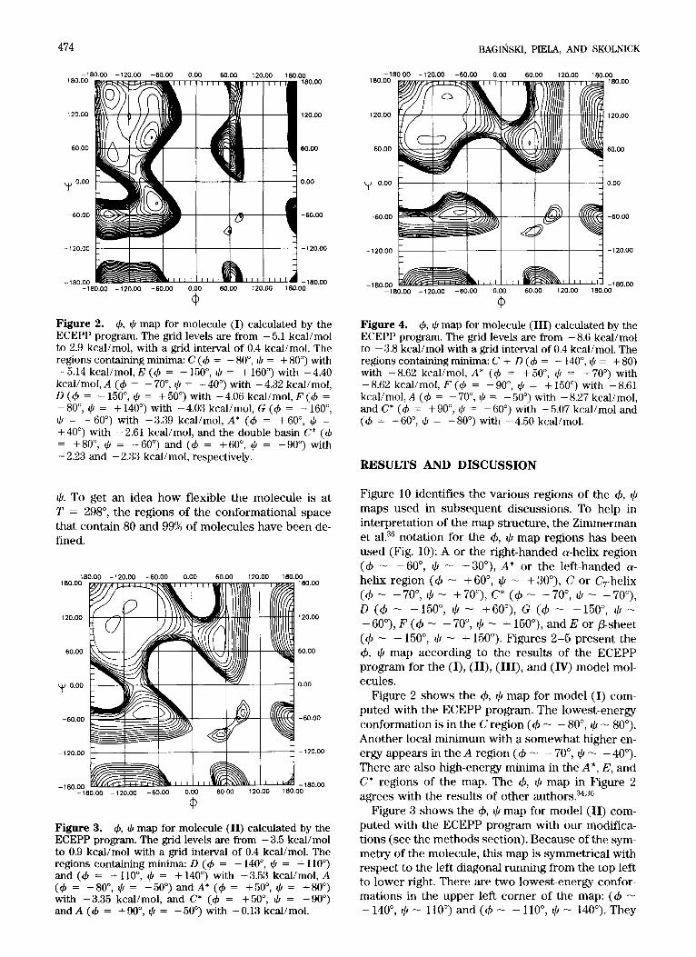
Figure 4. 4, J, map for molecule (111) calculated by the ECEPP program. The grid levels are from - 8.6 kcal/mol to - 3.8 kcal/mol with a grid interval of 0.4 kcal/mol. The regions containing minima: C + D (4 = - 140", $ = + SO) with -8.62 kcal/mol, A* (4 = + 50°, J, = +70") with -8.62 kcal/mol, F (4 = -go", J, = + 150") with - 8.61 kcal/mol, A (4 = - 70°, J, = - 50') with - 8.27 kcal/mol, and C* (4 = +go", J, = - SOo) with - 5.07 kcal/mol and (4 = +60", J, = -800) with -4.50 kcal/mol.
RESULTS AND DISCUSSION
Figure 10 identifies the various regions of the +, $ maps used in subsequent discussions. To help in interpretation of the map structure, the Zimmerman et al.36 notation for the 4, $ map regions has been used (Fig. 10): A or the right-handed a-helix region (d - -6O", $ - -3O"), A* or the left-handed a- helix region (+ - +60", $ - +30"), C or C,-helix ( 4 - -70°, $ - +70°),C* ( 4 - +70", 4 - -70"), D (+ - -150°, $ - +60"), G (4 - -150", q!/ - - 6V), F (4 - - 70", $ - + 150"), and E or P-sheet (+ - - 150", $ - + 150"). Figures 2-5 present the +, $ map according to the results of the ECEPP program for the (I), (II), (III), and (W) model mol- ecules.
Figure 2 shows the 4, $ map for model (JJ com- puted with the ECEPP program. The lowest-energy conformation is in the C region (+ - - 80", Ilr - 80"). Another local minimum with a somewhat higher en- ergy appears in the A region (+ - - 70°, $ - - 40"). There are also high-energy minima in the A", E, and C* regions of the map. The +, 9 map in Figure 2 agrees with the results of other author^?^,^^
Figure 3 shows the +, $ map for model (II) com- puted with the ECEPP program with our modifica- tions (see the methods section). Because of the sym- metry of the molecule, this map is symmetrical with respect to the left diagonal running from the top left to lower right. There are two lowest-energy confor- mations in the upper left corner of the map: (+ - - 140", $ - 110") and (+ - - 110", $ - 140"). They

PEPTIDE BOND MIMICKING UNIT 475
Figure 5. 4, J, map for molecule (IV) calculated by the ECEPP program. The grid levels are from - 13.0 kcal/mol to - 6.2 kcal/mol with a grid interval of 0.4 kcal/mol. The regions containing minima: A (4 = - 70°, J, = - 50") with - 12.99 kcalimol, D + E (4 = - 150°, I) = + 70") with - 12.87 kcal/mol, F + C (4 = -8O", I) = + 140") with - 12.78 kcal/mol, G (4 = - 150", I) - 60') with - 12.52 kcal/mol, and A* (I$ = +50°, I) = +60") with - 11.76 kcal/mol. In the region (4 = -110", I) = -120') is a maximum.
are separated by a small barrier and together with the neighboring region form a kind of basin similar to that formed by the C, D, E, and F subbasins for molecule (I). Two other local minima having the same energy (because of the symmetry) appear in t h e A ( 4 - -SO",$- -50")andA*(4-5Oo,$- 80") regions. One also sees a double-well basin in the C* region (4 - 50", + - - 90') and ( 4 - 90",
-180.00 -120.00 -60.00 0.00 60.00 120.00 180.00
" I I 1 1 120.00
60.00
y/ 0.00
-60.00
- 120.00
-180.00
- 120.00
- 1 80.00 -180.00 -120.00 -60.00 0.00 60.00 120.00 180.00
Q, Figure 6. 4, J, probability map for molecule (I) corre- sponding to Figure 2. (a). Contour defining the region of conformational space containing 80% of molecules at 25°C. (b). Contour defining the region of conformational space containing 99% of molecules at 25°C.
-180.00 -120.00 -60.00 0.00 60.00 120.00 180.00 180.00 180.00
120.00 120.00
60.00 60.00
y 0.00 0.00
-60.00 -60.00
-120.00 / \ 3 -120.00
Figure 7. 4, J, probability map for molecule (11) cor- responding to Figure 3. (a). Contour defining the region of conformational space containing 80% of molecules at 25°C. (b). Contour defining the region of conformational space containing 99% of molecules at 25°C.
$ - -5O"), similar to the ECEPP map for mole- cule (I).
Figure 4 presents the 4, $ map for model (111) computed with our modified ECEPP program (see the methods section). In this case, the deepest min- imum is located in the merged C + D area (4 - - 140°, I) - SO'), also similar to model molecule (I).
A bit higher-energy basin corresponds to the Fregion (4 - - 90", $ - 150"). The whole region close to the above minima may be seen as a single broad basin without distinct minima C, D, E, and F, which are present in model (I) in Figure 2. There is also another
-180.00 -120.00 -60.00 0.00 60.00 120.00 180.00
-60.00 m, '4 -60.00
L \ -I
1 -120.00 b \
/ \
-120.00 1 - 180.00 / I I I I I I I I I I I I j -180.00 I I fi-1 I I I TT-h
-180.00 -120.00 -60.00 0.00 60.00 120.00 180.00
0 Figure 8. 4, J, probability map for molecule (III) cor- responding to Figure 4. (a). Contour defining the region of conformational space containing 80% of molecules at 25°C. (b). Contour defining the region of conformational space containing 99% of molecules at 25°C.

476
F F'
BAGINSKI, PIELA, AND SKOLNICK
E F
-180.00 -120.00 -60.00 0.00 60.00 180.00
120.00
60.00
I ' I I 1 1 I -180.00
( I y 0.00
-60.00
-120.00
120.00 m
120.00
-60.00
- 120.00
I I I 1 I I 1 I I I I I I I 1 1 1 I -180.00 -180.00 -120.00 -60.00 0.00 60.00 120.00 180.00
0 Figure 9. 4, $ probability map for molecule (IV) cor- responding to Figure 5. (a). Contour defining the region of conformational space containing 80% of molecules at 25°C. (b). Contour defining the region of conformational space containing 99% of molecules at 25°C.
low-energy conformation of equal energy in region A* (4 - 50", $ - 70"). This basin is larger and a little deeper than that of region A (+ - - 70", $ - - 50"). The later basin has a different shape than the cor- responding one in Figure 2. There is also a double- well basin in region C* (+ - 60", @ - -80') and (4 - go", $ - -60"), similar to molecule (I) (Fig. 2). Although in general the +, $ map for model mol- ecule (111) resembles that for reference molecule (I), there is a small but significant preference in (111) for the left-handed helical conformation whereas in (I) for the right-handed one.
Figure 5 represents the +, $ map for model (IV) computed with the ECEPP program with our mod-
L 1 . I I I 4
120.00 1 1 120.00 1 t I I l l I I
- 120.00 I 1 1-1 -120.00
Figure 10. Subregions of the 4, $ map labeled as pro- posed by Zimmerman et a1?6 Only the regions taken into account in this article (containing minima) are labeled by capital letters.
ifications (see the methods section). This map ap- pears to be similar to the 4, $map in Figure 2 except the lowest minimum coresponds to the A (+ - - 70", @ - -50") region and not to the C region as for model (I). The difference, however, is small, being of the order of 0.1 kcal/mol. For model (IV), the minimum from C region moved up a bit and merged with that of the F area, giving one minimum in the region (+ - - 80", $ - 140"). There is another broad basin with a slightly higher energy in the merged D and E regions with the minimum at (4 - -150", $ - 70"). Both basins are reminiscent of the separate C, D, E, and F basins found for molecule (I). The A* basin (4 - 70", @ - 60") is also present as in Figure 2; however, the minimum in region C* has disap- peared.
These results for model (IV) agree well with the calculation of Deschrijver and Tourwe?O In the com- putations of these authors, as well as in our results (Fig. 5) , three basins are the most important: one in regions D + E, the second in C + F, and the third in A. The positions of the minima of these basins agree within 5". We reach the same general conclu- sion that model molecule (IV) is slightly more flex- ible than reference molecule (I). The reason for that is the replacement of the oxygen in the peptide bond by the hydrogen atom with smaller steric volume.
More compact information about flexibility of the molecules under study comes from the probability 4, (CI maps calculated at room temperature. The prob- ability 4, @ maps in Figures 6-9 correspond to the +, @ maps in Figures 2-5, respectively.
It can be seen in Figure 6 that among the ensemble of all molecules (I) at room temperature 80% are confined to the broad basin in the C, D, E, F, and A regions; 99% of the ensemble occupies a similar re- gion that in addition includes regions G and A*. All these regions together are a small part of the full 4, $ space; this indicates that the allowed conforma- tions are restricted. This picture is consistent with the distribution of 4, @ values for residues from 16 high-resolution protein crystal structures except that the G basin is not represented there?7 The absence of G conformations in nature despite their presence in the theoretical description in the ECEPP and AMBER38 force fields is seen by other authors as a systematic error appearing in these meth0ds.3~
Figures 7-9 correspond to the pseudopeptides' probability maps. It is seen that all populated basins represented in these figures are also present in Fig- ure 6. This is especially true when one takes into account the 80% contour. The 99% contours in Fig- ures 7-9 are slightly broader, especially in the A and A* regions, when compared with what happens for model molecule (I), Figure 6. The above observation is in particular valid in model (IV), Figure 9. Figures 7 and 8 contain an additional feature, namely, the 99% contour also extends into the F* region (+ - 60", $ - 150") not represented in Figure 6. However,

PEPTLDE BOND MIMICKING UNIT 477
the 80% contour alone does not extend to this area. The accessibility of A* and F' regions in models (11) and (III) pseudopeptides allows these systems to form not only right-handed but also left-handed hel- ical conformations. The reason for this is especially clear for model (11), which is symmetrical; therefore, both helical conformations are equally accessible.
To get a more quantitative measure of similarity of two molecules, a similarity index K (0 I K 5 1) has been defined in the accompanying article. K equal 0 means no similarity; K equal 1 corresponds to identity of the two molecules. In our case, the calculated K indices are
K,,II = 0.54, KI,III = 0.61, K1,p = 0.51
These values may be interpreted as though pseu- dopeptides (11), (110, and (IV) were similar to pep- tide molecule (I) by 54, 61, and 51%, respectively.
CONCLUSIONS
Based upon the present series of model calculations, one may conclude that a single substitution of the peptide bond by the ethylene bond is not likely to introduce any dramatic changes in conformational preferences. The neighborhood of the substituted site, when looking to the C terminus, is not expected to change significantly. This is in particular evident in Figures 5 and 9, which definitely and closely re- semble Figures 2 and 6 for model molecule (I). On the other hand, however, when looking to the N ter- minus the neighborhood of the point of substitution may have some additional possibility of adopting the A* (left-handed helix) conformation, as may be seen in Figures 4 and 8. A double-sequential substitution would not be expected to appreciably change the conformational restrictions, the only possible differ- ence perhaps being the equal preference of the right- and left-handed helical conformations.
This article is supported in part by the University of Warsaw and in part by the Ministry of National Education (Poland), as well as by the Polish National Council for Scientific Research (K.B.N.). L.P. is grateful to the Research Institute of Scripps Clinic for making possible his stay at the Institute.
References
1.
2.
3.
4.
5. 6.
IUPAC-IUB Commission on Biochemical Nomencla- ture, J. Mol. Biol., 52, 1 (1970). G.E. Schulz and R.H. Schrimer, In Principles of Protein Structure, Charles R. Cantor, Ed. Springer-Verlag, New York, 1979, pp. 18-26. J. Skolnick and A. Kolinski, Annu. Rev. Phys. Chem., 40, 207 (1990). T. Head-Gordon, M. Head-Gordon, M.J. Frish, C.L. Brooks 111, and J.A. Pople, J. Am. Chem. SOC., 113, 5989 (1991). H.A. Scheraga, Biopolymers, 22, 1 (1983). A.F. Spatola, In Chemistry and Biochemistry of Amino Acids, Peptides and Proteins, Vol. 7, B. Wein-
7.
8. 9.
10.
11.
12 13.
stein, Ed. Marcel Dekker, New York, 1983, pp. 267- 357. A. Aubry, G. Boussard, M.T. Cung, M. Marraud, and B. Vitoux, J. Chem. Phys. Phys. Chim. Biol., 85, 345 (1988). A.Aubry andM. Murraud,Biopolymers, 28,109 (1989). M.M. Shemyakin, N.A. Aldanova, E.I. Vinogradova, and M.Y. Feiniga, Tetrahedron Lett., 28, 1921 (1963). Y.A. Ovchinnikov, V.T. Ivanov, V.T. Evstratov, 1.1. Mik- haleva,V.F. Bystrov, S.L. Portnova, T.A. Balashova, E.N. Mershcheryakova, and V.M. Tulchinsky, Znt. J. Peptide Protein Res., 6,465 (1974). R.M. Wenger, Angew. Chem. Znt. Ed. Engl., 24, 77 (1985). R. Grupe and H. Niedrich, Chem. Berl., 99,3914 (1966). W. Ried and E. Schmidt, Liebigs Ann. Chem., 695,217 (1 966).
14. D. Hudson, R. Sharpe, and M. Szelke, Znt. J. Peptide Protein Res., 15, 122 (1980).
15. M.M. Hann, P.G. Sammers, P.D. Kennewell, and J.B. Taylor, J. Chem. SOC. Chem. Comm., 5, 234 (1980).
16. A.F. Spatola, H. Saneii, J.V. Edwards, A.L. Bettag, M.K. Anwer, P. Rowell, B. Browne, R. Lahti, and P. Von Voigtlander, Life Sci., 38, 1243 (1986).
17. M. Goodman and M. Chorev, Acc. Chem. Res., 12, 1 (1979).
18. A.E. Tonelli, Biopolymers, 15, 1615 (1976). 19. P. Manavalan and F.A. Momany,Biopolymers, 19,1943
(1980). 20. P. Deschrijver and D. Tourwe, FEBS Lett., 146, 353
(1982). 21. P.S. Stern, M. Chorev, M. Goodman, and A.T. Hagler,
Biopolymers, 22, 1901 (1983). 22. M. Hassan and M. Goodman, Biochemistry, 25, 7596
(1986). 23. M.T. Cox, D.W. Heaton, and J. Horbury, J. Chem. Soc.
Chem. Comm., 17, 799 (1980). 24. M.T. Cox, J.J. Gormley, C.E Hayward, and N.N. Petter,
J. Chem. SOC. Chem. C m m . , 17, 800 (1980). 25. M.M. Hann, P.G. Sammers, P.D. Kennewell, and J.B.
Taylor, J. Chem. SOC. Perkin Pans . Z., 307 (1982). 26. E. Ralston and J.L. De Coen, J. Mol. Biol., 84, 393
(1974). 27. F.A. Momany, R.F. McGuire, A.W. Burgess. and H.A.
Scheraga, J. Phys. Chem., 79,2361 (1975). 28. G. Nemethy, M.S. Pottle, and H.A. Scheraga, J. Phys.
Chem., 87, 1883 (1983). 29. J.A. Pople and G.A. Segal, J. C h m . Phys., 44, 3289
(1966). 30. J.E. Douglas, B.S. Rabinovitch, and F.S. Looney, J.
Chem. Phys., 23, 315 (1955). 31. N.L. Allinger and Y.H. Yuh, QCPE 395. Department of
Chemistry, University of Georgia, Athens, GA (1983); see also U. Burkert and N.L. Allinger, Molecular Me- chanics. ACS Monograph 177, American Chemical So- ciety, Washington, DC, 1982.
32. A. Almenigen, I.M. Anfinsen, and A. Haaland, Acta Chem. Scand., 24, 43 (1970).
33. I. Tokue, T. Fukuyama and K. Kuchitsu, J. Mol. Struct., 17, 207 (1973).
34. I.K. Roterman, M.H. Lambert, K.D. Gibson, and H.A. Scheraga, J. Biomol. Struct. Dynam., 7, 421 (1990).
35. W.G. Richards, Quantum Pharmacology, Butterworth & Co., London, UK, 1983, pp. 143-147.
36. S.S. Zimmerman, M.S. Pottle, G. Nemethy, and H.A. Scheraga, Macromolecules, 10, 1 (1977).
37. F.C. Bernstein, T.F. Koetzle, G.J.B. Williams, E.F. Meyer Jr., M.D. Brice, J.R. Rodgers, 0. Kennard, T. Schiman- ouchi, and M. Tasumi, J. Mol. Biol., 112, 535 (1977).
38. S.J. Weiner, P.A. Kollman, D.T. Nguyen, and D.A. Case, J. Comp. Chem., 7, 230 (1986).