the expression of the receptor for advanced glycation ... · the expression of the receptor for...
TRANSCRIPT
The expression of the receptor for advanced glycationendproducts (RAGE) is permissive for earlypancreatic neoplasiaRui Kanga, Tara Louxa, Daolin Tanga,1, Nicole E. Schapiroa, Philip Vernona, Kristen M. Liveseya, Alyssa Krasinskasb,Michael T. Lotzea,1, and Herbert J. Zeh IIIa,1
aDepartment of Surgery, Hillman Cancer Center, University of Pittsburgh Cancer Institute, Pittsburgh, PA 15219; and bDepartment of Pathology, University ofPittsburgh Medical Center, Pittsburgh, PA 15213
Edited by Mina J. Bissell, E. O. Lawrence Berkeley National Laboratory, Berkeley, CA, and approved March 23, 2012 (received for review August 23, 2011)
Pancreatic cancer is an almost uniformly lethal disease, character-ized by late diagnosis, early metastasis, resistance to chemother-apy, and early mutation of the Kras oncogene. Here we show thatthe receptor for advanced glycation endproducts (RAGE) is re-quired for the activation of interleukin 6 (IL-6)–mediated mitochon-drial signal transducers and activators of transcription 3 (STAT3)signaling in pancreatic carcinogenesis. RAGE expression correlateswith elevated levels of autophagy in pancreatic cancer in vivo andin vitro, and this heightened state of autophagy is required forIL-6–induced STAT3 activation. To further explore the intersectionof RAGE, autophagy, and pancreatic carcinogenesis, we createda transgenic murine model, backcrossing RAGE-null mice to a spon-taneous mouse model of pancreatic cancer, Pdx1-Cre:KrasG12D/+
(KC). Targeted ablation of Rage in KC mice delayed neoplasia de-velopment, decreased levels of autophagy, and inhibited mito-chondrial STAT3 activity and subsequent ATP production. Ourresults suggest a critical role for RAGE expression in the earlieststages of pancreatic carcinogenesis, potentially acting as the “auto-phagic switch,” regulating mitochondrial STAT3 signaling.
oncogenesis | bioenergetics | inflammation | metabolism | high-mobilitygroup box 1
Pancreatic cancer ranks as the fourth leading cause of cancerdeath, accounting for 6–7% of all cancer-related deaths in the
United States, in 2011 (1). Most pancreatic ductal adenocarcino-mas (PDA) are thought to arise from well-defined precursor le-sions, termed pancreatic ductal intraepithelial neoplasia (PanIN)(2). Many human PanIN lesions do not progress to invasive car-cinomas, so defining the events that drive carcinogenesis in theemergent tumor microenvironment is of critical importance.Studies into human pancreatic carcinogenesis have been greatlyfacilitated by the development of a genetically engineered mousemodel that expresses oncogenicKras under a pancreatic promoterPdx1-Cre:KrasG12D/+ (KC) (3). A more detailed understandingof how these pathways accelerate pancreatic carcinogenesis mayallow improved therapeutic strategies.The receptor for advanced glycation endproducts (RAGE) is
a member of the Ig superfamily. RAGE and its ligands, includinghigh-mobility group box 1 (HMGB1) and S100, are linked to thedevelopment and progression of several cancers by facilitating themaintenance of a chronic inflammatory state (4) and/or by pro-motion of metastases (5). We previously observed that RAGEsustains autophagy and limits apoptosis, promoting pancreatic tu-mor cell survival during chemotherapy and oxidative stress in vivoand in vitro (6, 7). Autophagy is an essential catabolic process bywhich cells break down old or damaged organelles and proteins (8).Autophagy promotes cell survival and supports metabolism duringcell stress (9). Conversely, apoptosis promotes tumor growth earlyin the development of cancer (10) during periods of inhibition ofautophagy with a subsequent switch to suppressed apoptosis, ac-quired later during the temporal development of more advancedcancers. Pancreatic tumors have elevated autophagy under basal
conditions compared with other epithelial cancers (11). Inhibitionof autophagy in pancreatic tumor cells augments production ofreactive oxygen species, increases DNA damage, and limits effec-tive metabolism with decreased mitochondrial oxidative phos-phorylation, resulting in significant inhibition of pancreatic tumorgrowth (12). Despite the growing body of literature supporting theimportance of autophagy andRAGE inpancreatic cancer (6, 7, 12),the role of RAGE-mediated autophagy in pancreatic carcinogen-esis and the underlying mechanisms for such a phenomenon havenot been addressed.We demonstrate here that RAGE expression is permissive for
the development of early pancreatic neoplasia by enhancing themitochondrial interleukin 6 (IL-6)/signal transducers and acti-vators of transcription 3 (STAT3) pathway in pancreatic tumorcells. These effects depend on autophagy and lead to enhancedATP production in vitro and in vivo.
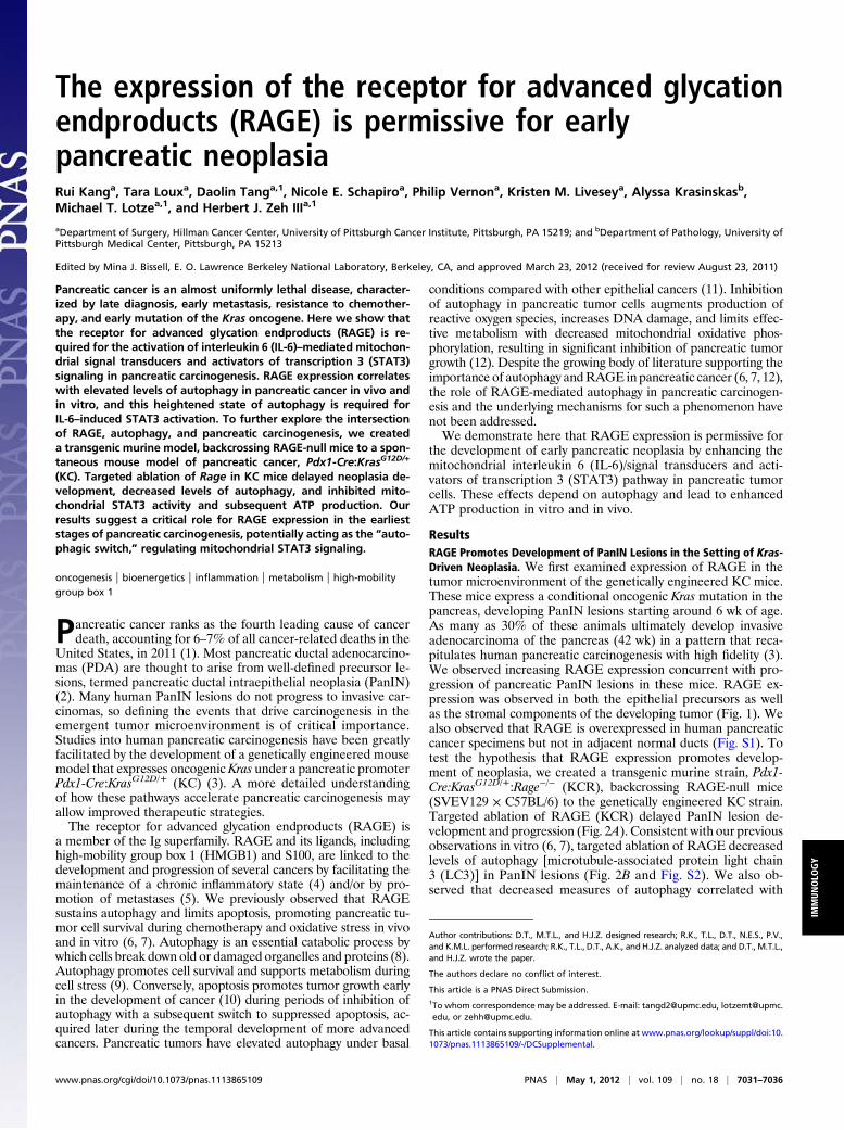
ResultsRAGE Promotes Development of PanIN Lesions in the Setting of Kras-Driven Neoplasia. We first examined expression of RAGE in thetumor microenvironment of the genetically engineered KC mice.These mice express a conditional oncogenic Kras mutation in thepancreas, developing PanIN lesions starting around 6 wk of age.As many as 30% of these animals ultimately develop invasiveadenocarcinoma of the pancreas (42 wk) in a pattern that reca-pitulates human pancreatic carcinogenesis with high fidelity (3).We observed increasing RAGE expression concurrent with pro-gression of pancreatic PanIN lesions in these mice. RAGE ex-pression was observed in both the epithelial precursors as wellas the stromal components of the developing tumor (Fig. 1). Wealso observed that RAGE is overexpressed in human pancreaticcancer specimens but not in adjacent normal ducts (Fig. S1). Totest the hypothesis that RAGE expression promotes develop-ment of neoplasia, we created a transgenic murine strain, Pdx1-Cre:KrasG12D/+:Rage−/− (KCR), backcrossing RAGE-null mice(SVEV129 × C57BL/6) to the genetically engineered KC strain.Targeted ablation of RAGE (KCR) delayed PanIN lesion de-velopment and progression (Fig. 2A). Consistent with our previousobservations in vitro (6, 7), targeted ablation of RAGE decreasedlevels of autophagy [microtubule-associated protein light chain3 (LC3)] in PanIN lesions (Fig. 2B and Fig. S2). We also ob-served that decreased measures of autophagy correlated with
Author contributions: D.T., M.T.L., and H.J.Z. designed research; R.K., T.L., D.T., N.E.S., P.V.,and K.M.L. performed research; R.K., T.L., D.T., A.K., andH.J.Z. analyzed data; and D.T., M.T.L.,and H.J.Z. wrote the paper.
The authors declare no conflict of interest.
This article is a PNAS Direct Submission.1To whom correspondence may be addressed. E-mail: [email protected], [email protected], or [email protected].
This article contains supporting information online at www.pnas.org/lookup/suppl/doi:10.1073/pnas.1113865109/-/DCSupplemental.
www.pnas.org/cgi/doi/10.1073/pnas.1113865109 PNAS | May 1, 2012 | vol. 109 | no. 18 | 7031–7036
IMMUNOLO
GY
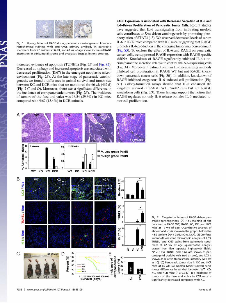
increased evidence of apoptosis (TUNEL) (Fig. 2B and Fig. S2).Decreased autophagy and increased apoptosis are associated withdecreased proliferation (Ki67) in the emergent neoplastic micro-environment (Fig. 2B). At the late stage of pancreatic carcino-genesis, we found a difference in animal survival and tumor sizebetween KC and KCR mice that we monitored for 66 wk (462 d)(Fig. 2 C and D). Moreover, there was a significant difference inthe incidence of extrapancreatic tumors (Fig. 2E). The incidenceof tumors of the face and vulva was 16/54 (29.6%) in KC micecompared with 9/67 (13.4%) in KCR animals.
RAGE Expression Is Associated with Decreased Secretion of IL-6 andIL-6–Driven Proliferation of Pancreatic Tumor Cells. Recent studieshave suggested that IL-6 transsignaling from infiltrating myeloidcells contributes to Kras-driven carcinogenesis by promoting phos-phorylation of STAT3 (13). We observed decreased levels of serumIL-6 in KCR mice compared with KC mice, suggesting that RAGEpromotes IL-6 production in the emerging tumormicroenvironment(Fig. S3). To explore the effect of IL-6 and RAGE on pancreaticcancer cells, we suppressed RAGE expression with RAGE-specificshRNA. Knockdown of RAGE significantly inhibited IL-6 auto-crine/paracrine secretion relative to control shRNA-expressing cells(Fig. 3A). Moreover, treatment with an IL-6 neutralizing antibodyinhibited cell proliferation in RAGE WT but not RAGE knock-down pancreatic cancer cells (Fig. 3B). In addition, knockdown ofRAGE inhibited exogenous IL-6–induced cell proliferation (Fig.3C). Colony-formation assays showed that IL-6 enhanced thelong-term survival of RAGE WT Panc02 cells but not RAGEknockdown cells (Fig. 3D). These findings support the notion thatRAGE regulates not only IL-6 release but also IL-6–mediated tu-mor cell proliferation.
Fig. 1. Up-regulation of RAGE during pancreatic carcinogenesis. Immuno-histochemical staining with anti-RAGE primary antibody in pancreaticspecimens from KC animals at 6, 24, and 48 wk of age shows increased RAGEexpression in peritumoral stroma and dysplastic ducts as lesions progress.
Fig. 2. Targeted ablation of RAGE delays pan-creatic carcinogenesis. (A) H&E staining of thepancreas in RAGE WT, RAGE KO, KC, and KCRmice at 12 wk of age. Quantitative analysis ofabnormal ducts is shown in the graphs below theH&E sections (*P < 0.05, KC vs. KCR). (B) Confocalimmunofluorescent microscopic analysis of LC3,TUNEL, and Ki67 stains from pancreatic speci-mens at 42 wk of age (quantitative analysisdrawn from five separate high-power fields;*P < 0.05). TUNEL and Ki67 are shown as per-centage of positive cells (red arrows), and LC3 isshown as relative fluorescence intensity (WT setas 10). (C) Pancreatic tumor size in KC and KCRmice at 66 wk. (D) Kaplan–Meier survival curveshows difference in survival between WT, KO,KC, and KCR mice (P = 0.037). (E ) Incidence oftumors of the face and vulva in KCR mice issignificantly decreased compared with KC.
7032 | www.pnas.org/cgi/doi/10.1073/pnas.1113865109 Kang et al.
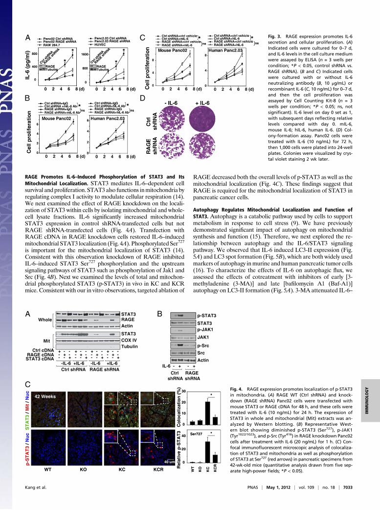
RAGE Promotes IL-6–Induced Phosphorylation of STAT3 and ItsMitochondrial Localization. STAT3 mediates IL-6–dependent cellsurvival and proliferation. STAT3also functions inmitochondria byregulating complex I activity to modulate cellular respiration (14).We next examined the effect of RAGE knockdown on the locali-zation of STAT3 within cells by isolating mitochondrial and whole-cell lysate fractions. IL-6 significantly increased mitochondrialSTAT3 expression in control shRNA-transfected cells but notRAGE shRNA-transfected cells (Fig. 4A). Transfection withRAGE cDNA in RAGE knockdown cells restored IL-6–inducedmitochondrial STAT3 localization (Fig. 4A). Phosphorylated Ser727
is important for the mitochondrial localization of STAT3 (14).Consistent with this observation knockdown of RAGE inhibitedIL-6–induced STAT3 Ser727 phosphorylation and the upstreamsignaling pathways of STAT3 such as phosphorylation of Jak1 andSrc (Fig. 4B). Next we examined the levels of total and mitochon-drial phosphorylated STAT3 (p-STAT3) in vivo in KC and KCRmice. Consistent with our in vitro observations, targeted ablation of
RAGE decreased both the overall levels of p-STAT3 as well as themitochondrial localization (Fig. 4C). These findings suggest thatRAGE is required for the mitochondrial localization of STAT3 inpancreatic cancer cells.
Autophagy Regulates Mitochondrial Localization and Function ofSTAT3. Autophagy is a catabolic pathway used by cells to supportmetabolism in response to cell stress (9). We have previouslydemonstrated significant impact of autophagy on mitochondrialsynthesis and function (15). Therefore, we next explored the re-lationship between autophagy and the IL-6/STAT3 signalingpathway. We observed that IL-6 induced LC3-II expression (Fig.5A) and LC3 spot formation (Fig. 5B), which are both widely usedmarkers of autophagy inmurine and human pancreatic tumor cells(16). To characterize the effects of IL-6 on autophagic flux, weassessed the effects of cotreatment with inhibitors of early [3-methyladenine (3-MA)] and late [bafilomycin A1 (Baf-A1)]autophagy on LC3-II formation (Fig. 5A). 3-MA attenuated IL-6–
Fig. 3. RAGE expression promotes IL-6secretion and cellular proliferation. (A)Indicated cells were cultured for 0–7 d,and IL-6 levels in the cell culture mediumwere assayed by ELISA (n = 3 wells percondition; *P < 0.05, control shRNA vs.RAGE shRNA). (B and C) Indicated cellswere cultured with or without IL-6neutralizing antibody (B, 10 μg/mL) orrecombinant IL-6 (C, 10 ng/mL) for 0–7 d,and then the cell proliferation wasassayed by Cell Counting Kit-8 (n = 3wells per condition; *P < 0.05; ns, notsignificant). IL-6 level on day 0 set as 1,with subsequent days reflecting relativelevels compared with day 0. mIL-6,mouse IL-6; hIL-6, human IL-6. (D) Col-ony-formation assay. Panc02 cells weretreated with IL-6 (10 ng/mL) for 72 h,then 1,000 cells were plated into 24-wellplates. Colonies were visualized by crys-tal violet staining 2 wk later.
Fig. 4. RAGE expression promotes localization of p-STAT3in mitochondria. (A) RAGE WT (Ctrl shRNA) and knock-down (RAGE shRNA) Panc02 cells were transfected withmouse STAT3 or RAGE cDNA for 48 h, and these cells weretreated with IL-6 (10 ng/mL) for 24 h. The expression ofSTAT3 in whole and mitochondrial (Mit) extracts was an-alyzed by Western blotting. (B) Representative West-ern blot showing diminished p-STAT3 (Ser727), p-JAK1(Tyr1022/1023), and p-Src (Tyr416) in RAGE knockdown Panc02cells after treatment with IL-6 (20 ng/mL) for 1 h. (C) Con-focal immunofluorescent microscopic analysis of colocaliza-tion of STAT3 and mitochondria as well as phosphorylationof STAT3 at Ser727 (red arrows) in pancreatic specimens from42-wk-old mice (quantitative analysis drawn from five sep-arate high-power fields; *P < 0.05).
Kang et al. PNAS | May 1, 2012 | vol. 109 | no. 18 | 7033
IMMUNOLO
GY
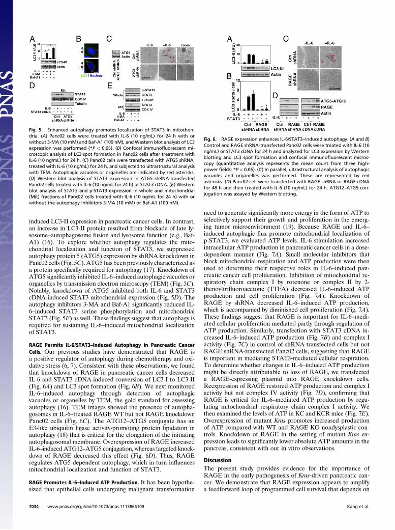
induced LC3-II expression in pancreatic cancer cells. In contrast,an increase in LC3-II protein resulted from blockade of late ly-sosome–autophagosome fusion and lysosome function (e.g., Baf-A1) (16). To explore whether autophagy regulates the mito-chondrial localization and function of STAT3, we suppressedautophagy protein 5 (ATG5) expression by shRNA knockdown inPanc02 cells (Fig. 5C). ATG5 has been previously characterized asa protein specifically required for autophagy (17). Knockdown ofATG5 significantly inhibited IL-6–induced autophagic vacuoles ororganelles by transmission electron microscopy (TEM) (Fig. 5C).Notably, knockdown of ATG5 inhibited both IL-6 and STAT3cDNA-induced STAT3 mitochondrial expression (Fig. 5D). Theautophagy inhibitors 3-MA and Baf-A1 significantly reduced IL-6–induced STAT3 serine phosphorylation and mitochondrialSTAT3 (Fig. 5E) as well. These findings suggest that autophagy isrequired for sustaining IL-6–induced mitochondrial localizationof STAT3.
RAGE Permits IL-6/STAT3–Induced Autophagy in Pancreatic CancerCells. Our previous studies have demonstrated that RAGE isa positive regulator of autophagy during chemotherapy and oxi-dative stress (6, 7). Consistent with these observations, we foundthat knockdown of RAGE in pancreatic cancer cells decreasedIL-6 and STAT3 cDNA-induced conversion of LC3-I to LC3-II(Fig. 6A) and LC3 spot formation (Fig. 6B). We next monitoredIL-6–induced autophagy through detection of autophagicvacuoles or organelles by TEM, the gold standard for assessingautophagy (16). TEM images showed the presence of autopha-gosomes in IL-6–treated RAGE WT but not RAGE knockdownPanc02 cells (Fig. 6C). The ATG12–ATG5 conjugate has anE3-like ubiquitin ligase activity-promoting protein lipidation inautophagy (18) that is critical for the elongation of the initiatingautophagosomal membrane. Overexpression of RAGE increasedIL-6–induced ATG12–ATG5 conjugation, whereas targeted knock-down of RAGE decreased this effect (Fig. 6D). Thus, RAGEregulates ATG5-dependent autophagy, which in turn influencesmitochondrial localization and function of STAT3.
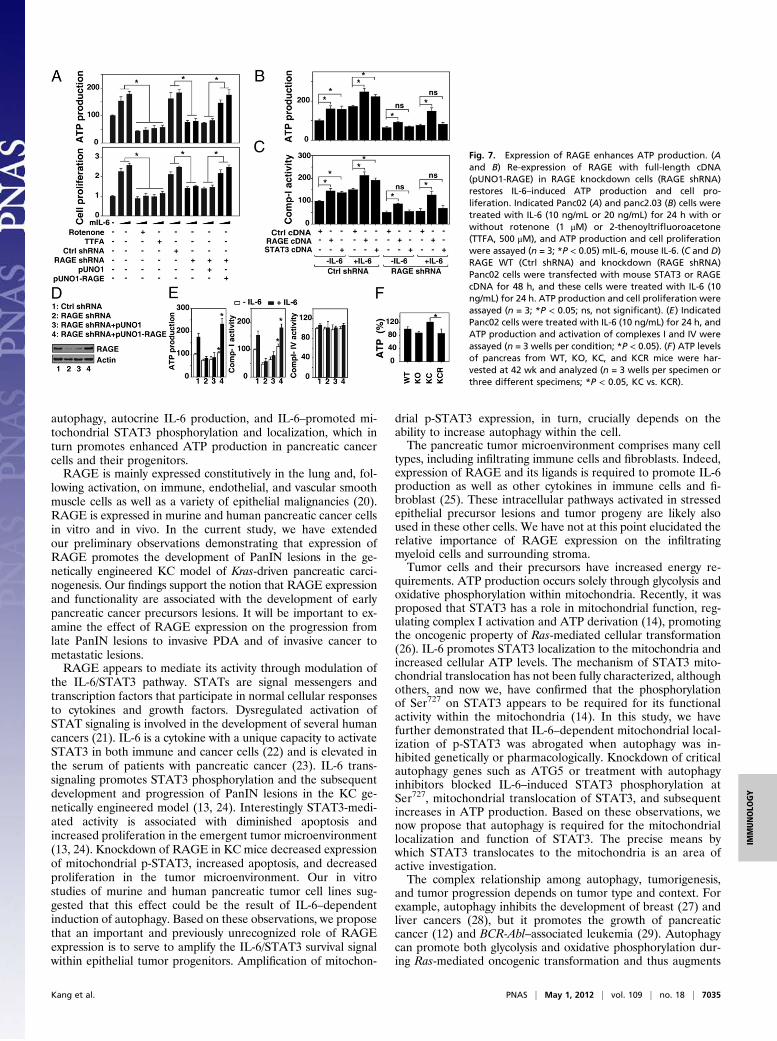
RAGE Promotes IL-6–Induced ATP Production. It has been hypothe-sized that epithelial cells undergoing malignant transformation
need to generate significantly more energy in the form of ATP toselectively support their growth and proliferation in the emerg-ing tumor microenvironment (19). Because RAGE and IL-6–induced autophagic flux promote mitochondrial localization ofp-STAT3, we evaluated ATP levels. IL-6 stimulation increasedintracellular ATP production in pancreatic cancer cells in a dose-dependent manner (Fig. 7A). Small molecular inhibitors thatblock mitochondrial respiration and ATP production were thenused to determine their respective roles in IL-6–induced pan-creatic cancer cell proliferation. Inhibition of mitochondrial re-spiratory chain complex I by rotenone or complex II by 2-thenoyltrifluoroacetone (TTFA) decreased IL-6–induced ATPproduction and cell proliferation (Fig. 7A). Knockdown ofRAGE by shRNA decreased IL-6–induced ATP production,which is accompanied by diminished cell proliferation (Fig. 7A).These findings suggest that RAGE is important for IL-6–medi-ated cellular proliferation mediated partly through regulation ofATP production. Similarly, transfection with STAT3 cDNA in-creased IL-6–induced ATP production (Fig. 7B) and complex Iactivity (Fig. 7C) in control of shRNA-transfected cells but notRAGE shRNA-transfected Panc02 cells, suggesting that RAGEis important in mediating STAT3-mediated cellular respiration.To determine whether changes in IL-6–induced ATP productionmight be directly attributable to loss of RAGE, we transfecteda RAGE-expressing plasmid into RAGE knockdown cells.Reexpression of RAGE restored ATP production and complex Iactivity but not complex IV activity (Fig. 7D), confirming thatRAGE is critical for IL-6–mediated ATP production by regu-lating mitochondrial respiratory chain complex I activity. Wethen examined the levels of ATP in KC and KCR mice (Fig. 7E).Overexpression of mutant Kras promotes increased productionof ATP compared with WT and RAGE KO nondysplastic con-trols. Knockdown of RAGE in the setting of mutant Kras ex-pression leads to significantly lower absolute ATP amounts in thepancreas, consistent with our in vitro observations.
DiscussionThe present study provides evidence for the importance ofRAGE in the early pathogenesis of Kras-driven pancreatic can-cer. We demonstrate that RAGE expression appears to amplifya feedforward loop of programmed cell survival that depends on
Fig. 5. Enhanced autophagy promotes localization of STAT3 in mitochon-dria. (A) Panc02 cells were treated with IL-6 (10 ng/mL) for 24 h with orwithout 3-MA (10 mM) and Baf-A1 (100 nM), andWestern blot analysis of LC3expression was performed (*P < 0.05). (B) Confocal immunofluorescent mi-croscopic analysis of LC3 spot formation in Panc02 cells after treatment withIL-6 (10 ng/mL) for 24 h. (C) Panc02 cells were transfected with ATG5 shRNA,treated with IL-6 (10 ng/mL) for 24 h, and subjected to ultrastructural analysiswith TEM. Autophagic vacuoles or organelles are indicated by red asterisks.(D) Western blot analysis of STAT3 expression in ATG5 shRNA-transfectedPanc02 cells treated with IL-6 (10 ng/mL for 24 h) or STAT3 cDNA. (E) Westernblot analysis of STAT3 and p-STAT3 expression in whole and mitochondrial(Mit) fractions of Panc02 cells treated with IL-6 (10 ng/mL for 24 h) with orwithout the autophagy inhibitors 3-MA (10 mM) or Baf-A1 (100 nM).
Fig. 6. RAGE expression enhances IL-6/STAT3–induced autophagy. (A and B)Control and RAGE shRNA-transfected Panc02 cells were treated with IL-6 (10ng/mL) or STAT3 cDNA for 24 h and analyzed for LC3 expression by Westernblotting and LC3 spot formation and confocal immunofluorescent micros-copy (quantitative analysis represents the mean count from three high-power fields; *P < 0.05). (C) In parallel, ultrastructural analysis of autophagicvacuoles and organelles was performed. These are represented by redasterisks. (D) Panc02 cell were transfected with RAGE shRNA or RAGE cDNAfor 48 h and then treated with IL-6 (10 ng/mL) for 24 h. ATG12–ATG5 con-jugation was assayed by Western blotting.
7034 | www.pnas.org/cgi/doi/10.1073/pnas.1113865109 Kang et al.
autophagy, autocrine IL-6 production, and IL-6–promoted mi-tochondrial STAT3 phosphorylation and localization, which inturn promotes enhanced ATP production in pancreatic cancercells and their progenitors.RAGE is mainly expressed constitutively in the lung and, fol-
lowing activation, on immune, endothelial, and vascular smoothmuscle cells as well as a variety of epithelial malignancies (20).RAGE is expressed in murine and human pancreatic cancer cellsin vitro and in vivo. In the current study, we have extendedour preliminary observations demonstrating that expression ofRAGE promotes the development of PanIN lesions in the ge-netically engineered KC model of Kras-driven pancreatic carci-nogenesis. Our findings support the notion that RAGE expressionand functionality are associated with the development of earlypancreatic cancer precursors lesions. It will be important to ex-amine the effect of RAGE expression on the progression fromlate PanIN lesions to invasive PDA and of invasive cancer tometastatic lesions.RAGE appears to mediate its activity through modulation of
the IL-6/STAT3 pathway. STATs are signal messengers andtranscription factors that participate in normal cellular responsesto cytokines and growth factors. Dysregulated activation ofSTAT signaling is involved in the development of several humancancers (21). IL-6 is a cytokine with a unique capacity to activateSTAT3 in both immune and cancer cells (22) and is elevated inthe serum of patients with pancreatic cancer (23). IL-6 trans-signaling promotes STAT3 phosphorylation and the subsequentdevelopment and progression of PanIN lesions in the KC ge-netically engineered model (13, 24). Interestingly STAT3-medi-ated activity is associated with diminished apoptosis andincreased proliferation in the emergent tumor microenvironment(13, 24). Knockdown of RAGE in KC mice decreased expressionof mitochondrial p-STAT3, increased apoptosis, and decreasedproliferation in the tumor microenvironment. Our in vitrostudies of murine and human pancreatic tumor cell lines sug-gested that this effect could be the result of IL-6–dependentinduction of autophagy. Based on these observations, we proposethat an important and previously unrecognized role of RAGEexpression is to serve to amplify the IL-6/STAT3 survival signalwithin epithelial tumor progenitors. Amplification of mitochon-
drial p-STAT3 expression, in turn, crucially depends on theability to increase autophagy within the cell.The pancreatic tumor microenvironment comprises many cell
types, including infiltrating immune cells and fibroblasts. Indeed,expression of RAGE and its ligands is required to promote IL-6production as well as other cytokines in immune cells and fi-broblast (25). These intracellular pathways activated in stressedepithelial precursor lesions and tumor progeny are likely alsoused in these other cells. We have not at this point elucidated therelative importance of RAGE expression on the infiltratingmyeloid cells and surrounding stroma.Tumor cells and their precursors have increased energy re-
quirements. ATP production occurs solely through glycolysis andoxidative phosphorylation within mitochondria. Recently, it wasproposed that STAT3 has a role in mitochondrial function, reg-ulating complex I activation and ATP derivation (14), promotingthe oncogenic property of Ras-mediated cellular transformation(26). IL-6 promotes STAT3 localization to the mitochondria andincreased cellular ATP levels. The mechanism of STAT3 mito-chondrial translocation has not been fully characterized, althoughothers, and now we, have confirmed that the phosphorylationof Ser727 on STAT3 appears to be required for its functionalactivity within the mitochondria (14). In this study, we havefurther demonstrated that IL-6–dependent mitochondrial local-ization of p-STAT3 was abrogated when autophagy was in-hibited genetically or pharmacologically. Knockdown of criticalautophagy genes such as ATG5 or treatment with autophagyinhibitors blocked IL-6–induced STAT3 phosphorylation atSer727, mitochondrial translocation of STAT3, and subsequentincreases in ATP production. Based on these observations, wenow propose that autophagy is required for the mitochondriallocalization and function of STAT3. The precise means bywhich STAT3 translocates to the mitochondria is an area ofactive investigation.The complex relationship among autophagy, tumorigenesis,
and tumor progression depends on tumor type and context. Forexample, autophagy inhibits the development of breast (27) andliver cancers (28), but it promotes the growth of pancreaticcancer (12) and BCR-Abl–associated leukemia (29). Autophagycan promote both glycolysis and oxidative phosphorylation dur-ing Ras-mediated oncogenic transformation and thus augments
Fig. 7. Expression of RAGE enhances ATP production. (Aand B) Re-expression of RAGE with full-length cDNA(pUNO1-RAGE) in RAGE knockdown cells (RAGE shRNA)restores IL-6–induced ATP production and cell pro-liferation. Indicated Panc02 (A) and panc2.03 (B) cells weretreated with IL-6 (10 ng/mL or 20 ng/mL) for 24 h with orwithout rotenone (1 μM) or 2-thenoyltrifluoroacetone(TTFA, 500 μM), and ATP production and cell proliferationwere assayed (n = 3; *P < 0.05) mIL-6, mouse IL-6. (C and D)RAGE WT (Ctrl shRNA) and knockdown (RAGE shRNA)Panc02 cells were transfected with mouse STAT3 or RAGEcDNA for 48 h, and these cells were treated with IL-6 (10ng/mL) for 24 h. ATP production and cell proliferation wereassayed (n = 3; *P < 0.05; ns, not significant). (E) IndicatedPanc02 cells were treated with IL-6 (10 ng/mL) for 24 h, andATP production and activation of complexes I and IV wereassayed (n = 3 wells per condition; *P < 0.05). (F) ATP levelsof pancreas from WT, KO, KC, and KCR mice were har-vested at 42 wk and analyzed (n = 3 wells per specimen orthree different specimens; *P < 0.05, KC vs. KCR).
Kang et al. PNAS | May 1, 2012 | vol. 109 | no. 18 | 7035
IMMUNOLO
GY
tumor growth (30, 31). Our findings indicate that RAGE-medi-ated autophagy is required for enhanced ATP production andproliferation in pancreatic cancer cells, mostly via regulation ofmitochondrial STAT3. Our findings may provide the missing linkbetween two factors that are known to drive pancreatic carci-nogenesis, enhanced autophagy (mediated by RAGE), and dys-functional metabolism (mediated by STAT3).RAGE is a positive regulator of autophagy during both anti-
cancer therapy and pancreatic carcinogenesis. Radiation therapyand some forms of chemotherapy, such as gemcitabine, rely onreactive oxygen species generation and toxicity to eradicate tumorcells. Reactive oxygen species, in turn, increase the activity of NF-κB, resulting inRAGEexpression (6). This up-regulation ofRAGEprotects pancreatic tumor cells from further oxidative injury andincreases drug resistance by enhancing autophagy and decreasingapoptosis (6, 7). We found that RAGE increased autophagic fluxby regulating ATG12–ATG5 conjugation. RAGE also regulatesactivation of the mammalian target of rapamycin (mTOR) andBeclin 1–PI3K class III complex (32), suggesting that there aremultiple roles for RAGE in the enhancement of autophagy withinpancreatic cancer cells. In vivo, targeted deletion of RAGE in theKrasG12D/+ spontaneous KC cancer model decreased Kras-drivenautophagy and subsequently mitochondrial STAT3 and ATPproduction, confirming the interaction between autophagy andSTAT3. Interestingly, intracellular HMGB1, the cognate ligandfor RAGE, regulates mitochondrial quality via mitophagy (15).Further studies are required to establish the relationship be-tween HMGB1 and RAGE in autophagy-mediated mitochon-drial STAT3 activation and their roles within the emergentpancreatic tumor microenvironment. We hypothesize thatRAGE expression serves as an ”autophagic switch,” enablingsuppression of apoptosis and promotion of survival pathwaysrequisite later during carcinogenesis (10).We have previously established that knockdown of RAGE with
shRNA significantly alters the biology of PDA (6, 7). DiNorcia et al.have recently shown in an accelerated compound p16-deleted Krasmutant tumor model that RAGE ablation limits development of
frank carcinoma (33). No mechanisms for this finding were pro-posed, and our study extends and confirms their study, now showingthe critical role of the IL-6/STAT3 signaling pathway. Interestingly,we have not been able to derive a PDA from a RAGE-null animalat this point but will continue to try. The principle findings relatenot to the phenotype of PDA in the setting of RAGE knockdown(6, 7) but rather its link to early precursor formation.Webelieve theobservation that knockdown of RAGE attenuates development ofPDA precursor lesions through an IL-6/STAT3 autophagic path-way is important, suggesting unique therapeutic strategies.In summary, we demonstrate here that RAGE is a critical
mediator of pancreatic carcinogenesis through its ability to am-plify IL-6–induced autophagic translocation of STAT3 to themitochondria. Ultimately, this amplification leads to increasedATP production by fully transformed tumor cells and early epi-thelial precursor lesions. We hypothesize that the RAGE/IL-6–dependent programmed cell survival pathways provide a meansfor epithelial tumor precursor cells to gain a selective growthadvantage within the tumor microenvironment.
Materials and MethodsCell Lines and Reagents. Human (Pan2.03) ormouse (Panc02) pancreatic tumorcells, mouse RAW264.7 cells, and human umbilical vein endothelial cells werepurchased fromAmericanTypeCultureCollection. Theneutralizing antibodiesto human or mouse IL-6 and RAGE were obtained from R&D Systems. Theantibodies to STAT3, p-STAT3 (Ser727), p-JAK1 (Tyr1022/1023), JAK1, p-Src(Tyr416), Src, COX IV, and ATG12–ATG5 were obtained from Cell SignalingTechnology. The antibodies to LC3 and ATG5 were obtained from Novus. Theantibodies to actin, tubulin, and RAGE were obtained from Sigma. The anti-body to Ki67 was from Abcam. Human and mouse IL-6 protein and ELISA kitwere obtained from R&D Systems. Other chemical reagents were from Sigma.
Human Samples. Human pancreatic adenocarcinoma specimens were col-lected from patients who underwent surgical procedures. Samples wereobtained according to an institutional review board-approved clinical pro-tocol at the University of Pittsburgh Medical Center.
Full methods and associated references are available in the SI Materialsand Methods.
1. Siegel R, Ward E, Brawley O, Jemal A (2011) Cancer statistics, 2011: The impact ofeliminating socioeconomic and racial disparities on premature cancer deaths. CACancer J Clin 61:212–236.
2. Hruban RH, Adsay NV (2009) Molecular classification of neoplasms of the pancreas.Hum Pathol 40:612–623.
3. Hingorani SR, et al. (2003) Preinvasive and invasive ductal pancreatic cancer and itsearly detection in the mouse. Cancer Cell 4:437–450.
4. Gebhardt C, et al. (2008) RAGE signaling sustains inflammation and promotes tumordevelopment. J Exp Med 205:275–285.
5. Taguchi A, et al. (2000) Blockade of RAGE-amphoterin signalling suppresses tumourgrowth and metastases. Nature 405:354–360.
6. Kang R, et al. (2011) The Receptor for Advanced Glycation End-products (RAGE)protects pancreatic tumor cells against oxidative injury. Antioxid Redox Signal 15:2175–2184.
7. Kang R, et al. (2010) The receptor for advanced glycation end products (RAGE) sus-tains autophagy and limits apoptosis, promoting pancreatic tumor cell survival. CellDeath Differ 17:666–676.
8. Yang Z, Klionsky DJ (2010) Eaten alive: A history of macroautophagy. Nat Cell Biol 12:814–822.
9. Rabinowitz JD, White E (2010) Autophagy and metabolism. Science 330:1344–1348.10. Tang D, Lotze MT, Kang R, Zeh HJ (2011) Apoptosis promotes early tumorigenesis.
Oncogene 30:1851–1854.11. Fujii S, et al. (2008) Autophagy is activated in pancreatic cancer cells and correlates
with poor patient outcome. Cancer Sci 99:1813–1819.12. Yang S, et al. (2011) Pancreatic cancers require autophagy for tumor growth. Genes
Dev 25:717–729.13. Lesina M, et al. (2011) Stat3/Socs3 activation by IL-6 transsignaling promotes pro-
gression of pancreatic intraepithelial neoplasia and development of pancreaticcancer. Cancer Cell 19:456–469.
14. Wegrzyn J, et al. (2009) Function of mitochondrial Stat3 in cellular respiration. Science323:793–797.
15. Tang D, et al. (2011) High-mobility group box 1 is essential for mitochondrial qualitycontrol. Cell Metab 13:701–711.
16. Mizushima N, Yoshimori T, Levine B (2010) Methods in mammalian autophagy re-search. Cell 140:313–326.
17. Kuma A, et al. (2004) The role of autophagy during the early neonatal starvationperiod. Nature 432:1032–1036.
18. Hanada T, et al. (2007) The Atg12-Atg5 conjugate has a novel E3-like activity forprotein lipidation in autophagy. J Biol Chem 282:37298–37302.
19. Hsu PP, Sabatini DM (2008) Cancer cell metabolism: Warburg and beyond. Cell 134:703–707.
20. Sparvero LJ, et al. (2009) RAGE (Receptor for Advanced Glycation Endproducts), RAGEligands, and their role in cancer and inflammation. J Transl Med 7:17.
21. Yu H, Pardoll D, Jove R (2009) STATs in cancer inflammation and immunity: A leadingrole for STAT3. Nat Rev Cancer 9:798–809.
22. Bromberg J, Wang TC (2009) Inflammation and cancer: IL-6 and STAT3 complete thelink. Cancer Cell 15:79–80.
23. Okada S, et al. (1998) Elevated serum interleukin-6 levels in patients with pancreaticcancer. Jpn J Clin Oncol 28:12–15.
24. Fukuda A, et al. (2011) Stat3 and MMP7 contribute to pancreatic ductal adenocarci-noma initiation and progression. Cancer Cell 19:441–455.
25. Sims GP, Rowe DC, Rietdijk ST, Herbst R, Coyle AJ (2010) HMGB1 and RAGE in in-flammation and cancer. Annu Rev Immunol 28:367–388.
26. Gough DJ, et al. (2009) Mitochondrial STAT3 supports Ras-dependent oncogenictransformation. Science 324:1713–1716.
27. Liang XH, et al. (1999) Induction of autophagy and inhibition of tumorigenesis bybeclin 1. Nature 402:672–676.
28. Takamura A, et al. (2011) Autophagy-deficient mice develop multiple liver tumors.Genes Dev 25:795–800.
29. Altman BJ, et al. (2011) Autophagy is essential to suppress cell stress and to allow BCR-Abl-mediated leukemogenesis. Oncogene 30:1855–1867.
30. Lock R, et al. (2011) Autophagy facilitates glycolysis during Ras-mediated oncogenictransformation. Mol Biol Cell 22:165–178.
31. Guo JY, et al. (2011) Activated Ras requires autophagy to maintain oxidative me-tabolism and tumorigenesis. Genes Dev 25:460–470.
32. Kang R, Tang D, Loze MT, Zeh HJ (2011) Apoptosis to autophagy switch triggered bythe MHC class III-encoded receptor for advanced glycation endproducts (RAGE).Autophagy 7:91–93.
33. DiNorcia J, et al. (2012) RAGE gene deletion inhibits the development and pro-gression of ductal neoplasia and prolongs survival in a murine model of pancreaticcancer. J Gastrointest Surg 16:104–112, discussion 112.
7036 | www.pnas.org/cgi/doi/10.1073/pnas.1113865109 Kang et al.