the-eye.euthe-eye.eu/public/books/biomed/diagnosis and treatment of human mycoses...the-eye.eu
TRANSCRIPT


Diagnosis and Treatment of Human Mycoses

In f e c t i o u s D i s e a s e
Series Editor: Vassil St. GeorgievNational Institute of Allergy and Infectious Diseases
National Institutes of Health
Diagnosis and Treatment of Human Mycoses, edited by Duane R. Hospenthal, MD,PhD and Michael G. Rinaldi, PhD, 2008
Foodborne Diseases, edited by Shabbir Simjee, PhD, 2007Reverse Transcriptase Inhibitors in HIV/AIDS Therapy, edited by Gail Skowron,
MD and Richard Ogden, PhD, 2006Vaccine Adjuvants: Immunological and Clinical Principles, edited by Charles J.
Hackett, PhD and Donald A. Harn, Jr., PhD, 2006Congenital and Perinatal Infections: A Concise Guide to Diagnosis, edited by
Cecelia Hutto, MD, 2006Drug Interactions in Infectious Diseases: Second Edition, edited by Stephen C.
Piscitelli, PharmD and Keith A. Rodvold, PharmD, 2005Biological Weapons Defense: Infectious Disease and Counterbioterrorism, edited
by Luther E. Lindler, PhD, Frank J. Lebeda, PhD, and George W. Korch,PhD, 2005
Microbial Genomes, edited by Claire M. Fraser, PhD, Timothy D. Read, PhD, andKaren E. Nelson, PhD, 2004
Management of Multiple Drug-Resistant Infections, edited by Stephen H. Gillespie,MD, 2004
Aging, Immunity, and Infection, by Joseph F. Albright, PhD and Julia W. Albright,PhD, 2003
Handbook of Cytokines and Chemokines in Infectious Diseases, edited by MalakKotb, PhD and Thierry Calandra, MD, PhD, 2003
Opportunistic Infections: Treatment and Prophylaxis, Vassil St. Georgiev, PhD, 2003Innate Immunity, edited by R. Alan B. Ezekowitz, MBChB, DPhil, FAAP, and Jules
A. Hoffmann, PhD, 2003Pathogen Genomics: Impact on Human Health, edited by Karen Joy Shaw, PhD,
2002Immunotherapy for Infectious Diseases, edited by Jeffrey M. Jacobson, MD, 2002Retroviral Immunology: Immune Response and Restoration, edited by Giuseppe
Pantaleo, MD and Bruce D. Walker, MD, 2001
I n f e c t i o u s D i s e a s e
Diagnosis andTreatment of Human
Mycoses
Edited by
Duane R. Hospenthal, MD, PhD
Infectious Disease Service, Department of Medicine, Brooke Army Medical Center,Fort Sam Houston, TX; F. Edward Hébert School of Medicine, Uniformed Services
University of Health Sciences, Bethesda, MD
Michael G. Rinaldi, PhD
Fungus Testing Laboratory, Department of Pathology, University of Texas HealthScience Center at San Antonio, San Antonio, TX

© 2008 Humana Press Inc.999 Riverview Drive, Suite 208Totowa, New Jersey 07512
All rights reserved.
www.humanapress.com
All rights reserved. No part of this book may be reproduced, stored in a retrieval system, or transmitted in any form orby any means, electronic, mechanical, photocopying, microfilming, recording, or otherwise without written permissionfrom the Publisher.
The content and opinions expressed in this book are the sole work of the authors and editors, who have warranted duediligence in the creation and issuance of their work. The publisher, editors, and authors are not responsible for errorsor omissions or for any consequences arising from the information or opinions presented in this book and make nowarranty, express or implied, with respect to its contents.
Due diligence has been taken by the publishers, editors, and authors of this book to assure the accuracy of theinformation published and to describe generally accepted practices. The contributors herein have carefully checked toensure that the drug selections and dosages set forth in this text are accurate and in accord with the standards acceptedat the time of publication. Notwithstanding, since new research, changes in government regulations, and knowledgefrom clinical experience relating to drug therapy and drug reactions constantly occur, the reader is advised to check theproduct information provided by the manufacturer of each drug for any change in dosages or for additional warningsand contraindications. This is of utmost importance when the recommended drug herein is a new or infrequently useddrug. It is the responsibility of the treating physician to determine dosages and treatment strategies for individualpatients. Further, it is the responsibility of the health care provider to ascertain the Food and Drug Administrationstatus of each drug or device used in their clinical practice. The publishers, editors, and authors are not responsiblefor errors or omissions or for any consequences from the application of the information presented in this book andmake no warranty, express or implied, with respect to the contents in this publication.
This publication is printed on acid-free paper. ∞
ANSI Z39.48-1984 (American Standards Institute) Permanence of Paper for Printed Library Materials
Cover design by Karen Schulz
Cover illustration: Septate, uniform hyphae of Aspergillus fumigatus (see Color plate 1, following p. 48).
For additional copies, pricing for bulk purchases, and/or information about other Humana titles, contact Humana atthe above address or at any of the following numbers: Tel.: 973-256-1699; Fax: 973-256-8341; or visit our Website:www.humanapress.com
Photocopy Authorization Policy:
Authorization to photocopy items for internal or personal use, or the internal or personal use of specific clients, isgranted by Humana Press Inc., provided that the base fee of US $30.00 per copy is paid directly to the CopyrightClearance Center at 222 Rosewood Drive, Danvers, MA 01923. For those organizations that have been granted aphotocopy license from the CCC, a separate system of payment has been arranged and is acceptable to Humana PressInc. The fee code for users of the Transactional Reporting Service is: [978-1-58829-822-5/08 $30.00].
e-ISBN 978-1-59745-325-7
Printed in the United States of America. 10 9 8 7 6 5 4 3 2 1
Library of Congress Control Number: 2007929353

Preface
With the rapid expansion of medical technologies and treatments over the pastseveral decades fungi have emerged as important causative agents of human infection(mycosis). Improved survival afforded by cancer and human immunodeficiency virus(HIV) therapies, intensive care units, and broad-spectrum antibacterial agents continueto increase the population at risk for these infections. Fortunately, our understandingof the fungi, and ability to diagnose and treat fungal infections, has also progressedover this time. Diagnosis and Treatment of Human Mycoses brings together globallyrecognized mycoses experts to guide readers in the use of the current knowledge in thefield of medical mycology to manage those who suffer from fungal infections (mycoses).
Diagnostic strategies and tests, including basic and directed culturing techniques,histopathology with standard and special stains, serological methods, and radiologicalstudies, often all need to be considered and commonly combined to make the diagnosisof fungal infection. This book introduces and reviews these tools first separately andlater as they pertain to specific infections or groups of diseases.
The antifungal armamentarium has almost exponentially expanded over the pastdecade alone. In addition to amphotericin B, multiple systemically active triazole andechinocandin antifungal agents are now available. Selecting which drug to use has nowprogressed beyond amphotericin B or no amphotericin B, allowing many options fortherapy, but also increasing the complexity of choosing which agent to employ. Withthe differing spectrums of these agents, diagnosis to species level has become morenecessary to provide the best care to those infected. This expansion and increases indisease also raises questions of prophylactic therapy and preemptive and combinationuse of these drugs. The development of standardized antifungal susceptibility testingpromises to help with questions of selecting the best antifungal for individual patients,but has also introduced questions of how and when to best use this still emergingtechnology.
Diagnosis and Treatment of Human Mycoses is meant to be a concise text that willprovide the busy infectious disease, hematology–oncology, pulmonology, or criticalcare specialist a practical tool to diagnose and manage fungal infections. In addition,the depth of the material in the text will provide these and other medical specialistsand trainees an excellent reference and learning resource.
The text is divided into four parts to guide the reader. Part I gives a generalintroduction with epidemiology and presents practical approaches for using patientrisk factors, exposures, and site of infection to direct diagnostic evaluations. Part IIintroduces the science of mycology and the current tools available to diagnose fungalinfections, in the clinical mycology laboratory, using histopathology and diagnosticimmunology, and with radiological technologies. Part III provides an in-depth review
v

vi Preface
of the available antifungal drugs, their use, and discussion of resistance and antifungalsusceptibility testing. After the reader is provided with the overview of diagnostic andtherapeutic tools in Parts I to III, Part IV presents the human mycoses in 15 uniform,easy to read chapters, with accompanying tables and figures. At the end of the text are18 instructive cases that provide the reader a review of many of the important conceptspresented in the book. Each case is presented as an unknown to test the knowledgeobtained or to emphasize an important or complex subject in the field.
Duane R. Hospenthal, MD, PhD
Michael G. Rinaldi, PhD
The views expressed herein are those of the authors and do not reflect the official policy or positionof the Department of the Army, Department of Defense, or the US Government.

Contents
Preface . . . . . . . . . . . . . . . . . . . . . . . . . . . . . . . . . . . . . . . . . . . . . . . . . . . . . . . . . . . . . . . . . . . . . . . . . . . . . . . v
Contributors . . . . . . . . . . . . . . . . . . . . . . . . . . . . . . . . . . . . . . . . . . . . . . . . . . . . . . . . . . . . . . . . . . . . . . . . . ix
Color Plates . . . . . . . . . . . . . . . . . . . . . . . . . . . . . . . . . . . . . . . . . . . . . . . . . . . . . . . . . . . . . . . . . . . . . . . . . . xiii
Companion CD . . . . . . . . . . . . . . . . . . . . . . . . . . . . . . . . . . . . . . . . . . . . . . . . . . . . . . . . . . . . . . . . . . . . . . xv
Part I: Approach to Patients
1 Approach to Patients with Suspected Fungal InfectionsClinton K. Murray and Duane R. Hospenthal . . . . . . . . . . . . . . . . . . . . . . . . . . . . . 3
Part II: Laboratory and Radiological Diagnosis
2 Basic MycologyDeanna A. Sutton . . . . . . . . . . . . . . . . . . . . . . . . . . . . . . . . . . . . . . . . . . . . . . . . . . . . . . . . . 15
3 Diagnostic HistopathologyMichael B. Smith and Michael R. McGinnis . . . . . . . . . . . . . . . . . . . . . . . . . . . . . . 37
4 Diagnostic ImmunologySamit S. Desai and Brian Wong. . . . . . . . . . . . . . . . . . . . . . . . . . . . . . . . . . . . . . . . . . . 53
5 Diagnostic RadiologyMaria Angela C. Hospenthal and Constanza J. Gutierrez . . . . . . . . . . . . . . . . . 81
Part III: Antifungal Agents
6 Antifungal AgentsRussell E. Lewis and Annette W. Fothergill . . . . . . . . . . . . . . . . . . . . . . . . . . . . . . . 105
Part IV: Mycoses
7 CandidiasisJack D. Sobel . . . . . . . . . . . . . . . . . . . . . . . . . . . . . . . . . . . . . . . . . . . . . . . . . . . . . . . . . . . . . 137
8 Infection Due to Non-Candidal YeastsJose A. Vazquez . . . . . . . . . . . . . . . . . . . . . . . . . . . . . . . . . . . . . . . . . . . . . . . . . . . . . . . . . . . 163
9 AspergillosisHelen W. Boucher and Thomas F. Patterson . . . . . . . . . . . . . . . . . . . . . . . . . . . . . . 181
10 Hyalohyphomycosis—Infection Due to Hyaline MouldsRhonda V. Fleming and Elias J. Anaissie . . . . . . . . . . . . . . . . . . . . . . . . . . . . . . . . . . 201
vii

viii Contents
11 Phaeohyphomycosis—Infection Due to Dark (Dematiaceous)Moulds
Sanjay G. Revankar . . . . . . . . . . . . . . . . . . . . . . . . . . . . . . . . . . . . . . . . . . . . . . . . . . . . . . 215
12 Zygomycosis (Mucormycosis)Charalampos Antachopoulos, Juan C. Gea-Banacloche
and Thomas J. Walsh . . . . . . . . . . . . . . . . . . . . . . . . . . . . . . . . . . . . . . . . . . . . . . . . . . . 227
13 PneumocystosisFrancis Gigliotti and Terry W. Wright . . . . . . . . . . . . . . . . . . . . . . . . . . . . . . . . . . . . 245
14 CryptococcosisMethee Chayakulkeeree and John R. Perfect . . . . . . . . . . . . . . . . . . . . . . . . . . . . . . 255
15 BlastomycosisStanley W. Chapman and Donna C. Sullivan . . . . . . . . . . . . . . . . . . . . . . . . . . . . . 277
16 CoccidioidomycosisRoyce H. Johnson and Shehla Baqi. . . . . . . . . . . . . . . . . . . . . . . . . . . . . . . . . . . . . . . . 295
17 HistoplasmosisL. Joseph Wheat and Nicholas G. Conger . . . . . . . . . . . . . . . . . . . . . . . . . . . . . . . . . 317
18 ParacoccidioidomycosisAngela Restrepo, Angela M. Tobón and Carlos A. Agudelo . . . . . . . . . . . . . . . . 331
19 SporotrichosisCarol A. Kauffman . . . . . . . . . . . . . . . . . . . . . . . . . . . . . . . . . . . . . . . . . . . . . . . . . . . . . . . . 343
20 Dermatophytosis (Tinea) and Other Superficial Fungal InfectionsAditya K. Gupta and Elizabeth A. Cooper . . . . . . . . . . . . . . . . . . . . . . . . . . . . . . . . 355
21 Subcutaneous Fungal Infections (Chromoblastomycosis,Mycetoma, and Lobomycosis)
Michael B. Smith and Michael R. McGinnis . . . . . . . . . . . . . . . . . . . . . . . . . . . . . . 383
Instructive Cases . . . . . . . . . . . . . . . . . . . . . . . . . . . . . . . . . . . . . . . . . . . . . . . . . . . . . . . . . . . . . . . 393Instructive Cases Discussion . . . . . . . . . . . . . . . . . . . . . . . . . . . . . . . . . . . . . . . . . . . . . . . . . . . . 415Index . . . . . . . . . . . . . . . . . . . . . . . . . . . . . . . . . . . . . . . . . . . . . . . . . . . . . . . . . . . . . . . . . . . . . . . . . . 423

Contributors
Carlos A. Agudelo, md • Facultad de Medicina, Universidad PontificiaBolivariana, Medellín, Colombia
Elias J. Anaissie, md • Myeloma Institute for Research and Therapy, Divisionof Cancer Supportive Care, University of Arkansas for Medical Sciences,Little Rock, AR
Charalampos Antachopoulos, md • Immunocompromised Host Section, PediatricOncology Branch, National Cancer Institute, National Institutes of Health,Bethesda, MD
Shehla Baqi, md • Division of Infectious Diseases, Department of Medicine, DavidGeffen School of Medicine at the University of California at Los Angeles; andDepartment of Medicine, Kern Medical Center, Bakersfield, CA
Helen W. Boucher, md • Division of Infectious Diseases, Department of Medicine,Tufts University–New England Medical Center, Boston, MA
Stanley W. Chapman, md • Division of Infectious Diseases, Departmentof Medicine, University of Mississippi Medical Center, Jackson, MS
Methee Chayakulkeeree, md • Division of Infectious Diseases, Departmentof Medicine, Duke University Medical Center, Durham, NC
Nicholas G. Conger, md • Division of Infectious Diseases, Landstuhl RegionalMedical Center, Landstuhl Germany
Elizabeth A. Cooper, hbsc • Mediprobe Research Inc., London, Ontario, CanadaSamit S. Desai, md • Infectious Diseases Section, Yale University School
of Medicine, New Haven; and Infectious Diseases Section, Veterans AffairsConnecticut Healthcare System, West Haven, CT
Rhonda V. Fleming, md • Division of Infectious Diseases, Department of InternalMedicine, Texas Tech University Health Sciences Center, El Paso, TX
Annette W. Fothergill, ma, mba • Fungus Testing Laboratory, Departmentof Pathology, University of Texas Health Science Center at San Antonio, SanAntonio, TX
Juan C. Gea-Banacloche, md • Experimental Immunology Branch, NationalCancer Institute, National Institutes of Health, Bethesda, MD
Francis Gigliotti, md • Division of Infectious Diseases, Department of Pediatrics;and Department of Microbiology and Immunology, University of Rochester Schoolof Medicine and Dentistry, Rochester, NY
Aditya K. Gupta, md, phd • Division of Dermatology, Department of Medicine,Sunnybrook Health Sciences Center, Toronto; University of Toronto, Toronto; andMediprobe Research, Inc., London, Ontario, Canada
ix

x Contributors
Constanza J. Gutierrez, md • Department of Radiology, University of TexasHealth Science Center at San Antonio, San Antonio, TX
Duane R. Hospenthal, md, phd • Infectious Disease Service, Departmentof Medicine, Brooke Army Medical Center, Fort Sam Houston, TX; F. EdwardHébert School of Medicine, Uniformed Services University of Health Sciences,Bethesda, MD
Maria Angela C. Hospenthal, md • Division of Pulmonary/Critical CareMedicine, Department of Medicine, University of Texas Health Science Center atSan Antonio, San Antonio, TX
Royce H. Johnson, md • Division of Infectious Diseases, Department of Medicine,David Geffen School of Medicine at the University of California at Los Angeles;and Department of Medicine, Kern Medical Center, Bakersfield, CA
Carol A. Kauffman, md • Division of Infectious Diseases, University of MichiganMedical School and Veterans Affairs Ann Arbor Healthcare System, Ann Arbor, MI
Russell E. Lewis, pharmd • University of Houston College of Pharmacy, Houston,TX
Michael R. McGinnis, phd • Department of Pathology; and Medical MycologyResearch Center, University of Texas Medical Branch, Galveston, TX
Clinton K. Murray, md • Infectious Disease Service, Department of Medicine,Brooke Army Medical Center, Fort Sam Houston, TX; F. Edward Hébert Schoolof Medicine, Uniformed Services University of Health Sciences, Bethesda, MD
Thomas F. Patterson, md • Division of Infectious Diseases, Department ofMedicine, University of Texas Health Science Center at San Antonio; and Audie L.Murphy Division, South Texas Veterans Health Care System, San Antonio, TX
John R. Perfect, md • Division of Infectious Diseases, Department of Medicine,Duke University Medical Center, Durham, NC
Angela Restrepo, phd • Corporación para Investigaciones Biológicas (CIB),Medellín, Colombia
Sanjay G. Revankar, md • Division of Infectious Diseases, Wayne State UniversitySchool of Medicine, Detroit, MI
Michael G. Rinaldi, phd • Fungus Testing Laboratory, Department of Pathology,University of Texas Health Science Center at San Antonio, San Antonio, TX
Michael B. Smith, md • Department of Pathology, University of Texas MedicalBranch, Galveston, TX
Jack D. Sobel, md • Division of Infectious Diseases, Wayne State University Schoolof Medicine, Detroit, MI
Donna C. Sullivan, phd • Division of Infectious Diseases, Departmentof Medicine, University of Mississippi Medical Center, Jackson, MS
Deanna A. Sutton, phd • Fungus Testing Laboratory, Department of Pathology,University of Texas Health Science Center at San Antonio, San Antonio, TX
Angela M. Tobón, md • Corporación para Investigaciones Biológicas (CIB);Facultad de Medicina, Universidad Pontificia Bolivariana; and Hospital LaMaría, Medellín, Colombia

Contributors xi
Jose A. Vazquez, md • Division of Infectious Diseases, Henry Ford Hospital; andDivision of Infectious Diseases, Wayne State University School of Medicine,Detroit, MI
Thomas J. Walsh, md • Immunocompromised Host Section, Pediatric OncologyBranch, National Cancer Institute, National Institutes of Health, Bethesda, MD
L. Joseph Wheat, md • MiraVista Diagnostics and Mirabella Technologies,Indianapolis, IN
Brian Wong, md • Division of Infectious Diseases, Department of Medicine,Oregon Health and Science University, Portland, OR
Terry W. Wright, phd • Division of Infectious Diseases, Department of Pediatrics;and Department of Microbiology and Immunology, University of Rochester Schoolof Medicine and Dentistry, Rochester, NY

Color Plates
Color Plates follow p. 48
Color Plate 1 Fig. 1, Chapter 3: Septate, uniform hyphae of Aspergillus fumigatus.Hyphae morphology is characteristic but not specific. GMS. Seediscussion on p. 37.
Color Plate 2 Fig. 2, Chapter 3: Uniform, yeast-like cells in chains of Lacazialoboi. GMS. (Photo courtesy of Dr. A. Padhye). See discussion onp. 37.
Color Plate 3 Fig. 3, Chapter 3: Large sporangium containing sporangiosporesand smaller trophocytes of Rhinosporidium seeberi. PAS. Seediscussion on p. 37.
Color Plate 4 Fig. 4, Chapter 3: Mucicarmine stain demonstrating positivestaining of capsule of Cryptococcus neoformans. Mayer’smucicarmine. See discussion on pp. 37, 41.
Color Plate 5 Fig. 5, Chapter 3: Pigmented brown sclerotic bodies seen in a caseof chromoblastomycosis. The pigment would be masked in a GMSstain. H&E. See discussion on p. 41.
Color Plate 6 Fig. 6, Chapter 3: Small intracellular yeast with budding, charac-teristic of Histoplasma capsulatum. GMS. See discussion on p. 42.
Color Plate 7 Fig. 7, Chapter 3: Penicillium marneffei. Yeast showing occasionaltransverse septa (center). Note absence of budding. GMS. Seediscussion on p. 42.
Color Plate 8 Fig. 8, Chapter 3: Broad-based budding of Blastomyces dermatitidisin a background of neutrophils and histiocytes. H&E. See discussionon p. 44.
Color Plate 9 Fig. 9, Chapter 3: Large spherule containing endospores of Coccid-ioides species. H&E. See discussion on p. 44.
Color Plate 10 Fig. 10, Chapter 3: Sporothrix schenckii with characteristic “cigar-shaped” buds attached by narrow base to the parent cell. GMS. Seediscussion on pp. 44, 45.
Color Plate 11 Fig. 11, Chapter 3: Hyphae of a Zygomycete. H&E. See discussionon p. 46.
Color Plate 12 Fig. 12, Chapter 3: Hyphae of Aspergillus terreus with small, lateralaleurioconidia. GMS. See discussion on p. 46.
xiii

Companion CD
Color versions of illustrations listed here may be found on the Companion CDattached to the inside back cover. The image files are organized into folders bychapter number and are viewable in most Web browsers. The number following “f ”at the end of the file name identifies the corresponding figure in the text. The CD iscompatible with both Mac and PC operating systems.
Chapter 2 Figs. 1–16
Chapter 3 Figs. 1–12
Chapter 6 Figs. 2–4 and 6
Chapter 8 Fig. 1
Chapter 9 Figs. 1 and 2
Chapter 10 Fig. 1
Chapter 11 Figs. 2 and 3
Chapter 12 Figs. 1, 2 and 3
Chapter 13 Fig. 1
Chapter 14 Figs. 4–7
Chapter 15 Figs. 1, 7 and 8
Chapter 16 Figs. 6–9
Chapter 17 Figs. 1, 2 and 6
Chapter 18 Figs. 1, 2 and 4–6
Chapter 19 Figs. 1–5
Chapter 20 Figs. 1–12
Chapter 21 Figs. 1–4
Instructive Case: Figs. 1–5, 7, 10, 12–14 and 17
xv

IApproach to Patients

1Approach to Patients with Suspected Fungal
Infections
Clinton K. Murray, MD and Duane R. Hospenthal, MD, PhD
1. INTRODUCTION
The incidence of fungal infections (mycoses) is increasing throughout the world asa result of modern medical advances that use immunosuppressive therapies, broad-spectrum antibiotics, and central venous access devices, as well as a rise in thepopulation of individuals at risk. Technology has led to the improved survival ofpersons with malignancies, transplanted organs, and human immunodeficiency virus(HIV) infection; those who have experienced trauma; and persons at the extremesof age. The medical community has met this challenge with the introduction of newantifungal agents, often with less toxicity and improved spectrums of activity. Inaddition, newer, more sensitive and specific diagnostic strategies such as improvedradiographic imaging and serological tests have provided clinicians with better toolsto detect fungal infections earlier, potentially influencing disease outcomes. Despitethese advances, the approach to the diagnosis and management of fungal infectionsstill relies on recognizing the interaction of the pathogen and the host. Although somefungal diseases have classic presentations, many of these occur so rarely that cliniciansmay not initially include them in their differential diagnoses. In the setting of immuno-suppression, mycoses may produce nonspecific signs and symptoms, making theirdiagnosis a challenge. Early recognition and treatment are fundamental to modifyingdisease outcomes in many fungal infections, especially those in immunocompromisedindividuals. Increased awareness of key risk factors and clinical presentations of thehuman mycoses may enable clinicians to develop an inclusive approach to the diagnosisof these diseases.
The views expressed herein are those of the authors and do not reflect the official policy or position ofthe Department of the Army, Department of Defense, or the US Government. The authors are employeesof the US government. This work was prepared as part of their official duties and, as such, there is nocopyright to be transferred.
From: Infectious Disease: Diagnosis and Treatment of Human MycosesEdited by: D. R. Hospenthal and M. G. Rinaldi © Humana Press Inc., Totowa, NJ
3

4 Clinton K. Murray and Duane R. Hospenthal
2. EPIDEMIOLOGY
Deaths associated with mycoses have increased in the United States, advancingfrom the 10th most common infectious disease cause of death in 1980 to the 7thin 1997 (1). Sepsis due to fungal infection increased more than 200% in the UnitedStates between 1979 and 2000 (2). Fungal sepsis is chiefly secondary to infection withCandida, which continues to be the fourth most common organism recovered frombloodstream infections in the United States, associated with an estimated mortalityof about 40% (3,4). Candidemia and disseminated (also termed systemic or invasive)candidiasis continue to be the most common nosocomial fungal infections, responsiblefor more than 80% of these infections and up to 15% of nosocomial infections overall.Infections with Candida have declined in patients with cancer and in those undergoinghematopoietic stem cell transplantation (HSCT), likely in association with antifungalprophylaxis. The incidence of candidemia, after surging in the 1980s, appears to havedeclined, at least in the intensive care setting (5). This overall decline is chiefly dueto fewer infections with C. albicans, as non-albicans Candida (NAC) candidemia hasincreased over this same period, 1989–1999.
The range of hosts who develop opportunistic mould infections, most commonlycaused by the Aspergillus species, continues to expand from severely neutropeniccancer patients to patients with other risk factors, including persons with prolongedimmunosuppressive therapies with corticosteroids and newer agents, including thosethat inhibit tumor necrosis factor-alpha (TNF-�) (6). Aspergillus is the second mostcommon cause of nosocomial fungal infection and the most common mould to causeinvasive mycosis. Other rare opportunistic moulds (e.g., the zygomycetes, Fusarium,and Scedosporium) and yeasts (e.g., Trichosporon and Malassezia) have emerged asmore frequent causes of disease in patients with a wide range of risks (7–13).
Outbreaks of endemic mycoses, including coccidioidomycosis in association withthe growing urbanization of the US Southwest, and on a smaller scale, histoplas-mosis, continue to be reported more frequently, often affecting greater numbersof persons. Outbreaks of endemic disease are occasionally diagnosed outside theirknown geographical areas, occurring in travelers to those locales. A localized outbreakof infection with the non-neoformans Cryptococcus, C. gattii, in immunocompetentpatients has recently been reported in southwest Canada (Vancouver Island) (14).
3. SUSPICION BASED ON RISK FACTORS
The risks for fungal infections are highly dependent on the combination of hostimmune competency and the specific exposures people have both within the healthcaresystem and in their communities.
3.1. Immunocompromise
Host immune status is probably the most important underlying factor determiningwhether people develop life-threatening, self-limiting, or no infection after exposure tofungi in their environment. Defense against invasive mycoses depends chiefly on intactmucosal barriers, the innate immunity provided by phagocytic cells, and cell-mediatedimmunity (CMI). The impact of humoral immunity appears limited and remains poorlydefined in defense against the fungi.

1. Approach to Suspected Fungal Infections 5
3.1.1. Neutropenia and Altered Phagocytic Function
Classically, neutropenia has been associated with candidemia and invasivecandidiasis. With prolonged neutropenia, Aspergillus species become more commoncauses of infection. Infection with the zygomycetes, Fusarium, Scedosporium,Trichosporon, and other rare species can also been seen with prolonged loss ofneutrophils. The incidence of candidiasis in the highest risk populations appears tohave declined over the past decade in association with antifungal prophylaxis of thesepatients. This decrease has been associated with an increase in aspergillosis and otherinvasive mould infections. In addition to insufficient numbers of neutrophils, declinein phagocytic function also raises the risk of mycoses. The phagocytic dysfunctionseen in chronic granulomatous disease (CGD) is associated with fungal infections,especially aspergillosis.
3.1.2. Impaired Cell-Mediated Immunity
Impaired CMI occurs in patients infected with HIV and in those receiving manyof the currently used immunosuppressive therapies. Impairment of CMI is associatedwith mucocutaneous candidiasis, Pneumocystis pneumonia, infection with Crypto-coccus, and more severe and/or disseminated endemic mycoses. The specific mycosesassociated with CD4+ T lymphocyte decline as seen in HIV/AIDS have been carefullydocumented, allowing the clinician to increase the level of suspicion for particularfungal infections based on CD4+ T lymphocyte counts of their patients (Table 1.1).
3.1.3. Organ Transplantation
Solid organ and HSCT recipients are at great risk for fungal infections (15–17). Inaddition to immunosuppressive therapies, the mucosal damage and intensive therapyassociated with these procedures place the persons who receive them at risk for theentire spectrum of fungal disease. Transplant medicine has seen substantial advance-ments in tailoring regimens to minimize the duration of neutropenia and to reduceimmunosuppressive treatments used to control rejection. Unfortunately, most of thesestill place patients at a substantial risk for opportunistic infections. In solid organtransplantation, the risk of fungal infection is associated with risk surrounding theinitial surgery and the use of immunosuppression to prevent rejection. This risk variesgreatly based on the organ transplanted and underlying condition of the recipient. As
Table 1.1Mycoses commonly associated with HIV infection
CD4+ T lymphocyte cell count (cells/μl) Fungal infections
>500 Candidal vaginitis200–500 Thrush (oropharyngeal candidiasis)<200 PCP, disseminated histoplasmosis,
disseminated coccidioidomycosis<100 Cryptococcosis, candidal esophagitis, penicilliosis
PCP, Pneumocystis pneumonia.

6 Clinton K. Murray and Duane R. Hospenthal
an example, in liver transplantation the substantial risk of Candida infection in thefirst month is associated mostly with surgical manipulation of the gastrointestinal tractand need for intensive care monitoring, as well as initial immunosuppressive agentsgiven to control rejection (Table 1.2). Lung transplants are at high risk for invasivepulmonary aspergillosis, likely secondary to the route of inoculation and immuno-suppression. Although a similar sequence of occurrence of fungal infection is seenin HSCT, the underlying factors creating risk differ from those of solid organ trans-plant (Table 1.3). In HSCT, initial conditioning commonly leads to neutropenia andbreakdown of the mucosal surfaces. This neutropenia can be prolonged and associatedwith life-threatening mould infections. In allogeneic HSCT, graft versus host disease(GvHD) and its treatment may put the patient at risk for fungal infection for a prolongedperiod of time after engraftment.
3.2. Healthcare Exposure (Nosocomial)
A multitude of risk factors for nosocomial fungal infections have been identified(Table 1.4) (6,18,19). Unfortunately, many of these healthcare-associated risk factorsoverlap with those associated with bacterial infections or are risks that are commonto many or most hospitalized patients. This is especially true for patients hospi-talized in intensive care units, the majority of whom have central venous cathetersand are receiving broad-spectrum antibiotics (20,21). In addition to the use of vascularcatheters, other procedures including urinary catheterization and intubation establishportals of entry for fungal pathogens. Other risk factors include immunosuppressionseen with the use of corticosteroids and chemotherapy, malnutrition and malignancy.
Table 1.2Fungi associated with solid organ transplantation
Time period Common fungi Other fungi
First month Candida1–6 months Aspergillus, Pneumocystis, Cryptococcus Endemic fungia
>6 months Endemic fungia Cryptococcus
aChiefly, Coccidioides and Histoplasma.Table produced from data in ref. 16.
Table 1.3Fungi associated with hematopoietic stem cell transplantation
Time period Common fungi Other fungi
Preengraftment (<30 days) Candida AspergillusPostengraphment(30–100 days)
Aspergillus,Candida,Pneumocystis
Zygomycetes, Fusarium,Pseudallescheria (Scedosporium)
Late (>100 days) Aspergillus, Pneumocystis
Table produced from data in ref. 15.

1. Approach to Suspected Fungal Infections 7
Table 1.4Risk factors commonly associated with healthcare-associated invasive mycoses
Mycosis Risk factors
Candidiasis Candida colonization, surgery (especially abdominal), acute renalfailure, parenteral nutrition, central venous catheters, neutropenia,broad spectrum antibacterial antimicrobials, mucosal surface disruption
Aspergillosis Prolonged neutropenia, corticosteroids, neutrophil dysfunction,hematologic malignancy, cytotoxic drugs, AIDS, HSCT (highest inallogeneic), solid organ transplantation (highest heart-lung), underlyinglung disease, GvHD, GvHD therapies (TNF-� blockers)
HSCT, hematopoietic stem cell transplantation; GvHD, graft versus host disease; TNF-�, tumornecrosis factor alpha.
Infusion of contaminated infusates, inclusion of lipids in parenteral nutrition, andconstruction within the hospital are additional exposures that can lead to fungal infec-tions. A few specific risks allow the clinician to suspect certain fungi. Ketoacidosisand deferoxamine therapy has been clearly shown to be a risk for zygomycosis(mucormycosis). Unfortunately, given the overlapping nature of most of these riskfactors with those associated with bacterial infections, it is often difficult to applythese risk factors to differentiate patients at higher risk of fungal versus bacterialinfection.
3.3. Community Exposure
The fungi that cause community-acquired infections commonly originate in theenvironment and are “true pathogens,” that is, cause disease in persons with normalimmune status. Most are restricted to certain geographic environments or exposurerisks (Table 1.5). The sources of disease include inhalation, ingestion, or traumaticinoculation of the fungi. Diseases most commonly afflict the lungs, paranasal sinuses,skin, and soft tissues. Rarely, disseminated, central nervous system, or osteoarticulardisease occurs. The most commonly recognized community-acquired infections arethe endemic mycoses, each with their limited geographical areas of exposure. Withthe extensive use of antibiotics, corticosteroids, and other immune modulators in thecommunity, as well as the increased number of elderly and populations of immunocom-promised persons receiving their care outside of the hospital, the boundaries betweencommunity-acquired and healthcare-associated infection have become blurred.
3.4. Other Risks
Other risks or probable risks associated with immune competency or genetic dispo-sition include gender and race. The role of gender, and potentially inhibitory effect ofestrogen, has been postulated to be important in the risk of clinical paracoccidioidomy-cosis. A clear risk for disseminated coccidioidomycosis has been seen in women whendisease is acquired in pregnancy. Disseminated and severe coccidioidomycosis has alsobeen associated with Filipino and African descent.

Tab
le1.
5G
eogr
aph
icar
eas
inw
hic
hth
een
dem
icm
ycos
esre
sid
e
Myc
osis
Reg
ion
Spec
ific
coun
trie
s/ar
eas
with
incr
ease
dpr
eval
ence
Ass
ocia
ted
expo
sure
risk
sa
Bla
stom
ycos
isN
orth
Am
eric
abSo
uthe
aste
rnan
dso
uth
cent
ral
Uni
ted
Stat
es,C
anad
aSo
ilex
posu
rene
arfr
esh
wat
er(f
ishi
ng,
hunt
ing,
farm
ing,
cons
truc
tion)
Coc
cidi
oido
myc
osis
Wes
tern
Hem
isph
ere
Sout
hwes
tern
Uni
ted
Stat
es,
Cen
tral
and
Sout
hA
mer
ica
Soil/
dust
expo
sure
(con
stru
ctio
n,ar
cheo
logy
)
His
topl
asm
osis
Wor
ldw
ide
Mis
siss
ippi
and
Ohi
oR
iver
valle
ys,W
este
rnA
fric
aSo
ilor
orga
nic
mat
eria
las
soci
ated
with
bird
orba
tgu
ano
(con
stru
ctio
n,de
mol
ition
,spe
lunk
ing)
Para
cocc
idio
idom
ycos
isL
atin
Am
eric
aB
razi
l,C
olum
bia,
Ven
ezue
la,
Ecu
ador
,Arg
entin
aFa
rmin
gor
othe
rou
tdoo
rem
ploy
men
t
Peni
cilli
osis
cSo
uthe
ast
Asi
aC
hina
,N
orth
east
Indi
a,T
aiw
an,
Tha
iland
,Vie
tnam
Ric
efa
rmin
g,ro
dent
burr
ows
Spor
otri
chos
isW
orld
wid
eN
orth
Am
eric
a,Ja
pan
Gar
deni
ng,s
phag
num
mos
s,ha
y,ro
ses/
thor
ns
a Not
all
wel
l-pr
oven
.bR
are
repo
rts
from
Afr
ica,
Cen
tral
and
Sout
hA
mer
ica,
Indi
a,an
dth
eM
iddl
eE
ast.
c Res
tric
ted
alm
ost
excl
usiv
ely
tope
rson
sw
ithA
IDS.

Tab
le1.
6M
ycos
isb
yor
gan
syst
emch
iefl
yaf
fect
ed
Focu
sof
dise
ase
onpr
esen
tatio
nC
omm
unity
-ass
ocia
ted
fung
iH
ealth
care
-ass
ocia
ted
fung
i
Pulm
onar
yB
last
omyc
es,
Coc
cidi
oide
s,H
isto
plas
ma,
Par
acoc
cidi
oide
sA
sper
gill
us,
zygo
myc
etes
,P
seud
alle
sche
ria
(Sce
dosp
oriu
m),
Fus
ariu
m,
Cry
ptoc
occu
s,P
neum
ocys
tis
Supe
rfic
ial/c
utan
eous
/sub
cuta
neou
sD
erm
atop
hyte
s(T
rich
ophy
ton,
Mic
rosp
orum
,E
pide
rmop
hyto
n),
Can
dida
,M
alas
sezi
a,ag
ents
ofm
ycet
oma,
agen
tsof
chro
mbl
asto
myc
osis
,B
last
omyc
es,
Par
acoc
cidi
oide
s,C
rypt
ococ
cus,
Spor
othr
ix,
zygo
myc
etes
,ph
aeoh
ypho
myc
etes
,Lac
azia
Can
dida
,Fus
ariu
m,T
rich
ospo
ron
Bon
ean
djo
int
Bla
stom
yces
,C
occi
dioi
des,
His
topl
asm
a,P
arac
occi
dioi
des,
Spor
othr
ixC
andi
da,C
rypt
ococ
cus
Cen
tral
nerv
ous
syst
emC
rypt
ococ
cus,
Coc
cidi
oide
s,B
last
omyc
es,
His
topl
asm
a,ph
aeoh
ypho
myc
etes
,P
seud
alle
sche
ria
(Sce
dosp
oriu
m)
Asp
ergi
llus
,Can
dida
Gen
itour
inar
yB
last
omyc
es,C
occi
dioi
des,
His
topl
asm
aC
andi
da,T
rich
ospo
ron
Ora
lH
isto
plas
ma,
Par
acoc
cidi
oide
s,C
andi
daC
andi
daE
yeK
erat
itis—
Can
dida
,A
sper
gill
us,
Fus
ariu
m,
phae
ohyp
hom
ycet
es,
othe
rhy
aloh
ypho
myc
etes
End
opht
halm
itis/
retin
itis—
Can
dida
Dis
sem
inat
eddi
seas
eC
occi
dioi
des ,
His
topl
asm
a,P
arac
occi
dioi
des,
Pen
icil
lium
mar
neff
eiC
andi
da,
Asp
ergi
llus
,F
usar
ium
,zy
gom
ycet
es,
Cry
ptoc
occu
s,T
rich
ospo
ron
and
othe
rra
reye
asts

10 Clinton K. Murray and Duane R. Hospenthal
The use of antifungal therapy or prophylaxis in populations at risk should also bekept in mind when evaluating patients for potential fungal infections. The last decadehas seen an emergence of NAC, non-fumigatus Aspergillus infections, and increasednumbers of infections with the more rare yeasts and moulds. This shift appears toreflect our greater use of antifungals and the newer agents. Included in this change inepidemiology is the emergence of fluconazole-resistant Candida (i.e., C. krusei) andthe recent increase in non-Aspergillus moulds (e.g., the zygomycetes, Fusarium, andScedosporium) that have decreased susceptibility or resistance to many of the currentlyavailable antifungal agents.
4. SUSPICION BASED ON ORGANS INVOLVED
Although the fungi may and often do cause disease in more than one organ system,many of these are associated with certain organ system infections. The presentationof disease (e.g., prolonged or chronic pneumonia with lymphadenopathy on chestradiography) can guide the clinician to the diagnosis. Disease localization and presen-tation can be altered based on the host immune system, route of pathogen inoculation(e.g., inhalation, cutaneous inoculation, ingestion), and quantity of inoculum. Themost common presentations are pulmonary, cutaneous/subcutaneous, and disseminateddiseases (Table 1.6). Other presentations include those localized or involving thecentral nervous system, bones, joints, genitourinary tract, oral cavity, eyes, or gastroin-testinal tract. Fungal infection can affect any organ or system, often after asymptomaticrespiratory system colonization and dissemination. The fungus recovered at a specificsite may portend varying diagnoses based on the combination of fungus and site,often modified by patient immune status. Oral lesions in histoplasmosis or paracoc-cidioidomycosis typically indicate the presence of disseminated disease. Oral lesionsfrom Candida in a patient recently given a short course of corticosteroids likely onlyindicate mild, transient, localized disease.
REFERENCES
1. McNeil MM, Nash SL, Hajjeh RA, et al. Trends in the mortality due to invasive mycoticdiseases in the United States, 1980–1997. Clin Infect Dis 2001;33:641–647.
2. Martin GS, Mannino DM, Eaton S, Moss M. The epidemiology of sepsis in the UnitedStates from 1979 through 2000. N Engl J Med 2003;348:1546–1554.
3. Edmund MB, Wallace SE, McClish DK, Pfaller MA, Jones RN, Wenzel RP. Nosocomialbloodstream infections in United States hospitals: a three-year analysis. Clin Infect Dis1999;29:239–244.
4. Wisplinghoff H, Bischoff T, Tallent SM, Seifert H, Wenzel RP, Edmond MB. Nosocomialbloodstream infections in US hospitals: analysis of 24,179 cases from a prospectivenationwide surveillance study. Clin Infect Dis 2004;39:309–317.
5. Trick WE, Fridkin SK, Edwards JR, Hajjeh RA, Gaynes RP, National Nosocomial Infec-tions Surveillance System hospitals. Secular trends in hospital-acquired candidemia amongintensive care unit patients in the United States during 1989–1999. Clin Infect Dis2002;35:627–630.
6. Cornillet A, Camus C, Nimubona S, et al. Comparison of epidemiological, clinical, andbiological features in invasive aspergillosis in neutropenic and nonneutropenic patients: a6-year survey. Clin Infect Dis 2006;43:577–584.

1. Approach to Suspected Fungal Infections 11
7. Chamilos G, Luna M, Lewis RE, et al. Invasive fungal infections in patients with hemato-logic malignancies in a tertiary care cancer center: an autopsy study over a 15-year period(1989–2003). Haematologica 2006;91:986–989.
8. Husain S, Alexander BD, Munoz P, et al. Opportunistic mycelial fungal infections in organtransplant recipients: emerging importance of non-Aspergillus mycelial fungi. Clin InfectDis 2003;37:221–229.
9. Jahagirdar BN, Morrison VA. Emerging fungal pathogens in patients with hematologicmalignancies and marrow/stem-cell transplant recipients. Semin Respir Infect 2002;17:113–120.
10. Marr KA, Carter RA, Crippa F, Wald A, Corey L. Epidemiology and outcome of mouldinfections in hematopoietic stem cell transplant recipients. Clin Infect Dis 2002;34:909–917.
11. Nucci M, Marr KA. Emerging fungal diseases. Clin Infect Dis 2005;41:521–526.12. Patterson TF. Advances and challenges in management of invasive mycoses. Lancet
2005;366:1013–1025.13. Walsh TJ, Groll A, Hiemenz J, Fleming R, Roilides E, Anaissie E. Infection due to emerging
and uncommon MEDICALLY important fungal pathogens. Clin Microbiol Infect 2004;10(Suppl 1): 48–66.
14. Hoang LMN, Maguire JA, Doyle P, Fyfe M, Roscoe DL. Cryptococcus neoformansinfections at Vancouver Hospital and Health Sciences Centre (1997–2002): epidemiology,microbiology and histopathology. J Med Microbiol 2004;53:935–940.
15. Centers for Disease Control and Prevention. Guidelines for preventing opportunistic infec-tions among hematopoietic stem cell transplant recipients. MMWR 2000;49(RR-10):1–125.
16. Fishman JA, Rubin RH. Infection in organ-transplant recipients. New Engl J Med 1998–338:1741–1751.
17. Singh N. Fungal infections in the recipients of solid organ transplantation. Infect Dis N Am2003;17:113–134.
18. Enoch DA, Ludlam HA, Brown NM. Invasive fungal infections: a review of epidemiologyand management options. J Med Microbiol 2006;55:809–818.
19. Fridkin SK. The changing face of fungal infections in health care settings. Clin Infect Dis2005;41:1455–1460.
20. Eggimann P, Garbino J, Pittet D. Epidemiology of Candida species infections in criticallyill non-immunosuppressed patients. Lancet Infect Dis 2003;3:685–702.
21. Blumberg HM, Jarvis WR, Soucie JM, et al. Risk factors for candidal bloodstream infectionsin surgical intensive care unit patients: the NEMIS prospective multicenter study. ClinInfect Dis 2001;33:177–186.
SUGGESTED READINGS
Clark TA, Hajjeh RA. Recent trends in the epidemiology of invasive mycoses. Curr Opin InfectDis 2002;15:569–574.
Enoch DA, Ludlam HA, Brown NM. Invasive fungal infections: a review of epidemiology andmanagement options. J Med Microbiol 2006;55:809–818.
Fridkin SK. The changing face of fungal infections in health care settings. Clin Infect Dis2005;41:1455–1460.
Husain S, Alexander BD, Munoz P, et al. Opportunistic mycelial fungal infections in organtransplant recipients: emerging importance of non-Aspergillus mycelial fungi. Clin Infect Dis2003;37:221–229.
Walsh TJ, Groll A, Hiemenz J, Fleming R, Roilides E, Anaissie E. Infection due to emergingand uncommon medically important fungal pathogens. Clin Microbiol Infect 2004;10 (Suppl 1):48–66.

12 Clinton K. Murray and Duane R. Hospenthal
OTHER KEY RESOURCES
Anaissie EJ, McGinnis MR, Pfaller MA, eds. Clinical mycology. Philadelphia: Churchill Living-stone, 2003:1–608.
Dismukes WE, Pappas PG, Sobel JD, eds. Clinical mycology. New York: Oxford UniversityPress, 2003:1–519.
Kwon-Chung KJ, Bennett JE. Medical mycology. Malvern, PA: Lea & Febiger, 1992:1–866.Section G. Mycoses. In: Mandell GL, Bennett JE, Dolin R, eds. Mandell, Douglas, and Bennett’s
principles and practice of infectious diseases. 6th ed. Philadelphia: Elsevier Churchill Living-stone, 2005:2935–3094.
www.doctorfungus.org is an excellent Internet resource for information about current taxonomyand other quick reference material.

IILaboratory and Radiological Diagnosis

2Basic Mycology
Deanna A. Sutton, PhD
1. INTRODUCTION
The mycology laboratory plays a vital role in the diagnosis of fungal infections byrecovery of the etiologic agent. Specimen collection from appropriate sites is critical,as is the proper transport, storage, and processing of samples. Fungal elements seen viadirect microscopy often provide the first clues to a fungal infection, and are the basis onwhich empiric therapy is initiated. To ensure recovery of the fungus, a sufficient numberand type of media should be utilized for primary isolation based on the clinical historyand any possible organisms being expected. Accurate fungal identification, in combi-nation with antifungal susceptibility testing, provides the basis for appropriate organism-directed antifungal therapy and is essential for conducting epidemiologic investigations.
Human and/or animal pathogens historically considered to be fungal are now placedin three kingdoms: Fungi, Straminipila, and Protoctista, with the bulk of the humanpathogens in the kingdom Fungi (1). Organisms within this kingdom are eukaryotic(have cells containing a membrane-bound nucleus); heterotrophic (lack chlorophyll orother pigments capable of photosynthesis for making food and therefore must obtainnourishment from an external food source); may be unicellular or filamentous; and havecells surrounded by cell walls containing glucan, chitin, or both. Unlike animals, fungipossess cell walls, but unlike in plants, the major cell wall component is not cellulose.In the past, medical problems attributed to these organisms, in comparison to thosecaused by the bacteria, viruses, and parasites, have been relatively few, and includedallergic symptoms, mushroom poisoning, mycotoxicoses from ingested fungal toxins,and occasional fungal infections (2). Fungal infections (mycoses) have increased overthe past decades, with the advent of modern medical advances utilizing immunosup-pressive regimens, and with an increase in diseases/underlying conditions significantlyaltering the human immune system. The recovery of these organisms from host tissueand their identification is often critical to the diagnosis and treatment of mycotic diseaseand is the classic method for documentation of pathogenicity. Histopathology, andother adjunctive tools such as antigen or antibody assays and molecular techniques,addressed elsewhere in this text, may also be relied on for empiric/preemptive thera-peutic decisions, when cultures are either not available or fail to provide unequivocal
From: Infectious Disease: Diagnosis and Treatment of Human MycosesEdited by: D. R. Hospenthal and M. G. Rinaldi © Humana Press Inc., Totowa, NJ
15

16 Deanna A. Sutton
information. The proper collection, transport, and processing of specimens; selectionof fungal stains and preliminary direct microscopy techniques; and use of appropriatemedia and incubation conditions are all important to the accurate identification of fungalinfection. This chapter provides a cursory review of the laboratory fundamentals as theyrelate to medical mycology. It also reviews basic taxonomy, classification, and nomen-clature regarding the kingdom Fungi, and a description of mycologic terms/featurescommon to the most frequently recovered etiologic agents in the teleomorphic (sexual)phyla Ascomycota, Basidiomycota, Zygomycota, and in the anamorphic (asexual)fungi. Fungi without known sexual states are referred to as “mitosporic” (based on theirreproductive mitotic processes). The mitosporic fungi are the most common etiologicagents of human and animal disease.
2. SPECIMEN COLLECTION, TRANSPORT, AND PROCESSING
The likelihood of recovering a fungal etiologic agent is directly proportional to thequality of methods employed in the collection, transport, and processing of clinicalspecimens. For all disease processes, recovery is highest from an active site of infection.Common (but not all-inclusive) specimen types include those from the respiratorytract (3), draining sites, aspirated abscess fluids, normally sterile body fluids, urine(4), vaginal secretions (5), corneal scrapings (6), surgical tissue specimens, intravenouscatheter tips (obtained by the Maki roll method (7)), and various surgically removedmedical devices (8). Although tissue may be homogenized for the recovery of Histo-plasma capsulatum, when the patient history suggests infection with a zygomycete orother filamentous fungi, tissue grinding should be avoided because it may be delete-rious to the growth in culture of fragile fungal hyphae (8). Specimens peripheral tothe site of infection, such as blood or bone marrow, may be diagnostic in dissemi-nated disease or when foci are not easily accessible. Several blood culture systemsreliably recover yeast pathogens. If manual blood cultures are used, a broth/agar biphasicsystem in which an agar paddle it attached to the bottle (Septi-Chek, BD DiagnosticSystems, Sparks, MD) may be preferred. Several automated, continuously monitoredblood culture systems are available for higher volume laboratories. These include theESP (Trek Diagnostics, Inc., Cleveland, Ohio), BacT/Alert (bio Mérieux, Durham, NC),and BACTEC (BD Diagnostic Systems, Sparks, MD) systems (9–15). Always follow themanufacturer’s recommendations for the specific system, using the maximum amountof blood samples recommended. The ratio of blood to broth is the most critical factorin fungal recovery, and should be near 1:5 in most systems (16). Lysis centrifugationmethods, either commercially available as the Isolator™ system (Wampole Labora-tories, Princeton, NJ) or manual methods (17,18), are recommended for dimorphic fungalpathogens and filamentous fungi (19,20). Intravascular catheter tips are also frequentlysubmitted, and should be cultured according to the semiquantitative method of Maki (8).Blood cultures should also be drawn at the time of catheter removal to correlate cathetercolony counts and organisms recovered with catheter-related septicemia. Catheter colonycounts of less than 15 are less likely to forewarn of septicemia. Specimens should betransported, at room temperature, to the laboratory as soon as possible, ideally within 2hours. Exceptions include storage of central nervous system specimens at 30ºC, and 4ºCextended storage for specimens likely to have bacterial contamination. Hair, skin, and

2. Basic Mycology 17
nails may be transported in clean paper envelopes. Several sources provide specific guide-lines for the collection, transport, and processing of various types of specimens for fungalculture (8,21,22). See Table 2.1 for common collection sites.
Table 2.1Common specimen collection sites for fungal culturesa
Collection site Comments
Abscesses, subcutaneous sites Aspirate abscess; sample base of subcutaneouslesions
Blood Use maximum amount of blood recommendedfor the system being used
Bone marrow Pediatric Isolator™ recommendedb
CSF Do not refrigerateDraining sinus tracts Search for granules of eumycotic mycetoma;
wash several times with saline containingantibiotics
Ear Rotate swab firmly in outer earEye Inoculate corneal scrapings directly only
plates in a “C” shapeHair Use forceps to collect several hairs with shaft
intact and sample any active lesionsIntravenous catheters Use Maki methodLower respiratory Process promptly for dimorphic pathogens
(BAL, brush, aspirate, wash, sputum)Medical devices (valves, hardware, etc.) Dislodge any biofilms before inoculation into
liquid mediumNails For dermatophytes, agents of
dermatomycoses, and Candida spp.; cleanwith 70% alcohol; collect subungual debrisand clip affected nails
Nasal sinus Surgical collection, commonly ethmoid andmaxillary sinuses
Open wound Aspirate or swab vigorouslyProstatic fluid Primarily for blastomycosisSkin For dermatophytes; clean with 70% alcohol
and scrape vigorouslySterile body fluids May be concentrated by centrifugation or
syringe filtrationTissue Surgical collection; use punch biopsies for
skin lesionsUrine Early morning midstream collectionVagina Primarily for refractory vaginal candidiasisVitreous fluid Needle aspirationUpper respiratory (oral) Swab lesions, use selective media for yeasts
aThis list is not all inclusive.bWampole Laboratories, Princeton, NJ.

18 Deanna A. Sutton
Before receipt in the mycology laboratory, a portion of all tissue samples submittedfor culture should also be placed in formalin for submission to the histology laboratory.Histopathologic examination with appropriate stains is usually necessary to documentfungal invasion. These may include the routine hematoxylin and eosin stain (H&E),Gomori methenamine silver stain (GMS), periodic acid-Schiff stain (PAS), and others.A discussion of the use of histopathology and of mycological stains is provided inChapter 3 of this book. As part of routine processing, the mycology laboratory shouldalso examine a portion of the specimen directly via microscopy, typically with the useof a potassium hydroxide (KOH) preparation, Gram stain, calcofluor white fluorescentstain, India ink stain (limited to cerebrospinal fluid examination for Cryptococcusneoformans), or some other method (Table 2.2). Observation of fungal structures viadirect microscopy and/or histopathology is essential to corroborate organism recoveryin culture (rule out contamination).
The media used for primary isolation may vary according to personal preference;however, certain basic tenets apply to all media used for primary recovery. Material fromnonsterile sites should be cultured on media that will support fungal growth but alsoinhibit bacteria. Antibacterial agents, alone or in combination, are added for this purpose.Common choices include chloramphenicol (<16 μg/ml), gentamicin (5 to 100 μg/ml),penicillin (20 U/ml), streptomycin (40 μg/ml), and ciprofloxacin (5 μg/ml). These agentsshould not be included, however, when actinomycetes are suspected. Media may alsobe made selective by the addition of the eukaryotic protein synthesis inhibitor cyclo-heximide at 0.5 μg/ml. This may be useful in the detection of dimorphic fungi anddermatophytes; however, many clinically significant, saprobic fungi may be suppressed,leading to failure in recovering opportunistic etiologic agents in compromised hosts.Therefore, media with and without this agent should routinely be employed. Enriched
Table 2.2Useful direct microscopy methods for the routine mycology laboratorya
Method Comments
Calcofluor white Requires fluorescence microscope; can be used with KOH todetect all fungi, including Pneumocystis
Gram stain Detects most fungi present, however Cryptococcus spp. mayexhibit only faint staining
Giemsa stain Several modifications; detects intracellular Histoplasmacapsulatum and intracystic bodies and trophozoites ofPneumocystis
India Ink stain Commonly used from demonstration of capsular material ofCryptococcus neoformans in CSF
Potassium hydroxide Clears debris so fungi more readily observed; stains may beadded for better visualization of fungal elements
Wright stain Useful to detect intracellular Histoplasma capsulatum inbone marrow and peripheral smears
aAdditional fungal stains are available through the histopathology laboratory. This list is not allinclusive.

2. Basic Mycology 19
media with 5% to 10% sheep erythrocytes may be incorporated into the battery forfastidious thermally dimorphic fungi such as Histoplasma capsulatum and Blastomycesdermatitidis. Peptone-based versus plant-based media may also be a consideration. Manyof the opportunistic filamentous fungi prefer plant-based media, producing more typicalcolony morphologies and more diagnostic structures, thus increasing the potential to makeidentification possible from primary plates. Plant-based media may also be made selectivewith antibacterial agents or cycloheximide. Table 2.3 lists several commercially availablemedia that may be used for both primary isolation and identification. The choice of tubedversus plated media is made based on space constraints, personal preference, and safety.The greater surface area provided by plates is preferred by many laboratorians (and alwayspreferred by the fungi!), as manipulation of cultures, isolation procedures, and so forth ismore easily performed on plates. When used, plate lids should be firmly attached with anair-permeablematerialorplatessealed inair-permeablebags toavoidcross-contaminationor laboratory worker exposure.
Optimally, cultures should be incubated at 30ºC (± 1ºC). If this temperature is notavailable, room temperature near 25ºC should be used. A 7-day incubation is generallyadequate when screening for yeasts from oropharyngeal or vaginal sites. Although
Table 2.3Media useful for primary isolation and identificationa
Medium Uses/comments
Sabouraud dextrose agar (SDA) For yeastsUsually adequate for aspergilliPoor color and conidiation for black
mouldsClassic morphologic descriptions for
dermatophytesCHROMagar Candidab Contains chromogenic substrates and
antimicrobial agents; for isolation andidentification of yeasts
Albicans IDc As abovePotato dextrose agar (PDA) Useful for all mould recovery/identificationPotato flakes agar (PFA)Brain heart infusion agar (BHI)Inhibitory mould agar (IMA)Yeast extract phosphate mediumSabhi agarMycosel agar™d or Mycobiotic agar SDA with chloramphenicol and
cycloheximideDermatophyte test medium (DTM)Dermatophyte identification medium (DIM)
aThis list is not all-inclusive. All are commercially available.bCHROMagar Microbiology, Paris, France.cbioMérieux, Marcy l’Etoile, France.dBD Diagnostic Systems, Sparks, MD.

20 Deanna A. Sutton
4-week incubation times have traditionally recommended, recent studies suggest that3 weeks is adequate to detect growth of a fungi from most other specimens, excludingthose from skin, hair, and nails, and in cultures requested specifically to attempt torecover dimorphic pathogens (23). The time required for development of diagnosticstructures, particularly for some coelomycetes and ascomycetes, may be considerablylonger, up to several weeks (24).
3. EXAMINING CULTURES
Cultures should be examined every day for the first 3 days and preferably twice aweek thereafter. Cultures of yeasts are typically creamy to waxy, while moulds appearvelvety to woolly to cottony. Some safety precautions common to both yeasts andmoulds include the careful handling of plates and tubes so as not to create aerosolsof infectious material and the prevention of contamination of patient cultures withubiquitous fungi from the work surroundings.
3.1. Yeast and Yeast-like Organism Identification
One may handle yeast cultures, consisting of unicellular organisms that replicate bybudding, on the open bench, adhering to the same safety precautions as for bacteria.Yeast and yeast-like fungi should be examined for their colony color (white to cream topink; brownish-black for the yeast synanamorph of Exophiala species when observedon Sabouraud dextrose agar; blue to green to pink for Candida species on CHROMagarCandida™ [CHROMagar Microbiology, Paris, France]), growth rate, temperaturerequirements (or preferences), macroscopic morphology (smooth, wrinkled, glabrous,moist, dry, etc.), and microscopic morphology (size and shape, presence of blasto-conidia, capsules, germ tubes, pseudohyphae, true hyphae, chlamydoconidia, etc.).Yeast morphology is most reliably observed on a cornmeal agar plate via the Dalmaumethod (25). This technique involves streaking a very small amount of yeast onto aplate in two parallel lines, streaking back and forth over these lines for better isolation,and covering the area with a flame-sterilized coverslip. The plate is incubated at roomtemperature for 18 to 24 hours and then examined microscopically for diagnostic struc-tures. Tease mounts may also provide useful information. Additional procedures thatmay be required for identification of yeast include the reduction of nitrate to nitrite,urease activity, the ability of the organism to grow on media containing cycloheximide,and assimilation and fermentation patterns. Many commercial systems, both manualand automated, are available to assist in yeast identification.
3.2. mould Identification
Any filamentous organisms recovered on culture should be examined and manip-ulated in a biological safety cabinet. While moulds can be recovered on a variety ofmedia, conidiation/sporulation is generally enhanced on plant-based media. If not usedin primary isolation, plant-based media should be employed in identification schema.moulds should be examined for their growth rate, temperature requirements, and macro-scopic morphology to include color (hyaline to brightly colored or phaeoid [brownishto blackish]), texture (velvety, woolly, granular, cottony, etc.), and the observation ofany diagnostic features visible to the naked eye. The microscopic detail may be studied

2. Basic Mycology 21
using tease mounts or temporary tape mounts (clear tape only) in lactophenol cottonblue. The preferred technique to demonstrate diagnostic structures and methods ofconidiogenesis for most filamentous fungi is the slide culture method. This method alsoprovides a permanent mount that can be preserved in a slide collection for future studiesand is extremely useful for comparison with other similar isolates or atypical strains.Slide cultures should not, however, be set up on moulds in which the clinical historysuggests a dimorphic pathogen such as Histoplasma capsulatum, Blastomyces dermati-tidis, Coccidioides species, Paracoccidioides brasiliensis (not commonly seen in theUnited States), or Penicillium marneffei (usually restricted to human immunodeficiencyvirus [HIV]-infected individuals from endemic areas of Southeast Asia). Tease mountsshould be prepared for these isolates in a mounting fluid known to kill the fungus,such as lactophenol cotton blue. Sporothrix schenckii, another dimorphic organism,poses less of an exposure risk, and may be examined via slide culture. Histoplasmacapsulatum, B. dermatitidis, and Coccidioides species may be definitively identifiedusing DNA GenProbe® (AccuProbe, San Diego, CA) methodology. Zygomycetes mayrapidly overgrow slide cultures, making the method less than optimal for studying thisgroup of fungi.
4. TAXONOMY, CLASSIFICATION, AND NOMENCLATURE
Many volumes have been dedicated to the taxonomy, classification, and nomen-clature of clinically significant fungi. Herein, this work highlights only some of thebasic concepts. The classification scheme accepted by most authorities is presentedfor the kingdom Fungi. The term classification, in the fungal sense, refers to theapplication of names for the categories into which the taxa (taxonomic groups) maybe grouped, with some subdivisions regarding their relative order. “Taxonomy” refersto this classification in a very systematic way, and nomenclature is the assigning ofnames to fungi that must abide by the rules of the International Code of BotanicalNomenclature (ICBN). The following is an abbreviated classification scheme for thekingdom Fungi:
Group Group EndingKingdom nonePhylum -mycotaClass -mycetesOrder -alesFamily -aceaeGenus -no specific endingSpecies -no specific endingVariety -no specific ending
The phyla in which the sexual or teleomorph forms of the majority of human/animalpathogens reside are the Ascomycota, Basidiomycota, and Zygomycota (1). Anexample of this classification scheme for the ascomycete Microascus cinereus, anetiologic agent of nail infections, maxillary sinusitis, endocarditis, and brain abscesswould look like this:

22 Deanna A. Sutton
Kingdom: FungiPhylum: AscomycotaOrder: MicroascalesFamily: MicroascaceaeGenus: MicroascusSpecies: Microascus cinereus
Microascus cinereus, a sexual fungus (or the teleomorph) that produces perithecia,asci, and ascospores in culture, also simultaneously produces an asexual form (theanamorph) that is microscopically quite different. Asexual fungi, previously given theprefix “Form-” in the classification scheme, such as Form-Class, Form-Order, etc.,are now commonly known as “mitosporic” fungi, or those reproducing my mitosisrather than meiosis. The anamorphic form of Microascus cinereus is the phaeoid fungusScopulariopsis cinereus. Anamorphic fungi are identified mostly on the basis of theirmethod of conidiogenesis (how they form their reproductive structures). Asexual repro-ductive propagules are referred to as conidia, hence the term conidiogenesis. Sexualfungi are identified mostly based on the method they use to form their sexual repro-ductive propagules (ascospores, basidiospores, or zygospores). Not all taxonomistsagree that we should apply different names to the anamorph and teleomorph of thesame fungus, the holomorph, or “whole fungus”; however, this is the current practice.Adding to this confusion, some fungi produce multiple anamorph forms, such as isseen with the fungus Pseudallescheria boydii. Pseudallescheria boydii is the teleo-morph, Scedosporium apiospermum is the anamorph, and Graphium eumorphum is thesynanamorph, or other anamorphic form of the “whole fungus.” Practically speaking,most etiologic agents are identified in the laboratory on the basis of structures formedby the anamorphic form of the fungus. Although many mitosporic fungi have knownteleomorphs, most require two mating strains to produce the sexual form. These arereferred to as heterothallic. A few clinically significant fungi require only one strain toproduce the teleomorph, and these are considered homothallic. Microascus cinereus andPseudallescheria boydii, cited in the preceding text, are examples of homothallic fungi.
5. FUNGAL IDENTIFICATION
Yeast identification is performed in a manner similar to that for bacterial identifi-cation, and easily lends itself to various compartmentalized and automated methods thatmeasure various physiologic characteristics. Mould identification, however, currentlyrelies more on the observation of macroscopic morphologies, such as color and colonialfeatures, growth rate, temperature maximums and minimums, and microscopic struc-tures. Some of these more common identifying characteristics are exemplified in theorganisms chosen in the thumbnail sketch of the kingdom Fungi as illustrated in Table 2.4.
5.1. Ascomycota
Under the phylum Ascomycota, the ascomycetous yeasts are usually identified by yeastmethods, while the moulds are identified based on the structures they produce. Someof the filamentous homothallic ascomycetes produce ascomata known as cleistothecia,perithecia,orgymnothecia inwhich theasciandascosporesarecontained(Figs.2.1 to2.4).

2. Basic Mycology 23
Table 2.4Simplified schematic of the kingdom Fungi for most human/animal pathogens
Phylum AscomycotaClass Hemiascomycetes—yeastsClass Euascomycetes—moulds; produce ascospores in a variety of sexual
structures known as ascomata (pl.), ascoma (sing.)Cleistothecium—round, closed ascoma
Example: Pseudallescheria boydii, Fig. 2.1Perithecium—pear-shaped ascoma, with an opening or ostiole
Example: Microascus cirrosus, Fig. 2.2Gymnothecium—ascoma with a loose network of hyphae
Example: Myxotrichum deflexum, Fig. 2.3Asci (pl.), ascus (sing.)—within the ascoma and containing ascosporesAscospores, various sizes, shapes, colors, ornamentation
Example: Sporomiella sp., Fig. 2.4Phylum Basidiomycota
Class Urediniomycetes—contains a few red yeastsClass Ustilaginomycetes—contains yeast-like members of smut fungiClass Hymenomycetes—contains mushrooms (basidiocarps) producing yeast
anamorphs (Cryptococcus species) and filamentous anamorphs that arefrequently sterile or may produce arthroconidia
Example: Schizophyllum commune, a human etiologic agent, producesspicules (small protrusions) along the hyphae, Fig. 2.5Basidiospores sometimes seen from basidiocarps of S. commune,Fig. 2.6
Phylum ZygomycotaClass Zygomycetes
Order Mucorales—asexual reproduction by multispored or few- (to one)spored sporangia (sporangiola)Heterothallic genera (require two mating strains) include some spp. of
Rhizopus, Absidia, Mucor, and others; produce sporangiosporesExample: Rhizopus microsporus var rhizopodiformis, Fig. 2.7
Homothallic genera/species (one mating strain required) producezygosporesExample: Cokeromyces recurvatus, Fig. 2.8
Order Entomophthorales—characterized by forcibly discharged conidia.Produce asexual primary conidia and smaller secondary conidiaExample: Conidiobolus incongruus, Fig. 2.9Example: Basidiobolus ranarum, produces zygospores
Mitosporic Fungi (formerly Fungi Imperfecti)Methods of conidiogenesis
Blasticconidia blown outPhialidic conidiogenous cell—often have discernible collarettes and
produce phialoconidiaExample: Phialophora americana, Fig. 2.10, and Aspergillus flavus,
Fig. 2.11Annellidic conidiogenous cells—have rings or annellations and become
longer and narrower with production of annelloconidiaExample: Scopulariopsis cirrosus, Fig. 2.12
(Continued)

24 Deanna A. Sutton
Table 2.4(Continued)
Some species blow out conidia through pores on geniculateconidiophoresExample: Bipolaris hawaiiensis, Fig. 2.13
Thallic—conidia formed from preexisting hyphaArthroconidia produced that may or may not have intervening
disjunctor cellsExample: Coccidioides species, Fig. 2.14, and dematiaceous
arthroconidia of Scytalidium dimidiatum, Fig. 2.15Hyphomycetes—bear their conidia free and display various colors, methods of
conidiogenesis, growth rates, etc.Example: Aspergillus flavus, Fig. 2.11
Coelomycetes—bear their conidia within some type of asexual structureknown as a conidioma (sing.) [ conidiomata (pl.)] and displayvarious colors, methods of conidiogenesis„ growth rates, etc.
Pycnidium—round conidioma with an opening (ostiole) and containedwithin;Example: Phoma species, Fig. 2.16
Acervulus—flat, cup-shaped conidioma, with conidia more or lessexposedExample: Colletotrichum species
Fig. 2.1. Globose ascoma (closed cleistothecium) of Pseudallescheria boydii. [Figure incolor on CD-ROM].

2. Basic Mycology 25
Fig. 2.2. Pear-shaped ascoma (perithecium with an opening or ostiole) of Microascuscirrosus. [Figure in color on CD-ROM].
5.2. Basidiomycota
Similarly, the red and white yeasts within the phylum Basidiomycota are commonlyidentified via yeast methodologies. The filamentous basidiomycetes pose identificationdilemmas, as they frequently remain sterile in culture, producing no unique reproductivestructures. Schizophyllum commune is one of the few that may sometimes be tentativelyidentified by its production of spicules along the sides of the hyphae, and occasionallyby clamp connections (Fig. 2.5), basidiocarps, and basidiospores (Fig. 2.6) whendikaryons (compartments of a hypha that contain two nuclei, each derived from adifferent parent) are present.
5.3. Zygomycota
Human and animal pathogens of the phylum Zygomycota are contained withintwo orders in the class Zygomycetes; the Mucorales and the Entomophthorales. TheMucorales contain the most common zygomycete genera such as Absidia, Rhizopus(Fig. 2.7), Mucor, Rhizomucor, Cunninghamella, and Cokeromyces (Fig. 2.8), while the

26 Deanna A. Sutton
Fig. 2.3. Gymnothecium (ascoma with a loose hyphal network surrounding centralascospores) of Myxotrichum deflexum. [Figure in color on CD-ROM].
Fig. 2.4. Asci containing dark ascospores of a Sporomiella species. [Figure in color onCD-ROM].

2. Basic Mycology 27
Fig. 2.5. Spicules and clamp connections on hyphae of Schizophyllum commune. [Figurein color on CD-ROM].
Fig. 2.6. Basidiospores produce by Schizophyllum commune. [Figure in color on CD-ROM].

28 Deanna A. Sutton
Fig. 2.7. Ramified rhizoids, short, dark sporangiophores, collapsed columellae, and sporan-giospores of Rhizopus microsporus var. rhizopodiformis. [Figure in color on CD-ROM].
Fig. 2.8. Central vesicle, recurving stalks with terminal sporangioles containing sporan-giospores, and thick-walled zygospores of Cokeromyces recurvatus. [Figure in color on CD-ROM].

2. Basic Mycology 29
Entomophthorales encompass the less frequently seen genera Conidiobolus (Fig. 2.9)and Basidiobolus (both characterized by forcibly discharged conidia).
5.4. Mitosporic Fungi
The group that contains the most human etiologic agents, by far, is one known as the“Mitosporic Fungi,” or previously, the “Fungi Imperfecti.” While these fungi may byrelated to various sexual phyla, these associations have not been yet demonstrated, andtherefore these fungi are identified on the basis of their asexual rather than sexual repro-ductive propagules (method of conidial formation or conidiogenesis). Two main groupsexist within the mitosporic fungi. The hyphomycetes bear their conidia free to the air,whereas the conidia of the coelomycetes are contained within some type of enclosed tosemi-enclosed structure. The hyphomycetes contain numerous common moniliaceous(hyaline) and phaeoid or dematiaceous (dark) genera and generally produce their conidiaby either blastic or thallic methods. Blastic conidia are “blown out” of some type of conid-iogenous cell. These include those produced from phialides, as in Phialophora species(Fig. 2.10) or Aspergillus species such as A. flavus (Fig. 2.11), or from annellides, as inScopulariopsis cirrosus (Fig. 2.12). Some species blow out their conidia through pores,such as in Bipolaris hawaiiensis (Fig. 2.13). Thallic conidia are formed from preexistinghyphae, as in Coccidioides species (Fig. 2.14), Malbranchea species, and Scytalidium
Fig. 2.9. Primary sporangiole giving rise to secondary sporangiole of Conidioboluscoronatus. [Figure in color on CD-ROM].

30 Deanna A. Sutton
Fig. 2.10. Phialides of Phialophora americana with deep collarettes producing phialo-conidia. [Figure in color on CD-ROM].
Fig. 2.11. Rough conidiophore and biseriate fruiting head of Aspergillus flavus. [Figure incolor on CD-ROM].

2. Basic Mycology 31
Fig. 2.12. Annellides and chains of annelloconidia produced by Scopulariopsis cirrosus.[Figure in color on CD-ROM].
Fig. 2.13. Geniculate conidiophores with pores through which the conidia of Bipolarishawaiiensis are blown out. [Figure in color on CD-ROM].

32 Deanna A. Sutton
Fig. 2.14. Hyphae and arthroconidia with disjunctor cells of Coccidioides species. [Figurein color on CD-ROM].
Fig. 2.15. Dematiaceous hyphae and arthroconidia of Scytalidium dimidiatum which lackdisjunctor cells. [Figure in color on CD-ROM].
dimidiatum (Fig. 2.15). The structures produced by coelomycetes to contain their conidiaare known as conidioma (sing.) or conidiomata (pl.). They may be round structures withan opening or ostiole known as a pycnidium, as in Phoma species (Fig. 2.16), or a flat,cup-shaped, semi-enclosed structure known as an acervulus. The conidiogenous cellswithin both of these conidiomata may be either phialidic or annellidic.

2. Basic Mycology 33
Fig. 2.16. Conidioma of a Phoma species containing a large central ostiole or opening.[Figure in color on CD-ROM].
REFERENCES
1. Howard DH. An introduction to the taxonomy of zoopathogenic fungi. In: Howard DH,ed. Pathogenic fungi in humans and animals. 2nd ed. Vol 16. New York: Marcel Dekker,2003:1–16.
2. Sutton DA, Fothergill AW, Rinaldi MG. Guide to clinically significant fungi. Baltimore:Lippincott Williams & Wilkins, 1998.
3. Bartlett JG, Ryan KJ, Smith TF, Wilson WR. Cumitech 7A, Laboratory diagnosis of lowerrespiratory tract infections. Washington II JA, Coordinating ed. Washington, DC: AmericanSociety of Microbiology, 1987.
4. Clarridge Je, Pezzlo MT, Vosti KL. Cumitech 2A. Laboratory diagnosis of urinary tractinfections. Weissfeld AW, coordinating ed. Washington, DC: American Society for Micro-biology, 1987.
5. Eschenbach D, Pollock HM, Schacter J. Cumitech 17. Laboratory diagnosis of femalegenital tracts infections. Rubin SJ, coordinating ed. Washington, DC: American Society forMicrobiology, 1983.
6. Jones DB, Liesegang TJ, Robinson NM. Cumitech 13. Laboratory diagnosis of ocularinfections. Washington JA, coordinating ed. Washington, DC: American Society for Micro-biology, 1981.

34 Deanna A. Sutton
7. Maki DG, Weise CD, Sarafin HW. A semiquantitative culture method for identifyingintravenous-catheter-related infection. N Engl J Med 1977;296:1303–1309.
8. Sutton DA. Specimen collection, transport, and processing: mycology. In: Murray PR,Baron EJ, Jorgensen JH, Landry ML, MA Pfaller MA, eds. Manual of clinical microbiology.9th ed. Washington, DC: American Society for Microbiology, 2007;1728–1736.
9. Jorgensen JH, Mirrett S, McDonald LC, et al. Controlled clinical laboratory comparisonof BACTEC Plus Aerobic/F resin medium with BacT/Alert aerobic FAM medium fordetection of bacteremia and fungemia. J Clin Microbiol 1997;35:53–58.
10. Mattia AR. FDA review criteria for blood culture systems. Clin Microbiol Newsl1993;15:132–136.
11. Horvath LL, George BJ, Murray CK, Harrison LS, Hospenthal DR. Direct comparison ofthe BACTEC 9240 and BacT/ALERT 3D automated blood culture systems for Candidagrowth detection. J Clin Microbiol 2004;42:115–118.
12. Horvath LL, Hospenthal DR, Murray CK, Dooley DP. Detection of simulated candidemiaby the BACTEC 9240 SYSTEM WITH plus Aerobic/F and Anaerobic/F blood culturebottles. J Clin Microbiol 2003;41:4714–4717.
13. Horvath LL, Hospenthal DR, Murray CK, Dooley DP. Direct isolation of Candida spp.from blood cultures on the chromogenic medium CHROMagar Candida. J Clin Microbiol2003;41:2629–2632.
14. McDonald LC, Weinstein MP, Fune J, Mirrett S, Reimer LG, Reller LB. Controlledcomparison of BacT/ALERT FAN aerobic medium and BACTEC fungal blood culturemedium for detection of fungemia. J Clin Microbiol 2004;39:622–624.
15. Meyer M-H, Letscher-Bru V, Jaulhac B, Waller J, Candolfi E. Comparison of MycosisIC/F and Plus Aerobic/F Media for diagnosis of fungemia by the BACTEC 9240 system.J Clin Microbiol 2004;42:773–777.
16. Auckenthaler R, Ilstrup DM, Washington II JA. Comparison of recovery of organisms fromblood cultures diluted 10% (volume/volume) and 20% (volume/volume). J Clin Microbiol1982;15:860–864.
17. Billi J, Stockman L, Roberts GD, Horstmeier CD, Istrup DM. Evaluation of a lysis-centrifugation system for recovery of yeasts and filamentous fungi from blood. J ClinMicrobiol 1983;18:469–471.
18. Bille J, Edson RS, Roberts GD. Clinical evaluation of the lysis-centrifugation blood culturesystem for detection of fungemia and comparison with a conventional biphasic broth bloodculture system. J Clin Microbiol 1984;19:126–128.
19. Guerra-Romero L, Edson RC, Dockerill FR, Horstmeier CD, Roberts GD. Comparisonof Dupont Isolator and Roche Septi-check for detection of fungemia. J Clin Microbial1987;25:1623–1625.
20. Lyon R, Woods G. Comparison of the BacT/Alert and ISOLATOR blood culture systemsfor recovery of fungi. Am J Clin Pathol 1995;103:660–662.
21. Hazen KC. Section 6. Mycology and aerobic actinomycetes. In: Isenberg HD, ed. Essentialprocedures for clinical microbiology. Washington, DC: American Society for Microbiology,1998:255–283.
22. Miller JM. A guide to specimen management in clinical microbiology. 2nd ed. Washington,DC: American Society for Microbiology Press, 1999.
23. Labarca JAI, Wagar EA, Grasmick AE, Kokkinos HM, Bruckner DA. Critical evaluationof a 4-week incubation for fungal cultures: is the fourth week useful? J Clin Microbiol1998; 36:3686–3685.
24. Sutton DA. Coelomycetous fungi in human disease. A review: clinical entities, pathogenesis,identification and therapy. Rev Iberoam Micol 1999;16:171–179.
25. McGinnis MR. Laboratory handbook of medical mycology. New York: Academic Press, 1980.

2. Basic Mycology 35
SUGGESTED READINGS
Chandler FW, Watts JC. Pathologic diagnosis of fungal infections. Chicago: American Societyof Clinical Pathologists Press, 1987.
deHoog GS, Guarro J, Gene J, Figueras MJ. Atlas of clinical fungi. 2nd ed. Utrecht, TheNetherlands: Centraalbureau voor Schimmelcultures, 2000.
Larone DH. Medically important fungi: a guide to identification. 4th ed. Washington, DC:American Society for Microbiology Press, 2002.
Mandell GL, ed. in Chief, Diamond RD, ed. Atlas of infectious diseases. Fungal infections.Philadelphia: Current Medicine, 2000.
Miller JM. A guide to specimen management in clinical microbiology. 2nd ed. Washington,DC: American Society for Microbiology Press, 1999.
Sutton DA. Specimen collection, transport, and processing: mycology. In: Murray PR, BaronEJ, Jorgensen JH, Landry ML, Pfaller MA, eds. Manual of clinical microbiology. 9th Ed.Washington, DC: American Society for Microbiology, 2007;1728–1736.
Sutton DA, Fothergill AW, Rinaldi MG. Guide to clinically significant fungi. Baltimore:Lippincott Williams & Wilkins, 1998.
www.doctorfungus.org is an excellent source for current names and morphology of fungi ofmedical importance.

3Diagnostic Histopathology
Michael B. Smith, MD and Michael R. McGinnis, PhD
1. INTRODUCTION
Fungal infections can be diagnosed on the basis of mycologic, immunologic, clinical,and histopathologic information. Of these procedures, histopathology can provideimportant diagnostic information in a relatively short period of time, but is limitedin that much of the information obtained from the examination of tissue sectionscan provide only tentative fungal identification, unless specialized techniques suchas immunofluorescence are used or when the etiologic agent has distinctly uniquestructures (such as spherules containing endospores). These limitations are particularlyevident in the case of filamentous fungi, as many of the opportunistic filamentouspathogens have similar tissue morphology (Fig. 3.1). Although this often makes identi-fication of these organisms from tissue sections essentially impossible, examinationof tissue sections is critical in determining whether fungi are involved in invasion,colonization, or simply have been recovered in culture as contaminants. The techniqueis rapid, inexpensive, and accurate, and the information it provides can have enormousand immediate patient care implications.
Even though the most specific method in identifying a fungus is to recover itin culture so that it can be properly and accurately identified, cultures may not besubmitted for various reasons. In addition, some organisms, such as Lacazia loboi(Fig. 3.2) and Rhinosporidium seeberi (Fig. 3.3), have not yet been grown in vitro. Inthe absence of culture recovery, the diagnosis of fungal infection via histology rests onthe size, morphology, and staining properties of fungal elements in tissue along withthe assessment of invasion of normally sterile tissue and body fluids.
2. STAINS
A number of specific stains are available that can assist in visualizing fungi intissue (Table 3.1). With the exception of immunofluorescence using species-specificantibodies, all stains are nonspecific and serve only to demonstrate the morphologicalstructures of the fungus, which usually allows categorization in most cases, or definitiveidentification to species in some cases. A good example is the ability of mucicarmineto stain the polysaccharide capsule of Cryptococcus neoformans (Fig. 3.4). Althoughnot completely specific, with the appropriate morphology of the yeast cells, the staining
From: Infectious Disease: Diagnosis and Treatment of Human MycosesEdited by: D. R. Hospenthal and M. G. Rinaldi © Humana Press Inc., Totowa, NJ
37

38 Michael B. Smith and Michael R. McGinnis
Fig. 3.1. Septate, uniform hyphae of Aspergillus fumigatus. Hyphae morphology is charac-teristic but not specific. GMS. [See color plate 1, following p. 48]. [Figure in color on CD-ROM].
Fig. 3.2. Uniform, yeast-like cells in chains of Lacazia loboi. GMS. (Photo courtesy ofDr. A. Padhye). [See color plate 2, following p. 48]. [Figure in color on CD-ROM].

3. Diagnostic Histopathology 39
Fig. 3.3. Large sporangium containing sporangiospores and smaller trophocytes ofRhinosporidium seeberi. PAS. [See color plate 3, following p. 48]. [Figure in color on CD-ROM].
Table 3.1Stains useful in the diagnosis of fungal infection
Stain Reaction Comments
Fontana-Masson Chromaffin reaction that oxidizesmelanin or melanin precursoras it reduces silver. Fungal cellwalls are black. Background ispale pink.
Useful for staining cell walls ofsuspected dematiaceousfungi when the natureof their cell wall is notevident. Useful for stainingCryptococcus neoformans.Natural color of cell wallsmasked by this stainingtechnique. Tissue responsecannot be studied.
Gomori methenaminesilver (Grocott’smodification) (GMS)
Chromic acid oxidizes cell wallpolysaccharides to aldehydes.Aldehyde products reducemethenamine silver nitrate tometallic silver. Erythrocytesand fungal cell walls arebrown-to-black. Background ispale green.
Enhances visualization of fungiand their morphology. Naturalcolor of cell walls masked bythe staining. Tissue responsecannot be studied. Idealspecial fungal stain toevaluate fungal structures.Cell nuclei can mimic yeasts.
(Continued)

40 Michael B. Smith and Michael R. McGinnis
Table 3.1Continued
Stain Reaction Comments
Gridley’s Fungus Chromic acid oxidizes adjacenthydroxyl groups of cell wallpolysaccharides to aldehydes.Aldehyde groups combinewith Schiff reagent. Aldehydefuchsin occupies uninvolvedlinkages of the Schiff reagent.Fungal cell walls are rose topurple and the background isyellow.
Enhances visualization of fungiand their morphology. Naturalcolor of cell walls masked.Tissue response cannot bestudied.
Hematoxylin andeosin (H&E)
Hematein-mordant stains DNAin the nucleus and nuclearproteins. Nuclei blue,cartilage and calciumdeposits blue; cytoplasm andother components shades ofred and erythrocytes brightred.
Visualizes host tissue responseto the fungus. Aspergillus andzygomycetes stain well.Difficult to see some fungi intissue, especially if they arepresent in small numbers.Dematiaceous nature of fungalcell wall can often be seen.
Immunofluorescence Direct technique: Specificfluorescein-labeled antibodyreacts with fungal cell wallantigens.
Specific and highlysensitive. Some useimmunoperoxidase technique.Can be used to detect andmeasure antibodies. Reagentsoften available only inspecialized laboratories.
Indirect technique: Unlabeledantibody complexes withfungal antigens.
Fluorescein-labeled conjugatereacts with globulins attachedto fungal antigens. Cell wallsfluoresce yellow-green.
Mayer’s Mucicarmine Aluminum binds acid groups ofthe mucopolysaccharides ofthe capsule where carmine isattached as a complex.Capsule stains deep rose tored. Background is yellow.
Alcian blue may also be used ascapsular stain. Not specific forCryptococcus neoformans.Rhinosporidium seeberiand Blastomyces dermatitidiscells are occasionally stained.
Periodic acid-Schiff(Hotchkiss-McManustechnique) (PAS)
Periodic acid oxidizes adjacenthydroxyl groups of cell wallpolysaccharides to aldehydes.Aldehyde combines withSchiff reagent. Fungi arered-purple. Background isgreen.
Enhances visualization of fungiand their morphology. Naturalcolor of cell walls is masked.Tissue response cannot bestudied.

3. Diagnostic Histopathology 41
Fig. 3.4. Mucicarmine stain demonstrating positive staining of capsule of Cryptococcusneoformans. Mayer’s mucicarmine. [See color plate 4, following p. 48]. [Figure in color on CD-ROM].
allows a presumptive diagnosis of C. neoformans. The ability of these fungal stains tohighlight the appearance of the organisms does, however, come with some disadvan-tages, as the stains mask the presence of pigment in the walls of the pigmented fungi(Fig. 3.5), and neither the host inflammatory reaction nor the viability of the tissue in
Fig. 3.5. Pigmented brown sclerotic bodies seen in a case of chromoblastomycosis. Thepigment would be masked in a GMS stain. H&E. [See color plate 5, following p. 48]. [Figurein color on CD-ROM].

42 Michael B. Smith and Michael R. McGinnis
which the fungus is present can be assessed. The latter may be crucial in determiningwhether the fungus is invasive.
The routine hematoxylin and eosin (H&E) stain is extremely valuable for studyingthe host’s response to the fungus. Frequently, fungi are difficult to see in H&E stainedtissue sections because they are not readily stained by this technique. The aspergilli andzygomycetes tend to be an exception because they usually stain very well. In additionto difficulties in recognizing fungi in tissue, especially when their numbers within thepathologic process are small, their presence can be overlooked. Because stains suchas the Gomori methenamine silver (GMS), Gridley fungus, and periodic acid-Schiff(PAS) stain the fungus, they provide the advantage of allowing the histopathologistto clearly see small numbers of fungi as well as their morphology. A number ofhistopathologists find a combined H&E and GMS stain to be an excellent techniquebecause it clearly distinguishes the fungus and at the same time allows visualizationof the tissue reaction.
3. FUNGAL MORPHOLOGY
Confusion may result from tissue components associated with pathologic processesthat resemble fungi. These include Russell bodies, karyorrhectic debris, calcifiedbodies, elastic fibers, small blood vessels, and other host components. Care mustalways be exercised when examining tissue. Multiple fields should always be examinedmicroscopically before reaching a diagnosis. When determining whether a structurerepresents a fungus, and if so, which species it represents, the morphology of structuresin tissue must always be carefully evaluated.
3.1. Yeasts in Tissue
Yeast cells are characterized by their size, budding (location and attachment ofdaughter cells), color (hyaline or dematiaceous), presence or absence of a capsule,number of nuclei, and thickness of their cell walls. Other associated forms, such aspseudohyphae (well developed or rudimentary), hyphae, and arthroconidia, are helpfulin identifying the etiologic agents. Dimorphic fungi, yeasts such as Candida albicans,and several other fungi may produce yeast cells in tissue. The location of yeast cells canprovide valuable data. For example, H. capsulatum forms solitary, budding 2 to 5 μmcells that are intracellular (Fig. 3.6). Care must be taken to avoid confusing H. capsu-latum with Penicillium marneffei, another intracellular dimorphic fungus (Fig. 3.7). Theyeast cells of P. marneffei divide by fission, and not by budding as in H. capsulatum.Geographically, infection with P. marneffei is limited to Southeast Asia.
3.2. Hyphae in Tissue
Hyphae are filaments that are characterized by their diameter, branching pattern,septation, color, consistency of shape, cell wall contour, and organizational pattern. Asclerotium (syn. grain, granule) is a macroscopic structure with a distinct, organized,hyphal architecture, whereas a fungus ball consists of a mass of disorganized hyphae.The fungus ball overall can be organized as repeating zones of hyphae and necrotictissue. Additional structures such as arthroconidia, blastoconidia, vesicles, chlamydo-

3. Diagnostic Histopathology 43
Fig. 3.6. Small intracellular yeast with budding, characteristic of Histoplasma capsulatum.GMS. [See color plate 6, following p. 48]. [Figure in color on CD-ROM].
conidia, and others are helpful for the recognition of different fungi and fungus-likepathogens.
3.3. Other Structures in Tissue
A definitive diagnosis can be made when the number of organisms present isadequate and they are typical of the etiologic agent. For example, a spherule containing
Fig. 3.7. Penicillium marneffei. Yeast showing occasional transverse septa (center). Noteabsence of budding. GMS. [See color plate 7, following p. 48]. [Figure in color on CD-ROM].

44 Michael B. Smith and Michael R. McGinnis
endospores is characteristic of Coccidioides. To an inexperienced person, spherulescould be confused with “parent bodies” that contain small, 5- to 7-μm round structures(which appear to represent altered erythrocytes) in myospherulosis. The sporangia ofPrototheca species, Russell bodies, and closely appressed endospores may resemblethe yeast form of B. dermatitidis. Chlorella species, which are green algae, may beconfused with Coccidioides and Prototheca species in H&E stained tissue sectionsbecause their chlorophyll is destroyed during the fixation and embedding process. Agreen color due to chlorophyll can be readily seen in smears of fresh tissue. BecauseCoccidioides may form hyphae that develop into arthroconidia, and subsequentlyspherules, transition forms might be confused with a number of hyalohyphomyceteswhen spherules containing endospores are not evident. The type of etiologic agentpresent, the quality of the staining, the stains selected, and the skill of the pathologistcontribute to the quality of the tissue diagnosis.
4. HOST RESPONSE
Although there is no specific inflammatory tissue reaction associated with aparticular fungus, some have characteristic inflammatory infiltrates or produce charac-teristic effects on tissue. For example, Blastomyces dermatitidis characteristicallyis seen with a mixed suppurative–granulomatous inflammatory infiltrate (Fig. 3.8).Several of the other thermally dimorphic pathogens, such as Coccidioides (Fig. 3.9)and Sporothrix schenckii (Fig. 3.10), can show similar reactions. Infections due toFusarium species are usually seen with a suppurative inflammatory infiltrate, and oftenshow angioinvasion with associated tissue infarction. A number of other opportunisticfilamentous fungi will produce these identical findings in tissue. The inflammatory
Fig. 3.8. Broad-based budding of Blastomyces dermatitidis in a background of neutrophilsand histiocytes. H&E. [See color plate 8, following p. 48]. [Figure in color on CD-ROM].

3. Diagnostic Histopathology 45
Fig. 3.9. Large spherule containing endospores of Coccidioides species. H&E. [See colorplate 9, following p. 48]. [Figure in color on CD-ROM].
infiltrate associated with an infection is influenced not only by the infecting organism,but also by the immune status of the patient. A characteristic inflammatory infiltrate,seen with infection by a given organism in an immunocompetent patient, may not bepresent or may be altered in a severely immunocompromised individual.
Fig. 3.10. Sporothrix schenckii with characteristic “cigar-shaped” buds attached by narrowbase to the parent cell. GMS. [See color plate 10, following p. 48]. [Figure in color onCD-ROM].

46 Michael B. Smith and Michael R. McGinnis
Pyogenic inflammation, abscess formation, suppurative necrosis, and a propensityto invade blood vessels associated with hyaline, sparsely septate hyphae with irregularbranching and large diameter are characteristic of Zygomycetes hyphae, such asAbsidia, Mucor, Rhizomucor, and others (Fig. 3.11). Rarely, chlamydoconidia may beformed by species such as Rhizopus arrhizus. Occasionally Zygomycetes hyphae willstain positive with the Fontana–Masson stain. In contrast, a mixed purulent necro-tizing inflammation with hyaline septate, dichotomously branching hyphae having aconsistent and uniform diameter is associated with Aspergillus species. If a cavity ispresent, the aspergilli often form conidial heads that allow for the identification of theAspergillus to species. Some species such as A. terreus form lateral one-celled aleuri-oconidia along their hyphae in tissue (Fig. 3.12). When calcium oxalate crystals thatoriginated from fungal oxalic acid are present near hyphae in necrotic tissue, A. nigershould be considered. Aspergillus and Pseudallescheria boydii resemble each otherin tissue, and often cannot be differentiated histologically. When conidial heads ofAspergillus species are seen in tissue, which usually occurs only in lesions exposed toair, such as in a lung cavity, a specific histopathologic diagnosis of aspergillosis can berendered.
Fig. 3.11. Hyphae of a Zygomycete. H&E. [See color plate 11, following p. 48]. [Figurein color on CD-ROM].

3. Diagnostic Histopathology 47
Fig. 3.12. Hyphae of Aspergillus terreus with small, lateral aleurioconidia. GMS. [Seecolor plate 12, following p. 48]. [Figure in color on CD-ROM].
5. OTHER IMPORTANT CONSIDERATIONS
The accuracy of a histopathologic examination of tissue sections is also impacted bya number of parameters that are out of the control of the histopathologist. The selectionof the sample by the clinician or surgeon is crucial. The central portion, as well asthe periphery of the lesion, is important because some fungi tend to be predominantlyin one portion rather than the other. H. capsulatum tends to be in the central portionof lesions, whereas Blastomyces dermatitidis appears at the edge. Because fixativesused in histology kill fungi, a portion of the specimen must be concurrently sent tothe mycology laboratory, where the fungi causing the infectious disease process canbe isolated and identified. Important fungal pathogens and their morphology in tissueare summarized in Table 3.2.
Often, presumptive identifications are made first via histopathology, with the definitiveidentification being delayed until the isolated fungus is identified in the laboratory(Fig. 3.13). Even if the etiologic agent cannot be identified, the tissue forms seen mayresult in the recognition of the disease and allow initiation of treatment. For example,granules are associated with mycetoma, muriform cells with chromoblastomycosis, andoval yeast cells and pseudohyphae with candidiasis. The fungal forms seen in tissueare extremely important in diagnosing the disease, even if they cannot be used toidentify the specific fungus causing the disease. Simply determining the presence or

48 Michael B. Smith and Michael R. McGinnis
Table 3.2Typical characteristics of fungal structures found in human tissue
Infection Fungal morphology Differential diagnosis
Adiaspiromycosis Solitary cells, 200–400 μm, cell wall10–80 μm, hyaline. Endosporulation,budding cells, and hyphae absent.
NA
Aspergillosis Hyphae, 2.5–6 μm, swollen cells up to12 μm, dichotomous branching,septate, parallel walls, hyaline.Calcium oxalate crystals maybepresent. Conidial heads may formwithin cavities.
Candidiasis (hyphae),hyalohyphomycosis(hyaline hyphae),phaeohyphomycosis(phaeoid hyphae),Pseudallescheria boydii,occasionally zygomycetes.
Blastomycosis Small yeast cells, 2–4 μm. Typical cells,6–15 μm. Large yeast cells, 20–30μm. Single blastoconidia, broadbase between parent and daughtercells, thick cell wall, consistentsize, hyaline. Extracellular, morethan one nucleus may be present.Pseudohyphae and hyphae may bepresent in areas of body with lowertemperature (ears, upper airway).Yeast tends to be at edge of lesions.
Coccidioidomycosis(endospores),cryptococcosis (smallcapsule form),histoplasmosis.
Candidiasis Yeast 2.5–6 μm, globose to ovoid.Pseudohyphae. Hyphae may bepresent, hyaline.
Aspergillosis (hyphae),blastomycosis (small yeastform), cryptococcosis(small capsule),phaeohyphomycosis(pseudohyphae).
Chromoblasto-mycosis
Muriform cells, 5–12 μm, chestnutbrown, crust-like material may bepresent on cells. Septate, branching,dematiaceous hyphae in epidermis.
Phaeohyphomycosis.
Coccidioidomycosis Spherules, 20–200 μm, endospores2–5 μm. Hyphae, arthroconidia,and developing spherules may bepresent, hyaline. Portions of spherulewall often present.
Blastomycosis,cryptococcosis(small capsule form),protothecosis.
Cryptococcosis Yeast, 2–20 μm, globose, ovoid,elliptical to crescent shaped, variablein size, capsule. Narrow neck betweenparent and daughter cells. Melanin incell wall stains with Fontana-Massonstain.
Blastomycosis (smallyeast form),candidiasis,histoplasmosis.
(Continued)

3. Diagnostic Histopathology 49
Table 3.2 (Continued)Typical characteristics of fungal structures found in human tissue
Infection Fungal morphology Differential diagnosis
Histoplasmosis Yeast 2–5 μm, globose to ovoid, singleblastoconidium, intracellular, singlenucleus, hyaline. Halo between cell walland fungal cytoplasm in H&E stainedsections. Poorly stained cell walls incalcified lesions (coin lesions). Yeasttends to be in center of lesions.
Candidiasis,cryptococcosis (yeastin histiocytes),Penicilliummarneffei.
Histoplasmosis(African variety)
Yeast 8–15 μm, globose to ovoid, singleblastoconidium, intracellular, singlenucleus, hyaline. Halo between cell walland fungal cytoplasm in H&E stainedsections.
Blastomycosis(especially largeyeast form).
Hyalohyphomycosis Hyphae 2.5–7 μm, septate, branching, mayhave swollen cells, hyaline. This isthe non-pigmented counter part tophaeohyphomycosis.
Phaeohyphomycosis.
Lobomycosis Series of globose cells, 5–12 μm, thickwalls, narrow tubular connections, hyaline.Hyphae absent. Melanin in cell wallstains with Fontana-Masson stain.Typically in cutaneous-subcutaneoustissue. Paracoccidioidomycosis andlobomycosis often occur together in somegeographic regions.
Paracoccidioidomycosis.
Mycetoma Sclerotia (syn. grains, granules) 1–4 mm,organized masses of hyphae. Hyphae 2–6μm, often with swollen cells. Variable incolor.
Actinomycoticmycetoma,botryomycosis.
Paracoccidioido-mycosis
Yeast 5–60 μm, globose to oval, multipleblastoconidia, attached by narrow tubularconnections; budded cells remain attached,hyaline. Cell wall thick, especially centralcells. Does not stain with Fontana-Massonstain.
Blastomycosis,cryptococcosis(small capsule form),histoplasmosis,lobomycosis.
Penicilliosis(Penicilliummarneffei)
Yeast 2.5–4.5 μm, some cells 1–2 × 3 to 6μm, globose to ovoid, division by fission,hyaline. Blastoconidia absent. Intracellular.
Histoplasmosis
Phaeohyphomycosis Yeast, pseudohyphae, hyphae, or anycombination. Hyphae 2.5–6 μm, often withswollen cells up to 25 μm, pale brown tohyaline. Morphology extremely variable.
Hyalohyphomycosis
(Continued)

50 Michael B. Smith and Michael R. McGinnis
Table 3.2Continued
Infection Fungal morphology Differential diagnosis
Protothecosis (algae) Sporangia 2–2.5 μm, 2–8 sporangiospores,often with one central spore, hyaline.Hyphae and blastoconidia absent.
Chlorella species,coccidioidomycosis.
Rhinosporidiosis(protozoan)
Sporangia, 6 to >300 μm, maturesporangiospores 6–7 μm, sporangialwall approximately 5 μm thick, hyaline.
Adiaspiromycosis,coccidioidomycosis.
Sporotrichosis Yeast 2–6 μm, rarely up to 20 μm,globose to ovoid (“cigar-shaped”),hyphae rare, hyaline.
Candidiasis,histoplasmosis.
Zygomycosis Hyphae, 3–25 μm, sparsely septate,irregular diameter, irregular branching,nonparallel walls, often distorted. Tendto invade blood vessels.
Aspergillosis,hyalohyphomycosis,pythiosis.
absence of invasion of fungal pathogens by histopathology can influence a patient’scourse tremendously. With close cooperation and communication between a patient’sphysician, surgeon, and histopathologist, the microscopic assessment of fungi in tissue canmake an important if not crucial contribution to a patient’s treatment and clinical course.
Hyphae only Budding yeasts only Hyphae (or pseudohyphae) and yeasts
Septation present
Blastomyces, Candida glabrata, Cryptococcus,
Histoplasma,Paracoccidioides,
Sporothrix, other rare yeasts
Arthroconidia present
no (aseptate)
yes (septate)
yes no
Agents of zygomycosis
Dark cell walls on H&E (or Fontana-Masson staining)
Trichosporon,Blastoschizomyces
Candida (most species), Malassezia
yes no
Agents of phaeohyphomycosis
Aspergillus or other agents of hyalohyphomycosis
Fig. 3.13. Simple flow algorithm for diagnosis of fungal infection by histopathology.

3. Diagnostic Histopathology 51
SUGGESTED READINGS
Chandler FW, Kaplan W, Ajello L. A colour atlas and textbook of the histopathology of mycoticdiseases. London: Wolfe Medical Publications, 1980.
Chandler FW, Watts JC. Pathologic diagnosis of fungal infections. Chicago: ASCP Press, 1987.Salfelder K. Atlas of fungal pathology. Boston: Kluwer Academic Publishers, 1990.Sheehan DC, Hrapchak BB. Theory and practice of histotechnology. 2nd ed. St Louis, MO:
CV Mosby, 1980.

4Diagnostic Immunology
Samit S. Desai, MD and Brian Wong, MD
1. INTRODUCTION
The frequency of invasive fungal infections has risen dramatically in recent years,primarily because of a larger population of at-risk patients who are immunocompro-mised, neutropenic, or critically ill. For clinicians evaluating these patients, it hasbecome increasingly important to make the diagnosis early so that timely antifungaltherapy can be instituted. Although culture of body fluids or tissue for the causativefungus continues to be the gold standard for definitive diagnosis, it may sometimestake several weeks to obtain results and the process often lacks sufficient sensitivity.For example, blood cultures are positive in only approximately 50% of cases ofinvasive candidiasis (IC) and in fewer than 10% of cases of invasive aspergillosis(IA) (1). A presumptive diagnosis can also be made on the basis of characteristichistopathology and special tissue stains. However, obtaining adequate samples fromprotected anatomical sites is often not feasible in the populations at highest risk for suchinfections. Non-culture-based diagnostic tests are classified into four groups accordingto which component of the invading pathogen or host immune response they target.These include detection of host antibody, fungal antigen, fungal metabolites, or fungalnucleic acid. Overall, despite these multiple potential targets and extensive effortstoward development, only a handful of non-culture-based tests have proven clinicallyuseful, and even fewer have reached commercial availability. As current diagnostictechniques are less than ideal, development of new methods is a priority in medicalmycology. This chapter outlines the available tests according to the component of theinvading pathogen or host immune response they target and provides some discussionof their strengths and weaknesses. Given the growing interest in this field, an intro-duction to newer assays that are currently being investigated is included. Specificrecommendations for utilizing the currently available tests in conjunction with theculture and histopathology are discussed for individual fungal species and specificdisease manifestations.
From: Infectious Disease: Diagnosis and Treatment of Human MycosesEdited by: D. R. Hospenthal and M. G. Rinaldi © Humana Press Inc., Totowa, NJ
53

54 Samit S. Desai and Brian Wong
1.1. Antibody Testing
Many tests in current use have been developed to detect host antibodies againstspecific fungal antigens. These require identification of one or more distinctive antigensto which host antibodies are directed, sufficient immunocompetence on the part of thehost to mount a specific antibody response, and the use of a variety of techniquesto detect the antibody, such as tube precipitin (TP) assays, immunodiffusion (ID)assays, complement fixation (CF) assays, radioimmunoassays (RIA), and enzyme-linked immunosorbent assays (ELISA/EIA). The major limitation of this generalapproach is that immunocompromised patients have impaired abilities to mount specificantibody responses. Moreover, these responses may be delayed and antibodies do notnecessarily distinguish acute infection from chronic infection or colonization.
1.2. Antigen Testing
A second common method to diagnose fungal infection includes tests that useimmunologic reagents to identify antigenic components of the fungus. These requirethe presence of circulating antigenemia with unique antigens and use of a varietyof techniques to detect the antigens, including latex agglutination (LA) assays, dotimmunobinding assays, ELISA, and RIA. Monoclonal or polyclonal antibody is oftenneeded in these assays to help detect the antigen of interest. The major limitations ofthis general approach are the low level and transient nature of antigenemia in somehosts, occasional cross-reactions between antigens derived from different fungal species,and lack of specificity for a particular antigen when polyclonal antibodies are used.
1.3. Metabolite Testing
Another methodology includes the direct detection of fungal metabolites in patientserum or other samples. They are usually by-products of a specific fungus that aredetected via gas chromatography, mass spectrometry, or an enzymatic reaction. Onelimitation of these tests is that the metabolic products are not unique to individualfungal species and may be present in small amounts in uninfected individuals. Further,the level of metabolite may not be present in sufficient quantity outside of the localtissue being invaded, making detection difficult.
1.4. Nucleic Acid Testing
Most recently, tests that use nucleic acid amplification techniques to identify specificfungal nucleic acid sequences have been explored. The basic steps required are samplepreparation, nucleic acid target selection, amplification, and detection of the amplifiedproducts. There is still significant debate among researchers as to how best to accom-plish each of these steps; however, there is consensus that this general approach hasnumerous advantages. First, fungal DNA or RNA may be amplified and detected evenif viable fungal cells are not present. Second, the results can be generated within 4 to 8hours of sample collection and even faster with some newer methods. Finally, the resultsmay correlate well with disease burden and subsequent clinical outcome. Potentiallimitations of the technique include its possible inability to distinguish colonizationfrom true infection and its potential false-positive results due to cross-contamination.
4. Diagnostic Immunology 55
2. PAN-FUNGAL (1,3-�-D-GLUCAN) TESTING
1,3-�-d-glucan (BG) is a major component of the cell walls of many different fungi.The ability of fungal BG to activate an enzyme in the clotting cascade of the horseshoecrab has led to the development of assays capable of detecting very small amountsof BG. Two separate assays, Fungitec-G test (Seikagaku Corp., Tokyo, Japan) andFungitell™ assay (Cape Cod Assoc., East Falmouth, MA), have been developed forthis purpose. The Fungitec-G test has shown high sensitivity and specificity whenused prospectively to predict a fungal etiology for fever in high-risk populations (2)and also when tested retrospectively in patients with known invasive aspergillosis (3).The Fungitell assay has also shown good correlation when used retrospectively inpatients with proven or probable fungal infections (4), but it has not been prospectivelystudied for its ability to predict or diagnose fungal infections. The major limitationof these tests is their inability to differentiate between different species of fungi,because BG is a component of the cell walls of Candida, Aspergillus, Trichosporon,Fusarium, and Saccharomyces species, though not of Cryptococcus or the agents ofzygomycosis. False-positive results occur in patients exposed to other sources of BGsuch as dialysis membranes and filters, cotton gauze and sponges used in surgery, andsome medications.
3. CANDIDIASIS
Candida species are now the fourth most common microorganism isolated from thebloodstream of hospitalized patients in the United States and the sixth most commonnosocomial pathogen overall (5). Rapid detection of IC is critical and warrants thedevelopment of nonculture diagnostic approaches. This goal has drawn much attention,but owing to limited results still requires further investigation.
3.1. Antibody Detection
Antibody detection assays were the area of earliest interest but yielded tests withpoor sensitivity and specificity. These tests have since fallen out of favor and are notrecommended for routine use.
3.2. Antigen Detection
Several tests that target a variety of cell-wall and cytoplasmic components havebeen developed to detect macromolecular Candida antigens (Table 4.1). Some of theseare no longer available, such as an assay to detect enolase antigen. Of the availabletests, the earliest was the Cand-Tec LA assay (Ramco Laboratories, Stafford, TX),which was designed to detect circulating Candida antigen in patients with serious,disseminated infection. Unfortunately, there are conflicting reports (6,7) on its overallsensitivity and specificity, especially in patients with renal failure or rheumatoid factorpositivity, making it difficult to confirm the diagnosis of candidiasis by the Cand-Tecassay alone.
The mannan component of the Candida cell wall is a major antigen and the targetof many serum detection assays. These assays vary in the laboratory method and typeof antibody used for detection. Two tests that use the same monoclonal antibody arethe Pastorex Candida LA test (Bio-Rad, Marnes-la-Coquette, France) and the Platelia

Tab
le4.
1S
tud
ies
ofim
mu
nol
ogic
alas
says
for
Can
dida
infe
ctio
n
Tar
get
Ass
ayPo
pula
tion
test
edSe
nsiti
vity
Spec
ific
ityC
omm
ents
Ref
.
Ant
igen
test
s
Unk
now
nC
and-
Tec
LA
a
Ret
rosp
ectiv
ean
alys
isof
patie
nts
with
cand
idem
iaan
dco
ntro
lpat
ient
sw
ithsu
perf
icia
lca
ndid
alco
loni
zatio
n,ot
her
deep
myc
oses
and
heal
thy
subj
ects
30/3
9(7
7%)
35/4
0(8
8%)
Tite
rof
1:4
cons
ider
edpo
sitiv
e
(6)
16/3
9(4
1%)
38/4
0(9
5%)
Tite
rof
1:8
cons
ider
edpo
sitiv
e
Pros
pect
ive
anal
ysis
ofpa
tient
sat
-ris
kfo
rdi
ssem
inat
edca
ndid
iasi
s
16/2
3(7
0%)
123/
179
(69%
)T
iter
of1:
4co
nsid
ered
posi
tive
(7)
7/23
(30%
)15
9/17
9(8
9%)
Tite
rof
1:8
cons
ider
edpo
sitiv
e
Man
nan
Past
orex
LA
bA
sab
ove
10/3
9(2
6%)
40/4
0(1
00%
)(6
)
Past
orex
LA
Ret
rosp
ectiv
ean
alys
isof
patie
nts
with
prov
enin
vasi
veca
ndid
iasi
san
dco
ntro
lpa
tient
sth
atin
clud
edho
spita
lized
,IC
Upa
tient
s,pa
tient
sw
ithot
her
deep
myc
oses
and
heal
thy
subj
ects
.
8/23
(35%
)15
0/15
0(1
00%
)C
andi
dem
iapa
tient
s
4/20
(20%
)C
andi
dain
fect
ion
ofot
her
ster
ilesi
tes
(8)
Plat
elia
EIA
cA
sab
ove
11/2
3(4
2%)
147/
150
(98%
)C
andi
dem
iapa
tient
s(8
)7/
20(3
5%)
Can
dida
infe
ctio
nof
othe
rst
erile
site
s

Plat
elia
Ag/
Ab
EIA
bA
sab
ove
19/2
3(8
3%)
140/
150
(93%
)C
andi
dem
iapa
tient
s
(8)
17/2
0(8
5%)
Can
dida
infe
ctio
nof
othe
rst
erile
site
s
Fung
itec-
Gte
std
Pros
pect
ive
anal
ysis
offe
brile
mos
tlyhe
mat
olog
y-on
colo
gypa
tient
san
da
smal
lpo
pula
tion
ofpa
tient
sw
ithot
her
chro
nic
illne
sses
(una
ble
todi
ffer
entia
teC
andi
dasu
b-gr
oup
info
rmat
ion)
6/7
(86%
)59
/59
(100
%)
Aut
opsy
prov
enin
vasi
vede
epm
ycos
is(2
)23
/24
(96)
Fung
emia
patie
nts
1/2
(50%
)Fu
ngal
cath
eter
-re
late
din
fect
ion
2/2
(100
%)
Fung
alm
enin
gitis
5/6
(83%
)O
ther
fung
alin
fect
ions
1,3-
�-d
-glu
can
76/9
2(8
3%)
148/
170
(87%
)C
andi
dem
iapt
s.at
BG
cut-
off
of60
pg/m
l
Fung
itelle
Ret
rosp
ectiv
ean
alys
isof
patie
nts
with
prov
enor
prob
able
inva
sive
fung
alin
fect
ions
and
heal
thy
cont
rols
(onl
ypr
oven
Can
dida
infe
ctio
nda
tash
own
here
)
(4)
11/1
5(7
3%)
157/
170
(92%
)O
ther
Can
dida
infe
ctio
nsat
BG
cut-
off
60pg
/ml
72/9
2(7
8%)
Can
dide
mia
pts.
atB
Gcu
t-of
fof
80pg
/ml
11/1
5(7
3%)
Oth
erC
andi
dain
fect
ions
atB
Gcu
t-of
f80
pg/m
l
(Con
tinu
ed)

Tab
le4.
1(C
onti
nued
)
Tar
get
Ass
ayPo
pula
tion
test
edSe
nsiti
vity
Spec
ific
ityC
omm
ents
Ref
.
Met
abol
icte
sts
d-a
rabi
nito
lE
nzym
atic
-ch
rom
ogen
ic
Pros
pect
ive
anal
ysis
ofhi
ghri
skon
colo
gypa
tient
san
dco
ntro
lpa
tient
sth
atin
clud
edth
ose
with
feve
r,ne
utro
peni
a,an
dm
ucos
alco
loni
zatio
nw
ithC
andi
dabu
tno
cultu
reev
iden
ceof
IC,a
ndth
ose
with
out
thes
ean
dal
sow
ithou
tcu
lture
evid
ence
ofIC
.
31/4
2(7
4%)
178/
206
(86%
)C
andi
dem
iapa
tient
s
25/3
0(8
3%)
Pers
iste
ntca
ndid
emia
patie
nts
(9)
4/10
(40%
)In
vasi
vetis
sue
cand
idia
sis
w/o
cand
idem
ia
7/16
(44%
)D
eep
muc
osal
cand
idia
sis
patie
nts
Enz
ymat
ic-
fluo
rom
etri
cR
etro
spec
tive
eval
uatio
nof
patie
nts
with
cand
idem
iaan
dhe
alth
yco
ntro
lpa
tient
s
63/8
3(7
6%)
89/1
00(8
9%)
Can
dide
mia
patie
nts
(10)
25/3
0(8
3%)
Pers
iste
ntca
ndid
emia
patie
nts
BG
,1,3
-�-d
-glu
can.
a Ram
coL
abor
ator
ies,
Staf
ford
,TX
.bB
io-R
ad,M
arne
s-la
-Coq
uette
,Fra
nce.
c Bio
-Rad
,Mun
ich,
Ger
man
y.dSe
ikag
aku
Cor
p.,T
okyo
,Jap
an.
e Cap
eC
odA
ssoc
.,E
ast
Falm
outh
,MA
.

4. Diagnostic Immunology 59
Candida EIA test (Bio-Rad, Munich, Germany). Although the EIA test is more sensitivethan the LA test, they are both limited by the rapid clearance of mannan antigenemia.In an effort to overcome this, a combination EIA test was developed to detect mannanantigen and anti-mannan antibodies simultaneously, marketed as the Platelia CandidaAntibody and Antigen test (Bio-Rad, Marnes-la-Coquette, France). When this test wasused retrospectively in high-risk patients with proven invasive candidiasis and controlpatients who included hospitalized patients without Candida infections, patients withother deep-seated mycoses and normal healthy patients, this combination test showeda sensitivity of 80% and specificity of 93% (8).
3.3. Detection of Fungal Metabolites
d-Arabinitol (DA) is a five-carbon polyol metabolite that is produced by severalpathogenic Candida species (except for C. krusei and C. glabrata). It has been shownto be present in higher serum concentrations in humans and animals with IC than inuninfected or colonized controls, making it potentially useful as a diagnostic markerfor IC. There are two general methods to measure DA: gas chromatography or anenzymatic method. The former is labor intensive and not readily available in mosthospital labs, while the latter is more suited to a commercial test kit, as is currentlymarketed in Japan as Arabinitec-Auto (Marukin Diagnostics, Osaka, Japan). This assayis also available for DA testing on urine samples. Several studies have shown that DAcan be detected earlier than Candida in blood cultures and that serial measurementscorrelate well with clinical response to therapy (9,10).
3.4. Nucleic Acid Detection
Many different methods have been developed and studied with promising results,but each has different limitations, such as sensitivity, specificity, reproducibility, cost,or commercial availability.
3.5. Conclusions and Recommendations
Tests being investigated are promising as additional diagnostic strategies in thedetection of IC. In general, there does seem to be utility in the tests for mannanantigen/antibody, serum BG, and serum DA as adjunctive tests in combination withcultures, histopathology, and radiology. As yet, no single test has been demonstratedto have optimal sensitivity and specificity. Several studies have compared these testswith one another, with variable results, differences in methodology, and small samplepopulations making it difficult to draw any conclusions. Their greatest value appearsto be in serial testing of high-risk populations in which the trend (rather than asingle value) will improve sensitivity, help make an earlier diagnosis, and monitor theeffectiveness of empirically instituted antifungal therapy. A potential strategy wouldbe the use of two or more of these tests in combination to further improve accuracyin making the correct diagnosis. For now, these modalities can only be recommendedas supplements and cannot be relied on as sole diagnostic methods. Further, rigorous,prospective clinical trials are needed to determine which will offer the greatest clinicalutility.

60 Samit S. Desai and Brian Wong
4. ASPERGILLOSIS
There are a variety of clinical manifestations of Aspergillus infection includingaspergillomas, allergic bronchopulmonary aspergillosis (ABPA), chronic invasiveaspergillosis, and IA. Different types of immunologic tests have shown different utilityfor this spectrum of disease.
4.1. Antibody Detection
The diagnosis of aspergilloma is made by combined radiologic and serologic testing,where immunoglobulin G (IgG) antibodies are usually positive. Similarly, for ABPA,a combination of routine blood tests, radiographic findings, skin testing for Aspergillussensitivity, and both IgG and IgE antibody positivity are used for diagnosis. Conversely,antibody detection is less useful and not recommended for invasive disease becausethe immunocompromised patients most at risk are less likely to mount a sufficientresponse.
4.2. Antigen Detection
Galactomannan (GM) is a polysaccharide component of the Aspergillus cell wall.It has been demonstrated in the sera of patients with IA and thus has been the targetof several serum detection assays. These assays vary in the laboratory method andantibody type used for detection. An earlier test called the Pastorex Aspergillus (Bio-Rad, Hercules, CA) utilized an LA method with a monoclonal antibody. It yieldeddisappointing results with low sensitivity unless multiple samples were used and false-positive reactions from cross-reactivity of the antibody with several other fungal species(11,12). A newer, commercially available test is the Platelia Aspergillus (Bio-Rad,Hercules, CA). This sandwich ELISA uses the same monoclonal antibody but has theability to detect GM at much lower limits, thereby improving the test’s sensitivityand allowing earlier detection of IA. In a retrospective review of stored specimens onbone marrow transplant and leukemia patients, the study leading to its FDA approval,this assay had a sensitivity of 81% and specificity of 89% (13). Several prospectivestudies have tested it on a variety of patients, from those with hematopoietic stemcell transplants to those with neutropenic fevers, with variable results (14,15). False-negative results can occur due to limited angioinvasion, low fungal load, high antibodytiters, or the use of prophylactic or preemptive antifungals. Alternatively, false positivesoccur due to cross-reactivity of the assay with other fungal species, certain bacterialexoantigens, or several fungal-derived antibiotics. Although it can be detected in otherbody fluids, the GM assay is validated only for serum samples at this time (Table 4.2).
4.3. Detection of Fungal Metabolites
Mannitol, a six-carbon acyclic polyol, is produced in large amounts by manydifferent fungi, including several Aspergillus species in culture. Unfortunately,available data do not support the usefulness of mannitol as a diagnostic marker.
4.4. Nucleic Acid Detection
While DNA detection assays for Aspergillus have had mixed results, mRNAdetection assays have shown more promise but still need further evaluation (17).

Tab
le4.
2S
tud
ies
ofA
sper
gill
usga
lact
oman
nan
anti
gen
det
ecti
on
Ass
ayPo
pula
tion
test
edSe
nsiti
vity
Spec
ific
ityC
omm
ents
Ref
.
Past
orex
LA
aR
etro
spec
tive
anal
ysis
ofat
-ris
kne
utro
peni
cpa
tient
sPr
ov/P
rob
7/10
(70%
)44
/51
(86%
)Si
ngle
posi
tive
test
was
cons
ider
edtr
uepo
sitiv
e(1
1)
Plat
elia
EL
ISA
aA
sab
ove
Prov
/Pro
b9/
10(9
0%)
36/5
1(7
1%)
EL
ISA
was
able
tode
tect
IAea
rlie
rth
enL
A(1
1)
Past
orex
LA
Ret
rosp
ectiv
ean
alys
isof
bone
mar
row
tran
spla
ntre
cipi
ents
Prov
4/25
(16%
)16
9/16
9(1
00%
)Si
ngle
posi
tive
test
was
cons
ider
edtr
uepo
sitiv
e(1
2)Pr
ob7/
15(4
7%)
Poss
0/8
(0%
)
Plat
elia
EL
ISA
As
abov
ePr
ov19
/25
(76%
)13
8/16
9(8
2%)
EL
ISA
was
able
tode
tect
IAea
rlie
rth
anL
Aea
rlie
rth
anL
A
(12)
Prob
14/1
5(9
3%)
Poss
8/8
(100
%)
Plat
elia
EL
ISA
Pros
pect
ive
mon
itori
ngof
hem
atop
oiet
icst
emce
lltr
ansp
lant
patie
nts
Prov
17/1
8(9
4%)
72/7
3(9
9%)
EL
ISA
was
able
tode
tect
IAea
rlie
rth
anby
othe
rm
eans
.C
onse
cutiv
epo
sitiv
esw
ere
cons
ider
edtr
uepo
sitiv
e
(14)
Prob
0/0
(0%
)Po
ss0/
6(0
%)
Plat
elia
EL
ISA
Ret
rosp
ectiv
ean
alys
isof
orth
otop
icliv
ertr
ansp
lant
reci
pien
ts
Prov
/Pro
b5/
9(5
6)31
/33
(94%
)C
onse
cutiv
epo
sitiv
ew
ere
cons
ider
edtr
uepo
sitiv
e(1
6)
(Con
tinu
ed)

Tab
le4.
2(C
onti
nued
)
Ass
ayPo
pula
tion
test
edSe
nsiti
vity
Spec
ific
ityC
omm
ents
Ref
.
Plat
elia
EL
ISA
Pros
pect
ive
mon
itori
ngof
hem
atol
ogy-
onco
logy
and
inte
nsiv
eca
reun
itpa
tient
s
Prov
0/3
(0%
)74
8/75
1(1
00%
)C
onse
cutiv
epo
sitiv
ew
ere
cons
ider
edtr
uepo
sitiv
e(1
5)Pr
ob17
/31
(55%
)Po
ss14
/22
(64%
)
Plat
elia
EL
ISA
Ret
rosp
ectiv
ean
alys
isof
bone
mar
row
tran
spla
ntan
dle
ukem
iapa
tient
s(i
nclu
ding
child
ren)
Prov
/Pro
b25
/31
(81%
)13
2/14
8(8
9%)
(13)
Prov
,pro
ven;
Prob
,pro
babl
e;Po
ss,p
ossi
ble.
a Bio
-Rad
,Her
cule
s,C
A.

4. Diagnostic Immunology 63
4.5. Conclusions and Recommendations
Despite the broad spectrum of disease caused by Aspergillus organisms, it is theinvasive disease that is most important and most challenging diagnostically. Whilenucleic acid detection assays seem to hold some promise, only the Platelia AspergillusGM assay can currently be recommended to support a diagnosis of IA in the appropriateclinical setting. This test has shown increased specificity with serial sampling andcan be used to screen patients on a twice weekly basis during periods of severeimmunosuppression or to monitor patients once or twice weekly while they are ontherapy. All clinicians should keep in mind the potential for false-positive and false-negative results and incorporate the GM results into the general clinical assessment ofthe patient, rather than as the sole basis on which to change management. For instance,a change in the assay from negative to positive in an immunosuppressed patient undersurveillance should prompt a more thorough investigation for IA, while a change frompositive to negative should lend support to other evidence that proper therapy has beeninstituted. In addition, it is important to remember that the positive predictive value ofthis test is highest in populations with a high pretest probability; using it for routinediagnosis in lower risk populations will likely increase the chance that a positive resultis a false positive. Despite its limitations, this assay is a suitable, noninvasive adjunctfor diagnosing and managing IA (Box 4.1).
Box 4.1. Recommendations for using Aspergillus galactomannan antigen assay
1. Twice weekly monitoring of patients during periods of severe immunosuppression2. Retesting of any positive results during this time period to improve specificity3. For patients presenting with compatible clinical findings, use it as an adjunct in
making a diagnosis4. Once treatment is initiated, use it to monitor response to treatment (usually once a
week)5. Always interpret the results in the context of other clinical and laboratory findings
5. CRYPTOCOCCOSIS
Culture and histopathology remain the gold standards for diagnosing cryptococcaldisease. An alternate method is serologic tests, which have the advantage of morerapidly establishing a diagnosis and allowing initiation of treatment.
5.1. Antibody Detection
Tests for cryptococcal antibodies are not useful and are not widely available forclinical use because they have high false-positive and false-negative rates.
5.2. Antigen Detection
Some of the most important and rapid serodiagnostic tests available for any fungiare those used to detect the cryptococcal capsular polysaccharide antigen (Table 4.3).These tests utilize a variety of different laboratory techniques for antigen detection.In a comparison study of four LA assays and one ELISA assay, they all performed

Tab
le4.
3S
tud
ies
ofC
rypt
ococ
cus
anti
gen
det
ecti
on
Ass
ayPo
pula
tion
test
edSe
nsiti
vity
Spec
ific
ityC
omm
ents
Ref
.
Cry
pto
LA
a18
2C
SF/9
0se
rum
sam
ples
.19
CSF
/30
seru
mpo
sitiv
eby
cultu
re(4
8fr
omH
IVpa
tient
s).
CSF
100%
Seru
m83
%C
SF98
%Se
rum
98%
(18)
Myc
o-Im
mun
eL
Ab
Ris
kfa
ctor
sof
patie
nts
with
nega
tive
resu
ltsno
tno
ted
CSF
100%
Seru
m83
%C
SF97
%Se
rum
100%
Imm
yL
atex
-Cry
pto
Ant
igen
cC
SF93
%C
SF93
%U
ses
pron
ase
onC
SF/s
erum
(18)
Seru
m97
%Se
rum
93%
Cal
asL
atex
-Cry
pto
Ant
igen
dC
SF10
0%C
SF96
%U
ses
pron
ase
onse
rum
Seru
m97
%Se
rum
95%
Prem
ier
EIA
dC
SF10
0%C
SF98
%Se
rum
93%
Seru
m96
%
Unk
now
nas
say
Cas
ere
port
—2
patie
nts
with
Cry
ptoc
occu
sin
pleu
ral
flui
dan
dan
alys
isof
pleu
ral
effu
sion
sin
agr
oup
ofco
ntro
lsfo
rcr
ypto
cocc
alan
tigen
2/2
(100
%)
12/1
2(1
00%
)
(20)

Cry
pto-
LA
aor
Cry
pto-
Tes
te
Ret
rosp
ectiv
ere
view
—m
ultip
lesa
mpl
es,
9im
mun
ocom
pete
nt/4
imm
unoc
ompr
omis
edpa
tient
sw
ithcr
ypto
cocc
alm
enin
gitis
and
ala
rge
grou
pof
unin
fect
edpa
tient
s
CSF
9/9
(100
%)
2997
/300
0(1
00%
)(O
f3
posi
tives
—1
imm
une
com
prom
ised
,2im
mun
eco
mpe
tent
)
Imm
unoc
ompr
omis
edpa
tient
s(2
1)
CSF
3/13
(23%
)Im
mun
ocom
pete
ntpa
tient
s
Unk
now
nas
say
Ret
rosp
ectiv
ere
view
—42
HIV
-neg
ativ
epa
tient
sw
ithpu
lmon
ary
Cry
ptoc
occu
s(6
patie
nts
with
diss
emin
ated
infe
ctio
n)
CSF
3/12
(25%
)Se
rum
7/15
(47%
)Im
mun
ocom
prom
ised
patie
nts
(22)
CSF
0/3
(0%
)Im
mun
ocom
pete
ntpa
tient
sSe
rum
0/3
(0%
)
a Inte
rnat
iona
lB
iolo
gica
lL
abs,
Cra
nbur
y,N
J.bA
mer
ican
Mic
roSc
an,M
ahw
ah,N
J.c Im
mun
o-M
ycol
ogic
s,N
orm
an,O
K.
dM
erid
ian
Dia
gnos
tics,
Cin
cinn
ati,
OH
.e B
iopr
oduc
ts,W
alke
rsvi
lle,M
D.

66 Samit S. Desai and Brian Wong
well when used to analyze cerebrospinal fluid (CSF) samples of predominantly humanimmunodeficiency virus (HIV)-positive patients (18). The same study also showedgood results for antigen detection in serum samples, but only with the ELISA and thetwo pronase pretreated LA assays. It is well known, however, that all of these assays areless sensitive when used in immunocompetent patients. One advantage in either patientpopulation is the quantitative nature of the antigen tests, with higher titers generallyindicating a higher burden of organisms. This also gives prognostic information suchthat titers of 1:32 or greater are found in 90% of patients with ultimately fatal infections,but in only 10% of those who are eventually cured of their infection; and titers of 1:8or greater at the end of treatment are associated with a higher incidence of relapse (19).These tests can also be used to analyze pleural fluid samples in patients with suspectedcryptococcal pneumonia (20). The only limitation of these assays is occasional false-positive results, generally in low titers, in patients with disseminated trichosporonosis,Capnocytophaga canimorsus sepsis, and Stomatococcus infection.
5.3. Detection of Fungal Metabolites
Cryptococcus species, like Aspergillus species, also produce large amounts ofmannitol, but it has not proven useful as a diagnostic marker for this disease either.
5.4. Nucleic Acid Detection
Several studies have shown the feasibility of using polymerase chain reaction (PCR)technology to diagnose Cryptococcus infections (23,24). Although these findings areencouraging, they have not been fully evaluated to establish sensitivity, specificity,and role in clinical management of patients with suspected cryptococcal disease.
5.5. Conclusions and Recommendations
Cryptococcal infection is the rare condition in which a serodiagnostic test has provennot only to have sufficient sensitivity and specificity, but also to have good clinicalcorrelation. Several kits are commercially available. Although a positive cryptococcalantigen result is highly suggestive of infection and can be the sole basis for initiatingtargeted therapy, definitive proof of disease still does require culture or histopathologyand efforts to prove the diagnosis by these means are always warranted. The clinicalutility of these antigen tests is different for acquired immunodeficiency syndrome(AIDS) patients and immunocompetent patients. For AIDS patients with suspectedmeningeal disease who are able to undergo a lumbar puncture (LP), the CSF antigen testshould be used to make a rapid, accurate diagnosis. In patients unable to undergo an LPor with vague central nervous system symptoms not warranting an LP, a serum antigentest may be used as a substitute for screening because it has sensitivity comparable tothat of a CSF test in this setting. If the serum test is positive, indicating disseminateddisease, the CSF opening pressure should be checked (if no other contraindicationsexist), as this has implications for management. A pleural fluid antigen titer can also bechecked if pneumonia is suspected. In addition, the baseline and end treatment antigentiters are helpful for prognostic purposes. However, serial measurement of antigentiters during therapy is not recommended in this patient population, in whom thesefluctuations do not accurately correlate with clinical response. This is especially truefor serum specimens.

4. Diagnostic Immunology 67
In immunocompetent patients, because both the CSF and serum assays have a lowersensitivity, both tests should be utilized if meningitis is suspected; pleural fluid canalso be tested if pneumonia is suspected. Further, because this population is morecapable of clearing antigenemia, serial measurement of antigen titers while on therapy(generally at 2-week intervals) may be useful for documenting therapeutic responseand predicting relapse. As is the case for AIDS patients, the baseline and end treatmentantigen titers provide useful prognostic information. In both patient populations, caremust be taken not to compare titers derived from different kits given the lack ofstandardization among manufacturers (Box 4.2).
Box 4.2. Recommendations for using cryptococcal antigen tests in differentpopulations AIDS patients
1. For meningeal symptoms, check a cerebrospinal fluid antigen. If unable to dolumbar puncture, can check serum antigen
2. For non-meningeal symptoms, check a serum or site-specific antigen (i.e., pleuralfluid)
3. A positive serum antigen suggests disseminated disease (including meningealinvolvement) and a lumbar puncture should be done to measure opening pressure,which is important for management
4. Antigen titers correlate with disease burden and provide prognostic information,but should not be followed serially during therapy
NON-IMMUNOSUPPRESSED PATIENTS
1. For meningeal symptoms, check both CSF and serum antigens2. For non-meningeal symptoms, check a serum or site-specific antigen (i.e., pleural
fluid)3. Antigen titers correlate with disease burden, provide prognostic information, and can
be followed at 2- week intervals during therapy to document therapeutic responseand predict relapse
6. HISTOPLASMOSIS
The standard method for the diagnosis of histoplasmosis remains isolation andspecific identification of the causative organism. The process can take 2 to 4 weeksand the necessary specimens can be difficult to obtain. Immunologic tests offer a morerapid alternative, and in some manifestations of the disease are the preferred means ofestablishing a diagnosis.
6.1. Antibody Detection
The two Histoplasma capsulatum species-specific antigens against which hostantibodies are made are the H and M antigens. Antibodies against H antigen formduring active histoplasmosis, while antibodies against M antigen may be formed inactive or chronic histoplasmosis and are usually the first to arise on seroconversion(Table 4.4). These antibodies can be detected via either ID or CF assays, where ID is

68 Samit S. Desai and Brian Wong
Table 4.4Studies of Histoplasma antigen and antibody detection
Assay Patients withAIDS andDH
Patientswithout AIDSwith DH
Patients withoutDH(limiteddisease)
Controls Specimen Ref.
Antigen
RIAa 19/27 (70)b 2/4 (50) 0/122 (0) BAL fluid(26)25/27 (93) 0/122 (0) Urine
23/26 (89) 0/122 (0) Serum
3/4 (75) 2/10 (20) 1/28 (4) CSF(27)3/4 (75) 4/10 (40) Urine
2/4 (50) 2/10 (20) Serum
75/79 (95) 22/27 (82) 24/82 (29) Urine(28)
54/63 (86) 7/11 (64) 6/26 (23) Serum
38/40 (95) 12/16 (75) 11/30 (37) 1/96 (1) Urine (25)
EIAa 38/40 (95) 12/16 (75) 11/30 (37) 1/96 (1) Urine (25)
Antibody
IDc 32/52 (62) 14/21 (67) 65/81(80) Serum (28)
17/21 (81) 210/255 (82) 4/767 (1) Serum (29)
CFd 29/46 (63) 14/22 (64) 75/83 (90) Serum (28)
0/3 (0) 7/10 (70) CSF (27)0/3 (0) 8/9 (89) Serum
12/21 (57) 212/255 (83) 15/357 (4) Serume (29)17/21 (81) 197/255 (77) 8/357 (2) Serumf
DH, disseminated histoplasmosis; RIA, radioimmunoassay; EIA, enzyme immunoassay; ID, immun-odiffusion; CF, complement fixation.aMira Vista Diagnostics, Indianapolis, IN.bNumber of positive tests/total number of subjects (%).cMeridian Diagnostics, Cincinnati, OH.dImmuno-Mycologics, Norman, OK; 1:8 or greater titer considered positive for CF testing.eAntibody to yeast phase antigen.f Antibody to mycelial phase antigen.
more specific and CF is more sensitive. Antibodies against M antigen are detected 6 to8 weeks after exposure in 50% to 80% of patients, but can persist for years in patientswho have recovered from infection; therefore their presence does not distinguish remoteinfection from current disease. On the other hand, antibodies against H antigen aredetected in only 10% to 20% of exposed patients, but their presence signifies an active

4. Diagnostic Immunology 69
infection. In general, asymptomatic patients are less likely to have detectable antibodylevels, and if present, they are usually in lower titers. This is evidenced by the lowlevels of antibody detected in approximately 10% of healthy patients residing in anendemic area. Antibody titers generally decline over several months after exposure,but may remain positive for years in some chronic forms of the disease. False-negativeresults can occur in immunocompromised patients approximately 50% of the time andless frequently in other patients during the early stages of infection. False-positiveresults occur in approximately 15% of patients, mainly due to cross-reaction with theagents of coccidioidomycosis or blastomycosis.
6.2. Antigen Detection
One of the major developments in diagnostic strategies for histoplasmosis was theintroduction of antigen detection assays that could recognize a histoplasmosis polysac-charide antigen (Table 4.4). Depending on the disease manifestation, this antigencan be present in urine, serum, CSF, or bronchoalveolar lavage (BAL) fluid. Theoriginal assay was an RIA that was costly and posed a risk to laboratory personnelbecause of its radioactivity. The newer assay is an EIA test (Mira Vista Diagnostics,Indianapolis, IN) that uses a monoclonal antibody and has shown results similar tothe RIA test (25). Antigen detection assays are especially useful for establishing adiagnosis in immunosuppressed patients and patients with acute disseminated forms ofdisease. Further, these tests are useful for monitoring antigen levels during treatment,where levels decrease with appropriate therapy and increase with disease relapse. Somecross-reactivity of the assay can be seen with penicilliosis, paracoccidioidomycosis,or blastomycosis. False-negative results can also occur depending on the populationtested and the severity of illness.
6.3. Detection of Fungal Metabolites
No tests of this nature are currently available.
6.4. Nucleic Acid Detection
Preliminary studies support the feasibility of molecular approaches to the diagnosisof histoplasmosis and suggest that PCR and DNA probes might improve the accuracyfor identifying H. capsulatum in tissues or body fluids. But these methodologies havenot been validated by comparison with culture or antigen detection tests (30).
6.5. Skin Testing
Skin testing with histoplasmin antigen is a useful epidemiologic tool to documentpast exposure and to investigate histoplasmosis outbreaks. It is of little use in thediagnosis of individual cases. Prior skin test positivity can be lost with disseminateddisease or immunosuppression.
6.6. Conclusions and Recommendations
Immunodiagnostic tests for histoplasmosis are a proven adjunct to the usualdiagnostic methods of culture and histopathology (Table 4.5). Because of the widespectrum of disease with histoplasmosis, there are different recommendations to help

70 Samit S. Desai and Brian Wong
Table 4.5Immunologic tests in different nondisseminated clinical manifestations ofhistoplasmosis
Diseasemanifestations
Antigen sensitivity Antibodysensitivity
Recommendations
1. Acute localizedpulmonaryhistoplasmosis (withpossible sequelaeof mediastinalgranuloma,pericarditis, andrheumatologicsyndromes)
<40% overall >80% when bothIDand CF testsare done (at 4–6weeks)
Antibody testing ispreferred method.
Send both ID and CFantibody tests.
2. Acute diffusereticulonodular ormiliary pulmonaryhistoplasmosis
∼90% in urine,∼70% in serum,less frequentlyin other sterilebody fluids
60–80% Antigen testing of serumand urine is preferredmethod.
Antibody testing willidentify remaining cases.
In routine clinical practice,should send both to speedthe diagnostic process.
3. Chronicpulmonaryhistoplasmosis
∼20% 90–100% Sputum or BAL fluid forculture are preferred.
Can use antibody test forremaining cases.
4. Fibrosingmediastinitis,broncholithiasis,asymptomatic lunggranulomas, andchronic mediastinallymphadenopathy
Antigen test isusually negative
50–65% Antibody testing ispreferred method.
Send both ID and CFantibody tests.
5. Histoplasmameningitis
∼20–70% inCSF, ∼40–70%in urine,∼20–50% inserum
0–70% in CSF,0–80% in serum
Antigen testing in CSF,serum, and urine plusantibody testing in serumand CSF (use CF assay).
Data and recommendations from refs. 24–29.

4. Diagnostic Immunology 71
guide the appropriate use of immunologic tests. Patients with acute localized diseaseand a low burden of organisms or patients with chronic sequelae of a prior histoplas-mosis infection should be diagnosed predominantly via antibody testing. Conversely,patients with acute diffuse disease and a high burden of organisms are most amenableto antigen testing. Although urinary antigen detection is the most sensitive for thesepatients as a whole, CSF and BAL fluid testing may prove more valuable in patientswith disease at those specific sites. A third category of patients, those with chronicpulmonary histoplasmosis, can usually be diagnosed by routine cultures of sputumor BAL fluid. Antibody tests can be used if these cultures are negative. Cliniciansshould use caution when using serial antigen testing to follow disease progression;concurrent testing of both the prior and the current specimens is essential to counterthe assay-to-assay variability.
7. BLASTOMYCOSIS
A high level of suspicion for Blastomyces infection is important to its successfuldiagnosis because no clinical syndrome is characteristic for infection with thisorganism. While definitive diagnosis requires the growth of the organism from clinicalspecimens, a presumptive diagnosis can be made by histological characteristics andfurther supportive evidence can be gained from immunologic tests.
7.1. Antibody Detection
Early serologic tests for blastomycosis were directed toward detecting hostantibodies against B. dermatitidis A antigen. These tests utilized different laboratorytechniques including ID, CF, and ELISA. The ID test has the most specificity and theELISA test has the most sensitivity. However, all the tests have limited sensitivity fordiagnosing acute disease because the mean peak seroprevalence of antibody occurs 50to 70 days after the onset of symptoms (31). An additional limitation is the presenceof detectable antibodies for 1 year or more even after successful treatment. Antibodydetection assays directed against the WI-1 antigen of the outer cell wall of B. dermati-tidis have also been explored with promising results, but are not currently availablecommercially (32).
7.2. Antigen Detection
An antigen detection assay for use on urine specimens of patients with suspectedblastomycosis is also available (Mira Vista Diagnostics, Indianapolis, IN). This assaytargets a glycoprotein antigen that unfortunately is not genus specific. Although it hasa sensitivity of 93%, the specificity is only 79% owing to significant cross-reactivityof the assay with histoplasmosis, paracoccidioidomycosis, and penicilliosis (33).
7.3. Detection of Fungal Metabolites
No tests of this nature are currently available.
7.4. Nucleic Acid Detection
While nucleic acid detection assays have been examined in epidemiologic studiesof blastomycosis, they have not been evaluated in clinical specimens of such cases.

72 Samit S. Desai and Brian Wong
7.5. Conclusions and Recommendations
Several clinical features of blastomycosis make serodiagnosis relatively lessimportant. Unlike histoplasmosis or coccidioidomycosis, in which many of the recog-nized cases are acute pulmonary infections with negative sputum smears and cultures,identified blastomycosis cases are usually chronic pulmonary infections or dissemi-nated infections of the skin and bones. In both of these conditions, histopathologyand cultures are usually positive and easy to acquire. Further, despite the availabilityof multiple assays to detect Blastomyces antibodies or antigens, their sensitivities andspecificities vary significantly and they are generally not helpful for diagnosing blasto-mycosis. Therefore, a negative test should never be used to rule out disease, nor shoulda positive test be an indication to start treatment.
8. COCCIDIOIDOMYCOSIS
Culture and histopathology are the gold standards for diagnosing coccidioidomy-cosis, but have several limitations. First, although Coccidioides species are readilycultured, the culture needs to be performed under Biosafety Level 2 conditions andposes a certain degree of risk to laboratory personnel. Second, because Coccid-ioides species are listed by the Centers for Disease Control (CDC) as potentialbioterrorism threats, laboratories working with these fungi must follow extensivesecurity practices (and work under Biosafety Level 3 practices). Third, directexamination of clinical specimens is an insensitive test because of the smallnumber of Coccidioides organisms present in most clinical specimens. Finally,the mycelial form of growth rarely allows microscopic identification of Coccid-ioides species, requiring further testing to detect coccidioidal antigen in thefungal extract or a specific ribosomal RNA sequence using a DNA probe. Thisoften has to be carried out by a reference laboratory. Given these limitations,immunologic tests are an important adjunct in helping to establish a diagnosis ofcoccidioidomycosis.
8.1. Antibody Detection
Serologic diagnosis of coccidioidomycosis is based on the detection of anti-coccidioidal antibodies via one of several different laboratory methods (Table 4.6).One such method, the tube precipitin (TP) assay, detects IgM antibodies directedagainst a heat-stable carbohydrate antigen of the fungal cell wall. These antibodies formrelatively early during infection, with approximately 90% of patients developing themin the first 3 weeks of symptomatic disease. For patients with a self-limited illness,these antibodies decline to less than 5 percent within 7 months. A second method,the CF assay, detects IgG antibodies to the chitinase antigen, an enzyme of the fungalcell wall. These antibodies are detected later and persist longer. This assay can alsobe applied to body fluids other than serum; for example, it detects antibodies in CSFspecimens of approximately 60% of patients with coccidioidal meningitis (34–36).The antibody concentrations measured by CF are expressed as titers and generallyreflect the extent of infection. As variation between testing results from facility tofacility exists, it is suggested that serial measurements be conducted using the same

Tab
le4.
6S
tud
ies
ofC
occi
dioi
des
anti
bod
yd
etec
tion
Ass
ayPo
pula
tion
test
edSe
nsiti
vity
Spec
ific
ityC
omm
ents
Ref
.
TP
Ret
rosp
ectiv
ean
alys
isof
antib
ody
dete
ctio
nin
seru
mof
ala
rge
grou
pof
patie
nts
with
vari
ous
form
sof
cocc
idio
idal
infe
ctio
n
2524
/321
9(7
8%)
Pulm
onar
ydi
seas
e(3
7)
89/2
26(3
9%)
Dis
sem
inat
eddi
seas
e33
/73
(45%
)M
enin
geal
dise
ase
Ret
rosp
ectiv
ean
alys
isof
HIV
patie
nts
with
cocc
idio
idal
infe
ctio
n3/
7(4
3%)
(38)
CF
As
abov
e17
90/3
219
(56%
)Pu
lmon
ary
dise
ase
(37)
222/
226
(98%
)D
isse
min
ated
dise
ase
69/7
3(9
5%)
Men
inge
aldi
seas
e
As
abov
e5/
7(7
1%)
(38)
Ret
rosp
ectiv
ean
alys
isof
patie
nts
with
chro
nic
rena
lfa
ilure
5/6
(83%
)D
ialy
sis
patie
nts
(39)
14/1
8(7
8%)
Ren
altr
ansp
lant
patie
nts
Ret
rosp
ectiv
ean
alys
isof
patie
nts
with
cocc
idio
idal
men
ingi
tis29
/30
(97%
)Se
rum
sam
ples
(34)
25/3
0(8
3%)
CSF
sam
ples
Ret
rosp
ectiv
ean
alys
isof
HIV
patie
nts
with
diss
emin
ated
cocc
idio
idal
infe
ctio
n6/
8(7
5%)
Seru
msa
mpl
es(3
5)
4/6
(67%
)C
SFsa
mpl
es
(Con
tinu
ed)

Tab
le4.
6(C
onti
nued
)
Ass
ayPo
pula
tion
test
edSe
nsiti
vity
Spec
ific
ityC
omm
ents
Ref
.
Ret
rosp
ectiv
ean
alys
isof
antib
ody
dete
ctio
nin
CSF
ofa
grou
pof
patie
nts
with
vari
ous
form
sof
cocc
idio
idal
infe
ctio
nan
dco
ntro
lpa
tient
sw
ithou
tco
ccid
ioid
omyc
osis
0/9
(0%
)13
/13
(100
%)
Pulm
onar
ydi
seas
e(3
6)
0/2
(0%
)D
isse
min
ated
dise
ase
14/3
3(4
2%)
Men
inge
aldi
seas
e
IDC
FaA
sab
ove
0/9
(0%
)13
/13
(100
%)
Pulm
onar
ydi
seas
e(3
6)1/
3(3
3%)
Dis
sem
inat
eddi
seas
e10
/19
(53%
)M
enin
geal
dise
ase
Ret
rosp
ectiv
ean
alys
isof
patie
nts
with
prov
enco
ccid
ioid
omyc
osis
infe
ctio
nan
dco
ntro
lpa
tient
sw
ithno
n-co
ccid
ioid
alpu
lmon
ary
illne
ss,
othe
rfu
ngal
illne
ss,
HIV
dise
ase
and
noill
ness
.
47/4
7(1
00%
)36
2/36
2(1
00%
)(4
0)
EIA
bA
sab
ove
43/4
7(9
2%)
352/
362
(97%
)Ig
Gal
one
(40)
36/4
7(7
7%)
354/
362
(98%
)Ig
Mal
one
47/4
7(1
00%
)34
7/36
2(9
6%)
IgG
and
IgM
toge
ther
LA
As
abov
e8/
9(8
9%)
0/13
(0%
)Pu
lmon
ary
dise
ase
(36)
3/3
(100
%)
Dis
sem
inat
eddi
seas
e31
/33
(94%
)M
enin
geal
dise
ase
TP,
tube
prec
ipiti
n;C
F,co
mpl
emen
tfi
xatio
n;ID
CF,
imm
unod
iffu
sion
usin
gco
mpl
emen
tfi
xatio
nan
tigen
;E
IA,
enzy
me
imm
unoa
ssay
;L
A,
late
xag
glut
inat
ion.
a Imm
uno-
Myc
olog
ics,
Nor
man
,OK
.bM
erid
ian
Dia
gnos
tics,
Cin
cinn
ati,
OH
.

4. Diagnostic Immunology 75
laboratory to allow comparison. Use of serial testing can help the clinician gaugedisease progression, remission, or cure.
Newer assays involve ID, ELISA, and LA techniques. The commercially availableIDTP and IDCF tests (Meridian Diagnostics, Newtown, OH and Immuno-Mycologics,Norman, OK) can use a single prepared specimen to detect (with greater sensitivity)the same IgM and IgG antibodies previously detected by the individual TP and CFassays. The ELISA assay (Meridian Diagnostics, Cincinnati, OH) is highly sensitivefor detecting IgM and IgG antibodies in serum or CSF, but its specificity is compro-mised by some false-positive reactions on CSF specimens and with IgM detection.The LA assay is commercially available, easy to use, and provides rapid results,but is limited by a higher number of false-positive reactions compared to the otherassays.
8.2. Antigen Detection
Research laboratories have demonstrated the ability to detect coccidioidomycosisantigen in both acute and chronic disease (41). Currently these tests remain a researchinterest rather than a clinically applicable procedure.
8.3. Detection of Fungal Metabolites
No tests of this nature are currently available.
8.4. Nucleic Acid Detection
Although studies have demonstrated the feasibility of DNA or RNA detection viaPCR techniques in the diagnosis of coccidioidomycosis, this is still of limited utilityas PCR positivity appears to be transient (42).
8.5. Skin Testing
Skin testing with coccidioidin antigen or spherulin antigen is a useful epidemiologictool to document past exposure. It may also be useful in patients in whom pulmonarycoccidioidomycosis has already been proven by other means. A negative skin testin such a patient may be a bad prognostic sign, suggesting current or impendingdissemination. Standardized, FDA-approved antigen is not currently available.
8.6. Conclusions and Recommendations
The manifestations of most early coccidioidal infections overlap with those ofother respiratory infections; therefore, specific laboratory testing is usually requiredto establish a diagnosis of coccidioidomycosis. Serologic tests are important in thediagnosis of these diseases as specific anti-coccidioidal antibodies develop in many, ifnot most, patients; though they are often not detectable in the first few weeks or evenmonths after disease onset or in severely immunocompromised hosts. For this reason,the absence of detectable anti-coccidioidal antibodies does not generally exclude thediagnosis of coccidioidomycosis. For most patients who resolve their infection, theantibody concentrations decrease to undetectable levels during the course of illness, someasurable antibodies are more likely to represent a recent or active illness. In general,the commercial ID kit is the most often used assay while the old CF assay is generally

76 Samit S. Desai and Brian Wong
reserved for specimens other than serum, such as CSF, pleural fluid, peritoneal fluid,or joint fluid. In any assay, repeat testing will improve the diagnostic yield.
9. PARACOCCIDIOIDOMYCOSIS
A definitive diagnosis of paracoccidioidomycosis requires either direct visualizationof the organism in body fluids or tissues or its isolation and growth in culture.Immunologic assays are useful and rapid adjuncts for diagnosing this infection, butunfortunately they are not available in the United States.
9.1. Antibody Detection
Initial efforts at antibody detection utilized cytoplasmic extracts and cell wall compo-nents as antigen targets. But these antigens caused significant cross-reactivity with otherfungal pathogens, leading to the discovery of more specific P. brasiliensis antigens.Using these newer antigens, many antibody detection assays have been developed. Themajor limitation of these different assays is that antibodies can be detected for yearsafter apparent successful therapy, so their presence does not help determine diseaseactivity. In addition, there is still some cross-reactivity with H. capsulatum antigens,and antibody responses are difficult to detect in AIDS patients.
9.2. Antigen Detection
The same cell wall and cytoplasmic components were also used as targets in antigendetection assays. Unfortunately, these assays were also limited because of significantcross-reactivity in sera from patients with other mycoses (i.e., aspergillosis and histo-plasmosis). To improve on this, a new target was sought, the 43-kDa glycoprotein(gp43) from culture filtrates; now believed to be a dominant antigen in this disease.An immunoblotting assay, performed on urine specimens, has demonstrated goodsensitivity and excellent specificity for the detection of this antigen (43). An ELISAtechnique with a monoclonal antibody has successfully detected this antigen in serum,CSF, and BAL fluid of patients with confirmed acute and chronic disease states (44).Further, these antigen levels can be followed as a marker of treatment response (45).
9.3. Detection of Fungal Metabolites
No tests of this nature are currently available.
9.4. Nucleic Acid Detection
Despite some early studies using PCR on clinical specimens to detect sequences ofthe gene that codes for gp43 antigen, no tests of this nature are currently available(46).
Conclusions and Recommendations
Immunologic tests are useful for rapid diagnosis in suspected cases of paracoccid-ioidomycosis, as approximately 90% of patients with clinical disease have specificantibodies at the time of diagnosis. Further, in disseminated disease, antibodyproduction is elevated and titers are high, providing useful prognostic information.Antibody testing is limited, though, as the presence of antibodies does not differentiate

4. Diagnostic Immunology 77
disease activity and their absence does not rule out disease, especially in patients withearly disease or those who are severely immunocompromised. These are the popula-tions in whom antigenemia may be detectable, before the development of immunecomplexes. It is currently advisable to use more than one test for the diagnosis ofparacoccidioidomycosis. Serum antibody and serum, urine, or site-specific antigen testsshould both be ordered and any positive results should be monitored while the patientis on treatment. There is a concern that the assays may be detecting infection with othermycoses, so the results should be evaluated in the context of the entire clinical picture.
10. OTHER MYCOSES
Immunodiagnostic tests have also been investigated for several other fungal infec-tions, namely mycetoma, zygomycosis (mucormycosis), penicilliosis, sporotrichosis,dermatophytoses, and pneumocystosis. While they target a variety of antigens,antibodies, and nucleic acids, they are unfortunately still limited by a lack ofprospective trials and commercial availability and cannot yet be recommended forroutine clinical use.
REFERENCES
1. Horvath JA, Dummer S. The use of respiratory tract cultures in the diagnosis of invasivepulmonary aspergillosis. Am J Med 1996;100:171–178.
2. Obayashi T, Yoshida M, Mori T, et al. Plasma (1,3)-beta-d-glucan determination in thediagnosis of invasive deep mycosis and fungal febrile episodes. Lancet 1995;345:17–20.
3. Yuasa K, Goto H, Iguchi M, Okamura T, Ieki R. Evaluation of the diagnostic value of themeasurement of (1,3)-beta-d-glucan in patients with pulmonary aspergillosis. Respiration1996;63:78–83.
4. Ostrosky-Zeichner L, Alexander BD, Kett DH, et al. Multicenter clinical evaluation of the(1,3)-beta-d-glucan assay as an aid to diagnosis of fungal infections in humans. Clin InfectDis 2005;41:654–659.
5. Walsh TJ, Chanock SJ. Diagnosis of invasive fungal infections: advances in nonculturesystems. Curr Clin Top Infect Dis 1998;18:101–153.
6. Mitsutake K, Miyazaki T, Tashiro T, et al. Enolase antigen, mannan antigen, Cand-Tecantigen, and beta-glucan in patients with candidemia. J Clin Microbiol 1996;34:1918–1921.
7. Sanchez ML, Pfaller MA, Cabezudo I, Bale M, Buschelman B. Diagnosis of dissemi-nated candidiasis in hospitalized patients using the Cand-Tec latex agglutination assay.Mycopathologia 1992;118:153–162.
8. Sendid B, Tabouret M, Poirot JL, Mathieu D, Fruit J, Poulain D. New enzyme immunoassaysfor sensitive detection of circulating Candida albicans mannan and antimannan antibodies:useful combined test for diagnosis of systemic candidiasis. J Clin Microbiol 1999;37:1510–1517.
9. Walsh TJ, Merz WG, Lee JW, et al. Diagnosis and therapeutic monitoring of invasivecandidiasis by rapid enzymatic detection of serum d-arabinitol. Am J Med 1995;99:164–172.
10. Yeo SF, Huie S, Sofair A, Campbell S, Durante A, Wong B. Measurement of serumd-arabinitol/creatinine ratios for initial diagnosis and for predicting outcomes in anunselected, population-based sample of patients with Candida fungemia. J Clin Microbiol2006;44:3894–3899.
11. Verweij PE, Stynen D, Rijs AJ, de Pauw BE, Hoogkamp-Korstanje JA, Meis JF. Sandwichenzyme-linked immunosorbent assay compared with Pastorex latex agglutination test

78 Samit S. Desai and Brian Wong
for diagnosing invasive aspergillosis in immunocompromised patients. J Clin Microbiol1995;33:1912–1914.
12. Sulahian A, Tabouret M, Ribaud P, et al. Comparison of an enzyme immunoassay and latexagglutination test for detection of galactomannan in the diagnosis of invasive aspergillosis.Eur J Clin Microbiol Infect Dis 1996;15:139–145.
13. Wheat LJ. Rapid diagnosis of invasive aspergillosis by antigen detection. Transpl InfectDis 2003;5:158–166.
14. Maertens J, Van Eldere J, Verhaegen J, Verbeken E, Verschakelen J, Boogaerts M. Useof circulating galactomannan screening for early diagnosis of invasive aspergillosis inallogeneic stem cell transplant recipients. J Infect Dis 2002;186:1297–1306.
15. Pinel C, Fricker-Hidalgo H, Lebeau B, et al. Detection of circulating Aspergillus fumigatusgalactomannan: value and limits of the Platelia test for diagnosing invasive aspergillosis. JClin Microbiol 2003;41:2184–2186.
16. Fortun J, Martin-Davila P, Alvarez ME, et al. Aspergillus antigenemia sandwich-enzymeimmunoassay test as a serodiagnostic method for invasive aspergillosis in liver transplantrecipients. Transplantation 2001;71:145–149.
17. Yoo JH, Choi JH, Choi SM, et al. Application of nucleic acid sequence-based amplificationfor diagnosis of and monitoring the clinical course of invasive aspergillosis in patients withhematologic diseases. Clin Infect Dis 2005;40:392–398.
18. Tanner DC, Weinstein MP, Fedorciw B, Joho KL, Thorpe JJ, Reller L. Comparison ofcommercial kits for detection of cryptococcal antigen. J Clin Microbiol 1994;32:1680–1684.
19. Davies SF, Sarosi GA. Role of serodiagnostic tests and skin tests in the diagnosis of fungaldisease. Clin Chest Med 1987;8:135–146.
20. Young EJ, Hirsh DD, Fainstein V, Williams TW. Pleural effusions due to Cryptococcusneoformans: a review of the literature and report of two cases with cryptococcal antigendeterminations. Am Rev Respir Dis 1980;121:743–747.
21. Berlin L, Pincus JH. Cryptococcal meningitis: false-negative antigen test results and culturesin nonimmunosuppressed patients. Arch Neurol 1989;46:1312–1316.
22. Aberg JA, Mundy LM, Powderly WG. Pulmonary cryptococcosis in patients without HIVinfection. Chest 1999;115:734–740.
23. Bialek R, Weiss M, Bekure-Nemariam K, et al. Detection of Cryptococcus neoformansDNA in tissue samples by nested and real-time PCR assays. Clin Diagn Lab Immunol2002;9:461–469.
24. Rappelli P, Are R, Casu G, Fiori PL, Cappuccinelli P, Aceti A. Development of a nestedPCR for detection of Cryptococcus neoformans in cerebrospinal fluid. J Clin Microbiol1998;36:3438–3440.
25. Durkin MM, Connolly PA, Wheat LJ. Comparison of radioimmunoassay and enzyme-linkedimmunoassay methods for detection of Histoplasma capsulatum var. capsulatum antigen. JClin Microbiol 1997;35:2252–2255.
26. Wheat LJ, Connolly-Stringfield P, Williams B et al. Diagnosis of histoplasmosis in patientswith the acquired immunodeficiency syndrome by detection of Histoplasma capsulatumpolysaccharide antigen in bronchoalveolar lavage fluid. Am Rev Respir Dis 1992;145:1421–1424.
27. Wheat LJ, Kohler RB, Tewari RP, Garten M, French ML. Significance of Histo-plasma antigen in the cerebrospinal fluid of patients with meningitis. Arch Intern Med1989;149:302–304.
28. Williams B, Fojtasek M, Connolly-Stringfield P, Wheat J. Diagnosis of histoplasmosisby antigen detection during an outbreak in Indianapolis, Ind. Arch Pathol Lab Med1994;118:1205–1208.

4. Diagnostic Immunology 79
29. Wheat J, French ML, Kohler RB, et al. The diagnostic laboratory tests for histoplasmosis.Ann Intern Med 1982;97:680–685.
30. Wheat LJ. Current diagnosis of histoplasmosis. Trends Microbiol 2003;11:488–494.31. Klein BS, Vergeront JM, Kaufman L, et al. Serological tests for blastomycosis: assessments
during a large point-source outbreak in Wisconsin. J Infect Dis 1987;155:262–268.32. Soufleris AJ, Klein BS, Courtney BT, Proctor ME, Jones JM. Utility of anti-WI-1 serological
testing in the diagnosis of blastomycosis in Wisconsin residents. Clin Infect Dis 1994;19:87–92.
33. Durkin M, Witt J, LeMonte A, Wheat B, Connolly P. Antigen assay with the potential toaid in diagnosis of blastomycosis. J Clin Microbiol 2004;42:4873–4875.
34. Bouza E, Dreyer JS, Hewitt WL, Meyer RD. Coccidioidal meningitis: an analysis of thirty-one cases and review of the literature. Medicine 1981;60:139–172.
35. Antoniskis D, Larsen RA, Akil B, Rarick MU, Leedom JM. Seronegative disseminatedcoccidioidomycosis in patients with HIV infection. AIDS 1990;4:691–693.
36. Pappagianis D, Krasnow RI, Beall S. False-positive reactions of cerebrospinal fluid anddiluted sera with the coccidioidal latex-agglutination test. Am J Clin Pathol 1976;66:916–921.
37. Smith CE, Saito MT, Beard RR, Kepp RM, Clark RW, Eddie BU. Serological tests in thediagnosis and prognosis of coccidioidomycosis. Am J Hyg 1950;52:1–21.
38. Bronnimann DA, Adam RD, Galgiani JN, et al. Coccidioidomycosis in the acquired immun-odeficiency syndrome. Ann Intern Med 1987;106:372–379.
39. Cohen IM, Galgiani JN, Potter D, Ogden DA. Coccidioidomycosis in renal replacementtherapy. Arch Intern Med 1982;142:489–494.
40. Kaufman L, Sekhon AS, Moledina N, Jalbert M, Pappagianis D. Comparative evaluationof commercial Premier EIA and microimmunodiffusion and complement fixation tests forCoccidioides immitis antibodies. J Clin Microbiol 1995;33:618–619.
41. Galgiani JN, Grace GM, Lundergen LL. New serologic tests for early detection of coccid-ioidomycosis. J Infect Dis 1991;163:671–674.
42. Johnson SM, Simmons KA, Pappagianis D. Amplification of coccidioidal DNA in clinicalspecimens by PCR. J Clin Microbiol 2004;42:1982–1985.
43. Salina MA, Shikanai-Yasuda MA, Mendes RP, Barraviera B, Mendes-Giannini MJ.Detection of circulating Paracoccidioides brasiliensis antigen in urine of paracoc-cidioidomycosis patients before and during treatment. J Clin Microbiol 1998;36:1723–1728.
44. Marques da Silva SH, Colombo AL, Blotta MH, Lopes JD, Queiroz-Telles F, Piresde Camargo Z. Detection of circulating gp43 antigen in serum, cerebrospinal fluidand bronchoalveolar lavage of patients with paracoccidioidomycosis. J Clin Microbiol2003;41:3675–3680.
45. Marques da Silva SH, Queiroz-Telles F, Colombo AL, Blotta MH, Lopes JD, PiresDe Camargo Z. Monitoring gp43 antigenemia in paracoccidioidomycosis patients duringtherapy. J Clin Microbiol 2004;42:2419–2424.
46. Bialek R, Ibricevic A, Aepinus C, et al. Detection of Paracoccidioides brasiliensis in tissuesamples by a nested PCR assay. J Clin Microbiol 2000;38:2940–2942.
SUGGESTED READINGS
Galgiani J. Laboratory diagnosis of coccidioidomycosis. UpToDate; www.uptodate.com.Pappagianis D, Zimmer BL. Serology of coccidioidomycosis. Clin Microbiol Rev 1990;3:
247–268.

80 Samit S. Desai and Brian Wong
Powderly WG, Cloud GA, Dismukes WE, Saag MS. Measurement of cryptococcal antigen inserum and cerebrospinal fluid: value in the management of AIDS-associated cryptococcalmeningitis. Clin Infect Dis 1994;18:789–792.
Wheat LJ. Rapid diagnosis of invasive aspergillosis by antigen detection. Transpl Infect Dis2003;5:158–166.
Yeo SF, Wong B. Current status of nonculture methods for diagnosis of invasive fungalinfections. Clin Microbiol Rev 2002;15:465–484.

5Diagnostic Radiology
Maria Angela C. Hospenthal, MDand Constanza J. Gutierrez, MD
1. INTRODUCTION
Although there are no pathognomonic radiological findings associated with humanmycoses, use of diagnostic imaging is integral to the diagnosis and management ofmost fungal infections. In conjunction with clinical data, including patient symptoms,duration of illness, underlying immune function, and other risk factors (includingendemic exposures, invasive devices, coexisting disease, surgeries, and other therapies),radiographic diagnosis can be directed to enhance its sensitivity and thus usefulness.In human fungal infections, imaging of the central nervous system (CNS), upper andlower respiratory tract, abdomen, and musculoskeletal system is generally the focus ofthese diagnostic studies.
2. CENTRAL NERVOUS SYSTEM IMAGING
Magnetic resonance imaging (MRI) is superior to computed tomography (CT) inevaluating fungal infections of the brain and has been shown to be more sensitivethan CT for detecting abnormalities. MRI is especially helpful in the early phasesof disease when the brain CT may be nondiagnostic. MRI takes advantage of theinherent properties of molecules, especially hydrogen, and manipulates their behaviorin an electromagnetic field to generate an image. The composition of tissues andtheir differences when pathology is present can therefore be distinguished by alteringparameters of the electromagnetic field to see the effect on the molecules of thetissue being evaluated. Terminology to include longitudinal relaxation time (T1) andtransverse relaxation time (T2) relate to signal intensities that offer details on specifictissue characteristics. Findings on MRI, such as edema and contrast enhancement,are affected by the inflammatory response, which itself is highly dependent on thecompetence of the immune system. Nevertheless, noncircumscribed, ill-defined areaswith little or no contrast enhancement should raise the suspicion for fungal infection(1). CT of the brain with contrast may be normal initially and thus is more helpful
From: Infectious Disease: Diagnosis and Treatment of Human MycosesEdited by: D. R. Hospenthal and M. G. Rinaldi © Humana Press Inc., Totowa, NJ
81

82 Maria Angela C. Hospenthal and Constanza J. Gutierrez
in assessing later stages of infection with eventual findings of focal ring enhancing orhemorrhagic lesions. Other brain imaging modalities include proton magnetic resonancespectroscopy with MRI, which has been reported to be useful in the evaluation ofinfection due to zygomycosis (mucormycosis) and cryptococcosis (2). A wide varietyof radiologic findings can be found, although intracerebral masses and meningealenhancement predominate in these infections (Table 5.1).
2.1. CNS Mass Lesions
Intracerebral masses are one of the more common findings in fungal brain infections.Predominantly, granulomas or solid enhancing lesions are reported. In Aspergillusinfections, these have sometimes been referred to as “aspergillomas.” Likewise, inpatients with cryptococcal infections, the term “cryptococcoma” has been used. Mostof these lesions are found in the basal ganglia. On T2 weighted images (T2WI),cryptococcomas can be single or multiple punctate hyperintense round lesions usuallyless than 3 mm in size (3,4). Intraparenchymal cryptococcomas show low signalintensity on T1 weighted images (T1W1) and high intensity on T2 WI (5,6). Persis-tence of cryptococcomas over a prolonged period of time has been documentedand found to be inconsistent with active disease (7). Single or multiple enhancingbrain lesions have also been reported in Histoplasma, Candida, and Paracoccidioidesinfections.
Abscesses are frequently found in fungal brain infections (8). These lesions can bemultiple, hypodense, and may exert little mass effect. They may or may not enhance (9)(Fig. 5.1). Although abscesses occur most commonly in the cerebral hemispheres, theyhave also been visualized in the cerebellum and brainstem (10). Organisms reported tocause abscess formation include Aspergillus, Coccidioides, Cryptococcus, and Candida.Candidal organisms tend to cause focal necrosis producing microabscesses (11,12).
Table 5.1Abnormalities more commonly seen in central nervous system imaging of fungalinfections
Radiological finding Fungus
Granulomas or solid enhancing lesions Aspergillus, Cryptococcus, Histoplasma, Candida,Paracoccidioides
Abscesses Aspergillus, Blastomyces (epidural), Coccidioides,Cryptococcus, Candida, dematiaceous fungi,Pseudallescheria boydii (Scedosporiumapiospermum)
Parenchymal/leptomeningealnodules; pseudocysts
Cryptococcus
Hemorrhagic/infarcted lesions AspergillusMeningeal enhancement Blastomyces, Coccidioides (chronic granulomatous),
Cryptococcus, Histoplasma, Paracoccidioides,Aspergillus
Hydrocephalus Cryptococcus, Coccidioides, Paracoccidioides

5. Diagnostic Radiology 83
Fig. 5.1. Contrast-enhanced MRI showing multiple ring-enhancing brain abscesses inimmunocompromised patient with disseminated aspergillosis. Note the lack of surroundingedema. (Courtesy of Dr. D. R. Hospenthal.).
Less commonly, the dematiaceous moulds and Pseudallescheria boydii have beenreported to cause one or multiple brain abscesses (13). CNS abscesses outside thebrain parenchyma are not common, although Blastomyces has been reported to causeepidural abscesses (14).
Other intracerebral masses associated with fungal pathogens include edematous,hemorrhagic, or infarcted lesions such as those seen in Aspergillus infections (9).The hemorrhagic lesion, usually a consequence of an area of infarction, is an earlyradiologic sign owing to the angioinvasive nature of certain fungi (8,15). A peripheral

84 Maria Angela C. Hospenthal and Constanza J. Gutierrez
ring of low signal intensity relates to a dense population of hyphal elements and smallareas of hemorrhage (16). On cross-sectional imaging, these lesions show little orno enhancement or mass effect (9). Less frequently seen intracerebral lesions includeparenchymal or leptomeningeal nodules, and non-enhancing, gelatinous pseudocystsand dilated Virchow-Robin (perivascular) spaces, found mainly in cryptococcal infec-tions (5,17) (Fig. 5.2).
2.2. Meningeal Enhancement
Diffuse enhancement of the meninges on MRI is another common radiological findingof fungal infection of the CNS, thought to be due to active inflammation (meningitis).Histoplasma, Blastomyces, Coccidioides, Paracoccidioides, Cryptococcus, as well asAspergillus have all been observed to produce meningeal enhancement. Coccidioides
Fig. 5.2. Contrast-enhanced TI transaxial MRI image of the brain demonstrating low signalintensity lesions in the bilateral basal ganglia (left greater than right) associated with nosignificant enhancement, consistent with gelatinous pseudocysts of cryptococcosis. Also notemild meningeal enhancement.

5. Diagnostic Radiology 85
Fig. 5.3. MRI of brain revealing leptomeningeal enhancement and hydrocephalus in apatient with coccidioidomycosis.
meningitis early in its course can cause focal or nodular enhancement in the basal cisternswhich represents focal organization of the fungus surrounded by inflammation (18).
2.3. Hydrocephalus
Hydrocephalus, a consequence of meningeal involvement, is an additional findingassociated with infections by Cryptococcus, Coccidioides, and Paracoccidioides (19)(Fig. 5.3). Although CT is helpful in identifying dilated ventricles, MRI appearsbetter in determining the patency of the aqueduct of Sylvius. Other nonspecific CNSradiological findings include early vascular enhancement and diffuse cerebral edema.
3. RESPIRATORY TRACT IMAGING
3.1. Sinus Imaging
CT imaging is a useful initial test to evaluate the extent of fungal sinus disease. TheCT scan defines soft tissue invasion, necrosis, early bone erosion, and cavernous sinusthrombosis (20). When findings are suggestive of fungal sinusitis but the diagnosis is

86 Maria Angela C. Hospenthal and Constanza J. Gutierrez
uncertain, MRI with or without gadolinium is the best radiological means to furtherevaluate the disease (21). Central areas of hyperattenuation on CT correspond tohypointense signals on T1WI and signal void with T2WI MRI. Early changes in majorvessels and intracranial extension are also best seen on MRI, as is possible cavernoussinus thrombosis and embolic phenomena. It is thought that fungal sinusitis also hascharacteristic high signal intensity on T1WI and very low signal intensity on T2WI.
The paranasal sinuses are the most frequently affected, with the maxillary andethmoid sinuses being the most commonly involved, followed by the sphenoid sinuses.Bilateral involvement is slightly more common than unilateral involvement (22).Radiologic findings include opacification of multiple paranasal sinuses, with possibledemonstration of sinus cavity expansion and erosion of the involved sinus wall. Bonedestruction, erosion, and osteomyelitis have been reported in both the invasive andallergic form of Aspergillus sinusitis, as well as in infections due to zygomycetes(21,23) (Fig. 5.4). A soft tissue mass or a sinus “aspergilloma” is reported as a major CTfinding of the invasive granulomatous form of fungal sinusitis from Aspergillus. It canappear as sinus opacification associated with flocculent calcifications (24) (Fig. 5.5).The mass may either present as a homogenous density or have components of lowerattenuation. Intraorbital and/or intracranial extension may occasionally occur. Air-fluidlevels may be found though these are rare in either the invasive or noninvasive formsof fungal sinusitis (20). Other findings include scattered intrasinus high attenuationareas amid mucosal thickening on unenhanced CT scans.
Fig. 5.4. Transaxial sinus CT of patient with zygomycosis demonstrating osteolysis of thehard palate (arrow) and left maxillary sinus mucosal thickening with surrounding soft tissue air.

5. Diagnostic Radiology 87
Fig. 5.5. Transaxial CT image through the sinuses demonstrating high-density opacificationof the maxillary sinuses consistent with fungal sinusitis.
3.2. Pulmonary Imaging
Definitive diagnosis of a pulmonary fungal infection by radiological imaging aloneis not possible as other infectious organisms, and likewise, noninfectious pulmonarysyndromes, can mimic radiological findings (25). The most useful tools to assess lunginfections include chest roentgenography and CT (26). Chest radiography in the earlierstages of fungal disease may be normal; thus CT is the superior imaging modality asit has been shown to reveal abnormalities much earlier than chest x-ray films. MRI,although reported to have been useful in the work-up for Pneumocystis disease, hasnot been recognized as a significant diagnostic tool for the majority of pulmonaryfungal infections. Fungal infection of the lung presents generally with a wide varietyof nonspecific radiographic patterns (Table 5.2).
3.2.1. Airspace and Interstitial Opacities
Nonspecific airspace opacities are the most frequent radiologic findings found withany pulmonary infectious process. Alveolar, “patchy,” “air-space,” or “mass-like”opacities have been identified in many fungal diseases, often progressing to areasof consolidation in the lung. Alveolar opacities have been noted in both endemic

88 Maria Angela C. Hospenthal and Constanza J. Gutierrez
Table 5.2Abnormalities more commonly seen in pulmonary imaging of fungal infections
Radiological finding Fungus
Alveolar infiltrates Aspergillus, Blastomyces, Candida, Coccidioides,Cryptococcus, Histoplasma, Pneumocystis,zygomycetes
Interstitial infiltrates Aspergillus, Coccidioides, Cryptococcus, Histoplasma,Paracoccidioides, Penicillium, Pneumocystis
Nodules Aspergillus/zygomycetes (halo sign), Candida,Coccidioides, Cryptococcus, Histoplasma,Paracoccidioides, Pneumocystis
Masses Aspergillus, Blastomyces, Coccidioides, Cryptococcus,zygomycetes
Cavitation Aspergillus/Zygomycetes (air crescent sign),Blastomyces, Coccidioides, Cryptococcus,Histoplasma, Paracoccidioides, Pneumocystis
Abscesses Candida, Pseudallescheria (Scedosporium),zygomycetes
Adenopathy Coccidioides, Cryptococcus, HistoplasmaPleural effusion Candida, Coccidioides, Cryptococcus, Histoplasma,
Pneumocystis
and opportunistic fungal infections. Airspace opacities have been noted as a frequentinitial pattern in invasive pulmonary aspergillosis (IPA). Opacities may be unifocalor multifocal and then progress to diffuse consolidation, although segmental areasof consolidation has been noted as one of the most common CT patterns in IPA(27,28) (Fig. 5.6). Other disease processes resulting from Aspergillus infection suchas bronchopneumonia, hypersensitivity pneumonitis, chronic necrotizing aspergillosis,and semi-invasive aspergillosis have also presented with alveolar opacities, oftenprogressing to consolidation (26,27,29).
Interstitial, “reticular,” “reticulonodular,” and “linear” opacities have also beenobserved in many fungal infections. Diffuse bilateral interstitial opacities in a perihilardistribution (25,30,31) is the most common pattern seen in Pneumocystis infection(Fig. 5.7). Chest CT often reveals perihilar ground glass opacity in a mosaic patternwith patchy distribution of affected lung interspersed with areas of normal lung,and noted thickening of the interlobular septa (26). Interstitial opacities are also themost common pattern seen in cryptococcosis (32). Ground-glass attenuation may beseen in acquired immunodeficiency syndrome (AIDS) patients with this infection.Aspergillus can affect the lung in a variety of ways, most of which can present in aninterstitial pattern. These range from nodular opacities of invasive and semi-invasivedisease, mimicking the radiologic findings of reactivation tuberculosis (TB), to coarsereticulation found in chronic hypersensitivity pneumonitis (26,27). A miliary orreticulonodular pattern is commonly seen in Blastomyces infection. Coccidioidespneumonia has been noted with diffuse reticulonodular lesions, especially in the

5. Diagnostic Radiology 89
Fig. 5.6. Transaxial CT image through the chest of patient with invasive pulmonaryaspergillosis demonstrating diffuse ground-glass opacity and bibasilar consolidation. (Courtesyof Dr. D. R. Hospenthal.).
setting of AIDS. Heavy exposure to Histoplasma can similarly present with diffusereticulonodular opacities, such as those occurring in acute disseminated disease (33).Other organisms commonly demonstrating interstitial opacities include Penicilliummarneffei in the setting of human immunodeficiency virus (HIV) infection andparacoccidioidomycosis. In Paracoccidioides infection, the “reversed halo sign” ofground-glass opacity, surrounded by denser air space consolidation of crescent andring shapes, has been reported in 10% of patients (34).
Distribution and location of opacities are also nonspecific. Opacities may be confinedto a lobe or they may be diffuse as seen in disseminated disease. Hematogenouscandidal spread can manifest as perivascular pulmonary opacities [35]. Pneumocystisinfection is observed with bilateral hilar opacities with a peripheral spread. For personson prophylactic aerosolized pentamidine, the infection may present as an upper lobeinfiltrative process suggestive of TB. Allergic bronchopulmonary aspergillosis oftenpresents with fleeting or migratory upper lobe opacities (27). Phantom opacities thatresolve in one segment and then reappear in another lung field have also been seen incoccidioidomycosis (35).
The shape of infiltrates can sometimes aid in the diagnosis. Wedge-shaped opacities,reflecting invasion of blood vessels with subsequent lung infarction, are suggestive ofinvasive Aspergillus or zygomycetes (36).
Uncommon pathogens that can cause infections presenting with nonspecificpulmonary opacities include Fusarium, Trichosporon, Malassezia furfur, and thephaeohyphomycetes (37).

90 Maria Angela C. Hospenthal and Constanza J. Gutierrez
Fig. 5.7. Chest radiograph showing interstitial prominence and ground-glass opacity in apatient with Pneumocystis pneumonia.
3.2.2. Nodules
A well defined nodule, either single or multiple, has been reported as a frequentinitial radiographic pattern of IPA (38). This nodular lesion may be surrounded by a rimof hemorrhage from thrombosis of fungi within pulmonary vessels (39). Days to weeksafter treatment of neutropenia, patients infected with Aspergillus may present withground-glass attenuation around the nodules recognized as the “halo sign” (27,40,41)(Fig. 5.8). The “halo sign” is highly suggestive of angioinvasive aspergillosis, but isnonspecific. It is thought that the surrounding ground-glass opacity may be relatedto hemorrhage from the vascular involvement. Other infectious processes includingthe zygomycosis, Candida, herpes simplex virus, and cytomegalovirus infections, aswell as noninfectious processes such as Wegener’s granulomatosis, Kaposi’s sarcoma,and hemorrhagic metastatic malignancies, have also presented with halo signs (42,43).Nodular lesions may also be observed with branching linear opacities recognized

5. Diagnostic Radiology 91
Fig. 5.8. Transaxial CT image of the chest with a right middle lobe nodule with surroundingground glass opacity (halo sign). This is consistent with invasive aspergillosis in an immuno-compromised patient.
as the “tree in bud” pattern seen in Aspergillus bronchiolitis (26). A tree in budpattern suggests a small airways process, a disease process that may be spreadendobronchially. Similar lesions are found in endobronchial spread of mycobacterial,viral, and mycoplasma pneumonia. Nodules from zygomycetes can be indistinguishablefrom IPA. The most common finding in pulmonary cryptococcosis are nodules, solitaryor multiple, with or without cavitation, ranging from 5 mm to 20 mm in size withsmooth or irregular margins, associated with other parenchymal findings such as massesand consolidation (44–46). Miliary nodules are less commonly found in AIDS patientswith cryptococcosis (47). Other organisms that can present with diffuse nonspecificnodular lesions include disseminated Candida and Histoplasma. Nodules can turnto “buck shot” calcifications in pulmonary histoplasmosis (36). Approximately 5%of persons who develop Coccidioides pneumonia may develop solitary pulmonarynodules. Infections with Pneumocystis, Scedosporium, and Paracoccidioides have alsodemonstrated pulmonary nodules.
3.2.3. Masses
Parenchymal masses may include aspergillomas, 3 to 5 cm mobile, round or ovalmasses, usually solitary, and seen in the upper lobe within a preexisting cavity (Fig. 5.9).These masses may be partially surrounded by a radiolucent crescent (Monod’s sign) of

92 Maria Angela C. Hospenthal and Constanza J. Gutierrez
Fig. 5.9. Transaxial CT image of the chest showing bilateral upper lobe bronchiectasis,right pleural thickening, and right upper lobe ground-glass opacity and aspergilloma (soft tissuemass in preexisting cavity).
varying thickness (27,48). Occasionally, coccidioidal infections may leave persistentlesions on chest x-ray exam, most commonly in the peripheral cavity (35). Theoccasional “fungus ball” can form inside the cavity, rupture into the pleural space,and produce an air-fluid level on an upright chest x-ray study. Nonspecific masslesions have been reported in infections with Cryptococcus, Pneumocystis, and thezygomycetes.
Nonparenchymal masses may be seen in the hilar or mediastinal areas. Chronicpulmonary blastomycosis might present with a single large perihilar mass that oftenwarrants a thoracotomy to rule out possible carcinoma. Other findings of cryptococcosisin AIDS patients include mediastinal masses.
3.2.4. Cavitation
Virtually any nodular lesion has the potential to cavitate. Nodular lesions seen inAspergillus and zygomycetes infections may progress to cavitate to what is recognizedas the “air crescent sign” (27,41,42,49). The air crescent sign represents cavitation ofnodules caused by resorption of necrotic tissue by returning neutrophils (39) (Fig. 5.10).It is usually unilateral and frequently in the upper lobes (26). Other nodules, singleor multiple, can cavitate and then proceed to either diffuse pulmonary consolidationor abrupt development of large wedge-shaped pleural based lesions mimicking blandinfarction. Thin- or thick-walled cavities as well as cavitary infiltrates can appear insubacute invasive aspergillosis and chronic progressive coccidioidomycosis, both of

5. Diagnostic Radiology 93
Fig. 5.10. Transaxial CT scan of the chest demonstrates a cavity in the left upper lobe withdependent soft tissue in a patient with invasive aspergillosis (air crescent sign). (Courtesy ofDr. D. R. Hospenthal.).
which can mimic TB (36). Approximately 5% of persons who develop Coccidioidespneumonia develop thin-walled solitary cavities, typically near the pleura (Fig. 5.11).A chronic form of Coccidioides pneumonia presents as a slowly progressive fibro-cavitary process of biapical fibronodular lesions with retraction and cavitation. Inpulmonary histoplasmosis, upper lobe cavities are common, except in persons with HIVinfection. Cavitary infiltrates have also been demonstrated in disseminated disease.Fibrotic apical infiltrates with cavitation have been reported in chronic pulmonaryhistoplasmosis which can be confused with TB infection or coinfection on chest x-rayexamination (36). Other fungal infections reported to cause cavitary disease includesporotrichosis and paracoccidioidomycosis; nodular areas are sometimes confluent,often in the lower lobes; cavitation occurs in one-third of cases. Cavitation from blasto-mycosis is unusual and not as commonly seen as in mycobacterial or Histoplasmainfections. Cryptococcal infection may present with cavitary masses or nodules, thoughthis is uncommonly seen in the setting of AIDS infection. In cancer patients infectedwith Pneumocystis previously given prophylactic aerosolized pentamidine, upper lobeinfiltrative disease suggestive of tuberculosis may be seen, but cavitary lesions are veryuncommon.
3.2.5. Adenopathy
Adenopathy has been observed commonly in histoplasmosis, coccidioidomycosis,and cryptococcosis. In acute histoplasmosis, a common finding in a low-levelexposure includes enlarged hilar or mediastinal lymphadenopathy. In heavy exposure,

94 Maria Angela C. Hospenthal and Constanza J. Gutierrez
Fig. 5.11. Transaxial CT scan through the chest and chest radiograph demonstrating athin-walled cavity in the left upper lobe in an asymptomatic patient proven to have coccid-ioidomycosis. (Courtesy of Dr. D. R. Hospenthal.).
the mediastinal adenopathy is usually accompanied by diffuse reticulonodular infil-trates as mentioned previously. Lymph nodes can potentially calcify. Occasionally,these lymph nodes coalesce and form granulomas which may rupture and result inchronic inflammation with subsequent fibrosis (50). This process, known as fibrosingmediastinitis, can partially obstruct airways, vessels, and the esophagus. Mediastinallymphadenopathy is uncommon in disseminated histoplasmosis, occurring at lessthan 10% in one series. Coccidioidomycosis is also noted to present with bilateralhilar adenopathy. Prominent hilar adenopathy is occasionally seen in cryptococcosis.Radiologic findings vary widely in Pneumocystis infections but lymphadenopathy isextremely rare.
3.2.6. Pleural Abnormalities
The effect of fungal infections on the pleura and pleural cavity is not as common asthe other previously described radiologic findings. Pleural thickening with concomitantupper lobe consolidation potentially progressing to cavitation over weeks to monthscan be seen in semi-invasive pulmonary aspergillosis (27). Pleural effusions have beennoted in candidal pneumonia. Large parapneumonic effusions have been documented incoccidioidomycosis. Other organisms that have demonstrated pleural effusions includeCryptococcus, Histoplasma, and Scedosporium. Effusions are unusual in Blastomycesand Pneumocystis infections.
3.2.7. Airway Abnormalities
Tracheal or bronchial wall mucosal thickening along with airway plaques can beseen in invasive aspergillosis (26,27). Cryptococcus infection of the larynx can presenton CT soft tissue images of the neck as vocal cord irregularities and asymmetricenlargement (51). Cylindric bronchiectasis in a central distribution, as well as traction

5. Diagnostic Radiology 95
bronchiectasis, have also been noted on CT images of various forms of Aspergillus aswell as Paracoccidioides infections (27,52).
3.2.8. Miscellaneous
Hematogenous spread of Candida can cause multiple abscesses in the body,including the lungs (53). Zygomycosis and pseudallescheriasis have also been reportedto cause pulmonary abscesses (54,55). Atelectasis, which may appear as bilaterallower lobe consolidation, has been noted in various pathologic processes caused byAspergillus (27). Thin-walled cysts or pneumatoceles can form in Pneumocystis infec-tions, especially in patients receiving prophylaxis with aerosolized pentamidine andtrimethoprim/sulfamethoxazole (TMP/SMX) (30). These upper lobe lesions increasethe risk of developing pneumothoraces. End-stage honeycombing can be seen in thechronic form of hypersensitivity pneumonitis secondary to Aspergillus (27). Pseudoa-neurysm of the aortic arch has been noted in IPA (56).
A new imaging modality has recently been used to study invasive aspergillosis.Multidetector CT (MDCT) angiography takes advantage of the angioinvasive natureof Aspergillus and allows direct detection of vessel occlusion up to a peripheral lesion,with high-resolution images demonstrating possibly the earliest sign of disease fromAspergillus (57).
4. ABDOMINAL IMAGING
CT or MRI should be the initial imaging modality used to evaluate the abdomenfor signs of fungal infection. Ultrasonography, a safer, low-cost method, may thenbe obtained to follow up noted disease processes. Serial ultrasounds every 3 to 4weeks may be used to monitor response to therapy, typically observed as decreasingsize and number of lesions, or may be useful in detecting evolution of new lesions(58). Once the ultrasound is clear, a repeat CT or MRI is suggested. Similar to otheraffected organs mentioned in the preceding text, radiologic findings of abdominalfungal infections are varied to include nonspecific lesions, organomegaly, andlymphadenopathy.
4.1. Target Lesions
Candida is one of the main fungi to cause abdominal disease. Involvement of theliver, biliary tree, pancreas, and spleen has been documented in disseminated disease(59). Target lesions seen in the spleen and liver resulting from candidal infection aremost commonly detected on CT or MRI after the resolution of neutropenic episodes(58) (Fig. 5.12). On abdominal CT, chronic disseminated (formerly hepatosplenic)candidiasis is characterized by small, round, low-attenuation lesions scattered throughthe liver and spleen with occasional peripheral enhancement (60). Occasionally,multiple small low attenuation lesions in the spleen and kidneys are seen withoutlymph node enlargement or hepatosplenomegaly (61). Four dominant findings on ultra-sound have been described. Most commonly, uniform hypoechoic lesions are notedand can be seen in conjunction with the other three patterns. A “wheel within a wheel”pattern can be seen representing an outer hypoechoic area of fibrosis surrounding ahyperechoic area of inflammation. A “bull’s eye” measuring from 1 to 4 cm may

96 Maria Angela C. Hospenthal and Constanza J. Gutierrez
Fig. 5.12. Contrast-enhanced CT scan through the abdomen showing multiple subcen-timeter low attenuation hepatic and splenic lesions in patient with chronic disseminatedcandidiasis. (Courtesy of Dr. D. R. Hospenthal.).
evolve from primary lesions. “Echogenic foci,” usually seen late, correlate with centralfibrosis, calcifications, or both (62). MRI has been reported to be superior to CTin characterizing chronic disseminated candidiasis. Coccidioides infections have alsobeen reported to present with splenic lesions with central areas of low attenuation onCT imaging (61).
4.2. Organomegaly
Moderate to marked enlargement of the liver, spleen, and adrenals have been noted indisseminated histoplasmosis (61,63). Cryptococcal infections have also been reportedto produce marked splenomegaly and mild hepatomegaly (61).
4.3. Lymphadenopathy
Enlarged lymph nodes with or without central or diffuse low attenuation are seen inthe majority of patients with abdominal histoplasmosis (61). On CT imaging, enlargedlymph nodes have also been noted in the case of cryptococcal infection.
4.4. Miscellaneous
Abdominal abscesses have been reported in deeply invasive candidiasis (59).Uncommon findings in disseminated histoplasmosis include colonic wall thickening,and omental and mesenteric infiltration (61). Adrenal masses, vascular occlusion, and

5. Diagnostic Radiology 97
extensive necrosis have also been noted. Multiple scattered low-attenuation foci canpersist from focal scarring and granulomatous change, which may eventually result incalcifications.
5. MUSCULOSKELETAL IMAGING
Bone scans are the imaging modality of choice in assessing fungal infections ofthe skeletal system (Fig. 5.13). Technetium uptake is dependent on blood flow, whilegallium uptake is dependent on the presence of leukocytes in the area of inflammation(64). Although positive bone scans may be seen as early as 24 hours after the onsetof infection, a normal scan may be the result of scanning before the onset of reactivehyperperfusion. Osteomyelitis is the most frequent radiologic finding associated withskeletal fungal infections, although radiographic appearance is nonspecific and indis-tinguishable among fungi or from bacterial or neoplastic disease. Bone MRI may besensitive for picking up early lesions of osteomyelitis.
5.1. Osteomyelitis
In blastomycosis, the vertebral column is infrequently involved. The skull, ribs, andthe epiphyseal ends of long bones are more commonly affected (65). In the tubular
Fig. 5.13. Bone scan demonstrates increased uptake at approximately T8, T11, and T12.Although this patient had coccidioidomycosis, these findings are nonspecific and could representanother infection or inflammatory or neoplastic process..

98 Maria Angela C. Hospenthal and Constanza J. Gutierrez
Fig. 5.14. Plain radiograph of the right ankle showing a lytic lesion in the medial aspectof the distal tibia secondary to coccidioidomycosis.
bones of the extremities, eccentric saucer-shaped erosions may be seen beneath acutaneous abscess. Epiphyseal or metaphyseal focal or diffuse osteomyelitis has beenreported as well as cystic foci or diffuse “moth-eaten” areas in the carpal or tarsal areas(66). Histoplasmosis similarly affects the pelvis, skull, ribs, and small tubular bones.

5. Diagnostic Radiology 99
Radiologically, osteoporosis, joint space narrowing, and bony erosion may beseen similar to tuberculosis. Candida osteomyelitis usually occurs in the setting ofdisseminated candidiasis, affecting primarily the axial spine of adults and the longbones of children (66). Soft tissue swelling, joint space narrowing, and irregularitiesof subchondral bone are noted (67). Coccidioidomycosis also primarily affects thevertebral column and ribs. There is a tendency to involve multiple segments of thevertebrae, sometimes with “skip lesions” (65). Radiographs reveal periostitis as wellas multiple well-demarcated lytic foci in the metaphyses of long tubular bones andin bony prominences. In the spine, one or more vertebral bodies may be involved,typically with paraspinal masses and contiguous rib lesions (67) (Fig. 5.14). Crypto-coccosis presents with nonspecific radiographic features to include osteolytic lesionswith discrete margins, mild or absent surrounding sclerosis, and little or no periostealreaction. Zygomycosis generally causes osteolytic changes to the skull or face (67).With Madura foot (eumycotic mycetoma), a chronic granulomatous disease of thesubcutaneous tissues and bone, standard x-ray films may reveal abnormalities, thoughCT has been reported to be more sensitive in the earlier stages of the disease. Typically,single or multiple bony defects with extensive soft tissue and bony disruption occurringwith sclerosis and periostitis are seen (67). Other organisms reported to cause radio-logic abnormalities of soft tissue and bone include Scedosporium, Paecilomyces,Pseudallescheria boydii, and Sporothrix schenckii (13,55,67). Osseous and disk spacedestruction and a paraspinal mass resembling those of TB have been reported inaspergillosis (67).
REFERENCES
1. Aribandi M, Bazan C, Rinaldi MG. Magnetic resonance imaging findings in fatal primarycerebral infection due to Chaetomium strumarium. Australas Radiol 2005;49:166–169.
2. Siegal JA, Cacayorinb ED, Nassif AS, et al. Cerebral mucormycosis: proton MR spectro-scopy and MR imaging. Magn Reson Imaging 2000;18:915–920.
3. Miszkiel KA, Hall-Craggs MA, Miller RF, et al. The spectrum of MRI findings in CNScryptococcosis in AIDS. Clin Radiol 1996;51:842–850.
4. Tan CT, Kuan BB. Cryptococcus meningitis, clinical–CT scan considerations. Neuroradi-ology 1987;29:43–46.
5. Ho TL, Lee H-J, Lee K-W, Chen W-L. Diffusion-weighted and conventional magneticresonance imaging in cerebral cryptococcoma. Acta Radiol 2005;46:411–414.
6. Kamezawa T, Shimozuru T, Niiro M, Nagata S, Kuratsu J. MRI of a cerebral cryptococcalgranuloma. Neuroradiology 2000;42:441–443.
7. Hospenthal DR, Bennett JE. Persistence of cryptococcomas on neuroimaging. Clin InfectDis 2000;31:1303–1306.
8. DeLone DR, Goldstein RA, Petermann G, et al. Disseminated aspergillosis involvingthe brain: distribution and imaging characteristics. AJNR Am J Neuroradiol 1999;20:1597–1604.
9. Okafuji T, Yabuuchi H, Nagatoshi Y, Hattanda Y, Fukuya T. CT and MR findings of brainaspergillosis. Comput Med Imaging Graph 2003;27:489–492.
10. Banuelos AF, Williams PL, Johnson RH, et al. Central nervous system abscesses due toCoccidioides species. Clin Infect Dis 1996;22:240–250.
11. Chaabane M, Krifa H, Ladeb MF, et al. Cerebral candidiasis: CT appearance. Pediatr Radiol1989;19:436.

100 Maria Angela C. Hospenthal and Constanza J. Gutierrez
12. Lai PH, Lin SM, Pan HB, Yang CF. Disseminated miliary cerebral candidiasis. Am JNeuroradiol 1997;18:1303–1306.
13. Fleming RV, Walsh TJ, Anaissie EJ. Emerging and less common fungal pathogens. InfectDis Clin North Am 2002;16:915–933.
14. Roos KL, Bryan JP, Maggio WW, Jane JA, Scheld WM. Intracranial blastomycosis.Medicine (Baltimore) 1979;66:224–235.
15. Yamada Y, Shrier DA, Rubio A, et al. Imaging findings in intracranial Aspergillosis. AcadRadiol 2002;9:163–171.
16. Cox J, Murtagh FR, Wilfong A, Brenner J. Cerebral aspergillosis: MR imaging andhistopathologic correlation. Am J Neuroradiol 1992;13:1489–1492.
17. Cheng YC, Ling JF, Chang FC, et al. Radiological manifestations of cryptococcal infectionin central nervous system. J Chin Med Assoc 2003;66:19–26.
18. Erly WK, Bellon RJ, Seeger JF, Carmody RF. MR Imaging of acute coccidioidal meningitis.Am J Neuroradiol 1999;20:509–514.
19. Gasparetto EL, Liu CB, de Carvalho Neto A, Rogacheski E. Central nervous system paracoc-cidioidomycosis: Imaging findings in 17 cases. J Comput Assist Tomogr 2003;27:12–17.
20. Blitzer A, Lawson W. Fungal infections of the nose and paranasal sinuses: part I.Otolaryngol Clin North Am 1993;26:1007–1035.
21. Manning SC, Merkel M, Kriesel K, Vuitch F, Marple B. Computed tomographyand magnetic resonance diagnosis of allergic fungal sinusitis. Laryngoscope 1997;107:170–176.
22. Dahniya MH, Makkar R, Grexa E, et al. Appearances of paranasal fungal sinusitis oncomputed tomography. Br J Radiol 1998;71:340–344.
23. Terk MR, Underwood DJ, Zee CS, Colletti PM. MR imaging in rhinocerebral andintracranial mucormycosis with CT and pathological correlation. Magn Reson Imaging1992;10:81–87.
24. Yoon JH, Na DG, Byun HS, Koh YH, Chung SK, Dong HJ. Calcification in chronicmaxillary sinusitis: comparison of CT findings with histopathologic results. Am J Neuro-radiol 1999;20:571–574.
25. Boiselle PM, Tocino I, Hooley RJ, et al. Chest radiograph interpretation of Pneumo-cystis carinii pneumonia, bacterial pneumonia, and pulmonary tuberculosis in HIV-positive patients: accuracy, distinguishing features, and mimics. J Thorac Imaging 1997;12:47–53.
26. Franquet T, Gimenez A, Hidalgo A. Imaging of opportunistic fungal infections in theimmunocompromised patient. Eur J Radiol 2004;51:130–138.
27. Gotway MB, Dawn SK, Caoili EM, Reddy GP, Araoz PA, Webb WR. The radiologicspectrum of pulmonary Aspergillus infections. J Comput Assist Tomogr 2002;26:159–173.
28. Kami M, Kishi Y, Hamaki T, et al. The value of the chest computed tomography halo signin the diagnosis of invasive pulmonary aspergillosis: an autopsy-based retrospective studyof 48 patients. Mycoses 2002;45:287–294.
29. Kim SY, Lee KS, Han J, et al. Semiinvasive pulmonary aspergillosis: CT and pathologicfindings in six patients. AJR Am J Roentgenol 2000;174:795–798.
30. Goodman PC. Pneumocystis carinii pneumonia. J Thorac Imaging 1991;6:16–21.31. Primack SL, Muller NL. High resolution computed tomography in acute diffuse lung disease
in the immunocompromised patients. Radiol Clin North Am 1994;32:731–744.32. Feigin DS. Pulmonary cryptococcosis: radiologic-pathologic correlates of its three forms.
AJR Am J Roentgenol 1983;141:1263–1272.33. Conces DJ, Stockberger SM, Tarver RD, Wheat LJ. Disseminated histoplasmosis in AIDS:
findings on chest radiographs. AJR Am J Roentgenol 1993;160:15–19.

5. Diagnostic Radiology 101
34. Gasparetto EL, Escuissato DL, Davaus T, et al. Reversed halo sign in pulmonary paracoc-cidioidomycosis. AJR Am J Roentgenol 2005;184:1932–1934.
35. Bayer AS. Fungal pneumonias: pulmonary coccidioidal syndromes (part I). Chest1981;79:575–583.
36. Davies SF. An overview of pulmonary fungal infections. Clin Chest Med 1987;8:495–512.37. Walsh TJ, Hiemenz JW, Anaissie E. Recent progress and current problems in treatment
of invasive fungal infections in neutropenic patients. Infect Dis Clin North Am 1996;10:365–400.
38. Orr DP, Myerowitz RL, Dubois PJ. Patho-radiologic correlation of invasive pulmonaryaspergillosis in the immunocompromised host. Cancer 1978;41:2028–2039.
39. Tanaka N, Matsumoto T, Miura G, Emoto T, Matsunaga N. HRCT findings of chestcomplications in patients with leukemia. Eur Radiol 2002;12:1512–1522.
40. Kim Y, Lee KS, Jung KJ, Han J, Kim JS, Suh JS. Halo sign on high resolution CT: findingsin spectrum of pulmonary diseases with pathologic correlation. J Comput Assist Tomogr1999;23:622–626.
41. Kuhlman JE, Fishman EK, Siegelman SS. Invasive pulmonary aspergillosis in acuteleukemia: characteristic findings on CT, the CT halo sign, and the role of CT in earlydiagnosis. Radiology 1985;157:611–614.
42. McAdams HP, Rosado de Christenson M, Strollo DC, Patz EF. Pulmonary mucormycosis:radiologic findings in 32 cases. AJR Am J Roentgenol 1997;168:1541–1548.
43. Primack SL, Hartman TE, Lee KS, Muller NL. Pulmonary nodules and the CT halo sign.Radiology 1994;190:513–515.
44. Khoury MB, Godwin JD, Ravin CE, Gallis HA, Halvorsen RA, Putman CE. Thoraciccryptococcosis: immunologic competence and radiologic appearance. AJR Am J Roentgenol1984;141:893–896.
45. Zinck SE, Leung AN, Frost M, Berry GJ, Muller NL. Pulmonary cryptococcosis: CT andpathologic findings. J Comput Assist Tomogr 2002;26:330–334.
46. Zlupko GM, Fochler FJ, Goldschmidt ZH. Pulmonary cryptococcosis presenting withmultiple pulmonary nodules. Chest 1980;77:575.
47. Cameron ML, Barlett JA, Gallis HA, Waskin HA. Manifestations of pulmonary crypto-coccosis in patients with acquired immunodeficiency syndrome. Rev Infect Dis 1991;13:64–67.
48. Irwin A. Radiology of the aspergilloma. Clin Radiol 1966;18:432–438.49. Lee FY, Mossad SB, Adal KA. Pulmonary mucormycosis: the last 30 years. Arch Intern
Med 1999;159:1301–1309.50. Sherrick AD, Brown LR Harms GF, Myers JL. The radiographic findings of fibrosing
mediastinitis. Chest 1994;106:484–489.51. McGregor DK ,Citron D, Shahab I. Cryptococcal infection of the larynx simulating laryngeal
carcinoma. South Med J 2003;96:74–77.52. Funari M, Kavakama J, Shikanai-Yasuda MA, et al. Chronic pulmonary paracoccidioidomy-
cosis (South American blastomycosis): high resolution CT findings in 41 patients. AJR AmJ Roentgenol 1999;173:59–64.
53. Masur H, Rosen PP, Armstrong D. Pulmonary disease caused by Candida species. Am JMed 1977;63:914–925.
54. Rinaldi MG. Zygomycosis. Infect Dis Clin North Am 1989;3:19–41.55. Travis LB, Roberts GD, Wilson WR. Clinical significance of Pseudallescheria boydii: a
review of 10 years’ experience. Mayo Clin Proc 1985;60:531–537.56. Koral K, Hall TR. Mycotic pseudoaneurysm of the aortic arch: an unusual complication of
invasive pulmonary aspergillosis. Clin Imaging 2000;24:279–282.

102 Maria Angela C. Hospenthal and Constanza J. Gutierrez
57. Sonnet S, Buitrago-Tellez CH, Tamm M, Christen S, Steinbrich W. Direct detection ofangioinvasive pulmonary aspergillosis in immunosuppressed patients: preliminary resultswith high-resolution 16-MDCT angiography. Am J Roentgenol 2005;184:746–751.
58. Karthaus M, Huebner G, Elser C, Geissler RG, Heil G, Ganser A. Early detection of chronicdisseminated Candida infection in leukemia patients with febrile neutropenia: value ofcomputer-assisted serial ultrasound documentation. Ann Hematol 1998;77:41–45.
59. Ostrosky-Zeichner L, Rex JH, Bennett J, Kullberg BJ. Deeply invasive candidiasis. InfectDis Clin North Am 2002;16:821–835.
60. Kontoyiannis DP, Luna MA, Samuels BI, Bodey GP. Hepatosplenic candidiasis: a manifes-tation of chronic disseminated candidiasis in infections of the liver. Infect Dis Clin NorthAm 2000;14:721–739.
61. Radin R. HIV infection: analysis in 259 consecutive patients with abnormal abdominal CTfindings. Radiol 1995;197:712–722.
62. Pastakia B, Shawker TH, Thaler M, O’Leary T, Pizzo PA. Hepatosplenic candidiasis:wheels within wheels. Radiology 1988;166:417–421.
63. Radin DR. Disseminated histoplasmosis: abdominal CT findings in 16 patients. AJR Am JRoentgenol 1991;157:955–958.
64. Bonakdar-pour A, Gaines VD. The radiology of osteomyelitis. Orthop Clin North Am1983;14:21–37.
65. Sapico FL, Montogmerie JZ. Vertebral osteomyelitis. Infect Dis Clin North Am1990;4:539–550.
66. Katzenstein D. Isolated Candida arthritis. Report of a case and definition of a distinctclinical syndrome. Arthritis Rheum 1985;28:1421–1424.
67. Chhem RK, Wang S, Jaovisidha S, et al. Imaging of fungal, viral, and parasitic muscu-loskeletal and spinal diseases. Radiol Clin North Am 2001;39:357–378.
SUGGESTED READINGS
Davies SF. An overview of pulmonary fungal infections. Clin Chest Med 1987;8:495–512.DeLone DR, Goldstein RA, Petermann G, et al. Disseminated aspergillosis involving the brain:
distribution and imaging characteristics. AJNR Am J Neuroradiol 1999;20:1597–1604.Franquet T, Gimenez A, Hidalgo A. Imaging of opportunistic fungal infections in the immuno-
compromised patient. Eur J Radiol 2004;51:130–138.Gotway MB, Dawn SK, Caoili EM, Reddy GP, Araoz PA, Webb WR. The radiologic spectrum
of pulmonary Aspergillus infections. J Comput Assist Tomogr 2002;26:159–173.Manning SC, Merkel M, Kriesel K, Vuitch F, Marple B. Computed tomography and magnetic
resonance diagnosis of allergic fungal sinusitis. Laryngoscope 1997;107:170–176.Radin R. HIV infection: analysis in 259 consecutive patients with abnormal abdominal CT
findings. Radiol 1995;197:712–722.Som PM. Imaging of paranasal sinus fungal disease. Otolaryngol Clin North Am 1993;26:
983–994.

IIIAntifungal Agents

6Antifungal Agents
Russell E. Lewis, PharmDand Annette W. Fothergill, MA, MBA
1. INTRODUCTION
Until the 1950s, relatively few drugs were available for the treatment of superficialor invasive mycoses. The era of antifungal chemotherapy effectively began in 1955,with the discovery of the polyene antifungals nystatin and amphotericin B, followedclosely by the discovery of the first topical azole antifungal agent, chlormidazole,in 1958 (Fig. 6.1). Although amphotericin B was to remain the mainstay of therapyfor serious fungal infections for more than 40 years, infusion-related side effects anddose-limiting nephrotoxicity associated with its use prompted continued the search forequally effective but less toxic alternatives. In the 1960s, a synthetic fluorinated pyrim-idine analogue originally developed as an antineoplastic agent, flucytosine, was foundto have potent antifungal activity against common yeasts. Unfortunately, resistanceto flucytosine developed rapidly when the drug was administered as monotherapy,thus restricting its use to combination therapy with amphotericin B. The next majormilestone in antifungal therapy was not realized until 1981, when the first orallybioavailable systemic azole, ketoconazole, was introduced into clinical practice. Foralmost a decade, it would be regarded as the drug of choice for chronic mucocutaneouscandidiasis, mild to moderate blastomycosis, histoplasmosis, paracoccidioidomycosisand coccidioidomycosis, and occasionally deep-seated Candida and Cryptococcusinfections in patients who could not tolerate amphotericin B (1). Because ketoconazoleis a highly lipophilic weak base, it has many undesirable physiochemical characteristicsthat increased its toxicity and limited its usefulness in critically ill patients including:
• Limited absorption of the drug at elevated gastric pH• Lack of an intravenous formulation• Requirement for extensive cytochrome P450 biotransformation before elimination,
resulting in a high propensity for drug–drug interactions• Dose-related gastrointestinal, hepatic, and adrenal toxicity
From: Infectious Disease: Diagnosis and Treatment of Human MycosesEdited by: D. R. Hospenthal and M. G. Rinaldi © Humana Press Inc., Totowa, NJ
105

106 Russell E. Lewis and Annette W. Fothergill
Fig. 6.1. Representative structures of systemic antifungal agents.
6. Antifungal Agents 107
• Limited penetration into anatomically restricted sites such as the cerebrospinal fluid(CSF)
In an attempt to address these limitations, a new chemical group of azoles wasdeveloped (triazoles) with improved physiochemical characteristics and spectrum ofactivity. Fluconazole, the first triazole introduced on the market in early 1990s,could be administered intravenously or orally and had predictable pharmacoki-netics, excellent oral bioavailability, and improved penetration into anatomicallyrestricted sites such as the vitreous humor and CSF. Importantly, fluconazole waswell tolerated and was associated with few serious drug interactions in critically-ill patients. As a result, during the 1990s fluconazole quickly became one of themost widely prescribed antifungal agents for superficial and life-threatening infectionsdue to yeast (1). The lack of activity against opportunistic moulds (i.e., Aspergillus,Fusarium, and the Zygomycetes) and intrinsic resistance among some non-albicansCandida species (Candida glabrata and Candida krusei), however, created a needfor broader-spectrum alternatives in the treatment of severely immunocompromisedpatients. The development of itraconazole and the broader-spectrum triazole deriva-tives, voriconazole and posaconazole, has largely addressed the spectrum limitationsof fluconazole among these high-risk patients. Yet, these broader spectrum triazolesstill carry a potential for cross-resistance with fluconazole (2) and exhibit morecomplex pharmacokinetic profiles, and a higher propensity for drug interactions.Hence, less toxic alternatives to triazole antifungal therapy would be desirable incritically ill patients, especially patients at higher risk of pharmacokinetic drug–druginteractions.
The final milestone of antifungal drug discovery during the 20th century was theidentification and development of the echinocandins, lipopeptide molecules that inhibitglucan synthesis leading to damage of the cell wall (3). Because of the importance of thefungal cell wall survival and the lack of this target in mammalian cells, echinocandinswere predicted to be well tolerated antifungal agents with little collateral toxicity inhumans. Yet, the first echinocandin tested in humans, cilofungin, had to be abandonedbefore large-scale clinical trials owing to difficulties in its preparation and the toxicityof its intravenous formulation (4). Subsequent semisynthetic echinocandin derivativesdemonstrated improved solubility and potency and were well tolerated even at highdosages. In 2001, caspofungin became the first echinocandin approved by the US Foodand Drug Administration (FDA) for the treatment of invasive fungal infections inhumans. Two other echinocandin derivatives with a similar spectrum as caspofungin,anidulafungin and micafungin, subsequently progressed through Phase III clinical trialsand have been approved or have pending approval for clinical use in patients withoroesophageal candidiasis or invasive candidiasis.
Although the arrival of new antifungal agents has clearly advanced the managementof invasive fungal infections, drug therapy failures are still common and many patientsmay not tolerate particular antifungal agents because of hypersensitivity reactions, renalor hepatic toxicity, or the potential for serious drug interactions. Therefore, no singleantifungal agent is appropriate for all patients for any given mycosis. Moreover, break-through infections with intrinsically resistant pathogens have become more common with

108 Russell E. Lewis and Annette W. Fothergill
prolonged treatment courses and improved survival of chronically immunocompromisedhosts. This trend has created an urgent need for laboratory support in the treatment ofinvasive fungal infections including (1) the rapid identification of fungal pathogens tothe species level, and where appropriate, (2) in vitro susceptibility testing of clinicalisolates to guide the selection of antifungal therapy. This chapter reviews key compo-nents of antifungal pharmacology with a special emphasis on systemic antifungalagents and common resistance patterns among opportunistic mycoses in humans.
2. TARGETS OF ANTIFUNGAL THERAPY
Despite differences in the composition of the cell membrane and the presence ofa cell wall, much of the cellular machinery of fungi shares remarkable homologyto mammalian cells. Consequently, development of drugs that selectively targetpathogenic fungi without producing collateral damage to mammalian cells is a dauntingpharmacological challenge. Indeed, many of the toxicities and drug interactionsobserved with contemporary antifungal therapies can be attributed to “nonselective”interactions with homologous enzyme or cell membrane systems found in mammalianhost cells (5).
With the exception of flucytosine, currently available systemic antifungals actprimarily through direct or indirect interactions with the fungal cell wall and plasmamembrane, and the fungal membrane sterol ergosterol and its biosynthetic pathways(Fig. 6.2). The fungal cell envelope has several properties that make it an ideal targetfor antifungal therapy (4):
Fig. 6.2. Targets of antifungal therapy. [Figure in color on CD-ROM].

6. Antifungal Agents 109
• In contrast to the cholesterol-rich cell membranes of mammalian cells, the predom-inant cell membrane sterol in pathogenic fungi is ergosterol. Indirect or direct targetingof ergosterol results in selective toxicity to the fungal cells.
• Mammalian cells lack a true cell wall. Drugs that target synthesis of the fungal cellwall have a low potential to cause collateral toxicity in mammalian cells.
• The cellular wall and membranes are important for ion exchange, filtration, and area critical area for localization of enzymes involved in the metabolism and catabolismof complex nutrients (6). Drugs that disrupt growth of the cell membrane and wallproduce a number of pleotropic effects are selectively lethal to fungi.
2.1. Polyene Mechanisms of Action
Polyene antifungals (amphotericin B) bind to ergosterol, the principal sterol in thefungal cell membrane, disrupting the structure of the fungal cell membrane to the pointof causing leakage of intracellular contents. Although this binding typically resultsin rapid cell death, the precise mechanism of fungicidal activity remains unknown.Structurally, the fungal sterol ergosterol exhibits a more cylindrical three-dimensionalstructure than the mammalian sterol cholesterol, which largely explains the greateraffinity of amphotericin B binding to ergosterol (Fig. 6.3) (4). However, amphotericinB also can bind to cholesterol in mammalian cell membranes; a mechanism that couldaccount for the direct toxicity of the drug to the distal tubules of the kidney (5).
2.2. Azole Mechanisms of Action
In contrast to the direct interactions of the polyene antifungals with ergosterol,azole antifungals indirectly affect the fungal cell membrane through inhibition ofergosterol biosynthesis. Azole antifungal compounds inhibit cytochrome P-450 sterol14�-demethylase (Erg11p or CYP51p depending on nomenclature), an enzyme thatcatalyzes the oxidative removal of 14�-methyl group of lanosterol in the ergosterol
Fig. 6.3. Amphotericin B, ergosterol and cholesterol visualized in three dimensions. [Figurein color on CD-ROM].

110 Russell E. Lewis and Annette W. Fothergill
biosynthetic pathway. Inhibition of 14�-demethylase by azoles results in an accumu-lation of 14�-methylated sterols in the cytoplasmic membrane, which disrupt phospho-lipid organization and impair membrane-bound enzyme systems such as ATPase andenzymes of the electron transport system, thus arresting fungal cell growth. CYP51penzyme binding is accomplished through coordination of the triazole nitrogen, N3 orimidazole N4 of the azole ring with the cytochrome P-450 heme target site, while theremainder of the drug molecule binds to the apoprotein in a manner dependent onthe individual structure of the azole (Fig. 6.4) (4). Differences in the exact confor-mation of the active site between fungal species and drug structure largely define thespectrum of each agent. For molecules derived from the ketoconazole pharmacophore(e.g., itraconazole, posaconazole), extension of the side chain enhances binding of theazole to the P450 apoprotein, and expands the potency and spectrum against both yeastand filamentous fungi. For molecules derived from fluconazole (e.g., voriconazole)inclusion of an �-O-methyl group confers activity against Aspergillus and otherfilamentous fungi (7).
One drawback of targeting fungal CYP-450 enzymes involved in ergosterol biosyn-thesis is the homology the fungal enzyme systems share with mammalian CYP 450enzymes involved in drug metabolism (Fig. 6.4). Indeed, all azoles inhibit to varyingdegrees the mammalian CYP P450 enzymes involved in drug metabolism (8). Azoletherapy can predispose patients to a number of clinically significant pharmacokineticdrug–drug interactions when these antifungals are administered concurrently with drugsthat are either substrates or inducers of CYP P450 enzymes in humans. Unfortunately,modifications of the azole pharmacophore designed to enhance binding to fungalCYP51p frequently enhance binding of mammalian CYP P450 enzymes. Therefore,
Fig. 6.4. Cyp51p and CYP P450 3A4 visualized in three dimensions highlighting the hemebinding site for the imidazole (ketoconazole) and triazole rings. [Figure in color on CD-ROM].

6. Antifungal Agents 111
improvement in the spectrum of azole antifungals is often accompanied by an increasedpotential for drug interactions.
2.3. Allylamine Mechanisms of Action
Similar to azoles, allylamines inhibit ergosterol biosynthesis before 14 �-demethylase by inhibition of the squalene monooxygenase (formally epoxidase). Thisenzyme is responsible for conversion of squaline to squalene epoxide, a precursor oflanosterol in the ergosterol biosynthetic pathway. After exposure to allylamines such asterbinafine, the fungal cell membrane accumulates squalene while becoming deficientin ergosterol, resulting in arrest of cell growth. Although allylamines do not appearto produce the same degree of cross-inhibition of mammalian CYP P450 enzymes asazole antifungals, strong inducers of mammalian CYP-P450 enzymes such as rifampinstill increase the metabolism of squalene monooxygenase inhibitors such as terbinafine.
2.4. Echinocandin Mechanisms of Action
Of the currently available antifungal agents, only one class of agents, the echino-candins, are known to specifically target fungal cell wall synthesis. Echinocandinsinhibit the synthesis of 1,3-�-d-glucan polymers, which serve as essential cross-linkingstructural components of the cell wall. Depletion of 1,3-�-d-glucan polymers in suscep-tible fungi leads to an structurally impaired cell wall, osmotic instability, and lysis inrapidly growing cells. The presumed target of the echinocandins is thought to be �-1,3-d-glucan synthase, although formal proof of this target in pathogenic fungi has beencomplicated by technical difficulties in studying the membrane-bound protein complex.In Saccharomyces cerevisiae, where the enzyme complex has been best studied, theechinocandins are known to bind to the Fks1p component of the two proteins (Fks1pand Fks2p) regulated by the GTP-binding peptide, Rho1p, that comprise the trans-membrane �-1,3-d-glucan synthase complex (9).
The degree of �-1,3-d-glucan polymerization in the cell wall and expression ofthe �-1,3-d-glucan synthase target chiefly defines the spectrum and lethality of theechinocandins in pathogenic fungi. In Candida species, the fungal cell wall is richin �-1,3-d-glucans and the enzyme complex is highly expressed during rapid cellgrowth. Hence, echinocandins exhibit fungicidal activity against most rapidly growingCandida species. However, echinocandins lack clinically useful activity against Crypto-coccus neoformans owing in part to the limited use of �-1,3-d-glucan in the cellwall of this species (10). Among hyaline moulds, the cell wall of Aspergillus speciescontain the greatest degree of �-1,3- and �-1,6-d-glucan polymers. The �-1,3-d-glucansynthase complex, is expressed predominantly on the growing apical tips of the hyphae.Therefore, echinocandins kill only the growing hyphal tips of the fungus, resultingin abnormal, hyperacute branching and aberrant growth, with minimal effects on theviability of subapical components (Fig. 6.5) (11). Other filamentous fungi such asFusarium species and Zygomycetes utilize �-1,3-glucans in the cell wall matrix andchitosan polymers (10). As such, echinocandins lack pronounced activity in vitroagainst these opportunistic fungi.

112 Russell E. Lewis and Annette W. Fothergill
Fig. 6.5. In vitro effects of caspofungin (CAS) on fungal morphology.
2.5. Pyrimidine Mechanisms of Action
Flucytosine (5-fluorocytosine, 5-FC) works as an antifungal agent after conversionto 5-fluorouracil within fungal cells (Fig. 6.2). Once inside cells, 5-fluorouracil inhibitsthmidylate synthase, a key enzyme in DNA synthesis, and incorporates into RNA,causing premature chain termination. The uptake and conversion of flucytosine requiresthe activity of two enzymes, cytosine permease and cytosine deaminase. Mammaliancells and many filamentous fungi lack or have very low activity of these enzymes, thusrestricting the activity of the flucytosine to pathogenic yeast (5). In humans, however,resident intestinal flora may convert flucytosine to fluorouracil, resulting in nausea,vomiting, diarrhea, and bone marrow suppression (5).
3. ANTIFUNGAL RESISTANCE
Antifungal resistance is a broad concept describing the failure of a fungal infectionto respond to antifungal therapy. Resistance has been traditionally classified as eitherprimary (intrinsic, i.e., present before exposure to antifungal) or secondary (acquired,i.e., that which develops after antifungal exposure owing to stable or transient genotypicalterations) (12,13). A third type of antifungal resistance could be described as “clinicalresistance,” which encompasses progression or relapse of infection by a pathogenicfungus that appears, by laboratory testing, to be fully susceptible to the antifungal agentused to treat the infection. Clinical resistance is most commonly a result of persistentand profound immune defects (e.g., AIDS, neutropenia, graft versus host disease andits treatment) or infected prosthetic materials (i.e., central venous catheters), whichbecome encased in protective biofilm, thus limiting drug activity (12,13). In somecases, suboptimal drug concentrations at the site of infection resulting from poor drugabsorption, drug interactions, or infrequent dosing may contribute to clinical resistance.
Primary or secondary antifungal resistance can arise through a number of complexmechanisms and may be expressed over a wide phenotypic spectrum (12). At oneextreme, fungi may be susceptible to the effects of an antifungal agent but growth maynot be completely inhibited in vitro. This so-called trailing growth may be observed

6. Antifungal Agents 113
for antifungals during laboratory testing (particularly azoles and flucytosine) even athigh concentrations, but is generally considered a testing artifact and not indicative oftrue resistance. Similarly, some echinocandins may exhibit a paradoxical attenuation ofactivity at higher drug concentrations without clear evidence of diminished drug activityat higher dosages in animal models or patients. Heterogeneous resistance, the presenceof subpopulations of fungal cells with varying degrees of resistance to an antifungalagent in a susceptible population, may indicate an increased propensity for the devel-opment of antifungal resistance. This type of resistance may not be detected unlessspecialized testing methods are used in the laboratory. Inducible or transiently expressed(epigenetic) antifungal resistance mechanisms have also been described in fungi, butlittle is known about the clinical significance of these resistance patterns in humaninfections (12). The other extreme in the phenotypic expression of antifungal resistanceis represented by isolates with stable and persistent growth even at high antifungalconcentrations. It is important to note that most studies of antifungal resistance focuson isolates with a stable resistance phenotype. Molecular mechanisms of resistancehave been best described in C. albicans isolates recovered from AIDS patients withchronic, recurring fluconazole-refractory oropharyngeal candidiasis (13). The chronicnature of these mucosal infections allows the longitudinal collection of serial, matchedCandida isolates that exhibit progressively stable, higher degrees of resistance toantifungals. By contrast, acute bloodstream candidiasis, aspergillosis, or other lesscommon life-threatening mycoses do not typically allow for the study of serial,matched isolates, thus complicating genotypic–phenotypic correlation of resistancedevelopment.
3.1. Laboratory Detection of Resistance
Standardization of in vitro tests used to determine the activity of antifungals hasbeen a long process. In 1982, the Clinical Laboratory and Standards Institute (CLSI,formerly NCCLS) established a subcommittee to assess the need for such testing. It wasnot until 1985 that the first report of this subcommittee was released. That document,NCCLS M20-CR, Antifungal Susceptibility Testing; Committee Report, was basedon responses from hospitals and reference laboratories from across the nation. Thecommittee found that approximately 20% of the laboratories that responded were infact conducting antifungal susceptibility testing. Many methods existed, but most of therespondents utilized a broth method and were testing yeast only. Comparison testingof isolates between collaborating laboratories was unacceptably low.
Based on this study, the decision was made to develop a standardized method, thegoal being to correlate not between isolate and patient outcome but rather betweenlaboratories. Methods that existed included broth, agar, and disk diffusion. Thecommittee decided that the standard method should be a macrobroth dilution methodand that only a synthetic medium should be chosen. Several centers collaborated anda preliminary method was introduced in 1992, M27-P (14). Parameters were set toinclude medium, inoculum preparation and size, incubation temperature and duration,and end point criteria. The procedure has been refined and is now an approved method,M27-A2 (15). Subsequent publications include M38-A (16) and M44-A (17). M38-Autilizes similar methods for mould testing while M44-A provides guidelines for disk

114 Russell E. Lewis and Annette W. Fothergill
diffusion testing of yeast. As a result of approved methods, industry now provideskits that enable routine microbiology laboratories to perform testing in-house ratherthan sending isolates off for reference testing. Before doing this, however, laboratoriesshould have sufficient requests for this testing to ensure the volume of work neededto maintain accuracy and reproducibility.
Interpretive guidelines have been established only for fluconazole, itraconazole, and5-fluorocytosine. Categories for 5-fluorocytosine include susceptible (S), intermediate(I), and resistant (R) while those for the azoles include susceptible (S), susceptible dose-dependent (SDD), and resistant (R). The susceptible dose-dependent category relates toyeast testing only and is not interchangeable with the intermediate category associatedwith bacterial and 5-fluorocytosine breakpoints. This category is in recognition thatyeast susceptibility is dependent on achieving maximum blood levels. By maintainingblood levels with higher doses of antifungal, an isolate with an SDD endpoint may besuccessfully treated with a given azole (17).
One important problem with any approach toward in vitro susceptibility testing isthe correlation of the minimum inhibitory concentration (MIC) with patient outcome.Some assumptions can be made, however, about MIC and patient outcome. Rex andPfaller proposed the “90–60 Rule” as a general guide for establishing clinically relevantinterpretative breakpoints for resistance (18). This rule states that infections causedby isolates that have MICs considered susceptible respond favorably to appropriatetherapy approximately 90% of the time whereas infections caused by isolates withMICs considered resistant respond favorably in approximately 60% of cases.
3.2. Mechanisms of Resistance
Many aspects of antifungal resistance are still poorly understood, particularly withrespect to the regulation and expression of resistance mechanisms after exposure toantifungal agents (secondary resistance). Nevertheless, advances in molecular biologyand genome sequencing of pathogenic fungi have yielded progress in our understandingof the mechanisms most frequently leading to antifungal resistance. These mechanismscan be grouped into five general categories (Fig. 6.6):
• Decreased drug import or increased drug export (efflux pumps)• Alteration in drug target binding site• Changes in biosynthetic pathways (particularly sterol synthesis) that circumvent or
attenuate the effects of antifungal inhibition• Alterations in intracellular drug processing• Upregulation of homeostatic stress-response pathways to deal with antifungal-
associated damage
It is important to note that multiple resistance mechanisms are often expressed simulta-neously after antifungal exposure and that a single mechanism is unlikely to result in aresistant strain. Depending on the mechanisms concurrently expressed, cross-resistancemay or may not be observed between different antifungals. Whole genome expressionprofiles of C. albicans have revealed transient upregulation of several resistance mecha-nisms (i.e., ergosterol biosynthesis— ERG 3, ERG11; efflux pumps—CDR1, CDR2)following a single exposure to azole antifungals (19). Development of resistance in
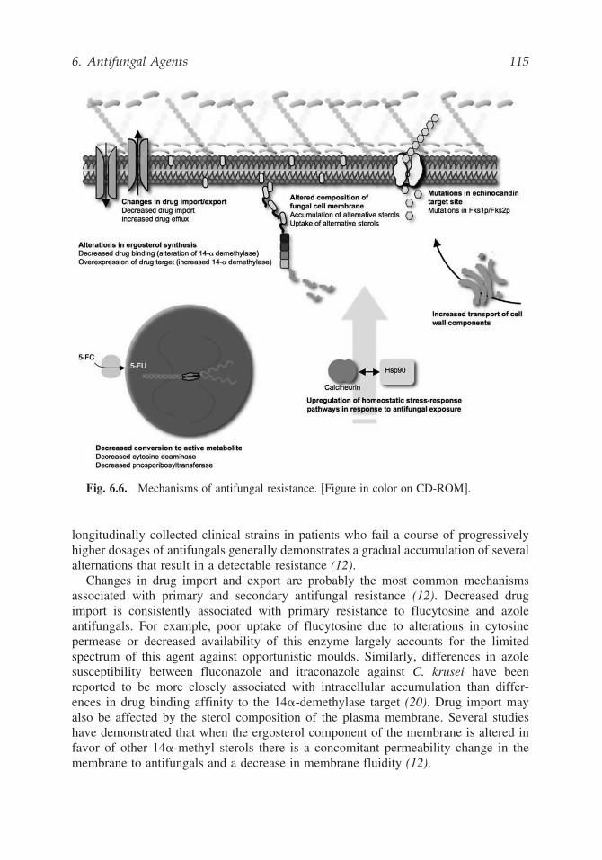
6. Antifungal Agents 115
Fig. 6.6. Mechanisms of antifungal resistance. [Figure in color on CD-ROM].
longitudinally collected clinical strains in patients who fail a course of progressivelyhigher dosages of antifungals generally demonstrates a gradual accumulation of severalalternations that result in a detectable resistance (12).
Changes in drug import and export are probably the most common mechanismsassociated with primary and secondary antifungal resistance (12). Decreased drugimport is consistently associated with primary resistance to flucytosine and azoleantifungals. For example, poor uptake of flucytosine due to alterations in cytosinepermease or decreased availability of this enzyme largely accounts for the limitedspectrum of this agent against opportunistic moulds. Similarly, differences in azolesusceptibility between fluconazole and itraconazole against C. krusei have beenreported to be more closely associated with intracellular accumulation than differ-ences in drug binding affinity to the 14�-demethylase target (20). Drug import mayalso be affected by the sterol composition of the plasma membrane. Several studieshave demonstrated that when the ergosterol component of the membrane is altered infavor of other 14�-methyl sterols there is a concomitant permeability change in themembrane to antifungals and a decrease in membrane fluidity (12).

116 Russell E. Lewis and Annette W. Fothergill
Similar to other eukaryotic cells, fungi are known to contain two types of effluxpumps that contribute to drug resistance: ATP binding cassette (ABC) transportersand major facilitators (MF). Overexpression of the ATP-dependent ABC transporterstypically confers a multidrug resistance phenotype. In contrast, MF pumps, whichexpel antifungal though protonmotive force (H+ gradient across membrane), have amuch narrower spectrum of substrate specificity. In Candida albicans, overexpressionof ATP-dependent efflux pumps CDR1 and CDR2 confer cross-resistance to all azoleantifungals (12). In contrast, overexpression of MF pump MDR1 affects only theaccumulation of fluconazole and does not result in cross-resistance to itraconazole orketoconazole. Overexpression of ATP-dependent efflux pumps is the most prevalentmechanism of efflux-mediated resistance reported in clinical isolates (12). Recently,overexpression of ABC transporters was reported to confer a degree of cross-resistancebetween azoles and echinocandins in a laboratory strain of C. albicans (21).
Besides drug efflux, the most common mechanism associated with antifungal resis-tance involves changes in the binding site of the drug. Several genetic alternationsin ERG11, the gene encoding 14�-demethylase, have been attributed to decreases inazole activity, including point mutations that result in changes in the active pocket siteor overexpression of ERG11. Similar alterations in other enzymes of the ergosterolbiosynthetic pathway, particularly ERG 3 (C-5-sterol desaturase), which is upregu-lated with inhibition of 14�-demethylase, have also been documented in azole-resistantclinical strains. Binding site alteration is also likely to be an important mechanismof echinocandin resistance. Point mutations in the FKS1 gene have been reported inlaboratory-derived caspofungin-resistant mutants of C. albicans (3).
Changes in the target expression in the ergosterol biosynthetic pathway alter thefungal cell membrane sterol content. Substitution of alternative sterols for ergosterol,or alterations in the sterol:phospholid ratio in the cell membrane, can decrease intra-cellular accumulation of azoles and reduce the binding of amphotericin B to the cellmembrane. Indeed, many polyene-resistant yeasts recovered from patients with clearmicrobiological failure on amphotericin B have dimished ergosterol concentrations intheir fungal cell membranes. Several studies have even suggested that pathogenic fungican scavenge free sterols for the cell membrane, including cholesterol, resulting inresistance to polyene and azole antifungals (22).
Alterations in intracellular drug processing and/or degradation and metabolismare probably the least well studied pathways of resistance in fungi, even thoughthese mechanisms are well characterized in other prokaryotic and eukaryoticsystems. Resistance to flucytosine has been associated with alterations in cystosinedeaminase, which results in decreased intracellular conversion of flucytosine to itsactive form.
Recent studies of antifungal resistance have begun to focus on homeostatic stress-response pathways in fungi that may be unpregulated after exposure to antifungals.Disruption of the evolutionarily conserved protein kinase C (PKC) cell wall integrityand calcineurin pathways enhances azole and echinocandin killing in fungi (23). Upreg-ulation of these pathways also diminishes the lethal effects of antifungals throughupregulation of ergosterol and glucan biosynthesis, increases in chitin content in thefungal cell wall, as well as increased export of cell wall components for cell wall repair.

6. Antifungal Agents 117
Recently, the molecular chaperone heat shock protein 90 (Hsp90) was reported toplay a critical role in regulating resistance to antifungal agents through the calcineurinpathway (24,25). Future efforts toward combating antifungal resistance are likely toexploit this important and evolutionarily conserved mechanism for maintaining andexpressing resistance mechanisms to antifungals.
4. AMPHOTERICIN B
Conventional amphotericin B (Fungizone®) has long been considered to be thecornerstone of therapy for deeply invasive fungal infections. Toxicity, includinginfusion-related fever, chills, rigors, headache, and dose-limiting nephrotoxicity, oftenlimits the effectiveness of this agent in severely ill patients. Consequently, threelipid-based formulations (Ambisome®, Abelcet®, Amphotec®) were developed thatoffer several advantages over conventional amphotericin B including (1) the abilityto administer higher daily dosages of drug, (2) decreased infusion-related side effects(especially for the liposomal formulation), and (3) a reduced rate of nephrotoxicity(Table 6.1). Despite the improved therapeutic index of these formulations, there isstill relatively few data from prospective clinical trials to suggest these formulationsare more effective than conventional amphotericin B. Moreover, the higher acquisitioncost of the lipid formulations has required many institutions to restrict the use of theseformulations to patients with preexisting renal failure, or in patients who are at highrisk for developing nephrotoxicity while receiving amphotericin B (e.g., patients onconcomitant nephrotoxic therapies). Currently, there is no consensus opinion on theclinical or pharmacoeconomic threshold for using lipid amphotericin B formulationsas first-line therapy for most invasive mycoses.
4.1. Spectrum and Susceptibility
Amphotericin B should be administered only to patients with progressive andpossibly fatal infections. Acceptable activity can be measured in vitro againstalmost all fungi including Candida, Cryptococcus, Aspergillus, Blastomyces, Histo-plasma, Coccidioides, Sporothrix, and agents of zygomycosis (mucormycosis,including Rhizopus, Mucor, and Absidia), as well as other less frequently recoveredstrains.
A few species exhibit elevated MICs when tested against amphotericin B andare known to possess innate resistance to this drug. Resistant species include bothScedosporium apiospermum and S. prolificans in addition to Paecilomyces lilacinus,Aspergillus terreus, and some Fusarium species. Early reports have revealed Candidalusitaniae resistance to amphotericin B and have shown that this species possessesthe ability to develop resistance while the patient is on treatment. The first reportinvolved a patient whose initial isolate was susceptible but whose subsequent isolateshad developed amphotericin B resistance (26). Later reports have shown ampho-tericin B resistance may exist even before exposure to amphotericin B (27). Theexpected rate of resistance for C. lusitaniae is 8% to 10% of any microbiology labstock collection.

Tab
le6.
1S
yste
mic
anti
fun
gal
ther
apie
s
Ant
ifun
gals
Tra
dena
me(
s)U
sual
adul
tdo
seM
echa
nism
ofac
tion
Tox
iciti
esSp
ectr
um/c
omm
ents
Poly
enes
Am
phot
eric
inB
Fung
izon
e0.
25–1
.5m
g/kg
IVq2
4hB
ind
toer
gost
erol
and
inte
rcal
ates
with
the
fung
alce
llm
embr
ane,
resu
lting
inin
crea
sed
mem
bran
epe
rmea
bilit
yto
univ
alen
tan
ddi
vale
ntca
tions
Acu
te—
Feve
r,ch
ills,
rigo
r,ar
thra
lgia
with
infu
sion
.T
hrom
boph
lebi
tis,d
yspn
ea(r
are)
,arr
hyth
mia
s(r
are)
Dru
gof
choi
cefo
rse
vere
infe
ctio
nsca
used
byen
dem
icdi
mor
phic
fung
i,m
ost
Can
dida
spec
ies,
and
com
mon
hyal
ohyp
hom
ycet
es(i
nclu
ding
Asp
ergi
llus
)an
dzy
gom
ycos
is.
Del
ayed
—A
zote
mia
(26%
),tu
bula
rac
idos
is,
hypo
kale
mia
,hy
pom
agne
sem
ia,a
nem
ia
Nep
hrot
oxic
ityis
the
dose
-lim
iting
side
-eff
ect,
redu
ced
with
lipid
amph
oter
icin
Bfo
rmul
atio
nsan
dsa
line
pre
and
post
hydr
atio
n.L
ipid
form
ulat
ions
ofam
phot
eric
inB
:L
ipos
omal
(L)-
AM
BA
mbi
som
e,3–
10m
g/kg
q24h
Infu
sion
rela
ted
reac
tions
:A
mbi
som
eA
BL
CA
belc
et,
5m
g/kg
/q24
h<
Abe
lcet
<A
mph
otec
AB
CD
Am
phot
ec3–
4m
g/kg
q24h

Azo
les
Ket
ocon
azol
eN
izor
al20
0–80
0m
gPO
q24h
Div
ided
dose
sre
com
men
ded
≥40
0m
g/da
y
Inhi
bitio
nof
cyto
chro
me
P450
14�
-dem
ethy
lase
,de
crea
sed
prod
uctio
nof
ergo
ster
ol,
accu
mul
atio
nof
lano
ster
olle
adin
gto
pert
urba
tion
offu
ngal
cell
mem
bran
e,fu
ngis
tatic
Gas
troi
ntes
tinal
(20–
50%
)in
clud
ing
naus
eaan
dvo
miti
ng,
anor
exia
,ra
sh(2
%),
tran
sien
tin
crea
ses
inhe
patic
enzy
mes
,se
vere
hepa
toto
xici
ty(r
are)
,al
opec
ia,
inhi
bitio
nof
adre
nal
ster
oid
synt
hesi
s(e
spec
ially
atdo
sage
s>
600
mg/
day)
Ora
lfo
rmul
atio
non
ly.
Inco
nsis
tenc
ies
inor
alab
sorp
tion/
poor
gast
roin
test
inal
tole
ranc
elim
itsus
efo
rtr
eatm
ent
ofde
epm
ycos
es.
Pote
ntin
hibi
tor
ofm
amm
alia
ncy
toch
rom
eP4
50ca
nle
adto
pote
ntia
llyse
vere
drug
inte
ract
ions
whe
nad
min
iste
red
conc
omita
ntly
with
othe
rP4
50-m
etab
oliz
eddr
ugs.
Itra
cona
zole
Spor
anox
200–
400
mg
POq2
4hIV
200–
400
mg
q12h
,th
enq2
4h
Div
ided
dose
sre
com
men
ded
≥40
0m
g/da
y
Sim
ilar
toke
toco
nazo
le,
but
mor
ese
lect
ive
for
fung
alP4
50de
met
hyla
se
Gas
troi
ntes
tinal
(20%
)in
clud
ing
naus
eaan
dvo
miti
ng,
and
diar
rhea
,ra
sh(2
%),
tast
edi
stur
banc
e(o
ral
solu
tion)
,tr
ansi
ent
incr
ease
sin
hepa
ticen
zym
es,
seve
rehe
pato
toxi
city
(rar
e),
alop
ecia
,in
hibi
tion
ofad
rena
lst
eroi
dsy
nthe
sis
(esp
ecia
llyat
dosa
ges
>60
0m
g/da
y).
Acc
umul
atio
nof
hydr
oxy-
prop
yl--
cycl
odex
tran
vehi
cle
inpa
tient
sw
ithC
rCl
<30
ml/m
in(i
ntra
veno
usfo
rmul
atio
n).
Con
gest
ive
hear
tfa
ilure
(rar
e)
Spec
trum
sim
ilar
tofl
ucon
azol
ew
ithen
hanc
edac
tivity
agai
nst
C.
krus
eian
dA
sper
gill
us.N
otac
tive
agai
nst
Fus
ariu
man
dzy
gom
ycos
is.D
rug
ofch
oice
for
mild
tom
oder
ate
infe
ctio
nsca
used
byen
dem
icdi
mor
phic
fung
i.B
ioav
aila
bilit
yof
oral
solu
tion
isim
prov
edov
erca
psul
esby
30%
unde
rfe
dco
nditi
ons
and
60%
infa
stin
gco
nditi
ons.
Pote
ntin
hibi
tor
ofm
amm
alia
ncy
toch
rom
eP4
50en
zym
es.
Seru
mle
vel
mon
itori
ngis
occa
sion
ally
reco
mm
ende
d,tr
ough
leve
lsm
easu
red
byH
PLC
shou
ldex
ceed
0.5
μg/m
l (Con
tinu
ed)

Tab
le6.
1(C
onti
nued
)
Fluc
onaz
ole
Dif
luca
n3–
12m
g/kg
PO/I
Vq2
4h
Dos
age
adju
stm
ent
requ
ired
inre
nal
impa
irm
ent
Sim
ilar
toke
toco
nazo
le,b
utm
ore
sele
ctiv
ein
hibi
tor
of14
�de
met
hyla
se
Gas
troi
ntes
tinal
(5–1
0%),
rash
,he
adac
he,
tran
sien
tin
crea
ses
inhe
patic
enzy
mes
,he
pato
toxi
city
(rar
e),a
lope
cia
Spec
trum
incl
udes
mos
tC
andi
dasp
ecie
s,C
rypt
ococ
cus
neof
orm
ans,
and
ende
mic
dim
orph
icfu
ngi.
Les
sac
tive
agai
nst
Can
dida
glab
rata
.C
andi
dakr
usei
isin
trin
sica
llyre
sist
ant.
Not
clin
ical
lyac
tive
for
deep
myc
oses
caus
edby
inva
sive
mou
lds.
Hig
her
daily
dosa
ges
are
reco
mm
ende
d(e
.g.,
12m
g/kg
per
day)
incr
itica
lly-i
llpa
tient
sor
inin
stitu
tions
whe
reC
andi
dagl
abra
tais
com
mon
Vor
icon
azol
eV
fend
6m
g/kg
IVq1
2h×
2do
ses,
then
4m
g/kg
q12h
200
mg
POq1
2hif
>40
kg,1
00m
gPO
q12h
if<
40kg
Sim
ilar
tofl
ucon
azol
e,bu
thi
gher
affi
nity
for
fung
al14
�-d
emet
hyla
se
Tra
nsie
ntph
otop
sia
(rep
orte
dup
to30
%),
rash
,ha
lluci
natio
ns(2
%),
tran
sien
tin
crea
ses
inhe
patic
enzy
mes
,se
vere
hepa
toto
xici
ty(r
are)
.Acc
umul
atio
nof
sulf
o-bu
tyl
este
rcy
clod
extr
anve
hicl
em
ayoc
cur
inpa
tient
sw
ithC
rCl
<50
ml/m
inre
ceiv
ing
intr
aven
ous
form
ulat
ion.
Spec
trum
sim
ilar
toitr
acon
azol
ew
ithen
hanc
edac
tivity
agai
nst
Asp
ergi
llus
,F
usar
ium
,and
Sced
ospo
rium
apio
sper
mum
(Pse
udal
lesc
heri
abo
ydii
).R
etai
nsac
tivity
agai
nst
som
efl
ucon
azol
e-re
sist
ant
C.
glab
rata
.In
hibi
tor
ofm
amm
alia
ncy
toch
rom
eP4
50en
zym
es.
Posa
cona
zole
Nox
afil
600–
800
mg/
day
indi
vide
ddo
ses
Sim
ilar
tovo
rico
nazo
leG
astr
oint
estin
al(5
–15%
),fe
ver,
head
ache
,m
uscu
losk
elet
alpa
in(5
%)
Spec
trum
sim
ilar
tovo
rico
nazo
lew
ithen
hanc
edac
tivity
agai
nst
Fus
ariu
m,
zygo
myc
osis
,an
dbl
ack
mou
lds
(pha
eohy
phom
ycet
es).
Inhi
bito
rof
mam
mal
ian
cyto
chro
me
P450
3A4
(Con
tinu
ed)

Ech
inoc
andi
ns
Cas
pofu
ngin
Can
cida
s70
mg
IVda
y1,
then
50m
gq2
4hIn
hibi
tion
ofce
llw
all
gluc
ansy
nthe
sis,
lead
ing
toos
mot
icin
stab
ility
offu
gal
cell.
Feve
r,ch
ills,
phle
bitis
/thro
mbo
phel
ibiti
s(p
erip
hera
llin
e),
rash
.D
rug
conc
entr
atio
nsde
crea
sed
with
P450
3A4
indu
cers
.D
ecre
ases
tacr
olim
usbl
ood
leve
lsby
∼25%
Spec
trum
incl
udes
mos
tC
andi
dasp
ecie
sin
clud
ing
fluc
onaz
ole-
resi
stan
tC
andi
dakr
usei
and
Can
dida
glab
rata
.H
ighe
rdo
sage
sm
aybe
requ
ired
for
C.
para
psil
osis
.A
ctiv
eag
ains
tA
sper
gill
ussp
ecie
s.N
otac
tive
agai
nst
Cry
ptoc
occu
sne
ofor
man
s,T
rich
ospo
ron,
Fus
ariu
m,z
ygom
ycos
is,o
rbl
ack
mou
lds
(pha
eohy
phom
ycet
es).
Mic
afun
gin
Myc
amin
e50
–150
mg
IVq2
4hSi
mila
rto
casp
ofun
gin
Cle
aran
ceno
taf
fect
edby
P450
3A4
indu
cers
Sim
ilar
toca
spof
ungi
n
Ani
dula
fung
inE
raxi
s20
0m
gIV
day
1th
en10
0m
g/da
ySi
mila
rto
casp
ofun
gin
Cle
aran
ceno
taf
fect
edby
P450
3A4
indu
cers
Sim
ilar
toca
spof
ungi
n
Fluo
ropy
rim
idin
es
Fluc
ytos
ine
(5-F
C)
Anc
obon
100
mg/
kgda
ilyPO
divi
ded
q6h
Dos
age
adju
stm
ent
requ
ired
inre
nal
impa
irm
ent
Dru
gis
tran
spor
ted
into
susc
eptib
lefu
ngi
bycy
tosi
nepe
rmea
se,
and
then
deam
inat
edto
activ
efo
rm(5
-FU
)by
cyto
sine
deam
inas
ew
here
the
drug
inte
rfer
esw
ithD
NA
/RN
Asy
nthe
sis
Incr
ease
inse
rum
tran
sam
inas
es(7
%),
naus
eaan
dvo
miti
ng(5
%);
diar
rhea
,ab
dom
inal
pain
,ra
sh,e
nter
colit
is(r
are)
Les
sco
mm
on-
leuc
open
ia,
thro
mbo
cyto
peni
a,an
emia
Nar
row
spec
trum
for
deep
myc
oses
:C
andi
daan
dC
rypt
ococ
cus
neof
orm
ans
only
.Res
ista
nce
isco
mm
onw
hen
used
asm
onot
hera
py.
Typ
ical
lyad
min
iste
red
inco
mbi
natio
nw
itham
phot
eric
inB
for
cryp
toco
ccal
men
ingi
tis.R
isk
ofbo
nem
arro
wsu
ppre
ssio
nin
crea
sed
with
pers
iste
ntfl
ucyt
osin
ele
vels
>100
μg/m
l.C
aref
uldo
sage
adju
stm
ent
isre
quir
edin
patie
nts
with
rena
ldy
sfun
ctio
n.
PO,O
rally
;IV
,int
rave
neou
sly;
q6h,
ever
y6
hour
s;q1
2h,e
very
12ho
urs;
q24h
,eve
ry24
hour
s;P4
50,c
ytoc
hrom
eP4
50.

122 Russell E. Lewis and Annette W. Fothergill
4.2. Pharmacokinetics
Amphotericin B deoxycholate has negligible oral absorption and must be admin-istered intravenously. After intravenous administration, the drug is released from itscarrier and is highly bound by plasma proteins (91% to 95%) including lipoproteins,erythrocytes, and cholesterol in the plasma. Amphotericin B then redistributes from thebloodstream into tissue with an apparent volume of distribution (Vd) of 4 liters/kg (5).In adults, infusion of 0.6 mg/kg of amphotericin B deoxycholate yields peak serumconcentrations of approximately 1 to 3 μg/ml (28). Concentrations in other bodyfluids outside the serum are less than 5% of concurrent serum concentrations, withpoor penetration into bronchial secretions, pleura, peritoneum, synovium, and aqueoushumor. Although amphotericin B poorly penetrates the CSF, fungal infections of thebrain have been successfully treated with amphotericin B (5).
Tissue concentrations of amphotericin B are highest in the kidney, followed by theliver, spleen, heart, skeletal, muscle, and brain. The formulation of amphotericin Binto phospholipid sheets (Abelcet), cholesterol disks (Amphotec), or liposome carriers(Ambisome) alters drug distribution (particularly to the kidney) and the eliminationprofile of the drug (5) (Table 6.2).
Recent studies have suggested that amphotericin B undergoes relatively littlemetabolism, with a terminal elimination half-life of greater than 11 to 15 days. After168 hours, approximately 60% of a single dose can be recovered from the feces(∼40%) and urine (20%) (29). Because a relatively lower fraction of the daily dosageis slowly excreted in urine and bile, dosage modification is not necessary to preventdrug accumulation in patients with renal or hepatic failure, but may be judicious ina patient with declining renal function. Because amphotericin B behaves as a colloidin aqueous solutions and is highly protein bound, hemodialysis does not removesignificant amounts of the drug unless the patient is hyperlipidemic; which enhancesamphotericin B binding to the dialysis membrane (5).
4.3. Adverse Effects
The most common acute toxicity of amphotericin B formulations is infusion-relatedreactions, which are characterized by fever, chills, rigors, anorexia, nausea, vomiting,myalgias, arthralgias, and headache. Hypotension, flushing and dizziness are lesscommon, but bronchospasm and true anaphylactic reactions have been reported withboth the conventional and lipid formulations of amphotericin B (5). Severe hypokalemiaand cardiac arrhythmias have also been described in patients with central venouscatheters who have received rapid infusions or excessive doses of conventional ampho-tericin B. Therefore, slower infusion rates (4 to 6 hours or more) and EKG monitoringshould be considered in patients with underlying cardiac conduction abnormalities.Thrombophlebitis is a common local side effect with infusion, which often necessi-tates the placement of a central venous line for therapy more than 1 week. Slowerinfusion rates, rotation of infusion sites, application of hot packs, low-dose heparin,and avoidance of concentrations greater than 1 mg/ml can minimize thrombophlebitis.
Acute reactions generally subside over time and with subsequent amphotericinB infusions. In the past, a test dose of amphotericin B deoxycholate (i.e., 1 to5 mg) was recommended before initiating therapy. This is no longer considered

Tab
le6.
2C
omp
arat
ive
ph
arm
acok
inet
ics
ofth
ean
tifu
nga
lag
ents
AM
BA
BC
DA
BL
CL
-AM
BFl
uIt
raa
Vor
iPo
saA
nid
Cas
Mic
a5F
C
Ora
lbi
oava
ilabi
lity
(%)
<5<5
<5<5
9550
9660
<5<5
<5>8
0D
istr
ibut
ion
Tot
alC
max
(μg/
ml)
40.
3–1
131
0.7
114.
67.
80.
830.
270.
2480
AU
C(m
g*
h/L
)17
4314
555
400
29.2
20.3
8.9
99e
119
158e
Prot
ein
bind
ing
(%)
>95
>95
>95
>95
1099
.858
9984
9799
4C
SF(%
)0–
4>6
0<1
060
<5<5
<575
Eye
(%)
0–38
b�c
0–38
b�c
0–38
b�c
0–38
b�c
28–7
5b�c
(10–
70μg
/ml)
10†(0
.22
μg/m
l)18
b(0
.81
μg/m
l)26
(0.2
5μg
/ml)
ND
ND
ND
ND
Uri
ne(%
)d3–
204.
590
1–10
<2<2
<2<2
<290
Met
abol
ism
Unk
Unk
Unk
Unk
+++
+++
++N
one
Hep
Hep
Non
eE
limin
atio
nU
rine
/bi
leU
nkU
nkU
nkR
enal
Hep
Ren
alFe
ces
Fece
sU
rine
Fece
sR
enal
Hal
f-lif
e(h
)50
3017
310
0–15
331
246
2524
3015
3–6
AM
B,
amph
oter
icin
Bde
oxyc
hola
te;
AB
CD
,am
phot
eric
inB
chol
este
rol
disp
ersi
on;
AB
LC
,am
phot
eric
inB
lipid
com
plex
;L
-AM
B,
lipos
omal
amph
oter
icin
B;
Flu,
fluc
onaz
ole;
Itra
,itr
acon
azol
e;V
ori,
vori
cona
zole
;Po
sa,p
osac
onaz
ole;
Ani
d,an
idul
afun
gin;
Cas
,cas
pofu
ngin
;M
ica,
mic
afun
gin;
5FC
,5-f
luor
ocyt
osin
eor
fluc
ytos
ine;
ND
,no
data
avai
labl
e;U
nk,u
nkno
wn;
Hep
,hep
atic
.a D
ata
are
for
oral
solu
tion.
bH
uman
.c A
nim
al.
d%
ofac
tive
drug
orm
etab
olite
s.e Fo
rdo
ses
of10
0m
g/da
y.

124 Russell E. Lewis and Annette W. Fothergill
useful for screening patients for hypersensitivity reactions. Premedications such aslow-dose hydrocortisone (1 mg/kg), diphenhydramine, meperidine (0.5 mg/kg), andnonsteroidal anti-inflammatory agents are often administered before amphotericin Binfusions to blunt symptoms of acute reactions. Premedication is also recommendedbefore infusions of the lipid amphotericin B formulations, despite the reduced rates ofinfusion reactions seen with these drugs. Nephrotoxicity is the most significant, delayedtoxicity of amphotericin B and can be classified into glomerular or tubular mecha-nisms. Amphotericin B directly constricts the afferent arterioles, resulting in decreasedrenal blood flow and a drop in glomerular filtration (increased serum creatinine),eventually leading to azotemia. Amphotericin-induced azotemia can be reduced byensuring patients are well hydrated before starting therapy and by sodium loading—the practice of administering intravenous normal saline (0.5 to 1 liters) before andafter amphotericin B infusion to maintain renal blood flow and adequate glomerularfiltration pressure. Two small nonrandomized studies have also suggested that theadministration of amphotericin B deoxycholate by continuous infusion can preserveglomerular function in the short term (30,31); however, this dosing approach has notbeen widely adopted. Azotemia with amphotericin B is generally reversible, although5% to 10% of patients may have persistent renal impairment after discontinuation oftherapy.
Amphotericin B is directly toxic to the distal tubules, resulting in impaired urinaryacidification, impaired urinary concentrating ability, and wasting of potassium andmagnesium. Hypokalemia is common in patients receiving either conventional or lipidformulations of the drug. Patients may require the administration of up to 15 mmol ofsupplemental potassium per hour (4). Hypokalemia and low serum magnesium levelsfrequently precede decreases in glomerular filtration (increased serum creatinine),especially in patients who are adequately hydrated or receiving lipid formulations ofamphotericin B. Continued tubular damage, however, eventually results in decreases inrenal blood flow and glomerular filtration through tubuloglomerular feedback mecha-nisms that constrict the afferent arteriole. Hence, sodium loading should still beconsidered for patients receiving lipid amphotericin B formulations.
Patients who receive prolonged courses of amphotericin B frequently developnormochromic, normocytic anemia due to the inhibitory effects of amphotericin B onrenal erythropoietin synthesis. Patients may experience decreases in hemoglobin of15% to 35% below baseline that return to normal within several months of discontin-uation of the drug. Administration of recombinant erythropoietin may be required inpatients with symptomatic anemia.
5. AZOLES
The availability of azole antifungals, particularly the oral triazoles itraconazole,fluconazole, and more recently, voriconazole and posaconazole, fulfills a critical needfor effective and better tolerated alternatives to amphotericin B. Miconazole was thefirst systemic azole approved for use in humans, but the relatively toxic intravenousformulation limited its use to severely ill patients. Similarly, ketoconazole was noteffective in critically ill patients because of its lack of an intravenous formulation anderratic absorption in patients with relative achlorhydria. The triazoles have proven to be

6. Antifungal Agents 125
much more effective in the prevention and treatment of both primary and opportunisticmycoses. All three currently approved triazoles are available in table/capsule, oralsolution, and intravenous formulations, providing clinicians with added flexibility intherapy selection. Because all azoles are potentially teratogenic, they should be avoidedduring pregnancy.
5.1. Spectrum and Susceptibility
The triazoles are more easily tolerated, but are primarily considered fungistatic asopposed to fungicidal (lethal) drugs. However, clear definitions of cidality are oftenmore difficult to ascertain with antifungal agents. Although similar in mechanisms ofaction, each agent has a slightly different spectrum of activity.
Fluconazole is principally used for yeast infections including those caused by mostCandida and Cryptococcus species. Although this drug has been used to successfullymanage meningitis caused by Coccidioides, it is not typically a drug of choice forinfections caused by other moulds.
Of primary concern is acquired resistance by yeasts. Candida krusei is welldocumented to possess intrinsic resistance to fluconazole, so much so that susceptibilitytesting against this isolate is not recommended. Some reports place the rate of outrightC. glabrata resistance at about 15% of any given population of isolates (32), butboth C. albicans and C. glabrata are capable of developing resistance after prolongedtherapy or after therapy with inappropriate dosing. Overall, by in vitro testing, about88% of Cryptococcus neoformans and 95% of C. albicans strains appear susceptibleto fluconazole.
Itraconazole possesses a wide spectrum of activity, including activity against bothyeasts and moulds. It is useful in treating aspergillosis, blastomycosis, coccidioidomy-cosis, histoplasmosis, and candidiasis. In addition, itraconazole possess low MIC endpoints against the dematiaceous fungi and may be considered the drug of choice fortreatment of infections caused by fungi from this group. Cross-resistance is of concernbetween drugs within the azole class. Comparison of resistance patterns betweenitraconazole and fluconazole reveal similar percentages of resistance among Candidaspecies.
Voriconazole is noted to have activity against Aspergillus, S. apiospermum, andFusarium solani. This is remarkable because both S. apiospermum and F. solani arenotoriously resistant to other antifungal agents. In addition, voriconazole may possesslethal activity against the aspergilli as opposed to the static activity expected with theazoles. Susceptibility patterns with the yeasts are similar to those of both itraconazoleand fluconazole. An important exception is the extremely low incidence of resistanceseen with C. krusei, in contrast to near 100% resistance of this species to fluconazoleand about 10% resistance to itraconazole.
Posaconazole is a very promising investigational azole with a broad spectrum ofactivity. Clinical trials are underway to assess activity in aspergillosis, candidiasis,fusariosis, coccidioidomycosis, and zygomycosis (mucormycosis). Results againstspecies such as Rhizopus and Mucor show that this drug may provide alternativetherapy to amphotericin B for infections caused by this group of fungi. Resistance hasnot been noted but some cross-resistance may occur.

126 Russell E. Lewis and Annette W. Fothergill
5.1.1. Fluconazole
Among the triazole antifungals, fluconazole (Diflucan®) is clearly the best toleratedagent and has the most desirable pharmacological properties including high bioavail-ability, high water solubility, low degree of protein binding, linear pharmacokinetics,and a wide volume of distribution including the CSF, eyes, and urine (27). Unlike otherazoles, fluconazole is eliminated primarily unchanged through the kidneys and is lesssusceptible to clinically significant drug interactions through mammalian cytochromeP450 enzymes at standard dosages used to treat superficial (100 to 200 mg/day) orsystemic (400 mg/day) infections.
5.1.2. Itraconazole
Itraconazole (Sporanox®) was initially introduced in the early 1990s as a capsuleformulation that was effective for superficial fungal infections and mild to moderatelysevere endemic mycosis, but erratic absorption in the critically ill patient limited itseffectiveness for opportunistic mycoses. The subsequent reformulation of this triazoleinto an oral and intravenous solution with hydroxy-�-propyl cyclodextran signifi-cantly improved the blood levels that could be reliably obtained in critically ill andimmunocompromised patients. Itraconazole is a relatively broad-spectrum triazole withactivity against many common fungal pathogens including most Candida, Crypto-coccus, endemic dimorphic fungi (Histoplasma, Blastomyces , and Coccidioides), andAspergillus. The drug is lipophilic, highly protein bound, and has a long half-life,nonlinear pharmacokinetics, and limited distribution into some body fluids, includingthe CSF and urine (Table 6.2). The drug is metabolized in the liver and to a lesserextent in the gut into more hydrophilic metabolites, one of which retains potentantifungal activity (hydroxyitraconazole). The most common adverse effects associatedwith itraconazole therapy are gastrointestinal (especially with the oral solution), rash,and transient increases in hepatic transaminases. Prolonged therapy can be associatedwith metabolic disturbances (suppression of adrenal steroid synthesis) and idiopathiccongestive heart failure. Itraconazole is a substrate and potent inhibitor of mammaliancytochrome P450 enzymes and is therefore susceptible to a number of clinically signif-icant drug interactions (see Section 5.2, Azole Drug Interactions).
5.1.3. Voriconazole
Voriconazole (Vfend®) is a methylated analogue of fluconazole with enhancedactivity against yeast as well as important opportunistic moulds including Aspergillusand Fusarium (Table 6.1). Like fluconazole, voriconazole is well absorbed orally,has limited protein binding, and distributes widely throughout the body, includingthe CSF. Like itraconazole, intravenous voriconazole is formulated in a cyclodextransolution (sulfobutylether cyclodextran) and has nonlinear pharmacokinetics in adults.Voriconazole is metabolized to inactive metabolites through the liver and is an inhibitorof mammalian cytochrome P450 enzymes (Table 6.2). In addition to the commonadverse effects seen with other triazole antifungals (gastrointestinal, rash, increasesin hepatic enzymes), voriconazole can cause transient visual disturbances in 15% of30% of patients that manifest as photophobia, perception of blinking or flashing lights(even with the eyes closed), and occasionally hazy or blurred vision. Symptoms tend

6. Antifungal Agents 127
to occur during the first week of therapy and disappear with continued therapy in mostpatients. Occasionally, visual disturbances are intensified by hallucinations—a separateside effect seen in 2% to 8% of patients receiving voriconazole (often with concomitantbenzodiazepines and narcotic analgesic therapy). Visual disturbances are thought to bea result of temporary alterations in electrical conduction of photoreceptors in the rodsand cones of the retina, which revert to normal once therapy is stopped. No permanentdamage to the retina has been noted in human or animal studies of voriconazole (33,34).
5.1.4. Posaconazole
Posaconazole (Noxafil®) is triazole analogue of itraconazole with enhanced activityagainst opportunistic moulds including Aspergillus, Fusarium, and (notably) the agentsof zygomycosis. Posaconazole will initially be available as an oral suspension with anintravenous formulation still in development. Absorption of posaconazole suspensionis dose limited at 800 mg/day and can be improved if the suspension is administeredwith a high-fat meal or in divided doses (twice to four times daily) (Table 6.1) (35).Once absorbed, posaconazole is widely distributed into tissues throughout the bodyand is highly protein bound (98%). Elimination of posaconazole occurs predominantly(90%) in the feces as unchanged drug and in the urine (10%) as an inactive metabolite(Table 6.2). Despite its lack of phase I metabolism, posaconazole is a potent inhibitorof mammalian cytochrome P450 3A4 and has a similar potential for drug interactionsas itraconazole when coadministered with drugs metabolized through this pathway.
5.2. Azole Drug Interactions
As mentioned previously, an inherent limitation of azole pharmacology is thatthe target of antifungal activity in pathogenic fungi, the cytochrome P450 enzyme14-�-demethylase, shares considerable homology with mammalian cytochrome P450enzymes involved in drug metabolism. As a result, azole antifungals can be bothsubstrates and inhibitors of cytochrome P450 systems in humans (8). Significant druginteractions with azole antifungals are summarized in Table 6.3. Many of these druginteractions are potentially severe and concomitant use should be avoided. Some azoledrug interactions are less predictable and possibly dosage dependent. For example,fluconazole is a weak inhibitor of cytochrome P450 3A4 at dosages of 50 to 200 mg/dayand is excreted primarily (80%) through the urine. However, as daily dosages areincreased, fluconazole has a greater potential for inhibition of cytochrome P450 3A4and a larger percentage of the drug is metabolized via the P450 system (8). CytochromeP450 3A4 inducers increase metabolism of all azoles to varying degrees regardlessof their primary excretion pathways. Coadministration of rifampin, for example, canreduce fluconazole serum concentrations by approximately 50% and concentrations ofitraconazole, voriconazole, and posaconazole by greater than 90% (8). Azole antifungaltherapy should be avoided, whenever possible, during use of high-dose conditioningchemotherapy with busulfan or cyclophosphamide owing to an increased risk of acuteliver toxicity and accumulation of toxic chemotherapy metabolites (36). Althoughazoles themselves do not appear to exert major effects on cardiac conduction, theircombined use with drugs that affect potassium channels and are metabolized throughCYP 450 mechanisms (e.g., cisapride, haloperidol, certain tricyclic antidepressants)has the potential to cause life-threatening arrhythmias.

128 Russell E. Lewis and Annette W. Fothergill
Table 6.3Drug interactions of the azole antifungals.
6. ECHINOCANDINS
Despite some modest differences in pharmacokinetics and potency, theechinocandins are pharmacologically similar and probably interchangeable (3). Allthree currently approved agents, caspofungin, micafungin, and anidulafungin, are largesemisynthetic lipopeptides that are available only as intravenous formulations. All havelinear pharmacokinetics, are widely distributed (with the possible exception of the CSFand urine), and have prolonged elimination half-lives that permit once-daily dosing.Slight differences in the metabolism and excretion are seen between the echinocandins,which may account for some differences in the drug interaction profile of theseagents.
6.1. Spectrum and Susceptibility
Caspofungin is indicated for candidiasis and for aspergillosis in patients who arerefractory to other therapies. Use for infections caused by other moulds has not beenas extensively studied. Activity is fungicidal against the yeasts while static against theaspergilli. End points against Aspergillus are determined differently than with otherantifungals. End points are determined as minimum effective concentration (MEC).While growth is substantial with in vitro systems, it is evident that the growth isgrossly abnormal. The MEC is considered the lowest concentration of drug that causes

6. Antifungal Agents 129
the abnormal growth of hyphae in this species (Fig. 6.5). End points for other mouldswould be read in like manner.
The echinocandins have not been evaluated by the CLSI (NCCLS) to standardizetesting parameters. Many investigators feel that they should be tested in RPMI butresults in RPMI are as high as six dilutions higher than results in antibiotic medium3 (M3). Investigational animal studies do not support the results seen with RPMI andsome researchers feel that the M3 results are more representative of true MICs. Resis-tance has not been widely reported for this class of antifungals. However, there area few occurrences of isolates with high MICs. Candida parapsilosis and C. guillier-mondii are two species with notoriously increased MICs. Some clinical isolates ofC. albicans and C. glabrata exist with elevated MICs but it is unclear if these isolatesare truly resistant in vivo.
Activity of the echinocandins is similar across the class. Micafungin has low MICsin both RPMI and M3 as opposed to caspofungin and anidulafungin, both of whichexhibit high MICs in RPMI but lower results in M3.
6.2. Pharmacokinetics
All three echinocandins are available as intravenous formulations only, have (mostly)linear pharmacokinetics, are widely distributed (with the possible exception of the CSFand urine), have prolonged elimination half-lives, and are metabolized by chemicaldegradation followed by hepatic metabolism (Table 6.2). Dosage adjustment is recom-mended for caspofungin in patients with severe hepatic dysfunction (Child Pugh score7 to 9), but is not required for micafungin or anidulafungin.
6.3. Adverse Effects
All three echinocandins were well tolerated in Phase II/III clinical trials, with themost common adverse effects being phlebitis/venous irritation, headache, fever, andrash. Infusion-related reactions analogous to the “red person’s syndrome” observedwith vancomycin infusions have been described with all three echinocandins due tohistamine release during infusions. The most common adverse effects reported withechinocandin therapy are venous irritation when infused through a peripheral veinand transient abnormalities in hepatic transaminases and bilirubin. The echinocandinsare neither substrates nor inhibitors of cytochrome P450 enzymes of P-glycoproteinenzymes. For reasons not completely understood, coadministration of caspofungin withinducers of P450 3A4 (e.g., rifampin, phenytoin) results in modest (25% to 50%)decrease in the area under the curve (AUC), which can be overcome with higherdosages. P450 inducers do not appear to have as pronounced effect on the clearanceof micafungin or anidulafungin. Caspofungin modestly (∼20%) decreases the AUC ofconcomitant tacrolimus therapy. Micafungin modestly increases the AUC of nifedipineand sirolimus. No clinically significant drug interactions have been identified thus farfor anidulafungin.
7. FLUOROPYRIMIDINES
Flucytosine (5-fluorocytosine, 5-FC) is the only agent among the fluoropyrimidineclass of antifungal agents approved for the treatment of invasive fungal infections. In

130 Russell E. Lewis and Annette W. Fothergill
the United States it is available only in oral capsule formulation. The usefulness offlucytosine for treating invasive mycoses is hampered by its relatively narrow spectrum,high rates of acquired resistance among common pathogens (i.e., Candida species),and significant potential for toxic effects. For these reasons, flucytosine is not used asmonotherapy and has a minimal role in the treatment of most mycoses.
7.1. Spectrum and Susceptibility
5-Fluorocytosine has activity against both Candida and Cryptococcus species and isnot recommended for the treatment of infections caused by other fungal species. Therate of resistance against Candida species is expected in about 5% of isolates whilefor Cryptococcus species resistance occurs in about 2% of isolates tested.
7.2. Pharmacokinetics
Because flucytosine widely distributes throughout the body, including the CSF, afteroral administration, it is a useful adjuvant agent for difficult to treat infections in theseanatomically restricted sites. Several randomized prospective studies of cryptococcalmeningitis in patients with AIDS have shown that the addition of flucytosine toamphotericin B therapy results in more rapid sterilization of the CSF, decreased earlymortality, and fewer relapses after completion of “induction” antifungal therapy.
7.3. Adverse Effects
Flucytosine was originally developed as an antitumor chemotherapy before it wasdiscovered to have antifungal activity against common yeasts. Not surprisingly, themost common side effects are nausea and vomiting, increases in serum transaminases,and bone marrow suppression. The risk of bone marrow suppression can be reduced ifserum levels are maintained at less than 100 μg/ml. Because flucytosine is eliminatedunchanged through the kidney, serum level monitoring and dosage adjustments arerequired in patients receiving flucytosine in combination with amphotericin B or othernephrotoxic agents.
Gastrointestinal side effects are seen in up to 6% patients receiving oral flucy-tosine including diarrhea, nausea, and vomiting. Reversible elevations in hepatic serumtransmaninases and alkaline phosphatase have also been reported in 4% to 10% ofpatients receiving flucytosine. The most serious toxicity associated with flucytosine,however, is bone marrow suppression, which occurs in 6% of patients. Some evidencehas accumulated in the last two decades that marrow toxicity is enhanced if serumconcentrations of flucytosine exceed 100 μg/ml.
8. COMBINATION ANTIFUNGAL THERAPY
Because of their unique mechanism of action, the introduction of the echinocandinshas renewed interest in the use of combination antifungal therapy for invasive mycoses.The most common reasons for consideration of combination therapy are to (1) broadenthe spectrum of antifungal coverage of opportunistic mycoses; particularly in severelyimmunocompromised patients; (2) to enhance the activity of an antifungal regimenthrough (presumably) synergistic antifungal effects, especially in severely immunocom-promised patients with progressive disease; and (3) to overcome the pharmacokinetic

6. Antifungal Agents 131
limitations of a single antifungal agent in the treatment of life-threatening mycoses inan anatomically restricted sites such as the CNS (e.g., combined use of flucytosineand amphotericin B for cryptococcal meningitis) (37). With the possible exceptionof cryptococcal meningitis, there are surprisingly few data from controlled clinicaltrials supporting the use of combination therapy for deep mycoses. The majorityof data suggesting a possible benefit for combination therapy with echinocandin-based combinations are still derived from in vitro studies and animal models. Forthis reason, combination antifungal therapy is not routinely recommended as a first-line strategy in the treatment of opportunistic mycoses, but may be considered as asalvage approach in patients with refractory or breakthrough infections on antifungalprophylaxis.
REFERENCES
1. Maertens JA. History of the development of azole derivatives. Clin Microbiol Infect 2004;10(Suppl 1):1–10.
2. Pfaller MA, Diekema DJ, Messer SA, Boyken L, Hollis RJ, Jones RN. In vitro susceptibil-ities of rare Candida bloodstream isolates to ravuconazole and three comparative antifungalagents. Diagn Microbiol Infect Dis 2004;48:101–105.
3. Denning DW. Echinocandin antifungal drugs. Lancet 2003;362:1142–1151.4. Odds FC, Brown AJ, Gow NA. Antifungal agents: mechanisms of action. Trends Microbiol
2003;11:272–279.5. Groll AH, Piscitelli SC, Walsh TJ. Clinical pharmacology of systemic antifungal agents:
a comprehensive review of agents in clinical use, current investigational compounds, andputative targets for antifungal drug development. Adv Pharmacol 1998;44:343–500.
6. Kurtz MB, Rex JH. Glucan synthase inhibitors as antifungal agents. Adv Protein Chem2001;56:423–475.
7. Sheehan DJ, Hitchcock CA, Sibley CM. Current and emerging azole antifungal agents.Clin Microbiol Rev 1999;12:40–79.
8. Gubbins PO, McConnell SA, Penzak SR. Antifungal agents. In: Piscitelli SC, Rodvold KA,eds. Drug interactions in infectious diseases. Totowa, NJ: Humana Press, 2001:185–217.
9. Denning DW. Echinocandin antifungal drugs. Lancet 2003;362:1142–1151.10. Hector RF. Compounds active against cell walls of medically important fungi. Clin
Microbiol Rev 1993;6:1–21.11. Kurtz MB, Heath IB, Marrinan J, Dreikorn S, Onishi J, Douglas C. Morphological effects
of lipopeptides against Aspergillus fumigatus correlate with activities against (1,3)-beta-D-glucan synthase. Antimicrob Agents Chemother 1994;38:1480–1489.
12. White TC. Mechanisms of resistance to antifungal agents. In: Murray PR, Baron EJ,Jorgensen JH, Pfaller MA, Yolken RH, eds. Manual of clinical microbiology. 8th Ed.Washington, DC: ASM Press, 2003:1869–1879.
13. Kontoyiannis DP, Lewis RE. Antifungal drug resistance of pathogenic fungi. Lancet2002;359:1135–1144.
14. NCCLS. Reference method for broth dilution antifungal susceptibility testing of yeasts;proposed standard. NCCLS document M27-P. Wayne, PA: National Committee for ClinicalLaboratory Standards, 1992.
15. NCCLS. Reference method for broth dilution antifungal susceptibility testing of yeasts;approved standard. NCCLS document M27-A2. Wayne, PA: National Committee forClinical Laboratory Standards, 2002.

132 Russell E. Lewis and Annette W. Fothergill
16. NCCLS. Reference method for broth dilution antifungal susceptibility testing of conidial-forming filamentous fungi. Approved standard. NCCLS M38-A. Wayne, PA: NationalCommittee for Clinical Laboratory Standards, 2002.
17. NCCLS. Reference Method for Antifungal Disk Diffusion Susceptibility Testing of Yeasts;Approved Guideline. NCCLS document M44-A. Wayne, PA: National Committee forClinical Laboratory Standards, 2004.
18. Rex JH. Pfaller MA. Has antifungal susceptibility testing come of age? Clin Infect Dis2002;35:982–989.
19. Liu TT, Lee RE, Barker KS, et al. Genome-wide expression profiling of the responseto azole, polyene, echinocandin, and pyrimidine antifungal agents in Candida albicans.Antimicrob Agents Chemother 2005;49:2226–2236.
20. Marichal P, Gorrens J, Coene MC, Le Jeune L, Vanden Bossche H. Origin of differencesin susceptibility of Candida krusei to azole antifungal agents. Mycoses 1995;38:111–117.
21. Schuetzer-Muehlbauer M, Willinger B, Krapf G, Enzinger S, Presterl E, Kuchler K. TheCandida albicans Cdr2p ATP-binding cassette (ABC) transporter confers resistance tocaspofungin. Mol Microbiol 2003;48:225–235.
22. Tsai HF, Bard M, Izumikawa K, et al. Candida glabrata erg1 mutant with increasedsensitivity to azoles and to low oxygen tension. Antimicrob Agents Chemother 2004;48:2483–2489.
23. Sanglard D, Ischer F, Marchetti O, Entenza J, Bille J. Calcineurin A of Candida albicans:involvement in antifungal tolerance, cell morphogenesis and virulence. Mol Microbiol2003;48:959–976.
24. Heitman J. Cell biology. A fungal Achilles’ heel. Science 2005;309:2175–2176.25. Cowen LE, Lindquist S. Hsp90 potentiates the rapid evolution of new traits: drug resistance
in diverse fungi. Science 2005;309:2185–2189.26. Pappagianis D, Collins MS, Hector R, Remington J. Development of resistance to
amphotericin B in Candida lusitaniae infecting a human. Antimicrob Agents Chemother1979;16:123–126.
27. Merz WG. Candida lusitaniae: frequency of recovery, colonization, infection, and ampho-tericin B resistance. J Clin Microbiol 1984;20:1194–1195.
28. Atkinson AJ, Bennett JE. Amphotericin B pharmacokinetics in humans. Antimicrob AgentsChemother 1978;13:271–276.
29. Bekersky I, Fielding RM, Dressler DE, Lee JW, Buell DN, Walsh TJ. Pharmacokinetics,excretion, and mass balance of liposomal amphotericin B (AmBisome) and amphotericinB deoxycholate in humans. Antimicrob Agents Chemother 2002;46:828–833.
30. Imhof A, Walter RB, Schaffner A. Continuous infusion of escalated doses of amphotericinB deoxycholate: An open-label observational study. Clin Infect Dis 2003;36:943–951.
31. Eriksson U, Seifert B, Schaffner A. Comparison of effects of amphotericin B deoxycholateinfused over 4 or 24 hours: randomised controlled trial. Br Med J 2001;322:579–582.
32. Pfaller MA, Messer SA, Hollis RJ, et al. Trends in species distribution and susceptibilityto fluconazole among blood stream isolates of Candida species in the United States. DiagnMicrobiol Infect Dis 1999;33:217–222.
33. Lewis RE. Pharmacotherapy of Candida bloodstream infections: new treatment options,new era. Expert Opin Pharmacother 2002;3:1039–1057.
34. Johnson LB, Kauffman CA. Voriconazole: a new triazole antifungal agent. Clin Infect Dis2003;36:630–637.
35. Ezzet F, Wexler D, Courtney R, Krishna G, Lim J, Laughlin M. Oral bioavailability ofposaconazole in fasted healthy subjects: comparison between three regimens and basis forclinical dosage recommendations. Clin Pharmacokinet 2005;44:211–220.

6. Antifungal Agents 133
36. Marr KA, Leisenring W, Crippa F, et al. Cyclophosphamide metabolism is affected byazole antifungals. Blood 2004;103:1557–1559.
37. Kontoyiannis DP, Lewis RE. Toward more effective antifungal therapy: the prospects ofcombination therapy. Br J Haematol 2004;126:165–175.
SUGGESTED READINGS
Boucher HW, Groll AH, Chiou CC, Walsh TJ. Newer systemic antifungal agents: pharmacok-ientics, safety and efficacy. Drugs 2004:64:1997–2020.
Rex JH, Pfaller MA. Has antifungal susceptibility testing come of age? Clin Infect Dis2002;35:982–989.
Sheehan DJ, Hitchcock CA, Sibley CM. Current and emerging azole antifungal agents. ClinMicrobiol Rev 1999;12:40–79.
Gonzales GM, Fothergill AW, Sutton DA, Rinaldi MG, Lobenberg D. In vitro activities of newand established triazoles against opportunistic filamentous and dimorphic fungi. Med Mycol2005;43:281–284.

IVMycoses

7Candidiasis
Jack D. Sobel, MD
1. INTRODUCTION
Candida species are ubiquitous fungi and the most common fungal pathogensaffecting humans (1,2). The growing problem of mucosal and systemic candidiasisreflects the enormous increase in the pool of patients at risk and the increased oppor-tunity for Candida to invade tissues normally resistant to invasion. Candida are trueopportunistic pathogens that exploit recent technological advances to gain access to thevascular circulation and deep tissues. Candida in particular affects high-risk patientswho are either immunocompromised or critically ill.
2. ETIOLOGIC AGENTS
Candida are yeast-like fungi that can form true hyphae and pseudohyphae. Theseyeasts are typically confined to human and animal reservoirs; however, they arefrequently recovered from the hospital environment, including from food, countertops,air conditioning vents, floors, respirators, and medical personnel. They are alsonormal commensals of diseased skin and mucosal surfaces of the gastrointestinal (GI),genitourinary, and respiratory tracts.
More than 100 species of Candida exist, but only a few are recognized as causingdisease in humans (1). The medically significant Candida species are shown inTable 7.1. Candida glabrata and Candida albicans account for 70% to 80% of yeastsisolated from patients with invasive candidiasis. C. glabrata has become importantbecause of its increasing worldwide incidence and because it is intrinsically less suscep-tible to azoles and amphotericin B. Two uncommon Candida species, C. lusitaniae andC. guilliermondii, are important because of their innate resistance to amphotericin B.C. krusei; although not as common as some Candida species, is clinically significantbecause of its intrinsic resistance to fluconazole and decreased susceptibility to allother antifungals, including amphotericin B (Table 7.1) (3).
From: Infectious Disease: Diagnosis and Treatment of Human MycosesEdited by: D. R. Hospenthal and M. G. Rinaldi © Humana Press Inc., Totowa, NJ
137

Tab
le7.
1G
ener
alp
atte
rns
ofsu
scep
tib
ilit
yof
Can
dida
spec
ies
Fluc
onaz
ole
Itra
cona
zole
Fluc
ytos
ine
Am
phot
eric
inB
Vor
icon
azol
eE
chin
ocan
dins
a
Can
dida
spec
ies
C.
albi
cans
SS
SS
SS
C.
trop
ical
isS
SS
SS
SC
.pa
raps
ilos
isS
SS
SS
S(t
oI?
)C
.gl
abra
taS-
DD
toR
S-D
Dto
RS
S-I
Sto
IS
C.
krus
eiR
S-D
Dto
RI-
RS-
IS
toI
SC
.lu
sita
niae
SS
SS
toR
Sto
IS
C.
Kef
yrS
SS
SS
SC
.gu
illi
erm
ondi
iS
SS
Sto
RS
SC
.du
blin
iens
isS
SS
SS
S
S,su
scep
tible
;R
,res
ista
nt;
S-D
D,s
usce
ptib
le-d
ose
depe
nden
t.a Su
scep
tibili
tym
etho
dsfo
rth
eec
hino
cand
inan
tifun
gala
gent
s(c
aspo
fung
in,m
icaf
ungi
n,an
dan
idul
afun
gin)
are
nots
tand
ardi
zed,
and
inte
rpre
tive
crite
ria
are
not
avai
labl
e.A
llth
ree
drug
ssh
owge
nera
llysi
mila
rsu
scep
tibili
typa
ttern
san
dth
eref
ore
are
show
nas
acl
ass.

7. Candidiasis 139
3. EPIDEMIOLOGY
Candida species are the most common cause of fungal infection, primarily affectingimmunocompromised patients (4–7). Oropharyngeal colonization is found in 30% to55% of healthy young adults, and Candida may be detected in 40% to 65% of normalfecal flora. Clinical and autopsy studies have confirmed the marked increase in theincidence of disseminated candidiasis, reflecting a parallel increase in the frequency ofcandidemia. This increase is multifactorial in origin, reflecting an increased recognitionas well as a growing population of patients at risk (i.e., patients undergoing complexsurgical procedures and those with indwelling vascular devices). The increase indisseminated candidiasis also reflects the improved survival of patients with underlyingneoplasms, collagen vascular disease, and immunosuppression. Candidiasis causesmore fatalities than any other systemic mycosis. Early studies observed that in febrileneutropenic patients who die of sepsis, there was a 20% to 40% chance of findingevidence of invasive candidiasis at autopsy. Bodey described 21% of fatal infectionsin leukemic patients as the result of invasive fungal disease, in contrast with 13% and6% of fatal infections in patients with lymphoma and solid tumors, respectively (8).Systemic candidiasis has been described in 20% to 30% of patients undergoing bonemarrow transplantation. Candida species are now the fourth most commonly isolatedpathogens from blood cultures in hospitals (4–7). A dramatic increase in the incidenceof candidemia has occurred in the last four decades. Epidemiologic data indicate thatat least 10% to 12% of all nosocomial infections and 8% to 15% of all nosocomialbloodstream infections are caused by Candida.
Candidemia and disseminated candidiasis mortality rates have not improvedmarkedly over the past few years and remain in the 30% to 40% range, resulting in aserious economic impact (9). Candidemia is associated with considerable prolongationof the length of hospital stay (70 days versus 40 days in matched patients) (10,11).Although mucocutaneous fungal infections such as oral thrush and Candida esophagitisare common in acquired immunodeficiency syndrome (AIDS) patients, candidemiaand disseminated candidiasis are not.
Within the hospital setting, areas with the highest rates of candidemia includeintensive care units (ICUs), surgical units, trauma units, and neonatal ICUs. In fact, 25%to 50% of all nosocomial candidemia occurs in critical care units. Neutropenic patients,formerly the highest risk group, are no longer the most vulnerable subpopulation, likelyas a result of the widespread use of fluconazole prophylaxis during neutropenia (12).In some tertiary care centers, C. albicans is no longer the most frequent bloodstreamisolate, having been replaced by C. glabrata, which has replaced C. tropicalis as themost prevalent non-albicans species, now causing 3% to 35% of all candidemias.Non-albicans Candida have also become an increasing problem in ICUs, attributed tothe more widespread use of fluconazole in this population (13).
Risk factors for Candida bloodstream infections include broad-spectrum antibioticuse, chemotherapy, corticosteroids, intravascular catheters, receipt of total parenteralnutrition (TPN), recent surgery, hospitalization in ICU, malignancy, neutropenia, andfungal colonization. The most important risk factor for invasive candidiasis is aprolonged stay in the ICU (11).

140 Jack D. Sobel
4. PATHOGENESIS AND IMMUNOLOGY
Host defects play a significant role in the development of candidal infections(1). The intact skin constitutes a highly effective, impermeable barrier to Candidapenetration. Disruption of the skin from burns, wounds, and ulceration permits invasionby colonizing opportunistic organisms. Similarly, indwelling intravascular devicesprovide an efficient conduit that bypasses the skin barrier. The major defense mecha-nisms operating at the mucosal level to maintain colonization and prevent invasioninclude normal protective bacterial flora and cell-mediated immunity. The impor-tance of the latter mechanism is highlighted by chronic mucocutaneous candidiasis, acongenital Candida antigen-specific deficiency manifested by chronic, intractable, andsevere mucocutaneous infection. However, candidemia and disseminated candidiasisare rare in the presence of an intact humoral and phagocytic system.
An effective phagocytic system is the critical defense mechanism that preventsCandida deep tissue invasion, thereby limiting candidemia and preventing dissemi-nation. Polymorphonuclear and monocytic cells are capable of ingesting and killingblastoconidia and hyphal phases of Candida, a process that is enhanced by serumcomplement and specific immunoglobulins. Severe leukocyte qualitative dysfunction(e.g., chronic granulomatous disease) is associated with disseminated, often life-threatening candidal infections. Myeloperoxidase deficiency also results in increasedsusceptibility to invasive infection.
Several Candida virulence factors contribute to their ability to cause infection,including surface molecules that permit adherence of the organism to other structures(human cells, extracellular matrix, prosthetic devices), acid proteases, phospholipase,and the ability to convert from yeast to hyphal form.
Candidal colonization is at the highest levels in patients at the extremes ofage—neonates and adults older than 65 years. Numerous risk factors are associatedwith increased colonization. Once the colonized mucosal surface is disrupted bychemotherapy or trauma, organisms penetrate the injured areas and gain access tothe bloodstream. Although the yeast phase of Candida is capable of penetratingintact mucosal cells, the more virulent hyphal phase is more often associated withtissue invasion. Indwelling central venous catheters appear to be a frequent route ofbloodstream invasion, accounting for at least 20% of candidemias. Hyperalimentation(TPN) constitutes an independent risk factor. The risk of fungemia is increased withprolonged duration of catheterization, which also increases the risk of local phlebitis,occasionally progressing to suppurative thrombosis. Tunneled catheters (e.g., Hickmanand Broviac) are less commonly the source of candidemia, but the intravascular portionmay become colonized and infected as the result of candidemia originating from asecond independent focus or portal of entry. Fungal invasion from colonized woundsoccurs rarely, except in patients with extensive burns. Similarly, the respiratory tract,although frequently colonized, is not a common site for Candida invasion and rarelyis a source of dissemination.
After invasion of the bloodstream, efficient phagocytic cell function rapidly clearsthe invading organisms, especially when the inoculum is small. More prolongedcandidemia is likely in granulocytopenic patients, especially when diagnosis and

7. Candidiasis 141
treatment are delayed. This results in increased risk of hematogenous spread andmetastatic seeding of multiple visceral sites, primarily the kidney, eyes, liver, skin,and central nervous system. Manifestations of metastatic infection may be apparentimmediately or may be delayed several weeks or even months, long after predisposingfactors (e.g., granulocytopenia) have resolved.
A third route for bloodstream invasion is persorption via the GI wall, followingmassive colonization with a high titer of organisms that pass directly into the blood-stream. Candidemia and disseminated candidiasis almost invariably follow seriousbacterial infections, especially bacteremia.
5. CLINICAL MANIFESTATION
Candida infections can present in a wide spectrum of clinical syndromes, dependingon the site of infection and the degree of immunosuppression of the host.
5.1. Cutaneous Candidiasis Syndromes
Generalized cutaneous candidiasis manifests as a diffuse eruption over the trunk,thorax, and extremities. Patients have a history of generalized pruritus with increasedseverity in the genitocrural folds, anal region, axillae, hands, and feet. Physical exami-nation reveals a widespread rash that begins as individual vesicles and spreads intolarge confluent areas.
Intertrigo affects any site where skin surfaces are in close proximity, providinga warm, moist environment. A red pruritic rash develops, beginning with vesiculo-pustules, enlarging to bullae, which then rupture causing maceration and fissuring.The area involved typically has a scalloped border, with a white rim consisting ofnecrotic epidermis that surrounds the erythematous macerated base. Satellite lesionsare frequently found. These may coalesce and extend into larger lesions. Candida folli-culitis is predominantly found in hair follicles and rarely becomes extensive. Paronychiaand onychomycosis are frequently associated with immersion of the hands in water,especially in patients with diabetes mellitus. These patients usually have a history of apainful and erythematous area around and underneath the nails and nail beds.
Chronic mucocutaneous candidiasis describes a unique group of individuals withCandida infections of the skin, hair, nails, and mucous membranes that tend to havea protracted and persistent course. Most infections begin in infancy or the first twodecades of life; whereas onset in people older than 30 years is rare. These chronic andrecurrent infections frequently result in a disfiguring form called Candida granuloma.Most patients survive for long periods and rarely experience disseminated fungalinfections. Chronic mucocutaneous candidiasis is frequently associated with multipleendocrinopathies. Examination reveals disfiguring lesions of the face, scalp, hands,and nails occasionally associated with oral thrush and vitiligo.
5.2. Oropharyngeal Candidiasis
Oropharyngeal candidiasis (OPC) occurs in association with serious underlyingconditions such as diabetes, leukemia, neoplasia, corticosteroid use, antimicrobialtherapy, radiation therapy, dentures, and human immunodeficiency virus (HIV)infection. Persistent OPC in infants may be the first manifestation of childhood AIDS or

142 Jack D. Sobel
chronic mucocutaneous candidiasis. Samonis et al. reported that 28% of cancer patientsnot receiving antifungal prophylaxis developed OPC (14). In a similar immunocom-promised, hospitalized population, Yeo et al. observed OPC in 57% of patients (15).
Approximately 80% to 90% of patients with HIV infection will develop OPC at somestage of their disease. The presence of OPC should alert the physician to the possibilityof underlying HIV infection. Untreated, 60% of HIV-infected patients develop anAIDS-related infection or Kaposi’s sarcoma within 2 years of the appearance of OPC.Many HIV-positive patients experience recurrent episodes of OPC and esophagealcandidiasis as HIV progresses, and multiple courses of antifungals administered maycontribute to the development of antifungal resistance. Antifungal agents are lesseffective and take longer to achieve a clinical response in HIV-positive patients than incancer patients. There has been a significant increase in the incidence of non-albicansCandida recovered from HIV-positive patients.
C. albicans remains the most common species responsible for OPC (80% to 90%).C. albicans adheres better in vitro to epithelial cells than non-albicans Candida does.
The manifestations of OPC (commonly called thrush) vary significantly, from noneto a sore, painful mouth, burning tongue, and dysphagia. Frequently, patients withsevere objective (examination) changes are asymptomatic. Clinical signs include adiffuse erythema with white patches (pseudomembranes) that appear as discrete lesionson the surfaces of the mucosa, throat, tongue, and gums. With some difficulty, theplaques can be wiped off, revealing a raw, erythematous, and sometimes bleeding base.OPC impairs quality of life and results in a reduction in fluid or food intake. The mostserious complication of untreated OPC is extension to the esophagus. Fungemia anddisseminated candidiasis are uncommon.
Chronic atrophic stomatitis or denture stomatitis is a very common form of OPC,with soreness and burning of the mouth. Characteristic signs are chronic erythemaand edema of the portion of the palate that comes into contact with dentures. Denturestomatitis is found in 24% to 60% of denture wearers and is more frequent in womenthan in men. Notably, C. glabrata has been identified in 15% to 30% of all cultures, ahigher prevalence than generally found in the mouth. Angular cheilitis (perlèche), alsocalled cheilosis, is characterized by soreness, erythema, and fissuring at the cornersof the mouth. Chronic hyperplastic candidiasis (Candida leukoplakia) produces oralwhite patches, or leukoplakia, which are discrete, transparent-to-whitish, raised lesionsof variable sizes found on the inner surface of the cheeks and, less frequently, on thetongue. Midline glossitis (median rhomboid glossitis, acute atrophic stomatitis) refersto symmetrical lesions of the center dorsum of the tongue characterized by loss ofpapillae and erythema.
5.3. Esophageal Candidiasis
Candida esophagitis occurs in predisposed individuals. C. albicans is the mostcommon cause. The prevalence of Candida esophagitis has increased because of AIDSand the increased numbers of transplant, cancer, and severely immunocompromisedpatients.
Esophageal candidiasis in an HIV-infected patient may be the first manifes-tation of AIDS. Candida esophagitis tends to occur later in the natural history

7. Candidiasis 143
of HIV infection and almost invariably at a much lower CD4 count. In cancerpatients, factors predisposing to esophagitis include recent exposure to radiation,cytotoxic chemotherapy, antibiotic and corticosteroid therapy, and neutropenia. Clinicalfeatures include dysphagia, odynophagia, and retrosternal pain. Constitutional findings,including fever, occur only occasionally. Rarely, epigastric pain is the dominantsymptom. Although esophagitis may occur as an extension of OPC, in more thantwo thirds of published reports, the esophagus was the only site involved; more ofteninfection involved the distal two thirds of the esophagus. Candida esophagitis in AIDSpatients may occur in the absence of symptoms despite extensive objective esophagealinvolvement. Kodsi classified Candida esophagitis on the basis of its endoscopicappearance (16). Type I cases refer to a few white or beige plaques up to 2 mm indiameter. Type II plaques are larger and more numerous. In the milder grades, plaquesmay be hyperemic or edematous, but there is no ulceration. Type III plaques maybe confluent, linear, nodular, and elevated, with hyperemia and frank ulceration, andtype IV plaques additionally have increased friability of the mucosa and occasionalnarrowing of the lumen. Uncommon complications of esophagitis include perforation,aortic–esophageal fistula formation, and rarely, candidemia or bacteremia.
A reliable diagnosis can be made only by histologic evidence of tissue invasionin biopsy material. Nevertheless, antifungal therapy is frequently initiated empiricallywith minimal criteria in a high-risk patient. The mere presence of Candida within anesophageal lesion as established by brushings, smear, or culture does not provide suffi-cient evidence to distinguish Candida as a commensal from Candida as the responsibleinvasive pathogen.
Radiographic studies have been replaced by endoscopy, which not only providesa rapid and highly sensitive diagnosis, but also is the only reliable method of differ-entiating among the various causes of esophagitis. The characteristic endoscopicappearance is described as yellow-white plaques on an erythematous background, withvarying degrees of ulceration. Differential diagnosis includes radiation esophagitis,reflux esophagitis, cytomegalovirus, or herpes simplex virus infection. In AIDSpatients, it is not uncommon to identify more than one etiologic agent causingesophagitis.
5.4. Respiratory Tract Candidiasis
Laryngeal candidiasis is seen primarily in HIV-infected patients and occasionallyin those with hematologic malignancies. The patient presents with a sore throat andhoarseness and the diagnosis is made by direct or indirect laryngoscopy.
Candida tracheobronchitis is a rare form of candidiasis seen in HIV-positive orseverely immunocompromised subjects complaining of fever, productive cough, andshortness of breath. Physical examination reveals dyspnea and scattered rhonchi. Thediagnosis generally is made during bronchoscopy.
Candida pneumonia is also a rare form of candidiasis. The most common formof infection appears to be multiple lung abscesses due to the hematogenous dissem-ination of Candida. As there may be a high degree of colonization and isolationof Candida from the upper respiratory tract, diagnosis requires the visualization ofCandida invasion on histopathology. Patient history usually reveals similar risk factors

144 Jack D. Sobel
for disseminated candidiasis, and patients complain of shortness of breath, cough, andfever. Sputum or endotracheal secretions positive for Candida usually indicate upperrespiratory tract colonization and have low predictive value for pneumonia.
5.5. Vulvovaginal Candidiasis
In the United States, Candida vaginitis is the second most common vaginal infection.During the childbearing years, 75% of women experience at least one episode ofvulvovaginal candidiasis (VVC), and 40% to 50% of these women experience a secondepisode. A small subpopulation of women experiences repeated, recurrent episodes ofCandida vaginitis. Candida may be isolated from the genital tract of about 10% to20% of asymptomatic, healthy women of childbearing age.
Candida vaginitis can be classified as complicated or uncomplicated, dependingon factors such as severity and frequency of infection and the causative Candidaspecies (Table 7.2). Increased rates of asymptomatic vaginal colonization with Candidaand Candida vaginitis are seen in pregnancy (30% to 40%), with the use of oralcontraceptives with a high estrogen content, and in uncontrolled diabetes mellitus.The hormonal dependence of the infection is illustrated by the fact that Candida isseldom isolated from premenarchal girls, and the prevalence of Candida vaginitis islower after menopause, except in women taking hormone replacement therapy (HRT).Other factors include corticosteroid and antimicrobial therapy, the use of an intrauterinedevice, high frequency of coitus, and refined-sugar eating binges.
Vulvar pruritus is the most common symptom of VVC and is present in mostsymptomatic patients. Vaginal discharge is often minimal and occasionally absent.Although described as being typically “cottage cheese-like” in character, the dischargemay vary from watery to homogeneously thick. Vaginal soreness, irritation, vulvarburning, dyspareunia, and external dysuria are common. Malodorous discharge ischaracteristically absent. Typically, symptoms are exacerbated during the week beforemenses, while the onset of menstrual flow frequently brings some relief.
Examination reveals erythema and swelling of the labia and vulva, often with discretepustulopapular peripheral lesions. The cervix is normal. Vaginal mucosal erythemawith adherent whitish discharge is typically present.
In most symptomatic patients, VVC is readily diagnosed by microscopic examinationof vaginal secretions. A wet mount of saline preparation has a sensitivity of only 40% to60%. A 10% potassium hydroxide preparation (KOH) is more sensitive in diagnosingthe presence of budding yeast. Patients with Candida vaginitis have a normal vaginal
Table 7.2Classification of Candida vaginitis
Uncomplicated (90%) Complicated (10%)
Severity Mild or moderate SevereFrequency Sporadic RecurrentOrganism Candida albicans Non-albicans species of CandidaHost Normal Abnormal (e.g., uncontrolled diabetes mellitus)

7. Candidiasis 145
pH (4.0 to 4.5). A pH of more than 4.5 suggests bacterial vaginosis, trichomoniasis, ormixed infection. Routine cultures are unnecessary, but in suspicious cases with negativemicroscopy cases vaginal culture should be performed. Although vaginal culture isthe most sensitive method available for detecting Candida, a positive culture does notnecessarily indicate that Candida is responsible for the vaginal symptoms.
5.6. Urinary Tract Candidiasis
Candiduria is rare in otherwise healthy people. Although epidemiologic studieshave documented candiduria in approximately 10% of individuals sampled, many ofthese culture results reverted to negative when a clean-catch technique was used.The incidence of fungal urinary tract infections (UTIs), specifically candiduria, hasdramatically increased recently, especially among patients with indwelling urinarycatheters.
Platt et al. reported that 26.5% of all urinary tract infections related to indwellingcatheters were caused by fungi. Candida are the organisms most frequently isolatedfrom the urine samples of patients in surgical ICUs and 10% to 15% of nosocomialUTIs are caused by Candida (17).
Diabetes mellitus may predispose patients to candiduria by enhancing Candidacolonization of the vulvovestibular area (in women), by enhancing urinary fungalgrowth in the presence of glycosuria, by lowering host resistance to invasion by fungias a consequence of impaired phagocytic activity, and by promoting stasis of urine inthose with neurogenic bladder.
Antibiotics also increase colonization of the GI tract by Candida, which are normallypresent in approximately 30% of immunocompetent adults. In patients receiving antibi-otics, colonization rates approach 100%. Candiduria is almost invariably preceded bybacteriuria. Indwelling urinary catheters serve as a portal of entry for microorganismsinto the urinary drainage system. Other risk factors include the extremes of age, femalesex, use of immunosuppressive agents, venous catheters, interruption of urine flow,radiation therapy, and genitourinary tuberculosis.
In a large multicenter study by Kauffman et al., C. albicans was found in 51.8%of 861 patients with funguria. The second most common pathogen (134 patients) wasC. glabrata (18). Other non-albicans Candida are also very common and far moreprevalent than in other sites (i.e., oropharynx and vagina), possibly as a function ofurine composition and pH selectivity for non-albicans species. In approximately 10%of patients, more than one species of Candida are found simultaneously.
Ascending infection is by far the most common route for infection of the bladder. Itoccurs more often in women because of a shorter urethra and frequent vulvovestibularcolonization with Candida (10% to 35%). Ascending infection that originates in thebladder can infrequently lead to infection of the upper urinary tract, especially ifvesicoureteral reflux or obstruction of urinary flow occurs. This may eventually result inacute pyelonephritis and, rarely, candidemia. A fungus ball consisting of yeast, hyphalelements, epithelial and inflammatory cells, and, sometimes, renal medullary tissuesecondary to papillary necrosis may complicate ascending or descending infections.
Hematogenous spread is the most common route for renal infection (i.e., renalcandidiasis). Candida have a tropism for the kidneys; one study revealed that 90%

146 Jack D. Sobel
of patients with fatal disseminated candidiasis had renal involvement at autopsy.Frequently, when renal candidiasis is suspected, blood cultures are no longerpositive.
The finding of Candida organisms in the urine may represent contamination,colonization of the drainage device, or infection. Contamination of a urine specimenis common, especially with suboptimal urine collection from a catheterized patient orfrom a woman who has heavy yeast colonization of the vulvovestibular area. Giventhe capacity of yeast to grow in urine, small numbers of yeast cells that migrate intothe collected urine sample may multiply quickly. Therefore, high colony counts couldbe the result of yeast contamination or colonization. Colonization usually refers tothe asymptomatic adherence and settlement of yeast, usually on drainage catheters orother foreign bodies in the urinary tract (i.e., stents and nephrostomy tubes), and itmay result in a high concentration of the organisms on urine culture. Simply culturingthe organism does not imply clinical significance, regardless of the concentration oforganisms in the urine. Accordingly, some clinicians require confirmation of Candidapresence by a second urine sample examination before they initiate treatment or furtherinvestigation.
Infection is caused by superficial or deep tissue invasion. Kozinn showed that colonycounts of greater than 104 colony-forming units (cfu)/ml of urine were associated withinfection in patients without indwelling urinary catheters, although clinically significantrenal candidiasis has been reported with colony counts of 103 cfu/ml of urine (19).Pyuria supports the diagnosis of infection in patients with a urinary catheter but canresult from mechanical injury of the bladder mucosa by the catheter or from coexistentbacteriuria. In summary, absence of pyuria and low colony counts tend to rule outCandida infection, but the low specificity of pyuria and counts greater than 103 cfu/mlrequire that results be interpreted in their clinical context. The number of yeast cells inurine has little value in localizing the anatomical level of infection. Rarely, a granularcast containing Candida hyphal elements is found in urine, allowing localization of theinfection to the renal parenchyma. Declining renal function suggests urinary obstructionor renal invasion. For candiduria patients with sepsis, is it not only necessary to obtainblood cultures, but also, given the frequency with which obstruction and stasis coexist,essential to perform radiographic visualization of the upper tract. Any febrile patientfor whom therapy for candiduria is considered necessary should be investigated for theanatomic source of candiduria. In contrast, patients without sepsis require no additionalstudies unless candiduria persists after the removal of catheters.
Candiduria is most often asymptomatic, usually in hospitalized or nursing homepatients with indwelling catheters. These patients usually show none of the signsor symptoms associated with UTI. Symptomatic Candida cystitis is uncommon.Cystoscopy, although rarely indicated, reveals soft, pearly white, elevated patches withfriable mucosa underneath and hyperemia of the bladder mucosa. Emphysematouscystitis is a rare complication of lower UTI, as is prostatic abscess.
Upper UTIs present with fever, leukocytosis, and costovertebral angle tenderness,indistinguishable from bacterial pyelonephritis and urosepsis. Ascending infectionalmost invariably occurs in the presence of urinary obstruction and stasis, especiallyin patients with diabetes or nephrolithiasis.

7. Candidiasis 147
A major complication of upper UTI is obstruction caused by fungus balls (bezoars),which can be visualized on ultrasonography. Renal colic may occur with the passageof fungal “stones,” which are actually portions of these fungus balls.
Patients with hematogenous seeding of the kidneys caused by candidemia maypresent with high fever, hemodynamic instability, and variable renal insufficiency.Blood culture results are positive for Candida in half of these patients. Retinal or skininvolvement may suggest dissemination, but candiduria and a decline in renal functionare often the only clues to systemic candidiasis in a febrile, high-risk patient.
5.7. Abdominal Candidiasis, Including Peritonitis
Candida infection has been increasingly recognized as a cause of abdominalsepsis and is associated with a high mortality. Peritoneal contamination with Candidafollows either spontaneous GI perforation or surgical opening of the gut. However,after contaminating the peritoneal cavity, Candida organisms do not inevitably resultin peritonitis and clinical infection. Risk factors for peritonitis, include recent orconcomitant antimicrobial therapy, inoculum size, and acute pancreatitis. Translocationof Candida across the intact intestinal mucosa has been shown experimentally inanimals and in a volunteer. Additional risk factors for invasive candidiasis includediabetes, malnutrition, ischemia, hyperalimentation, neoplasia, and multiple abdominalsurgeries. Pancreatic transplantation, especially with enteric drainage, is associatedwith intraabdominal Candida abscess formation. Candida have a unique affinity forthe inflamed pancreas, resulting in intrapancreatic abscesses or infecting accompa-nying pseudocysts. In Candida peritonitis, Candida usually remains localized to theperitoneal cavity, with the incidence of dissemination at about 25%.
The clinical significance of Candida isolated from the peritoneal cavity during orafter surgery has been controversial. Several earlier studies concluded that a positiveculture did not require antifungal therapy. Calandra et al., in a review of Candidaisolates from the peritoneal cavity, determined that Candida caused intraabdominalinfection in 19 of 49 (39%) patients (20). In 61% of patients, Candida isolation occurredwithout signs of peritonitis. Accordingly, in each patient, clinicians should considerthe clinical signs of infection and other risk factors when deciding whether to initiateantifungal therapy (21).
Candida peritonitis as a complication of continuous ambulatory peritoneal dialysis(CAPD) is more common, but it infrequently results in positive blood cultures orhematogenous dissemination. In a series of CAPD patients followed for 5 years,fungal peritonitis, most commonly due to Candida, accounted for 7% of episodes ofperitonitis. Seventeen cases of fungal peritonitis were reported, with eight associateddeaths. Few risk factors have emerged except for recent hospitalization, previousepisodes of peritonitis, and antibacterial therapy. Clinically, fungal peritonitis cannotbe differentiated from bacterial peritonitis except by Gram stain and culture ofdialysate.
Yeast in the bile is not uncommon, especially after biliary surgery, and has the samesignificance as asymptomatic bactibilia (i.e., colonization only); however Candidais an infrequent cause of cholecystitis and cholangitis. Other risk factors includediabetes, immunosuppression, abdominal malignancy, and the use of biliary stents.

148 Jack D. Sobel
Biliary infection is usually polymicrobial, and when isolated, Candida is a pathogenthat should not be ignored.
5.8. Candida Osteomyelitis and Arthritis
Although previously rare, Candida osteomyelitis is now not uncommon, usually asthe result of hematogenous dissemination, with seeding of long bones in children andthe axial skeleton in adults. Sites of bone infection include the spine (vertebral andintravertebral disk), wrist, femur, humerus, and costochondral junctions.
Osteomyelitis may present weeks or months after the causal candidemic episode;therefore, at presentation, blood cultures are usually negative and radiologic findingsnonspecific. A bone biopsy is usually required for diagnosis .
Occasionally, postoperative wound infections may spread to contiguous bone such asthe sternum and vertebrae. Regardless of the source, manifestations resemble bacterialinfection but run a more insidious course, with a significant delay in diagnosis.
Candida arthritis generally represents a complication of hematogenous candidiasisand rarely follows local trauma, surgery, or intra-articular injections. Patients withunderlying joint disease (e.g., rheumatoid arthritis, prosthetic joints) are at increasedrisk. Candida arthritis can occur in any joint, is usually monoarticular (knee), but hasbeen reported to affect multiple joints in up to 25% of cases. Infection resemblesbacterial septic arthritis, but chronic infection often develops with secondary boneinvolvement because of the delay in diagnosis and suboptimal treatment.
5.9. Candidemia and Disseminated Candidiasis
Clinical presentation of candidemia varies from fever alone and absence of anyorgan-specific manifestations to a wide spectrum of manifestations, including fulminantsepsis (22). Accordingly, acute candidemia is indistinguishable from bacterial sepsisand septic shock. In general, there are no specific clinical features associated withindividual Candida species.
Candidemia may also present with manifestations of systemic and invasivemetastatic candidiasis, although when these occur, blood cultures have frequentlybecome negative. Accordingly, candidemia is a marker, although insensitive, of deepinvasive candidiasis. Only 50% of patients with disseminated candidiasis will havepositive blood cultures, and an antemortem diagnosis is even lower (15% to 40%).Dissemination to multiple organs may occur with candidemia, especially to the kidney,eye, brain, myocardium, liver, and spleen in leukemia patients, but infection can alsoinvolve the lungs, skin, vertebral column, and endocardium.
The possibility of asymptomatic disseminated infection drives the treatmentprinciples of candidemia. Transient candidemia can occur from any source but mostoften follows intravascular catheter infection, with prompt resolution of candidemiafollowing catheter removal. Prolonged candidemia, especially when blood culturesremain persistently positive on appropriate antifungal treatment, suggests a persistentfocus or source (e.g., intravascular catheter, abscess, suppurative thrombophlebitis,endocarditis, severe neutropenia) or antifungal resistance, which albeit rare, is morecommon with some of the non-albicans Candida. When candidemia is diagnosed,a general physical examination rarely reveals clinical signs of dissemination, but a

7. Candidiasis 149
thorough examination, including a dilated funduscopic examination, is mandatory. Thecrude mortality rate reported in patients with candidemia ranges from 40% to 60%,with an attributable mortality of 38%, exceeding that of most bacteremias. McNeil et al.reported a 50% reduction in national mortality rates for invasive candidiasis since 1989after a steady increase in mortality in the previous decades, reaching 0.62 death/100,000population (9). The decrease in mortality, despite increased invasive disease, may berelated to increased awareness, earlier diagnosis, and increased therapeutic options,primarily fluconazole and echinocandins.
5.10. Ocular Candidiasis
Candida organisms gain access to the eye by one of two routes: direct inoculationduring eye surgery or trauma, or as the result of hematogenous spread (endogenous).Once endophthalmitis occurs, therapy, especially if delayed, is often insufficient toprevent blindness. Given the recent increased incidence of nosocomial candidemia,a parallel increase in endophthalmitis has occurred. Endophthalmitis should raise thesuspicion of concomitant, widely disseminated candidiasis. Estimates of the incidenceof eye involvement during candidemia have been as high as 37%, but recent studiesindicate a reduced rate of less than 10%. Only half of patients diagnosed with endoph-thalmitis have a history of recent candidemia.
Symptoms of chorioretinitis vary; may be absent in patients too ill to complain; andinclude visual blurring, floaters, scotomata, and blindness. Funduscopic examinationreveals white, cotton ball-like lesions situated in the chorioretinal layer that mayprogress rapidly to extend into the posterior vitreous. Indirect ophthalmoscopy withpupillary dilation is necessary to achieve complete visualization. To be visible, thelesions require the presence of leukocytes; thus, in the presence of neutropenia, ocularlesions may be absent.
5.11. Cardiac and Endovascular Candidiasis
Candida myocarditis is the result of hematogenous dissemination with formationof microabscesses within the myocardium usually detected only on autopsy. Franklinet al. reported that 62% of 50 patients with disseminated candidiasis had myocardialinvolvement at autopsy (23).
Candida may reach the pericardium from adjacent endocarditis or myocarditis, butpericardial involvement is most often the result of hematogenous seeding or directinoculation during cardiac surgery. Pericarditis is purulent in nature, resembles bacterialinfection, and may be complicated by constrictive pericarditis.
The advent of prosthetic cardiac valve replacement surgery and the increase inintravenous drug abuse have resulted in a dramatic increase in the incidence of Candidaendocarditis, which previously had been rare. Fungal endocarditis is responsible forfewer than 10% of all cases of infective endocarditis.
Endocarditis following prosthetic valve surgery (PVE) remains the most commonform of Candida endocarditis (>50%). Most episodes occur within 2 months ofsurgery, although endocarditis can also occur much later (>12 months). Specific riskfactors for PVE include complicated surgery, antibiotics, prolonged postoperative useof catheters, and candidemia, even if transient. Non-albicans Candida species are

150 Jack D. Sobel
increasingly responsible for prosthetic valve endocarditis, especially C. parapsilosis.Damaged endocardium and prosthetic material, especially suture lines, serve as foci forCandida adherence. Pacemaker-associated endocarditis from Candida has also beenreported.
Clinical findings and complications in Candida endocarditis are similar to those seenin bacterial endocarditis, with the exceptions of increased frequency of large vegetationsand large emboli to major vessels. Aortic and mitral valve involvement is the mostcommon. The higher incidence of embolization is frequently manifested as focal andglobal neurologic deficits. Some studies have found a reduced incidence of cardiacfailure, charging heart murmurs, and splenomegaly. Prosthetic valve endocarditis mayrecur several years after a putative cure with medical therapy, so long-term follow upis necessary.
Most patients with Candida endocarditis have positive blood cultures. Improveddiagnosis is the result of greater awareness of the significance of candidemia,newer blood culture techniques, and echocardiography. Visualizing large vegeta-tions via echocardiogram in patients with negative blood culture is strong circum-stantial evidence of Candida endocarditis. Mycologic examination including cultureand histopathology should be performed on all surgically removed emboli.
Candida endocarditis mortality remains high. Before cardiac surgery was available,mortality exceeded 90%. With combined treatment using surgery and aggressiveantifungal therapy, mortality rates of approximately 45% are now typical.
Phlebitis due to Candida is common and often associated with subcutaneouscatheters. Delay in treatment often results in extensive vascular thrombosis and suppu-ration. Prolonged candidemia, despite adequate antifungal treatment, is not uncommon.Venous thrombi, even after removal of responsible catheters, impair drug penetrationand contain persistent microabscesses, with resultant prolonged candidemia. Surgicalexcision of thrombi is often required in addition to prolonged antifungal therapy.Complications include superior vena cava obstruction, tricuspid valve endocarditis,right-sided mural endocarditis, and pulmonary vein thrombosis.
5.12. Chronic Systemic Candidiasis
Hepatosplenic candidiasis (HSC) is a chronic form of disseminated candidiasisthat develops as a complication of invasive candidiasis during granulocytopenia.Many now prefer the term chronic systemic candidiasis because other organs (eyes,skin, soft tissue) may also be involved. In the last 2 decades, reports of HSC haveincreased, probably as a result of improved diagnostic imaging and increased ratesof candidemia. Candidemia, although frequently secondary to intravascular catheterinfection, generally follows Candida colonization of the gut, together with disruptionof the GI mucosa, organism reach the submucosal blood vessels that drain into theportal venous system and into the liver, where focal lesions are established. Thus, manypatients with chronic systemic candidiasis have no history of documented candidemia.As patients recover from neutropenia, the lesions that were established during theneutropenic phase become more prominent, especially in the liver, spleen, and kidneys.
Clinically, most patients have a history of a hematologic malignancy, cytotoxicchemotherapy, or recent recovery from neutropenia, during which time they were febrile

7. Candidiasis 151
andreceivedantibacterial therapy.Onrecovery fromneutropenia, symptomsofantibiotic-resistant fever and abdominal pain begin and worsen as the neutrophils infiltrate fociof Candida in the liver and spleen. Serum alkaline phosphatase increases, parallelingthe increase in leukocytes, although hepatic transaminases are not commonly elevated.
6. DIAGNOSIS
In superficial candidiasis, wet mount smears use scrapings or smears from skin,nails, oral or vaginal mucosa examined under the microscope to identify hyphae,pseudohyphae, or budding yeast cells. Potassium hydroxide smear, gram stain, ormethylene blue stain may help directly demonstrate fungal cells. For diagnosis ofinvasive candidiasis, laboratory studies are nonspecific and lack sensitivity. Cliniciansare required to act definitively based on a high index of suspicion. In the past, manypatients with life-threatening candidiasis died without receiving antifungal therapy. Fortherapy to be effective, clinicians must act early, often empirically in patients who arefebrile and at risk.
In candidemia and disseminated candidiasis, blood cultures are helpful, but theyare positive in only 40% to 60% of cases of disseminated disease. Urinalysis maybe helpful and may be indicative of colonization or renal candidiasis. Non-culture,antigen-based diagnostic assays are not available in the United States. Occasionally,blood cultures obtained via central catheters may indicate contamination. Nevertheless,febrile patients with a single positive blood culture for Candida should always initiallybe considered to have a proven infection. Given the low sensitivity of blood cultures, aswell as the lack of an adequate test for the diagnosis of invasive candidiasis, detectionof hematogenous dissemination remains poor.
Cultures of nonsterile sites, although not useful in establishing a diagnosis, maydemonstrate high degrees of candidal colonization. This may be useful in decidingwhether to initiate antifungal therapy in patients with fever unresponsive to broad-spectrum antimicrobials. Positive blood cultures and cultures from sterile sites, onthe other hand, are indicative of definite infection using recently established inter-national definitions of the European Organization for Research and Treatment ofCancer/Mycoses Study Group (EORTC/MSG). Lesions of HSC may be detected byimaging techniques such as computed tomography (CT) scan, ultrasonography, andmagnetic resonance imaging (see Fig. 5.12, Chapter 5). The characteristic “bull’s-eye” lesions seen on ultrasound and/or CT are not detectable until neutrophil recovery.However, the lesions are not specific for HSC. As they resolve during therapy, theymay either disappear completely or calcify. Ultrasonography appears to be less sensitivebut possibly more specific than CT scanning in demonstrating these lesions. Diagnosismay be confirmed by histopathologic examination and culture of hepatic tissue obtainedby either percutaneous biopsy or laparoscopy. The appearance of hyphae in a granulo-matous lesion is itself not specific for Candida and may be caused by other fungi suchas Trichosporon, Fusarium, and Aspergillus. In addition, metastatic tumors may simulatethe appearance of HSC. Diagnosis of Candida endophthalmitis is usually made on thebasis of clinical context and characteristic funduscopic picture. Aspiration of the anteriorchamber is justified, but often culture-negative; vitrectomy is often helpful. Polymerasechain reaction (PCR) studies on the aspirate may prove the presence of Candida.

152 Jack D. Sobel
Species identification of Candida is critically important because of the increasein non-albicans Candida infections. CHROMagar Candida media allows for thepresumptive identification of several Candida species by using enzymatic reactionsin specialized media that produce differing colony colors. Several biochemical assays,usually based on fermentation reactions, can be used to identify the different Candidaspecies with more accuracy. Assays that evaluate the assimilation of a number ofcarbon substrates and generate profiles are used in the identification of different fungalspecies.
Recently, a new sensitive commercial test for diagnosis of fungal infection hasbeen introduced. The Fungitell™ assay measures the amount of �-d-glucan releasedfrom the fungal cell wall. Sensitivity for Candida infections of greater than 80% hasbeen reported. The test often provides a positive test days before clinical signs andsymptoms appear, allowing earlier initiation of therapy (24).
The CLSI (formerly NCCLS) microbroth dilution methodology has standardizedantifungal susceptibility testing for Candida species. Although not used as a standard ofcare, it may be helpful in guiding difficult therapeutic decisions. Most of these difficultdecisions involve antifungal therapy of refractory oral or esophageal candidiasis inpatients with AIDS or of patients with persistent candidemia, including infectiveendocarditis.
7. TREATMENT
Treatment of Candida infections varies considerably and is based on the anatomiclocation of the infection, the patient’s underlying disease and immune status, thepatient’s risk factors for infection as well as the species of Candida responsiblefor infection, and, in some cases, the susceptibility of the strain to antifungal drugs(Tables 7.1, 7.3, 7.4, and 7.5). In 2004, the Infectious Diseases Society of Americapublished updated practice guidelines for the treatment of candidiasis (26).
Azoles have become the mainstay of therapy, including many topical and systemicagents. Polyenes include amphotericin B, lipid-based amphotericin B formulations, and
Table 7.3In vitro susceptibility of Candida species to azoles antifungal agentsa
Fluconazole Voriconazole Itraconazole PosaconazoleCandida species (MIC50) (MIC50) (MIC50) (MIC50)
C. albicans 1 0�06 0�5 0�13C. tropicalis 1 2 1 1C. glabrata 16 0�5 0�25 1C. parapsilosis 1 0�06 0�13 0�13C. krusei 64 1 0�5 0�5C. lusitaniae 2 0�06 0�25 0�13
MIC50, median minimum inhibitory concentration (μg/ml).aBased on 2047 blood culture isolates collected from January 1997 through December 2000. Suscep-tibilities were calculated on the basis of NCCLS methodology. Pfaller et al.(25).

7. Candidiasis 153
Table 7.4In vitro susceptibility of Candida species to other antifungal agentsa
Amphotericin Bb Flucytosineb Caspofunginc Anidulafunginc Micafunginc
Candidaspecies
(MIC50) (MIC50) (MIC50) (MIC50) (MIC50)
C. albicans 0�5 < 0�25 0�5 0�03 0�03C. tropicalis 0�25 < 0�25 1 0�06 0�06C. glabrata 0�5 < 0�25 2 0�06 0�06C. parapsilosis 0�25 < 0�25 2 2 2C. krusei 0�25 16 2 0�25 0�13C. lusitaniae >1 < 0�25 1 1 0�25
MIC50, median minimum inhibitory concentration (μg/ml).aSusceptibilities were calculated on the basis of NCCLS methodology.bPfaller et al.(5).cOstrosky-Zeichner L et al.(3).
Table 7.5Interpretive breakpoints for Candida species
Minimum inhibitory concentration (MIC, μg/ml)
Drug S S-DD or I R
Fluconazole ≤8 S-DD, 16–32 >32Itraconazole ≤0.125 S-DD, 0.25–0.5 >0.5Flucytosine ≤4 I, 8–16 >16Voriconazole ≤1 S-DD, 2 >4
S, susceptible; R, resistant; S-DD, susceptible-dose dependent; I, intermediate.
topical nystatin. The echinocandin class of antifungals has excellent fungicidal activityagainst Candida species.
7.1. Cutaneous Candidiasis
Most localized, cutaneous candidiasis infections can be treated with topicalantifungal agents, such as clotrimazole, econazole, ciclopirox, miconazole,ketoconazole, and nystatin. If the infection is a paronychium, the most important aspectof the therapy is drainage of the abscess, followed by oral antifungal therapy witheither fluconazole or itraconazole. In cases of extensive cutaneous infections, infectionsin immunocompromised patients, folliculitis, or onychomycosis, systemic antifungaltherapy is recommended. For Candida onychomycosis, oral itraconazole appears to bethe most efficacious of azoles. Two treatment regimens are available: a single dailydose of itraconazole taken for 3 to 6 months or a pulsed-dose regimen that requires aslightly higher dose daily for 7 days, followed by 3 weeks off therapy. The cycle is

Tab
le7.
6O
rop
har
ynge
alan
des
oph
agea
lca
nd
idia
sis
trea
tmen
top
tion
s
Dru
g/fo
rmul
atio
nD
ose
Com
men
ts
Nys
tatin
Past
illes
orlo
zeng
eSu
spen
sion
200,
000
Uqi
da
500,
000
Uby
swis
han
dsw
allo
wqi
dU
nple
asan
tta
ste;
may
caus
ena
usea
and
gast
roin
test
inal
dist
urba
nces
Vag
inal
tabl
et10
0,00
0U
diss
olve
1ta
blet
tidV
agin
alta
blet
sin
com
bina
tion
with
unsw
eete
ned
min
tsor
chew
ing
gum
bette
rto
lera
ted;
not
reco
mm
ende
dfo
res
opha
gitis
Clo
trim
azol
eT
roch
esD
isso
lve
inm
outh
5tim
esea
chda
yM
ore
pala
tabl
eth
anny
stat
inbu
tco
ntai
nsde
xtro
se,
whi
chm
aypr
omot
ede
ntal
cari
es;
not
reco
mm
ende
dfo
res
opha
gitis
Fluc
onaz
ole
Ora
lsu
spen
sion
orT
able
t
100
mg/
day;
load
ing
dose
of20
0m
gfo
rse
vere
dise
ase
Supe
rior
tony
stat
in,c
lotr
imaz
ole,
keto
cona
zole
.Hig
hdo
ses
(up
to80
0m
g/da
y)ca
nbe
used
indi
ffic
ult
case
s.Su
cces
sha
sbe
enob
tain
edev
enin
case
sw
ithin
vitr
ore
sist
ance
Itra
cona
zole
Solu
tion
200
mg
(20
ml)
bysw
ish
and
swal
low
daily
Solu
tion
has
been
test
edon
lyam
ong
HIV
patie
nts,
but
ism
uch
bette
rab
sorb
edan
dha
ssh
own
effi
cacy
equi
vale
ntto
that
offl
ucon
azol
eC
apsu
le20
0m
g/da
y(w
ithfo
od)
×14
–28
days
Lim
ited
bioa
vaila
bilit
y;ab
sorp
tion
impr
oved
ifta
ken
with
fatty
mea
l;ef
fica
cyof
caps
ules
isth
ough
teq
ual
toth
atof
keto
cona
zole
Ket
ocon
azol
eT
able
t20
0–40
0m
g/da
yL
imite
dbi
oava
ilabi
lity;
requ
ires
acid
icen
viro
nmen
tfo
rbe
stab
sorp
tion;
liver
toxi
city
;le
ssef
fica
ciou
sth
anfl
ucon
azol
ean
ditr
acon
azol
ean
dle
ssfr
eque
ntly
used
Am
phot
eric
inB
Susp
ensi
onL
ozen
geT
able
t
1m
l(1
mg/
ml)
swis
han
dsw
allo
wqi
d10
0m
gqi
d10
mg
qid
Age
ntco
nsid
ered
seco
nd-l
ine
optio
n;re
serv
edfo
rse
vere
case
san
ddo
cum
ente
dfa
ilure
sto
azol
es;
pare
nter
aldo
sing
nece
ssar
yfo
res
opha
gitis
Pare
nter
al0.
4–0.
6m
g/kg
per
day
IV
U,u
nits
;qi
d,fo
urtim
esda
ily;
tid,t
hree
times
daily
;IV
,int
rave
nous
ly.
a All
give
nfo
r7–
14da
ysfo
ror
opha
ryng
eal
cand
idia
sis
and
upto
21da
ysfo
res
opha
geal
cand
idia
sis
oral
ly,u
nles
sot
herw
ise
stat
ed.

7. Candidiasis 155
repeated every month for 3 to 6 months. Also effective and well-tolerated is terbinafine250 mg daily for 6 weeks.
7.2. Gastrointestinal Candidiasis
Oropharyngeal candidiasis may be treated with topical antifungal agents (nystatin,clotrimazole, amphotericin B oral suspension) or with systemic oral azoles (fluconazole,itraconazole) (Table 7.6).
Candida esophagitis requires systemic therapy, usually with fluconazole oritraconazole for at least 14 to 21 days. Parenteral therapy with fluconazole may berequired initially if the patient is unable to take oral medications. Daily suppressiveantifungal therapy with fluconazole 100 to 200 mg/day is effective in preventingrecurrent episodes, but it should be used only if the recurrences become frequent or areassociated with malnutrition from poor oral intake and wasting syndrome. In patientswith advanced AIDS and severe immunodeficiency, recurrent candidal esophagitis dueto azole-resistant C. albicans or C. glabrata can be treated effectively with voriconazoleor caspofungin (27,28).
Table 7.7Azole therapy for vaginal candidiasis
Drug Formulation Dosage
Butoconazole 2% cream2% vaginal suppository
5 g × 3 d (single dose)1 suppository (5g) once daily ×7–14 d
Clotrimazole 1% cream10% cream100-mg vaginal tablet100-mg vaginal tablet500-mg vaginal tablet
5 g × 7–14 days5 g single application1 tablet × 7 days2 tablets × 3 days1 tablet once
Econazole 150-mg vaginal tablet 1 tablet × 3 daysFenticonazole 2% cream 5 g × 7 daysMiconazole 2% cream
100-mg vaginal suppository200-mg vaginal suppository1200-mg vaginal suppository
5 g ×7 days1 suppository × 7 days1 suppository × 3 days1 suppository once
Tioconazole 2% cream6.5% cream
5 g × 3 days5 g single dose
Terconazole 0.4% cream0.8% cream80-mg vaginal suppository
5 g × 7 days5 g × 3 days80 mg × 3 days
Fluconazole Oral tablet 150 mg single doseKetoconazole 200-mg tablet 400 mg × 5 daysItraconazole 100-mg tablet 200 mg × 3 days

156 Jack D. Sobel
7.3. Genital Tract Candidiasis
Vulvovaginal candidiasis can be managed with either topical antifungal agents orsingle-dose oral fluconazole in uncomplicated infections (Table 7.7). Single-dose (150mg) oral fluconazole is the preferred method of treatment and typically preferred bywomen. This therapy has been shown to have clinical and microbiologic efficacy asgood as that of topical antifungal agents.
A small percentage of women (<5%) suffer from chronic recurrent VVC infections,which often require chronic or prophylactic oral azole therapy for control. In womenwho suffer from recurrent attacks, the recommended regimen is fluconazole at a doseof 150 mg every third day for three doses, followed by weekly fluconazole at a dose of150 mg for 6 months. This regimen prevents recurrent infections in more than 90% ofwomen, although symptomatic recurrence is common after cessation of maintenancesuppressive prophylaxis (29).
7.4. Urinary Tract Candidiasis (Candiduria)
Asymptomatic candiduria in urinary catheterized patients is extremely common andmost commonly reflects yeast colonization of the catheter and lower urinary tract andhence no antifungal therapy is not indicated. Symptomatic candiduria reflects deeptissue or parenchymal invasion and results in organ specific as well as constitutionalsymptoms (e.g., fever, frequency, dysuria [lower urinary tract] or fever, renal anglepain, nausea, vomiting, and even sepsis [pyelonephritis]). While amphotericin B IV hasbeen the mainstay of indicated therapy, accompanying drug nephrotoxicity limits itsuse. Fluconazole, 400 mg daily, achieves high urinary concentrations and has emergedas the drug of first choice with small dose adjustments required for coexistent renalinsufficiency. None of the other azoles, including voriconazole, are excreted in urine.Similarly, the echinocandins achieve minimal subtherapeutic urine concentrations. Auseful agent for eradicating non-albicans candidemia, especially C. glabrata, is oralflucytosine in the absence of renal failure. Deep tissue invasion of kidneys or bladdercan be treated by all the systemically active antifungals.
7.5. Candidemia and Acute Disseminated Candidiasis
Candidemia requires treatment in all patients (Tables 7.8 and 7.9) and is related to thepresence of an intravascular catheter in up to 80% of non-neutropenic patients. Removalof intravascular catheters shortens the duration of candidemia and has been associatedwith reduced mortality. Although some patients have been cured by catheter removalalone, even transient episodes of candidemia can be associated with hematogenousspread and subsequent diagnosis of endophthalmitis or osteomyelitis. Thus, all episodesof candidemia mandate antifungal therapy. A dilated retinal examination is importantin all candidemic patients.
Although amphotericin B has been the standard approach, two prospectiverandomized trials and two retrospective reviews compared amphotericin B withfluconazole (2). The studies demonstrated that amphotericin B at 0.5 to 0.6 mg/kg perday and fluconazole at 400 mg/day are equivalent as effective therapy of candidemiain non-neutropenic patients. In all studies, most isolates were C. albicans. The strength

7. Candidiasis 157
Table 7.8First-line therapy for candidemia and invasive candidiasis
PolyenesAmphotericin B deoxycholate 0.5–0.7 mg/kg IV dailyLiposomal amphotericin B 3–5 mg/kg IV dailyLipid complex amphotericin B 3–5 mg/kg IV daily
AzolesFluconazole 400–800 mg IV dailyItraconazole 200 mg IV q12h × 4 doses, followed by 200 mg IV dailyVoriconazole 6 mg/kg IV q12h × 2 doses, followed by 3 mg/kg IV q12hPosaconazole PO (not yet approved)
EchinocandinsCaspofungin 70 mg IV × 1 dose, followed by 50 mg IV dailyMicafungin 100 mg IV dailya
Anidulafungin 200 mg IV × 1 dose, followed by 100 mg IV daily
IV, intravenously; PO, by mouth; q12h, every 12 hours.aSuggested, but not finalized dose.
of the study data is equally convincing for non-albicans Candida species. Certain non-albicans Candida, especially C. glabrata, have higher fluconazole minimum inhibitoryconcentrations, so higher antifungal doses may be required for optimal outcome (3).Caspofungin was also approved for candidemia and invasive candidiasis, based ondata from one study that showed caspofungin to be as good as, if not superior to,amphotericin B in patients with candidemia (30). The broad-spectrum anti-Candidaactivity of echinocandins together with rapid fungicidal action makes the echinocandinclass highly suitable for therapy, especially given their safety profile and ease of usein renal failure and liver disease: In a recent study, anidulafungin was found to benon-inferior to fluconazole in candidemic patients (31). Voriconazole has been shownto be equivalent to a strategy of amphotericin B therapy followed by fluconazole (32).
Table 7.9Management of candidemia and disseminated candidiasis
For C. albicans, C. parapsilosis, C. tropicalis, C. lusitaniae, C. dubliniensisFluconazole 800 mg IV × 1 dose, followed by 400 mgCaspofungin 70 mg IV × 1 dose, followed by 50 mg IV daily
For C. glabrata (select therapy on the basis of MICs)Caspofungin (as above)Voriconazole 6 mg/kg IV q12h × 2 doses, followed by 3 mg/kg IV q12hAmphotericin B 0.7–1.0 mg/kg IV dailyFluconazole 800 mg IV daily
For C. kruseiCaspofungin (as above)Voriconazole (as above)
IV, intravenously; PO, by mouth; q12h, every 12 hours.

158 Jack D. Sobel
Accordingly, a number of potent antifungal agents can be empirically selected in theinitial therapy candidemia.
Many clinicians treat C. glabrata fungemia with intravenous (IV) fluconazole 800mg/d (12 mg/kg) in adults with normal renal function (26,33). The results of only onenoncomparative study suggest that 800 mg/day may produce a better response rate than400 mg/day for C. albicans fungemia. Choosing between the initial polyene, triazoleor echinocandin, is somewhat arbitrary; however, in unstable, critically ill patientswith little margin for error, or in patients previously exposed to fluconazole, initiatingtreatment with amphotericin B or an echinocandin is recommended (33). Moreover,a study by the Mycoses Study Group suggests a possible advantage in initiatingtreatment with a combination of fluconazole and amphotericin B. Combinations ofeither fluconazole or amphotericin B with flucytosine at 100 to 150 mg/kg per daymay be useful in some patients, but the precise role of this combination is unclear.The required duration of antifungal therapy is undetermined, but therapy is usuallycontinued for about 2 weeks after the last positive blood culture (26). With thisapproach, the rate of subsequent recurrent infection at a hematogenously seeded site isabout 1%.
Although the gut has been implicated as a potential source of candidemia onlyin non-neutropenic patients, it appears likely that the GI tract is the most commonsource of candidemia in neutropenic patients. In these patients, removal of intravenouscatheters may still be important. One notable exception is C. parapsilosis fungemia,which is highly associated with intravascular catheters in cancer patients. Recovery ofmarrow function is critical, and no therapeutic approach is consistently successful inthe face of persistent leukopenia. In this setting, most available experience is with theuse of amphotericin B at 0.6 to 1.0 mg/kg per day, given until recovery of marrowfunction. The optimal dose of amphotericin B is not certain, but non-albicans Candidarequire higher doses (0.8 to 1.0 mg/kg per day) of amphotericin B. This appears to beespecially true of C. krusei and C. glabrata. Use of flucytosine in neutropenic patientsis generally limited because of its potential for marrow suppression and the lack of areadily available intravenous formulation.
Patients may develop candidemia while receiving antifungal therapy, includingprophylactic antifungals. Such breakthrough candidemia may be the result of aninfected unremoved intravascular catheter. In cancer patients, breakthrough candidemiahas been associated with a higher mortality and has occurred more often during anICU stay, during prolonged neutropenia, and with the use of corticosteroids. In thissetting, immunosuppression should be reduced and factors that might alter antifungaldrug delivery or clearance excluded. Intravenous catheters should be changed and thepossibility of drug resistance considered, especially since non-albicans Candida arefrequently responsible. Antifungal drug susceptibility tests should be performed andtherapy should be changed to an antifungal of a different class.
Central tunneled catheters in febrile neutropenic patients do not require mandatoryremoval because alternate vascular access sites are less available, removal is moredifficult, and, most importantly, such catheters are less likely to be the source ofcandidemia, although they may become infected secondarily to bloodstream infection.

7. Candidiasis 159
Occasionally, these valuable access sites can be salvaged using the controversialantibiotic lock method using amphotericin B, but results are unpredictable.
7.6. Chronic Disseminated Candidiasis
Therapy of chronic disseminated candidiasis or HSC traditionally consists ofprolonged therapy with amphotericin B alone, but this approach has not been uniformlysuccessful. Amphotericin B 0.5 to 1.0 g, followed by a prolonged course of fluconazole200 to 400 mg/day for 2 to 14 months, is associated with cure rates of greater than90%. Use of fluconazole is sometimes successful when therapy with amphotericin Bis not. Lipid-based amphotericin B has also been used successfully. If the lesions havestabilized and the patient is clinically improved, antineoplastic therapies (includingthose that induce neutropenia) may be restarted, while antifungal therapy is continued.The duration of antifungal therapy is determined by imaging studies of the liver andspleen.
8. PREVENTION
8.1. Prophylaxis of Candidiasis in Transplant Patients
Invasive candidal infections are a concern in these high-risk groups. Institutionswith recipients of solid organ and bone marrow transplants usually consider prophy-laxis with fluconazole for the prevention of candidiasis in selected patients only (34).Fluconazole is generally started 1 day before neutropenia, and although controversial,some investigators support its use for 75 to 100 days after bone marrow transplan-tation. In liver transplants, short-term fluconazole prophylaxis is indicated in selectedhigh-risk patients.
8.2. Prophylaxis of Superficial Candidiasis in HIV-Positive Patients
There is little support for primary or secondary prevention of OPC, esophagealcandidiasis, or vaginal candidiasis in HIV-positive patients. Concern about potentialdevelopment of resistance or colonization by resistant species or strains of Candidaexists. Prophylaxis may be indicated in a select group of patients with recurrent episodesof symptomatic candidiasis only.
8.3. Empirical Anti-Candida Treatment
Empirical use of antifungal agents in febrile patients in ICUs is widely used withoutdata to support its use. Given the existent difficulties in diagnosing invasive candidiasis,it appears reasonable to recommend empirical antifungal therapy in selected febrile,high-risk patients with persistent antibiotic resistant fever. Although caspofungin withits broad spectrum may be preferable, less expensive, empirical fluconazole is recom-mended. The use of empirical antifungals in low-risk patients is not justified (35).
REFERENCES
1. Calderone RA. Candida and candidiasis. Washington, DC: ASM Press, 2001.2. Edwards JE. Candida species. In: Mandell GL, Bennett JE, Dolin R, eds. Mandell, Douglas,
and Bennett’s principles and practice of infectious diseases. 6th ed. Philadelphia: ElsevierChurchill Livingstone, 2005;2938–2957.

160 Jack D. Sobel
3. Ostrosky-Zeichner L, Rex JH, Pappas PG, et al. Antifungal susceptibility survey of2,000 bloodstream Candida isolates in the United States. Antimicrob Agents Chemother2003;47:3149–3154.
4. Jarvis WR. Epidemiology of nosocomial fungal infections, with emphasis on Candidaspecies. Clin Infect Dis 1995;20:1526–1530.
5. Morgan J. Global trends in candidemia: review of reports from 1995–2005. Curr InfectDis Rep 2005;7:429–439.
6. Pfaller MA, Jones RN, Messer SA, Edmond MB, Wenzel RP. National surveillance ofnosocomial bloodstream infection due to Candida albicans: frequency of occurrence andantifungal susceptibility in the SCOPE Program. Diagn Microbiol Infect Dis 1998;31:327–332.
7. Pappas PG, Rex JH, Lee J, et al. A prospective observational study of candidemia: epidemi-ology, therapy, and influences on mortality in hospitalized adult and pediatric patients.Clin Infect Dis 2003;37:634–643.
8. Bodey GP, Anaissie EJ, Edwards JE. Definition of Candida infections. In: Bodey GP,ed. Candidiasis: pathogenesis, diagnosis, and treatment. New York: Raven Press Ltd,1993:407–409.
9. McNeil MM, Nash SL, Hajjeh RA, et al. Trends in mortality due to invasive mycoticdiseases in the United States, 1980–1997. Clin Infect Dis 2001;33:641–647.
10. Gudlaugsson O, Gillespie S, Lee K, et al. Attributable mortality of nosocomial candidemia,revisited. Clin Infect Dis 2003;37:1172–1177.
11. Pittet D, Tarara D, Wenzel RP. Nosocomial bloodstream infection in critically ill patients.Excess length of stay, extra costs, and attributable mortality. JAMA 1994;271:1598–1601.
12. Marr KA, Seidel K, White TC, Bowden RA. Candidemia in allogeneic blood and marrowtransplant recipients: evolution of risk factors after the adoption of prophylactic fluconazole.J Infect Dis 2000;181:309–316.
13. Abi-Said D, Anaissie E, Uzun O, Raad I, Pinzcowski H, Vartivarian S. The epidemi-ology of hematogenous candidiasis caused by different Candida species. Clin Infect Dis1997;24:1122–1128.
14. Samonis G, Anaissie EJ, Rosenbaum B, Bodey GP. A model of sustained gastrointestinalcolonization by Candida albicans in healthy adult mice. Infect Immun 1990;58:1514–1517.
15. Yeo E, Alvarado T, Fainstein V, Bodey GP. Prophylaxis of oropharyngeal candidiasis withclotrimazole. J Clin Oncol 1985;3:1668–1671.
16. Kodsi BE, Wickremesinghe C, Kozinn PJ, Iswara K, Goldberg PK. Candida esophagitis:a prospective study of 27 cases. Gastroenterology 1976;71:715–719.
17. Platt R, Polk BF, Murdock B, Rosner B. Risk factors for nosocomial urinary tract infection.Am J Epidemiol 1986;24:977–985.
18. Kauffman CA, Vazquez JA, Sobel JD, et al. Prospective multicenter surveillance study offunguria in hospitalized patients. The National Institute for Allergy and Infectious Diseases(NIAID) Mycoses Study Group. Clin Infect Dis 2000;30:14–18.
19. Kozinn PJ, Taschdjian CL, Goldberg PK, Wise GJ, Toni EF, Seelig MS. Advances in thediagnosis of renal candidiasis. J Urol 1978;19:184–187.
20. Calandra T, Bille J, Schneider R, Mosimann F, Francioli P. Clinical significance of Candidaisolated from peritoneum in surgical patients. Lancet 1989;2:1437–1440.
21. Eggimann P, Garbino J, Pittet D. Management of Candida species infections in criticallyill patients. Lancet Infect Dis 2003;:772–785.
22. Pappas PG, Rex JH, Lee J, et al. A prospective observational study of candidemia: epidemi-ology, therapy, and influences on mortality in hospitalized adult and pediatric patients.Clin Infect Dis 2003;3:634–643.

7. Candidiasis 161
23. Franklin WG, Simon AB, Sodeman TM. Candida myocarditis without valvulitis. Am JCardiol 1976;38:924–928.
24. Odabasi Z, Mattiuzzi G, Estey E, et al. Beta-d-glucan as a diagnostic adjunct for invasivefungal infections: validation, cutoff development, and performance in patients with acutemyelogenous leukemia and myelodysplastic syndrome. Clin Infect Dis 2004;39:199–205.
25. Pfaller MA, Diekema DJ, Jones RN, Messer SA, Hollis RJ, SENTRY Participants Group.Trends in antifungal susceptibility of Candida spp. isolated from pediatric and adult patientswith bloodstream infections: SENTRY Antimicrobial Surveillance Program, 1997 to 2000.J Clin Microbiol 2002;40:852–856.
26. Pappas PG, Rex JH, Sobel JD, et al. Guidelines for treatment of candidiasis. Clin InfectDis 2004;38:161–189.
27. Ally R, Schurmann D, Kreisel W, et al. A randomized, double-blind, double-dummy,multicenter trial of voriconazole and fluconazole in the treatment of esophageal candidiasisin immunocompromised patients. Clin Infect Dis 2001;33:1447–1454.
28. Villanueva A, Gotuzzo E, Arathoon EG, et al. A randomized double-blind study ofcaspofungin versus fluconazole for the treatment of esophageal candidiasis. Am J Med2002;113:294–299.
29. Sobel JD, Wiesenfeld HC, Martens M, et al. Maintenance fluconazole therapy for recurrentvulvovaginal candidiasis. N Engl J Med 2004;351:876–883.
30. Mora-Duarte J, Betts R, Rotstein C, et al. Comparison of caspofungin and amphotericin Bfor invasive candidiasis. N Engl J Med 2002;347:2020–2029.
31. Reboli A, Robstein C, Pappas P, Schrantz J, Krause D, Walsh T. Anidulafungin versusfluconazole for treatment of candidemia and invasive candidiasis. 45th InterscienceConference of Antimicrobial Agents and Chemotherapy, Washington DC, 2005, Dec16–19, Abstract M-718, p 418.
32. Kullberg BJ, Sobel JD, Ruhnke M, et al. Voriconazole versus a regimen of amphotericinB followed by fluconazole for candidemia in non-neutropenic patients: a randomizednon-inferiority trial. Lancet 2005;366:1435–1442.
33. Spellberg BJ, Filler SG, Edwards Jr JE. Current treatment strategies for disseminatedcandidiasis. Clin Infect Dis 2006;42:244–251.
34. Goodman JL, Winston DJ, Greenfield RA, et al. A controlled trial of fluconazole toprevent fungal infections in patients undergoing bone marrow transplantation. N Engl JMed 1992;326:845–851.
35. Golan Y, Wolf MP, Pauker SG, Wong JB, Hadley S. Empirical anti-Candida therapyamong selected patients in the intensive care unit: a cost-effectiveness analysis. Ann InternMed 2005;143:857–869.
SUGGESTED READINGS
Calderone RA. Candida and candidiasis. Washington, DC: ASM Press, 2001.Edwards JE. Candida species. In: Mandell GL, Bennett JE, Dolin R, eds. Mandell, Douglas,
and Bennett’s principles and practice of infectious diseases. 6th ed. Philadelphia: ElsevierChurchill Livingstone, 2005:2938–2957.
Pappas PG, Rex JH, Lee J, et al. A prospective observational study of candidemia: epidemiology,therapy, and influences on mortality in hospitalized adult and pediatric patients. Clin InfectDis 2003;3:634–643.
Pappas PG, Rex JH, Sobel JD, et al. Guidelines for treatment of candidiasis. Clin Infect Dis2004;38:161–189.
Spellberg BJ, Filler SG, Edwards Jr JE. Current treatment strategies for disseminated candidiasis.Clin Infect Dis 2006;42:244–251.

8Infection Due to Non-Candidal Yeasts
Jose A. Vazquez, MD
1. INTRODUCTION
Yeasts exist throughout nature in association with soil, plants, mammals, fish, andinsects, and as a result, humans are constantly exposed to many yeast genera throughvarying routes. Depending on the interaction between host defense mechanisms andfungal virulence factors, the association between yeast and humans can be eithertransient or persistent, and can associated with either local infection or systemic disease.Most yeast organisms are of low virulence and generally require significant alterationsor reductions in host defenses before tissue invasion. Recently, however, because ofthe increased population of immunocompromised patients, the frequency of invasiveinfections due to yeast as well as the number of organisms causing disease continueto grow (1,2) (Table 8.1).
2. TRICHOSPORON
Trichosporon asahii was first described in 1865 by Beigel, who identified it asthe causative agent of hair infections (3). Infections due to Trichosporon may beclassified as superficial or deep. Disseminated infections are increasingly recognizedin the compromised host over the past decade and are frequently fatal (1,2). One ofthe first reported cases of disseminated disease was described in a 39-year-old womanwith lung cancer who subsequently developed a brain abscess (4).
2.1. Etiologic Agents
The genus Trichosporon was first reported by Behrend (5). Gueho and colleagueshave suggested that the species known as T. asahii may include several differentTrichosporon species with epidemiological and pathogenic differences (6). Kemkeret al., using isoenzyme delineation and polymerase chain reaction (PCR) DNA finger-printing suspect that strains that produce superficial infections are distinctly differentfrom those strains that produce invasive infection (7). There are currently seven speciesof Trichosporon. These include T. asahii (formerly, T. beigelii), the most frequentlyrecovered species from invasive infections, and T. mucoides and T. inkin, also known
From: Infectious Disease: Diagnosis and Treatment of Human MycosesEdited by: D. R. Hospenthal and M. G. Rinaldi © Humana Press Inc., Totowa, NJ
163

164 Jose A. Vazquez
Table 8.1Yeasts other than Candida and Cryptococcusthat occasionally cause human infection
TrichosporonT. asahii (T. beigelii)T. inkinT. Mucoides
SaccharomycesS. cerevisiaeS. boulardii
RhodotorulaR. mucilaginosa (rubra)R. glutinis
MalasseziaM. furfurM. pachydermatis
Blastoschizomyces capitatus(Geotrichum capitatum, Trichosporon
capitatum)Sporobolomyces
S. SalmonicolorS. holsaticus
to cause systemic infections (1,2,8). T. asteroides and T. cutaneum generally producesuperficial skin infections, while T. ovoides generally causes white piedra of the scalpand T. inkin, white piedra of the pubic hair. Trichosporon capitatum is now known asBlastoschizomyces capitatus (5,6).
Trichosporon species are characterized by true hyphae, pseudohyphae, arthroconidia,and blastoconidia (5) (Fig. 8.1). T. asahii grows readily on Sabouraud dextrose agar,producing smooth, shiny gray to cream colored yeast-like colonies with cerebriformradiating furrows that become dry and membranous with age. All Trichosporon speciesare easily identified via commercially available carbohydrate assimilation assays.
2.2. Epidemiology
T. asahii is generally found in the soil, but may also be recovered from air, rivers andlakes, sewage, and bird droppings (1,2). It rarely colonizes the inanimate environment,but can colonize the mucosal surfaces of the oropharynx, the lower gastrointestinaltract, and the skin of humans (9).
More than 100 documented cases of disseminated infection due to Trichosporonspecies have been reported, most due to T. asahii (1,2,5). The major risk factorsassociated with infection include hematologic malignancies (acute leukemia, chronicleukemia, multiple myeloma), solid tumors, and neutropenia (1,2). In non-neoplastic,non-neutropenic cases, the major risk factors include corticosteroids, prosthetic valvereplacement, solid organ transplantation, chronic active hepatitis, and occasionallyintravenous drug use (Table 8.2). The most common portal of entry appears to be the

8. Infection Due to Non-Candidal Yeasts 165
Fig. 8.1. Trichosporon species produce yeast-like colonies in culture and are unique intheir production of hyphae, pseudohyphae, arthroconidia, and blastoconidia (budding) both inculture and in disease. (Courtesy of D. R. Hospenthal.) [Figure in color on CD-ROM].
either the respiratory or gastrointestinal tracts. Infrequently, central venous cathetersand other vascular devices have also been implicated (10,11).
2.3. Clinical Manifestations
Trichosporonosis is classified into superficial infections (white piedra [hair shaftinfection], onychomycosis, and otomycosis) and invasive infections.
Table 8.2Risk factors associated with Trichosporon infection
Hematologic malignancySolid organ transplantation (kidney, heart, liver)NeutropeniaBroad-spectrum antibioticsCorticosteroidsUse of intravenous lipidsBone marrow transplantChronic active hepatitisIVDUCentral venous cathetersCAPDBurns

166 Jose A. Vazquez
Deep tissue infections may involve either a single organ or multiple organs. Themost commonly infected tissue is the lungs, accounting for approximately 33% of alllocalized deep tissue infections (1,2,10,12,13). Other sites of infection may includethe peritoneum, heart valves (natural and prosthetic), retina, liver, spleen, kidneys,gallbladder, and central nervous system (brain abscess and chronic fungal meningitis)(1,2,10,12,13).
The signs and symptoms of disseminated infection resemble those of systemiccandidiasis and include fungemia with associated organ infection. Moreover, dissem-inated infections may present as either acute or chronic disease. Acute dissemi-nated trichosporonosis often has a sudden onset and progresses rapidly, especially inneutropenic patients. Patients may develop skin lesions (∼33%), pulmonary infiltrates(∼30% to 60%), or renal and ocular involvement.
The metastatic cutaneous lesions generally begin as an erythematous rash with raisedpapules on the trunk and the extremities. The rash eventually evolves into macronodularlesion, followed by central necrosis of the nodules and occasionally formation ofhemorrhagic bullae. The pulmonary infiltrates may present as lobar consolidations,bronchopneumonia, or reticulonodular patterns.
Renal involvement occurs in more than 75% of the disseminated infection cases.Renal disease may manifest as proteinuria, hematuria, red blood cell casts, with eitheracute renal failure or acute glomerulonephritis (10). Urine cultures are frequentlypositive for Trichosporon and suggest disseminated disease, especially in immuno-compromised patients.
Chorioretinitis is not uncommon in disseminated infection and may be a causeof visual alterations due to retinal vein occlusion or retinal detachment (10). Forunexplained reasons, Trichosporon has been found to have tropism for the choroid andretina. However, unlike candidal endophthalmitis, Trichosporon infects uveal tissuesincluding the iris, but spares the vitreous (14).
During disseminated infection, any tissue in the body may become infected. Theorgans most frequently include the liver, spleen, gastrointestinal tract, lymph nodes,myocardium, bone marrow, pleura, brain, adrenal gland, and thyroid gland (1,2,10,11).
In chronic disseminated infection subtle manifestations may be present for severalweeks and frequently include persistent fever of unknown etiology (1,2,10). Theinfection is similar to the entity known as chronic disseminated (hepatosplenic)candidiasis. It is generally a chronic infection of the liver, spleen, and other tissues afterrecovery from neutropenia. Laboratory studies frequently reveal an elevated alkalinephosphatase. Computed tomography (CT) scan or magnetic resonance imaging (MRI)frequently reveals hepatic or splenic lesions compatible with abscesses. A tissue biopsyis needed to confirm the diagnosis.
2.4. Diagnosis
The diagnosis is made with a biopsy of the skin or involved organs. Blood culturesmay occasionally be useful in deep tissue infection, but are positive only late inthe course of infection. Trichosporon grows readily in conventional blood cultureand on standard fungal media including Sabouraud dextrose agar (5). The presence

8. Infection Due to Non-Candidal Yeasts 167
of Trichosporon in the urine of a high-risk patient should increase the suspicion ofdisseminated infection.
Although there are no standardized serologic assays, the serum latex agglutinationtest for C. neoformans may be positive. A potential usefulness of this assay hasbeen postulated based on the report of positive serum latex agglutination test forC. neoformans in several patients with disseminated Trichosporon infection (15,16).
2.5. Treatment
Disseminated trichosporonosis has a mortality rate of approximately 60% to 70%(1,2). In most cases, however, the underlying disease contributes greatly to the overallmortality. First-line, optimal antifungal therapy has not been established. The initialstep in the management of disseminated Trichosporon infection should be to decreaseor reverse immunosuppression.
In vitro susceptibility studies of Trichosporon species are limited (Table 8.3). Invitro susceptibility assays of T. asahii, reveal fluconazole MIC90 (minimal inhibitoryconcentration for 90% of isolates) of 4.0 μg/ml, itraconazole MIC90 of 0.25 μg/ml, andan amphotericin B MIC90 of 4.0 μg/ml. In general, most strains have relatively highMICs for polyenes, flucytosine, and echinocandins, with relatively low MICs for theazoles. Among the newer triazoles, voriconazole and posaconazole have demonstratedexcellent in vitro activity (17). In vitro and animal models suggest that azoles andnot polyenes are more effective in the eradication of Trichosporon species (1,2,13,14).Suggested therapy for the treatment of disseminated disease includes the use of eithervoriconazole 3 mg/kg IV or 200 mg orally twice daily, fluconazole 400 to 800 mg/day,or itraconazole 400 to 600 mg/day (Table 8.4). A potential option in patients failingazole therapy may also include a combination of an azole with an echinocandin.Serena et al. demonstrated in vitro synergy and improved outcomes in an animalmodel of trichosporonosis with either the combination of amphotericin B–micafunginor fluconazole–micafungin (18). However, because of the high MICs to polyenes andechinocandins, these antifungals should not be used alone or as first-line therapy. In apatient with disseminated infection and poor response to therapy, in vitro susceptibilitytesting of recovered isolates may be a helpful adjunct.
3. SACCHAROMYCES
Saccharomyces is an ascomycetous yeast found throughout nature. Saccharomycesis commonly known as “brewer’s yeast” or “baker’s yeast.” It is best known for itscommercial use in beer and wine production, in health food supplements, and morerecently, its use in DNA recombinant technology. Occasionally, these yeasts have beenreported to cause severe infection in immunocompromised hosts (5). Species include S.cerevisiae, S. boulardii (a subtype of S. cerevisiae), S. fragilis, and S. carlsbergensis.Saccharomyces may occasionally be part of the normal flora of the gastrointestinaland genitourinary tracts (1,2,19). Recently, S. cerevisiae has been found to causemucosal and disseminated infection in humans, primarily in immunocompromisedhosts (19–22).

Tab
le8.
3In
vitr
oan
tifu
nga
lac
tivi
tyag
ain
stem
ergi
ng
yeas
tin
fect
ion
s
Min
imum
inhi
bito
ryco
ncen
trat
ion
(MIC
;μg
/ml)
rang
ea
Org
anis
mFl
uIt
raV
ori
Posa
Mic
aC
asA
nid
AM
B5F
C
Tri
chos
poro
n1–
160.
06–
0.25
0.03
–>16
0.06
–>16
16–6
544–
>16
16–>
161–
816
–>51
2Sa
ccha
rom
yces
0.5–
640.
03–4
0.01
6–2
0.12
–1.0
NA
0.25
–10.
25–1
0.03
2–4
<0.
125–
1R
hodo
toru
la0.
5–>6
40.
25–>
160.
25–>
80.
25–>
8.0
>64
8–>6
4>6
40.
12–1
.00.
6–0.
25M
alas
sezi
a1.
0–16
0.03
–0.2
50.
03–0
.125
0.03
–32
NA
NA
NA
0.3–
2.5
>100
Bla
stos
chiz
omyc
es1–
320.
03–0
.50
0.03
–0.5
00.
12–0
.25
NA
NA
1–4
0.6–
0.25
0.12
–16
Spor
obol
omyc
es1.
25–>
641.
0–2.
00.
25–4
.0N
A>6
4N
AN
A0.
14–1
.0N
A
Flu,
fluc
onaz
ole;
Itra
,itr
acon
azol
e;V
ori,
vori
cona
zole
;Pos
a,po
saco
nazo
le;M
ica,
mic
afun
gin;
Cas
,cas
pofu
ngin
;Ani
d,an
idul
afun
gin;
AM
B,a
mph
oter
icin
B;
5FC
,flu
cyto
sine
;N
A,d
ata
not
avai
labl
e.a C
linic
alL
abor
ator
ySt
anda
rds
Inst
itute
(CL
SI)
test
ing
crite
ria
and
brea
kpoi
nts
have
not
been
esta
blis
hed
for
any
ofth
ese
fung
i.

8. Infection Due to Non-Candidal Yeasts 169
Table 8.4Suggested antifungal agents for use in treatment of emergingyeast infections
Yeast Antifungal therapya
Trichosporon Fluconazole 400 mg/dayVoriconazole 200 mg bidItraconazole 400–600 mg /day
Saccharomyces Amphotericin B5-FlucytosineAzoles (ketoconazole, clotrimazole, miconazole)
Rhodotorula Amphotericin B + 5-flucytosineMalassezia Fluconazole 400 mg/day
Voriconazole 200 mg bidAmphotericin B 0.7 mg/kg/day
Blastoschizomyces Amphotericin B 1–1.5 mg/kg/daySporobolomyces Amphotericin B
Azoles (ketoconazole, itraconazole, fluconazole)
aNo antifungal agents have specific FDA approval for therapy of any of theseinfections. No randomized clinical trials have been performed. Selections arebased on in vitro data, limited animal studies, and/or individual case reports.
3.1. Etiologic Agents
Cells are oval to spherical and exist as either haploids or diploids. When present,ascospores, one to four in number, are in either tetrahedral or linear arrangementand stain gram negative; vegetative cells stain gram positive. Colonies are smooth,moist, and either white or cream-colored. Saccharomyces are generally nonpathogenicbecause of innate low virulence (23–25). Investigators evaluating more than 3300 yeastcultures obtained from cancer patients found only 19 isolates of S. cerevisiae. Recentstudies by Clemons et al. using an animal model have been able to show that somestrains of S. cerevisiae, when introduced into CD-1 mice, can proliferate and resistclearance in vivo, supporting the role of S. cerevisiae as a cause of clinical infectionin humans (25).
3.2. Epidemiology
Isolation of Saccharomyces species from human surfaces is rarely of any clinicalsignificance. It has been recovered from the bloodstream, lungs, peritoneal cavity,esophagus, urinary tract and vagina (19,23,26). Genotyping studies evaluating the relat-edness between clinical strains and commercial strains of S. cerevisiae have demon-strated that commercial products may occasionally be a contributing factor in humancolonization and infection (27). Nyirjesy et al. reported that four women sufferingfrom recurrent S. cerevisiae vaginitis had also experienced exposure to bread doughthat contained identical strains S. cerevisiae (22).
The risk factors associated with Saccharomyces infections are similar to therisk factors associated with candidemia and systemic candidiasis, including central

170 Jose A. Vazquez
venous catheters, neutropenia, use of antimicrobials, gastrointestinal tract surgery, andoccasionally HIV (21,28–30). The portal of entry for invasive disease is most likelythe oropharynx or gastrointestinal tract (29).
3.3. Clinical Manifestations
Manifestations are generally nonspecific and indistinguishable from those associatedwith candidemia and invasive candidiasis. In addition, Saccharomyces has beenassociated with bloodstream infections, endocarditis, peritonitis, disseminated diseaseand vaginitis (1,2,19,22,26,28–32)
Fungemia is the most common form of infection, occurring in approximately 70%of reported cases. As in invasive candidiasis, it is seen primarily in the immuno-compromised host and tends to be associated with use of intravascular catheters,chemotherapy, and/or antimicrobials (20,21,29). Manifestations are similar to those ofsystemic candidiasis and candidemia. Overall, fever unresponsive to broad spectrumantimicrobials is the most frequent manifestation. Unlike patients with infections dueto Candida species, most patients with Saccharomyces infections survive.
In addition, it is not uncommon for other organ systems to become infected, includingthe respiratory tract, with several documented episodes of pneumonia and empyema.Diagnosis is generally established by histopathology, because Saccharomyces cancolonize the respiratory tract without producing invasive disease (20,29,33). Saccha-romyces has also been reported to produce peritonitis, cholecystitis, and endocarditis(20,29). All cases of endocarditis were associated with prosthetic valves and intra-venous heroin use. Further, two out of the three patients were cured with antifungaltherapy, while only one patient had their valve replaced. There have also been severaldocumented cases of urinary tract infections due to S. cerevisiae (26,29). All patientshad urologic abnormalities that were underlying or associated with fungemia (29).
Mucosal infections due to S. cerevisiae have also been reported. Sobel et al. reportedon 17 women with difficult to manage vaginitis due to S. cerevisiae (19,22). In fact,the women with symptomatic vaginitis had manifestations indistinguishable from thosecaused by C. albicans. All patients had a history of chronic vaginitis unresponsive toconventional antifungals and all but two had systemic or local predisposing factors.
3.4. Diagnosis
Because of the fact that Saccharomyces species have a tendency to be nonpathogenic,the decision to attribute a causal role to S. cerevisiae is difficult. Diagnostic diffi-culty occurs when the organism is recovered from body sites that may be colonizedby Saccharomyces, especially in the absence of symptoms of infection. Unless theorganism is found in the bloodstream, it is frequently necessary to determine whetherthese yeasts are causing true infection versus colonization. This is generally done viaa histopathologic examination. S. cerevisiae readily grows from blood culture bottlesand on Sabouraud dextrose media.
3.5. Treatment
It is often difficult to assess the role of antifungal therapy in patients with infectiondue to Saccharomyces. There are several reports that document resolution of fungemia

8. Infection Due to Non-Candidal Yeasts 171
and infection just by removing the intravascular catheter without providing antifungaltherapy (28,29). Most experts advocate removing the focus of infection, whether it isan indwelling or tunneled intravenous catheter and the concurrent use of antifungalagents (29). In vitro susceptibility studies reveal that S. cerevisiae, when compared toC. albicans isolates, are less susceptible to most antifungals, including azoles (2,20)(Table 8.3). Although clinical trials have not be conducted and in vitro suscepti-bility assays are not standardized, Saccharomyces species appear to be susceptible tomost antifungals including amphotericin B, 5-flucytosine, ketoconazole, clotrimazole,miconazole, and terconazole (1,2,20,34) (Table 8.4).
4. RHODOTORULA
Yeasts of the genus Rhodotorula are found worldwide from a variety of sourcesand is generally considered a contaminant when identified. Infections are occasionallyseen primarily in immunocompromised hosts.
4.1. Etiologic Agents
Yeasts of the genus Rhodotorula are imperfect basidiomycetous yeasts belongingto the family Cryptococcaceae. Currently, eight species in the genus Rhodotorula arerecognized (2,34,35). Rhodotorula mucilaginosa (formerly R. rubra) is the speciesmost frequently associated with human infections. The other species include R. glutinis,R. pilimanae, R. pallida, R. aurantiaca, and R. minuta. Most Rhodotorula speciesproduce red-to-orange colonies due to the presence of carotenoid pigments (5,36). Theyeast is mucoid, encapsulated, and readily grows on many types of culture media.
4.2. Epidemiology
Rhodotorula can be isolated from a variety of sources including seawater, plants, air,food, fruit juices, and occasionally from humans (1,2,5,36). It is not unusual to recoverit as an airborne laboratory contaminant. Rhodotorula can also be recovered fromshower curtains, bathtub-wall junctions, and toothbrushes. In humans, Rhodotorulacan be been recovered from skin, nails, respiratory tract, urinary tract, gastrointestinaltract, and bloodstream (1,2,37–41).
R. mucilaginosa and R. glutinis account for approximately 0.5% of yeast isolatedfrom the oral cavity and more than 12% of yeast isolates recovered from stool andrectal swabs (42). The recovery of Rhodotorula from nonsterile human sources suchas mucosal sites has been of questionable clinical significance. Although a number ofinvasive infections have been documented, risk factors include underlying immuno-suppression (malignancy, neutropenia, corticosteroids, collagen vascular disease, anduncontrolled diabetes mellitus), use of broad-spectrum antimicrobials, and centralvenous catheters.
4.3. Clinical Manifestations
Manifestations are generally nonspecific, and may vary from subtle and mild, tosevere, including septic shock. Rhodotorula have been incriminated in a wide spectrumof infections including bloodstream infections, endocarditis, peritonitis, meningitis, anddisseminated disease (34,38,41,43–46) (Table 8.5)
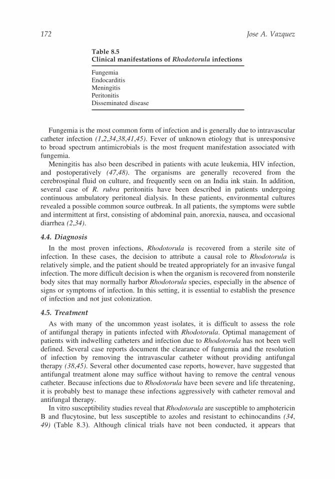
172 Jose A. Vazquez
Table 8.5Clinical manifestations of Rhodotorula infections
FungemiaEndocarditisMeningitisPeritonitisDisseminated disease
Fungemia is the most common form of infection and is generally due to intravascularcatheter infection (1,2,34,38,41,45). Fever of unknown etiology that is unresponsiveto broad spectrum antimicrobials is the most frequent manifestation associated withfungemia.
Meningitis has also been described in patients with acute leukemia, HIV infection,and postoperatively (47,48). The organisms are generally recovered from thecerebrospinal fluid on culture, and frequently seen on an India ink stain. In addition,several case of R. rubra peritonitis have been described in patients undergoingcontinuous ambulatory peritoneal dialysis. In these patients, environmental culturesrevealed a possible common source outbreak. In all patients, the symptoms were subtleand intermittent at first, consisting of abdominal pain, anorexia, nausea, and occasionaldiarrhea (2,34).
4.4. Diagnosis
In the most proven infections, Rhodotorula is recovered from a sterile site ofinfection. In these cases, the decision to attribute a causal role to Rhodotorula isrelatively simple, and the patient should be treated appropriately for an invasive fungalinfection. The more difficult decision is when the organism is recovered from nonsterilebody sites that may normally harbor Rhodotorula species, especially in the absence ofsigns or symptoms of infection. In this setting, it is essential to establish the presenceof infection and not just colonization.
4.5. Treatment
As with many of the uncommon yeast isolates, it is difficult to assess the roleof antifungal therapy in patients infected with Rhodotorula. Optimal management ofpatients with indwelling catheters and infection due to Rhodotorula has not been welldefined. Several case reports document the clearance of fungemia and the resolutionof infection by removing the intravascular catheter without providing antifungaltherapy (38,45). Several other documented case reports, however, have suggested thatantifungal treatment alone may suffice without having to remove the central venouscatheter. Because infections due to Rhodotorula have been severe and life threatening,it is probably best to manage these infections aggressively with catheter removal andantifungal therapy.
In vitro susceptibility studies reveal that Rhodotorula are susceptible to amphotericinB and flucytosine, but less susceptible to azoles and resistant to echinocandins (34,49) (Table 8.3). Although clinical trials have not been conducted, it appears that

8. Infection Due to Non-Candidal Yeasts 173
amphotericin B with or without flucytosine is the best recommendation for antifungaltherapy at this time (1,2). In view of the intrinsic resistance of Rhodotorula to theazoles and echinocandins, these agents should not be used as monotherapy unless invitro susceptibility activity has been assessed.
5. MALASSEZIA
Malassezia furfur is a yeast commonly found on human skin. It has been welldocumented to cause superficial skin infections such as pityriasis (tinea) versicolorand folliculitis. In addition, in immunocompromised host it may occasionally causeinvasive infection.
5.1. Etiologic Agents
The genus Malassezia consists of several species; the two most frequently isolatedspecies include M. furfur and M. pachydermatis (1,2,5). The other less commonlyisolated species include M. sympodialis, M. slooffiae, M. globosa, M. obtuse, andM. restricta. M. furfur is the dominant species recovered in humans as a fungalpathogen. M. furfur is a dimorphic, lipophilic yeast that is unable to synthesize mediumor long-chain fatty acids and thus has a strict in vitro requirement for exogenous fattyacids of the C12 and C14 series (50). Although it exists primarily in the yeast form,it may also form filamentous structures on the skin when the organism is associatedwith superficial infections (51). Because of its nutritional requirements, M. furfur isdifficult to recover from clinical specimens unless its presence is suspected and specialpreparations are made by the microbiology laboratory. The second most commonspecies is M. pachydermatis, which is generally associated with infections in dogsproducing otitis externa (50). Occasionally, however, it has been implicated in humaninfections (51,52). Both Malassezia species, when grown under favorable conditionsproduce clusters of oval to round, thick-walled yeast cells, with unipolar buds that formrepeatedly from the same pole of the parent cell. This gives rise to the characteristic“collarette” at the bud site. Media such as Sabouraud dextrose agar, chocolate agar, andtrypticase soy agar with 5% sheep blood all require the addition of supplements suchas olive oil to permit the growth of this organism (53). M. pachydermatis, however,does not require exogenous lipids for growth, can be recovered on conventional fungalmedia, and its colonies tend to be dry and white to creamy in color.
5.2. Epidemiology
Malassezia is frequently found on normal individuals colonizing the skin. Distri-bution of this colonization tends to correlate with the more oily areas, most likelybecause of the organisms’ requirement of exogenous fatty acids found in those areas.Thus, it is found primarily on the scalp, shoulders, chest, and back (53). The highestcolonization rates are found in teenagers (>90%).
The isolation of M. furfur from newborns is reported to be less than 10% in non-intensive care settings. However, it has been reported to be greater than 80% in neonatalintensive care units (ICUs) (51,53). Risk factors associated with increased colonizationrates in neonates include prematurity, duration of hospitalization in the ICU, use ofocclusive dressings, and prolonged antimicrobial use (51). Although the epidemiology

174 Jose A. Vazquez
Table 8.6Risk factors associated with Malassezia infections
PrematurityLonger duration of hospitalizationUse of occlusive dressingsAdministration of antibioticsUse of central venous cathetersUse of intravenous lipids
of disseminated infection in adults has not been well studied, there appear to be severalrisk factors that are frequently associated with deep-seated infections (Table 8.6). Theseinclude prematurity, central venous catheters, total parenteral nutrition, parenteral lipidpreparations, and immunocompromised state (54–57). Molecular epidemiologic studiesusing DNA fingerprinting have concluded that within the neonatal ICUs there islongitudinal persistence of both M. furfur and M. pachydermatis strains (58).
5.3. Clinical Manifestations
Malassezia generally produces superficial skin infections, such as pityriasis (tinea)versicolor or folliculitis. From time to time, Malassezia may produce a deep-seatedor hematogenous infections (54–57). The first reported case of systemic infection wasdescribed in 1981 in a premature neonate who developed fungemia and vasculitis whileon lipid therapy (57). Since then, numerous reports describing disseminated infectionhave been published (51,55). The manifestations of disseminated infection vary fromsubclinical and mild symptomatology, such as fever, to sepsis with associated multi-organ dysfunction (51,55). The majority of the Malassezia infections are diagnosedin premature infants. Occasionally, they may be seen in adults. The most commonlyreported manifestations of systemic infection include fever unresponsive to broad-spectrum antimicrobials, bradycardia, respiratory distress, hepatosplenomegaly, andlethargy.
5.4. Diagnosis
Laboratory findings include leukocytosis and thrombocytopenia. Chest x-ray exami-nation frequently reveals bilateral pulmonary infiltrates (>50%) (1,2,54). Occasionally,the diagnosis of disseminated infection can be made by a Gram stain of the buffy coat ofblood. The budding yeast cells may be observed using different stains such as Giemsa,periodic acid Schiff (PAS), Gomori’s methenamine silver (GMS), or Calcofluor white.Blood cultures are usually negative unless the infection is initially suspected and thelaboratory uses a lipid-enriched media. The recovery of the organisms is enhancedby using the lysis centrifugation blood culture technique (59). Palmitic acid (3%)supplementation may also improve the recovery of Malassezia (59).
5.5. Treatment
Management of M. furfur fungemia and disseminated infection are controversial.Most authorities recommend prompt removal of the central venous catheter and discon-tinuation of intravenous lipids (54,55). In most cases without a deep-seated infection,

8. Infection Due to Non-Candidal Yeasts 175
removal of the central venous catheter and discontinuation of lipids is all that isneeded to clear the infection. This treatment modality accomplishes two objectives:it eradicates the nidus of infection, and removes the nutritional requirements of theorganism. If fungemia persists or there is evidence of deep-seated infection, it isprudent to initiate antifungal therapy. Fortunately, Malassezia species are suscep-tible to azoles and polyenes (1,2,34) (Table 8.3). In vitro susceptibility assays ofM. furfur strains demonstrate that most of the isolates are susceptible to ampho-tericin B, ketoconazole (MIC range ≤0.05 to 0.4 μg/ml), miconazole (MIC range0.4≥50 μg/ml), and fluconazole. Most of the isolates are intrinsically resistant to flucy-tosine (MIC90>100 μg/ml) (1,2,34). Although randomized clinical trials have not beenconducted, in most situations, either fluconazole 400 mg/day or amphotericin B 0.7mg/kg per day is sufficient to eradicate the infection (Table 8.4). Based on excellentin vitro activity, itraconazole and voriconazole are also alternate choices.
6. OTHER YEASTS
In addition to the yeasts discussed thus far, fewer reports have been publishedof infection due to other yeasts. These include Blastoschizomyces, Sporobolomyces,Pichia (formerly Hansenula), and Exophiala.
6.1. Blastoschizomyces
Blastoschizomyces capitatus (formerly Geotrichum capitatum or Trichosporoncapitatum) infections, although less common than those due to T. asahii, have beenwell described in the literature (60). B. capitatus is found in wood and poultry, buthas also been recovered from sputum and normal intact skin (5). Geographically, itappears to be the opposite of T. asahii, with B. capitatus infections found primarily inEurope and T. asahii found in North America (61). In most cases, the major risk factorsinclude neutropenia and underlying hematologic malignancies. Although the portal ofentry is unknown, it is suspected to be the either the respiratory tract, gastrointestinaltract, or central venous catheters (35,61).
Infection may involve a single organ or multiple organs and may be associated withfungemia. The clinical spectrum of disseminated infection is similar to that of systemiccandidiasis and includes fungemia with or without organ infection (1,2,61). By andlarge, the manifestations begin with fever of unknown etiology and unresponsive toantimicrobials. Diagnosis can be made with blood cultures or on biopsy of the skin oraffected organs. Blood cultures have been reported to be positive in more than 80%of cases (61). B. capitatus easily grows in blood culture bottles and on fungal specificmedia (5). Although skin lesions are commonly seen, fungal stains and cultures frombiopsied skin lesions are frequently negative (5,61).
Mortality rates between 60% and 80% are generally described (61). However,underlying disease, persistent neutropenia and concurrent infections are significantcontributing factors to this overall mortality rate. Optimal therapy has not yet beenestablished. Until recently, however, most patients have received amphotericin B (2).As with all fungal infections, the initial step is to decrease or reverse the immuno-compromised state. In vitro susceptibility studies demonstrate that the organism issusceptible to amphotericin B (MIC90 0.12 �g/ml), and less susceptible to azoles such

176 Jose A. Vazquez
as fluconazole and ketoconazole (0.04 to 32�g/ml), but appears to be susceptible toitraconazole and voriconazole. Most isolates are resistant to flucytosine (5) (Table 8.3).The current recommendation is to use amphotericin B at a dose of 1 to 1.5 mg/kgper day (1,2). However, since the newer azoles, voriconazole and posaconazole, havedemonstrated good in vitro activity they may also be suitable alternatives (62).
6.2. Sporobolomyces
Sporobolomyces are yeast-like organisms that belong to the family Sporobolomyc-etaceae. These yeast are found throughout the world in soil, bark, and decaying organicmaterial. They have occasionally been associated with infections in humans. There areseven known species of Sporobolomyces, but only three have been documented to causedisease—S. salmonicolor, S. holsaticus, and S. roseus. To date, there have been only sixdocumented cases of Sporobolomyces infections: a nasal polyp, one case of dermatitis,one case of infected skin blisters, one case of mycetoma, and two cases of disseminatedinfection in patients with acquired immunodeficiency syndrome (AIDS; lymph nodeand bone marrow) (2,63). In vitro susceptibility studies show that S. salmonicolor issusceptible to amphotericin B and the imidazoles (1,2,5) (Table 8.3). Despite the factthat these organisms are saprophytic, the case reports indicate their potential ability toproduce invasive infection in humans, especially in a compromised host.
REFERENCES
1. Hospenthal DR. Uncommon fungi. In: Mandell GL, Bennett JE, Dolin R, eds. Mandell,Douglas, and Bennett’s principles and practice of infectious diseases. 6th ed. Philadelphia:Elsevier Churchill Livingstone, 2005;3068–3079.
2. Vazquez JA. Rhodotorula, Malassezia, Trichosporon, and other yeast-like fungi. In:Dismukes WE, Pappas PG, Sobel JD, eds. Clinical mycology. New York: OxfordUniversity Press, 2003:206–217.
3. Beigel H. The human hair: its growth and structure (1865), as cited by Langenon M. InDarier S, et al: Nouvelle Praqt Dermatol (Paris, Masson, Cie) 1936;2:377.
4. Watson KC, Kallichurum S. Brain abscess due to Trichosporon cutaneum. J Med Microbiol1970;3:191–193.
5. Kwon-Chung KJ, Bennett JE. Infections due to Trichosporon and other miscellaneousyeast-like fungi. In: Medical mycology. Philadelphia: Lea & Febiger, 1992:768–794.
6. Gueho E, de Hoog GS, Smith MT, Meyer SA. DNA relatedness, taxonomy, and medicalsignificance of Geotrichum capitatum. J Clin Microbiol 1997;25:1191–1194.
7. Kemeker BJ, Lehman PF, Lee JW, Walsh TJ. Distinction of deep vs. superficial clinical andnon-clinical environmental isolates of Trichosporon beigelii by isoenzymes and restrictionfragment length polymorphisms of rDNA generated by the polymerase chain reaction. JClin Microbiol 1991;29:1677–1683.
8. Behrend G. Ubertrichomycosis nodosa. Berlin Lin Wochenschr 1890;27:464.9. Haupt HM, Merz WG, Beschorner WE, Vaughan WP, Saral R. Colonization and infection
with Trichosporon species in the immunosuppressed host. J Infect Dis 1983;147:199–203.10. Walsh TJ. Trichosporonosis. Infect Dis Clin North Am 1989;3:43–52.11. Ebright JR, Fairfax MR, Vazquez JA. Trichosporon asahii, a non-Candida yeast that caused
fatal septic shock in a patient without cancer or neutropenia. Clin Infect Dis 2001;33:28–30.12. Keay S, Denning D, Stevens DA. Endocarditis due to Trichosporon beigelii: in vitro
susceptibility of isolates and review. Rev Infect Dis 1991;13:383–386.

8. Infection Due to Non-Candidal Yeasts 177
13. Sheikh HA, Mahgoub S, Badi K. Postoperative endophthalmitis due to Trichosporoncutaneum. Brit J Ophthalmol 1986;58:591–594.
14. Walsh TJ, Lee JW, Melcher GP, et al. Experimental Trichosporon infection in persistentlygranulocytopenia rabbits: Implications for pathogenesis, diagnosis, and treatment of anemerging opportunistic mycosis. J Infect Dis 1992;166:121–133.
15. Seeliger HPR, Schroter R. A serologic study on the antigenic relationships of the formgenus Trichosporon. Sabouraudia 1963;2:248–263.
16. Campbell CK, Payne AL, Teall AJ, Brownell A, Mackenzie DW. Cryptococcal latexantigen test positive in a patient with Trichosporon beigelii. Lancet 1985;2:43–44.
17. Paphitou NI, Ostrosky-Zeichner L, Paetznick VL, Rodriguez JR, Rex JH. In vitro antifungalsusceptibilities of Trichosporon species. J Clin Microbiol 2003; 46:1144–1146.
18. Serena C. Pastor FJ, Gilgado F, Mayayo E, Guarro J.. Efficacy of micafungin in combi-nation with other drugs in a murine model of disseminated Trichosporonosis. AntimicrobAgents Chemother 2005;49:497–502.
19. Sobel JD, Vazquez J, Lynch M, Meriwether C, Zervos MJ.. Vaginitis due to Saccharomycescerevisiae: epidemiology, clinical aspects, and therapy. Clin Infect Dis 1993;16:93–99.
20. Enache-Angoulvant A, Hennequin C. Invasive Saccharomyces Infection: a comprehensivereview. Clin Infect Dis 2005;41:1559–1568.
21. Nielsen H, Stenderup J, Bruun B. Fungemia with Saccharomycetaceae. Report of fourcases and review of the literature. Scand J Infect Dis 1990;22:581–584.
22. Nyirjesy P, Vazquez JA, Ufberg DD, Sobel JD, Boikov DA, Buckley HR. Saccharomycescerevisiae vaginitis: transmission from yeast used in baking. Obstet Gynecol 1995;86:326–329.
23. Morrison VA, Haake RJ, Weisdorf DJ. The spectrum of non-Candida fungal infectionsfollowing bone marrow transplantation. Medicine 1993;72:78–89.
24. Holzschu DL, Chandler FW, Ajello L, Ahearn DG. Evaluation of industrial yeast forpathogenicity. Sabouradia 1979;17:71–78.
25. Clemons KV, McCusker JH, Davis RW, Stevens DA. Comparative pathogenesis of clinicaland non-clinical isolates of Saccharomyces cerevisiae. J Infect Dis 1994;169:859–867.
26. Eng RH, Drehmel R, Smith SM, Goldstein EJ. Saccharomyces cerevisiae infections inman. Sabouraudia 1984;22:403–407.
27. McCullough MJ, Clemons KV, Farina C, McCusker JH, Stevens DA. Epidemiologicalinvestigation of vaginal Saccharomyces cerevisiae isolates by a genotypic method. J ClinMicrobiol 1998;36:557–562.
28. Cimolai N, Gill MJ, Church D. Saccharomyces cerevisiae fungemia: case report and reviewof the literature. Diagn Microbiol Infect Dis 1987;8:113–117.
29. Aucott JN, Fayen J, Grossnicklas H, Morrissey A, Lederman MM, Salata RA. Invasiveinfection with Saccharomyces cerevisiae: report of three cases and review. Rev Infect Dis1990;12:406–411.
30. Sethi N, Mandell W. Saccharomyces fungemia in a patient with AIDS. NY State J Med1988;88:278–279.
31. Rubinstein E, Noriega ER, Simberkoff MS, Holzman R, Rahal Jr JJ. Fungal endocarditis:analysis of 24 cases and review of the literature. Medicine 1975;54:331–344.
32. Canafax DM, Mann HJ, Dougherty SH. Postoperative peritonitis due to Saccharomycescervisiae treated with ketoconazole. Drug Intell Clin Pharm 1982;16:698–699.
33. Chertow GM, Marcantonio ER, Wells RG. Saccharomyces cerevisiae empyema in apatient with esophagopleural fistula complicating variceal sclerotherapy. Chest 1991;99:1518–1519.
34. Hazen KC. New and emerging yeast pathogens. Clin Microbiol Rev 1995;8:462–478.

178 Jose A. Vazquez
35. Fell JW, Tallman AS, Ahearn DG. Genus Rhodotorula Harrison. In: Kreger-van RijNJW, ed. The yeasts: a taxonomic study. 3rd ed. Amsterdam: Elsevier Science Publishers,1984:893–905.
36. Rippon JW. Medical mycology. The pathogenic fungi and pathogenic actinomycetes. 3rded. Philadelphia: W.B. Saunders, 1988:610–611.
37. Mackenzie DWH. Yeasts from human sources. Sabouraudia 1961;1:8–14.38. Kiehn TE, Gorey E, Brown AE, Edwards FF, Armstrong D. Sepsis due to Rhodotorula
related to use of indwelling central venous catheters. Clin Infect Dis 1992;14:841–846.39. Ahearn DG, Jannach JR, Roth FJ. Speciation and densities of yeasts in human urine
specimens. Sabouraudia 1966;5:110–119.40. Saez H. Etude ecologique sur les Rhodotorula des homotherms. Rev Med Vet
1979;130:903–908.41. Anaissie E, Bodey GP, Kantarjian H, et al. New spectrum of fungal infections in patients
with cancer. Rev Infect Dis 1989;11:369–378.42. Rose HD, Kurup VP. Colonization of hospitalized patients with yeast-like organisms.
Sabouraudia 1977;15:251–256.43. Leeber DA, Scher I. Rhodotorula fungemia presenting as “endotoxic” shock. Arch Intern
Med 1969;123:78–81.44. Rusthoven JJ, Feld R, Tuffnell PG. Systemic infection by Rhodotorula spp. in the immuno-
compromised host. J Infect 1984;8:241–246.45. Braun DK, Kauffman CA. Rhodotorula fungemia: a life-threatening complication of
indwelling central venous catheters. Mycoses 1992;35:305–308.46. Louria DB, Greenberg SM, Molander DW. Fungemia caused by certain nonpathogenic
strains of the family Cryptococcaceae. N Engl J Med 1960;263:1281–1284.47. Pore RS, Chen J. Meningitis caused by Rhodotorula. Sabouraudia 1976;14:331–335.48. Gyaurgieva OH, Bogomolova TS, Gorshkova GI. Meningitis caused by Rhodotorula rubra
in an HIV-infected patient. J Med Vet Mycol 1996;34:357–359.49. Diekema DJ, Petroelje B, Messer SA, Hollis RJ, Pfaller MA. Activities of available and
investigational agents against Rhodotorula species. J Clin Microbiol 2005;43:476–478.50. Nazzaro-Porro MN, Passi S, Caprilli F, Nazzaro P, Morphogo G. Growth requirements
and lipid metabolism of Pityrosporum orbiculare. J Invest Dermatol 1976;66:178–182.51. Marcon MJ, Powell DA. Human infections due to Malassezia spp. Clin Microbiol Rev
1992;5:101–119.52. Gueho E, Simmons RB, Pruitt WR, Meyer SA, Ahearn DG. Association of Malassezia
pachydermatis with systemic infections of humans. J Clin Microbiol 1987;25:1789–1790.53. Ingham E, Cunningham AC. Malassezia furfur. J Med Vet Mycol 1993;31:265–288.54. Marcon MJ, Powell DA. Epidemiology, diagnosis and management of M. furfur systemic
infection. Diagn Microbiol Infect Dis 1987;10:161–175.55. Klotz SA. Malassezia furfur. Infect Dis Clin Am 1989;3:53–63.56. Barber GR, Brown AE, Kiehn TE, Edwards FF, Armstrong D. Catheter-related Malassezia
furfur fungemia in immunocompromised patients. Am J Med 1993;95:365–370.57. Weiss SJ, Schoch PE, Cunha BA. Malassezia furfur fungemia associated with central
venous catheter lipid emulsion infusion. Heart Lung 1991;20:87–90.58. van Belkum A, Boekhout T, Bosboom R. Monitoring spread of Malassezia infections
in a neonatal intensive care unit by PCR-medicated genetic typing. J Clin Microbiol1994;32:2528–2532.
59. Nelson SC, Yau YC, Richardson SE, Matlow AG. Improved detection of Malasseziaspecies in lipid-supplemented peds plus blood culture bottles. J Clin Microbiol1995;33:1005–1007.

8. Infection Due to Non-Candidal Yeasts 179
60. Gemeinhardt H. Lungenpathogenitat von Trichosporon capitatum beim menschen. Zentra-blatt fur Bakteriolgie (Series A) 1965;196:121–133.
61. Martino P, Venditti M, Micozzi A, et al. Blastoschizomyces capitatus: an emerging causeof invasive fungal disease in leukemia patients. Rev Infect Dis 1990;12:570–582.
62. Espinel-Ingroff A. Comparison of in vitro activities of the new triazole SCH56592 andthe echinocandins MK-0991 (L-743,872) and LY303366 against opportunistic filamentousand dimorphic fungi and yeast. J Clin Microbiol 1998;36:2950–2956.
63. Morris JT, Beckius M, McAllister CK. Sporobolomyces infection in an AIDS patient.J Infect Dis 1991;164:623–624.
SUGGESTED READINGS
Diekema DJ, Petroelje B, Messer SA, Hollis RJ, Pfaller MA. Activities of available andinvestigational agents against Rhodotorula species. J Clin Microbiol 2005;43:476–478.
Enache-Angoulvant A, Hennequin C. Invasive Saccharomyces infection: a comprehensivereview. Clin Infect Dis 2005;41:1559–1568.
Espinel-Ingroff A. Comparison of in vitro activities of the new triazole SCH56592 and theechinocandins MK-0991 (L-743,872) and LY303366 against opportunistic filamentous anddimorphic fungi and yeast. J Clin Microbiol 1998;36:2950–2956.
Hospenthal DR. Uncommon fungi. In: Mandell GL, Bennett JE, Dolin R, eds. Mandell, Douglas,and Bennett’s principles and practice of infectious diseases. 6th ed. Philadelphia: ElsevierChurchill Livingstone, 2005:3068–3079.
Marcon MJ, Powell DA. Human infections due to Malassezia spp. Clin Microbiol Rev1992;5:101–119.
Martino P, Venditti M, Micozzi A, et al. Blastoschizomyces capitatus: an emerging cause ofinvasive fungal disease in leukemia patients. Rev Infect Dis 1990;12:570–582.
Vazquez JA. Rhodotorula, Malassezia, Trichosporon, and other yeast-like fungi. In: DismukesWE, Pappas PG, Sobel JD, eds. Clinical mycology. New York: Oxford University Press,2003:206–217.

9Aspergillosis
Helen W. Boucher, MD and Thomas F. Patterson, MD
1. INTRODUCTION
Aspergillosis is caused by Aspergillus, a hyaline responsible not only for invasiveaspergillosis, but also for a variety of noninvasive or semi-invasive conditions. Thesesyndromes range from colonization to allergic responses to Aspergillus includingallergic bronchopulmonary aspergillosis (ABPA) to semi-invasive or invasive infec-tions, spanning a spectrum from chronic necrotizing pneumonia to invasive pulmonaryaspergillosis.
The genus Aspergillus was first recognized in 1729 by Micheli, in Florence, whonoted the resemblance between the sporulating head of an Aspergillus species and anaspergillum used to sprinkle holy water (Fig. 9.1). In 1856, Virchow published thefirst complete microscopic descriptions of the organism (1).
The frequency and severity of invasive fungal infections in immunocompromisedpatients have increased steadily over the past 2 decades with the growing populationof patients undergoing transplantation and the persistent challenges in preventing,diagnosing and treating these infections (2). Mortality due to documented invasiveaspergillosis approaches 80% to 100% in high-risk patients, including those with under-lying hematologic malignancy or bone marrow or solid organ transplantation, and maybe related to several factors, including diagnostic and therapeutic inadequacies (3–5).Apart from organ transplant recipients, individuals with acquired immunodeficiencysyndrome (AIDS), and patients hospitalized with severe illnesses, major increases ininvasive fungal infections have been observed in patients with hematologic malig-nancies who receive induction or consolidation chemotherapy and those who undergohematopoietic stem cell transplantation (HSCT) (5).
Successful therapy depends not only an early diagnosis—which is often difficult toestablish—but even more importantly, on reversal of underlying host immune defects,such as neutropenia or high-dose immunosuppressive therapy (2). Non-culture-basedtests and radiological approaches can be used to establish an early diagnosis of infectionand may result in improved outcomes of infection (2,6,7). Even when therapy is begun
From: Infectious Disease: Diagnosis and Treatment of Human MycosesEdited by: D. R. Hospenthal and M. G. Rinaldi © Humana Press Inc., Totowa, NJ
181

182 Helen W. Boucher and Thomas F. Patterson
Fig. 9.1. Microscopic morphology of Aspergillus fumigatus showing a single roleof phialides (uniseriate) bearing smooth conidia in a columnar fashion. (Courtesy ofwww.doctorfungus.org). [Figure in color on CD-ROM].
promptly, efficacy of many treatment regimens, including amphotericin B deoxy-cholate, is poor, particularly in patients with disseminated or central nervous systemdisease (2,3,5). New diagnostic approaches have been introduced and new antifungalagents have been developed for this disease including the newer azoles, lipid formula-tions of amphotericin B, and a new drug class—the echinocandins (8).
2. ETIOLOGIC AGENTS
Aspergillus fumigatus is one of the most ubiquitous of the airborne saprophyticfungi (9). A. fumigatus has emerged worldwide as a frequent cause of nosocomialinfection and may be regarded as the most important airborne pathogenic fungus (10).As Aspergillus species can be readily found in the environment, invasive aspergillosisis widely believed to occur as a consequence of exogenous acquisition of the conidia(spores) of the species (10). The most common route of transmission of Aspergillusinfection is the airborne route. Aspergillus conidia are resilient and may survive forlong periods in fomites (any substance that can absorb, retain, and transport infec-tious species, e.g., woolen clothes or bedding) (11). Aspergillus infection occursless frequently through damaged mucocutaneous surfaces (e.g., following surgery orthrough contaminated dressings). However, the sources of Aspergillus may be broaderthan have traditionally been thought, as waterborne transmission of Aspergillus conidiathrough contaminated aerosols has been suggested (12).

9. Aspergillosis 183
Table 9.1Characteristics of common Aspergillus species
Aspergillusspecies
Mycologicalcharacteristics
Clinicalsignificance
Mycoses
A. flavus Olive to limegreen colonies
Second most commonspecies, producesaflatoxin, may beless susceptible topolyenes
Sinusitis, cutaneousinfection, pulmonary anddisseminated disease
A. fumigatus Smoky, blue- orgray green,small, smoothconidia (2–2.5μm)
Most common speciescausing invasiveinfection
Invasive pulmonaryaspergillosis, disseminatedinfection, CNS, others
A. niger Typically blackcolonies, radiateconidial head,large roughconidia
Common cause ofotomycosis,produces oxalatecrystals which maybe seen in host
Otomycosis, cutaneous,endophthalmitis,aspergilloma, invasivepulmonary or disseminateddisease less common
A. terreus Beige to buffcolonies,globoseaccessoryconidia alonghyphae
Increasing frequency,associated with soil,usually resistant topolyenes
Pulmonary, disseminated,cutaneous, keratitis, CNS
A. lentulus Poorly sporulatingvariant of A.fumigatus
May be multidrugresistant, recentlydescribedvariant, may beunderdiagnosed
Invasive pulmonary,disseminated, other sites
The most common species causing invasive aspergillosis include Aspergillusfumigatus, by far the most common, A. flavus, A. terreus, and, less commonly forinvasive infection, A. niger (5) (Table 9.1). Recent studies have shown emergenceof less common species, including A. terreus (which is frequently resistant topolyenes) and other unusual less pathogenic species as the etiologic agents of invasiveinfection (13).
3. EPIDEMIOLOGY
The incidence of invasive aspergillosis has increased substantially during the last fewdecades because of the use of more intensive cytotoxic anticancer chemotherapy andthe introduction of novel immunosuppressive therapies for organ transplant recipients,both of which have prolonged the period of risk for many individuals. The increasingnumber of patients undergoing solid organ, bone marrow, and hematopoietic stem celltransplantation, and the implementation of aggressive surgical interventions has also

184 Helen W. Boucher and Thomas F. Patterson
contributed to the increased incidence (10). The changes in epidemiology of invasiveaspergillosis may also be the result of growing awareness of aspergillosis among clini-cians, the introduction of noninvasive diagnostic tools, and improved microbiologicallaboratory techniques.
Invasive fungal infections are an important cause of morbidity and mortalityamong patients with severely compromised immune systems. Although there havebeen significant advances in the management of immunosuppressed patients, invasiveaspergillosis remains an important life-threatening complication, and is the leadingcause of infection-related mortality in many immunocompromised individuals.
Immunosuppression and breakdown of anatomical barriers, such as the skin, are themajor risk factors for fungal infections (7). Individuals at risk for invasive aspergillosisinclude those with severely comprised immune systems as a result of anticancerchemotherapy, solid organ or bone marrow transplantation, AIDS, or use of high-dosecorticosteroids. Patients with hematological disorders, such as prolonged and severeneutropenia, those undergoing transplantations, and those treated with corticosteroidsand newer immunosuppressive therapies such as the tumor necrosis factor-� antagonists(e.g., inflixamab) are considered to be at highest risk for invasive aspergillosis (7,14).
4. PATHOGENESIS AND IMMUNITY
Invasive aspergillosis most frequently originates via inhalation of Aspergillus conidiainto the lungs, although other routes of exposure such as inhalation of water aerosolscontaminated with Aspergillus conidia have been suggested (12).
In the absence of effective pulmonary host defenses, the inhaled small resting conidiaenlarge and germinate, then transform into hyphae with subsequent vascular invasionand eventual disseminated infection. The incubation period for conidial germinationin pulmonary tissue is variable, ranging from 2 days to months (15). Hydrocortisonesignificantly increases the growth rates of Aspergillus; likely one of the reasons corti-costeroids pose a risk factor for invasive disease (16).
Although infection in apparently normal hosts can occur, invasive aspergillosisis extremely uncommon in immunocompetent hosts (5). Normal pulmonary defensemechanisms usually contain the organism in a host with intact pulmonary defenses.The first line of defense against Aspergillus is ciliary clearance of the organism fromthe airways and limited access to the alveoli due to conidia size. This feature isone reason for the increased pathogenicity of A. fumigatus as compared with otherspecies of Aspergillus (16). Once conidia reach the alveoli, pulmonary macrophages aregenerally capable of ingesting and killing Aspergillus conidia (17). When macrophagesfail to kill the conidia (e.g., high fungal inoculum, decreased number or function ofmacrophages), conidia germinate and begin to form hyphae. Polymorphonuclear leuko-cytes are recruited via complement activation and production of neutrophil chemotacticfactors and extracellularly kill both swollen conidia and hyphae (18). Antibodies againstAspergillus are common due to the ubiquitous nature of the organism, although theyare not protective nor are they useful in the diagnosis of infection in high-risk patientsdue to the lack of consistent seroconversion after exposure or infection (19).
Corticosteroids play a major role in increasing susceptibility to Aspergillusby decreasing oxidative killing of the organism by pulmonary macrophages and

9. Aspergillosis 185
by increasing the linear growth rate by as much as 30% to 40% and cell synthesis bygreater than 150% (16).
Many Aspergillus species produce toxins including aflatoxins, ochratoxin A,fumagillin, and gliotoxin. Gliotoxin works in several ways to help evade host defenses:
• Inhibition of phagocyte NADPH oxidase activation (key in host defense versusfilamentous fungi)
• Inhibition of macrophage ingestion of Aspergillus• Suppression of functional T cell responses (9,20)
In tissues, invasive aspergillosis causes extensive destruction across tissue planes viavascular invasion with resulting infarction and necrosis of distal tissues.
5. CLINICAL MANIFESTATIONS
The clinical syndromes associated with aspergillosis are diverse, ranging fromallergic responses to the organism including allergic bronchopulmonary aspergillosis(ABPA), asymptomatic colonization, superficial infection, and acute or subacute andchronic invasive disease. The clinical presentation generally corresponds to the under-lying immune defects and risk factors associated with each patient group, with greaterimmune suppression correlating with increased risk for invasive disease. Althoughthis chapter focuses on invasive aspergillosis, a brief description of other presenta-tions follows. The reader is encouraged to reference other sources for more in-depthdiscussion of those conditions (1).
5.1. Allergic Bronchopulmonary Aspergillosis
Allergic bronchopulmonary aspergillosis (ABPA) is a chronic allergic response toAspergillus characterized by transient pulmonary infiltrates due to atelectasis. Theincidence of ABPA is estimated to range from 1% to 2% in patients with persistentasthma and in approximately 7% (with a range from 2% to 15%) of patients withcystic fibrosis (21). Specific criteria are used to establish the diagnosis of ABPA as nosingle finding except for central bronchiectasis in a patient with asthma is diagnostic forthe condition (21–23). ABPA typically progresses through a series of remissions andexacerbations but can eventually lead to pulmonary fibrosis, which is associated witha poor long-term prognosis (23). Management of ABPA is directed at reducing acuteasthmatic symptoms and avoiding end-stage fibrosis. Corticosteroid therapy is commonlyused for treating exacerbations, although few randomized trials have been conductedfor their use (24). The role for antifungal therapy was evaluated with a randomizeddouble-blind, placebo-controlled trial that showed itraconazole at 200 mg/day for 16weeks significantly reduced daily corticosteroid use, reduced levels of immunoglobulinE (IgE), and improved exercise tolerance and pulmonary function (22,25).
5.2. Aspergilloma
A pulmonary fungus ball due to Aspergillus or “aspergilloma” is a solid mass ofhyphae growing in a previously existing pulmonary cavity, typically in patients withchronic lung disease such as bullous emphysema, sarcoidosis, tuberculosis, histoplas-mosis, congenital cyst, bacterial lung abscess or, very rarely, in a pulmonary bleb

186 Helen W. Boucher and Thomas F. Patterson
from Pneumocystis pneumonia in AIDS (26,27). On chest radiograph, a pulmonaryaspergilloma appears as a solid round mass in a cavity (see Fig. 5–9, Chapter 5). Inmany patients the fungus ball due to Aspergillus remains asymptomatic, but in a signif-icant number, hemoptysis occurs and can be fatal (28). Surgical resection is consideredthe definitive therapy but the dense pleural adhesions adjacent the fungus ball and thepoor pulmonary reserve of most patients makes surgery hazardous. Contamination ofthe pleural space with Aspergillus and the common complication of bronchopleuralfistula in the postoperative period can lead to chronic Aspergillus empyema. Denseadhesions make pleural drainage difficult, often requiring pleural stripping or anEloesser procedure, further compromising lung function (28).
Aspergillus can also be associated with fungal balls of the sinuses without tissueinvasion (27). The maxillary sinus is the most common site for a sinus aspergilloma tooccur (27). Clinical presentation is similar to that for any chronic sinusitis. Managementis usually directed at surgical removal and a generous maxillary antrostomy for sinusdrainage, along with confirmation that invasive disease has not occurred.
5.3. Other Superficial or Colonizing Syndromes
Other superficial or colonizing syndromes of aspergillosis include otomycosis, acondition of superficial colonization typically due to A. niger (29); onychomycosiswhich, although rare, can become chronic and respond poorly to antifungal agents(30); and keratitis, particularly following trauma or corneal surgery (31).
5.4. Chronic Pulmonary Aspergillosis
Denning and colleagues have described three distinct syndromes of chronicpulmonary aspergillosis to better characterize patients who develop chronic pulmonarydisease related to Aspergillus (32). These conditions include chronic cavitarypulmonary aspergillosis, which is characterized by the formation and expansion ofmultiple cavities, which may contain fungus balls, chronic fibrosing aspergillosis,which as its name suggests involves extensive fibrosis, and chronic necrotizingaspergillosis or subacute aspergillosis, in which slowly progressive infection occursusually in a single thin-walled cavity. In all of these conditions, the diagnosis issuggested by radiological and clinical features and the role for therapy remains specu-lative, although it appears that long-term antifungal therapy may be beneficial in asubset of patients, perhaps even with the extended spectrum triazole antifungals (32,33).
5.5. Invasive Pulmonary Aspergillosis
Invasive pulmonary aspergillosis is the most common form of invasive aspergillosisin immunocompromised patients and occurs after approximately 2 weeks of neutropenia(34) or during the course of graft versus host disease, now the most common risk factorin hematopoietic stem cell transplant recipients (35). Symptoms include fever (may beabsent in the presence of high-dose corticosteroid therapy), dry cough, shortness ofbreath, pleuritic chest pain, hemoptysis as well as pulmonary infiltrates all of which lagbehind disease progression. In lung transplant patients and those with AIDS, Aspergillustracheobronchitis can present with cough, wheezing, and shortness of breath and chestradiographs show normal lungs with or without atelectasis (36).

9. Aspergillosis 187
5.6. Disseminated Aspergillosis
A variety of signs and symptoms are seen with disseminated invasive aspergillosisaccording to the organs involved. Involved organs include the kidneys, liver, spleen, andcentral nervous system (signs and symptoms of stroke or meningitis) most frequently,followed by the heart, bone, skin, and other organs (1). Aspergillosis of the skin canoccur either as a manifestation of disseminated disease or by direct extension from alocal inoculation, for example, from an intravenous catheter (37).
5.7. Sinusitis
Aspergillosis of the sinuses presents in a clinically like rhinocerebral zygomycosis,but is more common in neutropenic patients than in those with diabetic ketoacidosis,and inflammatory signs may thus be less frequent. Fever, nasal congestion, and facialpain can progress to visual changes, proptosis, and chemosis if the infection spreads tothe orbit. Posterior extension to the brain can lead to cranial nerve palsies, other focalneurologic deficits, as well as a depressed level of consciousness (38).
5.8. Endocarditis
Aspergillus endocarditis is the second most common form of fungal endocarditis afterthat caused by Candida species and occurs in prosthetic valve recipients and in nativecardiac valves in intravenous drug users and patients with indwelling central venouscatheters (39). Clinically, these patients present with fever and embolic complications.Blood cultures are rarely positive even with extensive disease (40).
6. DIAGNOSIS
Current diagnostic modalities are limited and the clinician must rely on the combi-nation of knowledge of risk factors, a high index of suspicion, clinical judgment,and the finding of fungi in tissue specimens and/or cultures from the presumedsite of infection. The diagnosis of proven invasive aspergillosis requires both tissuebiopsy demonstrating invasion with hyphae and culture positive for Aspergillusspecies (41). Aspergillus produce hyaline, 3 to 6 μm wide septate hyphae that typicallybranch at acute angles (42) (Fig. 9.2). In tissue these features can often distinguishAspergillus from agents of zygomycosis, but they cannot distinguish Aspergillus froma large number of other opportunistic moulds, including Fusarium and Scedosporium(Pseudallescheria). Thus, culture is needed to confirm the diagnosis (42). Unfortu-nately, invasive, or even less invasive, procedures such as bronchoscopy are oftencontraindicated in immunosuppressed patients, many of whom have low platelets asa result of chemotherapy and other complications. In this setting, positive culture cansupport the diagnosis of invasive aspergillosis.
Plain chest radiography is of limited utility in invasive aspergillosis, as it has lowsensitivity and specificity in this disease6. In contrast, chest computed tomography(CT) scans have proven useful in early diagnosis of invasive pulmonary aspergillosis,as the “halo sign” of low attenuation surrounding a pulmonary nodule has been usedsuccessfully as a marker for early initiation of therapy in high-risk patients withneutropenia or who have undergone hematopoietic stem cell transplantation (43–45)(see Fig. 5.8, Chapter 5). Of note, these radiographic findings are also consistent with

188 Helen W. Boucher and Thomas F. Patterson
Fig. 9.2. Periodic acid-Schiff (PAS) stained tissue section of lung showing dichotomouslybranched, septate hyphae of Aspergillus fumigatus. (Courtesy of www.doctorfungus.org).[Figure in color on CD-ROM].
other infections such as Nocardia species, and may increase over the first week oftherapy even when the patient is improving; follow-up scans should be ordered andinterpreted cautiously with full attention to the clinical progress of the patient (43).
Non-culture diagnostic tests have also been used to diagnose aspergillosis andin attempts to preempt difficult to treat proven disease. A sandwich enzymeimmunoassay (EIA) that utilizes a monoclonal antibody to Aspergillus galactomannan(Platelia Aspergillus, Sonofi Diagnostics Pasteur, Marnes-la-Coquette, France; BioRad,Redmond, WA) is currently US Food and Drug Administration (FDA)-approved andavailable and is being used with varying success around the world (46,47). Questionsremain regarding the value of routine surveillance testing, frequency of testing, roleof false positives, importance of prior antifungal therapy, and correlation of serumgalactomannan results with clinical outcome.
Several reports demonstrate the potential for using polymerase chain reaction (PCR)as an early diagnostic marker, which appears more sensitive than other methodsincluding galactomannan (48,49). These assays may be associated with false-positiveresults due to the ubiquitous nature of Aspergillus conidia, are not standardized, andremain investigational at the present time (50).
Other non-culture-based methods for the diagnosis of invasive aspergillosis includedetection of the nonspecific fungal marker 1,3-�-d-glucan using a variation of theLimulus amebocyte assay. This assay (Fungitell™, Associates of Cape Cod, Falmouth,MA) has been approved for diagnostic purposes by the FDA and is a colorimetricassay that can indirectly determine the concentration of 1,3-�-d-glucan in serumsamples (51). The test appears promising as an indicator of infection due to many

9. Aspergillosis 189
Table 9.2Diagnosis of invasive aspergillosis
Diagnostic method Comment
Respiratory culture Not frequently positive early in course of infection; positive resultin high risk patient (bone marrow transplant, neutropenia) highlycorrelates with infection; may indicate colonization in otherpopulations (chronic pulmonary diseases, lung transplant)
Galactomannan Aspergillus Platelia system (Bio-Rad, Redmond, WA) with variablesensitivity - low (∼40%) with single samples or prior antifungaltherapy or prophylaxis; better yield with reduced threshold forpositivity, serial samples, testing on BAL samples. False positiveswith pipercillin–tazobactam, dietary, neonates
1,3-�-d-glucan Nonspecific detection of cell wall glucan. Commercially availableFungitell™ assay (Associates of Cape Cod, Falmouth, MA),limited validation and availability.
PCR Remains investigational due to lack of standardized reagents andmethods, both false positives and negatives may occur, somerecent studies have suggested less sensitive than other assays
Computed tomography In high-risk patient, “halo” sign and/or pulmonary nodules withoutother documented cause may be a frequent and early sign ofinvasive pulmonary aspergillosis
BAL, bronchoalveolar lavage.
fungi, including Aspergillus and Candida but not Cryptococcus or Zygomycetes (whichcontain little or no �-d-glucan). One study suggested the utility of the assay in earlydiagnosis of invasive fungal infection in a leukemic population, but validation remainslimited (52).
Diagnosis of invasive aspergillosis is summarized in Table 9.2.
7. TREATMENT
The goals of treatment of patients with invasive aspergillosis are to control infectionand to reverse any correctable immunosuppression. Patients at high risk of developinginvasive aspergillosis should be treated based on clinical or radiological criteria aloneif microbiological or histological diagnosis would significantly delay treatment (2).
Treatment of Aspergillus infection is challenging due to difficulty in diagnosis, thepresence of advanced disease in many by the time of diagnosis and the presence ofsevere, often irreversible, immunosuppression. Mortality rates are high in patients withinvasive aspergillosis and the efficacy of currently available treatments is limited byspectrum of activity and serious toxicity. Treatment failure with currently availableantifungal medication in patients with invasive aspergillosis has been reported to be atleast 50% (3,4). New antifungal therapies with activity against Aspergillus have beendeveloped, including lipid formulations of amphotericin B, the broad-spectrum triazoles(voriconazole, posaconazole, and ravuconazole), and the echinocandins (caspofungin,

190 Helen W. Boucher and Thomas F. Patterson
micafungin, and anidulafungin), all of which offer new options for therapy of thisdisease (53,54) (Table 9.3).
7.1. Amphotericin B
Amphotericin B deoxycholate has been the “gold standard” therapy in patientswith invasive aspergillosis for more than 40 years (2). A number of recent studieshave consistently documented the limited efficacy and substantial toxicity with
Table 9.3Antifungal agents for treating invasive aspergillosis
Agent Typical dose/route ofadministration
Comments
Polyene
AmphotericinBdeoxycholate
1.0–1.5 mg/kg IVdaily
Prior “gold standard”; associated with significanttoxicity and limited efficacy in severelyimmunosuppressed patients; attempts toreduce toxicity by 24-hour infusions withlimited efficacy data and concerns regardingconcentration dependent killing (80)
LiposomalamphotericinB
3–6 mg/kg IV daily Well tolerated; limited nephrotoxicity or infusionrelated reactions; anecdotal reports of efficacywith higher doses (7.5 mg/kg per day or more),but limited clinical data (59); indicated forsalvage therapy and empirical use (54)
AmphotericinB lipidcomplex
5 mg/kg IV daily Indicated for salvage therapy or intolerance tostandard agents, generally well tolerated (81)
AmphotericinB colloidaldispersion
3–6 mg/kg IV daily Less nephrotoxic than amphotericin Bdeoxycholate, but associated with more infusionrelated and pulmonary toxicity than other lipidformulations (63)
Azole
Itraconazole 200 mg IV q12h × 4doses, then 200 mgIV daily or 200 mgPO bid (oralsuspension)
Erratic bioavailability, improved with oralsolution; drug interactions includingchemotherapeutic agents; limited data withintravenous formulation (2)
Voriconazole 6 mg/kg IV q12 h ×2 doses, then 4mg/kg IV q12 h;200 mg PO bid(step down after 2weeks IV)
Better efficacy and improved survival comparedwith amphotericin B deoxycholate; currentrecommended primary therapy for invasiveaspergillosis; drug interactions common, hepatictoxicity (10–15%) may be dose limiting; visualeffects common (∼30%) but not usually doselimited and no long-term toxicity reported (82)
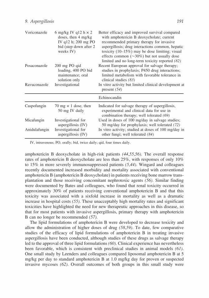
9. Aspergillosis 191
Voriconazole 6 mg/kg IV q12 h × 2doses, then 4 mg/kgIV q12 h; 200 mg PObid (step down after 2weeks IV)
Better efficacy and improved survival comparedwith amphotericin B deoxycholate; currentrecommended primary therapy for invasiveaspergillosis; drug interactions common, hepatictoxicity (10–15%) may be dose limiting; visualeffects common �∼30%) but not usually doselimited and no long-term toxicity reported (82)
Posaconazole 200 mg PO qidloading, 400 PO bidmaintenance; oralsolution only
Recent European approval for salvage therapy;studies in prophylaxis; P450 drug interactions;limited metabolism with favorable tolerance inclinical studies (83)
Ravuconazole Investigational In vitro activity but limited clinical development atpresent (54)
Echinocandin
Caspofungin 70 mg × 1 dose, then50 mg IV daily
Indicated for salvage therapy of aspergillosis,experimental and clinical data for use incombination therapy; well tolerated (69)
Micafungin Investigational foraspergillosis (IV)
Used in doses of 100 mg/day in salvage studies;50 mg/day for prophylaxis; well tolerated (72)
Anidulafungin Investigational foraspergillosis (IV)
In vitro activity; studied at doses of 100 mg/day inother fungi; well tolerated (84)
IV, intravenous; PO, orally; bid, twice daily; qid, four times daily.
amphotericin B deoxycholate in high-risk patients (44,55,56). The overall responserates of amphotericin B deoxycholate are less than 25%, with responses of only 10%to 15% in more severely immunosuppressed patients (5,44). Wingard and colleaguesrecently documented increased morbidity and mortality associated with conventionalamphotericin B (amphotericin B deoxycholate) in patients receiving bone marrow trans-plantation and those receiving concomitant nephrotoxic agents (56). Similar findingwere documented by Bates and colleagues, who found that renal toxicity occurred inapproximately 30% of patients receiving conventional amphotericin B and that thistoxicity was associated with a sixfold increase in mortality as well as a dramaticincrease in hospital costs (55). These unacceptably high mortality rates and significanttoxicities have highlighted the need for new therapeutic approaches in this disease, sothat for most patients with invasive aspergillosis, primary therapy with amphotericinB can no longer be recommended (57).
The lipid formulations of amphotericin B were developed to decrease toxicity andallow the administration of higher doses of drug (58,59). To date, few comparativestudies of the efficacy of lipid formulations of amphotericin B in treating invasiveaspergillosis have been conducted, although studies of these drugs as salvage therapyled to the approval of three lipid formulations (60). Clinical experience has neverthelessbeen favorable, which is consistent with preclinical studies in animal models (61).One small study by Leenders and colleagues compared liposomal amphotericin B at 5mg/kg per day to standard amphotericin B at 1.0 mg/kg day for proven or suspectedinvasive mycoses (62). Overall outcomes of both groups in this small study were
192 Helen W. Boucher and Thomas F. Patterson
similar, but analysis of patients with proven invasive aspergillosis favored the lipidpreparation of amphotericin B. A recent study evaluated amphotericin B colloidaldispersion for primary therapy for invasive aspergillosis (63). In this study of severelyimmunosuppressed patients with invasive aspergillosis, success rates with the lipidformulation were not better than those for conventional amphotericin B, althoughtoxicity was minimally decreased. These results suggest that although lipid formulationsof amphotericin B are dramatically more expensive than standard amphotericin B,hidden costs of standard amphotericin B in terms of morbidity and mortality as wellas resource utilization may justify the use of lipid formulation of amphotericin B incertain high-risk patients (55).
7.2. Azoles
Voriconazole is a potent, broad-spectrum, triazole that has cidal activity againstmany Aspergillus species, including A. terreus; is approved for therapy of invasiveaspergillosis; and has become the recommended primary therapy for most patientswith invasive aspergillosis (53,64). This recommendation is based on data from arandomized trial that compared voriconazole to conventional amphotericin B forthe primary treatment of invasive aspergillosis, with each agent followed by otherlicensed antifungal therapy if needed for intolerance or progression of disease, inseverely immunocompromised patients with invasive aspergillosis (44). In this trial,voriconazole was superior to amphotericin B, with successful outcomes in 52% ofpatients as compared to only 31% in those receiving amphotericin B. In addition,voriconazole demonstrated a survival advantage to amphotericin B, with an absolute13% difference in mortality between treatment groups.
In clinical trials, voriconazole has been adequately tolerated and the drug exhibits afavorable pharmacokinetic profile. There are a number of issues to consider, includingimportant drug interactions, especially those with immunosuppressive agents such ascyclosporine, tacrolimus, and sirolimus, the latter of which is contraindicated for usewith voriconazole, and intolerance to the drug. The most common adverse event hasbeen a transient and reversible visual disturbance described as an altered perception oflight which has been reported in approximately 30% of treated patients, but has notbeen associated with pathologic changes (44). Other less frequently reported adverseevents include liver function test abnormalities in 10% to 15%, and skin rash in 6%(sometimes associated with sun exposure).
Among the other azole antifungal agents, itraconazole is approved for use as salvagetherapy of aspergillosis. Its utility has been limited because until recently it has beenavailable only in an oral formulation that is poorly absorbed; drug interactions furthercomplicate use of this agent. An intravenous formulation of itraconazole has onlyrecently been approved for up to 2 weeks of clinical use (65). For these reasons,itraconazole is more frequently used in less immunosuppressed patients who are ableto take oral therapy and for use as sequential oral therapy (5). Other second-generationtriazoles, including posaconazole and ravuconazole, were developed with an expandedspectrum of activity to include Aspergillus (64,66). Posaconazole is available in onlyan oral formulation, has demonstrated in vitro and in vivo activity against Aspergillus,and has shown clinical activity in an open-label trial which led to its 2005 approval

9. Aspergillosis 193
in the EU for salvage therapy of invasive aspergillosis (67). Ravuconazole has beenevaluated in early phase clinical trails and has also shown activity in animal modelsof invasive aspergillosis (68).
7.3. Echinocandins
Echinocandins are natural cyclic hexapeptide antifungal compounds that noncom-petitively inhibit 1,3-�-d-glucan synthase, an enzyme complex that is unique to anumber of fungi, which forms glucan polymers in the fungal cell wall (54). Theseagents are active against Candida species and Pneumocystis. Specific modificationsto the N-acyl aliphatic or aryl side chains expand the antifungal spectrum to includeAspergillus (54). These agents are all poorly bioavailable and produced in intravenousformulation only.
Caspofungin is approved for treating patients refractory to or intolerant of standardtherapies for invasive aspergillosis based on an open-label trial that demonstratedtherapeutic efficacy in 22 of 54 (41%) patients studied (69). Caspofungin has beenvery well tolerated in clinical trials; in the aspergillosis study, only approximately 5%of patients discontinued therapy. Drug interactions with cyclosporine may occur, buthave not been a significant issue (69,70). In March 2005, micafungin was approvedfor the treatment of esophageal candidiasis and prevention of Candida infections.In the one prophylaxis study used to support this approval, micafungin may havereduced the number of Aspergillus infections as compared to standard prophylaxiswith fluconazole (71). Micafungin also demonstrated efficacy when used as salvagetherapy (72). Anidulafungin is another echinocandin with activity against Aspergillusspp. that appears to have a favorable toxicity profile similar to the other echinocandins.It was approved by the FDA in February 2006 for candidemia and other Candidainfections (including abdominal abscess, peritonitis, and esophagitis). Notably, theseagents are neither classically fungicidal nor fungistatic for Aspergillus, but exerttheir effect on the growing hyphal tips where the glucan synthase target is located(73). For this reason, they have not frequently been used for primary therapy andhave been more frequently used as salvage therapy or more recently in combinationregimens (74,75).
7.4. Other Therapies and Therapeutic Approaches
Outcomes for patients with invasive aspergillosis remain poor despite the advent ofnewer antifungal agents. This, together with the availability of several antifungal drugsand drug classes against Aspergillus, has increased interest in combination antifungaltherapy for this infection (76,77). Marr and colleagues reported on a historical controlstudy of caspofungin and voriconazole compared with voriconazole alone in patientswho failed amphotericin formulations in 2004. In this study, the use of combinationsalvage therapy was associated with an improved 3-month survival rate and the authorsconclude that further studies of this strategy are warranted (75). Adjuvant therapiesincluding surgical resection or use of granulocyte transfusions and growth factors ininvasive aspergillosis can augment antifungal therapy, although their utility has notbeen established in randomized trials. Surgical resection of isolated pulmonary nodulesprior to additional immunosuppressive therapies has been shown to improve outcome of

194 Helen W. Boucher and Thomas F. Patterson
infection (6,78). Recent studies suggest that the majority of patients will have bilateralinfection when the diagnosis is first made, limiting the utility of this approach. Surgicalresection may also be indicated in patients with severe hemoptysis or lesions near thehilar vessels or pericardium. The guidelines for treating invasive aspergillosis publishedby the Infectious Diseases Society of America were produced before the introduction ofthe newest available agents; a revised document is currently in press (2). Unfortunately,few randomized, controlled trials exist to be used to formulate specific guidelines fortherapy. A prompt diagnosis and aggressive initial therapy are both critical in improvingthe outcome of this infection (79). Radiography and use of galactomannan EIA mayfacilitate an early detection of aspergillosis in high risk patients, for whom outcomes areespecially poor (45). Most patients should receive primary therapy with voriconazole,which has been shown to be superior to amphotericin B, the other agent approvedfor primary therapy of this infection (44). However, in patients who are intolerantof voriconazole, have a contraindication to the drug, or have progressive infection,alternative agents include lipid formulations of amphotericin B, the echinocandins, oranother triazole (58,61,69). Primary use of combination therapy is not recommendedat the present time because of lack of prospective clinical trial data, but the additionof another agent in a salvage setting may be considered, owing to the poor outcomesof a single agent in progressive infection (75). Sequential therapy with oral azolesafter initial intravenous therapy may be a useful option (5). Although the optimalduration of antifungal therapy is not known, improvement in underlying host defensesis crucial to successful therapy. While substantial advances have recently been madein the management of invasive aspergillosis, newer approaches to therapy includingthe potential of combination therapy and newer diagnostic tools are needed to improvethe outcome of this disease.
REFERENCES
1. Patterson TF. Aspergillus species. In: Mandell GL, Bennett JE, Dolin R, eds. Mandell,Douglas, and Bennett’s principles and practice of infectious diseases. 6th ed. Philadelphia:Elsevier Churchill Livingstone, 2005:2958–2973.
2. Walsh TJ, Anaissie EJ, Denning DW, et al. Treatment of aspergillosis: clinical practiceguidelines of the Infectious Diseases Society of America (IDSA). Clin Infect Dis (In press).
3. Lin SJ, Schranz J, Teutsch SM. Aspergillosis case-fatality rate: systematic review of theliterature. Clin Infect Dis 2001;32:358–366.
4. McNeil MM, Nash SL, Hajjeh RA, et al. Trends in mortality due to invasive mycoticdiseases in the United States, 1980–1997. Clin Infect Dis 2001;33:641–647.
5. Patterson TF, Kirkpatrick WR, White M, et al. Invasive aspergillosis. Disease spectrum,treatment practices, and outcomes. I3 Aspergillus Study Group. Medicine (Baltimore)2000;79:250–260.
6. Caillot D, Casasnovas O, Bernard A, et al. Improved management of invasive pulmonaryaspergillosis in neutropenic patients using early thoracic computed tomographic scan andsurgery. J Clin Oncol 1997;15:139–147.
7. Maertens J, Vrebos M, Boogaerts M. Assessing risk factors for systemic fungal infections.Eur J Cancer Care (Engl) 2001;10:56–62.
8. Patterson TF. Advances and challenges in management of invasive mycoses. Lancet2005;366:1013–1025.
9. Latge JP. Aspergillus fumigatus and aspergillosis. Clin Microbiol Rev 1999;12:310–350.

9. Aspergillosis 195
10. Hajjeh RA, Warnock DW. Counterpoint: invasive aspergillosis and the environmentrethinking our approach to prevention. Clin Infect Dis 2001;33:1549–1552.
11. Woodcock AA, Steel N, Moore CB, Howard SJ, Custovic A, Denning DW. Fungal contam-ination of bedding. Allergy 2006;61:140–142.
12. Anaissie EJ, Stratton SL, Dignani MC, et al. Pathogenic moulds (including Aspergillusspecies) in hospital water distribution systems: a 3-year prospective study and clinicalimplications for patients with hematologic malignancies. Blood 2003;101:2542–2546.
13. Iwen PC, Rupp ME, Langnas AN, Reed EC, Hinrichs SH. Invasive pulmonary aspergillosisdue to Aspergillus terreus: 12-year experience and review of the literature. Clin Infect Dis1998;26:1092–1097.
14. Warris A, Bjorneklett A, Gaustad P. Invasive pulmonary aspergillosis associated withinfliximab therapy. N Engl J Med 2001;344:1099–1100.
15. Walsh TJ, Petraitis V, Petraitiene R, et al. Experimental pulmonary aspergillosis due toAspergillus terreus: pathogenesis and treatment of an emerging fungal pathogen resistantto amphotericin B. J Infect Dis 2003;188:305–319.
16. Brummer E, Kamberi M, Stevens DA. Regulation by granulocyte-macrophage colony-stimulating factor and/or steroids given in vivo of proinflammatory cytokine and chemokineproduction by bronchoalveolar macrophages in response to Aspergillus conidia. J InfectDis 2003;187:705–709.
17. Kan VL, Bennett JE. Lectin-like attachment sites on murine pulmonary alveolarmacrophages bind Aspergillus fumigatus conidia. J Infect Dis 1988;158:407–414.
18. Levitz SM, Selsted ME, Ganz T, Lehrer RI, Diamond RD. In vitro killing of spores andhyphae of Aspergillus fumigatus and Rhizopus oryzae by rabbit neutrophil cationic peptidesand bronchoalveolar macrophages. J Infect Dis 1986;154:483–489.
19. Washburn RG, Gallin JI, Bennett JE. Oxidative killing of Aspergillus fumigatusproceeds by parallel myeloperoxidase-dependent and -independent pathways. Infect Immun1987;55:2088–2092.
20. Stanzani M, Orciuolo E, Lewis R, et al. Aspergillus fumigatus suppresses thehuman cellular immune response via gliotoxin-mediated apoptosis of monocytes. Blood2005;105:2258–2265.
21. Greenberger PA. Allergic bronchopulmonary aspergillosis. J Allergy Clin Immunol2002;110:685–692.
22. Stevens DA, Moss RB, Kurup VP, et al. Allergic bronchopulmonary aspergillosis in cysticfibrosis–state of the art: Cystic Fibrosis Foundation Consensus Conference. Clin Infect Dis2003;37 (Suppl 3):S225–S264.
23. Patterson R, Greenberger PA, Radin RC, Roberts M. Allergic bronchopulmonaryaspergillosis: staging as an aid to management. Ann Intern Med 1982;96:286–291.
24. Wark PA, Gibson PG, Wilson AJ. Azoles for allergic bronchopulmonary aspergillosisassociated with asthma. Cochrane Database Syst Rev 2004:CD001108.
25. Stevens DA, Schwartz HJ, Lee JY, et al. A randomized trial of itraconazole in allergicbronchopulmonary aspergillosis. N Engl J Med 2000;342:756–762.
26. Gillespie MB, O’Malley BW. An algorithmic approach to the diagnosis and managementof invasive fungal rhinosinusitis in the immunocompromised patient. Otolaryngol ClinNorth Am 2000;33:323–334.
27. Ferguson BJ. Fungus balls of the paranasal sinuses. Otolaryngol Clin North Am2000;33:389–398.
28. Kauffman CA. Quandary about treatment of aspergillomas persists. Lancet 1996;347:1640.29. Kaur R, Mittal N, Kakkar M, Aggarwal AK, Mathur MD. Otomycosis: a clinicomycologic
study. Ear Nose Throat J 2000;79:606–609.

196 Helen W. Boucher and Thomas F. Patterson
30. Torres-Rodriguez JM, Madrenys-Brunet N, Siddat M, Lopez-Jodra O, Jimenez T.Aspergillus versicolor as cause of onychomycosis: report of 12 cases and susceptibilitytesting to antifungal drugs. J Eur Acad Dermatol Venereol 1998;11:25–31.
31. Kuo IC, Margolis TP, Cevallos V, Hwang DG. Aspergillus fumigatus keratitis after laserin situ keratomileusis. Cornea 2001;20:342–344.
32. Denning DW, Riniotis K, Dobrashian R, Sambatakou H. Chronic cavitary and fibrosingpulmonary and pleural aspergillosis: case series, proposed nomenclature change, andreview. Clin Infect Dis 2003;37 (Suppl 3):S265–S280.
33. Jain LR, Denning DW. The efficacy and tolerability of voriconazole in the treatment ofchronic cavitary pulmonary aspergillosis. J Infect 2006;52:e133–e137.
34. Gerson SL, Talbot GH, Hurwitz S, Strom BL, Lusk EJ, Cassileth PA. Prolonged granulo-cytopenia: the major risk factor for invasive pulmonary aspergillosis in patients with acuteleukemia. Ann Intern Med 1984;100:345–351.
35. Marr KA, Carter RA, Crippa F, Wald A, Corey L. Epidemiology and outcome ofmould infections in hematopoietic stem cell transplant recipients. Clin Infect Dis2002;34:909–917.
36. Miller WT, Sais GJ, Frank I, Gefter WB, Aronchick JM, Miller WT. Pulmonaryaspergillosis in patients with AIDS. Clinical and radiographic correlations. Chest1994;105:37–44.
37. Allo MD, Miller J, Townsend T, Tan C. Primary cutaneous aspergillosis associated withHickman intravenous catheters. N Engl J Med 1987;317:1105–1108.
38. Mylonakis E, Rich J, Skolnik PR, De Orchis DF, Flanigan T. Invasive Aspergillus sinusitisin patients with human immunodeficiency virus infection. Report of 2 cases and review.Medicine (Baltimore) 1997;76:249–255.
39. Ellis ME, Al-Abdely H, Sandridge A, Greer W, Ventura W. Fungal endocarditis: evidencein the world literature, 1965–1995. Clin Infect Dis 2001;32:50–62.
40. Denning DW, Stevens DA. Antifungal and surgical treatment of invasive aspergillosis:review of 2,121 published cases. Rev Infect Dis 1990;12:1147–1201.
41. Ascioglu S, Rex JH, de Pauw B, et al. Defining opportunistic invasive fungal infectionsin immunocompromised patients with cancer and hematopoietic stem cell transplants: aninternational consensus. Clin Infect Dis 2002;34:7–14.
42. Sutton DA, Fothergill AW, Rinaldi MG. Guide to Clinically Significant Fungi. Baltimore,MD: Lippincott Williams & Wilkins, 1998.
43. Caillot D, Couaillier JF, Bernard A, et al. Increasing volume and changing characteristicsof invasive pulmonary aspergillosis on sequential thoracic computed tomography scans inpatients with neutropenia. J Clin Oncol 2001;19:253–259.
44. Herbrecht R, Denning DW, Patterson TF, et al. Voriconazole versus amphotericin B forprimary therapy of invasive aspergillosis. N Engl J Med 2002;347:408–415.
45. Herbrecht R. Improving the outcome of invasive aspergillosis: new diagnostic tools andnew therapeutic strategies. Ann Hematol 2002;81 (Suppl 2):S52–S53.
46. Maertens J, Theunissen K, Verbeken E, et al. Prospective clinical evaluation of lower cut-offs for galactomannan detection in adult neutropenic cancer patients and haematologicalstem cell transplant recipients. Br J Haematol 2004;126:852–860.
47. Maertens J, Theunissen K, Verhoef G, et al. Galactomannan and computed tomography-based preemptive antifungal therapy in neutropenic patients at high risk for invasive fungalinfection: a prospective feasibility study. Clin Infect Dis 2005;41:1242–1250.
48. Loeffler J, Hebart H, Cox P, Flues N, Schumacher U, Einsele H. Nucleic acidsequence-based amplification of Aspergillus RNA in blood samples. J Clin Microbiol2001;39:1626–1629.

9. Aspergillosis 197
49. White PL, Linton CJ, Perry MD, Johnson EM, Barnes RA. The evolution and evalu-ation of a whole blood polymerase chain reaction assay for the detection of invasiveaspergillosis in hematology patients in a routine clinical setting. Clin Infect Dis 2006;42:479–486.
50. Donnelly JP. Polymerase chain reaction for diagnosing invasive aspergillosis: getting closerbut still a ways to go. Clin Infect Dis 2006;42:487–489.
51. Odabasi Z, Mattiuzzi G, Estey E, et al. Beta-d-glucan as a diagnostic adjunct forinvasive fungal infections: validation, cutoff development, and performance in patientswith acute myelogenous leukemia and myelodysplastic syndrome. Clin Infect Dis 2004;39:199–205.
52. Ostrosky-Zeichner L, Alexander BD, Kett DH, et al. Multicenter clinical evaluation of the(1–>3) beta-D-glucan assay as an aid to diagnosis of fungal infections in humans. ClinInfect Dis 2005;41:654–659.
53. Steinbach WJ, Stevens DA. Review of newer antifungal and immunomodulatory strategiesfor invasive aspergillosis. Clin Infect Dis 2003;37 (Suppl 3):S157–S187.
54. Boucher HW, Groll AH, Chiou CC, Walsh TJ. Newer systemic antifungal agents: pharma-cokinetics, safety and efficacy. Drugs 2004;64:1997–2020.
55. Bates DW, Su L, Yu DT, et al. Mortality and costs of acute renal failure associated withamphotericin B therapy. Clin Infect Dis 2001;32:686–693.
56. Wingard JR, Kubilis P, Lee L, et al. Clinical significance of nephrotoxicity in patientstreated with amphotericin B for suspected or proven aspergillosis. Clin Infect Dis1999;29:1402–1407.
57. Ostrosky-Zeichner L, Marr KA, Rex JH, Cohen SH. Amphotericin B: time for a new “goldstandard.” Clin Infect Dis 2003;37:415–425.
58. Walsh TJ, Hiemenz JW, Seibel NL, et al. Amphotericin B lipid complex for invasivefungal infections: analysis of safety and efficacy in 556 cases. Clin Infect Dis 1998;26:1383–1396.
59. Walsh TJ, Goodman JL, Pappas P, et al. Safety, tolerance, and pharmacokinetics of high-dose liposomal amphotericin B (AmBisome) in patients infected with Aspergillus speciesand other filamentous fungi: maximum tolerated dose study. Antimicrob Agents Chemother2001;45:3487–3496.
60. Rex JH, Walsh TJ, Nettleman M, et al. Need for alternative trial designs and evaluationstrategies for therapeutic studies of invasive mycoses. Clin Infect Dis 2001;33:95–106.
61. Barrett JP, Vardulaki KA, Conlon C, et al. A systematic review of the antifungal effec-tiveness and tolerability of amphotericin B formulations. Clin Ther 2003;25:1295–1320.
62. Leenders AC, Daenen S, Jansen RL, et al. Liposomal amphotericin B compared withamphotericin B deoxycholate in the treatment of documented and suspected neutropenia-associated invasive fungal infections. Br J Haematol 1998;103:205–212.
63. Bowden R, Chandrasekar P, White MH, et al. A double-blind, randomized, controlled trialof amphotericin B colloidal dispersion versus amphotericin B for treatment of invasiveaspergillosis in immunocompromised patients. Clin Infect Dis 2002;35:359–366.
64. Espinel-Ingroff A, Boyle K, Sheehan DJ. In vitro antifungal activities of voriconazoleand reference agents as determined by NCCLS methods: review of the literature.Mycopathologia 2001;150:101–115.
65. Caillot D. Intravenous itraconazole followed by oral itraconazole for the treatmentof amphotericin-B-refractory invasive pulmonary aspergillosis. Acta Haematol2003;109:111–118.
66. Sheehan DJ, Hitchcock CA, Sibley CM. Current and emerging azole antifungal agents.Clin Microbiol Rev 1999;12:40–79.

198 Helen W. Boucher and Thomas F. Patterson
67. Raad I, Chapman S, Bradsher R. Posaconazole (POS) salvage therapy for invasive fungalinfections (IFI) (abstract M-699), 44th Interscience Conference on Antimicrobial Agentsand Chemotherapy, Washington, DC, 2004.
68. Kirkpatrick WR, Perea S, Coco BJ, Patterson TF. Efficacy of ravuconazole (BMS-207147) in a guinea pig model of disseminated aspergillosis. J Antimicrob Chemother2002;49:353–357.
69. Maertens J, Raad I, Petrikkos G, et al. Efficacy and safety of caspofungin for treatmentof invasive aspergillosis in patients refractory to or intolerant of conventional antifungaltherapy. Clin Infect Dis 2004;39:1563–1571.
70. Marr KA, Hachem R, Papanicolaou G, et al. Retrospective study of the hepatic safetyprofile of patients concomitantly treated with caspofungin and cyclosporin A. TransplInfect Dis 2004;6:110–116.
71. van Burik JA, Ratanatharathorn V, Stepan DE, et al. Micafungin versus fluconazole forprophylaxis against invasive fungal infections during neutropenia in patients undergoinghematopoietic stem cell transplantation. Clin Infect Dis 2004;39:1407–1416.
72. Denning DW, Marr KA, Lau WM, et al. Micafungin (FK463), alone or in combinationwith other systemic antifungal agents, for the treatment of acute invasive aspergillosis.J Infect 2006;53:337–349.
73. Bowman JC, Hicks PS, Kurtz MB, et al. The antifungal echinocandin caspofungin acetatekills growing cells of Aspergillus fumigatus in vitro. Antimicrob Agents Chemother2002;46:3001–3012.
74. Kirkpatrick WR, Perea S, Coco BJ, Patterson TF. Efficacy of caspofungin alone and incombination with voriconazole in a Guinea pig model of invasive aspergillosis. AntimicrobAgents Chemother 2002;46:2564–2568.
75. Marr KA, Boeckh M, Carter RA, Kim HW, Corey L. Combination antifungal therapy forinvasive aspergillosis. Clin Infect Dis 2004;39:797–802.
76. Kontoyiannis DP, Hachem R, Lewis RE, et al. Efficacy and toxicity of caspofungin incombination with liposomal amphotericin B as primary or salvage treatment of invasiveaspergillosis in patients with hematologic malignancies. Cancer 2003;98:292–299.
77. Aliff TB, Maslak PG, Jurcic JG, et al. Refractory Aspergillus pneumonia in patientswith acute leukemia: successful therapy with combination caspofungin and liposomalamphotericin. Cancer 2003;97:1025–1032.
78. Yeghen T, Kibbler CC, Prentice HG, et al. Management of invasive pulmonary aspergillosisin hematology patients: a review of 87 consecutive cases at a single institution. Clin InfectDis 2000;31:859–868.
79. Patterson TF, Boucher HW, Herbrecht R, et al. Strategy of following voriconazoleversus amphotericin B therapy with other licensed antifungal therapy for primarytreatment of invasive aspergillosis: impact of other therapies on outcome. Clin Infect Dis2005;41:1448–1452.
80. Imhof A, Walter RB, Schaffner A. Continuous infusion of escalated doses of amphotericinB deoxycholate: an open-label observational study. Clin Infect Dis 2003;36:943–951.
81. Wingard JR, White MH, Anaissie E, Raffalli J, Goodman J, Arrieta A. A randomized,double-blind comparative trial evaluating the safety of liposomal amphotericin B versusamphotericin B lipid complex in the empirical treatment of febrile neutropenia. LAmph/ABLC Collaborative Study Group. Clin Infect Dis 2000;31:1155–1163.
82. Keating GM. Posaconazole. Drugs 2005;65:1553–1567.83. Murdoch D, Plosker GL. Anidulafungin. Drugs 2004;64:2249–2258.84. Johnson LB, Kauffman CA. Voriconazole: a new triazole antifungal agent. Clin Infect Dis
2003;36:630–637.

9. Aspergillosis 199
SUGGESTED READINGS
Boucher HW, Groll AH, Chiou CC, Walsh TJ. Newer systemic antifungal agents: pharmacoki-netics, safety and efficacy. Drugs 2004;64:1997–2020.
Caillot D, Casasnovas O, Bernard A, et al. Improved management of invasive pulmonaryaspergillosis in neutropenic patients using early thoracic computed tomographic scan andsurgery. J Clin Oncol 1997;15:139–147.
Herbrecht R, Denning DW, Patterson TF, et al. Voriconazole versus amphotericin B for primarytherapy of invasive aspergillosis. N Engl J Med 2002;347:408–415.
Keating GM. Posaconazole. Drugs 2005;65:1553–1567.Patterson TF. Aspergillus species. In: Mandell GL, Bennett JE, Dolin R, eds. Mandell, Douglas,
and Bennett’s principles and practice of infectious diseases. Philadelphia: Elsevier ChurchillLivingstone, 2005;2958–2973.
Patterson TF. Advances and challenges in management of invasive mycoses. Lancet 2005;366:1013–1025.
Patterson TF, Kirkpatrick WR, White M, et al. Invasive aspergillosis. Disease spectrum,treatment practices, and outcomes. Medicine (Baltimore) 2000;79:250–260.
Maertens J, Theunissen K, Verhoef G, et al. Galactomannan and computed tomography-based preemptive antifungal therapy in neutropenic patients at high risk for invasive fungalinfection: a prospective feasibility study. Clin Infect Dis 2005;41:1242–1250.
Marr KA, Boeckh M, Carter RA, Kim HW, Corey L. Combination antifungal therapy forinvasive aspergillosis. Clin Infect Dis 2004;39:797–802.
Walsh TJ, Anaissie EJ, Denning DW, et al. Treatment of aspergillosis: clinical practice guide-lines of the Infectious Diseases Society of America (IDSA). Clin Infect Dis (In press).
Walsh TJ, Petraitis V, Petraitiene R, et al. Experimental pulmonary aspergillosis due toAspergillus terreus: pathogenesis and treatment of an emerging fungal pathogen resistant toamphotericin B. J Infect Dis 2003;188:305–319.

10Hyalohyphomycosis—Infection Due
to Hyaline Moulds
Rhonda V. Fleming, MD and Elias J. Anaissie, MD
1. INTRODUCTION
Hyalohyphomycosis is the term used to designate infections caused by fungi noted tohave hyaline (colorless) septate hyphae microscopically in clinical samples, similar to theuse of phaeohyphomycosis for those infections caused by pigmented fungi (Chapter 11).This distinction is clinically useful when hyphal elements are seen on tissue examinationbut fail to grow. Hyalohyphomycosis typically includes infections caused by species ofFusarium, Scedosporium, Acremonium, Paecilomyces, Scopulariopsis, Beauveria, andPenicillium. Although Aspergillus produces similar hyaline septate hyphae microscopi-cally, and is thus considered a member of this grouping of fungi, infection caused by thisgenus (aspergillosis) is generally discussed as a separate disease (Chapter 9). These agentsmay cause superficial or localized infection in immunocompetent hosts (usually as a resultof direct inoculation of the fungus after trauma) and invasive or disseminated infectionsin immunocompromised hosts. In the latter setting, the clinical infection may be indis-tinguishable from that of invasive aspergillosis. A remarkable feature of some of thesehyaline moulds is their ability to cause fungemia and to disseminate hematogenously,causing numerous embolic skin lesions. These infections may be clinically suspected onthebasisofaconstellationofclinicaland laboratoryfindings.Definitivediagnosis requiresisolation of the organism because histopathological examination reveals branchinghyaline septate hyphae regardless of the pathogen, similar to the findings with Aspergillus.Anaccuratediagnosisat thespecies level is importantbecauseof thevariablesusceptibilityto antifungal agents. An important component of therapy of localized infection is surgeryand removal of infected prosthetic devices. Outcome is usually favorable in immuno-competent hosts, while usually very poor in the setting of persistent profound immuno-suppression. We herein describe the most relevant characteristics of these organisms andthe clinical spectrum and diagnosis and treatment of infections caused by these agents.
From: Infectious Disease: Diagnosis and Treatment of Human MycosesEdited by: D. R. Hospenthal and M. G. Rinaldi © Humana Press Inc., Totowa, NJ
201

202 Rhonda V. Fleming and Elias J. Anaissie
2. FUSARIUM
2.1. Introduction
Fusarium species recently emerged as a cause of disseminated infections inneutropenic patients and in patients undergoing allogeneic hematopoietic stemcell transplantation (HSCT). Fusarium represents the second most common fungalpathogen, after Aspergillus, as the cause of life-threatening infection in recipientsof hematopoietic transplant (1). Fusarium causes a broad spectrum of infections inhumans, including superficial and local infections in immunocompetent hosts, whiledisseminated infection is seen almost exclusively in immunosuppressed patients.
2.2. Etiologic Agents
Four species are most commonly involved in human infections: F. solani (the mostcommon), F. oxysporum, F. moniliforme, and F. proliferatum (2). Fusarium speciesare septate filamentous fungi that produce conidiophores, phialides, macroconidia, andmicroconidia. Fusarium species grow easily and rapidly in almost all fungal media.On potato dextrose agar (PDA), the colonies have a velvety or cottony surface, and arewhite, yellow, pink, purple salmon or gray on the surface, with a pale, red, violet, brownor sometimes blue reverse. The characteristic sickle- or banana-shaped multiseptatemacroconidia with a foot cell at the base are used in identifying the genus and speciesof Fusarium (Fig. 10.1). Molecular methods may also be used for rapid identification ofFusarium to the species level. In tissue, the hyphae are similar to those of Aspergillusspecies, with hyaline and septate filaments that typically dichotomize in acute and rightangles. In the absence of microbial growth, distinguishing fusariosis from aspergillosisand other hyalohyphomycoses is difficult, and requires the use of in situ hybridizationin paraffin-embedded tissue specimens (3). Fusarium species are toxigenic, and maycause mycotoxicosis in animals and humans (2).
2.3. Epidemiology
Fusarium is ubiquitous in soil and water, taking part in water biofilms and is ahuman and plant pathogen (4). Fusarium species are causative agents of superficialand localized infections in immunocompetent hosts, most commonly onychomycosisand cutaneous and subcutaneous infections including mycetoma and keratitis, thelatter in contact lens wearers (5). A recent large outbreak of Fusarium keratitis wasreported in contact lens wearers in the United States and was linked to contaminatedcontact lens rinse solutions (6). Other risk factors for keratitis are trauma and useof topical corticosteroids and antibiotics. Fusarium endophthalmitis may arise fromkeratitis or by direct inoculation after cataract surgery or trauma (7). Fusariosis mayalso result from skin breakdown, such as burns and wounds, or the presence of foreignbodies, such as peritonitis in patients receiving continuous ambulatory peritonealdialysis (CAPD), and catheter-associated fungemia, and thrombophlebitis (8–10). Otherinfections include sinusitis, pneumonia, cutaneous and subcutaneous infections, septicarthritis, and osteomyelitis (11–15).
Immunosuppressed patients may develop locally invasive and disseminated fusar-iosis (16). Risk factors include prolonged neutropenia such as following chemotherapy

10. Hyalohyphomycosis—Infection Due to Hyaline Moulds 203
Fig. 10.1. Fusarium oxysporum macroconidia. Characteristic fusiform to sickle-shaped,multiseptate, mostly with an attenuated apical cell and a foot-shaped basal cell. (Courtesy ofwww.doctorfungus.org.) [Figure in color on CD-ROM].
for acute leukemia and T-cell immunodeficiency which occurs most commonly afterHSCT (16–18). In HSCT infection may develop early during neutropenia or monthsafter neutrophil recovery, following treatment of chronic extensive graft versus hostdisease (GvHD). Localized infections may also develop among solid organ transplantrecipients (SOT), usually as a late infection (19).
Portals of entry are the respiratory tract and skin, the latter playing a significant rolein patients with tissue breakdown such as onychomycosis. Hospital water systems area potential reservoir for Fusarium; transmission may occur from inhalation of conidiaaerosolized in the shower or from direct contact of contaminated water with sites ofskin breakdown (20–22).
2.4. Pathogenesis and Immunology
Similar to Aspergillus, this organism is highly angioinvasive and leads to tissueinfarction. In contrast to Aspergillus however, Fusarium is frequently isolated from thebloodstream, likely as a result of intravascular adventitious sporulation (23). Phagocytesappear to be the predominant line of defense against fusarial infections (16–18).
2.5. Clinical Manifestations
Infection with Fusarium in immunocompetent hosts may be superficial or locallyinvasive, involving the skin, eyes, sinuses, lungs, and joints and bones. In immuno-suppressed patients, the infection may be locally invasive, usually pneumonia and/orsinusitis, or more commonly disseminated (16–18). The clinical picture resembles thatof invasive aspergillosis. Unlike aspergillosis, however, fungemia and skin lesions arecommon (up to 40% of patients with disseminated disease). Skin lesions may represent

204 Rhonda V. Fleming and Elias J. Anaissie
the primary site of infection (onychomycosis) or secondary to disseminated infection(16–18). Metastatic skin lesions evolve from subcutaneous painful lesions, to erythe-matous induration followed by ecthyma gangrenosum-like with necrotic center, whichmay be surrounded by a rim of erythema (16–18).
2.6. Diagnosis
Two characteristics suggest the diagnosis of disseminated fusariosis in the severelyimmunocompromised host: metastatic skin lesions and positive blood cultures formould (16–18). Definitive diagnosis relies on cultures (tissue and/or blood) andhistopathology which shows a pattern common to all hyalohyphomycosis (invasion byacute-branching, septate hyaline hyphae). The use of polymerase chain reaction (PCR)techniques and/or in situ hybridization may be required to reach the correct diagnosis intissues (3,24). The 1,3-�-d-glucan test is usually positive in invasive fusarial infections,but it cannot distinguish Fusarium from other fungal infections (Candida, Aspergillus,Trichosporon and others) that are also detected by the assay (25).
2.7. Treatment
Localized infections, particularly in immunocompetent hosts, usually respond wellto treatment consisting of topical therapy for fungal keratitis or excision of involvedtissue (sinuses, eye, soft tissue, bone). Removal of an infected intravascular cathetermay be needed in the rare cases of catheter-related fungemia.
The outcome of disseminated fusariosis in severely immunosuppressed patientsremains poor despite aggressive antifungal therapy, with very high mortality rates(16–18). Predictors of poor outcome are persistent neutropenia and recent therapywith corticosteroids for chronic GvHD (17). Treatment options are limited by thelack of reliable and consistent activity of antifungal agents against Fusarium species.F. moniliforme is the most susceptible species, typically inhibited by amphotericin Band the newer triazoles voriconazole and posaconazole; F. verticilloides is commonlyresistant to both antifungal classes. F. solani and F. oxysporum have an interme-diate susceptibility to amphotericin B, but are resistant to the triazoles (26). Hence,rapid species identification is needed and antifungal susceptibility testing should beconsidered because of this variable in vitro susceptibility among Fusarium species(Table 10.1). The echinocandins do not appear to be active against Fusarium.
3. SCEDOSPORIUM
3.1. Introduction
Two Scedosporium species, S. apiospermum (sexual state name, Pseudallescheriaboydii) and S. prolificans (formerly S. inflatum) cause human disease (27,28). Aspectrum of disease, ranging from respiratory tract colonization (cystic fibrosis or SOT)to superficial and deep infections, in both immunocompetent and immunosuppressedhosts, has been reported. Rarely, disseminated infection with high mortality is seen inthe setting of severe immunosuppression. S. prolificans belongs to the group of fungithat cause phaeohyphomycosis (dematiaceous fungi), but are briefly discussed becauseof its relationship to S. apiospermum .

Tab
le10
.1O
vera
llsu
scep
tib
ilit
yof
hya
lin
efu
ngi
toav
aila
ble
anti
fun
gals
a
Gen
us/s
peci
esA
mB
Itra
cona
zole
Vor
icon
azol
ePo
saco
nazo
leA
nidu
lafu
ngin
Cas
pofu
ngin
Ref
.
Fus
ariu
msp
p.++
+++
b++
00
(50,
51)
F.
oxys
poru
m++
+++
++++
ND
ND
(50,
52)
F.
sola
ni+
++
+N
DN
DSc
edos
pori
umsp
p.S.
prol
ific
ans
00
00
0N
D(5
1)S.
apio
sper
mum
0++
++++
+++
00
(51,
52)
Pae
cilo
myc
essp
p.++
+++
+++
+++
++++
+(5
0,51
)A
crem
oniu
msp
p.++
0++
ND
0N
D(5
1)
++++
,dru
gof
choi
ce;
+++,
alte
rnat
ive
choi
ce;
++,s
ome
stra
ins
are
susc
eptib
le(<
50%
);+,
rare
lysu
scep
tible
(<10
%);
0,al
way
sre
sist
ant;
ND
,no
data
avai
labl
e.a Su
scep
tibili
tyba
sed
onM
IC50
.bIn
the
Uni
ted
Stat
es,t
heon
lyag
ent
indi
cate
dfo
rin
vasi
vefu
sari
osis
.
206 Rhonda V. Fleming and Elias J. Anaissie
3.2. Etiologic Agents
Scedosporium species are identified by their characteristic macroscopic (wooly tocottony, dark gray to dark brown) and microscopic (characteristic conidia, conidio-phores, and hyphae) appearance. S. prolificans is distinguished from S. apiospermumby the production of terminal annelloconidia with inflated bases (cylindrical inS. apiospermum) and growth inhibition by cycloheximide or actidione. In tissuesections, Scedosporium appear as septate hyaline hyphae that cannot be reliably distin-guished from Aspergillus or Fusarium unless conidia are present.
3.3. Epidemiology
Scedosporium have been isolated from soil, potting mix, compost, and animalmanure and stagnant or polluted water. Infections occur worldwide, though a largenumber of reports come from Northern Spain (27). Patients at risk for invasiveand/or disseminated infection include those with human immunodeficiency virus (HIV)infection, acute leukemia and recipients of allogeneic HSCT or SOT (29,30).
Infection is thought to be secondary to direct inoculation (such as aftertrauma) or inhalation of airborne conidia. In normal hosts, S. apiospermum causesinfection after penetrating trauma, including keratitis, endophthalmitis, cutaneous andsubcutaneous infections, bursitis, arthritis, and osteomyelitis. After near drowningaccidents, sinusitis, pneumonia, meningoencephalitis, and brain abscesses may develop.Allergic bronchopulmonary disease due to S. apiospermum may also occur. Like S.apiospermum, S. prolificans causes localized infections (usually of bone or soft tissue)in immunocompetent patients after trauma, and deeply invasive infections in immuno-compromised hosts, sometimes as a nosocomial outbreak (27,29,30).
3.4. Clinical Manifestations
Mycetoma is the most common S. apiospermum infection in normal hosts (seeChapter 21), usually occurring after penetrating injury and presenting as lowerextremity swelling with draining sinuses. Other infections include non-mycetomacutaneous and subcutaneous infections, keratitis, and endophthalmitis (29). InvasiveS. apiospermum infection is usually seen in immunocompromised patients, mostcommonly as pneumonia. Disseminated infection is mainly associated with S. prolif-icans, and is characterized by refractory fever, pulmonary infiltrates (diffuse ornodular), central nervous system involvement (present in one third of patients),fungemia, renal failure and erythematous, and nodular skin lesions with central necrosis.
3.5. Diagnosis
The diagnosis relies on the combination of clinical signs and symptoms and recoveryof Scedosporium from blood and/or infected tissue, with or without demonstration ofcolorless septate hyphae.
3.6. Treatment
Localized infections, particularly in immunocompetent hosts, usually respond wellto surgical débridement. Scedosporium apiospermum is resistant to fluconazole andflucytosine, but susceptible to the newer azoles voriconazole, posaconazole, and

10. Hyalohyphomycosis—Infection Due to Hyaline Moulds 207
ravuconazole (Table 10.1). Voriconazole is approved for use in S. apiospermum infec-tions (31,32). Caspofungin appears to be more active than itraconazole or amphotericinB. Variable strain-to-strain susceptibility to amphotericin B can be seen (33). Surgicalresection remains the only definite therapy for S. prolificans infections, as this organismis resistant to all available antifungal agents in vitro. In vitro synergism betweenterbinafine and either voriconazole or itraconazole has been reported (34).
4. PAECILOMYCES
4.1. Introduction
Paecilomyces species are frequent airborne contaminants in clinical microbiologylaboratories but have been increasingly reported as cause of human infection.
4.2. Etiologic Agents
Two Paecilomyces species, P. lilacinus (most common) and P. varioti, account formost human infections; although recent reports have described infection secondaryto P. marquandii and P. javanicus. Paecilomyces grow rapidly on various agarmedia including blood, chocolate, Sabouraud dextrose (SDA), and PDA. P. variotiis thermophilic and grows well at temperatures as high as 50°C. The color of thecolony and certain microscopic features help differentiate Paecilomyces species fromeach other. The colonies are flat and velvety. The color is initially white and becomesyellow-green, yellow-brown, pink or violet according to species.
4.3. Epidemiology
Paecilomyces are found worldwide in soil, food products, and water and causesinfection in both immunocompetent and immunosuppressed patients. A strong associ-ation of Paecilomyces with prosthetic implants may be due to their inherent resistanceto most sterilizing techniques. Prosthetic implant-related infections include keratitisin contact lens wearers and after corneal implants (rarely endophthalmitis), and inrecipients of CAPD, cardiac valves, and ventriculoperitoneal shunts. Other infectionsinvolve the nails, skin, and subcutaneous tissues, bones and joints, sinuses and lungs,while disseminated infections only occur in immunosuppressed patients (35).
4.4. Clinical Manifestations
The most common infections due to Paecilomyces involve the eye and eye struc-tures (keratitis and endophthalmitis), followed by the nails (onychomycosis) and skinand soft tissues. Skin infections are characterized by erythematous macules, nodules,pustules, vesicular lesions, or necrotic crusts (35). Sporotrichosis-like skin infectionhas also been described. Other reported infections in the competent host includeperitonitis in CAPD, prosthetic-valve endocarditis, catheter-related fungemia, andarthritis/osteomyelitis. In immunocompromised patients, pneumonia and disseminateddisease are most commonly observed.
4.5. Diagnosis
Paecilomyces infections are diagnosed with routine tissue culture. These organismsare easily cultured on SDA and on histopathology, hyaline septate hyphae can be seen

208 Rhonda V. Fleming and Elias J. Anaissie
with periodic acid-Schiff (PAS) staining. Paecilomyces may exist in various forms intissue (conidia and phialides) and can therefore be misdiagnosed as candidiasis.
4.6. Treatment
Treatment of invasive Paecilomyces infections relies on surgical debridementand removal of infected prosthetic materials. Because of different susceptibilities toantifungal agents, Paecilomyces should be identified to the species level (Table 10.1).P. varioti is susceptible to amphotericin B, flucytosine, itraconazole, voriconazole, andposaconazole, whereas P. lilacinus is susceptible only to the latter two triazoles (36).
5. ACREMONIUM
5.1. Introduction
Acremonium species are filamentous fungi of low pathogenicity commonly isolatedfrom the environment (soil, insects, sewage, plants, and water) (37).
5.2. Etiologic Agents
Seven species of Acremonium have been reported to cause human infection withmore than 80% of infections caused by A. strictum (the most common), A. falciforme,A. recifei, and A. kiliense. Acremonium species grow on SDA, forming white, salmon,or yellowish-green colonies that are usually velvety or cottony with slightly raisedcenters. This genus is distinguished by formation of narrow hyphae bearing solitary,unbranched needle-shaped phialides. As in other hyaline moulds, septate colorlesshyphae are found in tissue.
5.3. Epidemiology
Most infections occur in immunocompetent hosts and include mycetoma followingtrauma, keratitis in contact lenses wearers, and endophthalmitis (37–39). Fungalcolonization of humidifier water in a ventilator system was thought to be the sourceof infections in an outbreak of endophthalmitis. Invasive disease is almost exclusivelyseen in immunocompromised patients.
5.4. Clinical Manifestations
Mycetoma is the most common infection due to Acremonium and presents ina manner similar to that of mycetoma caused by S. apiospermum. Keratitis andendophthalmitis constitute the second most common infections. Colonization of softcontact lenses may proceed to corneal invasion. Other reported infections includeonychomycosis, peritonitis, dialysis fistulae infection, pneumonia, empyema, septicarthritis, osteomyelitis, meningitis (following spinal anesthesia in an otherwise healthyindividual), cerebritis in an intravenous drug abuser, and prosthetic valve endocarditis.Disseminated infection occurs exclusively in immunosuppressed hosts and has beencharacterized by endocarditis, meningitis, and bloodstream infection. In vivo sporu-lation can occur, facilitating dissemination and perhaps explaining the high rate ofmetastatic skin lesions and positive blood cultures with Acremonium.

10. Hyalohyphomycosis—Infection Due to Hyaline Moulds 209
5.5. Diagnosis
Acremonium species grow slowly on SDA. Hence, cultures must be kept at least2 weeks. Blood cultures may isolate Acremonium in cases of disseminated disease. Likeother hyaline moulds, septate colorless hyphae are found in histopathologic examinationthat stain with PAS (39). Acremonium may be difficult to identify in tissue because ofmorphologic similarities with other moulds, such as Fusarium.
5.6. Treatment
Acremonium species have a variable susceptibility to antifungal agents (37)(Table 10.1). In vitro activity of amphotericin B and itraconazole against Acremoniumis variable, while resistance to fluconazole and 5-fluorocytosine is uniform. The newerazoles, voriconazole and posaconazole, appear promising.
6. PENICILLIUM MARNEFFEI
6.1. Introduction
Most Penicillium species are frequent laboratories contaminants, but P. marneffeihas emerged as a significant pathogen, and can cause disseminated infection in HIV-infected patients residing or traveling to Southeast Asia where the disease is endemic(40).
6.2. Etiologic Agents
Penicillium marneffei is a facultative intracellular pathogen and the only knownthermally dimorphic fungus of the genus Penicillium. At room temperature,P. marneffei exhibits the characteristic morphology of the genus; in contrast, it growsas a yeast when found in infected tissue or at 37°C.
6.3. Epidemiology
Infection due to Penicillium marneffei constitutes the third most common oppor-tunistic infection in HIV-infected patients in certain parts of Southeast Asia and isendemic in the Guangxi province of China, Hong Kong, and Taiwan (41). The incidenceof penicilliosis has increased significantly, paralleling the incidence of HIV infection.Although penicilliosis is most commonly seen in adults infected with HIV, the diseasehas also been detected in children and adults without immunodeficiency. The modeof transmission is thought to be due to ingestion or inhalation of the fungus. Soilexposure, especially during rainy season, has been suggested to be a critical factor.
6.4. Clinical Manifestations
Penicilliosis marneffei can clinically resemble tuberculosis, molluscum contagiosum,cryptococcosis, and histoplasmosis. The most common clinical manifestation in penicil-liosis includes low-grade fever, anemia, weight loss, cough, lymphadenopathy, andhepatosplenomegaly (42–44). Skin lesions are present in up 70% of the cases and arecharacterized by a central necrotic umbilication resembling molluscum contagiosum.Palatal and pharyngeal lesions can also be present. Bloodstream infection is presentin approximately 50% of cases. Pulmonary involvement has been described as being

210 Rhonda V. Fleming and Elias J. Anaissie
diffuse or focal with either a reticulonodular or alveolar pattern. The mean number ofCD4+ T lymphocytes at presentation is 64 cells/μl(41).
6.5. Diagnosis
Diagnosis of P. marneffei infections is usually made by identification of the organismfrom smear, culture, or histopathologic sections. Rapid diagnosis of suspected infectioncould be obtained by direct examination of bone marrow aspirate, lymph node, or skinbiopsy. Microscopic examination of Wright-stained smears reveal yeasts forms bothwithin phagocytes and extracellularly. The intracellular forms resemble those seenwith Histoplasma capsulatum infection. The demonstration of characteristic centralseptation and elongated “sausage-shaped” forms by methenamine silver stain, clearlydistinguish P. marneffei from H. capsulatum (see Fig. 3.7, Chapter 3).
6.6. Treatment
Penicillium marneffei is usually susceptible to both amphotericin B and the azoleantifungals. Amphotericin B is effective in the majority of the cases, whereas the azolesare preferred for mild to moderate infections. In a nonrandomized trial of 74 HIV-infected patients with disseminated penicilliosis (45), high response rate (97%) wasdemonstrated with a regimen of amphotericin B (0.6 mg/k per day) for 2 weeks,followed by itraconazole (400 mg/day) for 10 weeks. Relapse is common 6 monthsafter discontinuation of therapy, as high as 50% in patients who do not receive mainte-nance antifungal therapy. Long-term suppressive therapy with itraconazole has beenrecommended in patients with HIV infection and penicilliosis (46). Recent noncon-trolled trials, however, demonstrated safe discontinuation of secondary prophylaxisfor penicilliosis in HIV-infected patients who were responding to highly active anti-retroviral therapy (HAART) (42,47).
7. OTHER AGENTS OF HYALOHYPHOMYCOSIS
Scopulariopsis species are common soil saprophytes and have been isolatedworldwide (see Fig. 2.12, Chapter 2). Five species have been associated with humaninfections: S. brevicaulis, S. brumptii, S. acremonium, S. fusca, and S. koningii.Disease in immunocompetent hosts include onychomycosis (most common), keratitis,and rarely, posttraumatic endophthalmitis or subcutaneous infection. Rare cases ofendocarditis associated with valvuloplasty or prosthetic valves have been described.Invasive and disseminated infections, particularly with S. brevicaulis, may occur inimmunosuppressed patients, manifesting as pneumonia or disseminated infection withskin lesions and fungemia. Patients at risk include those with acute leukemia and HSCT(48). Prognosis is related to immune reconstitution and the ability to perform surgicaldébridement on localized infections.
Beauveria are ubiquitous fungi commonly found in soil. Because of theirpathogenicity to many insect species, the organisms are incorporated into pesticidesworldwide. Rarely, Beauveria may cause infections in humans, including keratitisand subcutaneous mycosis. Disseminated infections have occurred in patients withleukemia and HSCT (49). The organism appears to be susceptible to itraconazole andamphotericin B.

10. Hyalohyphomycosis—Infection Due to Hyaline Moulds 211
REFERENCES
1. Marr KA, Carter RA, Crippa F, Wald A, Corey L. Epidemiology and outcome ofmould infections in hematopoietic stem cell transplant recipients. Clin Infect Dis 2002;34:909–917.
2. Nelson PE, Dignani MC, Anaissie EJ. Taxonomy, biology, and clinical aspects of Fusariumspecies. Clin Microbiol Rev 1994;7:479–504.
3. Hayden RT, Isotalo PA, Parrett T, et al. In situ hybridization for the differentiation ofAspergillus, Fusarium, and Pseudallescheria species in tissue section. Diagn Mol Pathol2003;12:21–26.
4. Elvers KT, Leeming K, Moore CP, Lappin-Scott HM. Bacterial-fungal biofilms in flowingwater photo-processing tanks. J Appl Microbiol 1998;84:607–618.
5. Doczi I, Gyetvai T, Kredics L, Nagy E. Involvement of Fusarium spp. in fungal keratitis.Clin Microbiol Infect 2004;10:773–776.
6. Fusarium keratitis–multiple states, 2006. MMWR Morb Mortal Wkly Rep 2006;55:400–401.
7. Gabriele P, Hutchins RK. Fusarium endophthalmitis in an intravenous drug abuser. Am JOphthalmol 1996;122:119–121.
8. Flynn JT, Meislich D, Kaiser BA, Polinsky MS, Baluarte HJ. Fusarium peritonitis in a childon peritoneal dialysis: case report and review of the literature. Perit Dial Int 1996;16:52–57.
9. Velasco E, Martins CA, Nucci M. Successful treatment of catheter-related fusarial infectionin immunocompromised children. Eur J Clin Microbiol Infect Dis 1995;14:697–699.
10. Murray CK, Beckius ML, McAllister K. Fusarium proliferatum superficial suppurativethrombophlebitis. Mil Med 2003;168:426–427.
11. Kurien M, Anandi V, Raman R, Brahmadathan KN. Maxillary sinus fusariosis in immuno-competent hosts. J Laryngol Otol 1992;106:733–736.
12. Madhavan M, Ratnakar C, Veliath AJ, Kanungo R, Smile SR, Bhat S. Primary disseminatedfusarial infection. Postgrad Med J 1992;68:143–144.
13. Nucci M, Anaissie E. Cutaneous infection by Fusarium species in healthy and immuno-compromised hosts: implications for diagnosis and management. Clin Infect Dis 2002;35:909–920.
14. Jakle C, Leek JC, Olson DA, Robbins DL. Septic arthritis due to Fusarium solani.J Rheumatol 1983;10:151–153.
15. Sierra-Hoffman M, Paltiyevich-Gibson S, Carpenter JL, Hurley DL. Fusariumosteomyelitis: case report and review of the literature. Scand J Infect Dis 2005;37:237–240.
16. Boutati EI, Anaissie EJ. Fusarium, a significant emerging pathogen in patients withhematologic malignancy: ten years’ experience at a cancer center and implications formanagement. Blood 1997;90:999–1008.
17. Nucci M, Marr KA, Queiroz-Telles F, et al. Fusarium infection in hematopoietic stem celltransplant recipients. Clin Infect Dis 2004;38:1237–1242.
18. Nucci M, Anaissie EJ, Queiroz-Telles F, et al. Outcome predictors of 84 patients withhematologic malignancies and Fusarium infection. Cancer 2003;98:315–319.
19. Sampathkumar P, Paya CV. Fusarium infection after solid-organ transplantation. ClinInfect Dis 2001;32:1237–1240.
20. Anaissie EJ, Kuchar RT, Rex JH, et al. Fusariosis associated with pathogenic Fusariumspecies colonization of a hospital water system: a new paradigm for the epidemiology ofopportunistic mould infections. Clin Infect Dis 2001;33:1871–1878.
21. Anaissie EJ, Stratton SL, Dignani MC, et al. Cleaning patient shower facilities: a novelapproach to reducing patient exposure to aerosolized Aspergillus species and other oppor-tunistic moulds. Clin Infect Dis 2002;35:E86–E88.

212 Rhonda V. Fleming and Elias J. Anaissie
22. Girmenia C, Arcese W, Micozzi A, Martino P, Bianco P, Morace G. Onychomycosis as apossible origin of disseminated Fusarium solani infection in a patient with severe aplasticanemia. Clin Infect Dis 1992;14:1167.
23. Schell WA. New aspects of emerging fungal pathogens. A multifaceted challenge. ClinLab Med 1995;15:365–387.
24. Hue FX, Huerre M, Rouffault MA, de Bievre C. Specific detection of Fusarium speciesin blood and tissues by a PCR technique. J Clin Microbiol 1999;37:2434–2438.
25. Ostrosky-Zeichner L, Alexander BD, Kett DH, et al. Multicenter clinical evaluation of the(1–>3) beta-D-glucan assay as an aid to diagnosis of fungal infections in humans. ClinInfect Dis 2005;41:654–659.
26. Cuenca-Estrella M, Gomez-Lopez A, Mellado E, Garcia-Effron G, Monzon A, Rodriguez-Tudela JL. In vitro activity of ravuconazole against 923 clinical isolates of nondermatophytefilamentous fungi. Antimicrob Agents Chemother 2005;49:5136–5138.
27. Idigoras P, Perez-Trallero E, Pineiro L, et al. Disseminated infection and colonizationby Scedosporium prolificans: a review of 18 cases, 1990–1999. Clin Infect Dis 2001;32:E158–E165.
28. Rainer J, De Hoog GS. Molecular taxonomy and ecology of Pseudallescheria, Petriellaand Scedosporium prolificans (Microascaceae) containing opportunistic agents on humans.Mycol Res 2006;110:151–160.
29. Castiglioni B, Sutton DA, Rinaldi MG, Fung J, Kusne S. Pseudallescheria boydii(anamorph Scedosporium apiospermum). Infection in solid organ transplant recipients in atertiary medical center and review of the literature. Medicine (Baltimore) 2002;81:333–348.
30. Montero A, Cohen JE, Fernandez MA, Mazzolini G, Gomez CR, Perugini J. Cerebralpseudallescheriasis due to Pseudallescheria boydii as the first manifestation of AIDS. ClinInfect Dis 1998;26:1476–1477.
31. Carrillo AJ, Guarro J. In vitro activities of four novel triazoles against Scedosporium spp.Antimicrob Agents Chemother 2001;45:2151–2153.
32. Radford SA, Johnson EM, Warnock DW. In vitro studies of activity of voriconazole(UK-109,496), a new triazole antifungal agent, against emerging and less-common mouldpathogens. Antimicrob Agents Chemother 1997;41:841–843.
33. Walsh TJ, Peter J, McGough DA, Fothergill AW, Rinaldi MG, Pizzo PA. Activities ofamphotericin B and antifungal azoles alone and in combination against Pseudallescheriaboydii. Antimicrob Agents Chemother 1995;39:1361–1364.
34. Gosbell IB, Toumasatos V, Yong J, Kuo RS, Ellis DH, Perrie RC. Cure of orthopaedicinfection with Scedosporium prolificans, using voriconazole plus terbinafine, without theneed for radical surgery. Mycoses 2003;46:233–236.
35. Fleming RV, Walsh TJ, Anaissie EJ. Emerging and less common fungal pathogens. InfectDis Clin North Am 2002;16:915–933.
36. Espinel-Ingroff A. Comparison of In vitro activities of the new triazole SCH56592 andthe echinocandins MK-0991 (L-743,872) and LY303366 against opportunistic filamentousand dimorphic fungi and yeasts. J Clin Microbiol 1998;36:2950–2956.
37. Guarro J, Gams W, Pujol I, Gene J. Acremonium species: new emerging fungal opportunists– in vitro antifungal susceptibilities and review. Clin Infect Dis 1997;25:1222–1229.
38. Fincher RM, Fisher JF, Lovell RD, Newman CL, Espinel-Ingroff A, Shadomy HJ. Infectiondue to the fungus Acremonium (Cephalosporium). Medicine (Baltimore) 1991;70:398–409.
39. Liu K, Howell DN, Perfect JR, Schell WA. Morphologic criteria for the preliminaryidentification of Fusarium, Paecilomyces, and Acremonium species by histopathology. AmJ Clin Pathol 1998;109:45–54.
40. Supparatpinyo K, Khamwan C, Baosoung V, Nelson KE, Sirisanthana T. DisseminatedPenicillium marneffei infection in southeast Asia. Lancet 1994;344:110–113.

10. Hyalohyphomycosis—Infection Due to Hyaline Moulds 213
41. Sirisanthana T, Supparatpinyo K. Epidemiology and management of penicilliosis in humanimmunodeficiency virus-infected patients. Int J Infect Dis 1998;3:48–53.
42. Sun HY, Chen MY, Hsiao CF, Hsieh SM, Hung CC, Chang SC. Endemic fungal infectionscaused by Cryptococcus neoformans and Penicillium marneffei in patients infected withhuman immunodeficiency virus and treated with highly active anti-retroviral therapy. ClinMicrobiol Infect 2006;12:381–388.
43. Supparatpinyo K, Chiewchanvit S, Hirunsri P, Uthammachai C, Nelson KE, SirisanthanaT. Penicillium marneffei infection in patients infected with human immunodeficiency virus.Clin Infect Dis 1992;14:871–874.
44. Duong TA. Infection due to Penicillium marneffei, an emerging pathogen: review of 155reported cases. Clin Infect Dis 1996;23:125–130.
45. Sirisanthana T, Supparatpinyo K, Perriens J, Nelson KE. Amphotericin B and itraconazolefor treatment of disseminated Penicillium marneffei infection in human immunodeficiencyvirus-infected patients. Clin Infect Dis 1998;26:1107–1110.
46. Supparatpinyo K, Perriens J, Nelson KE, Sirisanthana T. A controlled trial of itraconazoleto prevent relapse of Penicillium marneffei infection in patients infected with the humanimmunodeficiency virus. N Engl J Med 1998;339:1739–1743.
47. Hung CC, Chen MY, Hsieh SM, Sheng WH, Hsiao CF, Chang SC. Discontinuation ofsecondary prophylaxis for penicilliosis marneffei in AIDS patients responding to highlyactive antiretroviral therapy. AIDS 2002;16:672–673.
48. Cuenca-Estrella M, Gomez-Lopez A, Mellado E, Buitrago MJ, Monzon A, Rodriguez-Tudela JL. Scopulariopsis brevicaulis, a fungal pathogen resistant to broad-spectrumantifungal agents. Antimicrob Agents Chemother 2003;47:2339–2341.
49. Tucker DL, Beresford CH, Sigler L, Rogers K. Disseminated Beauveria bassiana infectionin a patient with acute lymphoblastic leukemia. J Clin Microbiol 2004;42:5412–5414.
50. Diekema DJ, Messer SA, Hollis RJ, Jones RN, Pfaller MA. Activities of caspofungin,itraconazole, posaconazole, ravuconazole, voriconazole, and amphotericin B against 448recent clinical isolates of filamentous fungi. J Clin Microbiol 2003;41:3623–3626.
51. Pfaller MA, Diekema DJ. Rare and emerging opportunistic fungal pathogens: concernfor resistance beyond Candida albicans and Aspergillus fumigatus. J Clin Microbiol2004;42:4419–4431.
52. Sabatelli F, Patel R, Mann PA, et al. In vitro activities of posaconazole, fluconazole,itraconazole, voriconazole, and amphotericin B against a large collection of clinicallyimportant moulds and yeasts. Antimicrob Agents Chemother 2006;50:2009–2015.
SUGGESTED READINGS
Dignani MC, Anaissie E. Human fusariosis. Clin Microbiol Infect 2004;10(Suppl 1):67–75.Fincher RM, Fisher JF, Lovell RD, Newman CL, Espinel-Ingroff A, Shadomy HJ. Infection
due to the fungus Acremonium (Cephalosporium). Medicine (Baltimore) 1991;70:398–409.Fleming RV, Walsh TJ, Anaissie EJ. Emerging and less common fungal pathogens. Infect Dis
Clin North Am 2002;16:915–933.Husain S, Alexander BD, Munoz P, et al. Opportunistic mycelial fungal infections in organ
transplant recipients: emerging importance of non-Aspergillus mycelial fungi. Clin Infect Dis2003;37:221–229.
Panackal AA, Marr KA. Scedosporium/Pseudallescheria infections. Semin Respir Crit CareMed 2004;25:171–181.
Schinabeck MK, Ghannoum MA. Human hyalohyphomycoses: a review of human infectionsdue to Acremonium spp., Paecilomyces spp., Penicillium spp., and Scopulariopsis spp. JChemother 2003;15 (Suppl 2):5–15.

11Phaeohyphomycosis—Infection Due to Dark
(Dematiaceous) Moulds
Sanjay G. Revankar, MD
1. INTRODUCTION
Dematiaceous, or darkly pigmented, fungi are a large heterogeneous group oforganisms that have been associated with a wide variety of clinical syndromes. Theseare uncommon causes of human disease, but can be responsible for life-threateninginfections in both immunocompromised and immunocompetent individuals. In recentyears, these fungi have been increasingly recognized as important pathogens, and thespectrum of diseases they are associated with has also broadened.
These fungi may cause hypersenstivity disorders and superficial infections, buttypically the term phaeohyphomycosis is limited to deeper infections. Two othermore classically described clinical syndromes caused by the dark-walled fungi,typically distinguished by characteristic histologic findings, are chromoblastomycosisand mycetoma. Chromoblastomycosis and mycetoma are caused by a small group offungi that are associated with characteristic structures in tissue and are usually seenin tropical areas (1). These are discussed in the chapter on subcutaneous mycoses(Chapter 21). Phaeohyphomycosis is a term introduced by Ajello et al. in 1974 thatliterally means “infection caused by dark walled fungi” (2). It is a catch-all termgenerally reserved for the remainder of clinical syndromes caused by dematiaceousfungi that range from superficial infections and allergic disease to brain abscess andwidely disseminated disease (3). These fungi are alternately called phaeoid, dematia-ceous, dark, or black moulds. While typically phaeohyphomycosis is a term limited toinfections caused by the dark moulds, there are dark yeasts that rarely cause infection,and these are also included under this grouping by many experts.
2. ETIOLOGIC AGENTS
More than 100 species and 60 genera of dematiaceous fungi have been implicatedin human disease (4). The common characteristic among these fungi is the presence ofmelanin in their cell walls, which imparts the dark color to their conidia or spores and
From: Infectious Disease: Diagnosis and Treatment of Human MycosesEdited by: D. R. Hospenthal and M. G. Rinaldi © Humana Press Inc., Totowa, NJ
215

216 Sanjay G. Revankar
Table 11.1Clinical spectrum and treatment of phaeohyphomycosis
Clinical syndrome Commonly associated fungi Therapy
Allergic fungalsinusitis/Allergicbronchopulmonarymycosis (disease)
Curvularia, Bipolaris Corticosteroids ± itraconazoleor voriconazole
Onychomycosis Onychocola, Alternaria Itraconazole or terbinafineKeratitis Curvularia, Bipolaris,
Exserohilum, LasiodiplodiaTopical natamycin ± itraconazole or
voriconazoleSubcutaneous
infectionExophiala, Alternaria,
PhialophoraSurgery ± itraconazole or voriconazole
Pneumonia Ochroconis, Exophiala,Chaetomium
Itraconazole or voriconazole,(amphotericin B if severe)
Brain abscess Cladophialophora (C.bantiana), Ramichloridium(R. mackenzei), Ochroconis
See text
Disseminateddisease
Scedosporium (S. prolificans),Bipolaris, Wangiella
See text
hyphae. Their colonies are typically brown to black in color as well. As the number ofpatients immunocompromised from diseases and medical therapy increases, additionalspecies are being reported as causes of human disease, expanding an already long listof potential pathogens. Common genera associated with specific clinical syndromesare listed in Table 11.1.
Guidelines are available regarding the handling of potentially infectious fungi inthe laboratory setting. It is suggested that work with cultures of certain well-knownpathogenic fungi, such as Coccidioides immitis and Histoplasma capsulatum, beconducted in a Biosafety Level 3 facility, which requires a separate negative pressureroom. Recently, certain agents of phaeohyphomycosis, in particular Cladophialophorabantiana, have been included in the list of fungi that should be kept under BiosafetyLevel 2 containment (5). This seems reasonable given their propensity, albeit rarely,for causing life-threatening infection in normal individuals.
3. EPIDEMIOLOGY
These fungi are typically soil organisms and generally distributed worldwide (6).However, there are species that do appear to be geographically restricted, such asRamichloridium mackenzei, which has only been seen in patients from the Middle East(7). Exposure is thought to be from inhalation or minor trauma, which may not evenbe noticed by the patient. Surveys of outdoor air for fungal spores routinely observedematiaceous fungi (8). This suggests that most if not all individuals are exposedto them, though they remain uncommon causes of disease. These fungi may also befound to be contaminants in cultures, making the determination of clinical significanceproblematic. A high degree of clinical suspicion as well as correlation with appropriateclinical findings and histopathology is required when interpreting culture results.

11. Phaeohyphomycosis—Infection Due to Dark Moulds 217
4. PATHOGENESIS AND IMMUNOLOGY
Little is known regarding the pathogenic mechanisms by which these fungi causedisease. One of the likely virulence factors is the presence of melanin in the cell wall,which is common to all dematiaceous fungi. It may confer a protective advantage byscavenging free radicals that are produced by phagocytic cells in their oxidative burstthat normally kill most organisms (9). In addition, melanin may bind to hydrolyticenzymes, thereby preventing their action on the plasma membrane (9). In the yeastsC. neoformans and W. dermatitidis, disruption of melanin production leads to markedlyreduced virulence in animal models (10,11). Melanin has also been associated withdecreased susceptibility of fungi to certain antifungals, possibly by binding these drugs(12,13). It is interesting to note that almost all allergic disease and eosinophilia is causedby two genera, Bipolaris and Curvularia , though the virulence factors responsible foreliciting allergic reactions are unclear at present (14).
5. CLINICAL MANIFESTATIONS
5.1. Allergic Disease
Relatively few species have been associated with allergic disease. Alternariaalternata is thought to be involved in some cases of asthma (15). Whether dematia-ceous fungi may be responsible for symptoms of allergic rhinitis is unclear, as it isdifficult to quantitate exposure and to distinguish them from other causes (16).
Bipolaris and Curvularia are responsible for most cases of allergic fungal sinusitis(AFS) and allergic bronchopulmonary mycosis (ABPM). Patients with AFS usuallypresent with chronic sinus symptoms that are not responsive to antibiotics. Previ-ously, Aspergillus was thought to be the most common fungus responsible for allergicsinusitis, but it is now appreciated that disease due to dematiaceous fungi actuallycomprises the majority of cases (17). Criteria have been suggested for this disease, andinclude (1) nasal polyps; (2) presence of allergic mucin, containing Charcot-Leydencrystals and eosinophils; (3) hyphal elements in the mucosa without evidence of tissueinvasion; (4) positive skin test to fungal allergens; and (5) on computed tomography(CT) scans, characteristic areas of central hyperattenuation within the sinus cavity(18). Diagnosis generally depends on demonstration of allergic mucin, with or withoutactual culture of the organism.
Allergic bronchopulmonary mycosis (ABPM) (or disease [ABPD]) is similar inpresentation to allergic bronchopulmonary aspergillosis (ABPA), which is typicallyseen in patients with asthma or cystic fibrosis (19). Criteria for the diagnosis of ABPAin patients with asthma include (1) asthma, (2) positive skin test for fungal allergens,(3) elevated IgE levels, (4) Aspergillus-specific IgE, and (5) proximal bronchiec-tasis (19). Similar criteria for ABPM are not established, but finding allergic mucin(Charcot-Leyden crystals and eosinophils) without tissue invasion, as in AFS, makesthis diagnosis highly likely (20).

218 Sanjay G. Revankar
5.2. Focal Infection
5.2.1. Superficial Infection
Superficial infections are the most common form of disease associated with phaeo-hyphomycosis. These include black piedra, tinea nigra, and onychomycosis.
Black piedra is a nodular disease of the scalp hair, typically seen in tropical climates,predominantly in South America. Caused by Piedraia hortae, this is chiefly a diseaseof only cosmetic impact to the sufferer.
Tinea nigra is primarily seen in tropical areas, and involves only the stratumcorneum of the skin. Patients are generally asymptomatic, presenting with brownish-black macular lesions, almost exclusively on the palms and soles. Hortaea werneckii isthe most commonly isolated species, though Stenella araguata has also been culturedfrom lesions (21). Tinea nigra may be confused with a variety of other diseases,including dysplastic nevi, melanoma, syphilis, or Addison’s disease. Diagnosis is madeby scrapings of lesions and culture. As it is a very superficial infection, simple scrapingor abrasion can be curative, though topical treatments such as keratolytics or imidazolecreams are also highly effective (21).
Dematiaceous fungi are rare causes of onychomycosis. Clinical features may includea history of trauma, involvement of only one or two toenails, and lack of response tostandard systemic therapy (22). Onychocola and Alternaria have been reported, withthe former being highly resistant to therapy.
5.2.2. Keratitis
Fungal keratitis is an important ophthalmologic problem, particularly in tropicalareas of the world. In one large series, 40% of all infectious keratitis was causedby fungi, almost exclusively moulds (23). The most common fungi are Fusariumand Aspergillus, followed by dematiaceous fungi (up to 8% to 17% of cases) (24).Approximately half the cases are associated with trauma; prior eye surgery, diabetes,and use of contact lens use have also been noted as important risk factors (24). Ina study from the United States of 43 cases of Curvularia keratitis, almost all wereassociated with trauma (25). Plants were the most common source, though severalcases involving metal injuries were seen as well.
5.2.3. Subcutaneous Infection
There are numerous case reports of subcutaneous infection due to a wide varietyof species (26,27). Minor trauma is the usual inciting factor, though it may be unrec-ognized by the patient. Lesions typically occur on exposed areas of the body andoften appear cystic or papular. Immunocompromised patients are at increased risk ofsubsequent dissemination. Occasionally, these infections may involve joints or bone.
5.2.4. Pneumonia
Nonallergic pulmonary disease is usually seen in immunocompromised patients, andmay be due to a wide variety of species, in contrast to allergic disease (14,28–31).Clinical manifestations include pneumonia, asymptomatic solitary pulmonary nodules,and endobronchial lesions which may cause hemoptysis.

11. Phaeohyphomycosis—Infection Due to Dark Moulds 219
5.2.5. Brain Abscess
This is a rare but frequently fatal manifestation of phaeohyphomycosis (32). Inter-estingly, more than half of reported cases have occurred in patients with no risk factorsor known immunodeficiency. Lesions are usually solitary. Symptoms may includeheadache, neurologic deficits, and seizures, though the classic triad seen in bacterialbrain abscess (fever, headache, and focal neurologic deficit) was not usually present.The most commonly isolated organism is Cladophialophora bantiana, particularly inimmunocompetent patients. The pathogenesis may be hematogenous spread from aninitial, presumably subclinical pulmonary focus. It remains unclear why these fungipreferentially cause CNS disease.
5.3. Disseminated Infection
This is the most uncommon manifestation of infection seen with dematiaceous fungi.Most patients are immunocompromised, though occasional patients without knownimmunodeficiency or risk factors have developed disseminated disease as well (33).In contrast to most invasive mould infections, blood cultures are often positive. Themost commonly isolated fungus, S. prolificans, may also be associated with septicshock. Peripheral eosinophilia, seen in 11% of these cases, was generally associatedwith Bipolaris or Curvularia.
6. DIAGNOSIS
In contrast to other common mycoses that cause human disease, there are noserologic or antigen tests available to detect these fungi in blood or tissue. The diagnosisof phaeohyphomycosis currently rests on pathologic examination of clinical specimensand careful gross and microscopic examination of cultures (Fig. 11.1). Hospital labora-tories can generally identify the most common genera associated with human disease
Clinical Suspicion
Obtain clinical material(tissue, blood, aspirate, etc.)
Microscopic examination CultureMacroscopic - observe for darkly pigmented colonies Microscopic - identify by
morphology (hyphae, yeasts, conidia)
Direct observation Observe for fungal elements
with dark cell walls
HistopathologyObserve for irregular hyphae and beaded yeast-like forms; positive staining by Fontana-
Masson method
Fig. 11.1. Diagnostic approach to phaeohyphomycosis.
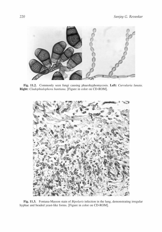
220 Sanjay G. Revankar
Fig. 11.2. Commonly seen fungi causing phaeohyphomycosis. Left: Curvularia lunata.Right: Cladophialophora bantiana. [Figure in color on CD-ROM].
Fig. 11.3. Fontana-Masson stain of Bipolaris infection in the lung, demonstrating irregularhyphae and beaded yeast-like forms. [Figure in color on CD-ROM].

11. Phaeohyphomycosis—Infection Due to Dark Moulds 221
(Fig. 11.2) (see also Fig. 2.13, Chapter 2), though referral to a reference laboratory isoften needed to identify unusual species. As many of these are rarely seen in practice,a high degree of clinical suspicion is required when interpreting culture results.
In tissue, these fungi will stain strongly with the Fontana-Masson stain, which isspecific for melanin (Fig. 11.3) (3). This can be helpful in distinguishing these fungifrom other species, particularly Aspergillus. In addition, hyphae typically appear morefragmented in tissue than seen with Aspergillus, with irregular septate hyphae andbeaded, yeast-like forms (3).
7. TREATMENT
Therapy is not standardized for any of these clinical syndromes, and randomizedtrials are unlikely given the sporadic nature of cases. Itraconazole and voriconazoledemonstrate the most consistent in vitro activity against this group of fungi, thoughfar more clinical experience has accumulated with itraconazole (34). Amphotericin Bmay be used for severe infections in unstable patients; high doses of lipid formulationsmay have a role in the treatment of refractory cases or in patients intolerant of standardamphotericin B. However, some species of dematiaceous fungi are resistant to thisagent. Once the infection is under control, longer term therapy with a broad-spectrumoral azole is often reasonable until a complete response is achieved, which may requireseveral weeks to months.
Other agents have limited roles in treating these fungi. Ketoconazole is not welltolerated, and fluconazole has poor activity against these fungi in general. Terbinafineand flucytosine have occasionally been used for subcutaneous infections in patientsrefractory to other therapy. Echinocandins do not appear to be very useful as singleagents. Combination therapy is a potentially useful therapeutic strategy for refractoryinfections, particularly brain abscess and disseminated disease, though it has notbeen well studied. Suggested therapies for specific infections are summarized inTable 11.1.
7.1. Allergic Disease
Steroids are the mainstay of treatment for allergic disease caused by these fungi,especially in asthma, though other modalities may have a role in specific clinicalsituations. For example, therapy for AFS consists of systemic corticosteroids andsurgery to remove the mucin, which is often tenacious. Antifungal therapy, usually inthe form of itraconazole, may play a role in reducing the requirement for corticosteroids,but this is not routinely recommended (35). Other azoles have only rarely been usedin the treatment of this disease.
Allergic bronchopulmonary mycosis (ABPM) can be treated with systemic corticos-teroids as in ABPA; prednisone at a dose of 0.5 mg/kg per day for 2 weeks, followed bya slow taper over 2 to 3 months or longer (19). Itraconazole has been used as a steroidsparing agent in APBA, but its efficacy is not clear, and routine use of itraconazole isnot generally recommended (19).

222 Sanjay G. Revankar
7.2. Focal Infection
7.2.1. Superficial Infection
Itraconazole and terbinafine are the most commonly used systemic agents foronychomycosis, and may be combined with topical therapy for refractory cases (36).There is no published experience with voriconazole.7.2.2. Keratitis
For keratitis, topical 5% natamycin is used almost exclusively, with only a fewsevere cases requiring adjunctive therapy, usually with an azole (23,37). Itraconazolehas the best in vitro activity. The majority of isolates are resistant to flucytosine.Surgery, including penetrating keratoplasty, is often needed. Enucleation is occasionallyrequired due to poor clinical response. Many patients do not recover complete visualacuity despite aggressive therapy.7.2.3. Subcutaneous Infection
Subcutaneous lesions will often respond to surgical excision alone (38). Oralsystemic therapy with a broad-spectrum azole antifungal agent in conjunction withsurgery is frequently employed and has been used successfully, particularly in immuno-compromised patients (39,40).7.2.4. Pneumonia
Therapy consists of systemic antifungal agents, usually amphotericin B oritraconazole initially, followed by itraconazole for a more prolonged period (14).Mortality rates are high in immunocompromised patients. Experience with voriconazoleis currently only anecdotal (41).7.2.5. Brain Abscess
Therapy published in the literature has varied greatly depending on the case report,and there is no standard treatment. A retrospective analysis of 101 reported casessuggested that the combination of amphotericin B (high-dose lipid formulation), flucy-tosine, and itraconazole may be associated with improved survival, though it was notfrequently used (32). Voriconazole may also prove useful. High doses of azoles havebeen suggested as an option, though there are no studies confirming this approach.Complete excision of brain abscesses may lead to better outcomes than aspiration orpartial excision. Overall mortality is greater than 70%.
7.3. Disseminated Infection
A recent literature review suggested the mortality rate is greater than 70%, despiteaggressive antifungal therapy (33). There were no antifungal regimens associated withimproved survival in disseminated infection. High-dose lipid amphotericin B may bereasonable for initial therapy, given its fungicidal activity for many fungi. Additionof a broad-spectrum azole or echinocandin could be considered in individuals failingtherapy. Infection with S. prolificans has been associated with a nearly 100% mortalityin the absence of recovery from neutropenia, as it is generally resistant to all availableantifungal agents. Recent reports have suggested that the combination of itraconazoleor voriconazole with terbinafine may be synergistic against this species, though theclinical relevance of this finding is unclear (42,43).

11. Phaeohyphomycosis—Infection Due to Dark Moulds 223
REFERENCES
1. McGinnis MR. Chromoblastomycosis and phaeohyphomycosis: new concepts, diagnosis,and mycology. J Am Acad Dermatol 1983;8:1–16.
2. Ajello L, Georg LK, Steigbigel RT, Wang CJ. A case of phaeohyphomycosis caused by anew species of Phialophora. Mycologia 1974;66:490–498.
3. Rinaldi MG. Phaeohyphomycosis. Dermatol Clin 1996;14:147–153.4. Matsumoto T, Ajello L, Matsuda T, Szaniszlo PJ, Walsh TJ. Developments in hyalohy-
phomycosis and phaeohyphomycosis. J Med Vet Mycol 1994;32(Suppl 1):329–349.5. Biosafety in microbiological and biomedical laboratories. 4th ed. Washington, DC: US
Government Printing Office, 1999:123–124.6. De Hoog GS. Significance of fungal evolution for the understanding of their pathogenicity,
illustrated with agents of phaeohyphomycosis. Mycoses 1997;40(Suppl 2):5–8.7. Sutton DA, Slifkin M, Yakulis R, Rinaldi MG. US case report of cerebral phaeohyphomy-
cosis caused by Ramichloridium obovoideum (R. mackenziei): criteria for identification,therapy, and review of other known dematiaceous neurotropic taxa. J Clin Microbiol1998;36:708–715.
8. Shelton BG, Kirkland KH, Flanders WD, Morris GK. Profiles of airborne fungi in buildingsand outdoor environments in the United States. Appl Environ Microbiol 2002;68:1743–1753.
9. Jacobson ES. Pathogenic roles for fungal melanins. Clin Microbiol Rev 2000;13:708–717.10. Dixon DM, Polak A, Szaniszlo PJ. Pathogenicity and virulence of wild-type and melanin-
deficient Wangiella dermatitidis. J Med Vet Mycol 1987;25:97–106.11. Kwon-Chung KJ, Polacheck I, Popkin TJ. Melanin-lacking mutants of Cryptococcus
neoformans and their virulence for mice. J Bacteriol 1982;150:1414–1421.12. van Duin D, Casadevall A, Nosanchuk JD. Melanization of Cryptococcus neoformans and
Histoplasma capsulatum reduces their susceptibilities to amphotericin B and caspofungin.Antimicrob Agents Chemother 2002;46:3394–3400.
13. Ikeda R, Sugita T, Jacobson ES, Shinoda T. Effects of melanin upon susceptibility ofCryptococcus to antifungals. Microbiol Immunol 2003;47:271–277.
14. Revankar SG. Dematiaceous fungi. Semin Respir Crit Care Med 2004;25:183–190.15. Bush RK, Prochnau JJ. Alternaria-induced asthma. J Allergy Clin Immunol 2004;113:
227–234.16. Bush RK, Portnoy JM, Saxon A, Terr AI, Wood RA. The medical effects of mould
exposure. J Allergy Clin Immunol 2006;117:326–333.17. Ferguson BJ. Definitions of fungal rhinosinusitis. Otolaryngol Clin North Am 2000;33:
227–235.18. Houser SM, Corey JP. Allergic fungal rhinosinusitis: pathophysiology, epidemiology, and
diagnosis. Otolaryngol Clin North Am 2000;33:399–409.19. Greenberger PA. Allergic bronchopulmonary aspergillosis. J Allergy Clin Immunol
2002;110:685–692.20. Hamilton BG, Humphreys CW, Conner WC, Hospenthal DR. Allergic bronchopulmonary
disease secondary to Bipolaris spicifera: Case report. J Bronchol 2006;13:77–79.21. Perez C, Colella MT, Olaizola C, de Capriles CH, Magaldi S, Mata-Essayag S. Tinea
nigra: report of twelve cases in Venezuela. Mycopathologia 2005;160:235–238.22. Gupta AK, Ryder JE, Baran R, Summerbell RC. Non-dermatophyte onychomycosis.
Dermatol Clin 2003;21:257–268.23. Gopinathan U, Garg P, Fernandes M, Sharma S, Athmanathan S, Rao GN. The epidemi-
ological features and laboratory results of fungal keratitis: a 10-year review at a referraleye care center in South India. Cornea 2002;21:555–559.
24. Srinivasan M. Fungal keratitis. Curr Opin Ophthalmol 2004;15:321–327.

224 Sanjay G. Revankar
25. Wilhelmus KR, Jones DB. Curvularia keratitis. Trans Am Ophthalmol Soc 2001;99:111–130.26. Sutton DA, Rinaldi MG, Kielhofner M. First US report of subcutaneous phaeohyphomy-
cosis caused by Veronaea botryosa in a heart transplant recipient and review of theliterature. J Clin Microbiol 2004;42:2843–2846.
27. Chuan MT, Wu MC. Subcutaneous phaeohyphomycosis caused by Exophiala jeanselmei:successful treatment with itraconazole. Int J Dermatol 1995;34:563–566.
28. Odell JA, Alvarez S, Cvitkovich DG, Cortese DA, McComb BL. Multiple lung abscessesdue to Ochroconis gallopavum, a dematiaceous fungus, in a nonimmunocompromisedwood pulp worker. Chest 2000;118:1503–1505.
29. Yeghen T, Fenelon L, Campbell CK, et al. Chaetomium pneumonia in patient with acutemyeloid leukaemia. J Clin Pathol 1996;49:184–186.
30. Mazur JE, Judson MA. A case report of a Dactylaria fungal infection in a lung transplantpatient. Chest 2001;119:651–653.
31. Manian FA, Brischetto MJ. Pulmonary infection due to Exophiala jeanselmei: successfultreatment with ketoconazole. Clin Infect Dis 1993;16:445–446.
32. Revankar SG, Sutton DA, Rinaldi MG. Primary central nervous system phaeohyphomy-cosis: a review of 101 cases. Clin Infect Dis 2004;38:206–216.
33. Revankar SG, Patterson JE, Sutton DA, Pullen R, Rinaldi MG. Disseminated phaeohy-phomycosis: review of an emerging mycosis. Clin Infect Dis 2002;34:467–476.
34. Sharkey PK, Graybill JR, Rinaldi MG, et al. Itraconazole treatment of phaeohyphomycosis.J Am Acad Dermatol 1990;23:577–586.
35. Kuhn FA, Javer AR. Allergic fungal rhinosinusitis: perioperative management, preventionof recurrence, and role of corticosteroid and antifungal agents. Otolaryngol Clin North Am2000;33:419–433.
36. Tosti A, Piraccini BM, Lorenzi S, Iorizzo M. Treatment of nondermatophyte mould andCandida onychomycosis. Dermatol Clin 2003;21:491–497.
37. Thomas PA. Fungal infections of the cornea. Eye 2003;17:852–862.38. Summerbell RC, Krajden S, Levine R, Fuksa M. Subcutaneous phaeohyphomycosis caused
by Lasiodiplodia theobromae and successfully treated surgically. Med Mycol 2004;42:543–547.
39. Kimura M, Goto A, Furuta T, Satou T, Hashimoto S, Nishimura K. Multifocal subcutaneousphaeohyphomycosis caused by Phialophora verrucosa. Arch Pathol Lab Med 2003;127:91–93.
40. Clancy CJ, Wingard JR, Hong NM. Subcutaneous phaeohyphomycosis in transplant recip-ients: review of the literature and demonstration of in vitro synergy between antifungalagents. Med Mycol 2000;38:169–175.
41. Diemert D, Kunimoto D, Sand C, Rennie R. Sputum isolation of Wangiella dermatitidisin patients with cystic fibrosis. Scand J Infect Dis 2001;33:777–779.
42. Meletiadis J, Mouton JW, Meis JF, Verweij PE. Combination chemotherapy for thetreatment of invasive infections by Scedosporium prolificans. Clin Microbiol Infect2000;6:336–337.
43. Howden BP, Slavin MA, Schwarer AP, Mijch AM. Successful control of disseminatedScedosporium prolificans infection with a combination of voriconazole and terbinafine.Eur J Clin Microbiol Infect Dis 2003;22:111–113.
SUGGESTED READINGS
Clancy CJ, Wingard JR, Hong NM. Subcutaneous phaeohyphomycosis in transplant recipients:review of the literature and demonstration of in vitro synergy between antifungal agents.Med Mycol 2000;38:169–175.

11. Phaeohyphomycosis—Infection Due to Dark Moulds 225
Jacobson ES. Pathogenic roles for fungal melanins. Clin Microbiol Rev 2000;13:708–717.Kuhn FA, Javer AR. Allergic fungal rhinosinusitis: perioperative management, prevention
of recurrence, and role of steroids and antifungal agents. Otolaryngol Clin North Am2000;33:419–433.
Revankar SG, Patterson JE, Sutton DA, Pullen R, Rinaldi MG. Disseminated phaeohyphomy-cosis: review of an emerging mycosis. Clin Infect Dis 2002;34:467–476.
Revankar SG, Sutton DA, Rinaldi MG. Primary central nervous system phaeohyphomycosis: areview of 101 cases. Clin Infect Dis 2004;38:206–216.
Sharkey PK, Graybill JR, Rinaldi MG, et al. Itraconazole treatment of phaeohyphomycosis.J Am Acad Dermatol 1990;23:577–586.
Srinivasan M. Fungal keratitis. Curr Opin Ophthalmol 2004;15:321–327.

12Zygomycosis (Mucormycosis)
Charalampos Antachopoulos, MD,Juan C. Gea-Banacloche, MD, and Thomas J. Walsh, MD
1. INTRODUCTION
The class Zygomycetes includes a variety of filamentous fungi that may cause life-threatening human disease and, over the past decade, have emerged as an increasinglyimportant cause of morbidity and mortality among immunocompromised patients (1,2).The first case of zygomycosis in humans was reported in 1885 by Platauf as MycosisMucorina. In many of the cases reported thereafter the infection was identified as“mucormycosis” or Mucor infection based solely on histological findings of wide,rarely septate hyphae, without culture confirmation. The use of the term “mucormy-cosis” was further promoted by the original classification of most of the pathogeniczygomycetes species as members of the genus Mucor. Many of these species were laterreassigned into different genera or families (3). Consequently the term “zygomycosis”should be preferred, instead of “mucormycosis,” for infections caused by any of thezygomycetes species. Some authors still employ the term “mucormycosis,” however,for infections caused by members of the order Mucorales (3,4). Those who preferthe use of “mucormycosis” stress that the opportunistic disease due to those fungi inthe order Mucorales differs substantially from disease caused by the other pathogeniczygomycetes order, Entomophthorales.
2. ETIOLOGIC AGENTS
The medically important Zygomycetes encompass two orders of filamentous fungiwith distinct morphologic, epidemiologic, and pathogenic characteristics, the Mucoralesand the Entomophthorales (3,5–8) (Table 12.1). The majority of cases of zygomycosisin humans are caused by members of the order Mucorales. Organisms of the genusRhizopus are by far the most common clinical isolates, with R. oryzae being the mostfrequently recovered species. Members of the genus Mucor are second to Rhizopusin order of frequency, while Cunninghamella, Apophysomyces, Absidia, Saksenaea,Rhizomucor and other genera each represent a significantly smaller percentage ofclinical isolates (1,3,4).
From: Infectious Disease: Diagnosis and Treatment of Human MycosesEdited by: D. R. Hospenthal and M. G. Rinaldi © Humana Press Inc., Totowa, NJ
227

228 Charalampos Antachopoulos, Juan C. Gea-Banacloche, and Thomas J. Walsh
Table 12.1Taxonomic classification of the Zygomycetes
Class Order Family Genus Species causinghuman disease
Zygomycetes Mucorales Mucoraceae Absidia A. corymbiferaApophysomyces A. elegansMucor M. circinelloides,
M. ramosissimus,M. racemosus,M. hiemalis,M. rouxianus
Rhizomucor R. pusillusRhizopus R. oryzae
(R. arrhizus),R. microsporus var.rhizopodiformis
Cunninghamellaceae Cunninghamella C. bertholletiaeMortierellaceae Mortierella (animal pathogens)Saksenaceae Saksenaea S. vasiformisSyncephalastraceae Syncephalastrum S. racemosumThamnidaceae Cokeromyces C. recurvatus
Entomophthoralesa Ancylistaceae Conidiobolus* C. coronatus,C. incongruus
Basidiobolaceae Basidiobolus* B. ranarum
aIt has been recently suggested that the order Entomophthorales should be divided into two phylogenet-ically distinct groups: the genus Basidiobolus should be classified in a new order, the Basidiobolales,while Conidiobolus appears to be closely related to the Mucorales and should form a distinct group (5,8).
The class of Zygomycetes is characterized in culture by broad, nonseptate or sparselyseptated hyphae and by the presence of sporangiophores supporting sporangia, whichcontain sporangiospores (see Fig. 2.7, Chapter 2). During sexual reproduction in culturezygospores may be produced. The Zygomycetes are characterized in tissue by theformation of wide, ribbonlike, hyaline, aseptate or sparsely septated hyphae with wide-angle (approximately 90°) branching. The substantial differences among these andother structures allow mycology laboratories to diagnose organisms by genus andspecies (3).
3. EPIDEMIOLOGY
The Zygomycetes are ubiquitous in soil and can be isolated from decaying organicmatter including hay, decaying vegetation, and a variety of food items. Human infectionis usually acquired through inhalation of sporangiospores from environmental sources.Acquisition via the cutaneous or percutaneous route is also common, either throughtraumatic disruption of skin barriers or with the use of catheters and injections. Lesscommonly, infection through the gastrointestinal route may occur (1,3,6).

12. Zygomycosis (Mucormycosis) 229
Zygomycosis is approximately 10- to 50- fold less common than invasive Candidaor Aspergillus infections, with a prevalence of 1 to 5 cases per 10,000 autopsies andan estimated incidence of 1.7 cases per million per year in the United States (4,9). Aclear male predisposition has been observed, as demonstrated by an approximate 2:1male to female ratio among cases (1). Unlike other filamentous fungi, targeting mainlyimmunocompromised patients, the Zygomycetes cause disease in a wider and moreheterogeneous population. While the Mucorales mainly affect patients with underlyingimmunosuppression or other medical conditions, the Entomophthorales largely afflictimmunocompetent hosts in tropical and subtropical areas of developing countries.Lately, however, infections caused by the Entomophthorales in immunocompromisedhosts have occasionally been documented (6,7,10).
The most common underlying condition for development of zygomycosis is diabetes,both type I and type II. A significant proportion of these patients will present withconcomitant ketoacidosis, while in others zygomycosis may even present as thediabetes-defining illness. Other significant underlying conditions include the presenceof hematological malignancy, solid organ or bone marrow transplantation, deferox-amine therapy, and injection drug use (1,6). During the 1980s and 1990s, the percentageof patients with hematological malignancy, solid organ or bone marrow transplan-tation, and injection drug use among all cases of zygomycosis increased significantly(1,11,12). In the aforementioned groups of hematological patients and transplant recip-ients, factors associated with this infection have been reported to include prolongedneutropenia, corticosteroid use, and graft versus host disease (GvHD) (6,12). Lesscommonly, the Zygomycetes may cause invasive disease in the presence of renalfailure, diarrhea, and malnutrition in low birth weight infants and in HIV patients.Occasionally zygomycosis has developed in patients with persistent metabolic acidosissecondary to causes other than diabetes (1,6).
A significant proportion of zygomycosis cases have as well been observed in personswith no primary underlying disease at the time of infection. In many of these casesthere was a history of penetrating trauma, surgery, or burn before the development ofinfection (1,6).
4. PATHOGENESIS AND IMMUNOLOGY
The epidemiologic profile of zygomycosis cases (patients with diabetes, hemato-logical malignancies or on deferoxamine therapy, transplant recipients) may in part beexplained by our current understanding of the pathogenesis of these infections.
As with other filamentous fungi, an effective immune response following inoculationof sporangiospores requires the presence of adequate phagocytic activity of the hosteffector cells, including tissue macrophages and neutrophils. The macrophages ingestthe sporangiospores to inhibit germination, while the neutrophils are involved in hyphaldamage (13). Consequently, the host immune response against the Zygomycetes maybe compromised if the phagocytic cells are insufficient in number as in the case ofchemotherapy-induced neutropenia, or dysfunctional, as in the case of corticosteroidtreatment or diabetes mellitus (3,13).
Experimental evidence also suggests an important role of iron in the pathogenesisof infections caused by Rhizopus species, whose growth is promoted in the presence of

230 Charalampos Antachopoulos, Juan C. Gea-Banacloche, and Thomas J. Walsh
increased iron uptake. Deferoxamine, an iron chelator, has siderophore activity for thesefungi, allowing significant increase in iron uptake. Further, the availability of serumiron is increased in the presence of acidic pH, suggesting an additional mechanism forthe development of zygomycosis in patients with diabetic ketoacidosis (4,13).
An almost universal feature in infections caused by the Mucorales is the presenceof extensive angioinvasion associated with thrombosis and ischemic necrosis (3).This is likely an important mechanism by which these organisms survive antifungaltherapy because adequate blood supply is necessary for the delivery of antifungalagents. Recent data also have demonstrated the ability of R. oryzae sporangiospores orhyphae to adhere to subenthodelial matrix proteins and human endothelial cells (4,14).Pregerminated sporangiospores of R. oryzae were able to damage endothelial cells invitro, following adherence to and phagocytocis by these cells. R. oryzae viability wasnot required for endothelial cell damage, suggesting that in the setting of establishedinfection even fungicidal therapy may not prevent subsequent tissue injury (14).
5. CLINICAL MANIFESTATIONS
The clinical presentations of zygomycosis in humans largely depend on the causativeagent (Mucorales versus Entomophthorales) and the patient’s condition (intact immuneresponse versus immunosuppresion or other underlying conditions). Thus, while theMucorales cause rapidly progressive disease characterized by angioinvasion, throm-bosis, tissue necrosis, and dissemination in susceptible hosts, infections caused bythe Entomophthorales usually lack these features, inducing a chronic inflammatoryresponse and following an indolent course in immunocompetent patients (3,6,7). Conse-quently the patterns of human disease are described separately for these two ordersof Zygomycetes, with more emphasis given to infections caused by the Mucorales,because organisms of this order are most likely to be encountered in developedcountries.
The clinical manifestations of human infection caused by the Mucorales can beclassified as sinus disease, localized or extended to the orbit and/or brain, pulmonary,cutaneous, gastrointestinal, disseminated and miscellaneous infection. Some of thesemanifestations may occur with increased frequency in patients with certain underlyingconditions (Table 12.2) (1,6). However, this is not always the case and zygomycosisin these patient groups may still present with any of the above patterns.
5.1. Sinus Infection
Sinus disease may be confined to the paranasal sinuses or may infiltrate theorbit (sino-orbital) and/or the brain parenchyma (rhinocerebral). This form representsapproximately two-thirds of all cases of zygomycosis in diabetic patients (1). Theinfection originates in the paranasal sinuses after inhalation of sporangiospores. Initialsymptoms may suggest sinusitis and include sinus pain, discharge, soft tissue swelling,and perinasal cellulitis/paresthesia. Fever is variable and may be absent in up to halfof cases (4,15,16). The tissues involved become red, violaceous, and finally black, asvascular thrombosis leads to tissue necrosis. A blood-tinged nasal discharge may bepresent. Extension of the infection to the mouth may produce painful necrotic ulcer-ations in the hard palate. Extension into the periorbital area and ultimately the orbit

12. Zygomycosis (Mucormycosis) 231
Table 12.2Predominant site of Mucorales infection according to thepatient’s underlying condition
Underlying condition Type of infection, by site
Diabetes SinusMalignancy PulmonarySolid organ transplantation PulmonaryBone marrow transplantation PulmonaryDeferoxamine therapy Pulmonary, sinus, disseminateda
Injection drug use CerebralNo underlying condition Cutaneous
Data from ref. 1.aNo clear predominance among the three sites.
may be manifested by periorbital edema, lacrimation, chemosis, and proptosis. Subse-quent ocular or optic nerve involvement may be suggested by pain, diplopia, blurringor loss of vision. Alteration of mental status and cranial nerve palsies may signifyinvasion of the central nervous system. Occasionally thrombosis of the cavernous sinusor the internal carotid artery may follow, with resultant neurological deficits, whiledissemination of the infection also may occur (4,6,16,17).
5.2. Pulmonary Infection
Pulmonary disease is most commonly observed in patients with hematologicalmalignancies, solid organ or bone marrow transplant recipients, and in those receivingdeferoxamine treatment (1). Not infrequently it may occur with concomitant sinusdisease (sinopulmonary infection) (18). Lung involvement may be manifested as infil-trates, consolidation, and solitary nodular or cavitary lesions (Fig. 12.1) (19,20).Fungal invasion of the pulmonary vessels may result in thrombosis and subsequentinfarcts in the lung parenchyma (Fig. 12.2). Angioinvasion may also lead to intra-parenchymal bleeding or even hemoptysis, which can be fatal if major vessels, suchas the pulmonary artery, are involved. Extension of the infection to the chest wall,pericardium, myocardium, mediastinum, and diaphragm has been described (4,6,19).A predilection for the upper lobes has been reported; however, any part of the lungmay be involved, and bilateral disease is not uncommon (19). Presenting signs andsymptoms are nonspecific and include fever, cough, chest pain, dyspnea, hemoptysis,tachypnea, crackles, decreased breath sounds, and wheezing (4,19,20).
5.3. Cutaneous Infection
Cutaneous zygomycosis is often observed in individuals with no underlyingcondition as a result of infection of a preexisting lesion, such as skin trauma or burn.Alternatively, it may occur in the context of disseminated disease or extensive localinfection in immunocompromised hosts (1,3,21,22). In the case of primary cutaneousinoculation, the lesion appears acutely inflamed with redness, swelling, induration, andfrequent progression to necrosis. Extensive local invasion may occur involving the

232 Charalampos Antachopoulos, Juan C. Gea-Banacloche, and Thomas J. Walsh
Fig. 12.1. Thoracic CT scan of profoundly neutropenic patient with pulmonary zygomycosisdemonstrates rapid evolution of pulmonary nodule to involve the pleural surface and to manifesta halo sign at the interface with radiologically normal lung. The two scans are separated by 5days. [Figure in color on CD-ROM].
Fig. 12.2. Histopathology of pulmonary zygomycosis. Characteristic broad nonseptatedribbonlike hyphae with nondichotomous branching invading a pulmonary blood vessel. Thespecimen was obtained from the lung lesion seen on CT scan in Figure 12.1. GMS. [Figure incolor on CD-ROM].

12. Zygomycosis (Mucormycosis) 233
Fig. 12.3. Development of zygomycosis in the skin and subcutaneous tissues of the rightlower extremity in a patient with cutaneous T-cell lymphoma. The top left panel depicts thelesions of cutaneous T-cell lymphoma, which were possibly infected by direct inoculation.The top right panel reveals the extensive necrosis and destruction of soft tissue caused bythe rapidly invading zygomycete. The bottom panel demonstrates the soft tissues followingextensive surgical debridement. [Figure in color on CD-ROM].
adjacent subcutaneous fat, muscle, bone tissues, and fascial layers (Fig. 12.3). Whencutaneous disease is the result of disseminated infection, it usually presents as nodularsubcutaneous lesions that may ulcerate (3,6,22).
5.4. Gastrointestinal Infection
Gastrointestinal disease is rare, occurring mainly in malnourished patients andpremature neonates, in whom it can present as necrotizing enterocolitis (1,23). Afteringestion of the sporangiospores, fungal invasion of the mucosa, submucosa, andvascular structures of the gastrointestinal tract may occur, often resulting in necroticulcers, rupture of the intestinal wall, and peritonitis. Symptoms are nonspecific,including fever, abdominal pain, distention, vomiting, and gastrointestinal hemorrhage(3,4,23).
5.5. Disseminated Infection
Disseminated infection refers to involvement of at least two noncontiguous sites andis commonly observed in patients receiving deferoxamine therapy (1). Disseminationoccurs through the hematogenous route and may originate from any of the above sitesof primary infection, although it seems to be more frequently associated with lung

234 Charalampos Antachopoulos, Juan C. Gea-Banacloche, and Thomas J. Walsh
disease. The most common site of dissemination is the brain, but other organs mayalso be involved (4,25).
5.6. Other Infection
Isolated cerebral zygomycosis is usually observed in injection drug users (1).Endocarditis is a potential complication of cardiac surgery. Isolated peritonitis is oftenassociated with peritoneal dialysis. Renal infection and external otitis also have beenreported (4,6).
5.7. Infection Due to the Entomophthorales
In contrast to the Mucorales infections, human disease caused by the Entomophtho-rales usually follows an indolent course, as previously mentioned. A chronic subcuta-neous disease, characterized by slowly enlarging subcutaneous nodules that eventuallyulcerate, is typically caused by B. ranarum. C. coronatus infections commonly presentas chronic sinusitis that usually does not extend to the central nervous system (3,7). Lesscommonly, involvement of other body sites or even aggressive disseminated infectionby members of the order Entomophthorales has been reported for immunocompromisedand immunocompetent patients (3,7,10,23).
6. DIAGNOSIS
As infections caused by the Zygomycetes, and particularly the Mucorales, in humansmay be rapidly fatal, timely diagnosis is crucial to avoid treatment delay. Whileconfirmation of the diagnosis and species identification of the causative organismshould be pursued, treatment should be initiated as soon as the diagnosis is suspected,owing to the severity of these infections.
Currently, the diagnosis of zygomycosis relies on a constellation of the following:high index of suspicion, assessment of presenting signs and symptoms, imaging studies,cultures of clinical specimens, and histopathology (Fig. 12.4).
6.1. Clinical Assessment
The high index of suspicion should be based on the knowledge of the underlyingconditions that predispose to zygomycosis and the usual presentation of the infection ineach of these conditions (Table 12.2). Nevertheless, less common manifestations of thedisease should not be excluded. A common scenario is the development of zygomycosisin oncological patients or transplant recipients who are receiving antifungal therapyfor prophylaxis or treatment of other opportunistic fungal infections, such as invasiveaspergillosis. If the antifungal agents that are being administered to the patient arenot active against the Zygomycetes (including, fluconazole, voriconazole, and theechinocandins), then clinical deterioration or appearance of new signs and symptomsin these patients should alert the clinician to the possibility of zygomycosis (18).
Most of the signs and symptoms that are associated with the clinical manifestationsof zygomycosis are nonspecific. However, their diagnostic significance may increase ifthey are interpreted in relation to the patient’s underlying condition. For example, thedevelopment of sinusitis in a leukemic or diabetic patient should raise the suspicionof zygomycosis. Other findings have probably greater specificity for this infection,

12. Zygomycosis (Mucormycosis) 235
Host factors
DiabetesMalignancyBone marrow transpl.Solid organ transpl.Deferoxamine therapyInjection drug use
Relevant clinical findings
SinusitisPneumoniaCutaneous ulcersCNS lesions
Rapid progression of infection
Presumed diagnosisof zygomycosis
Physician alertness,awareness of risk
factors & presentation
Diagnostic steps
Imaging
Cultures &Histopathology ofbiopsy material
Treatment
Rapid initiationReversal of predisposingconditionAntifungal agentsSurgical debridement
Final diagnosisOptimization of treatment
Fig. 12.4. Diagnosis and management of zygomycosis.
such as the presence of blood-tinged nasal discharge or necrotic eschars in the hardpalate. In addition, the presence of hemoptysis in a susceptible host is consistent withangioinvasion and should raise the possibility of zygomycosis (19). An alarming signshould also be the rapid spread of the infection. Finally, even after the diagnosis hasbeen made, careful periodic clinical assessment should be performed in order to detectprogression of the disease. For example, in a patient with pulmonary zygomycosis,palpation of the skin for subcutaneous nodules and neurological evaluation for changesin mental status and focal neurological signs should be performed repeatedly to detectdissemination to the skin and brain, respectively.
6.2. Diagnostic Imaging
Imaging studies are helpful in assessing the burden of the disease, involvement ofadjacent tissues, and response to treatment. They are also helpful in guiding moreinvasive procedures to obtain biopsy specimens for histopathology and culture (26).Although imaging findings may be suggestive of zygomycosis in the appropriateclinical setting, they are not sufficiently specific to establish the diagnosis. In sinusdisease, computerized tomography (CT) detects subtle mucosal thickening or bonyerosions of the sinuses, but it is less sensitive than magnetic resonance imaging (MRI)for the detection of extension of the infection to the soft tissues of the orbit (15,27).

236 Charalampos Antachopoulos, Juan C. Gea-Banacloche, and Thomas J. Walsh
In the case of pulmonary disease, high-resolution CT is more sensitive than chestradiograph for early diagnosis of the infection and can more accurately determinethe extent of pulmonary involvement. Radiographic features may be consistent withinfliltrate, cavity, or consolidation (Fig. 12.1). The air crescent and halo signs, whichare recognized radiologic features of invasive aspergillosis, have been reported as wellfor zygomycosis (19,20,28). In patients with pulmonary zygomycosis the presence ofan air crescent sign seems to be associated with increased risk for massive hemoptysis(19). Another suggestive finding could be expansion of a mass or consolidation acrosstissue planes, in particular toward the great vessels in the mediastinum (4,29). In thecase of cutaneous disease, MRI is superior to CT scan for assessment of extension ofthe infection to the adjacent soft or bone tissues.
6.3. Culture
Recovery of Zygomycetes from cultures of clinical specimens would allow not onlyestablishment of diagnosis, but also identification of the causative organism to thespecies level. Although the Zygomycetes may contaminate laboratory material, theirisolation from clinical specimens of susceptible hosts should not be disregarded ascontamination. Despite the ability of these organisms to invade tissues, they are rarelyisolated from cultures of blood, urine, cerebrospinal fluid, feces, sputum, paranasalsinuses secretions, bronchoalveolar lavage, or swabs from infected areas (3,15,19). Therecovery of Zygomycetes from biopsy material may be compromised if processing ofthe specimens involves tissue grinding, a procedure that kills the nonseptate hyphaeof these fungi. The recovery rate is enhanced, however, if thin slices of minimallymanipulated tissue are placed onto the culture medium. Consequently, for properhandling of the specimens, the laboratory should be notified of the possibility ofzygomycosis. In any case, negative cultures do not rule out the infection (3,6).
6.4. Endoscopic Findings and Histopathology
Given the above limitations of cultures or imaging studies, diagnosis of zygomy-cosis is almost always based on histopathologic examination of appropriately collectedsamples. The latter should be pursued in the presence of strong suspicion for zygomy-cosis if the cultures or imaging studies are negative or nonspecific. Depending on thepresentation of the disease, the samples may be collected by fiberoptic bronchoscopy,radiographically guided transthoracic needle aspiration, open lung biopsy, nasalendoscopy, paranasal sinus biopsy or débridement, and biopsies of skin or other infectedtissues (4,6,19,26). In the case of lung disease, endobronchial findings include stenosisor airway obstruction, erythematous mucosa, fungating or polypoid mass, and, lessoften, granulation tissue or mucosal ulceration (19). In sinus disease, nasal endoscopymay show black necrotic crusts on the nasal septum and turbinates; in the early phasesthe mucosa may still look pink and viable (4). We refer to these necrotic ulcers alongthe nasal mucosa or turbinates as “sentinel eschars,” as they may represent an earlyphase of infection or may be more amenable to biopsy than a deep maxillary sinusinfection.
Because the hyphae of Zygomycetes in tissue specimens may stain poorly withhematoxylin and eosin (H&E), a second more fungus-specific tissue stain should

12. Zygomycosis (Mucormycosis) 237
also be used, such as Gomori methenamine silver (GMS) or periodic acid Schiff(PAS) (3). As previously mentioned, the hallmark of zygomycosis is the demonstrationof wide, ribbonlike, aseptate (nonseptate or rarely septate) hyphae with wide-anglebranching in biopsy specimens (Fig. 12.2) (see also Fig. 3.11, Chapter 3). For Mucoralesinfections, the hyphae are seen to invade the adjacent blood vessels. Mycotic embolimay thrombose small vessels in which they are lodged. Extensive tissue necrosis orhemorrhage may be observed (3). Although histopathology is sensitive and reliable fordiagnosing zygomycosis, obtaining biopsy material from hematological patients maynot always be feasible owing to concomitant thrombocytopenia. Finally, because fixingand staining of the biopsy specimens takes time, a promising method for acceleratingdiagnosis by use of frozen sections in tissue specimens has been proposed recently (30).
6.5. Other Non-Culture Diagnosis
Unfortunately, no reliable molecular or antigen detection methods are availableto date for primary diagnosis of zygomycosis. A number of molecular techniquesare currently used by research laboratories for species identification of zygomycetesisolates, epidemiologic studies, or determination of taxonomic assignments (3,18).
7. TREATMENT
There are four cornerstones of successful management of zygomycosis: (1) rapidinitiation of therapy, (2) reversal of the patient’s underlying predisposing condition, (3)administration of appropriate antifungal agents, and (4) surgical débridement of infectedtissues (Fig. 12.4) (4,6). If not treated or if diagnosed with delay, infections causedby the Mucorales in humans are typically fatal (1,11,12). Even if timely diagnosisis made, treatment is challenging due to a number of reasons such as the underlyingcondition of the patient, the rapid progression of the disease, and the high degree ofangioinvasion and thrombosis that compromises the delivery of antifungal agents activeagainst the causative organisms. As mentioned previously, in the presence of certainconditions such as diabetes, immunosuppression, and others (Table 12.2), treatmentfor zygomycosis should be initiated as soon as a strong suspicion for this infection israised, without awaiting formal confirmation of the diagnosis, which may take time.Meanwhile of course, all the required actions to establish the diagnosis should beundertaken, as previously described (Fig. 12.4).
Reversal of the underlying condition can be fairly quickly achieved in certaincircumstances, such as diabetic ketoacidosis, which should be promptly corrected,or deferoxamine therapy, which should be discontinued. However, timely reversal ofthe disease- or treatment-related immunosuppression in patients with hematologicalmalignancies or transplant recipients is challenging. In these patients, temporary discon-tinuation of corticosteroid treatment or myelotoxic chemotherapy should be stronglyconsidered until the infection is brought under control. However, even with thesemeasures, spontaneous restoration of phagocytic activity or recovery from neutropeniaare likely to occur after several days, during which time the infection may progress. Invitro and in vivo studies, as well as case reports, have suggested that the administrationof cytokines, such as granulocyte colony-stimulating factor (G-CSF), granulocyte-macrophage colony-stimulating factor (GM-CSF), and interferon-�, may accelerate

238 Charalampos Antachopoulos, Juan C. Gea-Banacloche, and Thomas J. Walsh
immune recovery (31,32). In support of these general recommendations for cytokineaugmentation of host defense are recent in vitro studies demonstrating enhancement ofneutrophil activity against R. oryzae and other species of Zygomycetes in the presenceof interferon-� and GM-CSF (33). An alternative approach has been the administrationof granulocyte transfusions in neutropenic patients with invasive fungal infections(31,32). These immunomodulatory interventions may be considered on an individualpatient basis as adjunctive therapy for zygomycosis in immunocompromised hosts.Nonetheless, as a caveat, there is a lack of adequately powered clinical trials to evaluatetheir clinical efficacy and potential complications (6,31,32).
7.1. Amphotericin B
Amphotericin B is the drug of choice for the treatment of zygomycosis. Thispolyene agent exerts good in vitro and in vivo activity against the Zygomycetes.However, apparent in vitro resistance, with elevated minimal inhibitory concentrations(MICs) of amphotericin B, may be observed among clinical isolates and is relativelycommon among Cunninghamella species (34–36). The efficacy of amphotericin B inthe treatment of zygomycosis was demonstrated in a recent review of 929 cases, wheresurvival was 61% for patients treated with amphotericin B deoxycholate versus 3% forthose who received no treatment (1). The lipid formulations of amphotericin B (mainlyamphotericin B lipid complex and the liposomal formulation) also have been used inthe treatment of zygomycosis. These formulations are associated with significantly lesstoxicity than amphotericin B deoxycholate and demonstrate at least equivalent clinicalefficacy (1,37,38). However, no randomized controlled trials have been conducted tocompare the efficacy of deoxycholate versus lipid formulations of amphotericin B inthe treatment of zygomycosis.
When treatment with amphotericin B is initiated for documented zygomycosis, fulldoses should be given from the onset, foregoing the past practice of dose escalation. Theoptimal dosage of amphotericin B formulations for the treatment of zygomycosis hasnot been systematically evaluated in clinical studies. A study of the safety, tolerance,and plasma pharmacokinetics of liposomal amphotericin B in patients with invasivefungal infections found no demonstrable dose-limiting nephrotoxicity or infusion-related toxicity over a dose range of 7.5 to 15 mg/kg per day (39). Plasma concentrationsof liposomal amphotericin B achieved an upper limit at 10 mg/kg per day and were notincreased by further dosage increases. Nevertheless, the efficacy of higher dosages ofliposomal amphotericin B compared to the US Food and Drug Administration (FDA)-approved dosage of 3 to 5 mg/kg per day for aspergillosis has not been investigatedthrough clinical trials in zygomycosis. In the absence of such studies, an increase ofdosage of liposomal amphotericin B to 7.5 or 10 mg/kg per day could be consideredon an individual basis for patients with zygomycosis progressing through liposomalamphotericin B at 5 mg/kg per day (39). Amphotericin B lipid complex has been usedin a dosage of 5 mg/kg per day in salvage treatment of zygomycosis with complete orpartial responses in 17 (71%) of 24 cases (38).

12. Zygomycosis (Mucormycosis) 239
7.2. Azoles
Of the azole agents, fluconazole and voriconazole have little or no activity againstthe Zygomycetes (40). Itraconazole is active in vitro against some of these organisms,but has demonstrated poor efficacy in animal models (41,42). The investigationalazole posaconazole is active in vitro and in vivo against many of the Zygomycetes(34,35,40–42). Results from two recent case series as well as a number of case reportson the use of posaconazole in patients with zygomycosis refractory to or intolerantof conventional antifungal treatment provide encouraging data regarding posaconazoleas an alternative salvage therapy for zygomycosis (43–48). The use of this azole,however, as monotherapy or in combination with amphotericin B, for the treatmentof zygomycosis awaits further evaluation in a randomized clinical trial versus deoxy-cholate amphotericin B or a lipid formulation of amphotericin B.
7.3. Surgery
Appropriate and early surgical débridement is a critical intervention for thesuccessful management of zygomycosis for a number of reasons: the infectionprogresses rapidly, vascular thrombosis compromises the delivery of antifungal agentsto the site of infection, and there is massive tissue necrosis. Several retrospectivestudies have demonstrated that the survival of patients treated with antifungal therapycombined with surgical débridement was significantly higher than that of patientstreated with antifungal therapy alone (1,4,6,19). Surgical treatment should aim inremoving all necrotic tissues and should be considered for any of the clinical presenta-tions of zygomycosis (sinus disease, pulmonary or cutaneous). It should be performedearly in the course of treatment and repeated if necessary. It may include excision ofthe infected sinuses, débridement of retroorbital space, or even enucleation in the caseof sinus/sinoorbital disease, and wedge resection, lobectomy, or pneumonectomy in thecase of pulmonary disease (15,16,19,22,29). If the patient survives the infection, plasticsurgery is likely to be needed to correct disfiguring resulting from débridement (4).
7.4. Hyperbaric Oxygen
Besides the aforementioned important aspects of management of zygomycosis,hyperbaric oxygen is a therapeutic modality that has been occasionally used asadjunctive treatment. Hyperbaric oxygen has a theoretical potential for being beneficialin the treatment of zygomycosis because it is known to inhibit fungal growth at highpressures, correct tissue hypoxia and lactic acidosis, promote healing, and enhancephagocytosis (49,50). In a number of case reports and small case series of zygomy-cosis, administration of hyperbaric oxygen was associated with a favorable outcome(49). Currently, however, the absence of randomized controlled clinical trials on theefficacy of hyperbaric oxygen in this setting does not allow firm recommendationsregarding its use as adjunctive treatment of zygomycosis.
7.5. Prognosis
The prognosis of zygomycosis largely depends on the patient’s underlying condition,the clinical presentation of the infection, the time of initiation of therapy, and the typeof treatment provided. Mortality may range from less than 10% for localized sinus

240 Charalampos Antachopoulos, Juan C. Gea-Banacloche, and Thomas J. Walsh
disease to approximately 100% for disseminated infection, with an overall percentageof 47% for zygomycosis cases reported in the 1990s (1,4).
8. PREVENTION
Prevention may be feasible for a proportion of cases through adequate control ofdiabetes and judicious use of deferoxamine and corticosteroids. For severely immuno-compromised hosts, measures to reduce the risk of exposure to airborne sporan-giospores should be undertaken, including Hepafiltration of air supply, positive roomair pressures, exclusion of plants from the wards, and wearing of masks when leavingthe room. Owing to the relatively low incidence of zygomycosis, the cost-effectivenessof prophylactic treatment is questionable; the development of a preemptive therapyapproach, however, based on validated early indicators of the disease and riskassumption, should be a target for research in the near future. In the meantime, physi-cians caring for susceptible patients should maintain a high level of suspicion andbe alert to the early signs and symptoms of zygomycosis in order to achieve earlydiagnosis and timely initiation of treatment.
REFERENCES
1. Roden MM, Zaoutis TE, Buchanan WL, et al. Epidemiology and outcome of zygomycosis:a review of 929 reported cases. Clin Infect Dis 2005;41:634–653.
2. Kauffman CA. Zygomycosis: reemergence of an old pathogen. Clin Infect Dis2004;39:588–590.
3. Ribes JA, Vanover-Sams CL, Baker DJ. Zygomycetes in human disease. Clin MicrobiolRev 2000;13:236–301.
4. Spellberg B, Edwards J, Jr., Ibrahim A. Novel perspectives on mucormycosis: pathophys-iology, presentation, and management. Clin Microbiol Rev 2005;18:556–569.
5. Jensen AB, Dromph KM. The causal agents of “entomophthoramycosis” belong to twodifferent orders: a suggestion for modification of the clinical nomenclature. Clin MicrobiolInfect 2005;11:249–250.
6. Gonzalez CE, Rinaldi MG, Sugar AM. Zygomycosis. Infect Dis Clin North Am2002;16:895–914.
7. Prabhu RM, Patel R. Mucormycosis and entomophthoramycosis: a review of the clinicalmanifestations, diagnosis and treatment. Clin Microbiol Infect 2004;10(Suppl 1):31–47.
8. Jensen AB, Gargas A, Eilenberg J, Rosendahl S. Relationships of the insect-pathogenicorder Entomophthorales (Zygomycota, Fungi) based on phylogenetic analyses of nuclearsmall subunit ribosomal DNA sequences (SSU rDNA). Fungal Genet Biol 1998;24:325–334.
9. Rees JR, Pinner RW, Hajjeh RA, Brandt ME, Reingold AL. The epidemiological featuresof invasive mycotic infections in the San Francisco Bay area, 1992–1993: results ofpopulation-based laboratory active surveillance. Clin Infect Dis 1998;27:1138–1147.
10. Walsh TJ, Renshaw G, Andrews J, et al. Invasive zygomycosis due to Conidiobolusincongruus. Clin Infect Dis 1994;19:423–430.
11. Kontoyiannis DP, Wessel VC, Bodey GP, Rolston KV. Zygomycosis in the 1990s in atertiary-care cancer center. Clin Infect Dis 2000;30:851–856.
12. Marr KA, Carter RA, Crippa F, Wald A, Corey L. Epidemiology and outcome ofmould infections in hematopoietic stem cell transplant recipients. Clin Infect Dis2002;34:909–917.

12. Zygomycosis (Mucormycosis) 241
13. Shoham S, Levitz SM. The immune response to fungal infections. Br J Haematol2005;129:569–582.
14. Ibrahim AS, Spellberg B, Avanessian V, Fu Y, Edwards Jr JE. Rhizopus oryzae adheres to,is phagocytosed by, and damages endothelial cells in vitro. Infect Immun 2005;73:778–783.
15. Dhiwakar M, Thakar A, Bahadur S. Improving outcomes in rhinocerebral mucormycosis—early diagnostic pointers and prognostic factors. J Laryngol Otol 2003;117:861–865.
16. Talmi YP, Goldschmied-Reouven A, Bakon M, et al. Rhino-orbital and rhino-orbito-cerebral mucormycosis. Otolaryngol Head Neck Surg 2002;127:22–31.
17. Thajeb P, Thajeb T, Dai D. Fatal strokes in patients with rhino-orbito-cerebral mucormy-cosis and associated vasculopathy. Scand J Infect Dis 2004;36:643–648.
18. Kontoyiannis DP, Lionakis MS, Lewis RE, et al. Zygomycosis in a tertiary-care cancercenter in the era of Aspergillus-active antifungal therapy: a case-control observationalstudy of 27 recent cases. J Infect Dis 2005;191:1350–1360.
19. Lee FY, Mossad SB, Adal KA. Pulmonary mucormycosis: the last 30 years. Arch InternMed 1999;159:1301–1309.
20. McAdams HP, Rosado de Christenson M, Strollo DC, Patz EF, Jr. Pulmonary mucormy-cosis: radiologic findings in 32 cases. AJR Am J Roentgenol 1997;168:1541–1548.
21. Hay RJ. Mucormycosis: an infectious complication of traumatic injury. Lancet2005;365:830–831.
22. Losee JE, Selber J, Vega S, Hall C, Scott G, Serletti JM. Primary cutaneous mucormycosis:guide to surgical management. Ann Plast Surg 2002;49:385–390.
23. Lyon GM, Smilack JD, Komatsu KK, et al. Gastrointestinal basidiobolomycosis in Arizona:clinical and epidemiological characteristics and review of the literature. Clin Infect Dis2001;32:1448–1455.
24. Woodward A, McTigue C, Hogg G, Watkins A, Tan H. Mucormycosis of the neonatal gut:a “new” disease or a variant of necrotizing enterocolitis? J Pediatr Surg 1992;27:737–740.
25. Cuvelier I, Vogelaers D, Peleman R, et al. Two cases of disseminated mucormycosis inpatients with hematological malignancies and literature review. Eur J Clin Microbiol InfectDis 1998;17:859–863.
26. Maschmeyer G. Pneumonia in febrile neutropenic patients: radiologic diagnosis. Curr OpinOncol 2001;13:229–235.
27. Fatterpekar G, Mukherji S, Arbealez A, Maheshwari S, Castillo M. Fungal diseases of theparanasal sinuses. Semin Ultrasound CT MR 1999;20:391–401.
28. Jamadar DA, Kazerooni EA, Daly BD, White CS, Gross BH. Pulmonary zygomycosis:CT appearance. J Comput Assist Tomogr 1995;19:733–738.
29. Reid VJ, Solnik DL, Daskalakis T, Sheka KP. Management of bronchovascular mucormy-cosis in a diabetic: a surgical success. Ann Thorac Surg 2004;78:1449–1451.
30. Hofman V, Castillo L, Betis F, Guevara N, Gari-Toussaint M, Hofman P. Usefulnessof frozen section in rhinocerebral mucormycosis diagnosis and management. Pathology2003;35:212–216.
31. Antachopoulos C, Roilides E. Cytokines and fungal infections. Br J Haematol2005;129:583–596.
32. Pappas PG. Immunotherapy for invasive fungal infections: from bench to bedside. DrugResist Updat 2004;7:3–10.
33. Gil-Lamaignere C, Simitsopoulou M, Roilides E, Maloukou A, Winn RM, Walsh TJ.Interferon-gamma and granulocyte-macrophage colony-stimulating factor augment theactivity of polymorphonuclear leukocytes against medically important zygomycetes. JInfect Dis 2005;191:1180–1187.
34. Dannaoui E, Meletiadis J, Mouton JW, Meis JF, Verweij PE. In vitro susceptibilities ofzygomycetes to conventional and new antifungals. J Antimicrob Chemother 2003;51:45–52.

242 Charalampos Antachopoulos, Juan C. Gea-Banacloche, and Thomas J. Walsh
35. Sun QN, Fothergill AW, McCarthy DI, Rinaldi MG, Graybill JR. In vitro activitiesof posaconazole, itraconazole, voriconazole, amphotericin B, and fluconazole against 37clinical isolates of zygomycetes. Antimicrob Agents Chemother 2002;46:1581–1582.
36. Singh J, Rimek D, Kappe R. In vitro susceptibility of 15 strains of zygomycetes to nineantifungal agents as determined by the NCCLS M38-A microdilution method. Mycoses2005;48:246–250.
37. Gleissner B, Schilling A, Anagnostopolous I, Siehl I, Thiel E. Improved outcomeof zygomycosis in patients with hematological diseases? Leuk Lymphoma 2004;45:1351–1360.
38. Walsh TJ, Hiemenz JW, Seibel NL, et al. Amphotericin B lipid complex for invasive fungalinfections: analysis of safety and efficacy in 556 cases. Clin Infect Dis 1998;26:1383–1396.
39. Walsh TJ, Goodman JL, Pappas P, et al. Safety, tolerance, and pharmacokinetics of high-dose liposomal amphotericin B (AmBisome) in patients infected with Aspergillus speciesand other filamentous fungi: maximum tolerated dose study. Antimicrob Agents Chemother2001;45:3487–3496.
40. Boucher HW, Groll AH, Chiou CC, Walsh TJ. Newer systemic antifungal agents: pharma-cokinetics, safety and efficacy. Drugs 2004;64:1997–2020.
41. Dannaoui E, Meis JF, Loebenberg D, Verweij PE. Activity of posaconazole in treatmentof experimental disseminated zygomycosis. Antimicrob Agents Chemother 2003;47:3647–3650.
42. Sun QN, Najvar LK, Bocanegra R, Loebenberg D, Graybill JR. In vivo activity ofposaconazole against Mucor spp. in an immunosuppressed-mouse model. AntimicrobAgents Chemother 2002;46:2310–2312.
43. Greenberg RN, Scott LJ, Vaughn HH, Ribes JA. Zygomycosis (mucormycosis): emergingclinical importance and new treatments. Curr Opin Infect Dis 2004;17:517–525.
44. Greenberg RN, Mullane K, van Burik JA, et al. Posaconazole as salvage therapy forzygomycosis. Antimicrob Agents Chemother 2006;50:126–133.
45. van Burik JA, Hare RS, Solomon HF, Corrado ML, Kontoyiannis DP. Posaconazole iseffective as salvage therapy in zygomycosis: a retrospective summary of 91 cases. ClinInfect Dis 2006;42:e61–65.
46. Tobon AM, Arango M, Fernandez D, Restrepo A. Mucormycosis (zygomycosis) in aheart-kidney transplant recipient: recovery after posaconazole therapy. Clin Infect Dis2003;36:1488–1491.
47. Garbino J, Uckay I, Amini K, Puppo M, Richter M, Lew D. Absidia posttraumatic infection:successful treatment with posaconazole. J Infect 2005;51:e135–e138.
48. Dupont B. Pulmonary mucormycosis (zygomycosis) in a lung transplant recipient: recoveryafter posaconazole therapy. Transplantation 2005;80:544–545.
49. John BV, Chamilos G, Kontoyiannis DP. Hyperbaric oxygen as an adjunctive treatmentfor zygomycosis. Clin Microbiol Infect 2005;11:515–517.
50. Gill AL, Bell CN. Hyperbaric oxygen: its uses, mechanisms of action and outcomes. QJM2004;97:385–395.
SUGGESTED READINGS
Gonzalez CE, Rinaldi MG, Sugar AM. Zygomycosis. Infect Dis Clin North Am 2002;16:895–914.
Greenberg RN, Scott LJ, Vaughn HH, Ribes JA. Zygomycosis (mucormycosis): emergingclinical importance and new treatments. Curr Opin Infect Dis 2004;17:517–525.
Lee FY, Mossad SB, Adal KA. Pulmonary mucormycosis: the last 30 years. Arch Intern Med1999;159:1301–1309.

12. Zygomycosis (Mucormycosis) 243
Prabhu RM, Patel R. Mucormycosis and entomophthoramycosis: a review of the clinicalmanifestations, diagnosis and treatment. Clin Microbiol Infect 2004;10 (Suppl 1):31–47.
Ribes JA, Vanover-Sams CL, Baker DJ. Zygomycetes in human disease. Clin Microbiol Rev2000;13:236–301.
Roden MM, Zaoutis TE, Buchanan WL, et al. Epidemiology and outcome of zygomycosis: areview of 929 reported cases. Clin Infect Dis 2005;41:634–653.
Spellberg B, Edwards Jr J, Ibrahim A. Novel perspectives on mucormycosis: pathophysiology,presentation, and management. Clin Microbiol Rev 2005;18:556–569.

13Pneumocystosis
Francis Gigliotti, MD and Terry W. Wright, PhD
1. INTRODUCTION
Pneumocystis is the classic opportunistic pathogen in that it does not produceany recognizable disease in an immunologically intact host, yet infection of the at-risk immunocompromised host results in a pneumonitis that is universally fatal ifuntreated. The organism was first identified in the early 1900s but was not appreciatedto be a significant human pathogen until after World War II, when outbreaks ofPneumocystis pneumonia (PCP) occurred in orphanages in Europe. These young infantswho developed what was termed “interstitial plasma cell pneumonitis” were suspectedto be immunosuppressed secondary to severe malnutrition. Two subsequent eventsfirmly established Pneumocystis as a major opportunistic pathogen; the developmentof successful cancer chemotherapy in the late 1950s and 1960s and the start of theacquired immunodeficiency syndrome (AIDS) epidemic in the early 1980s. In fact itwas the recognition of a cluster of this “rare” pneumonia, PCP, in gay men over a shortperiod of time that led to the recognition that a new syndrome (AIDS) and infection(human immunodeficiency virus [HIV]) had emerged (1,2).
At present the population of patients at risk to develop PCP is growing steadily aswe develop new modalities of therapy and potent immunosuppressive drugs to treatmalignancies, organ failure, and autoimmune and inflammatory diseases. For example,in solid organ transplant recipients, as survival improves so does the recognition thatthese patients are at risk of developing PCP if not on specific prophylaxis. Mostrecently, the addition of antitumor necrosis factor (TNF) therapy to the management ofpatients with Crohn’s disease has resulted in the occurrence of PCP in this populationthat had previously not been considered to be at risk for the development of PCP.
2. ETIOLOGIC AGENT
All strains of Pneumocystis are extracellular organisms found in the lungs ofmammals. The taxonomic placement of these organisms has not been unequivocallyestablished, largely owing to the inability to adequately culture the organism. However,nucleic acid homologies indicate it is most closely related to the fungi, despite its
From: Infectious Disease: Diagnosis and Treatment of Human MycosesEdited by: D. R. Hospenthal and M. G. Rinaldi © Humana Press Inc., Totowa, NJ
245

246 Francis Gigliotti and Terry W. Wright
morphologic features and susceptibility to drugs that are similar to those of protozoa.Both phenotypic and genotypic analysis demonstrates that each mammalian speciesis infected by a unique strain of Pneumocystis (3–5). A biological correlate for thesedifferences is evidenced by animal experiments that have shown organisms are nottransmissible from one mammalian species to another (6). This restricted host rangeis the one biologic characteristic of Pneumocystis that might achieve the level ofuniqueness sufficient to define species of Pneumocystis.
Two forms of Pneumocystis are found in the alveolar spaces, thick-walled cysts(Fig. 13.1) that are 5 to 8 μm in diameter and may contain up to eight pleomorphicintracystic sporozoites, and trophozoites, which are 2 to 5 μm diameter cells with amore typical cell membrane, thought to be derived from excysted sporozoites. Theterminology sporozoites and trophozoites are based on the morphological similaritiesto protozoa, because there are not exact correlates for these forms of the organismamong the fungi. Sporozoites are also called intracystic bodies and trophozoites arereferred to as trophic forms.
As noted in the preceding text, the host-species specificity of Pneumocystis hasled some to propose the division of P. carinii into multiple unique species, with thenomenclature P. jirovecii being used to refer to human P. carinii (7). The proposal fora change in nomenclature is controversial because it also calls for species distinction
Fig. 13.1. Silver-stained bronchoalveolar lavage specimen showing characteristic clustersof Pneumocystis cysts. [Figure in color on CD-ROM].

13. Pneumocystosis 247
based on variation in gene sequences not known to result in a unique phenotype.Opposing opinions have also been published calling for the nomenclature P. cariniito be retained for all P. carinii or at least for P. carinii infecting humans (8). Untilconsensus is achieved, P. carinii can also be clearly defined using “special form”nomenclature (e.g., P. carinii f. sp. hominus for human P. carinii) or simply by identi-fying the mammalian source (e.g., mouse P. carinii to describe P. carinii isolatedfrom mice).
3. EPIDEMIOLOGY
PCP occurs only in patients who are significantly immunosuppressed, typically withabnormalities in CD4+ T lymphocytes or B cells. Serologic studies have demonstratedthat a high proportion of the population has evidence of infection and that serocon-version typically occurs during childhood. A recent prospective longitudinal studydemonstrated that seroconversion began in the first few months of life and by 20months of age 85% of the infants in the study had seroconverted (9).
Aside from the serologic data, Pneumocystis was not known to actually infect theimmunologically normal host. However, animal studies have proved that Pneumocystisproduces a typical pattern of infection, transmission, and resolution in the normalhost (10). The other important biological feature of Pneumocystis infection is that thestrain (or species) of Pneumocystis from any given mammalian host is transmissibleonly to members of the same host species. Cross-species transmission has never beenconvincingly demonstrated. Because of the finding of early seroconversion followedby disease later in life, PCP was postulated to be the result of reactivation of latentinfection. However, no evidence for latency has ever been demonstrated, and mouseand rat models of PCP have shown that latency does not develop after infection.Considering all of these features it would seem most likely that PCP is the resultof new infection rather than reactivation of a latent infection. Person-to-person trans-mission is likely, based on the cumulative experience in animal models, but difficultto prove.
Without prophylaxis, PCP develops in approximately 70% of adults and 40% ofinfants and children with AIDS, and 10% of patients with organ transplants. It is oftenthe sentinel event identifying infants with severe congenital immunodeficiencies suchas severe combined immunodeficiency syndrome. PCP also is a frequent occurrence inpatients being treated for malignancies, occurring with an overall frequency of 10% to15%. The actual incidence for any given malignancy depends on the treatment regimenand is positively correlated with number of chemotherapeutic agents and intensity oftreatment.
4. PATHOGENESIS AND IMMUNOLOGY
Control of infection is dependant on normally functioning CD4+ T lymphocytes.Studies in patients with AIDS show an increase in the occurrence of Pneumocystispneumonia as CD4+ T lymphocytes drop. For adults and children older than 6 yearsof age a CD4+ T cell count of 200 cells/μl or lower is a marker of very high risk fordevelopment of PCP. Based on the occurrence of PCP in some patients and mousestrains with various immunologic defects that result in defective antibody production,

248 Francis Gigliotti and Terry W. Wright
a possible role for CD4+ T lymphocytes could be to provide help for the productionof specific antibody. Passively administered antibody has been shown to aid in theclearance of Pneumocystis in mouse models. Thus antibody could be involved inthe clearance of organisms through interaction with complement, phagocytes, and/orT lymphocytes.
The mechanism by which Pneumocystis damages the lung is not yet fully defined.Animal models have been valuable in helping us understand the immunopathogenesis ofPCP (11). Infection of severe combined immunodeficiency (SCID) mice with Pneumo-cystis produces very little alteration in lung histology or function until very late in thecourse of the disease. However, if Pneumocystis-infected SCID mice are immunolog-ically reconstituted with normal splenocytes there is a rapid onset of an inflammatoryresponse that results in an intense cellular infiltrate, markedly reduced lung compliance,and significant hypoxia, all changes seen in humans with PCP. These inflammatorychanges are associated with marked disruption of surfactant function. T-cell subsetanalysis has shown that CD4+ T lymphocytes produce an inflammatory response thatclears the organisms but also results in lung injury. In contrast, CD8+ T lymphocytesare ineffective in the eradication of Pneumocystis, but do produce a marked injuriousinflammatory response, especially in the absence of CD4+ T lymphocytes.
Immune reconstitution inflammatory syndrome (IRIS), also called immune resti-tution disease or immune reconstitution syndrome, is a recently described manifestationof pulmonary infection in AIDS patients with Pneumocystis, Mycobacterium tuber-culosis, and other pulmonary pathogens who are experiencing rapid reconstitution oftheir immune system due to administration of effective antiretroviral therapy (12).In general, the severity of IRIS is directly related to the degree and rapidity ofT-cell recovery. Mouse models of PCP suggest that CD8+ T lymphocytes helpmodulate the inflammation produced by CD4+T lymphocytes, but as mentioned in thepreceding text, their ineffectual inflammatory response can also contribute significantlyto lung injury. These various T-cell effects may be responsible for the variations inpresentation and outcome of Pneumocystis pneumonia observed in different patientpopulations.
The inflammatory processes taking place during PCP do not appear to result inmajor long-term damage to the lung in those who recover. A long-term follow up of23 children with cancer and PCP showed a return to normal lung function by 6 monthsin all 18 survivors. Similar studies in adults are complicated by the fact that adultpatients, especially those with AIDS, might have multiple pulmonary insults. Whilesome studies, primarily of adult AIDS patients, suggest long-term pulmonary damageafter PCP, other studies of renal transplant recipients have shown pulmonary functionreturned to nearly normal after recovery from PCP.
5. CLINICAL MANIFESTATIONS
5.1. Pneumocystis Pneumonia
There are at least three distinct clinical presentations of PCP. In patients withprofound immunodeficiency, such as young infants with congenital immunodefi-ciency, severe malnutrition, or in AIDS patients with very few CD4+ T lympho-cytes, the onset of hypoxia and symptoms is subtle, with cough, dyspnea on exertion,

13. Pneumocystosis 249
or tachypnea, often without fever. Infants may show progression to nasal flaringand intercostal, suprasternal, and infrasternal retractions. As the disease progresses,patients develop hypoxia, with cyanosis in severe cases. In the sporadic form ofPCP, occurring in children and adults with underlying immunodeficiency, the onsetof hypoxia and symptoms is usually more abrupt, with fever, tachypnea, dyspnea,and cough, progressing to severe respiratory compromise. This latter type accountsfor the majority of cases, although the severity of clinical expression may vary. Ralesare usually not detected on physical examination. The third pattern of disease is thatassociated with rapid restoration of immune function referred to as IRIS. It has beenbest described in newly diagnosed AIDS patients who are severely immunocompro-mised and present with PCP as their initial manifestation of AIDS (12). These patientsappear to respond well to therapy for PCP but 3 to 6 weeks after beginning treatmentthey experience an unexpected recurrence of pulmonary symptoms and chest x-rayabnormalities that coincide with return of immune function. IRIS may also occur inbone marrow transplant patients who engraft while infected with Pneumocystis.
5.2. Extrapulmonary Infections
Extrapulmonary infection with Pneumocystis is rare. The incidence is not welldefined, but is estimated to be 1000-fold less likely than PCP itself (13). The mostcommonly reported sites of infection include the ear and eye. Why these two sitesseem to predominate is unclear but may reflect the fact that infection at these sitesmay quickly produce readily apparent signs and symptoms. Other sites of involvementare the thyroid gland, liver, kidney, bone marrow, lymph nodes, spleen, muscle, andgastrointestinal tract. How the organism arrives at these sites is unknown. Responseto treatment is usually good when extrapulmonary infections occur in the absence ofpulmonary infection.
6. DIAGNOSIS
Pulmonary symptoms in at risk patients should always raise the suspicion of PCP.The classic chest radiograph reveals bilateral diffuse alveolar disease with a granularpattern (see Fig. 5.7, Chapter 5). The earliest densities are perihilar, and progressionproceeds peripherally, typically sparing the apical areas until last. Less common chestradiograph appearances in PCP include cystic lesions, pneumothorax, or isolated focalinfiltrates. In patients receiving aerosolized pentamidine for prophylaxis there maybe a predisposition for upper lobe infiltrates. The arterial oxygen tension (Pao2) isinvariably decreased.
A clinical pearl is that an elevated lactate dehydrogenase (LDH) may be a hint thatone is dealing with PCP, because LDH is a useful marker of alveolar and inflam-matory cell damage. Because Pneumocystis is a diffuse alveolar infection it tends toresult in higher and more often elevated levels of LDH than some other opportunisticpulmonary infections. For example, a recent analysis of LDH and pulmonary oppor-tunistic infections in AIDS patients showed that about 90% of those with definite PCPhad elevated serum LDH (14). Thus while not specific for PCP, very high LDH levelsshould raise one’s suspicion for PCP, and normal levels make the diagnosis of PCPmuch less likely.

250 Francis Gigliotti and Terry W. Wright
PCP can be definitively diagnosed only by demonstrating Pneumocystis in the lungsof a patient with compatible pulmonary signs and symptoms. Appropriate specimensfor analysis include bronchoalveolar lavage, tracheal aspirate, transbronchial lungbiopsy, bronchial brushings, percutaneous transthoracic needle aspiration, and openlung biopsy. Induced sputum samples are gaining popularity, but are helpful only ifpositive; the absence of Pneumocystis in an induced sputum sample does not excludeinfection. The open lung biopsy is the most reliable method, although bronchoalveolarlavage is generally more practical. Estimates of the diagnostic yield of the variousspecimens are as follows: induced sputum 20% to 40%, tracheal aspirate 50% to 60%,bronchoalveolar lavage 75% to 95%, transbronchial biopsy 75% to 95%, and openlung biopsy 90% to 100%. Once obtained, the specimens are typically stained withone of four commonly used stains: Gomori methenamine silver (GMS) and toluidineblue stain only cyst forms; polychrome stains such as Giemsa stain for both tropho-zoites and sporozoites; and the fluorescein-labeled monoclonal antibody stains forboth trophozoites and cysts. Pneumocystis can also be visualized by Papanicolaou stain.Polymerase chain reaction (PCR) analysis of respiratory specimens offers promise asa rapid diagnostic method, but a standardized system for clinical use has not beenestablished.
7. TREATMENT
The clear drug of choice for the treatment of PCP is trimethoprim-sulfamethoxazole(TMP-SMX) (Table 13.1). Generally TMP-SMX is administered intravenously, but itmay be given orally if disease is mild and no malabsorption or diarrhea is present.The duration of treatment is generally 3 weeks for patients with AIDS and 2 weeksfor other patients. Adverse reactions occur frequently, more so in adults than children,with TMP-SMX. These include rash, fever, and neutropenia in patients with AIDS.These side effects are less common in non-AIDS patients. For patients who cannottolerate or fail to respond to TMP-SMX after 5 to 7 days, pentamidine isethionate maybe used. Adverse reactions are frequent with pentamidine and include renal and hepaticdysfunction, hyperglycemia or hypoglycemia, rash, and thrombocytopenia. Atovaquoneis an alternative treatment that has been used primarily in adults with mild to moderatedisease. For adults and adolescents atovaquone is given twice a day with food. Lessinformation is available for the treatment of younger children with this agent.. Othereffective therapies include trimetrexate glucuronate or combinations of trimethoprimplus dapsone and of clindamycin plus primaquine.
Administration of corticosteroids in addition to anti-Pneumocystis drugs increasesthe chances for survival in moderate and severe cases of PCP (15). The recommendedregimen of corticosteroids for adolescents older than 13 years of age and for adults isoral prednisone, 80 mg/day divided in two doses on days 1 to 5, 40 mg/day once dailyon days 6 to 10, and 20 mg/day once daily on days 11 to 21. Although specific studiesof adjunctive corticosteroid therapy in young children are not available, a reasonableregimen for children is oral prednisone, 2 mg/kg per day for the first 7 to 10 days,followed by a tapering regimen for the next 10 to 14 days.

13. Pneumocystosis 251
Table 13.1Recommended treatment for Pneumocystis pneumoniaa
Adults Children
Treatment of first choice
Trimethoprim-sulfamethoxazole(TMP-SMX)
TMP 15–20 mg/kg/d withSMX 75–100 mg/kgper day IV divided into3 or 4 doses; PO formild disease
TMP 15–20 mg/kg per daywith SMX 75–100 mg/kg perday IV divided into 4 doses;PO for mild disease
Alternate treatment regimens
Pentamidine 4 mg/kg per day IV assingle dose
4 mg/kg per day as single dose
Atovaquone 750 mg PO bid 3–24 mo of age: 45 mg/kg perday PO divided into 2 doses;1–3 mo and over 24 mo: 30mg/kg per day in 2 divideddoses (max. daily dose 1500mg)
Dapsone plustrimethoprim
Dapsone 100 mg, POonce daily;TMP 15mg/kg per day PO in 3divided doses
Dapsone 2 mg/kg per day (100mg max.) PO once daily;TMP 15 mg/kg per day PO in3 divided doses
Primaquine plusclindamycin
Primaquine 15–30mg, PO oncedaily;clindamycin 600mg IV every 8 hours
Primaquine 0.3 mg/kg (max30 mg) PO once daily;clindamycin 40 mg/kg perday IV in 4 divided doses (nopediatric data)
Trimetrexate
Trimetrexate plusleucovorin
<50 kg: 1.5 mg/kg perday IV once daily
50–80 kg: 1.2 mg/kg perday IV once daily>
80 kg: 1.0 mg/kg per dayIV once daily
45 mg/m2 IV once daily
Leucovorin (continue 3 days beyond trimetrexate)
<50 kg: 0.8 mg/kg per dayIV or PO every 6 hours
>50 kg: 0.5 mg/kg perday IV or PO every 6hours
20 mg/m2 IV or PO every 6hours
IV, intravenous; PO, orally; mg/kg, milligrams/kilogram; mg/kg perday, milligrams/kilogram per day;mo, months of age; bid, twice daily.aDuration of therapy is typically 3 weeks in patients with AIDS and 2 weeks in other immunosup-pressed patients.

252 Francis Gigliotti and Terry W. Wright
8. PREVENTION
PCP is effectively prevented by the use of antimicrobial prophylaxis; thus all patientsat high risk for PCP should be placed on chemoprophylaxis. As noted in the precedingtext, CD4+ T-cells are the key cell in determining susceptibility to PCP. However,defining the risk for PCP is not always clear. In AIDS patients there is clear-cutcorrelation between cell number and function so that firm cutoffs can be given. Inadults with AIDS, prophylaxis is indicated at CD4+ T-cell counts of below 200 cells/μl.Because of rapid changes in CD4+ T-cell counts in young infants prophylaxis isrecommended for all HIV-infected children during their first year of life. Thereafterprophylaxis is started at CD4+T-cell counts drop below 750 cells/μl for infants 12 to23 months of age, 500 cells/μl for children from 2 to 6 years of age, and 200 cells/μl forthose 6 years of age and older. Prophylaxis is also recommended for all ages if CD4+T-cell percentages drop below 15%. In other disease states in which patients are placedat risk of PCP from being on immunosuppressive drugs, both lymphocyte numberand function will be affected. Thus while a patient may have a lymphocyte countabove the threshold for susceptibility to develop PCP, suppressed function of remaininglymphocytes may place him or her at risk for PCP. Because of the demonstratedincreased risk of PCP with increasing intensity of chemotherapy in patients with cancer,it would seem prudent, in our opinion, to consider prophylaxis for patients receivingprolonged (more than 6 to 8 weeks) therapy with two immunosuppressive agentsand to give prophylaxis to all patients receiving three or more immunosuppressiveagents.
Table 13.2Recommended antibiotic prophylaxis for Pneumocystis pneumonia
Drug Adults Children
Trimethoprim-sulfamethoxazole(TMP-SMX)
1 single or doublestrength tablet dailyor 3 days/week
TMP 5 mg/kg per day withSMX 25 mg/k per daygiven once daily ordivided into 2 doses
Dapsone 100 mg daily or twiceweekly
2 mg/kg per day as singledose (max. 100 mg/dose)
Atovaquone 1500 mg once daily 30 mg/kg per day as singledose for children aged 1–3months and older than 24mo; 45 mg/kg per day assingle dose for children4–23 mo
Aerosolizedpentamidine
300 mg monthly givenby Respigard IInebulizer
For children > 5years—same as for adults
IV, intravenous; PO, orally; mg/kg per day, milligrams/kilogram per day; mo, months of age.

13. Pneumocystosis 253
TMP-SMX is the drug of choice for Pneumocystis prophylaxis and may be given for3 days each week, or, alternatively, each day (Table 13.2). The original study testing lessthan daily administration of TMP-SMX used a schedule of 3 consecutive days on TMP-SMX and 4 days off with the idea of reducing potential bone marrow suppression fromthe TMP-SMX. Subsequent studies have used alternate day schedules such as dosingon Monday, Wednesday, and Friday. The double strength tablet is preferred for adultsreceiving 3-days-a-week dosing. Alternatives for prophylaxis, all of which are inferiorto TMP-SMX, include dapsone, atovaquone, and aerosolized pentamidine Prophylaxismust be continued as long as the patient remains immunocompromised. Studies inadult AIDS patients who reconstitute adequate immune response during antiretroviraltherapy show that prophylaxis may be withdrawn without risk of developing PCP.Small studies in children have provided similar results. Criteria of maintaining theCD4+ T-cell count at or above 200 cells/μl for at least 3 months have been establishedfor discontinuation of both primary and secondary prophylaxis (16).
REFERENCES
1. Masur H, Michelis MA, Greene JB, et al. An outbreak of community-acquired Pneumocystiscarinii pneumonia: initial manifestation of cellular immune dysfunction. N Engl J Med1981;305:1431–1438.
2. Gottlieb MS, Schroff R, Schanker HM, et a. Pneumocystis carinii pneumonia and mucosalcandidiasis in previously healthy homosexual men: evidence of a new acquired cellularimmunodeficiency. N Engl J Med 1981;305:1425–1431.
3. Gigliotti F. Host species-specific antigenic variation of a mannosylated surface glycoproteinof Pneumocystis carini. J Infect Dis 1992;165:329–336.
4. Gigliotti F, Haidaris PJ, Haidaris CG, Wright TW, Van der Meid KR. Further evidenceof host species-specific variation in antigens of Pneumocystis carinii using the polymerasechain reaction. J Infect Dis 1993;168:191–194.
5. Wakefield AE. Genetic heterogeneity in Pneumocystis carinii: an introduction. FEMSImmunol Med Microbiol 1998;22:5–13.
6. Gigliotti F, Harmsen AG, Haidaris CG, Haidaris PJ. Pneumocystis carinii is not universallytransmissible between mammalian species. Infect Immun 1993;61:2886–2890.
7. Stringer JR, Cushion MT, Wakefield AE. New nomenclature for the genus Pneumocystis.J Eukaryot Microbiol 2001;Suppl:184S–189S.
8. Gigliotti F. Pneumocystis carinii: has the name really been changed? Clin Infect Dis2005;41:1752–1755.
9. Vargas SL, Hughes WT, Santolaya ME, et al. Search for primary infection by Pneumocystiscarinii in a cohort of normal, healthy infants. Clin Infect Dis 2001;32:855–861.
10. Gigliotti F, Harmsen AG, Wright TW. Characterization of transmission of Pneumo-cystis carinii f. sp. muris through immunocompetent BALB/c mice. Infect Immun2003;71:3852–3856.
11. Wright TW, Gigliotti F, Finkelstein JW, McBride JT, An CL, Harmsen AG. Immune-mediated inflammation directly impairs pulmonary function, contributing to the patho-genesis of Pneumocystis carinii pneumonia. J Clin Invest 1999;104:1307–1317.
12. Cheng VC, Yuen KY, Chan WM, Wong SS, Ma ES, Chan RM. Immunorestitution diseaseinvolving the innate and adaptive response. Clin Infect Dis 2000;30:882–892.
13. Ng VL, Yajko DM, Hadley WK. Extrapulmonary pneumocystosis. Clin Microbiol Rev1997;10:401–418.

254 Francis Gigliotti and Terry W. Wright
14. Butt AA, Michaels S, Kissinger P. The association of serum lactate dehydrogenase levelwith selected opportunistic infections and HIV progression. Int J Infect Dis 2002;6:178–181.
15. Briel M, Bucher HC, Boscacci R, Furrer H. Adjunctive corticosteroids for Pneumo-cystis jiroveci pneumonia in patients with HIV-infection. Cochrane Database Syst Rev2006:3:CD006150.
16. Centers for Disease Control and Prevention. Guidelines for preventing opportunistic infec-tions among HIV-infected persons - 2002. Recommendations of the U.S. Public HealthService and the Infectious Diseases Society of America. MMWR 2002;51 (RR-8):1–52.
SUGGESTED READINGS
Gigliotti F, Wright TW. Immunopathogenesis of Pneumocystis carinii pneumonia. Exp RevMolec Med 2005;7:1–16, www.expertreviews.org.
Steele C, Shellito JE, Kolls JK. Immunity against the opportunistic fungal pathogen Pneumo-cystis. Med Mycol 2005;43:1–19.
Thomas CF, Limper AH. Pneumocystis pneumonia. N Engl J Med 2004;350:2487–2498.Walzer PD, Cushion MT. Pneumocystis pneumonia. 3rd ed. New York: Marcel Dekker, 2005.

14Cryptococcosis
Methee Chayakulkeeree, MD and John R. Perfect, MD
1. INTRODUCTION
Cryptococcosis is an infectious disease caused by pathogenic encapsulated yeastsin the genus Cryptococcus. Currently, two species of these fungi commonly causedisease in humans: C. neoformans which cause cryptococcosis in both immunocom-petent and immunocompromised hosts, and C. gattii , which is primarily a pathogenin apparently immunocompetent patients. C. neoformans was identified as a humanpathogen in 1894 by two German physicians, Otto Busse and Abraham Buske, whenthey described a circular yeastlike microorganism in a lesion on the tibia of a woman;the microorganism was initially named Saccharomyces hominis (1). The name Crypto-coccus neoformans has been consistently adopted in both the mycology and medicalliterature since 1950 (2). In the mid-1970s, when Kwon-Chung discovered two matingtypes of C. neoformans that produced fertile basidiospores, the organisms were subse-quently separated into two varieties, var. neoformans (serotypes A and D) and var.gattii (serotypes B and C). These two varieties were recently separated into two species,C. neoformans and C. gattii, based on their genetic background and phylogeneticdiversity, as proposed by Kwon-Chung in 2002 (3). It is possible, as more molecularinformation is gathered from genome sequencing, that C. neoformans var. neoformans(serotype D) and C. neoformans var. grubii (serotype A) will be divided into separatespecies as well.
The incidence of cryptococcosis began to rise by the late 1970s. Early case reports ofcryptococcal infections were primarily associated with cancer, autoimmune diseases,organ transplantation, and receipt of corticosteroids as these immunocompromisedpopulations enlarged (4). A major surge in new cases of cryptococcosis occurred inthe mid-1980s to 1990s. In the first two decades of the human immunodeficiencyvirus (HIV) infection pandemic, cryptococcal infection was an important opportunisticinfection in all parts of the world. Further, C. gattii has recently caused a localizedoutbreak of cryptococcosis in apparently immunocompetent individuals on VancouverIsland (5). As a result, these fungi are not only major pathogens in immunocompromisedpatients such as those with acquired immune deficiency syndrome (AIDS), cancer, and
From: Infectious Disease: Diagnosis and Treatment of Human MycosesEdited by: D. R. Hospenthal and M. G. Rinaldi © Humana Press Inc., Totowa, NJ
255

256 Methee Chayakulkeeree and John R. Perfect
immunosuppressive therapies, but also cause disease in apparently immunocompetenthosts. Despite the development of highly active antiretroviral therapy (HAART), whichhas decreased the rate of HIV-related cryptococcosis in developed countries, its preva-lence is still very high in developing countries and in individuals without access tohealthcare.
2. ETIOLOGIC AGENTS
Cryptococcus is a genus of basidiomycetous fungi containing more than 30 species.However, the common pathogenic organisms of cryptococcosis currently consist of twospecies that can be classified further into three varieties, five serotypes (based on capsularagglutination reactions), and eight molecular types (Table 14.1). C. neoformans has beenclassified into serotype A, D, and the hybrid strain, AD, whereas serotype B and C strainswere classified as C. gattii. Serotype A strains have also been named C. neoformans var.grubii and serotype D strains were named C. neoformans var. neoformans. Recently, bothC. neoformans and C. gattii were further classified into four molecular types for eachspecies, VN I-IV and VG I-IV, respectively.
The life cycle of C. neoformans and C. gattii involve asexual (yeast) and sexual(basidiospores/hyphae) forms. The asexual stage is the encapsulated yeast form foundin clinical specimens, whereas the sexual stage, which exists in one of two matingtypes, “alpha” or “a,” is observed only under certain conditions resulting in meiosisto form basidiospores. Since the sexual stage of C. neoformans and C. gattii hasbeen described, their teleomorphs were named Filobasidiella neoformans and Filoba-sidiella bacillospora, respectively. The majority of cryptococcal infections are causedby serotype A strains worldwide. However, cryptococcal diseases caused by serotypeB and C strains, mostly VG II molecular type, are endemic in some subtropical areas,and serotype D is commonly found in Europe (6).
C. neoformans and C. gattii usually appear as white-to-cream, opaque, and mucoidcolonies that grow to several millimeters in diameter on most routine agar within 48 to72 hours. With some strains, a few colonies occasionally develop sectors with differentpigmentation. Both cryptococcal species can readily grow on most fungal culturemedia without cycloheximide at 30 � to 37 �C under aerobic conditions. However, C.neoformans is generally more thermotolerant to higher temperature than C. gattii, andserotype A is generally more tolerant than serotype D. Besides the ability to growat 37 �C, the yeast can produce a thick shedding polysaccharide capsule, melanin
Table 14.1Classification of Cryptococcus neoformans and Cryptococcus gattii
Serotype Species and varieties Molecular types
A C. neoformans var. grubii VN I, VN IIB C. gattii VG I, VG II, VG III, VG IVC C. gattii VG I, VG II, VG III, VG IVD C. neoformans var. neoformans VN IVAD C. neoformans VN III

14. Cryptococcosis 257
pigments, and the enzymes urease and phospholipase, which are considered to beyeast virulence factors. Cryptococcus asexually reproduces by budding, but underspecific conditions, it can have sexual reproduction or haploid fruiting in which theformation of the basidium and basidiospores occurs. The vast majority of infectionsand environmental isolates are caused by mating locus alpha strains.
3. EPIDEMIOLOGY
Cryptococcosis was considered to be an uncommon infection before the AIDSepidemics, most associated with malignancies and immunosuppressive treatments.Since 1981, when HIV was rapidly becoming prevalent, the incidence of cryptococ-cosis in certain patients increased significantly and between 6% and 10% of AIDSpatients developed cryptococcosis (7,8). In fact, HIV/AIDS was found to be associatedwith cryptococcosis in about 80% of cases worldwide. Cryptococcal infection became amajor opportunistic infection in HIV-infected patients as their CD4+ cell count droppedbelow 100 cells/μl, especially those without access to HAART. Now that antiretro-viral treatment has been widely implemented, in many well developed countries theincidence of cryptococcosis has fallen significantly. The incidence of cryptococcalinfection in persons not infected with HIV has not changed during this time. Never-theless, in developing countries with HIV epidemics and limited resources for HAART,cryptococcosis is still associated with a high incidence of disease and death. BesidesHIV infection, other risk factors for acquiring cryptococcal infections include manyconditions which result in an immunocompromised status (Table 14.2). Although bothC. neoformans and C. gattii can cause cryptococcosis in apparently normal hosts, the
Table 14.2Predisposing factors of cryptococcosis
HIV infectionMalignanciesa (e.g., Hodgkin’s disease, other lymphomas and chronic lymphocytic leukemia)Lymphoproliferative disordersa
Idiopathic CD4+ T cell lymphopeniaRheumatologic or immunologic diseasesa
SarcoidosisSystemic lupus erythematosusRheumatoid arthritisHyper-IgM syndrome or hyper-IgE syndrome
Monoclonal antibodies (etanercept, infliximab, alemtuzumab)Corticosteroid and/or immunosuppressive therapiesDiabetes mellitusSolid organ transplantationa
Chronic pulmonary diseasesRenal failure and/or peritoneal dialysisChronic liver diseasesb
aImmunosuppressive therapies add to the risk.bPoor prognosis.

258 Methee Chayakulkeeree and John R. Perfect
Table 14.3Distribution of C. neoformans and C. gattii
Cryptococcus species Primary areas of distribution
C. neoformans var. grubii serotype A Worldwide; pigeon guano, tree hollowsC. gattii Tropical and subtropical regions: southern
California, Hawaii, Brazil, Australia,Southeast Asia, and central Africa;Eucalyptus trees, firs, and oak trees
C. neoformans var. neoformans serotype D Europe: Denmark, Germany, Italy, France,Switzerland; less common in theenvironment than serotype A
percentage of C. gattii infections causing disease in such patients is significantly higherthan for C. neoformans.
C. neoformans is found throughout the world in association with excreta from certainbirds such as pigeons and in tree hollows. C. gattii has been commonly associated withseveral species of eucalyptus trees and other trees (9). The link between environmentalsources of infection and cryptococcosis cases is not well established although there issome evidence of cryptococcosis and positive serologies associated with intense birdexposures. Recently, there has been a strong link between the C. gattii outbreak inhumans on Vancouver Island and common environmental yeast exposures. Althoughthese fungi can be detected as endobroncheal colonization in humans without disease,clinicians should be alert for subclinical disease or potential for disease when theseyeasts are isolated from any clinical specimens.
Approximately 95% of cryptococcal infections are caused by serotype A strains(C. neoformans var. grubii) with the remaining 4% to 5% of infections caused byserotype D (C. neoformans var. neoformans) or serotype B and C strains (C. gattii).Whereas C. neoformans serotype A is found worldwide, serotypes B and C are foundprimarily in tropical and subtropical regions such as southern California, Hawaii, Brazil,Australia, Southeast Asia, and central Africa, and the serotype D is predominantlyfound in European countries (Table 14.3) (10). In Australia and New Zealand, serotypesB and C caused up to 15% of all cryptococcosis cases in one study, but the serotypeA was still the predominant serotype (6). Only C. gattii strains have been reported tocause a widespread defined outbreak of disease (5).
4. PATHOGENESIS AND IMMUNOLOGY
Cryptococcosis occurs primarily by inhalation of the infectious propagules, eitherdehydrated (poorly encapsulated) yeasts or basidiospores, into the alveoli withinthe lungs. Direct inoculation into tissue due to trauma can be a portal of entryin occasional cases, and potentially the yeast might enter through gastrointestinaltract. After the yeasts are inhaled into the lungs of a susceptible host, they comein contact with the alveolar macrophages, and other inflammatory cells are recruitedthrough release of cytokines and chemokines such as interleukin-12 (IL-12), IL-18,

14. Cryptococcosis 259
monocyte chemotactic protein (MCP)-1, and macrophage inflammatory protein (MIP)-1�. Cryptococcal infection primarily involves granulomatous inflammation which is aresult of a helper T-cell (Th1) response with cytokines including tumor necrosis factor,interferon-�, and IL-2 (11). In many circumstances, the yeasts can remain dormant inhilar lymph nodes or pulmonary foci of an asymptomatic individual for years and thendisseminate outside those complexes when the local immunity is suppressed (10). Ina patient with a severely compromised cell-mediated immunity, the yeasts reactivateand proliferate at the site of infection and then disseminate to other sites, causing aprogression in clinical manifestations.
Recent advances in molecular biologic research into cryptococcal pathogenesis haveconfirmed several virulence factors in C. neoformans. The three classical virulencefactors of C. neoformans include capsule formation, melanin pigment production, andability to grow well at 37 �C (9,11). The prominent antiphagocytic polysaccharidecapsule, which is composed of glucuronoxylomannan (GXM), is unique to Crypto-coccus species and is considered an essential virulence factor that has multiple effectson host immunity. In addition, C. neoformans possesses an enzyme that catalyzes theconversion of diphenolic compounds to form melanin, which may have a biologicalrole in protecting the yeasts from host oxidative stresses. Finally, its ability to grow at37�C is a basic part of the virulence composite for most of the human pathogenic fungiincluding Cryptococcus, as high-temperature growth has been shown to be linked withcertain signaling pathways and enzymes through molecular studies. Other virulencefactors include phospholipase and urease production and enzymes associated withprotection against oxidative stresses.
5. CLINICAL MANIFESTATIONS
Infections caused by C. neoformans and C. gattii have a predilection for establish-ing clinical disease in the lungs and central nervous system (CNS). Other organsthat may be involved in cryptococcosis include skin, prostate, eyes, bone, and blood(2,8,10,12). In fact, this yeast may cause disease in any organ of the human body,and widely disseminated cryptococcal infection may affect multiple organs in severelyimmunosuppressed patients (Table 14.4).
5.1. Pulmonary Infection
The respiratory tract serves as the most important portal of entry for this yeast andthus there are many clinical manifestations of pulmonary cryptococcosis, ranging fromasymptomatic colonization of the airway or nodule on radiograph to a life-threateningfungal pneumonia (2,8). In a normal host with cryptococcal infection, asymptomaticpulmonary cryptococcosis can occur in about one third of patients with pulmonaryinfection, and patients may present to healthcare with only an abnormal chest radio-graph. The most common radiologic findings of cryptococcosis include well-definedsingle or multiple nodules (Fig. 14.1) and pulmonary infiltrates (Fig. 14.2), but otherless frequent radiographic findings include pleural effusions, hilar lymphadenopathy,and lung cavitation. Patients with pulmonary cryptococcosis can present with symptomsof acute onset of fever, productive cough, respiratory distress, chest pain, and weight

260 Methee Chayakulkeeree and John R. Perfect
Table 14.4Clinical manifestations of cryptococcosis
Organs Common clinical manifestations
Central nervous system Acute/subacute/chronic meningoencephalitisCryptococcomas
Lung Asymptomatic with abnormal chest radiographs; pulmonarynodule(s), hilar lymphadenopathy, lobar/interstitial infiltrates,lung cavities, miliary infiltrates
Acute/subacute pneumoniaAcute respiratory distress syndromeEndobronchial lesionsPleural effusion/pneumothorax
Skin Papules with central ulceration (molluscum contagiosum-like)AbscessesNodules/papulesCellulitisDraining sinusesUlcers
Eyes PapilledemaEndophthalmitisOptic nerve atrophyChorioretinitis
Genitourinary tract ProstatitisCryptococcuriaRenal abscess
Bones and joints Osteolytic lesionsArthritis
Cardiovascular system CryptococcemiaEndocarditisMycotic aneurysm
Other organs PeritonitisMyositisHepatitisThyroiditisAdrenal mass
loss (13). The outbreak of C. gattii infections in Vancouver Island had involved severalcases of severe symptomatic pulmonary cryptococcosis in apparently immunocom-petent individuals. In an immunocompromised patient, especially with HIV infection,cryptococcal pneumonia is usually symptomatic and can progress rapidly to an acuterespiratory distress syndrome. However, most immunocompromised patients withcryptococcal infection usually present with CNS rather than pulmonary symptoms. Infact, more than 90% of HIV/AIDS patients with cryptococcal infection already haveCNS cryptococcosis at the time of diagnosis. The findings in chest radiographs ofimmunocompromised patients with pulmonary cryptococcosis are the same as those in

14. Cryptococcosis 261
Fig. 14.1. Chest radiograph of pulmonary cryptococcosis presents as a single nodule in thelung at right lower lung field. (Reproduced with permission from A. Casadevall and J.R. Perfect,Cryptococcus neoformans, ASM Press, Washington, DC, 1998.).
Fig. 14.2. Chest radiograph of pulmonary cryptococcosis presents as a left lobar infiltrates.(Reproduced with permission from A. Casadevall and J.R. Perfect, Cryptococcus neoformans,ASM Press, Washington, DC, 1998.).

262 Methee Chayakulkeeree and John R. Perfect
immunocompetent patients, but alveolar and interstitial infiltrates tend to be morefrequent and can mimic Pneumocystis pneumonia. In pulmonary cryptococcosis, if theinfection is confined to the lung, serum cryptococcal polysaccharide antigen is usuallynegative and a positive serum polysaccharide antigen may indicate the disseminationof the yeast from the lung and therefore a lumbar puncture needs to be considered torule out CNS cryptococcosis without symptoms. In immunocompromised individualswith pulmonary cryptococcosis, a lumbar puncture to rule out CNS disease is suggestedregardless of the patient’s symptoms or serum polysaccharide antigen test results. Theonly setting in which a screening lumbar puncture may not necessarily need to beperformed is in asymptomatic, immunocompetent patients with disease that appearslimited to the lungs.
5.2. CNS Infection
Clinical manifestations of CNS cryptococcosis include headache, fever, cranialneuropathy, alteration of consciousness, lethargy, memory loss, and meningeal irritationsigns (2,8). These findings are usually present for several weeks and therefore cause aclinical syndrome of subacute meningitis or meningoencephalitis. However, on someoccasions, patients can present with acute and/or intermittent headaches, or even withan altered mental status without headache. In HIV-infected patients with CNS crypto-coccosis, the burden of fungal organisms in the CNS is usually higher. Therefore,these patients may have a shorter onset of signs and symptoms, higher CSF polysac-charide antigen titers, higher intracranial pressures, and slower CSF sterilization afterstarting antifungal treatment. Further, patients who receive antiretroviral treatment canhave a syndrome called immune reconstitution inflammatory syndrome (IRIS) withcryptococcal infections (14). This syndrome usually develops in the few months afterHAART is introduced and when the CD4+ cells rise and the immune status improves.This syndrome can also occur in patients with solid organ transplants associated withallograft loss or altered immunosuppression (15). It has been hypothesized that crypto-coccal infections are made clinically apparent as inflammation is mobilized to interactwith yeasts and/or their capsular antigen. Patients with cryptococcal IRIS can presentwith signs and symptoms that are indistinguishable from progressive cryptococcalmeningitis/meningoencephalitis (i.e., worsening headaches, new inflammation notedon magnetic resonance imaging [MRI] scan, increased intracranial pressure). However,cultures from clinical specimens are always negative for cryptococci although yeastsmay be present on a smear. Identification of IRIS has major implications for treatmentstrategies because it is not an antifungal treatment failure but a host immunity responseissue.
Different cryptococcal species may produce differences in clinical manifestations.For instance, one species may have a predilection to cause disease in brain parenchymarather than the meninges. In certain areas of the world, C. gattii tend to cause cerebralcryptococcomas (Fig. 14.3) and/or hydrocephalus with or without large pulmonarymass lesions in immunocompetent hosts more frequently than C. neoformans. Thesepatients with brain parenchymal involvement usually have high intracranial pressure,cranial neuropathies, and a poor response to antifungal therapy.

14. Cryptococcosis 263
Fig. 14.3. CT scan of the brain showing multiple cryptococcomas in an apparentlynormal host. (Reproduced with permission from A. Casadevall and J.R. Perfect, Cryptococcusneoformans, ASM Press, Washington, DC, 1998.).
5.3. Skin Infection
Cutaneous infections are the third most common clinical manifestations of crypto-coccosis. Patients can manifest several types of skin lesions. One common skin lesionis a papule or maculopapular rash with central ulceration that may be described as“molluscum contagiosum-like” lesions. These lesions cannot be distinguished fromthose found in other fungal infections caused by Histoplasma capsulatum, Coccidioides,and Penicillium marneffei. Other cutaneous lesions of cryptococcosis include acneiformlesions, purpura, vesicles, nodules, abscesses, ulcers (Fig. 14.4), granulomas, pustules,plaques, draining sinus, and cellulitis. Because there are many skin manifestations incryptococcosis that mimic other infections, skin biopsy with culture and histopathologyare essential for definitive diagnosis. Skin lesions of cryptococcosis usually occur as asign of disseminated cryptococcal infection. Primary cutaneous cryptococcosis is veryrare and is usually associated with skin injury and direct inoculation of the yeasts.Patients with solid organ transplants receiving tacrolimus seem to be more likely todevelop skin, soft tissue, and osteoarticular cryptococcal infections (16). Tacrolimus

264 Methee Chayakulkeeree and John R. Perfect
Fig. 14.4. Skin ulceration and cellulitis as cutaneous cryptococcosis. [Figure in color onCD-ROM].
has anticryptococcal activity at high temperatures, but loses this activity as environ-mental temperatures decrease, which potentially explains the increased frequency ofcutaneous cryptococcosis in these solid organ transplant recipients. Despite this seriesof patients, the most common site of disseminated infection in solid organ transplantrecipients still remains the CNS, including in patients receiving tacrolimus.
5.4. Prostate Infection
Prostatic cryptococcosis is usually asymptomatic and, in fact, the prostate gland isconsidered to be a sanctuary site for this yeast from the full impact of antifungal treatment.Itmaybean important reservoir for relapseofcryptococcosis inpatientswithahighburdenof yeasts (17). A latent C. neoformans infection has even been recognized to spread intothe blood during urological surgery on the prostate (18). Cultures of urine or seminal fluidmay still be positive for Cryptococcus after initial antifungal treatment of cryptococcalmeningoencephalitis in AIDS patients (19), strongly supporting a need for prolongedantifungal treatment to clear the prostate in these severely immunocompromised patients.
5.5. Eye Infection
In the early reports of cryptococcal meningoencephalitis before the AIDS epidemic,ocular signs and symptoms were noted in approximately 45% of cases (20). The mostcommon manifestations are ocular palsies and papilledema. However, in the presentHIV era, several other manifestations of ocular cryptococcosis have been identified,including the presence of extensive retinal lesions with or without vitritis which can leadto blindness. Further, catastrophic loss of vision without evidence of endophthalmitishas also been reported (21). Visual loss may be due to one of two pathogenic processes.

14. Cryptococcosis 265
The first is caused by infiltration of the optic nerve with the yeasts, producing rapidvisual loss with few effective treatments. The second is due to increased intracranialpressure. In this setting patients have slower visual loss, and treatment with seriallumbar punctures or ventricular shunts can prevent or slow down visual loss.
5.6. Infection at Other Body Sites
In addition to lung, CNS, skin, prostate, and eye, C. neoformans can cause diseasein many other organs (Table 14.4). Cryptococcemia can occur in severely immunosup-pressed patients but rarely causes endocarditis. Bone involvement of cryptococcosistypically presents as one or more circumscribed osteolytic lesions in any bone of thebody and can be associated with sarcoidosis. Cryptococcal peritonitis (22) and crypto-coccuria are also reported in several case series. Any organ of the human body can bea site of cryptococcal infections.
6. DIAGNOSIS
Several methods are used for diagnosis of cryptococcosis, including direct exami-nation of the fungus in body fluids, histopathology of infected tissues, serologicalstudies, and culture of body fluids or tissues.
6.1. Direct Examination
The most rapid method for diagnosis of cryptococcal meningitis is direct microscopicexamination for encapsulated yeasts by an India ink preparation of cerebrospinal fluid(CSF). Cryptococcus can be visualized as a globular, encapsulated yeast cell with orwithout budding, ranging in size from 5 to 20 μm in diameter. It is easily distinguishedin a colloidal medium of India ink when mixed with CSF (Fig. 14.5). Approximately1 to 5 ml of specimen is recommended for use in the India ink preparation. India inkexamination can detect encapsulated yeasts in a CSF specimen with a threshold between103 and 104 colony-forming units of yeasts per milliliter of fluid. The sensitivity of Indiaink preparation technique is 30% to 50% in non-AIDS-related cryptococcal meningitisand up to 80% sensitive in AIDS-related cryptococcal meningitis. Some false-positiveresults can be found from intact lymphocytes, myelin globules, fat droplets, and othertissue cells. Also, dead yeast cells can remain in the CSF and be visualized via Indiaink preparation for varying periods of time during and after appropriate antifungaltreatment. This is a limitation of direct microscopy of CSF during the management ofcryptococcal meningitis (23).
6.2. Cytology and Histopathology
Cryptococcus can be prominently identified by histological stains of tissues fromlung, skin, bone marrow, brain, or other organs (24). Histopathological staining ofa centrifuged CSF sediment has proven to be more sensitive for rapid diagnosisof cryptococcal meningitis than the India ink method (25). Peritoneal fluid fromchronic ambulatory peritoneal dialysis (CAPD), seminal fluid, bronchial wash, orbronchoalveolar lavage fluid can also be used for cytology preparations in the diagnosisof cryptococcal infections (26,27). Fine-needle aspiration (FNA) for cytology ofperipheral lymph nodes, adrenal glands; or vitreous aspiration, percutaneous transtho-racic biopsy under real-time ultrasound guidance; or video-assisted thorascopic lung

266 Methee Chayakulkeeree and John R. Perfect
Fig. 14.5. India ink preparation showing budding encapsulated yeasts of C. neoformans.[Figure in color on CD-ROM].
biopsy on pulmonary nodules, masses, or infiltrative lesions can be used to obtaintissues for cytology/histopathology (28).
A variety of positive staining methods have been described to demonstrate the yeastcells in tissue or fluids; ranging from the nonspecific Papanicolaou or hematoxylinand eosin stains, to the more specific fungal stains such as Calcofluor, which bindsfungal chitin, or Gomori methenamine silver (GMS), which stains the fungal cell wall(2,26) (Fig. 14.6). Several stains can identify the polysaccharide capsular materialsurrounding the yeasts. These stains can be especially useful in presumptively identi-fying Cryptococcus when cultures do not grow or are not obtained. They includeMayer’s mucicarmine, periodic acid-Schiff (PAS), and alcian blue stains (2) (Fig. 14.6)(also see Fig. 3.4, Chapter 3). The Fontana-Masson stain appears to identify melaninin the yeast cell wall. The fungus is observed as a yeast that reproduces by formationof narrow-based budding with a prominent capsule. Gram stain is not optimal foridentification of this yeast, but may show C. neoformans as a poorly stained grampositive budding yeast (Fig. 14.7) (2). The recognition of C. neoformans in Gram-stained smears of purulent exudates may be hampered by the presence of the largegelatinous capsule, which apparently prevents definitive staining of the yeast-like cells.
6.3. Serology
Diagnosis of cryptococcosis has been significantly improved over the last severaldecades by the development of serological tests for cryptococcal polysaccharide antigenand/or antibody. Using serum cryptococcal antibodies as the only diagnostic tool for

14. Cryptococcosis 267
Fig. 14.6. Mouse tissues stained with various stains used to identify cryptococcal infection.Upper left panel is of brain stained with H&E showing meningoencephalitis with encapsulatedyeast cells of C. neoformans. The upper right panel is of kidney stained with GMS. The middleleft panel demonstrates lung stained with Mayer’s mucicarmine. Note orange-red staining ofpolysaccharide capsular material of C. neoformans. The middle right panel is liver tissue stainedwith PAS. Lung stained with Alcian blue stain is seen in the bottom left panel. Lung stainedby Fontana-Masson method is seen in the bottom right. Melanin pigment in the cell wall ofC. neoformans stains dark with this stain. (Courtesy of Dr. W.A. Schell.). [Figure in color onCD-ROM].
cryptococcosis has not been adopted for early diagnosis of cryptococcosis. In contrast,detection of cryptococcal capsular polysaccharide antigen in serum or body fluids via alatex agglutination technique has been robust in its performance and is the most usefuldiagnostic serological test available for cryptococcosis. This test uses latex particlescoated with polyclonal cryptococcal capsular antibodies or antiglucuronoxylomannanmonoclonal antibodies and has overall sensitivities and specificities of 93% to 100%

268 Methee Chayakulkeeree and John R. Perfect
Fig. 14.7. Gram stain of sputum of a patient with pulmonary cryptococcosis. C. neoformansappears as poorly stained gram-positive budding yeasts. (Courtesy of Dr. W.A. Schell, DukeUniversity Medical Center.). [Figure in color on CD-ROM].
and 93% to 98%, respectively (29,30). The false-positive rate of cryptococcal capsularpolysaccharide antigen testing is only 0% to 0.4% (31). These can be caused bytechnical error, presence of rheumatoid factor or interference proteins, and infectionswith Trichosporon beigelii or some bacterial species. However, most of the false-positive results of latex agglutination testing for cryptococcal polysaccharide antigenhave initial reciprocal titers of 8 or less (29). Therefore results of such low titersmust be carefully interpreted within the clinical context. False-negative results of latexagglutination test for cryptococcal polysaccharide antigen in cryptococcal meningitisare unusual but can be due to prozone effect and therefore high-risk negative specimensmay need to be diluted and retested (32). Low fungal burden, as in chronic low-gradecryptococcal meningitis or in a very early stage of cryptococcal infection, and improperstorage of patient sera can also cause false-negative results in latex cryptococcalpolysaccharide antigen agglutination tests (33).
Enzyme immunoassay (EIA) for detection and quantification of cryptococcalpolysaccharide antigen of all four serotypes of C. neoformans in sera and CSF has beendeveloped to detect the major component of the polysaccharide capsule, glucuronoxy-lomannan (GXM), with sensitivities and specificities of 85.2% to 99% and 97%,respectively (29,34). Previous studies compared EIA and latex cryptococcal polysac-charide antigen agglutination tests and revealed that there was no significant differencebetween these tests. EIA for cryptococcal polysaccharide antigen did not give discrepantresults with rheumatoid factor, syneresis fluid, or serum macroglobulins, and is notaffected by prozone reactions.

14. Cryptococcosis 269
Although the presence of cryptococcal polysaccharide antigen in serum isundoubtedly suggestive for dissemination of cryptococcal infection outside the lung, theprecise value of cryptococcal polysaccharide antigen for diagnosis of nondisseminatedpulmonary cryptococcosis remains less certain. Generally, a positive or negative serumcryptococcal polysaccharide antigen will not prove or disprove limited pulmonarydisease, but detectable antigen in serum should make clinicians consider that infectionis now also located outside the lung. In a high-risk patient with meningitis, identifi-cation of cryptococcal antigen in CSF or serum is rapid, specific, noninvasive, and isvirtually diagnostic of meningoencephalitic or disseminated cryptococcosis even whenthe India ink examination or culture is negative (35,36). The latex agglutination testfor serum cryptococcal polysaccharide antigen is widely used for detecting crypto-coccal infection in patients with AIDS, as an initial screening test for those with feverof unclear etiologies or neurological symptoms. If financially feasible, this test hasbecome a part of routine clinical practice for suspected cases of cryptococcal infec-tions in geographical areas where a high density of cryptococcal disease is present. Insome patients, it may represent the only means of achieving an etiologic diagnosis ofinvasive cryptococcosis or an early diagnosis prior to CNS involvement.
Likely because of its sensitivity, the detection of cryptococcal polysaccharideantigen in the serum may precede clinically obvious disseminated cryptococcal disease(“isolated cryptococcal polysaccharidemia”) in severely immunosuppressed patients(37,38). The management of these cases, in which there is a positive serum antigen andother nonspecific clinical findings in HIV-infected patients with negative fluid or tissuecultures, is uncertain. Isolated cryptococcal antigenemia in persons in high-risk groupsprobably do benefit from antifungal therapy given to prevent or delay the developmentof overt cryptococcosis (37). Generally, positive serum antigen tests at titers of 1:4 ormore strongly suggest cryptococcal infections in these patients.
Baseline cryptococcal polysaccharide antigen titers in serum and CSF have beenshown to be factors that may be used to predict outcome of patients with crypto-coccal meningitis (39). A study in HIV-related acute cryptococcal meningitis indicatedthat a baseline titer of CSF cryptococcal polysaccharide antigen of 1:1024 or greaterwas a predictor of death during systemic antifungal treatment (40). After initiation ofsystemic antifungal therapy, patients may respond to treatment and titers of crypto-coccal polysaccharide antigen fall. Similarly, a rise in CSF cryptococcal polysaccharideantigen titers during suppressive therapy has been associated with relapse of crypto-coccal meningitis (41). However, it is important to emphasize that the use of changingantigen titers to make therapeutic decision is not precise and should be done withcaution. The kinetics of polysaccharide elimination remains unclear and despite theaccuracy of commercial kits for general diagnosis, the accuracy of specific titers canvary from kit to kit even with the same specimen.
6.4. Culture and Identification
Cryptococcus can be easily grown from biologic samples such as CSF, sputum, andskin biopsy specimens on routine fungal and bacterial culture media. Colonies canusually be observed on solid agar plates after 48 to 72 hours incubation at 30 � to 35 �Cin aerobic conditions. Antibacterial agents, preferably chloramphenicol, can be added

270 Methee Chayakulkeeree and John R. Perfect
to the media when bacterial contamination is considered. The yeasts, however, do notgrow in the presence of cycloheximide at the concentration used in selective fungalisolation media (25 μg/ml). Despite relatively rapid growth for most strains, culturesshould be held for 3 to 4 weeks before discarding, particularly for patients alreadyreceiving antifungal treatment. On the other hand, there may be negative culturesdespite positive microscopic examinations (India ink) due to nonviable yeast cellswhich may have prolonged persistence at the site of infection. Positive blood culturesare frequently reported in AIDS patients, and this may actually be the first positivetest for this infection in a febrile high-risk patient.
C. neoformans colonies appear on routine fungal media as opaque, white, creamycolonies that may turn orange-tan or brown after prolonged incubation. The mucoidappearance of the colony is related to the capsule size around the yeasts. Cryptococcusdoes not routinely produce hyphae or pseudohyphae, or ferment sugars, but is ableto assimilate inositol and hydrolyze urea (42). C. neoformans and C. gattii do notassimilate nitrate but have the ability to use galactose, maltose, galactitol, and sucrose(42). There are special media such as canavanine–glycine–bromthymol blue (CGB)agar which can be used to differentiate C. gattii strains from C. neoformans strains (43).
7. TREATMENT
The 2000 practice guidelines for the management of cryptococcal disease from theInfectious Diseases Society of America (summarized in Table 14.5) provide a goodstarting point for therapeutic decision making (44). Patients with pulmonary cryptococ-cosis with or without HIV infection can be treated with an oral regimen of fluconazole.However, in patients with severe symptoms, treatment similar to cryptococcal menin-gitis is recommended (44).
In brief, amphotericin B deoxycholate remains the drug of choice for disseminatedcryptococcosis. A standard induction dose for amphotericin B is 0.7 to 1 mg/kg per day.Liposomal amphotericin B (AmBisome) at 4 mg/kg per day can be used as an alternativetreatment with a similar outcome to that of amphotericin B deoxycholate but with lessnephrotoxicity (45). Flucytosine (5-FC) is primarily used in combination therapy withamphotericin B for first-line therapy in cryptococcal meningitis or severe pulmonarycryptococcosis at the dosage of 100 mg/kg per day in divided doses in patients withnormal renal function (46,47). The combination of amphotericin B and 5-FC representsthe most potent fungicidal regimen which produces consistently negative CSF culturesat 2 weeks of treatment in non-AIDS patients. 5-FC levels should be monitored tokeep 2-hour postdose levels under 100 μg/ml to reduce its primary side effect of bonemarrow suppression.
A three-stage approach is employed in the treatment of cryptococcal meningitisin HIV-infected patients; this can be also followed in non-HIV patients (46). Initialinduction treatment usually begins with amphotericin B plus 5-FC for 2 weeks, followedby clearance treatment with fluconazole 400 to 800 mg/day for a minimum of 10 weeks.Finally, a long-term suppressive/maintenance therapy (secondary prophylaxis) usuallybegins with oral fluconazole, 200 to 400 mg given once daily. Secondary prophylaxiscan be discontinued after 2 years in patients who respond to HAART with rise inCD4+ cell counts to greater than 100 cells/μl and decline in viral load (HIV RNA)

14. Cryptococcosis 271
Table 14.5Treatments for cryptococcal disease
Cryptococcal disease in HIV-negative patientsPulmonary
Mild-to-moderate symptoms:Fluconazole, 200–400 mg/day for 6–12 monthsItraconazole, 200–400 mg/day for 6–12 monthsAmphotericin B, 0.5–1 mg/kg per day (total 1–2 g)Severe symptoms and immunocompromised hosts:Treat like CNS disease
Central nervous systemInduction/consolidation or clearance therapy:Amphotericin B, 0.7–1 mg/kg per day (preferably 0.7 mg/kg per day) plus flucytosine,
100 mg/kg per day (assuming normal renal function) for 2 weeks, then fluconazole,400–800 mg/day for minimum 10 weeks
Alternative regimens:Amphotericin B, 0.3 mg/kg per day plus flucytosine, 100 mg/kg per day for 6–10 weeksAmphotericin B, 0.4–1 mg/kg per day for 6–10 weeksLipid formulation of amphotericin B, 4–6 mg/kg per day for 6–10 weeks with or without
2 weeks of flucytosineSuppressive therapy:Fluconazole 200–400 mg/day for completion of 1 year of therapy.
Cryptococcal disease in HIV-infected patientsPulmonary
Mild-to-moderate symptomsFluconazole, 200–400 mg/day, for 1–2 years depending on response to HAARTAlternative regimen:Itraconazole, 200–400 mg/day, for 1–2 years depending on response to HAARTSevere symptoms:Treat like CNS disease
Central nervous systema
Induction/consolidation or clearance therapy:Amphotericin B, 0.7–1 mg/kg per day (preferably 0.7 mg/kg per day) plus flucytosine,
100 mg/kg per day for 2 weeks, then fluconazole, 400–800 mg/day for a minimumof 10 weeks
Alternatives regimens:Fluconazole, 400–800 mg/d ay for 10–12 weeksFluconazole, 400–800 mg/day plus flucytosine, 100–150 mg/kg per day for 6–10 weeksLipid formulation of amphotericin B, 4–6 mg/kg per day for 6–10 weeks with or without
flucytosineMaintenance or suppressive therapy: 1–2 years and may consider stopping if response to
HAARTFluconazole, 200–400 mg/dayAlternatives regimens:Itraconazole, 200 mg/dayAmphotericin B, 1 mg/kg IV 1–3 times/week
Adapted from the 2000 IDSA Practice Guideline for the Management of Cryptococcal Diseases withpersonal suggestions (44).aStart HAART 8–10 weeks after beginning antifungal regimen. Control IRIS with corticosteroids.

272 Methee Chayakulkeeree and John R. Perfect
to undetectable levels for at least 3 months (48,49). Criteria for stopping treatment innon-AIDS patients with cryptococcal meningitis include resolution of symptoms, atleast two negative CSF cultures, and a normal CSF glucose; generally following atleast 1 year of suppressive therapy. Patients may have prolonged positive cryptococcalpolysaccharide antigen tests and/or slightly abnormal CSF findings for months duringsuccessful therapy.
Itraconazole can be used as an alternative azole treatment for cryptococcosis, butfirst-line therapy is with fluconazole. Itraconazole has poor CSF penetration and incon-sistent oral bioavailability but has been successfully used in the treatment of crypto-coccal meningitis (50). It has been shown to have efficacy inferior to fluconazoleduring suppressive therapy (51).
In patients with HIV-associated cryptococcal diseases, HAART has a major impacton the patient’s long-term prognosis. However, treatment with HAART can causecryptococcal IRIS. There are limited data for formal recommendations in preventionand treatment of cryptococcal IRIS in AIDS. However, at present, a delay in initiationof HAART for 8 to 10 weeks after starting antifungal therapy for cryptococcal diseasesis generally recommended to reduce the complexity of dealing with IRIS and theincreased intracranial pressure that may occur during induction therapy. Interruption ofHAART and/or corticosteroids treatment may be used to control symptoms if severecryptococcal IRIS occurs. IRIS in cryptococcosis is not limited to HIV and HAART,although it has been reported in up to 30% of these patients (14). It can occur in anypatient in whom immune status changes rapidly; IRIS has been reported in 5% of solidorgan transplantations with cryptococcosis (52).
Along with the use of antifungal therapy, management of increased intracranialpressure is also important. An intracranial pressure of 250 mm H2O or more isconsidered elevated intracranial pressure (ICP). HIV-infected patients with crypto-coccal meningitis and high ICP after 2 weeks of treatment have been shown to havepoorer clinical responses (44). Attempts to control increased ICP generally occurafter development of symptoms (increasing headache, mental status changes, and newneurological findings). Treatment options recommended for managing acutely elevatedICP include repeated lumbar punctures, lumbar drain insertion, ventriculostomy, orventriculoperitoneal shunt (Table 14.6). Medical treatments such as corticosteroids(unless there is a component of IRIS linked to the increased ICP), mannitol, or aceta-zolamide have some clinical data, but are generally not recommended for use inmanagement of increased ICP pressure in cryptococcal meningitis (53). Some patientsmay develop symptoms of obstructive hydrocephalus requiring a permanent ventricu-loperitoneal shunt during the first 1 to 2 years of treatment, and sometimes at initialpresentation. The shunt can be put in once a patient is receiving appropriate antifungaltherapy; delaying placement until cultures are sterile is not required.
8. PREVENTION
Prevention of cryptococcal diseases can be definitely achieved by use of HAARTin HIV-infected patients to improve their immunity. Fluconazole prophylaxis hasbeen shown to be effective for preventing cryptococcosis in AIDS patients who have

14. Cryptococcosis 273
Table 14.6Management of elevated intracranial pressure in HIV-infected patients withcryptococcosis
Initial lumbar puncturePositive focal neurological signs or altered conscious status
Radiographic imaging before lumbar puncture is indicatedNormal opening pressure
Initiate medical therapy, with follow-up lumbar puncture at 2 weeksOpening pressure ≥ 250 mmH2O with signs or symptoms
Lumbar drainage sufficient to achieve closing pressure <200 mm H2Oor 50% of initial opening pressurea
Follow-up for elevated pressureRepeated drainage daily until opening pressure <200 mm H2O and symptoms/signs
are stableIf elevated pressure persists, consider
Lumbar drainVentriculoperitoneal shunt
This table based on the 2000 IDSA Practice Guideline for the Management of Cryptococcal Diseases(44).aRecommendations are not evidence-based and provided as a guide only.
persistently low CD4+ cell counts (below 100 cells/μl), but this is not currently recom-mended, and HAART remains the best strategy for prevention in HIV infection.Although cryptococcal GXM-tetanus toxoid conjugate vaccine and specific monoclonalantibodies to cryptococci have been developed, clinical trials have not been initiatedto determine their usefulness.
REFERENCES
1. Knoke M, Schwesinger G. One hundred years ago: the history of cryptococcosis in Greif-swald. Medical mycology in the nineteenth century. Mycoses 1994;37:229–233.
2. Casadevall A, Perfect JR. Cryptococcus neoformans. Washington, DC: ASM Press, 1998.3. Kwon-Chung KJ, Boekhout T, Fell JW, Diaz M. Proposal to conserve the name Crypto-
coccus gattii against C. hondurianus and C. bacillisporus. Taxon 2002;51:804–806.4. Mitchell TG, Perfect JR. Cryptococcosis in the era of AIDS—100 years after the discovery
of Cryptococcus neoformans. Clin Microbiol Rev 1995;8:515–548.5. Kidd SE, Hagen F, Tscharke RL, et al. A rare genotype of Cryptococcus gattii caused the
cryptococcosis outbreak on Vancouver Island (British Columbia, Canada). Proc Natl AcadSci USA 2004;101:17258–17263.
6. Chen S, Sorrell T, Nimmo G, et al. Epidemiology and host- and variety-dependent charac-teristics of infection due to Cryptococcus neoformans in Australia and New Zealand.Australasian Cryptococcal Study Group. Clin Infect Dis 2000;31:499–508.
7. Hajjeh RA, Conn LA, Stephens DS, et al. Cryptococcosis: population-based multistateactive surveillance and risk factors in human immunodeficiency virus-infected persons.Cryptococcal Active Surveillance Group. J Infect Dis 1999;179:449–454.
8. Perfect JR, Casadevall A. Cryptococcosis. Infect Dis Clin North Am 2002;16:837–874.

274 Methee Chayakulkeeree and John R. Perfect
9. Hull CM, Heitman J. Genetics of Cryptococcus neoformans. Annu Rev Genet 2002;36:557–615.
10. Perfect JR. Cryptococcus neoformans. In: Mandell GL, Bennett JE, Dolin R, eds. Mandell,Douglas, and Bennett’s principles and practice of infectious diseases. 6th ed. Philadelphia:Elsevier Churchill Livingstone, 2005:2997–3012.
11. Perfect JR. Cryptococcus neoformans: a sugar-coated killer with designer genes. FEMSImmunol Med Microbiol 2005;45:395–404.
12. Lewis JL, Rabinovich S. The wide spectrum of cryptococcal infections. Am J Med1972;53:315–322.
13. Warr W, Bates JH, Stone A. The spectrum of pulmonary cryptococcosis. Ann Intern Med1968;69:1109–1116.
14. Shelburne SA, Darcourt J, White AC, et al. The role of immune reconstitution inflammatorysyndrome in AIDS-related Cryptococcus neoformans disease in the era of highly activeantiretroviral therapy. Clin Infect Dis 2005;40:1049–1052.
15. Singh N, Lortholary O, Alexander BD, et al. Allograft loss in renal transplant recipients withCryptococcus neoformans associated immune reconstitution syndrome. Transplantation2005;80:1131–1133.
16. Singh N, Gayowski T, Wagener MM, Marino IR. Clinical spectrum of invasive cryptococ-cosis in liver transplant recipients receiving tacrolimus. Clin Transplant 1997;11:66–70.
17. Larsen RA, Bozzette S, McCutchan JA, Chiu J, Leal MA, Richman DD. Persistent Crypto-coccus neoformans infection of the prostate after successful treatment of meningitis.California Collaborative Treatment Group. Ann Intern Med 1989;111:125–128.
18. Allen R, Barter CE, Chachoua LL, Cleeve L, O’Connell JM, Daniel FJ. Disseminatedcryptococcosis after transurethral resection of the prostate. Aust NZ J Med 1982;12:296–299.
19. Staib F, Seibold M, L’Age M. Persistence of Cryptococcus neoformans in seminal fluidand urine under itraconazole treatment. The urogenital tract (prostate) as a niche forCryptococcus neoformans. Mycoses 1990;33:369–373.
20. Okun E, Butler WT. Ophthalmologic complications of cryptococcal meningitis. ArchOphthalmol 1964;71:52–57.
21. Rex JH, Larsen RA, Dismukes WE, Cloud GA, Bennett JE. Catastrophic visual loss dueto Cryptococcus neoformans meningitis. Medicine (Baltimore) 1993;72:207–224.
22. Albert-Braun S, Venema F, Bausch J, Hunfeld KP, Schafer V. Cryptococcus neoformansperitonitis in a patient with alcoholic cirrhosis: case report and review of the literature.Infection 2005;33:282–288.
23. Diamond RD, Bennett JE. Prognostic factors in cryptococcal meningitis. A study in 111cases. Ann Intern Med 1974;80:176–181.
24. Shibuya K, Coulson WF, Wollman JS, et al. Histopathology of cryptococcosis and otherfungal infections in patients with acquired immunodeficiency syndrome. Int J Infect Dis2001;5:78–85.
25. Sato Y, Osabe S, Kuno H, Kaji M, Oizumi K. Rapid diagnosis of cryptococcal meningitisby microscopic examination of centrifuged cerebrospinal fluid sediment. J Neurol Sci1999;164:72–75.
26. Kanjanavirojkul N, Sripa C, Puapairoj A. Cytologic diagnosis of Cryptococcus neoformansin HIV-positive patients. Acta Cytol 1997;41:493–496.
27. Malabonga VM, Basti J, Kamholz SL. Utility of bronchoscopic sampling techniques forcryptococcal disease in AIDS. Chest 1991;99:370–372.
28. Lee LN, Yang PC, Kuo SH, Luh KT, Chang DB, Yu CJ. Diagnosis of pulmonary crypto-coccosis by ultrasound guided percutaneous aspiration. Thorax 1993;48:75–78.

14. Cryptococcosis 275
29. Tanner DC, Weinstein MP, Fedorciw B, Joho KL, Thorpe JJ, Reller L. Comparisonof commercial kits for detection of cryptococcal antigen. J Clin Microbiol 1994;32:1680–1684.
30. Wu TC, Koo SY. Comparison of three commercial cryptococcal latex kits for detection ofcryptococcal antigen. J Clin Microbiol 1983;18:1127–1130.
31. Kauffman CA, Bergman AG, Severance PJ, McClatchey KD. Detection of crypto-coccal antigen. Comparison of two latex agglutination tests. Am J Clin Pathol 1981;75:106–109.
32. Stamm AM, Polt SS. False-negative cryptococcal antigen test. JAMA 1980;244:1359.33. Bloomfield N, Gordon MA, Elmendorf DF. Detection of Cryptococcus neoformans antigen
in body fluids by latex particle agglutination. Proc Soc Exp Biol Med 1963;114:64–67.34. Gade W, Hinnefeld SW, Babcock LS, et al. Comparison of the PREMIER cryptococcal
antigen enzyme immunoassay and the latex agglutination assay for detection of crypto-coccal antigens. J Clin Microbiol 1991;29:1616–1619.
35. Chuck SL, Sande MA. Infections with Cryptococcus neoformans in the acquired immun-odeficiency syndrome. N Engl J Med 1989;321:794–799.
36. Shih CC, Chen YC, Chang SC, Luh KT, Hsieh WC. Cryptococcal meningitis in non-HIV-infected patients. QJM 2000;93:245–251.
37. Feldmesser M, Harris C, Reichberg S, Khan S, Casadevall A. Serum cryptococcal antigenin patients with AIDS. Clin Infect Dis 1996;23:827–830.
38. Tassie JM, Pepper L, Fogg C, et al. Systematic screening of cryptococcal antigenemia inHIV-positive adults in Uganda. J Acquir Immune Defic Syndr 2003;33:411–412.
39. Bindschadler DD, Bennett JE. Serology of human cryptococcosis. Ann Intern Med1968;69:45–52.
40. Saag MS, Powderly WG, Cloud GA, et al. Comparison of amphotericin B with fluconazolein the treatment of acute AIDS-associated cryptococcal meningitis. The NIAID MycosesStudy Group and the AIDS Clinical Trials Group. N Engl J Med 1992;326:83–89.
41. Powderly WG, Cloud GA, Dismukes WE, Saag MS. Measurement of cryptococcal antigenin serum and cerebrospinal fluid: value in the management of AIDS-associated cryptococcalmeningitis. Clin Infect Dis 1994;18:789–792.
42. Viviani MA, Tortorano AM, Ajello L. Cryptococcus. In: Anaissie EJ, McGinnis MR,Pfaller MA, eds. Clinical mycology. Philadelphia: Churchill Livingstone, 2003:240–259.
43. Min KH, Kwon-Chung KJ. The biochemical basis for the distinction between the twoCryptococcus neoformans varieties with CGB medium. Zentralbl Bakteriol Mikrobiol Hyg[A] 1986;261:471–480.
44. Saag MS, Graybill RJ, Larsen RA, et al. Practice guidelines for the management ofcryptococcal disease. Infectious Diseases Society of America. Clin Infect Dis 2000;30:710–718.
45. Leenders AC, Reiss P, Portegies P, et al. Liposomal amphotericin B (AmBisome) comparedwith amphotericin B both followed by oral fluconazole in the treatment of AIDS-associatedcryptococcal meningitis. AIDS 1997;11:1463–1471.
46. van der Horst CM, Saag MS, Cloud GA, et al. Treatment of cryptococcal meningitisassociated with the acquired immunodeficiency syndrome. National Institute of Allergyand Infectious Diseases Mycoses Study Group and AIDS Clinical Trials Group. N Engl JMed 1997;337:15–21.
47. Dismukes WE, Cloud G, Gallis HA, et al. Treatment of cryptococcal meningitis withcombination amphotericin B and flucytosine for four as compared with six weeks. N EnglJ Med 1987;317:334–341.
48. Vibhagool A, Sungkanuparph S, Mootsikapun P, et al. Discontinuation of secondaryprophylaxis for cryptococcal meningitis in human immunodeficiency virus-infected patients

276 Methee Chayakulkeeree and John R. Perfect
treated with highly active antiretroviral therapy: a prospective, multicenter, randomizedstudy. Clin Infect Dis 2003;36:1329–1331.
49. Mussini C, Pezzotti P, Miro JM, et al. Discontinuation of maintenance therapy for crypto-coccal meningitis in patients with AIDS treated with highly active antiretroviral therapy:an international observational study. Clin Infect Dis 2004;38:565–571.
50. Denning DW, Tucker RM, Hanson LH, Hamilton JR, Stevens DA. Itraconazole therapyfor cryptococcal meningitis and cryptococcosis. Arch Intern Med 1989;149:2301–2308.
51. Saag MS, Cloud GA, Graybill JR, et al. A comparison of itraconazole versus fluconazoleas maintenance therapy for AIDS-associated cryptococcal meningitis. National Instituteof Allergy and Infectious Diseases Mycoses Study Group. Clin Infect Dis 1999;28:291–296.
52. Singh N, Lortholary O, Alexander BD, et al. An immune reconstitution syndrome-likeillness associated with Cryptococcus neoformans infection in organ transplant recipients.Clin Infect Dis 2005;40:1756–1761.
53. Newton PN, Thai le H, Tip NQ, et al. A randomized, double-blind, placebo-controlledtrial of acetazolamide for the treatment of elevated intracranial pressure in cryptococcalmeningitis. Clin Infect Dis 2002;35:769–772.
SUGGESTED READINGS
Casadevall A, Perfect JR. Cryptococcus neoformans. Washington, DC: ASM Press, 1998.Kidd SE, Hagen F, Tscharke RL, et al. A rare genotype of Cryptococcus gattii caused the
cryptococcosis outbreak on Vancouver Island (British Columbia, Canada). Proc Natl AcadSci USA 2004;101:17258–17263.
Mitchell TG, Perfect JR. Cryptococcosis in the era of AIDS–100 years after the discovery ofCryptococcus neoformans. Clin Microbiol Rev 1995;8:515–548.
Mussini C, Pezzotti P, Miro JM, et al. Discontinuation of maintenance therapy for crypto-coccal meningitis in patients with AIDS treated with highly active antiretroviral therapy: aninternational observational study. Clin Infect Dis 2004;38:565–571.
Perfect JR. Cryptococcus neoformans. In: Mandell GL, Bennett JE, Dolin R, eds. Mandell,Douglas, and Bennett’s principles and practice of infectious diseases. 6th Ed. Philadelphia,PA: Elsevier Churchill Livingstone, 2005:2997–3012.
Perfect JR, Casadevall A. Cryptococcosis. Infect Dis Clin North Am 2002;16:837–874.Powderly WG, Cloud GA, Dismukes WE, Saag MS. Measurement of cryptococcal antigen in
serum and cerebrospinal fluid: value in the management of AIDS-associated cryptococcalmeningitis. Clin Infect Dis 1994;18:789–792.
Saag MS, Graybill RJ, Larsen RA, et al. Practice guidelines for the management of cryptococcaldisease. Infectious Diseases Society of America. Clin Infect Dis 2000;30:710–718.
Saag MS, Powderly WG, Cloud GA, et al. Comparison of amphotericin B with fluconazole inthe treatment of acute AIDS-associated cryptococcal meningitis. The NIAID Mycoses StudyGroup and the AIDS Clinical Trials Group. N Engl J Med 1992;326:83–89.

15Blastomycosis
Stanley W. Chapman, MD and Donna C. Sullivan, PhD
1. INTRODUCTION
Blastomycosis is the systemic mycosis, primarily involving the lungs, caused bythe thermally dimorphic fungus Blastomyces dermatitidis. First described by Gilchristas a cutaneous disease (1), later analysis showed that the lung was the primary routeof infection (2) and that skin disease or other organ involvement was secondaryto hematogenous dissemination. Blastomycosis of the lung may be asymptomaticor manifest as acute or chronic pneumonia. Hematogenous spread of the organismfrequently results in extrapulmonary disease. Blastomycosis has been reported in NorthAmerica, Africa, India, and parts of Europe, but the majority of cases are from theendemic region around the Mississippi and Ohio Rivers and in areas of southernCanada near the Great Lakes (3–5).
2. ETIOLOGIC AGENT
Blastomyces dermatitidis is the imperfect (asexual) stage of Ajellomyces dermati-tidis, which exhibits thermal dimorphism growing as a mould (mycelial) form at 25º to30°C and as a yeast form at 37°C (Fig. 15.1). The mycelia produce terminal conidia,which, when disturbed in the environment, easily become airborne. Human and animalinfections typically occur after the inhalation of conidia, which convert to large buddingyeast cells inside the lungs associated with the temperature shift to 37°C (6).
Primary isolation of B. dermatitidis from clinical specimens is most reliable whengrown as the mycelial form at 30°C. Mycelial colonies, which are white to brownin color, grow on agar in 1 to 3 weeks. Positive identification of B. dermatitidis,however, generally requires conversion to the yeast form at 37°C or nucleic acidamplification methods that allow early identification of mycelial phase growth. Yeast-like colonies are wrinkled and cream to tan in color. Asexual reproduction is bybudding of single, broad-based, thick-walled, multinucleated daughter cells (Fig. 15.1).The same morphologic characteristics are observed in tissue samples from infectedindividuals, and in the right clinical situation can be used to make a presumptivediagnosis of blastomycosis (6).
From: Infectious Disease: Diagnosis and Treatment of Human MycosesEdited by: D. R. Hospenthal and M. G. Rinaldi © Humana Press Inc., Totowa, NJ
277

278 Stanley W. Chapman and Donna C. Sullivan
Fig. 15.1. The mycelial phase of B. dermatitidis (left) produces no unique character-istics that allow organism identification. As a yeast (right), B. dermatitidis has characteristicthick-walled, multinucleated cells with single broad-based daughter cells. [Figure in color onCD-ROM].
Two serotypes of B. dermatitidis have been identified based on the presence orabsence of the A exoantigen (7); A antigen deficient serotypes are restricted toAfrica (8). Recent serologic differences in B. dermatitidis isolates from differentregions of the United States and Africa have been noted via an enzyme-linkedimmunoassay (9). Restriction fragment length polymorphism analysis of isolates fromdifferent geographic regions of North America reveals a high degree of geneticsimilarity among isolates (10,11). Using a polymerase chain reaction (PCR)-basedtyping system, three major groups have been identified. These results indicate thatdifferent genotypic groups may exist (10,11). These methods of analysis may be usefulin future epidemiological studies.
3. EPIDEMIOLOGY
The ecological niche of B. dermatitidis has been difficult to conclusively establish.Environmental isolations of the organism associated with disease outbreaks indicatethat the organism grows as microfoci in warm, moist soil in wooded areas that isenriched in organic material (12,13). Analysis of sporadic cases in humans and dogs,point source outbreaks, and infrequent environmental isolations has provided the majorbasis for the definition of endemic regions in North America (5,6). B. dermatitidis isendemic to the eastern United States, the Mississippi and Ohio River valleys, extendingnorthward to the Great Lakes and southern Canada. Most cases have been reportedin Mississippi, Arkansas, Kentucky, Tennessee, and Wisconsin. Within these endemicregions are hyperendemic areas with exceptionally high attack rates (14–16).
Dogs are the most commonly infected animal although infection in other mammalsalso occurs (16–18). Although early studies of endemic cases indicated middle-agedmen with outdoor occupations were at greatest risk for blastomycosis, subsequentreview of reported outbreaks indicate no predilection for sex, age, race, occupation, or

15. Blastomycosis 279
seasonal exposure (6). Exposure to soil at work and at play appears to be a commonfactor associated with both endemic and epidemic disease.
4. PATHOGENESIS AND IMMUNOLOGY
Blastomycosis is initiated by the inhalation of the conidia of B. dermatitidis. Afterinhalation, the infectious conidia may be nonspecifically phagocytosed and killed bypolymorphonuclear leukocytes (PMNs), monocytes, and alveolar macrophages. Thisphagocytic response represents natural or innate immunity and may in part explain theasymptomatic cases in analysis of outbreaks. Conidia that escape the initial phagocyticresponse rapidly convert to a yeast form that is more resistant to phagocytosis andkilling. Several virulence factors have been associated with the pathogenicity of B.dermatitidis. The thick cell wall of the yeast has been proposed to have antiphagocyticproperties while higher concentrations of lipids and phospholipids have been associatedwith increased virulence in some strains (19,20). Conversion of B. dermatitidis to theyeast form also induces the expression of a yeast phase specific gene designated BAD-1 (formerly WI-1). BAD-1 (WI-1) is a 120-kDa glycoprotein adhesion and immunemodulator that has a number of essential properties, including CR3 and CD14+ bindingand an epidermal growth factor (EGF)-like domain (21–24).
Cellular immunity in humans, as monitored by antigen-induced lymphocyte prolif-eration, has been documented using whole yeast phase organisms, an alkali-soluble,water-soluble yeast extract, and BAD-1 (25–27). As with other endemic fungi,B. dermatitidis seems to require type 1-dependent cell mediated immunity (CMI)(28–31). Recent vaccine studies in animal models have shown that CMI can bemediated by both vaccine-induced CD4+ (30) and CD8+ T cells (32) which producetype 1 cytokines such as interferon-gamma (IFN-�) and tumor necrosis factor-alpha(TNF-�). In addition, the CD28+ T cell receptor has been shown to be required for theinduction of protective T cell responses to B. dermatitidis infection (33). CD4+ cellsrequire interleukin-12 (IL-12) for the development of CMI while CD8+ cells wereless dependent on IL-12 for this process (34). These animal studies are promising andindicate that the development of a vaccine to prevent disease in humans is possible.
5. CLINICAL MANIFESTATIONS
The clinical manifestations of blastomycosis are varied and include asymptomaticinfection, acute or chronic pneumonia, and extrapulmonary disease. Extrapulmonarydisease results from the hematogenous spread of the fungus from a primary pulmonaryinfection. Although B. dermatitidis infection has been reported to involve almost everyorgan of the human body, the skin, bones, and genital urinary system are most common(Table 15.1). It is important to note that blastomycosis mimics many other diseaseprocesses whether acute or chronic (35). For example, acute pulmonary blastomycosismay be diagnosed as bacterial community-acquired pneumonia or influenza. Chronicpulmonary blastomycosis most commonly mimics a malignancy or tuberculosis. Skinlesions are often misdiagnosed as pyoderma gangrenosa or keratoacanthoma. Blasto-mycosis of the larynx is frequently misdiagnosed as carcinoma. Thus, a high index ofsuspicion and a careful histologic evaluation of secretions or biopsy material shouldbe performed in difficult cases.

280 Stanley W. Chapman and Donna C. Sullivan
Table 15.1Organ involvement in blastomycosis
Organ system involved No. involved/total patients (%)
Pulmonary 369/534 (69)Cutaneous 306/534 (57)Osseous 116/534 (22)Genitourinary 92/534 (17)Central nervous system 29/534 (5)
Modified from ref. 6. Data obtained from clinical and autopsyfindings compiled from seven studies (35–41).
5.1. Pulmonary Blastomycosis
5.1.1. Acute Infection
Initial infection occurs after inhalation of conidia into the lungs. Unless associatedwith an outbreak or group exposure, acute infection is frequently unrecognized. Clinical
Fig. 15.2. Acute pulmonary blastomycosis. Chest radiograph reveals peripheral alveolarinfiltrate that appear to be pleural based. Although this patient clinically improved with antibi-otics sputum culture grew B. dermatitidis.

15. Blastomycosis 281
studies involving point source outbreaks of infection indicate that symptomatic acutepulmonary disease occurs in only 50% of individuals, usually after an incubation periodof 30 to 45 days (12–14). Signs and symptoms of acute pulmonary blastomycosisare similar to those of influenza or bacterial pneumonia. Fever, chills, pleuritic chestpain, arthralgias, and myalgias usually occur abruptly. Cough begins as nonproductivebut frequently becomes purulent as disease progresses. Chest radiographs usuallyreveal alveolar infiltrates with consolidation (Fig. 15.2) (42,43). Pleural effusionsare uncommon and, if present, are typically small in volume. Hilar adenopathy isuncommon and is a useful sign in distinguishing acute blastomycosis from acute histo-plasmosis. Spontaneous cures of symptomatic acute infection have been documented,but the exact frequency of these cures has not been clearly established (44,45).
Fig. 15.3. Progressive pulmonary disease showing extensive left mid-lung alveolar infil-trate. This patient failed multiple courses of oral and intravenous antibiotics over a 2-monthperiod before diagnosis of blastomycosis.
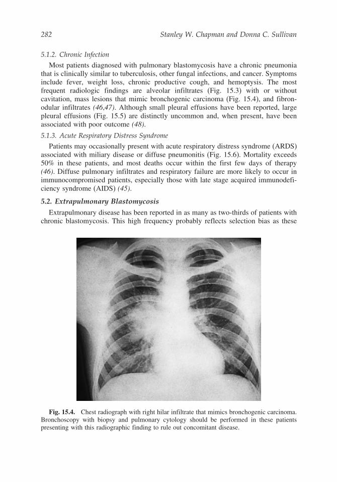
282 Stanley W. Chapman and Donna C. Sullivan
5.1.2. Chronic Infection
Most patients diagnosed with pulmonary blastomycosis have a chronic pneumoniathat is clinically similar to tuberculosis, other fungal infections, and cancer. Symptomsinclude fever, weight loss, chronic productive cough, and hemoptysis. The mostfrequent radiologic findings are alveolar infiltrates (Fig. 15.3) with or withoutcavitation, mass lesions that mimic bronchogenic carcinoma (Fig. 15.4), and fibron-odular infiltrates (46,47). Although small pleural effusions have been reported, largepleural effusions (Fig. 15.5) are distinctly uncommon and, when present, have beenassociated with poor outcome (48).
5.1.3. Acute Respiratory Distress Syndrome
Patients may occasionally present with acute respiratory distress syndrome (ARDS)associated with miliary disease or diffuse pneumonitis (Fig. 15.6). Mortality exceeds50% in these patients, and most deaths occur within the first few days of therapy(46). Diffuse pulmonary infiltrates and respiratory failure are more likely to occur inimmunocompromised patients, especially those with late stage acquired immunodefi-ciency syndrome (AIDS) (45).
5.2. Extrapulmonary Blastomycosis
Extrapulmonary disease has been reported in as many as two-thirds of patients withchronic blastomycosis. This high frequency probably reflects selection bias as these
Fig. 15.4. Chest radiograph with right hilar infiltrate that mimics bronchogenic carcinoma.Bronchoscopy with biopsy and pulmonary cytology should be performed in these patientspresenting with this radiographic finding to rule out concomitant disease.

15. Blastomycosis 283
Fig. 15.5. Patient with life-threatening pulmonary disease whose chest radiograph revealsbilateral alveolar infiltrates and large left-sided pleural effusion.
figures were reported in earlier studies before effective therapy was available and wereautopsy based (6). More recent studies have documented extrapulmonary disease inonly 25 to 40 percent of patients with blastomycosis (15,47). Extrapulmonary diseaseis usually seen in conjunction with active pulmonary disease.
5.2.1. Skin Disease
Skin disease is the most common extrapulmonary manifestation of blastomycosis.Two types of skin lesions occur, verrucous and ulcerative (Fig. 15.7). The verrucouslesion is most common, typically with well-demarcated borders and color from gray toviolaceous hues. These lesions often mimic squamous cell carcinoma. Microabscessesdevelop at the periphery of these lesions, and specimen samples taken from the marginsusually reveal the diagnostic yeast form on wet preparation (Fig. 15.8). The second typeof lesion is ulcerative. These ulcers, which bleed easily, usually have well-demarcated,

284 Stanley W. Chapman and Donna C. Sullivan
Fig. 15.6. Diffuse pulmonary infiltrates in a patient with ARDS. Patients presenting withthis syndrome have a mortality rate greater than 50%.
heaped up borders. The ulcers of blastomycosis develop from subcutaneous pustularlesions that spontaneously drain.
Regional lymphadenopathy is usually not present in cases of pulmonary dissem-ination. Patients with inoculation blastomycosis occurring after dog bite or autopsyaccidents often have lymphadenopathy/adenitis as a prominent feature (49,50). Lesionsmay also appear on the mucosa of the nose, mouth, and larynx. Laryngeal blastomycosismimics well differentiated squamous cell carcinoma both clinically and histopatholog-
Fig. 15.7. Cutaneous blastomycosis typically produces verrucous (left) or ulcerative (right)lesions. [Figure in color on CD-ROM].

15. Blastomycosis 285
Fig. 15.8. Diagnosis of blastomycosis. This figure shows the characteristic yeast forms ina wet preparation of a skin scraping. Scraping of the edges of the verrucous and ulcerativelesions yield the best diagnostic results. [Figure in color on CD-ROM].
ically (35). Subcutaneous nodules or cold abscesses are usually seen in patients withmultiorgan disease.
5.2.2. Osseous
Osteomyelitis is associated with as many as one-fourth of B. dermatitidis infections(51). The vertebrae, pelvis, sacrum, skull, ribs, or long bones are most frequentlyinvolved, although any bone may be infected. Granuloma, suppuration, or necrosismay be observed in biopsy specimens. A well-circumscribed osteolytic lesion may beobserved on x-ray examination, although such lesions cannot be distinguished fromother fungal, bacterial, or neoplastic disease. Patients with B. dermatitidis osteomyelitisusually present with contiguous soft tissue abscesses or chronic draining sinuses.Although most bone lesions resolve with prolonged antifungal therapy, some mayrequire surgical débridement for cure.
5.2.3. Genitourinary
In men, 10% to 30% of blastomycosis involves the genitourinary tract, primarilythe prostate and epididymis (52). Prostatic involvement is frequently associated withsymptoms of obstruction, an enlarged tender prostate, and pyuria. Urine culturesobtained following prostate massage are frequently positive.
5.2.4. Central Nervous System
Central nervous system (CNS) involvement normally occurs in fewer than 5% ofcases in immunocompetent patients. Persons with AIDS have been reported to have
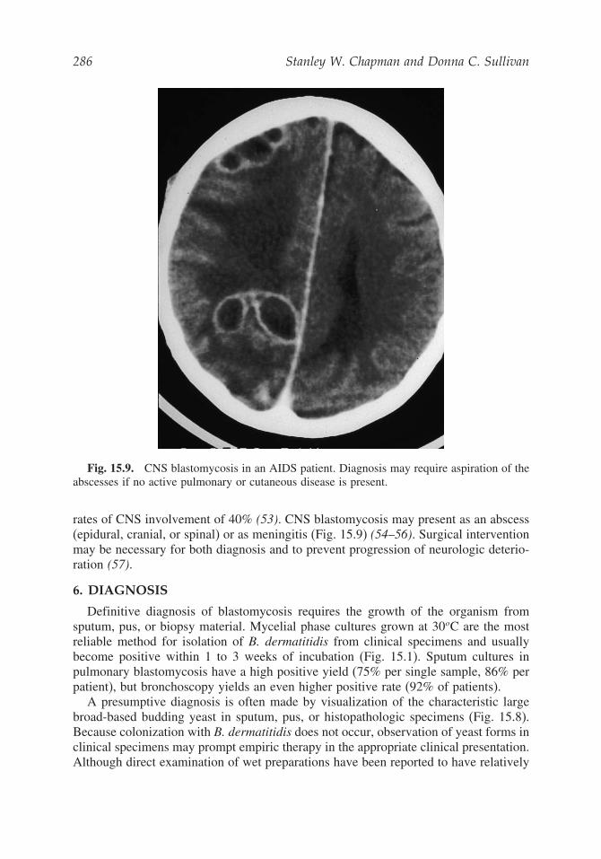
286 Stanley W. Chapman and Donna C. Sullivan
Fig. 15.9. CNS blastomycosis in an AIDS patient. Diagnosis may require aspiration of theabscesses if no active pulmonary or cutaneous disease is present.
rates of CNS involvement of 40% (53). CNS blastomycosis may present as an abscess(epidural, cranial, or spinal) or as meningitis (Fig. 15.9) (54–56). Surgical interventionmay be necessary for both diagnosis and to prevent progression of neurologic deterio-ration (57).
6. DIAGNOSIS
Definitive diagnosis of blastomycosis requires the growth of the organism fromsputum, pus, or biopsy material. Mycelial phase cultures grown at 30oC are the mostreliable method for isolation of B. dermatitidis from clinical specimens and usuallybecome positive within 1 to 3 weeks of incubation (Fig. 15.1). Sputum cultures inpulmonary blastomycosis have a high positive yield (75% per single sample, 86% perpatient), but bronchoscopy yields an even higher positive rate (92% of patients).
A presumptive diagnosis is often made by visualization of the characteristic largebroad-based budding yeast in sputum, pus, or histopathologic specimens (Fig. 15.8).Because colonization with B. dermatitidis does not occur, observation of yeast forms inclinical specimens may prompt empiric therapy in the appropriate clinical presentation.Although direct examination of wet preparations have been reported to have relatively

15. Blastomycosis 287
low diagnostic yield (58), the simplicity of the procedure, low cost, and potential forrapid diagnosis warrant the use of this method. Cytology has been shown to have ahigher diagnostic yield (59).
Serologic diagnosis of blastomycosis is of limited usefulness. Complement fixationantibodies have been used for epidemiologic purposes, but are severely limited intheir specificity because of cross-reactivity to antigens of other fungi, particularlyHistoplasma capsulatum and Coccidioides immitis. Immunodiffusion tests for precip-itating antibodies to B. dermatitidis are more specific than complement fixationbut lack sensitivity in early disease (60). Commercial radioimmunoassays, enzymeimmunoassays, and enzyme-linked immunosorbent assays have been developed whichoffer the promise of higher sensitivity but with specificities similar to complementfixation (61,62). Immunoassays employing the BAD-1 yeast phase specific proteindiscussed in the preceding text are not currently commercially available.
A commercially available Blastomyces assay that detects antigen in urineand serum has been developed by MiraVista Diagnostics (Indianapolis, IN;www.miravistalabs.com). Antigenemia is detected in 70% to 80% of patientswith disseminated disease. Antigen detection in the urine was higher than serum,approaching 100%. However, specificity is reduced by the presence of cross-reactiveantigens present in specimens obtained from patients with other fungal infections,especially histoplasmosis. Antigen levels are reported to decline with successfultreatment and increase in treatment failure or relapse of disease.
Nucleic acid detection techniques, both target and signal amplification methods,have been developed (63–65), including the GEN-PROBE® AccuProbe® culture identi-fication test. These facilitate early identification of B. dermatitidis in mycelial cultureswithout the requirement of conversion to the yeast form. PCR amplification of therRNA gene along with specific probe hybridization has been used to identify yeastphase organisms in tissue specimens. These molecular techniques offer great promisefor the rapid diagnosis of blastomycosis, but have not yet been evaluated in largeprospective studies.
7. TREATMENT
Most patients who have blastomycosis require therapy. Before the availability ofazoles, amphotericin B was the mainstay of treatment. However, a series of clinicaltrials performed by the NIAID Mycoses Study Group have shown ketoconazole,itraconazole, and fluconazole to be effective, relatively nontoxic agents when comparedto amphotericin B for treatment of patients with mild to moderate non-CNS disease(66–68). Other than its use in diagnosis, the role of surgery is limited. Along withspecific antifungal therapy, surgery may be helpful for the drainage of large abscesses,resection of cerebral blastomycomas, and débridement of devitalized bone.
Selection of an appropriate therapeutic regimen must be based on three majorconsiderations: the clinical form and severity of the disease, the immune status of thepatient, and the toxicity of the antifungal agent. Specific recommendations of dose andduration of therapy in defined clinical settings are listed in Table 15.2 (69,70). For moredetail concerning drug interactions, toxicities, adverse reactions and pharmacokinetics

288 Stanley W. Chapman and Donna C. Sullivan
Table 15.2Treatment guidelines for Blastomyces dermatitidis infections
Type of disease Primary therapy Alternate therapy
PulmonarySevere Amphotericin B
0.7–1.0 mg/kgper day;a totaldose of1.5–2.5 g
Switch to itraconazole 200–400 mg/day oncepatient stabilized
Mild to moderate Itraconazole200–400mg/day
Ketoconazole 400–800 mg/day, or fluconazole400–800 mg/day
DisseminatedCNS Amphotericin
B, 0.7–1.0mg/kg perday; total doseof at least 2 g
If intolerant to full course of amphotericin B,fluconazole 800 mg/day
Non-CNS diseaseSerious Amphotericin B
0.5–0.7 mg/kgper day; totaldose of1.5–2.5 g
Switch to itraconazole 200–400 mg/day oncepatient stabilized
Mild to moderate Itraconazole200–400mg/day
Ketoconazole 400–800 mg/day, or fluconazole400–800 mg/day
Immunocompromised Amphotericin B0.3–0.6 mg/kgper day;a totaldose of1.5–2.5 g
Selected patients with non-CNS disease maybe switched to itraconazole, 200–400mg/day, once clinically improved.Suppressive therapy with itraconazoleshould be considered in patients whoseimmunocompromised state continues. Forpatients with CNS disease or intolerant toitraconazole, consider fluconazole, 800mg/day
Modified from ref. 69.aA lipid formulation of amphotericin B (3.0–5.0 mg/kg per day) may be substituted for conventionalamphotericin B.
about individual antifungal agents, the reader is referred to the chapter discussingindividual agents in this text (Chapter 6).
Amphotericin B (including lipid-based formulations) is currently reserved forthe initial treatment of patients with life-threatening disease, immunocompromisedpatients, and those with CNS disease. In selected patients initially presenting with

15. Blastomycosis 289
life-threatening disease, itraconazole has been successfully substituted following aninduction course of amphotericin B (70). Patients with CNS disease and severelyimmunocompromised patients should be treated with a full course of amphotericin B.Most experts recommend a total dose of 1.5 to 2.5 grams.
Fluconazole has been used in only a limited number of patients, but appears effica-cious at doses of 400 to 800 mg/day. Two factors may eventually lead to moreextensive use of fluconazole: it has fewer side effects and adverse drug interactionsand it has excellent penetration into the CNS, suggesting a role for this drug in thetreatment of CNS blastomycosis. Voriconazole and posaconazole have activity againstB. dermatitidis in vitro and have been shown to be effective in murine models ofblastomycosis (71,72). The authors are aware of anecdotal cases of patients failingitraconazole therapy, including cases of cerebral blastomycosis, successfully treatedwith voriconazole as salvage therapy. Caspofungin and micafungin have shown variableactivity against B. dermatitidis and have not been studied extensively in animal modelsor in human cases of blastomycosis (72).
In some immunocompetent patients with acute pulmonary blastomycosis, thedifficult and controversial decision to withhold therapy has been proposed following thedescription of self-limiting infections. When the decision to withhold therapy is made,patients should be evaluated for the presence of extrapulmonary disease and carefullymonitored for progression of pulmonary infection. These patients should be followedfor many years for evidence of reactivation of pulmonary or extrapulmonary disease.
In contrast, all patients with progressive pulmonary infection or extrapulmonarydisease and all immunocompromised patients should be treated (69,70). In immuno-logically normal patients with mild to moderate pulmonary or extrapulmonary diseasethat does not involve the CNS, the azole antifungal agents, itraconazole, ketoconazole,and fluconazole, administered for 6 months have proven to be effective, less toxicalternatives to amphotericin B. Although no randomized, blinded studies have beenperformed to compare different azoles, and only a few comparative trials for blasto-mycosis therapy have been reported, itraconazole appears to be the best tolerated andmost effective azole. Itraconazole is considered the drug of choice for patients withnon-life-threatening, non-CNS blastomycosis (69,70).
Amphotericin B is the drug of choice for blastomycosis occurring during pregnancy.Azoles are contraindicated (69). The clinical spectrum of blastomycosis in pediatricpatients is similar to that seen in adults. Recent reports indicate blastomycosis inchildren is more difficult to diagnosis and less likely to respond to oral therapy. Childrenwith life-threatening disease should be treated with amphotericin B. Itraconazole hasbeen used successfully at a dosage of 5 to 7 mg/kg per day in a small cohort of pediatricpatients (69). CNS disease occurs in approximately 40% of patients with AIDS orother diseases or therapies associated with immunosuppression. Likewise, disseminateddisease and life-threatening pulmonary disease also appear more common in the clinicalsetting of immunosuppression. Hence, the recommendation that amphotericin B is thedrug of choice for treatment of immunocompromised patients. Frequent relapses havebeen reported in patients whose immunosuppression persists and chronic suppressivetherapy with an oral azole has been recommended by some experts (69,70).

290 Stanley W. Chapman and Donna C. Sullivan
Relapse rates of 10% to 14% have been reported in patients treated withketoconazole. Patients should be followed for many years for evidence of relapse,especially in the CNS. Relapse rates of less than 5% are reported in patients treatedwith amphotericin B and itraconazole. Owing to the problems with bioavailability oforal itraconazole and ketoconazole, serum blood levels may be clinically useful inguiding treatment of patients whose disease progresses on either of these azoles (69).
REFERENCES
1. Gilchrist TC. Protozoan dermatitis. J Cutan Gen Dis 1894;12:496–499.2. Schawartz J, BaumGL. Blastomycosis. Am J Clin Pathol 1951;21:999–1029.3. Bradsher RW. Clinical features of pulmonary blastomycosis. Semin Respir Infect
1997;12:229–234.4. DiSalvo AF. The epidemiology of Blastomyces dermatitidis. In: Al-Doory Y, DiSalvo AF,
eds. Blastomycosis. New York: Plenum Press, 1992:83–90.5. DiSalvo AF. The ecology of Blastomyces dermatitidis. In: Al-Doory Y, DiSalvo AF, eds.
Blastomycosis. New York: Plenum Press, 1992:43–69.6. Chapman SW. Blastomyces dermatitidis. In: Mandell GL, Bennett JE, Dolin R, eds.
Mandell, Douglas, and Bennett’s principles and practices of infectious diseases. 6th ed.Philadelphia: Elsevier Churchill Livingstone, 2005:3026–3040.
7. Kaufman L, Standard PG, Weeks RJ, et al. Detection of two Blastomyces dermatitidisserotypes by exoantigen analysis. J Clin Microbiol 1983;18:110–114.
8. Turner S, Kaufman L. Immunodiagnosis of blastomycosis. Semin Respir Infect1986;1:22–28.
9. Axtell RC, Scalarone GM. Serological differences in two Blastomyces dermatitidis isolatesfrom different geographic regions of North America. Mycopathologia 2002;153:141–144.
10. Yates-Ciilata KE, Sander DM, Keith EJ. Genetic diversity in clinical isolates of thedimorphic fungus Blastomyces dermatitidis detected by PCR based random amplifiedpolymorphic DNA assay. J Clin Microbiol 1995;33:2171–2175.
11. McCullough MJ, DiSalvo AF, Clemons KV, Park P, Stevens DA. Molecular epidemiologyof Blastomyces dermatitidis. Clin Infect Dis 2000;30:328–335.
12. Klein BS, Vergeront JM, Weeks RJ, et al. Isolation of Blastomyces dermatitidis insoil associated with a large outbreak of blastomycosis in Wisconsin. N Engl J Med1986;314:529–534.
13. Klein BS, Vergeront JM, DiSalvo AF, et al. Two outbreaks of blastomycosis along riversin Wisconsin: Isolation of Blastomyces dermatitidis from riverbank soil and evidence ofits transmission along waterways. Am Rev Respir Dis 1987;136:1333–1338.
14. Klein BS, Vergeront JM, Davis JP. Epidemiologic aspects of blastomycosis, the enigmaticsystemic mycosis. Semin Respir Infect 1986;1:29–39.
15. Proctor ME, Klein BS, Jones JM, Davis JP. Cluster of pulmonary blastomycosis in arural community: evidence for multiple high-risk environmental foci following a sustainedperiod of diminished precipitation. Mycopathologia 2002;153:113–120.
16. Bumgardner DM, Buggy BP, Mattson BJ, Ludwig D. Epidemiology of blastomycosis in aregion of high endemicity of north central Wisconsin. Clin Infect Dis 1992;15:629–635.
17. Baumgardner DJ, Paretsky DP, Yopp AC. The epidemiology of blastomycosis in dogs:north central Wisconsin, USA. J Med Vet Mycol 1995;33:171–176.
18. Legendre AM. Blastomycosis in animals. In: Al-Doory Y, DiSalvo AF, eds. Blastomycosis.New York, NY: Plenum Medical Books, 1992:249–266.
19. DiSalvo AF, Denton JF. Lipid content of four strains of Blastomyces dermatitidis ofdifferent mouse virulence. J Bacteriol 1963;85:927–931.

15. Blastomycosis 291
20. Cox RA, Best GK. Cell Wall composition of two strains of Blastomyces dermatitidisexhibiting differences in virulence for mice. Infect Immunol 1972;5:449–453.
21. Rooney PJ, Sullivan TD, Klein BS. Selective expression of the virulence factorBAD1 upon morphogenesis to the pathogenic yeast form of Blastomyces dermati-tidis: evidence for transcriptional regulation by a conserved mechanism. Mol Microbiol2001;39:875–889.
22. Brandhorst TT, Wuthrich M, Warner T, Klein B. Targeted gene disruption revealsan adhesin indispensable for pathogenicity of Blastomyces dermatitidis. J Exp Med1999;189:1207–1216.
23. Newman SL, Chaturvedi S, Klein BS. The WI-1 antigen of Blastomyces dermatitidisyeast mediates binding to human macrophage CD11b/CD18(CR3) and CD14. J Immunol1995;154:753–761.
24. Hogan LH, Josvai S, Klein BS. Genomic cloning, characterization and functional analysisof the major surface adhesion WI-1 on Blastomyces dermatitidis yeast. J Biol Chem1995;270:30725–30732.
25. Klein BS, Sondel PM, Jones JM. WI-1, a novel 120 kilodalton surface protein on Blasto-myces dermatitidis yeast cells, is a target antigen of cell-medicated immunity in humanblastomycosis. Infect Immunol 1992;60:4291–4300.
26. Bradsher RW. Live Blastomyces dermatitidis yeast induced responses of immune andnon-immune human mononuclear cells. Mycopathologia 1984;87:159–166.
27. Klein BS, Bradsher RW, Vergeront JM, et al. Development of long-term specific cellularimmunity after acute Blastomyces dermatitidis infection: assessments following a largepoint-source outbreak in Wisconsin. J Infect Dis 1990;161:97–101.
28. Cozad GC. Experimental blastomycosis. In Al-Doorey Y, DiSalvo AF, eds. Blastomycosis.New York: Plenum Press, 1992:221–236.
29. Cozad GC, Change CT. Cell mediated immunoprotection in blastomycosis. Infect Immun1980;28:398–403.
30. Wuthrich M, Filutowicz HI, Klein BS. Mutation of the WI-1 gene yields an atten-uated Blastomyces dermatitidis strain that induces host resistance. J Clin Invest 2000;106:1381–1389.
31. Wuthrich M, Filutowicz Warner T, Klein BS. Requisite elements in vaccine immunity toBlastomyces dermatitidis: plasticity uncovers vaccine potential in immune-deficient hosts.J Immunol 2002;169:6969–6976.
32. Wuthrich M, Filutowicz HI, Warner T, Deepe GS, Klein BS. Vaccine immunity topathogenic fungi overcomes the requirements for CD4 help in exogenous antigen presen-tation to CD8+ T cells: implications for vaccine development in immune-deficient hosts.J Exp Med 2003;197:1405–1416.
33. Wuthrich M, Warner T, Klein BS. CD28 is required for optimal induction, but notmaintenance, of vaccine-induced immunity to Blastomyces dermatitidis. Infect Immun2005;73:7436–7441.
34. Wuthrich M, Warner T, Klein BS. IL-12 is required for induction but not maintenanceof protective, memory responses to Blastomyces dermatitidis: implications for vaccinedevelopment in immune-deficient hosts. J Immunol 2005;175:5288–5297.
35. Bradsher RW, Chapman SW, Pappas PG. Blastomycosis. Infect Dis Clin N Am2003;17:21–40.
36. Cherniss EI, Waisbren BA. North American blastomycosis: a clinical study of 40 cases.Ann Intern Med 1956;44:169–123.
37. Abernathy RS. Clinical manifestations of pulmonary blastomycosis. Ann Intern Med1959;51:707–727.

292 Stanley W. Chapman and Donna C. Sullivan
38. Blastomycosis Cooperative Study of the Veterans Administration. Blastomycosis I: areview of 198 collected cases in Veterans Administration hospitals. Am Rev Respir Dis1964;89:659–672.
39. Witorsch P, Utz JP. North American blastomycosis: a study of 40 patients. Medicine(Baltimore) 1968;47:169–200.
40. Lockwood WR, Allison F, Batson BE, et al. The treatment of North American blastomy-cosis: ten years experience. Am Rev Respir Dis 1969;100:314–320.
41. Duttera MJ, Osterhout S. North American blastomycosis: a survey of 63 cases. South MedJ 1969;62:295–301.
42. Sheflin JR, Campbell JA, Thompson GP. Pulmonary blastomycosis: findings on chestradiographs in 63 patients. AJR Am J Roentgenol 1990;154:1177–1180.
43. Brown LR, Sweasen SJ, VanScovy RE, et al. Roentgenologic features of pulmonaryblastomycosis. Mayo Clin Proc 1991;66:29–38.
44. Sarosi GA, Davies SF, Phillips JR. Self-limited blastomycosis: a report of 39 cases. SeminRespir Infect 1986;1:40–44.
45. Sarosi GA, Hammerman KJ, Tosh FE, et al. Clinical features of acute pulmonary blasto-mycosis. N Engl J Med 1974;290:540–543.
46. Meyer KC, McManus EJ, Maki DG. Overwhelming pulmonary blastomycosis associatedwith the adult respiratory distress syndrome. J Engl J Med 1993;329:1231–1236.
47. Chapman SW, Lin AC, Hendricks DA, et al. Endemic blastomycosis in Mississippi:epidemiological and clinical studies. Semin Respir Infect 1997;12:219–228.
48. Kinasewitz GT, Penn RL, George RB. The spectrum and significance of pleural disease inblastomycosis. Chest 1984;86:580–584.
49. Larson DM, Eckman MR, Alber RL, et al. Primary cutaneous (inoculation) blastomycosis:an occupational hazard to pathologists. Am J Clin Pathol 1983;79:253–255.
50. Gnann JW, Bressler GS, Bodet CA, et al. Human blastomycosis after a dog bit. Ann InternMed 1983;98:48–49.
51. Moore RM, Green NE. Blastomycosis of bone. J Bone Joint Surg 1982;64:1094–1101.52. Sarosi GA, Davies SF. Blastomycosis: state of the art. Am Rev Respir Dis
1979;120:911–938.53. Pappas PG, Pottage JC, Powderly WG, et al. Blastomycosis in patients with acquired
immunodeficiency syndrome. Ann Intern Med 1992;116:847–853.54. Gonyea EF. The spectrum of primary blastomycotic meningitis: a review of central nervous
system blastomycosis. Ann Neurol 1978;3:26–39.55. Kravitz GR, Davies SF, Eckman MR, et al. Chronic blastomycotic meningitis. Am J Med
1981;71:501–505.56. Roos KL, Bryan JP, Maggio WW, et al. Intracranial blastomycoma. Medicine (Baltimore)
1987;66:224–235.57. Ward BA, Parent AD, Raila F. Indications for the surgical management of central nervous
system blastomycosis. Surg Neurol 1995;43:379–388.58. Martynowicz MA, Prakash UBS. Pulmonary blastomycosis: an appraisal of diagnostic
techniques. Chest 2002;121:768–773.59. Lemos LB, Guo M, Baliga M. Blastomycosis: organ involvement and etiologic diagnosis.
A review of 123 patients from Mississippi. Ann Diagn Pathol 2000;4:391–406.60. Klein BS, Vergeront JM, Kaufman L, et al. Serological test for blastomycosis: assess-
ments during a large point-source outbreak in Wisconsin. J Infect Dis 1987;155:262–268.
61. Bradsher RW, Pappas PG. Detection of specific antibodies in human blastomycosis byenzyme immunoassay. South Med J 1995;88:1256–1259.

15. Blastomycosis 293
62. Sekhorn AS, Kaufman L, Kobayashi AS, et al. The value of the Premier enzymeimmunoassay for diagnosing Blastomyces dermatitidis infections. J Med Vet Mycol1995;33:123–125.
63. Yeo SF, Wong B. Current status of nonculture methods for diagnosis of invasive fungalinfections. Clin Microbiol Rev 2002;15:465–484.
64. Walsh TJ, Larone DH, Schell WA, Mitchell TG. Histoplasma, Blastomyces, Coccidioides,and other dimorphic fungi causing systemic mycoses. In: Murray PR, Barron EJ, JorgensenJH, Pfaller MA, Yolken RH, eds. Manual of clinical microbiology. 8th Ed. Washington,DC: ASM Press, 2003:1781–1797.
65. Lindsey MD, Hurst SF, Iqbal NJ, Morrison CJ. Rapid identification of dimorphic and yeast-like fungal pathogens using specific DNA probes. J Clin Microbiol 2001;39:3505–3511.
66. Treatment of blastomycosis and histoplasmosis with ketoconazole. Results of a prospectiverandomized clinical trial. National Institute of Allergy and Infectious Diseases MycosesStudy Group. Ann Intern Med 1985;103:861–872.
67. Dismukes WE, Bradsher RW, Cloud GC, et al. Itraconazole therapy for blastomycosis andhistoplasmosis. Am J Med 1992;93:489–497.
68. Pappas PG, Bradsher RW, Kauffman CA, et al. Treatment of blastomycosis with higherdose fluconazole. Clin Infect Dis 1997;25:200–205.
69. Chapman SW, Bradsher RW, Campbell GD, Pappas PG, Kauffman CA. Practice guidelinesfor the management of patients with blastomycosis. Clin Infect Dis 2000;30:679–683.
70. Bradsher RW. Histoplasmosis and blastomycosis. Clin Infect Dis 1996;22:S102–S111.71. Sugar AM, Liu XP. Efficacy of voriconazole in treatment of murine pulmonary blastomy-
cosis. Antimicrob Agents Chemother 2001;45:601–604.72. Espinel-Ingroff A. Comparison of in vitro activities of the new triazole SCH56592 and
the echinocandins MK-0991 (L-743,872) and LY303366 against opportunistic filamentousdimorphic fungi and yeasts. J Clin Microbiol 1998;36:2950–2956.
SUGGESTED READINGS
Bradsher RW. Blastomycosis. In: Dismukes WE, Pappas PG, Sobel JD, eds. Clinical mycology.New York: Oxford University Press, 2003:299–310.
Bradsher RW, Chapman SW, Pappas PG. Blastomycosis. Infect Dis Clin N Am 2003;17:21–40.Chapman SW. Blastomyces dermatitidis. In: Mandell GL, Bennett JE, Dolin R, eds. Mandell,
Douglas, and Bennett’s principles and practices of infectious diseases. 6th Ed. Philadelphia:Elsevier Churchill Livingstone, 2005:3026–3040.

16Coccidioidomycosis
Royce H. Johnson, MD and Shehla Baqi, MD
1. INTRODUCTION
Coccidioidomycosis was first described in 1892, in Buenos Aires by Posadas andWernicke (1,2). They thought that the individual in their case report suffered from amalignant disease with a likely infectious cause. Organisms seen microscopically weremistakenly thought to be parasites. The disease was next described in San Franciscoin 1896 by Rixford and Gilchrist, whose paper was the first extensive study of coccid-ioidomycosis (3). They better understood that this was an infectious illness and werethe first to appreciate the importance of the parasite as the agent of a new and distinctivedisease. In 1900, Williams Ophuls began his work on coccidioidal disease. Althoughhe noted the “protozoa” of Rixford and Gilchrist in pathological sections, he discoveredthat culture of the organism always produced colonies of a mould, what we now know tobe the mycelial (saprobic) growth of Coccidioides. The life cycle was roughly outlinedin a preliminary report and the fungus given the name of Coccidioides immitis (4).During 1925–1936, the early pathologic, epidemiologic, and mycologic studies werecompleted. Montenegro reported the first recovery of Coccidioides immitis fromblood (5). Coccidioidal infection in farm animals was described by Beck (6). Menin-gitis was described first pathologically and subsequently clinically in the early part ofthe 20th century (7,8). Two important observations were also made during this period;that the lung is the portal of entry and that Coccidioides immitis can be isolated fromsoil (9,10).
Coccidioidomycosis was considered to be a rare and fatal infection until an accidentallaboratory exposure of a medical student at Stanford University resulted in only atransient pulmonary infection. This led to a reassessment of the natural history ofcoccidioidal infection. The work in Kern County by Dr. Myrnie Gifford on a local respi-ratory illness in the San Joaquin Valley of California, known as Valley Fever, eventuallyelucidated the primary infection as being predominantly pulmonary (11). During thelatter part of the 1930s and 1940s, the natural history of the primary illness and theutility of the skin test and serology were developed by Charles E. Smith and co-workers.William Winn and Hans Einstein made further contributions to disease descriptionand therapy with amphotericin B deoxycholate, both intravenously and intrathecally
From: Infectious Disease: Diagnosis and Treatment of Human MycosesEdited by: D. R. Hospenthal and M. G. Rinaldi © Humana Press Inc., Totowa, NJ
295

296 Royce H. Johnson and Shehla Baqi
for meningitis. By the 1950s, the clinical spectrum of coccidioidal infection was welldescribed, with the publication of an excellent monograph by Fiese (12).
2. ETIOLOGIC AGENTS
Kingdom: FungiPhylum: Ascomycota
Class: EuascomycetesOrder: Onygenales
Family: OnygenaceaeGenus: Coccidioides
Coccidioides was originally described as noted above as one species, C. immitis. Morerecently, two genetically distinct populations of Coccidioides have been described,Coccidioides immitis and Coccidioides posadasii, correlating to separate endemicregions. Currently the C. immitis is maintained as the name of isolates that are
Fig. 16.1. Life cycle of Coccidioides immitis depicting saprophytic (soil) and parasitic(host) phases.

16. Coccidioidomycosis 297
predominately found in California. The new species, C. posadasii, is predominatelyfound in Texas, Mexico, Central America, and South America. Both species arefound in Arizona (13). Coccidioides of both species, however, show few phenotypicdifferences and are mycologically and clinically indistinguishable. Coccidioides is athermal dimorphic fungus that exists either as a mycelium or a spherule. The fungusis found as far North as the northern central valley in California and as far Southas Argentina, the place of its original description. The fungus grows in conditionswhere the soil has a relatively high salinity, and in a climate that has mild winterswith few freezes and hot dry summers (14). Under these ideal conditions, the fungusgrows in isolated pockets as a mycelium by apical extension. These mycelia producespecialized aerial hyphae that segment and form arthroconidia. The connecting linksbetween arthroconidia are quite fragile and separate easily with minimal mechanicalforce or air turbulence. The arthroconidia become airborne in a form capable ofdeposition in the lungs if inhaled, and can travel substantial distances, perhaps asfar as 75 miles or more. These arthroconidia, if they find an appropriate soil niche,can reestablish the saprophytic phase. However, if they are inhaled by an appro-priate host, they undergo transformation from arthroconidia into spherules. Spherulesreproduce by endosporulation, a process whereby the growing spherule is subdividedinto numerous subcompartments, each of which become viable daughter cells orendospores. The spherule eventually ruptures, releasing endospores, each of which maycontinue to propagate in tissue or revert to mycelial growth in soil or on growth media(Fig. 16.1).
3. EPIDEMIOLOGY
The disease was first described in Argentina, but other foci of infection in SouthAmerica and Central America also exist. Coccidioides species are found solely in theWestern Hemisphere, in the “lower Sonoran life zone” (15). The majority of the soilsthat support the organism are found in North America, particularly in the southwesternUnited States and northwestern Mexico. The areas of greatest endemicity are in theSouthern San Joaquin Valley and south central Arizona. The disease extends to theNorthern Central Valley in California and as far as Utah in the Great Plains (16). Thetotal number of infections per year is not known, but prevalence surveys in the 1950s ofschool-age children in California’s central valley suggested an annual risk of infectionof 15%. More recent estimates from California and Arizona have indicated that the riskhas declined to 3% or less (17,18). It has been estimated that in the United States thereare approximately 100,000 infections annually (19). There has been a recent increasein the reported number of patients per year that is believed to be due to changes indemographics and medicine. Regions in which coccidioidomycosis is endemic haveexperienced a tremendous increase in their population. In 2005, it was estimated thepopulations of southern Arizona and southern California increased by greater than 7million inhabitants. In addition, there is greater recognition of the disease in patientswith compromised cellular immunity and in the elderly, who are more likely to havemore severe disease. It appears, however, that the absolute incidence of disease hasalso increased, particularly during the epidemics described in the 1990s and in the first2 years of the new millennium (20–23).

298 Royce H. Johnson and Shehla Baqi
4. PATHOGENESIS AND IMMUNOLOGY
Virtually all infections result from inhalation of arthroconidia. Inhaled arthroconidiatransform into spherules, an inflammatory response ensues, and a local pulmonarylesion develops. In some infections, the Coccidioides species gain access to the vascularspace, leaving the lungs and disseminating to other parts of the body. Control ofcoccidioidomycosis is predominantly cell mediated, with more severe infections seenin T-cell deficient patients (24–26). In addition, in vitro observations have shown thatinnate cellular responses, mediated by mononuclear cells or natural killer cells, mayslow fungal proliferation after infection (27). It is conceivable that interleukin (IL)-12,IL-23, and interferon gamma may play an important role in protective immunity incoccidioidomycosis as recently demonstrated in paracoccidioidomycosis and histoplas-mosis (28,29).
5. CLINICAL MANIFESTATIONS
5.1. Pulmonary
Coccidioidomycosis most commonly presents as primary pulmonary disease. Thefirst symptoms of primary infection usually appear 7 to 21 days after infection, althoughinfection is asymptomatic 60% of the time. In patients presenting with symptomaticdisease, the majority present with an influenza-like syndrome. Of the 40% of totalinfections with symptoms, only one out of four is diagnosed; the majority of thesepresent with pneumonic or pleural disease. There are a number of pulmonary compli-cations of primary coccidioidomycosis. The most common is severe and persistentpneumonia, with radiographic and clinical findings of pneumonic disease for greaterthan 6 weeks. Progressive primary coccidioidomycosis is a syndrome in which thepatient has resolution of his or her pulmonary parenchymal disease with persistenceof his or her hilar and mediastinal lymphadenitis. Rare cases of progressive fibro-cavitary coccidioidomycosis, which often resembles pulmonary tuberculosis, are alsodescribed. Solitary thin-walled pulmonary cavities are a frequent complication (seeFig. 5.11, Chapter 5). Residual nodules are often confused with a neoplasm, partic-ularly when individuals with unrecognized primary coccidioidomycosis present witha residual nodule on routine chest radiograph long after the time of infection (30).Modest amounts of pleural fibrosis, a residual of the primary infection, may alsobe seen. Cavitary disease may rupture into the pleural space causing coccidioidalempyema, not to be confused with simple pleural effusions which may occur aspart of the primary disease process. Symptoms prevalent in primary coccidioidomy-cosis include fever (76%), cough (73%), chest pain (44%), fatigue (38%), erythemanodosum (26 %), myalgias (23%), shortness of breath (22%), sputum production(22%), chills (21%), headache (21%), night sweats (21%), and other rashes (14%) (31).Radiographic findings in primary coccidioidomycosis typically include infiltrate only(70%), infiltrate with hilar adenopathy (10%), or infiltrate with effusion (10%)(Figs. 16.2 through 16.5). Lung cavities are present in about 8% of adults, but areless common in children. Approximately 10% of individuals will have a negativechest radiograph at diagnosis. Skin manifestations develop as part of the primaryillness, most often as a transient nonpruritic fine papular rash. Erythema nodosum is

16. Coccidioidomycosis 299
Fig. 16.2. Right upper lobe dense consolidation with associated right hilar adenopathy.
fairly common in primary coccidioidal infection, with a strong predilection for women(Fig. 16.6). Less commonly, erythema multiforme and erythema sweetobullosum areseen (32,33). Migratory arthralgias are common; the triad of fever, erythema nodosum,and arthralgias has been referred to as “desert rheumatism.” Rarely, pulmonary coccid-ioidomycosis may present as a bronchial mass found on bronchoscopy. Chronicfibrocavitary pneumonia can occur, commonly in association with diabetes or preex-isting pulmonary fibrosis (34). Miliary disease with coccidioidomycosis is seen withsignificant frequency in the endemic area, where miliary coccidioidomycosis may be10 times as frequent as miliary tuberculosis. Overwhelming miliary and/or alveolarcoccidioidomycosis can result in respiratory failure. Human immunodeficiency virus(HIV)-infected patients often have a fulminant presentation, particularly when CD4+ Tlymphocyte counts are less than 100 cells/μl. Probably the most common cause of deathin the endemic area from pulmonary coccidioidomycosis is respiratory failure, althoughmost coccidioidal respiratory infections resolve within several weeks to months withoutcomplications.
5.2. Disseminated Disease
The rate of dissemination of coccidioidomycosis is highly dependent on the infectedhost. The majority of disseminated disease occurs in individuals with antecedentsymptomatic pulmonary infection. However, in a minority of patients disseminateddisease presents without obvious primary pulmonary infection. Risk factors fordissemination include the extremes of age, male sex, African American or Filipinoancestry, tobacco smoking, and low social economic status (35,36). Persons with

300 Royce H. Johnson and Shehla Baqi
Fig. 16.3. Fibrocavitary changes involving both upper lobes. Right upper lobe cavity withairfluid level.
Fig. 16.4. Right upper lobe pleural-based mass with surrounding infiltrate.

16. Coccidioidomycosis 301
Fig. 16.5. Right middle lobe giant cavity with air-fluid level and associated infiltrate.
immunodeficiency, including that seen with advanced HIV infection, high-dose corti-costeroid therapy, Hodgkin’s lymphoma and solid organ or bone marrow transplan-tation, are at greater risk of dissemination (26,37–41). Pregnancy also predisposes toindividuals to disseminated disease (42,43). The majority of dissemination is to skin,subcutaneous tissue, bone and joints. These sites taken together represent more than50% of disseminated disease. Unfortunately, the single most common site of dissem-ination is the meninges. Cutaneous dissemination has a variable clinical appearance;perhaps the most characteristic is one or more verrucous lesions, which may varyin size from a few millimeters to a few centimeters (Fig. 16.7). Subcutaneous tissueinfection, which usually presents as a cold abscess, is also seen with some frequency.Infections of virtually all joints have been described. Infections of the knee, elbow,wrist, and ankle are seen, with the knee most commonly involved (44–46). Dissem-ination similarly has been described in almost every bone. Particularly common areinfections of the axial skeleton, the pelvic bones, tibia, and femur. It is not unusualto see osteomyelitis and joint involvement in the same patient. Single bone or jointinfections are most common but multiple sites may be involved, particularly in AfricanAmerican males. The most severe disseminated manifestation of coccidioidomycosisis meningitis. This is the single most common dissemination site in Caucasian andLatino males. Untreated, meningitis is fatal within a few months, although there arerare reports of survival for 2 or more years (47). Meningitis usually develops within

302 Royce H. Johnson and Shehla Baqi
Fig. 16.6. Erythema nodosum affecting the lower extremities. [Figure in color on CD-ROM].
Fig. 16.7. Characteristic lesions of cutaneous coccidioidomycosis. [Figure in color onCD-ROM].

16. Coccidioidomycosis 303
6 months of the initial infection (48). The cerebrospinal fluid has an elevated whiteblood cell count and protein, with depressed glucose. Eosinophils are not common butwhen present are highly suggestive of the diagnosis of coccidioidal meningitis (49).Finally, Coccidioides may disseminate to virtually any site in the body. Coccidioidalendophthalmitis, peritonitis, and prostatitis have all been described.
6. DIAGNOSIS
The diagnosis of coccidioidomycosis is dependent on a compatible clinical illnesswith positive laboratory confirmation by culture, histopathology, or serology. It isessential to obtain a detailed travel history for exposure to an endemic area. Exposuredoes not need to be over a prolonged period of time and infection has occurred afteronly briefly passing through an endemic area.
6.1. Culture
Suitable material for culture is sputum, tissue aspirates, or biopsy specimens. Coccid-ioides species grow well on most culture media after 5–7 days of incubation inaerobic conditions at 25º, 30º, or 35°C. Typically these fungi produce a white mould,although more pigmented strains have been observed. Laboratory cultures are highlyinfectious when mature and arthroconidia have formed (see Fig. 2.14, Chapter 2).Typically it takes about 10 to 20 days for Coccidioides to mature and produce arthro-conidia. Because of their size, the arthroconidia are easily dispersed in the air andinhaled; therefore Coccidioides presents an extreme hazardous when cultured in thelaboratory. At a minimum, Biosafety Level 2 practices and facilities are recommendedfor handling and processing of clinical specimens. When working with known Coccid-ioides, Biosafety Level 3 is required (50). Accidental percutaneous inoculation of thespherule form may result in local granuloma formation. Clinical specimens, prior toculture, however, are not infectious to personnel. The much larger size spherules areconsiderably less effective as airborne pathogens. Coccidioides will grow in mostof the media used in the microbiology laboratories including 5% sheep blood agar,chocolate agar, Sabouraud dextrose agar, Mycosel agar, and brain heart infusion agarwith or without blood. Growth on 5% sheep blood agar and chocolate agar incubatedat 35°C can be seen in as little as 24 hours. Growth on Sabouraud dextrose agarand Mycosel agar incubated at 25°C (room temperature) can be seen after 3 to 4days. Accidental recovery of Coccidioides on 5% sheep blood agar and chocolate agarfrom respiratory, tissue, aspirates and biopsy specimens can be extremely hazardous tolaboratory personnel. Specimens from known or suspected cases should not be culturedon unsealed plated media. Tubed media must be used (Fig. 16.8). Presumptive identi-fication may be made based on colony morphology, growth rate, and the production ofalternating arthroconidia. Care must be taken when attempting to identify Coccidioidesbecause other mycoses may have similar macroscopic and microscopic morphologies,especially if arthroconidia are not abundant (Fig. 16.9). Laboratories that are notexperienced with working with Coccidioides should refer these suspected isolates toqualified reference laboratories. Coccidioides species are dimorphic fungi that havethe ability to grow vegetatively at 25°C as moulds, and at 37°C in tissue or in specialmedium (Converse liquid medium) in 10% CO2 incubator as spherules. Confirmation

304 Royce H. Johnson and Shehla Baqi
Fig. 16.8. Growth of Coccidioides in tubed fungal medium. [Figure in color on CD-ROM].

16. Coccidioidomycosis 305
Fig. 16.9. Mycelial form of Coccidioides immitis. [Figure in color on CD-ROM].
traditionally was performed by animal inoculation with identification of endosporu-lating spherules on histopathology. Exoantigen tests and the production of spherules inConverse liquid medium could also be used. These methods have now been supplantedby molecular testing; a DNA probe is available commercially.
6.2. Histopathology
Diagnosis may also be confirmed histopathologically with the demonstration ofendosporulating spherules usually in the setting of granulomatous inflammation(see Fig. 3–9, Chapter 3).
6.3. Serologic Testing
The most common method of diagnosis is serologic testing in individuals who havetypical clinical features or based on suspicion. Correctly performed serologic tests areboth sensitive and specific for the disease. A negative serologic test, however, does notexclude the presence of infection, especially if recently acquired, and should be repeatedover the course of several weeks to months. Serologic tests for Coccidioides are many.The most commonly used currently are enzyme immunoassay (EIA), immunodiffusion(ID), and complement fixation (CF) antibody tests. The EIA allows the detection ofimmunoglobulin M (IgM) antibodies for the determination of recent infection. Althoughthis test suffers from many false positives, it is probably the most sensitive test for earlyinfection. The ID IgM test has somewhat less sensitivity but a better specificity thanthe EIA test. The EIA IgG test appears to have a significant number of false negatives,which limit its utility in the diagnosis of severe and advanced disease. The ID IgGtest, while somewhat less sensitive, has a high degree of specificity and fewer falsenegatives in individuals with significant or disseminated disease. Complement fixationtests are both sensitive and specific in the diagnosis of coccidioidal infections. Thequantitative CF test is expressed as a titer and has the additional advantage of being notonly diagnostic, but prognostic. There is an inverse relationship of the IgG antibodytiter to prognosis. Individuals with low amounts of IgG antibodies tend to have modestprimary infections. Individuals with high amounts of IgG antibodies are more likely

306 Royce H. Johnson and Shehla Baqi
to have extensive primary infection or disseminated disease. It must be understood,however, that this holds true for a population of patients. In a given individual, theextent and severity of disease cannot be accurately predicted solely on the measurementof IgG antibodies.
The majority of patients with disseminated disease will eventually have a titer greaterthan or equal to 1:16. At present, EIA results should be confirmed with the moreestablished immunodiffusion or complement fixation tests (51,52). Immunodiffusionand complement fixation IgG tests will have a false-negative rate of approximately1% in disseminated disease. The majority of individuals with disseminated diseasewho have falsely negative Coccidioides serology are HIV infected. The complementfixation titer in individuals with meningitis is higher than in individuals with primarydisease but lower than in those with other forms of disseminated disease (53).
6.4. Skin Testing
Skin test antigens derived from both mycelia and spherules have been marketed inthe past. No skin testing reagents are currently commercially available. Skin testingdetects the delayed-type hypersensitivity reaction to Coccidioides (54). Because skintests commonly remain positive for life in most people, a positive result may notbe related to current illness, analogous to tuberculin skin testing. The diagnosis ofcoccidioidomycosis can be made by demonstrating the conversion of the skin testfrom negative to positive. False-negative skin tests can occur in immunocompromisedindividuals and in the setting of overwhelming infection. Thus a negative skin testcannot exclude a diagnosis of current or past coccidioidal infection. Skin testingtherefore is limited as a screening procedure for recent infection, but may be useful inepidemiologic studies.
6.5. Other Laboratory Findings
Nonspecific laboratory tests such as the complete blood count and chemistry testsoccasionally offer clues to coccidioidal infection. In an endemic area, an individualpresenting with what appears to be community-acquired pneumonia who has anabsolute eosinophilia (greater than 350 cells/μl) is more likely to have primarycoccidioidal infection. It has also been noted that individuals who present withcoccidioidomycosis and elevated alkaline phosphatase may also have liver involvement.
6.6. Radiologic Imaging
Radiographic imaging may be of great help in defining the extent and severity ofboth pleural pulmonary and disseminated disease. Chest radiograph is mandatory inevaluation of primary disease. Computed tomography of the chest may be helpfulin selected cases, especially with cavitary disease. Bone scan is the most frequentlyutilized test for osteomyelitis and plain radiograph is frequently utilized (see Figs. 5.13and 5.14, Chapter 5). Magnetic resonance imaging (MRI) of bone and joint will helpdefine problematic cases. Approximately 50% of patients with meningitis will havea normal study, but neuroimaging of the brain and spinal cord with MRI may revealmeningeal enhancement, hydrocephalus, or vasculitic infarction (55) (see Fig. 5.3,Chapter 5).

16. Coccidioidomycosis 307
7. TREATMENT
7.1. General Approach
The treatment of coccidioidomycosis is both complex and at times controversial. Atreatment guideline published by the Infectious Disease Society of America (IDSA)in 2005 gives a consensus framework (56). It is clear that many individuals infectedwith Coccidioides species recover. This is especially true when one notes that 60%of infections are asymptomatic and, by definition, go undiagnosed and untreated. Ofsymptomatic individuals, large numbers also go undiagnosed and most recover unevent-fully. There is some controversy as to whether individuals who are diagnosed withsymptomatic primary disease need to be treated. Some experts believe that the majorityof these persons will recover without treatment and therefore treatment ought not tobe offered. Other authorities note that a small but significant percentage of individualswith primary disease will have either pulmonary or disseminated complications and itis difficult to predict with certainty who these individuals might be; thus the majorityshould be treated. Unfortunately, there is no evidence-based study of primary diseasethat has examined whether improvement in the primary symptom complex, rate ofpulmonary complications, or frequency of dissemination, is affected by treating ornot treating. What is generally agreed upon is that individuals with significant riskfactors for dissemination or poor outcome should receive treatment for primary disease(Table 16.1). Thus advanced age; male sex; vulnerable race; presence of associatedcomorbid diseases such as diabetes, liver disease, and underlying lung disease; andelevated complement fixation titers should favor treatment of primary disease. HIVinfection and other conditions associated with immunodeficiency, such as lymphoma,cancer chemotherapy, and organ transplantation, mandate early and aggressive therapyof primary disease. Pregnancy represents a special circumstance. The development ofprimary disease during the middle trimester through the early postpartum period puts anotherwise low-risk group of individuals at much higher risk. Coccidioidomycosis hadbeen the leading cause of maternal mortality in Kern County, California for more than50 years (57). Despite the risk, many women will have favorable outcomes withoutdrug treatment, and abortions or early delivery in subjects with active infection israre (58).
7.2. Primary Coccidioidomycosis
Most individuals with uncomplicated acute coccidioidal pneumonia, if treated, areinitiated on oral azole therapy (Fig. 16.10). Fluconazole 400 mg daily has beenprescribed most often. Alternatively, itraconazole 200 mg twice daily is also commonlyprescribed (59). Some institutions are initiating higher doses as primary therapy. Thishas especially been true with the recent availability of generic fluconazole. It shouldbe noted that fluconazole and itraconazole are not approved for coccidioidomycosisby the Food and Drug Administration (FDA), nor are doses greater than 400 mg daily,in any disease. Despite lack of an FDA indication, these two drugs have become themainstay in treatment of primary disease. Duration of recommended treatment rangesfrom 3 to 6 months, although longer courses may be prescribed in diabetics, persons

308 Royce H. Johnson and Shehla Baqi
Table 16.1Antifungal therapy of coccidioidomycosis
Disease Primary therapy Alternate therapy Duration
Acute pulmonary infectionUncomplicated 3–6 months
Low risk Observation Oral azolea
High riskb Oral azolec Amphotericin B
Diffuse/severe Amphotericin Bd High dose fluconazolee ≥12 months
Chronic infection
Chronic infectionPneumonia
Oral azole Surgical resection(cavitary diseaseonly)
≥12 months
DisseminatedNonmeningeal
Oral azole Amphotericin B Years
Meningeal Oral fluconazolee�f Voriconazole, oritraconazole, orintrathecalamphotericin B
Life-long
OtherNodule Observation
CavityAsymptomatic ObservationSymptomatic Oral azoleRuptured Surgery and antifungal
therapy>12 months
Modified from Infectious Diseases Society of America guidelines (56).aOral azoles include fluconazole and itraconazole, typically 400 mg or more daily.bSee text for description of patients at high risk.cAmphotericin B should be used in pregnant patients due to the teratogenicity of azoles.dTypically replaced with azole therapy once patient is clinically improving and stable.eHigh-dose fluconazole (800–1200 mg daily) is often used in severe disease, at least initially.f Consider early shunting if hydrocephalus is present and corticosteroids if vasculitis is present.
of African American or Filipino descent, and in immunocompromised patients. Inindividuals presenting with severe, diffuse pulmonary coccidioidomycosis or miliarydisease with respiratory failure, azoles are not the initial drugs of choice. In this circum-stance, amphotericin B deoxycholate, liposomal amphotericin B, or amphotericin Blipid complex continue to be preferred. It appears that there is a more rapid responseto the amphotericin than to the azole drugs. There does not seem to be a difference inefficacy between amphotericin B compounds, albeit in other diseases there has been ademonstrable difference in toxicity (60). Several weeks of therapy with amphotericinB are often required for improvement, after which oral azole therapy is employed.A brief initial course of corticosteroids is considered beneficial by some in case offulminant diffuse pneumonia with hypoxia (61).

16. Coccidioidomycosis 309
Hypoxia (PaO2 < 70 mmHg) yes Amphotericin
no
Nonpregnant female or male
Pregnant female
Minimal disease, < 1 risk factor*
Moderate to severe disease, large
infiltrate, multiple risk factors
CF titer > 1:8 CF titer < 1:8, mild disease
Observation Azole Amphotericin Observation
Fig. 16.10. Approach to the patient with acute pulmonary coccidioidomycosis. CF,complement fixation antibody. See text for listing of the risk factors associated with poorerprognosis. These include age greater than 40, African or Asian ancestry, and immunodeficiency..
7.3. Pulmonary Nodule
Antifungal therapy or resection is unnecessary for stable pulmonary nodules with anestablished diagnosis. If enlargement of the nodule occurs, reevaluation with sputumcultures and measurement of serum coccidioidal antibodies should be done to determineif the infection is active and warrants treatment.
7.4. Pulmonary Cavity
Asymptomatic, cavitary disease caused by Coccidioides seldom requires inter-vention. Symptomatic solitary cavity coccidioidomycosis may benefit from azoletherapy. A course of varying duration until symptoms are resolved is appropriate.Resolution of fever, cough and hemoptysis, improvement in appetite, and decreasein complement fixation titers, if any, may be used to guide therapy. Approximatelyone-half of cavities smaller than 3 cm will resolve in 6 to 12 months. If the cavitypersists but the symptoms abate, a trial of withdrawal of azole therapy can be under-taken. If symptoms recrudesce, reinstitution of therapy for a longer period of time issuggested. Indications for resection of the cavity include recurrent bacterial superin-fections and recurrent or life-threatening hemoptysis. Rupture of the cavity into thepleural space, with development of empyema often requires surgical as well as medicaltherapy.
7.5. Chronic Progressive Fibrocavitary Pneumonia
Fibrocavitary pneumonia of coccidioidomycosis in the pre-azole era often resultedin death from respiratory failure and pulmonary hypertension. Since the advent ofazoles, death is less common. Fluconazole or itraconazole at 400 mg or more per day isthe most common therapy. At this time, amphotericin has little role in the managementof this subacute illness.

310 Royce H. Johnson and Shehla Baqi
7.6. Coccidioidomycosis in Pregnancy
Because of demonstrated concerns of teratogenicity of azole compounds, ampho-tericin is the drug of choice in pregnancy for those requiring therapy. Pregnant femaleswith mild disease of limited extent and with low complement fixation titers are usuallyfollowed very closely, as often as weekly, without initiation of any therapy. Thosewith greater extent of disease or with high complement fixation titers are immediatelyplaced on amphotericin. This should be done in concert with an obstetrician who dealswith high-risk pregnancies.
7.7. Disseminated Disease (Extrapulmonary)
Therapy of disseminated disease requires more expertise and judgment than doesuncomplicated pulmonary disease (Fig. 16.11). It has been noted that minimalcutaneous disseminated disease may remit without specific antifungal therapy. At thistime however, no expert recommends that treatment of disseminated disease not includeantifungal therapy. Disseminated disease of the skin, soft tissue, joints and bones thatis limited and not life or limb threatening is usually treated with azole therapy. Someexperts prefer itraconazole for disseminated disease, particularly bony dissemination,because of a trend toward superior resolution at 1 year with itraconazole 200 mg every12 hours when compared with fluconazole 400 mg daily (59). However, both drugs areused, albeit fluconazole is now commonly used at doses greater than 400 mg per day.A duration of therapy substantially longer than a year is frequently recommended. Someexperts are recommending therapy for 3 years for significant disseminated disease.Severe multifocal osseous disease that affects the axial skeleton or a major long bonemay be treated with azole therapy, though many experts prefer to use amphotericininitially in this circumstance. If the disease is amenable to surgical debridement, thismay be a valuable adjunct. After individuals undergo treatment with amphotericin,secondary therapy with azoles is undertaken for protracted periods. Doses higher than400 mg of fluconazole or itraconazole are frequently administered.
In coccidioidal meningitis, fluconazole is the preferred drug, given at doses of800–1200 mg daily as a single dose (62,63). Itraconazole is not as commonly used
Meningitis Nonmeningeal disease
Minimal extent and serverity
Fluconazole 800-1200 mg daily*
yes no
Azole† Amphotericin
Fig. 16.11. Approach to the patient with disseminated coccidioidomycosis. ∗ Alternativesfor treatment failure include voriconazole, itraconazole, and intrathecal amphotericin B.† Fluconazole 400 to 1000 mg daily (as one dose) or itraconazole 400 to 800 mg daily (individed doses). Voriconazole and posaconazole may also have a role.

16. Coccidioidomycosis 311
as fluconazole but has had reported success. In patients failing high dose fluconazoletherapy, voriconazole has been used and has significant theoretical appeal as rescuetherapy at a dose of 4 mg/kg every 12 hours (64,65). Intrathecal amphotericin B was theprimary therapy of CNS coccidioidomycosis until supplanted by azole therapy. Thistherapy can and has been given by direct cisternal injection, via ventricular or cisternalreservoir, or via intrathecal lumbar injection or reservoir. It is now used primarilyin those failing other initial or secondary therapies. Coccidioidal meningitis is oftencomplicated by hydrocephalus (Fig. 16.12), which is treated by ventriculoperitonealshunting (66). Therapy for coccidioidal meningitis is usually life-long.
7.8. Monitoring Therapy
Patients with primary coccidioidomycosis should be monitored at 1- to 3-monthintervals, both with laboratory and radiologic studies. If there is suspicion for dissemi-nation by history or on examination, biopsy and culture of suspected sites of infectionshould be performed. Lumbar puncture should be performed in patients who developheadaches after the initial primary infection or other neurologic signs at any time. Bonescan is indicated to evaluate bony or joint involvement.
Fig. 16.12. Hydrocephalus in coccidioidal meningitis.

312 Royce H. Johnson and Shehla Baqi
8. PREVENTION
Developing a vaccine has been a goal for many years. A formalin-killed, whole-cellspherule vaccine was used in a human field trial but was not found to be protective (67).New research on a subcellular vaccine has been initiated and is ongoing.
ACKNOWLEDGMENT
The authors wish to thank D. Caldwell, H. Einstein, J. Pusavat, and C. Burke fortheir help and/or advice in the preparation of this chapter.
REFERENCES
1. Posadas A. Un nuevo caso de micosis fungoidea con psorospermias. Annales del CirculoMedico Argentina 1892;5:585–597.
2. Wernicke R. Ueber einen Protozoenbefund bei Mycosis fungoides. Zentralb Bakt1892;12:859–861.
3. Rixford E, Gilchrist TC. Two cases of protozoan (coccidioidal) infection of the skin andother organs. John Hopkins Hosp Rep 1896;1:209–268.
4. Ophuls W, Moffitt HC. A new pathogenic mould (formerly described as a protozoan:Coccidioides immitis): preliminary report. Phila Med J 1900;5:1471–1472.
5. Montenegro J. Septicemia por Coccidioides immitis. Brasil-med 1925;1:69–70.6. Beck MD. Occurrence of Coccidioides immitis in lesions of slaughtered animals. Proc Soc
Exper Biol Med 1929;26:534–536.7. Rand CW. Coccidioidal granuloma: report of two cases simulating tumor of the spinal cord.
Arch Neurol Psychiat 1930;23:502–511.8. Abbott KH, Cutler OI. Chronic coccidioidal meningitis: review of the literature and report
of seven cases. Arch Path 1936;21:320–330.9. Pulford DS, Larson EE. Coccidioidal granuloma; report of a case treated by intravenous dye,
colloidal lead, and colloidal copper, with autopsy observations. JAMA 1929;93:1049–1056.(Discussed by William Ophuls)
10. Stewart RA, Meyer KF. Isolation of Coccidioides immitis (Stiles) from the soil. Proc SocExper Biol Med 1932;29:937–938.
11. Gifford MA. San Joaquin Fever. Kern County Dept Public Health Annu Rep 1936:22–23.12. Fiese MJ. Coccidioidomycosis. Springfield, IL: Charles C Thomas, 1958.13. Fisher MC, Koenig GL, White TJ, Taylor JW. Molecular and phenotypic description of
Coccidioides posadasii sp. nov., previously recognized as the non-California population ofCoccidioides immitis. Mycologia 2002;94:73–84.
14. Centers for Disease Control and Prevention. Coccidioidomycosis in workers at an arche-ologic site–Dinosaur National Monument, Utah, June–July 2001. MMWR Morb MortalWkly Rep 2001;50:1005–1008.
15. Abuodeh RO, Orbach MJ, Mandel MA, Das A, Galgiani JN. Genetic transformation ofCoccidioides immitis facilitated by Agrobacterium tumefaciens. J Infect Dis 2000;181:2106–2110.
16. Smith CE. Diagnosis of pulmonary coccidioidal infections. California Med 1951;75:385–391.
17. Dodge RR, Lebowitz MD, Barbee RA, Burrows B. Estimates of C. immitis infection byskin test reactivity in an endemic community. Am J Public Health 1985;75:863–865.
18. Larwood TR. Coccidioidin skin testing in Kern County, California: decrease in infectionrate over 58 years. Clin Infect Dis 2000;30:612–613.
19. Stevens DA. Current concepts: coccidioidomycosis. N Engl J Med 1995;332:1077–1082.

16. Coccidioidomycosis 313
20. Centers for Disease Control and Prevention. Coccidioidomycosis-United States, 1991–1992.MMWR Morb Mortal Wkly Rep 1993;42:21–24.
21. Leake JA, Mosley DG, England B, et al. Risk factors for acute symptomatic coccidioidomy-cosis among elderly persons in Arizona, 1996–1997. J Infect Dis 2000;181: 1435–1440.
22. Komatsu K, Vaz V, McRill C, et al. Increase in coccidioidomycosis-Arizona, 1998–2001.JAMA 2003;289:1500–1502 (Reprinted from MMWR Morb Mortal Weekly Rep2003;52:109–112).
23. Park BJ, Sigel K, Vaz V, et al. An epidemic of coccidioidomycosis in Arizona associatedwith climate changes, 1998–2001. J Infect Dis 2005;191:1981–1987.
24. Beaman L, Pappagianis D, Benjamini E. Mechanisms of resistance to infection with Coccid-ioides immitis in mice. Infect Immun 1979;23:681–685.
25. Blair JE, Logan JL. Coccidioidomycosis in solid organ transplantation. Clin Infect Dis2001;33:1536–1544.
26. Woods CW, McRill C, Plikaytis BD, et al. Coccidioidomycosis in human immunodeficiencyvirus-infected persons in Arizona, 1994–1997: incidence, risk factors, and prevention. JInfect Dis 2000;181:1428–1434.
27. Ampel NM, Bejarano GC, Galgiani JN. Killing of Coccidioides immitis by human peripheralblood mononuclear cells. Infect Immun 1992;60:4200–4204.
28. De Moraes-Vasconcelos D, Grumach AS, Yamaguti A, et al. Paracoccidioides brasiliensisdisseminated disease in a patient with inherited deficiency in the beta 1 subunit of theinterleukin (IL)-12/IL-23 receptor. Clin Infect Dis 2005;41:e31–e37.
29. Zerbe CS, Holland SM. Disseminated histoplasmosis in persons with interferon gammareceptor 1 deficiency. Clin Infect Dis 2005;41:e38–e41.
30. Forseth J, Rohwedder JJ, Levine BE, Saubolle MA. Experience with needle biopsy forcoccidioidal lung nodules. Arch Intern Med 1986;146:319–320.
31. Johnson RH, Caldwell JW, Welch G, et al. The great coccidioidomycosis epidemic: clinicalfeatures. In: Einstein HE, Catanzaro A, eds. Coccidioidomycosis. Proceedings of the FifthInternational Conference. Washington DC: National Foundation for Infectious Diseases;1996;77–87.
32. Quimby SR, Connolly SM, Winkelmann RK, Smilack JD. Clinicopathologic spectrumof specific cutaneous lesions of disseminated coccidioidomycosis. J Am Acad Dermatol1992;26:79–85.
33. Elbaum DJ. Erythema sweetobullosum: the missing link in San Joaquin Valley Fever.Proceedings of the 42nd Annual Coccidioidomycosis Study Group Meeting, Visalia, CA,April 4, 1998.
34. Sarosi GA, Parker JD, Doto IL, Tosh FE. Chronic pulmonary coccidioidomycosis. N EnglJ Med 1970;283:325–329.
35. Pappagianis D. Epidemiology of coccidioidomycosis. Curr Top Med Mycol 1988;2:199–238.
36. Louie L, Ng S, Hajjeh R, et al. Influence of host genetics on the severity of coccidioidomy-cosis. Emerg Infect Dis 1999;5:672–680.
37. Ampel NM. Coccidioidomycosis among persons with human immunodeficiency virusinfection in the era of highly active antiretroviral therapy (HAART). Semin Respir Infect2001;16:257–262.
38. Deresinski SC, Stevens DA. Coccidioidomycosis in compromised hosts: experience atStanford University Hospital. Medicine (Baltimore) 1974;54:377–395.
39. Rutala PJ, Smith JW. Coccidioidomycosis in potentially compromised hosts: the effect ofimmunosuppressive therapy in dissemination. Am J Med Sci 1978;275:283–295.
40. Blair JE, Logan JL. Coccidioidomycosis in solid organ transplantation. Clin Infect Dis2001;33:1536–1544.

314 Royce H. Johnson and Shehla Baqi
41. Logan JL, Blair JE, Galgiani JN. Coccidioidomycosis complicating solid organ transplan-tation. Semin Respir Infect 2001;16:251–256.
42. Walker MP, Brody CZ, Resnik R. Reactivation of coccidioidomycosis in pregnancy. ObstetGynecol 1992;79:815–817.
43. Peterson CM, Schuppert K, Kelly PC, Pappagianis D. Coccidioidomycosis and pregnancy.Obstet Gynecol Surv 1993;48:149–156.
44. Bisla RS, Taber TH. Coccidioidomycosis of bone and joints. Clin Orthop 1976;121:196–204.
45. Bried JH, Galgiani JN. Coccidioides immitis infections in bones and joints. Clin Orthop1986;211:235–243.
46. Lund PJ, Chan KM, Unger EC, Galgiani JN, Pitt MJ. Magnetic resonance imaging incoccidioidal arthritis. Skeletal Radiol 1996;25:661–665.
47. Rosen E, Belber JP. Coccidioidal meningitis of long duration: report of a case of four yearsand eight months duration, with necropsy findings. Ann Intern Med 1951;34:796–808.
48. Vincent T, Galgiani JN, Huppert M, Salkin D. The natural history of coccidioidal meningitis:VA-Armed Forces cooperative studies, 1955–1958. Clin Infect Dis 1993;16:247–254.
49. Ragland AS, Arsura E, Ismail Y, Johnson RH. Eosinophilic pleocytosis in coccidioidalmeningitis: frequency and significance. Am J Med 1993;95:254–257.
50. Biosafety in Microbiological and Biomedical Laboratories. CDC. Fourth ed, May 1999.51. Kaufman L, Sekhon AS, Moledina N, Jalbert M, Pappagianis D. Comparative evaluation
of commercial Premier EIA and microimmunodiffusion and complement fixation tests forCoccidioides immitis antibodies. J Clin Microbiol 1995;33:618–619.
52. Wieden MA, Lundergan LL, Blum J, Jalbert M, Pappagianis D. Detection of coccidioidalantibodies by 33-kDa spherule antigen, Coccidioides EIA, and standard serologic tests insera from patients evaluated for coccidioidomycosis. J Infect Dis 1996;173:1273–1277.
53. Smith CE, Saito MT, Beard RR, Keep RM, Clark RW, Eddie BU. Serological tests in thediagnosis and prognosis of coccidioidomycosis. Am J Hyg 1950;52:1–21.
54. Drutz DJ, Catanzaro A. Coccidioidomycosis: Part 1. Am Rev Respir Dis 1978;117:559–585.55. Penrose J, Johnson R, Einstein H, et al. Neuroimaging in coccidioidal meningitis.
Proceedings of the 39th Annual Coccidioidomycosis Study Group Meeting, Bakersfield,CA, April 1, 1995.
56. Galgiani JN, Ampel NM, Blair JE, et al. Coccidioidomycoses. Clin Infect Dis 2005;41:1217–1223.
57. Vaughn JE, Ramirez H. Coccidioidomycosis as a complication of pregnancy. Calif Med J1951;74:121–125.
58. Caldwell JW, Arsura EL, Kilgore WB, Garcia AL, Reddy V, Johnson RH. Coccidioidomy-cosis in pregnancy during an epidemic in California. Obstet Gynecol 2000;95:236–239.
59. Galgiani JN, Catanzaro A, Cloud GA, et al. Comparison of oral fluconazole and itraconazolefor progressive, nonmeningeal coccidioidomycosis: a randomized, double-blind trial.Mycoses Study Group. Ann Intern Med 2000;133:676–686.
60. Walsh TJ, Finberg RW, Arndt C, et al. Liposomal amphotericin B for empirical therapy inpatients with persistent fever and neutropenia. National Institute of Allergy and InfectiousDiseases Mycoses Study Group. N Engl J Med 1999;340:764–771.
61. Shibli M, Ghassibi J, Hajal R, O’Sullivan M. Adjunctive corticosteroids therapy in acuterespiratory distress syndrome owing to disseminated coccidioidomycosis. Crit Care Med2002;30:1896–1898.
62. Classen DC, Burke JP, Smith CB. Treatment of coccidioidal meningitis with Fluconazole.J Infect Dis 1988;158:903–904.
63. Galgiani JN, Catanzaro A, Cloud GA, et al. Fluconazole therapy for coccidioidal meningitis.Ann Intern Med 1993;119:28–35.

16. Coccidioidomycosis 315
64. Proia LA, Tenorio AR. Successful use of voriconazole for treatment of Coccidioides menin-gitis. Antimicrobial Agents Chemother 2004;48:2341.
65. Cortez KJ, Walsh TJ, Bennett JE. Successful treatment of coccidioidal meningitis withvoriconazole. Clin Infect Dis 2003;36:1619–1622.
66. Johnson RH, Einstein HE. Coccidioidal Meningitis. Clin Infect Dis 2006;42:103–107.67. Pappagianis D. Valley Fever Vaccine Study Group: Evaluation of the protective efficacy
of the killed Coccidioides immitis spherule vaccine in humans. Am Rev Respir Dis1993;148:656–660.
SUGGESTED READINGS
Galgiani JN. Coccidioides species. In: Mandell GL, Bennett JE, Dolin R, eds. Mandell, Douglas,and Bennett’s principles and practice of infectious diseases. 6th ed. Philadelphia: ElsevierChurchill Livingstone, 2005:3040–3051.
Galgiani JN, Ampel NM, Blair JE, et al. Coccidioidomycosis. Clin Infect Dis 2005; 41:1217–1223.
Johnson RH, Einstein HE. Coccidioidal Meningitis. Clin Infect Dis 2006;43:103–107.Stevens DA. Current concepts: coccidioidomycosis. N Engl J Med 1995;332:1077–1082.

17Histoplasmosis
L. Joseph Wheat, MD and Nicholas G. Conger, MD
1. INTRODUCTION
Histoplasma capsulatum is a dimorphic fungus primarily found in the Americas,Africa, and Asia, but may be found worldwide, particularly in travelers and immigrantsfrom the endemic areas (1). Among the endemic mycoses, histoplasmosis is the leadingcause of hospitalization and death in the United States (2). Darling first described theorganism in 1906, believing it to be Leishmania. First thought to cause a progressiveand fatal disseminated disease, subsequently histoplasmosis was shown to be verycommon and usually asymptomatic or clinically self-limited.
2. ETIOLOGIC AGENT
Histoplasma is a dimorphic fungus, defined by its ability to grow as a mould in theenvironment and yeast at 37ºC. Clinical specimens viewed via potassium hydroxide(KOH) preparation, calcofluor white, Giemsa, or hematoxylin and eosin (H&E) stainsmay demonstrate the 2 to 4 μm budding yeast cells (Fig. 17.1). Histoplasma capsu-latum var. capsulatum causes the vast majority of clinical disease, while its closelyrelated variant Histoplasma capsulatum var. duboisii is the etiologic agent of Africanhistoplasmosis.
3. EPIDEMIOLOGY
Histoplasmosis is most commonly reported to occur in and around the Mississippiand Ohio River valleys of the United States, South and Central America, and less soin parts of Africa and Asia. This is thought to be due to factors such as the climate,humidity, and soil acidity. Large amounts of bird and bat excreta enrich the soil in whichthe fungi are found, facilitating growth and accelerating sporulation. When disturbed,
Dr. Wheat is the president of MiraVista Diagnostics, the company that developed and performsHistoplasma antigen testing. The views expressed herein are those of the authors and do not reflect theofficial policy of the Department of the US Air Force, Department of Defense, or other departments ofthe US Government.
From: Infectious Disease: Diagnosis and Treatment of Human MycosesEdited by: D. R. Hospenthal and M. G. Rinaldi © Humana Press Inc., Totowa, NJ
317

318 L. Joseph Wheat and Nicholas G. Conger
Fig. 17.1. Hematoxylin and eosin stain of yeast in macrophages. [Figure in color onCD-ROM].
microfoci or niches harboring a large number of infective particles may lead to highinfectivity rates or large outbreaks. In most cases, however, the exposure is small andusually unrecognized. Cases outside the endemic area usually occur in individuals whohave traveled or previously lived in endemic area (3). However, microfoci containingthe organism can sometimes be found outside the endemic area and may be the sourcefor exposures.
4. PATHOGENESIS AND IMMUNOLOGY
Infection occurs when aerosolized microconidia (spores) are inhaled (Fig. 17.2).Infection usually is asymptomatic in healthy individuals after low-level exposure.Further, infection usually is self-limited except after heavy exposure or in patients with
Fig. 17.2. Lactophenol cotton blue stain of the mould grown at 25ºC. Note microconidiaand tuberculate macroconidia. [Figure in color on CD-ROM].

17. Histoplasmosis 319
underlying diseases that impair immunity. In addition, pulmonary infection may beprogressive in patients with underlying obstructive lung disease. Also, for unknownreasons, pulmonary infection may rarely elicit exuberant mediastinal fibrosis.
In the alveoli, the conidia attract phagocytic cells including macrophages,neutrophils, and dendritic cells (4). Within a few days the conidia transform to yeast,which multiply within the nonactivated macrophages and disseminate via the blood-stream to extrapulmonary organs. Neutrophils and dendritic cells inhibit proliferationof the organism, and dendritic cells present antigen to T lymphocytes as the initialstep in development of specific cell-mediated immunity. Consequently, tumor necrosisfactor-� and interferon-� are induced, which activate macrophages to inhibit the growthof the organism, leading to spontaneous recovery and immunity against reinfection inmost individuals (5). Traditionally, humoral immunity is not believed to be important,but recent studies using monoclonal antibodies suggest otherwise.
Reactivation of latent infection has been proposed as the mechanism for progressivedisseminated histoplasmosis (PDH) in immunocompromised patients. However, therarity of PDH in immunosuppressed patients (0.1% to 1%) argues against reactivation.Among more than 600 patients undergoing solid organ or bone marrow transplantationin Indianapolis, a hyperendemic area in which three outbreaks occurred between 1978and 1993, none developed PDH during the year after transplantation (6). More likely,PDH in this population occurs because low-grade histoplasmosis was present at the timeimmunosuppression was initiated, or infection was acquired exogenously. In endemicareas, repeated exposure to Histoplasma spores probably occurs, permitting reinfectionin immunosuppressed individuals whose immunity to H. capsulatum has waned.
5. CLINICAL MANIFESTATIONS
5.1. Asymptomatic Infection
The infection is asymptomatic in most otherwise healthy individuals who experiencelow inoculum exposure, as indicated by skin test positivity rates above 50% to 80% inthe endemic areas (7). Asymptomatic infection also may be identified via radiographicfindings of pulmonary nodules or mediastinal lymphadenopathy, which eventuallycalcify, by splenic calcifications, or via positive serologic tests performed duringscreening for organ or bone marrow transplantation or epidemiologic investigation. Inendemic areas about 5% of healthy subjects have positive complement fixation testsfor anti-Histoplasma antibodies.
5.2. Acute Pulmonary Histoplasmosis
Healthy individuals who experience a heavy exposure usually present with acutediffuse pulmonary disease 1 to 2 weeks after exposure. Fever, dyspnea, and weightloss are common, and physical examination may demonstrate hepatomegaly orsplenomegaly as evidence of extrapulmonary dissemination. In most cases afterheavy exposure the illness is sufficiently severe to require hospitalization, with someindividuals experiencing respiratory failure. Chest radiographs usually show diffuseinfiltrates, which may be described as reticulonodular or miliary (Fig. 17.3).

320 L. Joseph Wheat and Nicholas G. Conger
Fig. 17.3. Chest radiograph showing diffuse infiltrate seen in acute pulmonary or dissemi-nated histoplasmosis.
5.3. Subacute Pulmonary Histoplasmosis
In symptomatic cases the most common syndrome is a subacute pulmonary infectionmanifested by respiratory complaints and fever lasting for several weeks, then resolvingspontaneously over 1 or 2 months. The chest radiograph or computed tomography(CT) scan usually shows focal infiltrates with mediastinal or hilar lymphadenopathy(Fig. 17.4). Symptoms caused by a mediastinal adenopathy may prevail, and insome cases persist for months to years (granulomatous mediastinitis). In somecases respiratory symptoms may be mild or absent, and findings of pericarditis orarthritis/arthralgia may be prominent. Pericarditis and this rheumatologic syndromerepresents inflammatory reactions to the acute infection, rather than infection of thepericardium or joints.
5.4. Chronic Pulmonary Histoplasmosis
Patients with underlying obstructive pulmonary disease develop chronic pulmonarydisease after infection with H. capsulatum. The underlying lung disease preventsspontaneous resolution of the infection. Chest radiographs reveal upper lobe infil-trates with cavitation, often misdiagnosed as tuberculosis (Fig. 17.5). The course ischronic and gradually progressive, highlighted by systemic complaints of fever andsweats, associated with shortness of breath, chest pain, cough, sputum production, and

17. Histoplasmosis 321
Fig. 17.4. Chest radiograph showing hilar lymphadenopathy seen in subacute pulmonaryhistoplasmosis.
occasional hemoptysis. Patients often experience repeated bacterial respiratory tractinfections, and occasionally superinfection with mycobacteria or Aspergillus.
5.5. Progressive Disseminated Histoplasmosis (PDH)
Hematogenous dissemination is common during acute pulmonary histoplasmosis,but is nonprogressive (8). With the development of specific cell-mediated immunity,the infection resolves in the lung and extrapulmonary tissues. In contrast, diseaseis progressive in individuals with defective cell-mediated immunity. In many casesthe cause for immune deficiency is easily identified, and often includes the extremesof age, solid organ transplantation (9), treatment with immunosuppressive medica-tions (10), acquired immunodeficiency syndrome (AIDS) (11), idiopathic CD4+
T-lymphocytopenia (12), deficiency in the interferon-�/interleukin-12 pathway (13),or malignancy (14). In other cases the cause for immunodeficiency remains unknown,awaiting a more complete understanding of immunity in histoplasmosis and devel-opment of better tests for diagnosis of immunodeficiency.
Clinical findings in PDH include progressive fever and weight loss, often associatedwith hepatomegaly or splenomegaly, and laboratory abnormalities including anemia,leukopenia, from the cytopenia, liver enzyme elevation, and ferritin elevation (8).An elevation in serum lactate dehydrogenase has been associated with disseminated

322 L. Joseph Wheat and Nicholas G. Conger
Fig. 17.5. Chest radiograph showing upper lobe infiltrates with cavitation seen in chronicpulmonary histoplasmosis.
histoplasmosis, particularly levels greater than 600 IU/liter. Other less frequent sites ofinvolvement include the central nervous system, gastrointestinal tract, skin, and adrenalglands.
5.6. Fibrosing Mediastinitis and Mediastinal Granuloma
Fibrosing mediastinitis is a rare complication of pulmonary histoplasmosis (7).The mechanism for this manifestation appears to be an excessive fibrotic responseto antigens released into the mediastinal tissues rather than progressive infection.It is unclear why the immune response from subclinical histoplasmosis can lead toeither mediastinal granuloma formation (with inflammation and caseating necrosis)or this fibrotic process. Genetic influences, inoculum size, and host immunity are alllikely factors. The clinical findings of fibrosing mediastinitis are caused by obstructionof mediastinal structures and may include involvement of the superior vena cava,airways, or pulmonary vessels. Infiltrative inflammatory mediastinal masses that donot respect fat or fascial planes are characteristic CT findings of fibrosing medias-tinitis. Fibrosing mediastinitis most commonly involves the right hemithorax, although

17. Histoplasmosis 323
bilateral involvement may occur. In most patients obstruction does not progress, butmild to moderate symptoms persist indefinitely. In fewer than one quarter of patientsthe illness is progressive, highlighted by repeated bouts of pneumonia, hemoptysis,respiratory failure, or pulmonary hypertension. No proven medical therapy existsfor fibrosing mediastinitis due to histoplasmosis, including antifungal and corticos-teroid therapy. Because the pathogenesis involves fibrosis rather than inflammationor infection, antifungal or anti-inflammatory therapy is not effective. Because fibrosisinfiltrates adjacent mediastinal structures, surgical therapy has been of little benefit andis associated with a high risk for surgical morbidity and mortality. Surgical therapyis rarely indicated, and should be considered only after careful consideration of therisks and benefits by experts in the management of patients with fibrosing medias-tinitis. Various procedures to relieve compression of vascular structures, and airwayand esophageal compression are often employed with mixed results.
Distinguishing fibrosing mediastinitis from mediastinal granuloma is key.Mediastinal granuloma represents persistent inflammation in mediastinal or hilar lymphnodes. Enlarged, inflamed nodes may cause chest pain with or without impingementupon soft mediastinal structures, such as the esophagus or superior vena cava. Fistulaemay develop between the lymph nodes, airways, or esophagus. Improvement may occurspontaneously or following antifungal therapy. The enlarged lymph nodes are usuallyencased in a discrete capsule, which can be dissected free from the adjacent tissueswith a low risk for surgical morbidity or mortality. Thus, surgery may be appropriatein patients with persistent symptoms despite antifungal therapy, weighing the risk ofsurgery with the severity of the clinical findings. However, studies establishing theeffectiveness of surgery for mediastinal granuloma are scant, and surgical therapy israrely necessary in such cases.
Often mediastinal lymphadenopathy is asymptomatic, identified on chest radiographsor CT scans performed for other reasons. In such cases concern arises if the massmay represent malignancy. Differentiation of mediastinal lymphadenopathy caused byhistoplasmosis or other granulomatous infection from that caused by a malignancyis best deferred to pulmonary disease consultants (7). The presence of calcificationstrongly suggests that lymphadenopathy is caused by granulomatous infection, butcannot rule out concomitant neoplasm. Conversely, the absence of calcification doesnot exclude granulomatous infection, and is quite typical of histoplasmosis duringthe first year or two after infection. Positron emission tomography (PET) scan hasbeen suggested as a method to distinguish malignancy from nonmalignant causes formediastinal or pulmonary masses, but PET scan is often positive in patients withhistoplasmosis. For most patients lacking risk factors for malignancy, follow-up CTscan at 3- to 6-month intervals for 1 year is appropriate; lack of progressive enlargementsupports the diagnosis of histoplasmosis. In others, especially those with risk factorsfor malignancy, biopsy may be necessary for definitive diagnosis. Choices includesurgical excision or mediastinoscopy, although many recommend avoiding the latterbecause of risk of excessive bleeding.

324 L. Joseph Wheat and Nicholas G. Conger
5.7. African Histoplasmosis
In addition to infection with Histoplasma capsulatum var. capsulatum, Histoplasmacapsulatum var. duboisii causes disease in Africa. Bony abscesses, more commonlyinvolving the axillary skeleton, and skin lesions are much more common with Africanhistoplasmosis. Pulmonary disease is rare, although infection with this variant alsolikely occurs via inhalation of spores. Disseminated African histoplasmosis resemblingPDH as described in the preceding text has been reported, with fever, multi-organinvolvement, and a progressive course. The yeasts of H. capsulatum var. duboisii are10 to 15 �m in diameter (much larger than var. capsulatum) and can be seen withingiant cells. This can be easily confused with Blastomyces dermatitis or Coccidioidesimmitis on histopathological examination. DNA and antigen tests with the Africanvariant should react similarly to H. capsulatum var. capsulatum. Likewise, therapy issimilar with the exception that isolated cutaneous disease, or “cold abscesses” mayheal spontaneously or with excisional surgery. Disseminated African histoplasmosis,especially with HIV coinfection, has a poor prognosis.
6. DIAGNOSIS
6.1. Culture
The only test specific for histoplasmosis is culture, but the sensitivity is low, anddelays up to 1 month may be required to isolate the organism (15). Identification asH. capsulatum can be determined by conversion from the mould to the yeast, exoantigendetection, or through use of the commercially available DNA probe. Despite theselimitations, culture should be performed unless the patient is already improving at thetime the diagnosis is suspected. In patients with pulmonary disease, bronchoscopy maybe required if patients are unable to produce sputum. In those with suspected PDH,fungal blood cultures should be obtained, but cultures of tissues requiring invasiveprocedures may be deferred if tests for antigen are positive. In such cases, failureto improve within 2 weeks after initiation of therapy would raise question about thediagnosis and support additional testing.
6.2. Antigen Detection
Antigen detection is most useful method for rapid diagnosis of the more severehistoplasmosis syndromes, including acute diffuse pulmonary histoplasmosis and PDH(15). The Histoplasma antigen assay has been revised to reduce false-positive (16)and false-negative results (17). Antigen may be detected in any body fluid, but urineand serum testing is recommended in all cases. In some patients with pulmonaryhistoplasmosis, antigen may be detected in the bronchoalveolar lavage fluid but notin urine or serum (18). Antigen may also be detected in the cerebrospinal fluid inpatients with central nervous system histoplasmosis (19), in pleural fluid, synovialfluid, peritoneal fluid, or in pericardial fluid in patients with infection localized to thosetissues. Antigen levels decline with effective therapy, persist with ineffective therapy,and rebound in patients who relapse during or following treatment (15).

17. Histoplasmosis 325
Fig. 17.6. Gomori methenamine silver (GMS) stain of yeast showing narrow neck budding.[Figure in color on CD-ROM].
6.3. Histopathology
Demonstration of yeast-like structures in body fluids or tissues may provide a rapiddiagnosis in some cases (7) (Fig. 17.6) (see also Fig. 3.6, Chapter 3). Limitations ofhistopathology include the need for performance of an invasive procedure and lowsensitivity. Histopathology may be falsely negative or falsely positive when performedby pathologists inexperienced with recognition of fungal pathogens. If histopathologyis inconsistent with clinical findings or other laboratory tests, the specimens should bereviewed by a pathologist experienced with recognition of fungal pathogens.
6.4. Serologic Tests
Serologic tests for antibodies are most useful in patients with subacute pulmonaryhistoplasmosis, chronic pulmonary histoplasmosis, granulomatous mediastinitis,pericarditis, or rheumatologic syndromes (15). Immunodiffusion and complementfixation tests should be performed in all cases of suspected histoplasmosis. Serologictests may be falsely negative during the first 2 months after acute exposure, limitingtheir usefulness in acute pulmonary histoplasmosis. Also, these tests may be falselynegative in immunosuppressed patients, limiting their role in PDH. Further, positiveserologic tests persist for several years after recovery from histoplasmosis, and thusmay provide misleading information in patients with other diseases.
6.5. Conclusions
Accurate diagnosis of histoplasmosis requires skilled use of all of these laboratorymethods. Except for culture, none of the tests are specific, and physicians must considerthat the positive result may be falsely positive. Further, none of the tests are positive inall cases, and the physician must consider the possibility that the test is falsely negative.

326 L. Joseph Wheat and Nicholas G. Conger
In cases in which the diagnosis is uncertain, or the laboratory tests are inconsistent,additional testing should be performed, including repetition of the antigen test orserology, and in some cases, invasive procedures to obtain tissues for histopathologyand culture; expert advise should be sought.
7. TREATMENT
Practice guidelines, published in 2000 (20), are currently being updated. Treatmentis indicated in patients with acute diffuse pulmonary histoplasmosis, chronic pulmonaryhistoplasmosis, and PDH (Table 17.1). Adjunctive treatment with methyl prednisoloneor prednisone may hasten recovery in otherwise healthy individuals with respiratorydistress caused by acute pulmonary histoplasmosis, and can be administered safely ifpatients also receive antifungal therapy.
Treatment also should be considered in patients with subacute pulmonary histo-plasmosis or granulomatous mediastinitis who are not improving within a month
Table 17.1Primary and alternate therapy for treatment for histoplasmosis
Presentation Primary therapy Alternate therapy
Acute pulmonaryMild
Itraconazole 200 mg qd or bidfor 6–12 weeks
Posaconazole 400 mg bid, orvoriconazole 200 mg bid, orfluconazole 800 mg qd
Moderatelysevere or severe
Liposomal amphotericin B3–5 mg/kg per day for1–2 weeks followed byitraconazole 200 mg bid for 12weeks;a methyl prednisoloneor prednisone 0.5–1 mg/kg perday for 1–2 weeksb
Amphotericin B lipid complex3–5 mg/kg per day, oramphotericin B deoxycholate0.7–1.0 mg/kg per day
Chronicpulmonary
Itraconazole 200 mg bid for atleast 12 months
Posaconazole 400 mg bid, orvoriconazole 200 mg bid, orfluconazole 800 mg qd
Disseminated Liposomal amphotericin B3–5 mg/kg per day for1–2 weeks followed byitraconazole 200 mg bid atleast 12 monthsa
Amphotericin B lipid complex3–5 mg/kg per day, oramphotericin B deoxycholate0.7–1.0 mg/kg per day
qd, once daily; bid, twice daily; tid, three times daily; mg/kg per day, milligram/kilogram per day.aItraconazole may be given 200 mg tid × 3 days as a loading dose to achieve steady-state levels morequickly. The capsule formulation should be administered with food whereas the solution should beadministered on an empty stomach. Measurement of drug concentrations is recommended becauseitraconazole drug exposure is highly variable. The intravenous formulation of itraconazole would bean alternative in a hospitalized patient with moderately severe or severe disease who cannot be treatedwith any amphotericin formulation.bAdjunctive treatment with methyl prednisolone or prednisone may hasten recovery in otherwisehealthy individuals with respiratory distress caused by acute pulmonary histoplasmosis.

17. Histoplasmosis 327
or two of the onset of symptoms; however, the effectiveness of therapy for thesemanifestations remains uncertain. Treatment for fibrosing mediastinitis is ineffective,and is not indicated except in cases in which granulomatous mediastinitis cannot bereasonably excluded. Treatment is not indicated in patients with calcified or noncal-cified pulmonary nodules or as prophylaxis before immunosuppression in patientswithout evidence of active histoplasmosis within the last 2 or 3 years.
Liposomal amphotericin B is the treatment of choice for patients with severemanifestations of histoplasmosis requiring hospitalization (21). In some cases, however,because of intolerance or cost, other lipid formulations may be used. In children,deoxycholate amphotericin B is well tolerated and preferred over the lipid formu-lations because of cost. Itraconazole is recommended for mild cases not requiringhospitalization and for continued therapy after response to liposomal amphotericin B.Treatment should be continued for 6 to 12 weeks in patients with acute pulmonaryhistoplasmosis and 1 year or longer in patients with chronic pulmonary histoplas-mosis or PDH. In patients with AIDS who achieve a good immunologic responseto antiretroviral therapy, itraconazole may be stopped after 1 year if the CD4+ Tlymphocyte count is above 200 cells/μl and the antigen concentration in urine andserum are below 4 ng/ml (22). However, in those with persistent immune deficiency,or who relapse after stopping an appropriate course of therapy, lifelong mainte-nance therapy may be required. Itraconazole blood levels should be monitored toensure adequate drug exposure, and the dosage should be increased or the capsuleformulation should be replaced with the solution if random concentrations are below2 μg/ml.
If the antigen test is positive, treatment should be continued until antigen levelsbecome undetectable or below 4 ng/ml. Further, antigen levels should be monitoredduring the first year following discontinuation of therapy, and at the time of recurrenceof symptoms suggesting relapse of histoplasmosis.
The best alternative oral therapy in patients unable to take itraconazole isposaconazole, which is highly active in vitro (23), in animal models (23), and inpatients (24). Fluconazole is less active in histoplasmosis, and relapse associatedwith development of resistance has been observed in patients with AIDS (25).Voriconazole is more active in vitro than fluconazole, but less active than itraconazoleor posaconazole. Although minimum inhibitory concentrations (MICs) are lower tovoriconazole than fluconazole, higher drug exposure with fluconazole offsets the lowerMICs. Further, prior exposure to fluconazole or voriconazole may induce resistanceto voriconazole (26). Voriconazole has not been studied in animal models or patientswith histoplasmosis, and offers no clear advantage over fluconazole. Measurementof voriconazole or posaconazole blood levels is recommended because of the widevariation in drug levels. Voriconazole exhibits a short half-life (approximately 6 hours)and concentrations decline in at least twofold from the peak to the trough time afteradministration. Accordingly, trough concentrations of voriconazole of at least 0.5μg/ml are recommended. Posaconazole exhibits a long half-life, similar to that ofitraconazole (approximately 24 hours), supporting a similar target random concen-tration of 2 μg/ml. The echinocandins are not active in histoplasmosis and should not beused (27).

328 L. Joseph Wheat and Nicholas G. Conger
REFERENCES
1. Panackal AA, Hajjeh RA, Cetron MS, Warnock DW. Fungal infections among returningtravelers. Clin Infect Dis 2002;35:1088–1095.
2. Chu JH, Feudtner C, Heydon K, Walsh TJ, Zaoutis TE. Hospitalizations for endemicmycoses: a population-based national study. Clin Infect Dis 2006;42:822–825.
3. Antinori S, Magni C, Nebuloni M et al. Histoplasmosis among human immunodeficiencyvirus-infected people in Europe: report of 4 cases and review of the literature. Medicine(Baltimore) 2006;85:22–36.
4. Newman SL. Interaction of Histoplasma capsulatum with human macrophages, dendriticcells, and neutrophils. Methods Mol Med 2005;118:181–191.
5. Deepe GS. Modulation of infection with Histoplasma capsulatum by inhibition of tumornecrosis factor-alpha activity. Clin Infect Dis 2005;41 (Suppl 3):S204–S207.
6. Vail GM, Young RS, Wheat LJ, Filo RS, Cornetta K, Goldman M. Incidence of histo-plasmosis following allogeneic bone marrow transplant or solid organ transplant in ahyperendemic area. Transpl Infect Dis 2002;4:148–151.
7. Wheat LJ, Conces DJ, Allen S, Blue-Hnidy D, Loyd J. Pulmonary histoplasmosis syndromes:recognition, diagnosis, and management. Semin Respir Crit Care Med 2004;25:129–144.
8. Wheat LJ, Kauffman CA. Histoplasmosis. Infect Dis Clin North Am 2003;17:1–19.9. Freifeld AG, Iwen PC, Lesiak BL, Gilroy RK, Stevens RB, Kalil AC. Histoplasmosis in
solid organ transplant recipients at a large Midwestern university transplant center. TransplInfect Dis 2005;7:109–115.
10. Wood KL, Hage CA, Knox KS, et al. Histoplasmosis after treatment with anti-TNF-alphatherapy. Am J Respir Crit Care Med 2003;167:1279–1282.
11. Wheat J. Histoplasmosis in the acquired immunodeficiency syndrome. Curr Top Med Mycol1996;7:7–18.
12. Duncan RA, Von Reyn CF, Alliegro GM, Toossi Z, Sugar AM, Levitz SM. IdiopathicCD4+ T-lymphocytopenia—four patients with opportunistic infections and no evidence ofHIV infection. N Engl J Med 1993;328:393–398.
13. Zerbe CS, Holland SM. Disseminated histoplasmosis in persons with interferon-gammareceptor 1 deficiency. Clin Infect Dis 2005;41:e38–e41.
14. Adderson EE. Histoplasmosis in a pediatric oncology center. J Pediatr 2004;144:100–106.15. Wheat LJ. Current diagnosis of histoplasmosis. Trends Microbiol 2003;11:488–494.16. Wheat LJ, Connolly P, Durkin M, et al. False-positive Histoplasma antigenemia caused by
antithymocyte globulin antibodies. Transpl Infect Dis 2004;6:23–27.17. Wheat LJ, Connolly P, Durkin M, Book BK, Pescovitz MD. Elimination of false-positive
Histoplasma antigenemia caused by human anti-rabbit antibodies in the second generationHistoplasma antigen test. Transpl Infect Dis 2006;8:219-221.
18. Hage CA, Davis TE, Egan L, et al. Diagnosis of pulmonary histoplasmosis and blastomy-cosis by detection of antigen in bronchoalveolar lavage fluid using an improved second-generation enzyme-linked immunoassay. Respir Med 2007;101:43–47.
19. Wheat LJ, Musial CE, Jenny-Avital E. Diagnosis and management of central nervoussystem histoplasmosis. Clin Infect Dis 2005;40:844–852.
20. Wheat J, Sarosi G, McKinsey D, et al. Practice guidelines for the management ofpatients with histoplasmosis. Infectious Diseases Society of America. Clin Infect Dis2000;30: 688–695.
21. Johnson PC, Wheat LJ, Cloud GA, et al. Safety and efficacy of liposomal amphotericinB compared with conventional amphotericin B for induction therapy of histoplasmosis inpatients with AIDS. Ann Intern Med 2002;137:105–109.

17. Histoplasmosis 329
22. Goldman M, Zackin R, Fichtenbaum CJ, et al. Safety of discontinuation of mainte-nance therapy for disseminated histoplasmosis after immunologic response to antiretroviraltherapy. Clin Infect Dis 2004;38:1485–1489.
23. Connolly P, Wheat LJ, Schnizlein-Bick C, et al. Comparison of a new triazole,posaconazole, with itraconazole and amphotericin B for treatment of histoplasmosisfollowing pulmonary challenge in immunocompromised mice. Antimicrob AgentsChemother 2000;44: 2604–2608.
24. Restrepo A, Tobon A, Clark B, et al. Salvage treatment of histoplasmosis with posaconazole.J Infect 2007;54:319–327.
25. Wheat LJ, Connolly P, Smedema M, Brizendine E, Hafner R. Emergence of resistanceto fluconazole as a cause of failure during treatment of histoplasmosis in patients withacquired immunodeficiency disease syndrome. Clin Infect Dis 2001;33:1910–1913.
26. Wheat LJ, Connolly P, Smedema M, et al. Activity of newer triazoles against Histoplasmacapsulatum from patients with AIDS who failed fluconazole. J Antimicrob Chemother2006;57:1235–1239.
27. Kohler S, Wheat LJ, Connolly P, et al. Comparison of the echinocandin caspofungin withamphotericin B for treatment of histoplasmosis following pulmonary challenge in a murinemodel. Antimicrob Agents Chemother 2000;44:1850–1854.
SUGGESTED READINGS
Goldman M, Zackin R, Fichtenbaum CJ, et al. Safety of discontinuation of maintenance therapyfor disseminated histoplasmosis after immunologic response to antiretroviral therapy. ClinInfect Dis 2004;38:1485–1489.
Johnson PC, Wheat LJ, Cloud GA, et al. Safety and efficacy of liposomal amphotericinB compared with conventional amphotericin B for induction therapy of histoplasmosis inpatients with AIDS. Ann Intern Med 2002;137:105–109.
Lee JH, Slifman NR, Gershon SK, et al. Life-threatening histoplasmosis complicatingimmunotherapy with tumor necrosis factor alpha antagonists infliximab and etanercept.Arthritis Rheum 2002;46:2565–2570.
Limaye AP, Connolly PA, Sagar M, et al. Transmission of Histoplasma capsulatum by organtransplantation. N Engl J Med 2000;343:1163–1166.
Vail GM, Young RS, Wheat LJ, Filo RS, Cornetta K, Goldman M. Incidence of histoplasmosisfollowing allogeneic bone marrow transplant or solid organ transplant in a hyperendemicarea. Transpl Infect Dis 2002;4:148–151.
Wheat LJ. Histoplasmosis: a review for clinicians from non-endemic areas. Mycoses2006;49:274–282.
Wheat LJ, Conces DJ, Jr., Allen S, Blue-Hnidy D, Loyd J. Pulmonary histoplasmosis syndromes:recognition, diagnosis, and management. Semin Respir Crit Care Med 2004;25:129–144.
Wheat LJ, Musial CE, Jenny-Avital E. Diagnosis and management of central nervous systemhistoplasmosis. Clin Infect Dis 2005;40:844–852.
Woods JP. Histoplasma capsulatum molecular genetics, pathogenesis, and responsiveness toits environment. Fungal Genet Biol 2002;35:81—97.

18Paracoccidioidomycosis
Angela Restrepo, PhD, Angela M. Tobón, MD,and Carlos A. Agudelo, MD
1. INTRODUCTION
Paracoccidioidomycosis (PCM), formerly known as South American blastomycosis,was first described by Lutz in Brazil in 1906. Lutz and Splendore described the etiologicagent but considered it to be a strain of Coccidioides immitis. It was not until 1930 when thede Almeida properly differentiated its etiologic agent and designated it Paracoccidioidesbrasiliensis. The Brazilian disease was soon diagnosed in other Latin American countriesand its peculiar geographic limitation to Latin American countries recognized (1).
PCM is an endemic and systemic disease caused by the thermally dimorphic fungus,Paracoccidioides brasiliensis, a microorganism exogenous to humans that has an asyet undiscovered habitat. The infection is acquired by the inhalation of mycelial formstructures (conidia, mycelial fragments) that settle into the lungs where they convert intothe tissue yeast form structure that characteristically reproduces by multiple budding.PCM exists in two forms, subclinical infection and the clinically manifested disease.The latter is usually chronic with involvement of the primary target, the lungs, andof other organs and systems (including the mucosa, skin, adrenal glands, and lymphnodes). The mycosis afflicts men more frequently than women and is more commonin adults. Latency is known to exist and is frequently prolonged (2).
2. ETIOLOGIC AGENT
P. brasiliensis is a thermally dimorphic fungus that at temperatures between 4ºCand 25°C grows as a white mould, microscopically composed of thin septated hyphae,occasional chlamydospores, and rare conidia; the latter are infectious. At 35º to 37ºCthe colony is soft and wrinkled and microscopically is comprised of oval to round yeastcells of varying sizes (4 to 40 μm) that reproduce by budding. The key distinguishingfeature is that of multiple budding yeast cells with a larger mother cell surroundedby multiple daughter cells (blastoconidia), a structure thought to resemble the pilotwheel of a ship. A thick refractive cell wall and prominent intracytoplasmic vacuoles
From: Infectious Disease: Diagnosis and Treatment of Human MycosesEdited by: D. R. Hospenthal and M. G. Rinaldi © Humana Press Inc., Totowa, NJ
331

332 Angela Restrepo, Angela M. Tobón, and Carlos A. Agudelo
Fig. 18.1. P. brasiliensis yeast cell surrounded by multiple budding daughter blastoconidia(“pilot’s wheel”). [Figure in color on CD-ROM].
Fig. 18.2. P. brasiliensis in sputum sample. Note multiple budding yeast cell and singleyeasts with prominent lipid vacuoles. KOH and blue ink. [Figure in color on CD-ROM].

18. Paracoccidioidomycosis 333
further characterize the organism at these temperatures (Fig. 18.1). The aforementionedmorphologic characteristics are also observed in clinical specimens (Fig. 18.2) (1,3).
Despite the absence of a sexual stage or teleomorph, P. brasiliensis has beenclassified in the phylum Ascomycota, order Onygenales, family Onygenaceae on thebasis of phylogenetic studies and on its sharing certain characteristics with otherdimorphic fungi (e.g., Blastomyces dermatitidis, Histoplasma capsulatum) exhibitingteleomorphic stages, genus Ajellomyces (4).
3. EPIDEMIOLOGY
Demographic data indicate that clinically manifested PCM predominates in adults,80% to 95% of cases. In highly endemic areas, skin testing with paracoccidioidin hasindicated that among healthy populations the prevalence of infection is close to 10%.The prevalence of disease in other areas of Latin America has been estimated to bemuch lower, 0.33 to 3 cases per 100,000 inhabitants (1,3). The disease is more oftendiagnosed in males than in females (ratio of 15:1), even though similar infection ratesare demonstrated by paracoccidioidin skin testing. Most patients (73%) have or havehad agriculture-related occupations (2,5–8).
One of the most relevant characteristics of PCM is its restricted geographic limitationto Central and South America from Mexico (23º North) to Argentina (34º South),sparing certain countries within these latitudes (Chile, Surinam, the Guyana, Nicaragua,Belize, most of the Caribbean Islands) (3). Also of note is the fact that within endemiccountries the mycosis is not diagnosed everywhere, but in areas with relatively well-defined ecologic characteristics. These characteristics include the presence of tropicaland subtropical forests, abundant watercourses, mild temperatures (<27ºC), high rainfall(2000 to 2999 mm), and coffee/tobacco crops (9). Risk factors include living andworking in these areas, as well as malnutrition. Alcoholism and smoking may also playa role. A direct relationship with underlying immunosuppressive conditions, includinghuman immunodeficiency virus (HIV) infection, has not been clearly demonstratedalthough sporadic cases have been reported (1,3,10). In the case of the PCM–HIVcoinfection, the total number of reported cases in the endemic areas appears to nothave exceeded 200 patients (1,3,11).
Approximately 60 cases of PCM have been reported outside of the endemic areas(North America, Europe, Asia); however all these patients had previously resided inrecognized endemic countries (3,12,13). These cases demonstrate that P. brasiliensiscan remain latent for long periods (mean 13 years) from the time of infection to diseasemanifestations (3). Latency may explain why the microniche of P. brasiliensis hasnot been precisely demonstrated, as with delay in presentation, the site and type ofactivities that led to infection are likely forgotten. No outbreaks have been reported andthe isolation of the fungus from nature (e.g., soils) has seldom been successful (14).
4. PATHOGENESIS AND IMMUNOLOGY
Lack of data on P. brasiliensis habitat and on the characteristics of the primaryinfection has hindered full understanding of the pathogenesis of PCM. Based onexperimental animal studies it is accepted that infection is acquired by inhalation of theconidia produced by the mycelial form, structures that are sufficiently small to reach

334 Angela Restrepo, Angela M. Tobón, and Carlos A. Agudelo
Dissemination via lymphatics/ blood
Distant foci
Host – parasite interaction
Adequate Inadequate - Disease
Symptomatic Progressive
Acute/subacute juvenile Chronic adult
form
DeathResponse
Residual form (sequelae)
Regressive form
Viable, latent fungi→ Relapse Fungi destroyed→ Cure
Treatment
Infection (subclinical)
Fig. 18.3. Natural history of paracoccidioidomycosis.
the alveoli, where they soon transform into yeast cells (15,16). The organism thenmultiplies in the lung parenchyma and disseminates by the venous/lymphatic routes toextrapulmonary organs (Fig. 18.3). Infection gives rise to an intense host response withalveolitis and abundant neutrophils engulfing the fungus, later on replaced by migratingmononuclear cells that convert into epithelial cells initiating granuloma formation andattracting CD8+ T lymphocytes (1,2,17).
The host immune response determines the course of the infection, with subclinicalinfection predominating in immunocompetent individuals and clinically manifesteddisease appearing in those whose cellular immunity is deficient. Thus, childrenand some HIV-infected individuals develop disseminated disease with predominantinvolvement of the reticuloendothelial system, the acute/subacute juvenile type disease(18–20). Adult patients tend to develop a chronic, progressive disease, marked bymore severe pulmonary damage accompanied in most cases by dissemination to thelymphatic system, the mucosa, the adrenals, skin, and other organs (1–3). Lung damageprogresses to fibrosis, leaving behind important sequelae (1–3). An efficient T helper1 (Th1) cellular immune response is required to control fungus invasion while a Thelper 2 (Th2) type response is reflected by abundant fungal multiplication and extra-pulmonary dissemination with progression of disease (21,22). Antibodies are detectedin most patients with either the acute or the chronic type of disease but their role inprotection or dissemination is still unclear (1,23).
5. CLINICAL MANIFESTATIONS
PCM is a disorder characterized by protean manifestations; usually it is a chronicprogressive disease involving various organs and systems. In untreated and patientswith chronic, advanced disease, mortality rates may be high; 1.4 per one millioninhabitants in Brazil (24). Most patients present with constitutional symptoms suchas general malaise, asthenia, weight loss and fever, as well as symptoms specific tothe organs infected by the fungus. The lungs are the site of primary infection, butoften neither the patient nor the clinical examiner will note abnormalities at this site.

18. Paracoccidioidomycosis 335
On the basis of the clinical presentation and the host immune response to PCM, thedisease is categorized as (1) subclinical infection or (2) symptomatic infection, whichis divided into two forms, the acute/subacute juvenile and the chronic adult type. Thelatter may involve one (unifocal) or various organs (multifocal) with the severity ofthe symptoms varying with the patient. The acute/subacute juvenile type is alwaysdisseminated. A third, residual form characterized by fibrotic sequelae has also beensuggested (1–3).
5.1. Subclinical Infection
Subclinical infection has no special characteristics and is detected mostly by areactive paracoccidioidin skin testing, and sometimes by chest radiograph abnormalities(25). However, P. brasiliensis may remain latent in the infected host and give rise tosymptomatic paracoccidioidomycosis years after the initial contact, as demonstratedby the cases diagnosed outside of the endemic areas for the mycosis (2,12,13).
5.2. Symptomatic Infection
The clinically manifested disease varies with patient age.
5.2.1. Juvenile Type Disease
The juvenile type disease is a serious disorder that afflicts children and immuno-compromised individuals of either sex; it represents fewer that 10% of all cases.Involvement of the reticuloendothelial system with lymphadenopathy, hepatomegaly,and splenomegaly characterize this form. Skin lesions, often multiple, may also bepresent, along with fever, marked weight loss, and general malaise. Bone involvementis frequent in the subacute severe cases. Respiratory symptoms are minimal but thefungus can be seen in respiratory secretions. High-resolution computed tomography(CT) studies reveal lung abnormalities in a significant proportion of these patients(8,18–20).
5.2.2. Chronic Adult Type Disease
The chronic, adult type disease predominates (80% to 90%) in all case series,typically occurring in patients older 30 years of age with agriculture-related occupa-tions; males are more frequently afflicted than females (15:1). The disease course ischaracterized by protracted pulmonary and extrapulmonary organ damage, mainly ofthe mucous membranes and the skin, where the lesions tend to be ulcerative, granulo-matous, and infiltrated (Fig. 18.4). Sialorrhea, dysphagia, and dysphonia are common.Regional lymph nodes are hypertrophied and may spontaneously drain forming fistulae.The adrenals glands may also become involved, with associated symptoms of adrenaldeficiency. Central nervous system (CNS) involvement is also considered important.Chest radiographs reveal infiltrates, mostly interstitial but at times also alveolar;these are located in the central and lower fields and are bilateral. Sequelae repre-sented mainly by pulmonary fibrosis can be observed in half of the cases (Fig. 18.5)(1,2,6,7,26–29). Usually, at time of the initial consultation, more than one organ isinvolved.

336 Angela Restrepo, Angela M. Tobón, and Carlos A. Agudelo
Fig. 18.4. Cutaneous and mucosal lesions in a patient with paracoccidioidomycosis. Notelip edema, ulceration, and scarring. [Figure in color on CD-ROM].
Fig. 18.5. Pulmonary paracoccidioidomycosis. Note abundant fibronodular infiltrates incentral fields and basal bullae. [Figure in color on CD-ROM].

18. Paracoccidioidomycosis 337
6. DIAGNOSIS
Diagnosis is based on directed diagnostic techniques in persons with the relevantsymptoms and risk factors/endemic exposures (Table 18.1). Direct potassium hydroxide(KOH) examination of clinical material including sputum, bronchoalveolar lavage(BAL) fluid, exudates, lymph node drainage, and biopsies offer a diagnosis in morethan 90% of the cases; the rather large yeast cells with their translucent wall andmultiple budding allow prompt identification. Most cases can be diagnosed by thehistopathological study of biopsies; although Gomori methenamine silver (GMS) stainis more reliable, hematoxylin and eosin (H&E), may reveal these fungi, especially inand around granulomas (Fig. 18.6).
Isolation of P. brasiliensis in culture is evidence of active disease, but mould culturesare positive in only of 80% of the cases and require between 20 to 30 days for growthadequate for microscopic diagnosis. Room temperature incubation is preferred fornonsterile site specimens, but requires a transfer to 36�–37�C for transformation of thefungus into its yeast form (with its multiple budding cells) once growth is noted forconfirmation. Primary isolation often requires multiple samples (e.g., sputum) and avariety of culture media (1,3).
Antibody detection is useful not only for diagnosis but also for disease monitoring;antibodies of the immunoglobulin classes G, M, and E are regularly detected. Becauseof its simplicity, the gel immunodiffusion (ID) test is typically used in endemiccountries. This test demonstrates circulating antibodies in 95% of the cases, and is veryspecific for PCM. Complement fixation (CF) testing allows quantification of antibodylevels, and indirectly aids in determination of the severity of illness and response totreatment. CF is reactive in more than 85% of the cases, but cross-reactivity with H.capsulatum antigens is seen. Other tests, such as immunofluorescence, counterimmu-noelectrophoresis, dot-blot, enzyme-linked immunosorbent assay, and immunoblotting,are also currently used by some centers (30–32). Improvements in serodiagnosis includedetection in more than 60% of the cases of circulating fungal antigens in patients’
Table 18.1Key factors in the consideration of the diagnosis of paracoccidioidomycosis
History of residence in an endemic country (even if many years previous tosymptoms/lesions)
History of working in agriculture or related occupationBeing an adult male with a chronic, progressive illnessComplaints related to external manifestations (mucous membrane, skin, and or lymph node
lesions)Pulmonary involvement is often ignored by the patient but is almost always demonstrated by
radiographic or other imaging studiesReticuloendothelial system (RES) involvement is commons in children and young adults.
Presence of multiple skin lesions or bone damage may also be observed in these age groupsCoexistence with immunodeficiency states is not a hallmark, but may occur.Remember: paracoccidioidomycosis is a disease of protean manifestations that may exhibit
peculiar clinical aspects hindering proper diagnosis

338 Angela Restrepo, Angela M. Tobón, and Carlos A. Agudelo
Fig. 18.6. Tissue biopsy with abundant P. brasiliensis yeasts enclosed within a granuloma.GMS. [Figure in color on CD-ROM].
sera. Most importantly, immunosupressed patients who do not raise antibodies can bediagnosed by antigen detection, and in addition, decreasing antigen titers correlate withtheir clinical improvement. Skin tests play no role in diagnosis (33,34).
7. TREATMENT
Antimicrobial agents from three different classes are currently used to treat PCM,with varying success. They include the sulfonamides, the polyene amphotericin B,and certain azoles (Table 18.2). Use of the newer agents, including voriconazole,posaconazole, and caspofungin, has been reported in isolated cases, but all appearpromising. It should be stated that additional measures such as adequate nutrition andsuspension of alcohol intake and smoking are required to accelerate recovery anddiminish fibrous sequelae. In advanced cases (approximately 10%) no response totreatment may occur (1–3).
Sulfonamides are of low cost and appear effective in 70% of patients. They requireprolonged periods of treatment (3 to 5 years) to avoid relapses and the development ofresistance. Rapidly absorbed sulfonamides such as sulfadiazine are given at a dose of4 g per day while those characterized by prolonged absorption are administered at halfthe dose (2 g per day). Trimethoprim–sulfamethoxazole also appears to be effectiveat a 160/800 mg per tablet, two tablets twice daily. Adverse effects more commonlyseen with the sulfonamides include rash, crystalluria photosensitivity, emesis, anddiarrhea (1,18).
Amphotericin B is effective in about 70% of cases, but its use is now limitedto severely ill patients, to those with CNS or cardiac disease, and those unable totolerate azole agents. A total cumulative dose of 1 to 2 g, based on clinical response,

18. Paracoccidioidomycosis 339
Table 18.2Antifungal use in paracoccidioidomycosis
Antifungals Dose (daily) Route Duration(mean)
Responserate (%)
Relapserate (%)
Sulfonamides 4 grams PO 3–5 years 70 20Amphotericin B 1 gram
(cumulativedose)a
IV Based onclinicalresponse
75 20
Ketoconazole 200–400 mg PO 6–18 months 70 10Itraconazole
Capsules 200 mg PO 6 months 95 5Suspension 100–200 mg PO 6 months 95 5Parenteral 200 mg IVb Based on
clinicalresponse
95 5
PO, oral; IV, intravenous.aTo be followed by an oral medication.bTo be followed by an oral azole.
is typically given, followed by maintenance therapy with oral medications. Infusionaltoxicity, electrolyte abnormalities, and renal dysfunction are common adverse effects.Lipid-based formulations of amphotericin B have been used in a few cases, limitedlikely by their high costs (1–3).
With the exception of fluconazole, most of the systemically absorbed azoleantifungals have proven effective in the therapy of PCM. Ketoconazole can be usedsuccessfully at a dose of 200 to 400 mg/day in adults or 5 mg/kg per day in childrenfor 6 to 18 months; relapses occur in approximately 10% of the cases. Complicationsinclude hepatitis, gonadal dysfunction, and gastrointestinal toxicity. Interactions withseveral medications that are metabolized through the P450 cytochrome also limit theuse of this drug. Since the introduction of itraconazole, now considered as the bestoption for the treatment of PCM, use of ketoconazole has decreased. Itraconazole istypically administered at a dose of 100 to 200 mg/day for approximately 6 monthsof therapy, based on clinical response and mycology laboratory data. Itraconazole hasbeen shown to be effective in 95% of the patients with less adverse effects; relapsesoccur in 5% of the cases. Despite this high response rate, itraconazole has not reducedthe fibrous pulmonary sequelae (35). Care should be taken with the administration ofantacids and H2 blockers as they hinder proper absorption; ingestion with a cola drink(acid) appears to improve absorption (1–3). Itraconazole is also available as an oralsolution with improved absorption compared to the capsular formulation. As for thesolution, a loading dose similar to that of capsular itraconazole (200 mg, three timesper day for 3–5 days) is administered and followed by a lower 200 mg/day dose. Anintravenous formulation has made it possible to treat severely ill patients at 200 mg bidfor four doses, followed by 200 mg daily until favorable changes occur in the patient’sclinical aspects (1,2,35,36).

340 Angela Restrepo, Angela M. Tobón, and Carlos A. Agudelo
8. PREVENTION
Preventive measures are difficult to establish because the source of infectionis unknown; the mycosis is not transmissible from person to person. Nonetheless,precaution against aerosols is recommended when falling trees or hunting armadillosin the forest (1).
REFERENCES
1. Lacaz CS, Porto E, Martins JEC, et al. Paracoccidioidomicose. In: Lacaz CS, Porto E,Martins JEC, et al., eds. Tratado de micologia médica lacaz 2002, 9th ed. Sao Paulo:Servier, 2002:639–729.
2. Restrepo A, Tobón AM. Paracoccidioides brasiliensis. In: Mandell GL, Bennett JE, DolinR, eds. Mandell, Douglas, and Bennett’s principles and practice of infectious diseases. 6thed. Philadelphia: Elsevier Churchill Livingstone, 2005:3062–3068.
3. Restrepo A. Paracoccidioidomycosis. In: Dismukes WE, Pappas PG, Sobel JD, eds. Clinicalmycology. New York: Oxford University Press, 2003:328–345.
4. San-Blas G, Niño-Vega G, Iturriaga T. Paracoccidioides brasiliensis and paracoccid-ioidomycosis: molecular approaches to morphogenesis, diagnosis, epidemiology, taxonomyand genetics. Med Mycol 2002;40:225–242.
5. Blotta MH, Mamoni RL, Oliveira SJ, et al. Endemic regions of paracoccidioidomycosis inBrazil: a clinical and epidemiologic study of 584 cases in the southeast region. Am J TropMed Hyg 1999;61:390–394.
6. Paniagoa AM, Aguiar JI, Aguiar ES, et al. Paracoccidioidomicose: a clinical and epidemi-ological study of 422 cases observed in Mato Grosso do Soul. Rev Soc Bras Med Trop2003;36:455–459.
7. Kalmar EM, Alencar FE, Alves FP, et al. Paracoccidioidomycosis: an epidemiologic surveyin a pediatric population from the Brazilian Amazon using skin tests. Am J Trop Med Hyg2004,71:82–86.
8. Rios-Gonçalves AJ, Londero AT, Terra GM, Rozenbaum E, Abreu TF, Nogueira SA.Paracoccidioidomycosis in children in the state of Rio de Janeiro (Brazil). Geographicdistribution and the study of a “reservarea”. Rev Inst Med Trop Sao Paulo 1998;40:11–13.
9. Calle D, Rosero DS, Orozco LC, Camargo D, Castañeda E, Restrepo A. Paracoccidioidomy-cosis in Colombia: an ecological study. Epidemiol Infect 2001;126:309–315.
10. Zavascki AP, Bienardt JC, Severo LC. Paracoccidioidomycosis in organ transplant recipient:case report. Rev Inst Med Trop Sao Paulo 2004;46:279–281.
11. Paniagoa AMM, Carli ACF, Aguiar JIA, et al. Paracoccidioidomycosis in patients withhuman immunodeficiency virus: review of 12 cases observed in an endemic region inBrazil. J Infect 2005;51:248–252.
12. Horre R, Schumacher G, Alpers K, et al. A case of imported paracoccidioidomycosis in aGerman legionnaire. Med Mycol 2002;40:213–216.
13. Mayr A, Kirchmair M, Rainer J, et al. Chronic paracoccidioidomycosis in a female patientin Austria. Eur J Clin Microbiol Infect Dis 2004;23:916–919.
14. Franco M, Bagagli E, Scapolio S, da Silva Lacaz C. A critical analysis of isolation ofParacoccidioides brasiliensis from soil. Med Mycol 2000,38:185–191.
15. McEwen JG, Bedoya V, Patiño MM, Salazar ME, Restrepo A. Experimental murineparacoccidioidomycosis induced by the inhalation of conidia. J Med Vet Mycol1987;25:165–175.
16. Cock AM, Cano LE, Vélez D, Aristizabal BH, Trujillo J, Restrepo A. Fibrotic sequelaein pulmonary paracoccidioidomycosis: histopathological aspects in BALB/c mice infected

18. Paracoccidioidomycosis 341
with viable and non-viable Paracoccidioides brasiliensis propagule. Rev Inst Med Trop SPaulo 2000;42:59–66.
17. Borges-Walmsley MI, Chen D, Shu X, Walmsley AR. The pathobiology of Paracoccid-ioides brasiliensis. Trends Microbiol 2002:10:80–87.
18. Pereira RM, Bucaretchi F, Barison Ede M, Hessel G, Tresoldi AT. Paracoccidioidomycosisin children: clinical presentation, follow-up and outcome. Rev Inst Med Trop S Paulo2004;46:127–131.
19. Benard G, Kavakama J, Mendes-Giannini MJ, Kono A, Duarte AJ, Shikanai-Yasuda MA.Contribution to the natural history of paracoccidioidomycosis: identification of the primarypulmonary infection in the severe acute form of the disease–a case report. Clin Infect Dis2005;40:e1–e4.
20. Benard G, Duarte AJ. Paracoccidioidomycosis: a model for evaluation of the effects ofhuman immunodeficiency virus infection on the natural history of endemic tropical diseases.Clin Infect Dis 2000;31:1032–1039.
21. Marques Mello L, Silva-Vergara ML, Rodrigues V. Patients with active infection withParacoccidioides brasiliensis present a Th2 immune response characterized by highInterleukin-4 and Interleukin-5 production. Hum Immunol 2002;63:149–154.
22. Mamoni RL, Blotta MH. Kinetics of cytokines and chemokines gene expression distin-guishes Paracoccidioides brasiliensis infection from disease. Cytokine 2005;32:20–29.
23. Souza AR, Gesztesi JL, del Negro GM, et al. Anti-idiotypic antibodies in patients with differentclinical forms of paracoccidioidomycosis. Clin Diagn Lab Immunol 2000;7: 175–181.
24. Coutinho ZF, da Silva D, Lazera M, et al. Paracoccidioidomycosis mortality in Brazil(1980–1995). Cad Saude Publica 2002;18:1441–1454.
25. dos Santos JW, Debiasi RB, Miletho JN, Bertolazi AN, Fagundes AL, Michel GT. Asymp-tomatic presentation of chronic pulmonary paracoccidioidomycosis: case report and review.Mycopathologia. 2004;157:53–57.
26. Fumari K, Kavakama J, Shikanai-Yasuda MA, et al. Chronic pulmonary paracoccidioidomy-cosis (South American blastomycosis): high resolution CT findings in 41 patients. Am JRoentgenol 1999;173:59–64.
27. Leal AM, Bellucci AD, Muglia VF, Lucchesi FR. Unique adrenal gland imaging featuresin Addison’s disease caused by paracoccidioidomycosis. Am J Roentgenol 2003;181:1433–1434.
28. Almeida SM, Queiroz-Telles F, Teive HA, Ribeiro CE, Werneck LC. Central nervoussystem paracoccidioidomycosis: clinical features and laboratorial findings. J Infect2004;48:193–198.
29. Godoy H, Reichart PA. Oral manifestations of paracoccidioidomycosis. Report of 21 casesfrom Argentina. Mycoses 2003;46:412–417.
30. San Blas G, Niño-Vega G. Paracoccidioides brasiliensis: virulence and host response. In:Cihlar RL, Caldernone RA, eds. Fungal pathogenesis: principles and clinical applications.New York, NY: Marcel Dekker, Inc.; 2000:206–226.
31. Do Valle ACF, Costa RLB, Fialho Monteiro PC, Von Helder J, Muniz MM, Zancopé-Oliveira RM. Interpretation and clinical correlation of serological tests in paracoccid-ioidomycosis. Med Mycol 2001;39:373–377.
32. Del Negro GMB, Pereira CN, Andrade HF, et al. Evaluation of tests for antibody responsein the follow-up of patients with acute and chronic forms of paracoccidioidomycosis. J MedMicrobiol 2000;49:37–46.
33. Marques da Silva SH, Colombo AL, Blotta MH, Lopes JD, Queiroz-Telles F, Pires deCamargo Z. Detection of circulating gp43 antigen in serum, cerebrospinal fluid, andbronchoalveolar lavage fluid of patients with paracoccidioidomycosis. J Clin Microbiol2003;41:3675–3680.

342 Angela Restrepo, Angela M. Tobón, and Carlos A. Agudelo
34. Gómez BL, Figueroa JI, Hamilton AJ, et al. Antigenemia in patients with paracoccid-ioidomycosis: detection of the 87-kilodalton determinant during and after antifungal therapy.J Clin Microbiol 1998;36:3309–3316.
35. Tobon AM, Agudelo CA, Osorio ML, et al. Residual pulmonary abnormalities in adultpatients with chronic paracoccidioidomycosis: prolonged follow-up after itraconazoletherapy. Clin Infect Dis 2003:37:898–904.
36. Restrepo A, Benard G. Paracoccidioidomycosis. In: Feigin R, Cherry J, Demmler G,Kaplan S, eds. Textbook of pediatric infectious diseases. 5th ed. Philadelphia: WB Saunders,2004:2592–2601.
SUGGESTED READINGS
Bethlem EP, Capone D, Maranhao B, Carvalho CR, Wanke B. Paracoccidoidiomycosis. CurrOpin Pulm Med 1999;5:319–325.
Bicalho RN, Santo MF, de Aguiar MCF, Santos VR. Oral paracoccidioidomycosis: a retro-spective study of 62 Brazilian patients. Oral Dis 2001;7:56–60.
de Camargo ZP, de Franco MF. Current knowledge on pathogenesis and immunodiagnosis ofparacoccidioidomycosis. Rev Iberoam Micol 2000;17:41–48.
Franco M, Lacaz CS, Restrepo-Moreno A, del Negro G, eds. Paracoccidioidomycosis. BocaRaton, FL: CRC Press, 1994:1–410.
Hahn RC, Hamdan JS. Effects of amphotericin B and three azole derivatives on the lipidsof yeast cells of Paracoccidioides brasiliensis. Antimicrob Agents Chemother 2000;44:1997–2000.
Kashino SS, Fazioli RA, Cafalli-Favati C, et al. Resistance to Paracoccidioides brasiliensisinfection is linked to a preferential Th1 immune response, whereas susceptibility is associatedwith absence of IFN-gamma production. J Interferon Cytokine Res 2000;20:89–97.
Manns BJ, Baylis BW, Urbanski SJ, Gibb AS, Rabin HR. Paracoccidioidomycosis: case reportand review. Clin Infect Dis 1996;23:1026–1032.
Tobon AM, Orozco B, Estrada S, et al. Paracoccidioidomycosis and AIDS: report of the firsttwo Colombian cases. Rev Inst Med Trop Sao Paulo 1998;40:377–381.
Yamaga LY, Benard G, Hironaka FH, et al. The role of gallium-67 scan in defining the extentof disease in an endemic deep mycosis, paracoccidioidomycosis: a predominant multifocaldisease. Eur J Nucl Med Mol Imaging 2003;30:888–894.

19Sporotrichosis
Carol A. Kauffman, MD
1. INTRODUCTION
Sporotrichosis is a subacute to chronic mycotic infection of skin and subcutaneoustissues. Most cases of sporotrichosis arise from direct inoculation of the organism fromsoil, vegetation, or wood into the subcutaneous tissues. Subsequent spread along thelymphatics draining the primary lesion is common, but hematogenous spread is rare.The organism is occasionally inhaled from the soil, causing pneumonia. Immunosup-pressed hosts can develop disseminated infection, and alcoholism and diabetes mellitusappear to be risk factors for locally invasive osteoarticular sporotrichosis.
The etiologic agent is Sporothrix schenckii, named after Dr. Schenck, who describedthe first case in Baltimore in 1898. The disease has a worldwide distribution,but most cases currently are reported from the Americas and Japan. Early in thetwentieth century, the most important work on the mycological and clinical aspectsof sporotrichosis was carried out in France by de Beurmann and Gougerot. Theirmonograph is still timely in its description of the disease (1). In 1903, they alsodeveloped the first effective antifungal therapy, potassium iodide, which although nowrelegated to a secondary role, is still useful for the treatment of lymphocutaneoussporotrichosis.
2. ETIOLOGIC AGENT
S. schenckii is a dimorphic fungus that exists as a mould in the environment andas a yeast in tissues. The dimorphism is temperature dependent. In the environmentand in the laboratory, at 25º to 27ºC, S. schenckii is a mould with thin, septate,branching hyphae that have conidia that can be either dark or hyaline and that tendto arrange themselves along the hyphae in “bouquet-like” arrangements (Fig. 19.1).In the laboratory, on Sabouraud’s dextrose agar, growth of a white to cream-coloredmould occurs within 1 to 2 weeks. The colony becomes brown or black and assumesa wrinkled appearance over the ensuing weeks (Fig. 19.2).
In tissues and in vitro at 37ºC, S. schenckii assumes a yeastlike form. The yeastsare 4 to 6 μm in diameter and may show budding; they are classically described as
From: Infectious Disease: Diagnosis and Treatment of Human MycosesEdited by: D. R. Hospenthal and M. G. Rinaldi © Humana Press Inc., Totowa, NJ
343

344 Carol A. Kauffman
Fig. 19.1. Microscopic view of the mould form of Sporothrix schenckii grown at 25ºCon Sabouraud dextrose agar. Note the thin septate hyphae with conidiophores that bear ovalconidia that appear “bouquet-like.” (Courtesy of Dr. D. R. Hospenthal.) [Figure in color onCD-ROM].
Fig. 19.2. Colony of Sporothrix schenckii grown at 25ºC on malt extract agar. Initiallycream-colored, the colony darkens over time. [Figure in color on CD-ROM].
being cigar-shaped although round and oval forms are also seen (Fig. 19.3). In thelaboratory, growth of the yeast phase is accomplished by incubation at 35º to 37ºCusing enriched media, such as brain heart infusion (BHI) agar. The colony morphologyof S. schenckii in the yeast phase is usually off-white and wrinkled. Some strains of S.schenckii do not grow well at 37ºC but do grow at 35ºC. These strains are generallyfound in fixed cutaneous lesions that do not manifest lymphangitic spread (2).

19. Sporotrichosis 345
Fig. 19.3. Smear of an ulcerated lesion caused by Sporothrix schenckii showing a largenumber of oval and cigar-shaped yeasts, 4 to 6 μm in diameter. Courtesy of Dr. K. Reed.[Figure in color on CD-ROM].
3. EPIDEMIOLOGY
S. schenckii is found throughout the world. Most cases are reported from theAmericas and Japan. In the environment, S. schenckii is found in sphagnum moss,decaying wood, vegetation, hay, and soil (3). For infection to occur, one must beexposed to an environmental source, and the organism must be inoculated throughthe skin. This can occur with motor vehicle accidents, hay baling, landscaping, and indeveloping countries, just the activities of daily living (4–7).
The typical person who develops sporotrichosis is a healthy man whose occupation orhobby takes him into the out-of-doors. Classically, landscapers and gardeners developsporotrichosis because they are exposed to contaminated materials and their activitiesfrequently lead to nicks and cuts on their extremities, allowing the organism easyaccess.
Zoonotic transmission also occurs from infected animals or from soil transferredfrom the nails of burrowing animals, such as armadillos (8,9). Cats develop ulceratedskin lesions due to sporotrichosis and many die of the infection. These ulcers areteeming with organisms and are highly infectious. The persons most often infectedare veterinarians, children, and their household caretakers, who are usually women.Sporotrichosis also has occurred in laboratory workers who, in the course of handlinginfected animals or culture material, have inoculated themselves or splashed materialinto their eyes (10).
Outbreaks of sporotrichosis are not uncommon. The largest outbreak involved morethan 3000 South African gold miners who were inoculated with S. schenckii fromcontaminated timbers in the mines (3). Other outbreaks have been traced back tocontaminated sphagnum moss packed around trees and bushes, contaminated seedlingsused in topiary creations, and hay used for Halloween parties (4,11–13). Outbreaks

346 Carol A. Kauffman
have also been traced back to transmission from cats. A large outbreak of cat-associatedsporotrichosis affecting mainly housewives in Rio de Janeiro has been ongoing since1998 (14).
4. PATHOGENESIS AND IMMUNOLOGY
Infection with S. schenckii is almost always initiated when the mould that is presentin the environment is inoculated into the skin, usually through minor trauma. Inhalationof the conidia of S. schenckii is the presumed method of transmission in the uncommonsyndrome of pulmonary sporotrichosis. Virulence factors of S. schenckii, other thanthe ability to grow at 37ºC, have not been clarified, but likely include extracellularproteinases and melanin (15). The host response is comprised primarily of neutrophils,monocytes, and macrophages, cells able to ingest and kill the yeast phase of S. schenckii(16). Antibody appears unimportant in immunity, but cell-mediated immunity is crucialin containing infection with S. schenckii (17,18). This appears further supported bythe clinical observation that S. schenckii causes disseminated infection in acquiredimmunodeficiency syndrome (AIDS) patients, a complication rarely noted in normalhosts (19). It now appears that tumor necrosis factor (TNF) also plays a role inimmunity, given the recent clinical observation that treatment with an antagonist forthis cytokine has led to disseminated sporotrichosis (20).
5. CLINICAL MANIFESTATIONS
The usual manifestation of sporotrichosis is localized lymphocutaneous infection.Most patients who present with typical lymphocutaneous sporotrichosis are healthyhosts. Extensive disseminated cutaneous lesions and spread to other structures,including joints, meninges, lungs, and other organs almost always occur in those whohave certain underlying illnesses. Alcoholism and diabetes mellitus are two consistentrisk factors for more severe sporotrichosis (3). Chronic obstructive pulmonary diseaseis almost always present in patients who have pulmonary sporotrichosis, and dissemi-nated sporotrichosis is rare unless cell-mediated immunity is suppressed (Table 19.1).
5.1. Lymphocutaneous Sporotrichosis
The first manifestation of infection generally occurs several days to weeks aftercutaneous inoculation of the fungus when a papule appears at the site of inoculation.This primary lesion becomes nodular, and most will eventually ulcerate. Drainagefrom the lesion is minimal, is not grossly purulent, and has no odor. Pain is generallymild, and most patients have no systemic symptoms. Over the next few weeks, newnodules, that often ulcerate, appear proximal to the initial lesion along the lymphaticdistribution (Fig. 19.4).
The differential diagnosis for this form of sporotrichosis includes infection withM. marinum or another atypical mycobacterium, Leishmania species, and Nocardiabrasiliensis (21). Rarely, other bacterial, fungal, and even viral infections cause asimilar lymphocutaneous syndrome (22).
Fixed cutaneous sporotrichosis is uncommon in North America, but common inSouth America (Fig. 19.5). Patients with this form of sporotrichosis manifest only asingle lesion, often on the face, which can be verrucous or ulcerative (6). The lesion

19. Sporotrichosis 347
Table 19.1Clinical manifestations of sporotrichosis
Clinical syndrome Known risk factors Initiation of infection
Lymphocutaneous None Local inoculationFixed cutaneous None Local inoculationOsteoarticular Alcoholism, diabetes Local inoculation
or hematogenousspread
Pulmonary COPD, alcoholism InhalationMeningitis AIDS Hematogenous spreadOther focal disease (eye,
breast, larynx, pericardium,epididymis, rectum, spleen,liver)
None known Hematogenousspread or localinoculation
Disseminated AIDS Hematogenous spread
COPD, chronic obstructive pulmonary disease.
Fig. 19.4. Typical skin lesions in lymphatic distribution seen in a patient who was ahorticulturist and had inoculation of Sporothrix schenckii in the subcutaneous tissue of thewrist. (Reproduced with permission from C. Watanakunakorn, Clinical Infectious Diseases,University of Chicago Press, 1996.) [Figure in color on CD-ROM].
may regress and flare periodically, and can be present for years until it is treated. Painand drainage are not prominent symptoms.
5.2. Pulmonary Sporotrichosis
Pulmonary sporotrichosis is usually a subacute to chronic illness (23). The symptomsmimic those of reactivation tuberculosis. Patients have fever, night sweats, weight loss,and fatigue; dyspnea, cough, purulent sputum, and hemoptysis also occur frequently.Chest radiography shows unilateral or bilateral fibronodular or cavitary disease; the

348 Carol A. Kauffman
Fig. 19.5. Fixed cutaneous skin lesion of sporotrichosis. In this form of the disease,lymphatic spread does not occur, and the lesion may remain for months to years until treated.(Courtesy of Dr. P. Pappas. Reproduced with permission from C.A. Kauffman, Clinical Infec-tious Diseases, University of Chicago Press, 1999.) [Figure in color on CD-ROM].
Fig. 19.6. Chest radiograph of a patient with pulmonary sporotrichosis. The patient was analcoholic who also had diabetes mellitus and chronic obstructive pulmonary disease.

19. Sporotrichosis 349
upper lobes are preferentially involved (Fig. 19.6). Sporotrichosis must be differentiatedfrom tuberculosis, chronic cavitary histoplasmosis or blastomycosis, and sarcoidosis.Some, but not all, patients with pulmonary sporotrichosis have disease elsewhere,especially in the skin and osteoarticular structures.
5.3. Osteoarticular Sporotrichosis
Osteoarticular sporotrichosis is an uncommon manifestation of infection withS. schenckii that can occur after local inoculation, but more often arises fromhematogenous spread. It is found most often in middle-aged men and appears to occurmore frequently in alcoholics. Overlying cutaneous lesions may or may not be present,and one or more joints may be involved. Most commonly, the knees, elbows, wrists,and ankles are infected (24). Bone involvement usually occurs contiguous to an infectedjoint (Fig. 19.7). Bursitis and tenosynovitis, the latter presenting as nerve entrapment,also have been described (25).
5.4. Meningitis and Disseminated Infection
Meningitis, a rare manifestation of sporotrichosis that occurs almost always inthose with cellular immune defects, is usually chronic and must be differentiated fromtuberculosis or cryptococcosis (26). Fever and headache are prominent symptoms, andthe cerebrospinal fluid (CSF) findings are those of a lymphocytic meningitis withmild hypoglycorrhachia. Meningitis may be an isolated finding or a manifestation ofwidespread dissemination. Disseminated sporotrichosis is very uncommon, with mostcases having been reported in patients with AIDS (27). S. schenckii has been reported
Fig. 19.7. Elbow radiograph of a patient who had osteoarticular sporotrichosis manifestedby infection of both elbows and one knee. There is destruction of the joint and adjacentosteomyelitis of the radius, ulna, and humerus.

350 Carol A. Kauffman
very rarely to cause infection of eye, larynx, breast, pericardium, spleen, liver, bonemarrow, lymph nodes, rectum, and epididymis (28).
6. DIAGNOSIS
Culture of S. schenckii is the gold standard for establishing the diagnosis of sporotri-chosis. Biopsy or aspiration material from a cutaneous lesion should be sent to thelaboratory for both culture and histopathology. Sputum, synovial fluid, or CSF should beobtained, when appropriate, for smear and culture. Material obtained for culture shouldbe inoculated onto Sabouraud’s agar or blood agar and incubated at room temperature toallow growth of the mould phase of S. schenckii. Growth usually occurs within a week,but can take several weeks. The characteristic arrangement of conidia on the hyphaemakes the diagnosis likely, but conversion to the yeast phase at 35º to 37ºC, whichmay take several weeks, allows definitive identification of the organism as S. schenckii.
The histopathology of sporotrichosis reveals a mixed granulomatous and pyogenicinflammatory process. The organism is an oval to cigar-shaped yeast, 3 to 5 μmin diameter, and can exhibit multiple buds. However, it is difficult to visualize theorganisms within tissues, even with the use of methenamine silver or periodic acidSchiff stains (see Fig. 3.10, Chapter 3). In some cases, a tissue reaction that mayrepresent antigen–antibody complexes, called an asteroid body, can be seen. In anasteroid body, the basophilic yeast is surrounded by eosinophilic material radiatingoutward like spokes on a wheel. This is also known as the Splendore–Hoeppliphenomenon, which is not specific for sporotrichosis, but can be seen in various parasitic,fungal, and bacterial infections, and may be due to antigen–antibody complexes.
Serology is not useful in the diagnosis of sporotrichosis. In the special case ofsporotrichal meningitis, a latex agglutination assay and an enzyme immunoassay onCSF have been reported to be both sensitive and specific, although neither test isreadily available (29).
7. TREATMENT
In general, most patients who have sporotrichosis are treated with oral antifungalagents. Those patients who have disseminated infection, meningitis, or severepulmonary involvement should be treated initially with intravenous amphotericin B.Guidelines for the management of the various forms of sporotrichosis have beenupdated by the Infectious Diseases Society of America (30). The suggestions thatfollow are modified from these Guidelines (Table 19.2).
7.1. Lymphocutaneous Sporotrichosis
Itraconazole is the drug of choice for the treatment of this form of sporotrichosis(30–32). The dosage should be 200 mg/day, which is best given as the oral solutionto achieve higher serum concentrations. If itraconazole capsules are used the patientcannot be taking any acid-inhibiting drugs, such as antacids, proton pump inhibitors,or H2 blockers, and should take the capsules with food to ensure adequate absorption.Treatment should continue until the lesions have resolved; this usually takes 3 to 6 months.
Saturated solution of potassium iodide (SSKI) has been used successfully to treatlymphocutaneous sporotrichosis for decades. It still is not clear how SSKI inhibits

19. Sporotrichosis 351
Table 19.2Treatment of sporotrichosis
Clinical syndrome Primary therapy Alternate therapy Duration
Lymphocutaneousand cutaneous
Itraconazole 100-200 mgdaily
SSKI, titrated doseFluconazole 400mg/dayTerbinafine1000 mg/dayHyperthermia
3–6 months
Pulmonary Itraconazole 400 mg/day Amphotericin B,1–2 g total
1–2 years
Osteoarticular Itraconazole 400 mg/day Amphotericin B,1–2 g total
1–2 yr
Meningitis Amphotericin B 1–2 gtotal or equivalentdosage of lipidformulation
Itraconazole 400mg/day
See belowa
Disseminated Amphotericin B 1–2 gtotal or equivalentdosage of lipidformulation
Itraconazole 400mg/day
See belowb
aTherapy for meningeal sporotrichosis has not been studied. Initial therapy should be with amphotericinB or a lipid formulation of amphotericin B; therapy can be changed to itraconazole after 4–6 weeksif the patient is doing well. The duration of treatment is unknown, and some patients may requirelife-long suppressive therapy.bIn patients with HIV infection and disseminated sporotrichosis, therapy can be changed to itraconazoleafter the patient’s condition has stabilized; life-long suppressive therapy with itraconazole is indicatedin these patients.
S. schenckii (33). The initial dose is 5 to 10 drops three times daily, increasing weeklyto a maximum of 40 to 50 drops three times daily, as tolerated. Side effects are verycommon and include nausea, rash, metallic taste, fever, and salivary gland swelling.
Several other options exist if the patient is unable to tolerate itraconazole or SSKI.Fluconazole at a dosage of 400 mg daily can be used (34). Voriconazole appears to beless active than itraconazole and should not be used to treat sporotrichosis (35). Highdoses of terbinafine (500 mg twice daily) appear to be effective for lymphocutaneoussporotrichosis (36). However, experience is limited, and this should be tried only inthose who fail standard therapy.
Local hyperthermia can be used to treat cutaneous sporotrichosis (37). A variety ofdifferent warming devices are available, but each must be used faithfully for monthsto effect improvement in cutaneous lesions (38).
7.2. Pulmonary Sporotrichosis
Pulmonary sporotrichosis can be quite recalcitrant to therapy. If the patient isseriously ill, amphotericin B, preferably as a lipid formulation, should be usedinitially (23,30). The daily dosage of amphotericin B is 0.7 mg/kg daily, and for the

352 Carol A. Kauffman
lipid formulations it is 3 to 5 mg/kg daily. When the patient is stable, therapy can bechanged to oral itraconazole, 200 mg twice daily. The duration of therapy should be atleast 1 year and perhaps longer for some patients. Surgical resection is an option forpatients who have a single focal lesion that has not responded to medical management.
7.3. Osteoarticular Sporotrichosis
This form of sporotrichosis, which is almost always chronic and not life-threatening,can be treated with an oral antifungal agent (24). Itraconazole is the agent of choice,and the dosage is 200 mg twice daily (30). Therapy should continue for 1 to 2years. Although inferior to itraconazole in the treatment of this infection, if the patientcannot tolerate itraconazole, fluconazole at a dosage of 800 mg daily can be tried(34). Amphotericin B (0.7 mg/kg per day) or a lipid formulation of amphotericin B (3to 5 mg/kg per day) is the only remaining option. Intraarticular, amphotericin B hasbeen reported to be effective, but is not recommended (39). Even if cure occurs, jointfunction rarely is recovered.
7.4. Meningitis and Disseminated Infection
Amphotericin B is the drug of choice for patients with these life-threatening formsof sporotrichosis (30). For meningitis, it is recommended that a lipid formulation ofamphotericin B be given at a dosage of 5 mg/kg daily for 4–6 weeks. Itraconazolecan be used after the patient has responded to amphotericin B if long-term suppressivetherapy is needed.
REFERENCES
1. de Beurmann L, Gougerot H. Les Sporotrichosis. Paris. Felix Alcan, 1912.2. Kwon-Chung KJ. Comparison of isolates of Sporothrix schenckii obtained from fixed
cutaneous lesions with isolates from other types of lesions. J Infect Dis 1979;139:424–431.3. Kauffman CA. Sporotrichosis. Clin Infect Dis 1999;29:231–237.4. Dixon DM, Salkin IF, Duncan RA, et al. Isolation and characterization of Sporothrix
schenckii from clinical and environmental sources associated with the largest U.S. epidemicof sporotrichosis. J Clin Microbiol 1991;29:1106–1113.
5. Zhang X, Andrews JH. Evidence for growth of Sporothrix schenckii on dead but not onliving sphagnum moss. Mycopathologia 1993;123:87–94.
6. Pappas PG, Tellez I, Deep AE, et al. Sporotrichosis in Peru: description of an area ofhyperendemicity. Clin Infect Dis 2000;30:65–70.
7. Lyon GM, Zurita S, Casquero J, et al. Population-based surveillance and a case-controlstudy of risk factors for endemic lymphocutaneous sporotrichosis in Peru. Clin Infect Dis2003;36:34–39.
8. Conti Diaz IA. Epidemiology of sporotrichosis in Latin America. Mycopathologia1989;108:113–116.
9. Reed KD, Moore FM, Geiger GE, Stemper ME. Zoonotic transmission of sporotrichosis:case report and review. Clin Infect Dis 1993;16:384–387.
10. Cooper CR, Dixon DM, Salkin IF. Laboratory-acquired sporotrichosis. J Med Vet Mycol1992;30:169–171.
11. Powell KE, Taylor A, Phillips BJ, et al. Cutaneous sporotrichosis in forestry workers.Epidemic due to contaminated sphagnum moss. JAMA 1978;240:232–235.
12. Hajjeh R, McDonnell S, Reef S, et al. Outbreak of sporotrichosis among tree nurseryworkers. J Infect Dis 1997;76:499–504.

19. Sporotrichosis 353
13. Dooley DP, Bostic PS, Beckius ML. Spook house sporotrichosis. A point-source outbreakof cutaneous sporotrichosis from hay bale props in a Halloween haunted-house. Arch InternMed 1997;157:1885–1887.
14. Barros MBL, Schubach AAO, Valle ACF, et al. Cat-transmitted sporotrichosis epidemicin Rio de Janeiro, Brazil: description of a series of cases. Clin Infect Dis 2004;38:529–535.
15. Tsuboi R, Sanada T, Takamori K, Ogawa H. Isolation and properties of extracellularproteinases from Sporothrix schenckii. J Bacteriol 1987;169:4104–4109.
16. Cunningham KM, Bulmer GS, Rhoades ER. Phagocytosis and intracellular fate ofSporothrix schenckii. J Infect Dis 1979;140:815–817.
17. Shiraishi A, Nakagaki K, Arai T. Role of cell-mediated immunity in the resistance toexperimental sporotrichosis in mice. Mycopathologia 1992;120:15–21.
18. Tachibana T, Matsuyama T, Mitsuyama M. Involvement of CD4+ T cells and macrophagesin acquired protection against infection with Sporothrix schenckii in mice. Med Mycol1999;37:397–404.
19. Bolao F, Podzamczer D, Ventin M, Gudiol F. Efficacy of acute phase and maintenancetherapy with itraconazole in an AIDS patient with sporotrichosis. Eur J Clin MicrobiolInfect Dis 1994;13:609–612.
20. Gottlieb GS, Lesser CF, Holmes KK, Wald A. Disseminated sporotrichosis associated withtreatment with immunosuppressants and tumor necrosis factor-alpha antagonists. Clin InfectDis 2003;37:638–640.
21. Kostman JR, DiNubile MJ. Nodular lymphangitis: a distinctive but often unrecognizedsyndrome. Ann Intern Med 1993;118:883–888.
22. Smego RA, Castiglia M, Asperilla MO. Lymphocutaneous syndrome. A review of non-Sporothrix causes. Medicine (Baltimore) 1999;78:38–63.
23. Pluss JL, Opal SM. Pulmonary sporotrichosis: review of treatment and outcome. Medicine(Baltimore) 1986;65:143–153.
24. Winn RE, Anderson J, Piper J, Aronson NE, Pluss J. Systemic sporotrichosis treated withitraconazole. Clin Infect Dis 1993;17:210–217.
25. Atdjian M, Granda JL, Ingberg HO, Kaplan BL. Systemic sporotrichosis polytenosynovitiswith median and ulnar nerve entrapment. JAMA 1980;243:1841–1842.
26. Donabedian H, O’Donnell E, Olszewski C, MacArthur RD, Budd N. Disseminatedcutaneous and meningeal sporotrichosis in an AIDS patient. Diagn Microbiol Infect Dis1994;18:111–115.
27. Al-Tawfiq JA, Wools KK. Disseminated sporotrichosis and Sporothrix schenckii fungemiaas the initial presentation of human immunodeficiency virus infection. Clin Infect Dis1998;26:1403–1406.
28. Wilson DE, Mann JJ, Bennett JE, Utz JP. Clinical features of extracutaneous sporotrichosis.Medicine (Baltimore) 1967;46:265–279.
29. Scott EN, Kaufman L, Brown AC, Muchmore HG. Serologic studies in the diagnosis andmanagement of meningitis due to Sporothrix schenckii. N Engl J Med 1987;317:935–940.
30. Kauffman CA, Bustamante B, Chapman SW, Pappas PG. Clinical practice guidelines forthe management of sporotrichosis: 2007 update by IDSA. Clin Infect Dis (In press).
31. Sharkey-Mathis PK, Kauffman CA, Graybill JR, et al. Treatment of sporotrichosis withitraconazole. Am J Med 1993;95:279–285.
32. Restrepo A, Robledo J, Gomez I, Tabares AM, Gutierrez R. Itraconazole therapy inlymphangitic and cutaneous sporotrichosis. Arch Dermatol 1986;122:413–417.
33. Rex JH, Bennett JE. Administration of potassium iodide to normal volunteers does notincrease killing of Sporothrix schenckii by their neutrophils and monocytes. J Med VetMycol 1990;28:185–189.

354 Carol A. Kauffman
34. Kauffman CA, Pappas PG, McKinsey DS, et al. Treatment of lymphocutaneous and visceralsporotrichosis with fluconazole. Clin Infect Dis 1996;22:46–50.
35. McGinnis MR, Nordoff N, Li R-K, Pasarell L, Warnock DW. Sporothrix schenckii sensi-tivity to voriconazole, itraconazole, and amphotericin B. Med Mycol 2001;39:369–371.
36. Chapman SW, Pappas P, Kauffman C, et al. Comparative evaluation of the efficacy andsafety of two doses of terbinafine (500 and 1000 mg day−1) in the treatment of cutaneousor lymphocutaneous sporotrichosis. Mycoses 2003;47:62–68.
37. Hiruma M, Kagawa S. The effects of heat on Sporothrix schenckii in vitro and in vivo.Mycopathologia 1998;84:21–30.
38. Hiruma M, Kawada A, Noguchi H, Ishibashi A, Conti Diaz IA. Hyperthermictreatment of sporotrichosis: experimental use of infrared and far infrared rays. Mycoses1992;35:293–299.
39. Downs NJ, Hinthorn DR, Mhatre VR, Liu C. Intra-articular amphotericin B treatment ofSporothrix schenckii arthritis. Arch Intern Med 1989;149:954–955
SUGGESTED READINGS
Kauffman CA. Old and new therapies for sporotrichosis. Clin Infect Dis 1995;21:981–985.Kauffman CA, Bustamante B, Chapman SW, Pappas PG. Clinical practice guidelines for the
management of sporotichosis: 2007 update by IDSA. Clin Infect Dis (In press).Kwon-Chung KJ, Bennett JE. Sporotrichosis. In: Medical Mycology. Philadelphia: Lea &
Febiger, 1992:707–729.
20Dermatophytosis (Tinea) and Other Superficial
Fungal Infections
Aditya K. Gupta, MD, PhD and Elizabeth A. Cooper, HBSc
1. INTRODUCTION
Superficial dermatophyte infection has been reported under a variety of differentterminologies since the early days of recorded human civilization (1). However, ithas only been during the past 200 years with the development of modern sciencethat the contagious nature of the disease could be related to the presence of fungalorganisms (1). The current taxonomy of dermatophytes used today, dividing organismsinto Trichophyton, Microsporum, and Epidermophyton species, was not developed until1934 (1). Topical preparations have been the historic method of treatment for dermato-phyte infections. The first effective oral medication for dermatophytes, griseofulvin,was not developed until 1958 (1). Thus, although the term “tinea” used for dermato-phyte infection has ancient roots (1), the current standards of medical treatment fordermatophyte infection are a recent development, and the field continues to developas more fungal knowledge and more effective therapies become available.
Dermatophytes require keratin for growth and therefore infect hair, nails, and super-ficial skin, with clinical manifestations named for the area affected: tinea capitis (scalp);tinea corporis (body); tinea cruris (groin); tinea pedis (feet); tinea manuum (hands); tineabarbae (affecting the beard area, in males); tinea faciei (face); and tinea unguium (nails)(2). Tinea infections have alternately been called “ringworm,” because of the lesions thatpresent as a circular or oval clearing surrounded by a red, scaly, elevated border (“ring”).Tinea unguium is the subset of onychomycosis infections caused by dermatophytes, asopposed to nail infections caused by Candida or nondermatophyte moulds.
Besides the dermatophytoses, superficial infections may also result from infectionwith other fungi, including the Malassezia species of yeast. Malassezia feed on lipidsfound in areas where sebaceous gland activity is highest and in individuals withhigh levels of sebaceous secretions. Although first recognized as a pathogen in 1846,laboratory culture was not successful until it was recognized that the organisms had alipid requirement, in 1927 (3). Initially only two species were described, under the genus
From: Infectious Disease: Diagnosis and Treatment of Human MycosesEdited by: D. R. Hospenthal and M. G. Rinaldi © Humana Press Inc., Totowa, NJ
355

356 Aditya K. Gupta and Elizabeth A. Cooper
name Pityrosporum, and only three species were recognized as of 1970 (3). Difficultyin identification was also complicated by the existence of both yeast and mycelialforms; conversion between forms was not induced in the laboratory until 1977. Geneticresearch in the 1990s confirmed at least seven species of Malassezia existed, and morehave been discovered since. Because of the issues of species identification, definitiveunderstanding of Malassezia infection has been difficult. Malassezia is known tocause pityriasis (tinea) versicolor and is associated with seborrheic dermatitis, andthus these superficial conditions will be included here along with the dermatophyteinfections.
Other rare superficial fungal infections, including those secondary to Candida(Chapter 7), white piedra (caused by Trichosporon, Chapter 8), black piedra, andtinea nigra (Chapter 11), are discussed elsewhere in this text, as are the nonsuperficialinfections caused by Malassezia (Chapter 8).
2. ETIOLOGIC AGENTS
Three fungal genera cause tinea infections: Microsporum, Trichophyton, and morerarely, Epidermophyton (2). Species may be grouped by their source of human infection,other humans (anthropophilic), animals (zoophilic), or less commonly, soil (geophilic).Infections may be transmitted between humans or, much more rarely, from animalsor soil to humans. The major causative species differ geographically and may changein prevalence over time owing to population movements from immigration or travel.Anthropophilic dermatophytes are the most frequent causes of onychomycosis andother superficial dermatophytoses, and the most frequently seen agents of infection areT. rubrum and T. mentagrophytes. T. tonsurans is currently the most frequent cause oftinea capitis in North America. M. canis is a zoophilic organism frequently picked upby humans from contact with animals such as dogs and cats.
The Malassezia yeast species are associated with the superficial fungal infec-tions pityriasis (tinea) versicolor (PV) and seborrheic dermatitis (SD). The currentlyrecognized species are M. furfur, M. pachydermatis, M. sympodialis, M. globosa, M.slooffiae, M. restricta, and M. obtusa (4). M. pachydermatis is typically associated withanimal infection rather than human infection (3). Up to five new species have beendescribed in the recent literature: M. dermatis, M. equi, M. japonica, M. yamatoensis,and M. nana (4). The most common Malassezia species contributing to PV lesions areM. globosa (50% to 60%), M. sympodialis (3% to 59%), M. furfur, and M. slooffiae(1% to 10%) (5).
3. EPIDEMIOLOGY
Dermatophytosis is a frequent diagnosis, and it has been estimated that the riskof acquiring such infection is 10% to 20% in an individual’s lifetime (6). Whilesome types of infections are well studied (tinea capitis, onychomycosis), other typesare less well defined. Overall rates of dermatophyte infection in subjects seekingoutpatient treatment in the United States have been measured using the NationalAmbulatory Medical Care Survey (NAMCS) from 1990 to 1994 (7). This surveymodeled an estimated 21.6 million physician office visits for fungal infections duringthis period, breaking down into types of infection as follows: tinea corporis—27.2%,

20. Dermatophytosis and Other Superficial Fungal Infections 357
tinea cruris—16.9%, tinea pedis—16.7%, tinea unguium—15.6%, tinea of hair andbeard—6.9%, and tinea manuum—1.0%.
3.1. Tinea Pedis/Manuum
Tinea pedis is estimated to affect 10% of the world population (8). Infections aremore frequent in tropical climates and may also be associated with use of occlusivefootwear (8). Males are more often affected than females for both tinea pedis and tineamanuum, with most infections affecting the web space between the fourth and fifthtoes (8). Children do not often develop tinea pedis. Patients with atopic dermatitis orimmunosuppressive disorders may be predisposed to developing tinea pedis. Predis-posing factors for tinea manuum include manual work that results in repeated traumato the hands, hyperhidrosis, and the frequent use of alkaline soaps.
3.2. Tinea Corporis/Cruris
Tinea corporis and tinea cruris are found commonly, with worldwide distribution(9). Little data on prevalence in North America has been published, but tinea corporiswas found to be the most common dermatophytosis for which patients sought treatmentduring the National Ambulatory Medical Care Survey from 1990 to 1994 (27.2% ofall dermatophytoses, with an estimated 2.3 million physician visits made) (7). A subsetof tinea corporis affecting only the nonbearded regions of the face, tinea faciei, makesup 3% to 4% of tinea corporis cases, and is more frequently seen in warm, humidclimates (10).
3.3. Tinea Capitis
In North America, the genus Trichophyton, particularly T. tonsurans, is the predom-inant cause of tinea capitis infection (1). In Western Europe, M. canis and T. violaceumare the most common pathogens of tinea capitis; T. tonsurans is dominant in theCaribbean and South America; M. canis, T. mentagrophytes, and T. violaceum dominatein the Middle East (1).
Tinea capitis is most prevalent in children older than 6 months of age and beforepuberty (1). African Americans develop tinea capitis at much higher frequencies thanthe general US population (1). Trichophyton species affect males and females equally,although M. audouinii and M. canis affect males more than females (1). Infectionspread may increase in conditions of overcrowding, poverty, and poor hygiene.
3.4. Onychomycosis
Onychomycosis has an estimated prevalence of 6.5% to 12.8% in North America,accounting for up to 50% of all nail disease (11,12). It is more common in males thanfemales, and in people older than 60 years of age. Other risk factors include traumato the nail, diabetes, peripheral arterial disease, and immunocompromised status (12).Tinea pedis may be present in patients with toenail onychomycosis (12).
3.5. Pityriasis Versicolor
Pityriasis versicolor (PV) has worldwide distribution. Prevalence in tropical climateshas been reported at 30% to 40%, compared to 1% to 4% in temperate climates (3,13).

358 Aditya K. Gupta and Elizabeth A. Cooper
PV does not typically affect prepubescent children, but is more frequent in adults whensebaceous gland activity is most active (5,13). Equal prevalence between the sexes hasbeen noted (5).
3.6. Seborrheic Dermatitis
Seborrheic dermatitis (SD) is a chronic, recurrent disorder affecting between 1% and5% of immunocompetent adults (3,13,14). Typically referred to as dandruff, the mildform affects a large proportion of the North American population, but reported numbersare likely underestimated as people to do not tend to seek medical advice for dandruff.Males are more frequently affected than females, and the disease is more severe inwinter, improving with summer sun exposure (15). SD chiefly affects adolescents,young adults, and adults older than age 50 (16). Incidence may increase in immuno-compromised populations such as in persons infected with human immunodeficiencyvirus (HIV), in whom estimates of incidence of SD are as high as 83% (3).
4. PATHOGENESIS AND IMMUNOLOGY
The dermatophytes colonize keratinized tissue of the stratum corneum; invasionby anthropophilic species usually result in less inflammation than that of zoophilicor geophilic species (17). The epidermis functions as a barrier to microorganisms,and commensal flora may also help reduce infection by pathogens (3). Entry into thestratum corneum may result from trauma to the skin or some other breach of theskin barrier. Excessive sweating and occlusive clothing/footwear aid in providing awarm, moist environment conducive to tinea infection. Infection may be transferredfrom one area of the body to another. Infection may also be transmitted betweenindividuals by direct or indirect contact with scales containing fungal arthroconidiafrom infected individuals, such as seen in individuals participating in contact sportsincluding wrestling and rugby (9).
Fungicidal proteins are present in the epidermis, and some skin lipids in the scalpand hair are fungicidal to certain, but not all, dermatophyte species (3). Dermatophyteglycopeptides prompt development of delayed hypersensitivity, although patients withinflammatory infection are more likely to demonstrate such a reaction than patientswith noninflammatory chronic infection (18). Dermatophytes and other microorganismscan activate the alternate pathway of the complement cascade of immune response,causing production of molecules that prompt the chemotactic activity of neutrophilsinto the skin (3). Immunoglobulins are secreted onto the skin surface via sweat, andcommensal organisms including Malassezia species have been found to be coated withsuch immunoglobulins (3).
Malassezia organisms are a normal part of human commensal skin flora, foundparticularly in sebaceous skin such as the chest, back, and head, and in the yeast formrather than the mycelial form (3). Malassezia species vary in the antigens presented,and can alter their expressed antigens throughout the growth cycle (3). The ability ofMalassezia to elicit activities of the human immune system is not clear (3).
People and animals, though uninfected, may still be asymptomatic pathogen carriers.Fomites also play a significant role in transmission. Autoinoculation may also occur,for example, tinea pedis spreading to tinea cruris, tinea capitis to tinea corporis, or

20. Dermatophytosis and Other Superficial Fungal Infections 359
onychomycosis to tinea pedis (19). High levels of perspiration may be predisposing toinfection, as fungal arthroconidia persist to a greater extent on the scalp with higherlevels of oils (9).
5. CLINICAL MANIFESTATIONS
Clinical presentations and differential diagnoses for the various superficial infectionsare summarized in Table 20.1.
Table 20.1Clinical presentations and differential diagnoses of common dermatophyte andsuperficial fungal infections
Condition Presentation Differential diagnosis
Tinea pedis Interdigital: Scaling, fissuring,maceration, erosions, hyperhidrosis,pruritus, odor
Candidiasis, erythrasma, bacterialinfection, psoriasis, contactdermatitis, dyshidrotic eczema,Reiter’s syndromeMoccasin: Fine silvery scales with
underlying pink or red skin on soles,heels, sides of feet
Vesicobullous: Inflammatory vesicularor bullous lesions, particularly atin-step
Tinea manuum Dry, scaly, hyperkeratotic skinparticularly of the palmar area,minimal erythema
Contact dermatitis, atopicdermatitis, pompholyx,psoriasis, lamellar dyshidrosis
Tinea corporis Annular erythematous plaques withraised leading edges and scaling,over glabrous skin of trunk; may becentral clearing
Impetigo, nummular dermatitis,secondary or tertiary syphilis,psoriasis, lichen planus,seborrheic dermatitis, pityriasisrosea, pityriasis rubra pilaris,candidal intertrigo, atopicdermatitis, cutaneous lupus,pityriasis versicolor
Tinea cruris Annular erythematous plaques withraised leading edges and scaling,over pubic area, perineal andperianal skin, typically not affectingthe scrotum or labia majora
Psoriasis, seborrheic dermatitis,candidiasis, erythrasma, lichensimplex chronicus, Darier’sdisease, pemphigus vegetans
Tinea capitis Noninflammatory: Erythematouspapules around hair shaft spreadingout with fine scaling in noticeablepatches and partial or completealopecia
Seborrheicdermatitis, psoriasis,atopic dermatitis, tineaamiantacia, alopeciaareata, trichotillomania,lupus erythematosus, lichenplanopilaris, tractionfolliculitis, bacterial pyoderma
(Continued)

360 Aditya K. Gupta and Elizabeth A. Cooper
Table 20.1(Continued)
Condition Presentation Differential diagnosis
Black dot: Noticeable black dotswhere hair breakage at scalplevel occurs, scaling with littleinflammation (particularly with T.tonsurans or T. violaceum)
Inflammatory: Kerion with pustules,loose hair, discharge of pus
Favic: Large yellow crusts on thescalp
Onychomycosis Distal lateral subungual (DLSO):Infection at the distal end of nailplate; discoloration and thickeningof nail plate, onycholysis, subungualdebris
Psoriasis, chronic onycholysis,chronic paronychia, trauma,hemorrhage, onychogryphosis,lichen planus, alopeciaareata, subungual malignantmelanoma, subungualsquamous cell carcinoma
Superficial white (SWO): White spotsor patches on the surface of the nailplate
Proximal subungual (PSO): Infectionof the proximal nail fold, andextending distally, typically whitishin color
Endonyx: Milky white discoloration ofthe nail plate without hyperkeratosis,onycholysis; may show lamellarsplitting of the nail plate (typicallycaused by T. soudanense or T.violaceum)
Ptyriasisversicolor
Well-defined, hyperpigmented orhypopigmented lesions of areas withhigh concentrations of sebaceousglands such as scalp, chest, back,upper arms and face; showing finescaling in most cases (caused byMalassezia species)
Vitiligo, chloasma, tineacorporis, pityriasis rotunda,erythrasma
Seborrheicdermatitis
Red, flaky, greasy-looking patches ofskin on scalp, nasolabial folds,eyebrows and ears: “dandruff’ of thescalp; may also affect groin, axillae,anterior chest; pruritus, irritationmay be associated (associated withMalassezia infection)
Psoriasis, atopic dermatitis,tinea capitis, rosacea, lupuserythematosus

20. Dermatophytosis and Other Superficial Fungal Infections 361
5.1. Tinea Pedis/Manuum
Also known as “athlete’s foot,” there are three common presentations recognized intinea pedis: interdigital, moccasin, and vesicobullous (8).
Interdigital is the most common presentation, and typically infects the toe webs,particularly between the fourth and fifth toes (Fig 20.1) (8). Interdigital infectionmay show fissuring, scaling, maceration, and erosions. Hyperhidrosis, pruritus, andfoul odor may also be present. Dermatophytosis simplex is an uncomplicated form ofinterdigital tinea pedis, contrasted with dermatophytosis complex which is associatedwith concomitant bacterial infection showing inflammation, maceration and odor, andmay be facilitated by breakdown of the skin in preliminary infection stages (8).
Moccasin tinea pedis manifests as fine silvery scales with underlying pink to redskin, on the soles, heels and sides of feet (Fig. 20.2) (8). More severe cases may showcracked, inflamed skin, erythema, and odor. This infection is most commonly producedby T. rubrum.
Fig. 20.1. Interdigital tinea pedis. [Figure in color on CD-ROM].

362 Aditya K. Gupta and Elizabeth A. Cooper
Fig. 20.2. Moccasin tinea pedis, with close-up of fine scaling. [Figure in color onCD-ROM].
Vesicobullous tinea pedis is the least common type, and shows as acute and highlyinflammatory vesicular or bullous lesions, typically at the in-step, but inflammationmay spread over the whole sole (8). This type of infection is associated with T.mentagrophytes infection.
Tinea manuum is a rare form that primarily affects the palmar areas of the hands,and presents as chronic, dry, scaly, hyperkeratotic skin with minimal erythema (8).Infections are most frequently caused by T. rubrum. Tinea manuum may accompanytinea pedis or onychomycosis, and a two feet–one hand syndrome has been noted tooccur (20). This syndrome consists of development of tinea manuum from excoriationof infected soles (tinea pedis) and/or toenails (onychomycosis). Typically one hand isdominant in the excoriation, leading to infection in that hand which may also lead tofingernail onychomycosis.

20. Dermatophytosis and Other Superficial Fungal Infections 363
5.2. Tinea Corporis/Cruris
Tinea corporis is a superficial dermatophyte infection of the glabrous skin, excludingthe scalp, beard, face, hands, feet, and groin (Fig. 20.3) (9). It is more common in menthan in women, and is also common in children (9). Tinea faciei is a subset of tineacorporis affecting only the facial area, excluding the beard region (10).
Also known as “jock itch,” tinea cruris is a dermatophyte infection of the genitalia,pubic area, perineal skin, and perianal skin (Fig. 20.4). The scrotum and labia majoraare typically not affected. Infection is more common in men than in women (9).
Tinea cruris and tinea corporis present as annular erythematous plaques with raisedleading edges and scaling; central clearing of the lesion may be noticed, but nodulesmay remain present throughout the lesion (9). Infection is typically associated withpruritus. Infection may also present as an erythematous papule or series of vesicles.Significant inflammation may result from infection with zoophilic organisms such asT. verrucosum, producing large pustular lesions or a kerion or associated with formationof frank bullae causing tinea corporis bullosa (9). Infection may also spread down thehair shaft into the dermis producing inflammatory papules and pustules (9).
Tinea faciei has a broad range of presentations. Infection may begin as flat, scalymacules that develop into a raised border advancing outward in all direction, with orwithout development of papules, vesicles, and crusts (Fig. 20.5) (10). Lesions may notbe annular. The central area may become either hypopigmented, or hyperpigmented.Lesions may occur singly or in multiple patches, and may extend to other parts of thebody (10).
Tinea imbricata or Tokelau is a chronic infection of glabrous skin caused bythe anthropophilic dermatophyte T. concentricum , presenting as distinctive scaly,concentric, overlapping plaques that may cover large areas of the body (21). Lesionstypically begin on the face, and spread to large areas of the body. The infection isendemic to Polynesia, Central and South America, particularly in rural areas.
Fig. 20.3. Tinea corporis. [Figure in color on CD-ROM].

364 Aditya K. Gupta and Elizabeth A. Cooper
Fig. 20.4. Tinea cruris. [Figure in color on CD-ROM].
5.3. Tinea Capitis
Infection of the scalp involves hyphal proliferation in the stratum corneum thatextends into the hair follicle orifice and hair shaft (1). Exposure of the scalp to theinoculum from an infected individual, animal, or other source of organism may resultin infection where trauma or some other factor (tight braiding exposing the scalp,application of hair oil that facilitates adherence of arthroconidia) aids arthroconidiaimplantation (1). Inflammatory tinea capitis is associated with zoophilic or geophilicspecies such as M. canis or M. gypseum, but may also occur with T. verrucosum, T.schoenleinii, T. tonsurans, and M. audouinii (1). A kerion may be produced: an oozing
Fig. 20.5. Tinea faciei. [Figure in color on CD-ROM].

20. Dermatophytosis and Other Superficial Fungal Infections 365
mass with pustules, loose hair and discharge of pus. Signs of systemic illness may bepresent, including fever and lymphadenopathy.
Noninflammatory or epidemic tinea capitis may begin as a small erythematouspapule around the hair shaft which spreads outwards, developing fine scaling innoticeable patches (Fig. 20.6) (1). Partial or complete alopecia may result where brittlehair breaks off a few millimeters from the scalp. Affected hair may appear greydue to coating with arthroconidia. Non-inflammatory infection is associated with M.audouinii and M. ferrugineum; however T. tonsurans and M. canis may infrequentlycause noninflammatory infection.
Black dot tinea capitis is most frequently associated with T. tonsurans or T.violaceum infection, and results from hair breakage at the level of the scalp, showingdiseased hair in the follicle as a “black dot” (1). Scaling is typically present with littleinflammation, although inflammatory kerions are possible (1).
Favic infection is rare in North America and most often caused by T. schoenleinii,leading to large yellow crusts (1,22).
5.4. Onychomycosis
The most common presentation of onychomycosis is distal lateral subungualonychomycosis (DLSO), which presents as a nail with discoloration and varyingdegrees of hyperkeratosis, onycholysis (separation of nail from nail bed), subungual
Fig. 20.6. Noninflammatory tinea capitis. [Figure in color on CD-ROM].

366 Aditya K. Gupta and Elizabeth A. Cooper
Fig. 20.7. Top left and right: onychomycosis; bottom left:onycholysis; bottom right:psoriasis. [Figure in color on CD-ROM].
debris, and thickening (23,24) (Fig. 20.7). DLSO begins at the distal edge of the nail(hyponychium) and travels proximally through the stratum corneum of the nail bed andinvolving the nail plate (Fig. 20.8). The most severe grades of DLSO may progressto total dystrophic onychomycosis (TDO), where the nail plate becomes friable andcrumbles away to varying degree, leaving exposed thickened nail bed and subungualdebris. Within the spectrum of DLSO presentations, infections may spread relativelyevenly across the nail plate. Alternatively, infection may penetrate only the lateral edgeor edges of the nail plate (lateral infection), or may penetrate longitudinally in a “spike”formation (25). Infection may also develop as a dermatophytoma, where debris andfungus clump densely to form a thick, hyperkeratotic mass (25). These presentationsmay not respond as well to therapy as a more diffuse DLSO presentation.
Infrequently, infection may present as superficial white onychomycosis (SWO),proximal subungual onychomycosis (PSO), or endonyx onychomycosis (23,24). SWOinvolves infection of the superficial nail plate, showing patches of white discolorationon the nail surface. Multiple nails may be affected, and varying degrees of nail platearea may be covered (Fig. 20.9). The rare presentation PSO results from invasion of theproximal nail fold and extending distally along the underside of the nail plate as a whitepatch of infection (Fig. 20.10). PSO is more common in HIV-infected persons than inthe healthy population, and may serve as a marker for degree of immunodeficiency(26). Endonyx infection presents as a diffuse milky white discoloration of the nail in

20. Dermatophytosis and Other Superficial Fungal Infections 367
Fig. 20.8. Routes of infection causing the typical presentations of onychomycosis. [Figurein color on CD-ROM].
the absence of hyperkeratosis and onycholysis, with nail plate surface and thicknessremaining normal (23). Alternately, the nail may show lamellar splitting of the nailplate, with invasion of superficial and deeper layers of the nail plate, without excessivethickening or discoloration of the nail unit (24). Endonyx infections are usually causedby T. soudanense or T. violaceum (23,24).
5.5. Pityriasis Versicolor
Pityriasis versicolor presents as well-defined lesions, with a fine scale from desqua-mation, that are either hyperpigmented (pink, tan, dark brown, or black) or hypopig-mented (white, or lighter than normal skin). Hypopigmentation may not always exhibit
Fig. 20.9. Typical presentation of superficial white onychomycosis (SWO) on the thirdtoenail with DLSO presented in the great toenail. [Figure in color on CD-ROM].

368 Aditya K. Gupta and Elizabeth A. Cooper
Fig. 20.10. Proximal subungual onychomycosis (PSO) developed during occlusion by theneighboring digit. [Figure in color on CD-ROM].
scaling (Fig. 20.11) (27). It is a cosmetic disorder that is largely asymptomatic, withthe exception of possible mild pruritus (5). There is a large variation in lesion size frommacules to entire trunk coverage (28). Lesions are predominant in areas with a highnumber of sebaceous glands such as the scalp, chest, and back, as well as upper armsand face (5,27). Facial lesions are more common in children than adults (3). Hypopig-mentation may occur independently or following the hyperpigmented stage (27).
5.6. Seborrheic Dermatitis
Seborrheic dermatitis presents as red, flaking, greasy-looking patches of skin on thescalp and hair-bearing areas of the face such as the nasolabial folds, eyebrows, andears (Fig. 20.12) (15). SD is a more severe form of dandruff involving body sites ofabundant sebaceous gland activity. It may occur on the groin, axillae, anterior chest, orinside/behind the ears (15,29). Dandruff can appear as loosely adherent white or grayflakes, while severe SD may be thick, oily, yellow-brown crusts. Pruritus, irritation, and

20. Dermatophytosis and Other Superficial Fungal Infections 369
Fig. 20.11. Tinea versicolor showing hyperpigmentated lesions (upper photos) andhypopigmented lesions (lower photo). [Figure in color on CD-ROM].
Fig. 20.12. Seborrheic dermatitis—severe presentation. [Figure in color on CD-ROM].
a tight, dry feeling may be associated with the afflicted area (30). Some cases presentwith little erythema, while others present as a sore scalp with occasional pustules (14).
6. DIAGNOSIS
Definitive diagnosis of tineas require confirmation of dermatophyte organisms bymicroscopic examination and laboratory fungal culture methods. For skin infections,scrapings or swabs can be taken from the leading edge of a lesion (1,29). Nail clippingsand subungual debris can similarly be investigated. Potassium hydroxide (KOH) is

370 Aditya K. Gupta and Elizabeth A. Cooper
added to the samples to dissociate hyphae from keratinocytes, and the samples areexamined by microscopy (1,31). Microscopy can indicate the presence of dermato-phytes via the presence of hyphae, however dermatophyte species cannot be distin-guished by microscopy, thus cultures are required to confirm the causative species.Malassezia species may show a distinctive “spaghetti and meatball” form (mixture ofyeasts and short hyphae) on microscopic examination (3).
Microscopic examination of hairs may help differentiate types of tinea capitisinfection: ectothrix infection can be distinguished from endothrix infection wherearthroconidia appear as chains of the surface of the hair shaft or as a mosaic sheatharound the hair (1). Inspection under the Wood’s light (filtered ultraviolet light witha peak of 365 nm) may aid in diagnosis (1). Ectothrix infections with M. audouinii,M. canis, and M. ferrugineum show bright green fluorescence under the Wood’s light. T.schoenleinii shows dull green fluorescence. T. tonsurans, however, does not fluoresce,and the utility of the Wood’s lamp for diagnosis is currently limited in countries wherethis is the major infecting agent.
7. TREATMENT
Treatment for superficial fungal infections varies widely, although the antifungalmedications used typically belong to either the azole or allylamines drug classes(Table 20.2). Topical use of these drugs can be effective in infections of limitedarea. Some of the newer topical antifungals exhibit anti-inflammatory and antibac-terial activities as well as antifungal activity, and may therefore be a suitable choicefor infections showing inflammation or concomitant bacterial infection. Five mainsystemic agents are available: terbinafine, itraconazole, fluconazole, griseofulvin, andketoconazole. Oral formulations may be required for infections of more severe or morewidespread presentation. Oral therapy may alternately be preferred for immunocom-promised patients, in whom prompt, thorough resolution of infection is mandatory.Oral therapy may also be a more convenient choice for the patient than daily topicaltherapy applications.
Safety of therapy is more of a concern for oral treatment than topical treatment,as serum absorption tends to be minimal with topical drug use for dermatophy-tosis. With topical agents, most adverse events are skin reactions at the applicationsite that are mild and transient. Owing to the large number of topical medications,adverse events will not be discussed here. Adverse events with oral medications arediscussed where relevant. The oral antifungal medications are commonly associatedwith the potential for severe hepatic toxicity, rare serious skin events such as Stevens–Johnson syndrome, and possible drug–drug interactions due to metabolism through thecytochrome P-450 system. Current country-specific prescribing information for anydermatophytosis medication should be consulted before providing any medication.
Relapse of therapy has been noted with most types of dermatophyte infection.Patients must be encouraged to complete a full treatment cycle, as infection canbe present without visible symptoms. Treatment must include microscopic exami-nation and culture to confirm elimination of the pathogen. Infection transmission fromsymptom-free carriers such as family members and pets may need to be controlled withadjunct therapies and techniques; fomites such as hats and combs must also be treated.

Tab
le20
.2T
reat
men
top
tion
sav
aila
ble
for
der
mat
oph
ytos
esan
dot
her
sup
erfi
cial
fun
gal
infe
ctio
nsa
Ter
bina
fine
Itra
cona
zole
Fluc
onaz
ole
Ket
ocon
azol
eG
rise
oful
vin
Top
ical
s
Tin
eape
dis/
man
uum
aC
ream
b:
App
lytw
ice
daily
×1–
4w
eeks
1%So
lutio
nb:
App
lytw
ice
daily
×1
wee
kO
ral:
250
mg/
day
×2
wee
ks
Ora
l:20
0m
gbi
d×
1w
eek
Ora
l:15
0m
gon
cew
eekl
y×
2–6
wee
ks
2%C
ream
b:
App
lyon
ceda
ily×
6w
eeks
Ora
lb:
200–
400
mg/
day
×>4
wee
ks
Mic
rosi
zeb:
1g/
day
Ultr
amic
rosi
ze:
660
or75
0m
g/da
y×
4–8
wee
ks
Cip
clop
iro
0.77
%cr
eam
orge
lb:
Tw
ice
daily
×4
wee
ksA
ntif
unga
lpo
wde
rfo
rpr
even
tion
Clo
trim
azol
eb
Mic
onaz
oleb
But
enaf
ineb
Eco
nazo
leb
Tin
ea corp
oris
/cru
ris
Cre
amb:
App
lytw
ice
daily
×1–
4w
eeks
1%So
lutio
nb:
App
lytw
ice
daily
×1
wee
kO
ral:
250
mg/
day
×2–
4w
eeks
Ora
l:20
0m
g/da
y×
1w
eek
Ora
l:150
–300
mg
once
wee
kly
×2–
4w
eeks
2%C
ream
b:
App
lyon
ceda
ily×
2w
eeks
Ora
lb:
200–
400
mg/
day
×4
wee
ks
Mic
rosi
zeb:
500
mg/
day
Ultr
amic
rosi
ze:
330–
375
mg/
day
×2–
4w
eeks
Cip
clop
iro
0.77
%cr
eam
orge
lb:
Tw
ice
daily
×4
wee
ks
Clo
trim
azol
eM
icon
azol
eB
uten
afin
eE
cona
zole
Tin
eaca
pitis
See
Tab
le20
.3fo
rpe
diat
ric
dosi
ng
See
Tab
le20
.3fo
rpe
diat
ric
dosi
ng
See
Tab
le20
.3fo
rpe
diat
ric
dosi
ng
Onl
yef
fect
ive
agai
nst
Tri
chop
hyto
n.2%
sham
poo
used
asad
junc
tth
erap
y
See
Tab
le20
.3fo
rpe
diat
ric
dosi
ng
Sele
nium
sulf
ide
sham
poo
1%as
adju
nct
ther
apy
Cor
ticos
tero
idad
junc
tth
erap
yfo
rse
vere
infl
amm
ator
yva
riet
ies
(Con
tinu
ed)

Tab
le20
.2(C
onti
nued
)
Ter
bina
fine
Itra
cona
zole
Fluc
onaz
ole
Ket
ocon
azol
eG
rise
oful
vin
Top
ical
s
Ony
chom
ycos
isO
ralb
:25
0m
g/da
yT
oena
il:
12w
eeks
Fin
gern
ail:
6w
eeks
Ora
lb:
Con
tinuo
usth
erap
yb:
200
mg/
day
×12
wee
ksPu
lse
ther
apy:
200
mg
bid
for
1w
eek,
follo
wed
by3
itrac
onaz
ole–
free
wee
ksT
oena
ils:
3pu
lses
Fin
gern
ails
bon
ly:
2pu
lses
Ora
l:15
0mg
once
wee
kly
Toe
nail
:9–
15m
onth
sF
inge
rnai
l:4–
9m
onth
s
Ora
l:20
0–40
0m
g/da
y×
6m
onth
s-N
otre
com
men
ded
due
tohe
pato
toxi
city
risk
Mic
rosi
zeb
1g/
day
Ultr
ami-
cros
ize
660
or75
0m
g/da
y×4–
12m
onth
s
†C
ipcl
opir
o8%
lacq
uer:
once
daily
×48
wee
ks
Am
orol
fine
5%la
cque
r–
Not
appr
oved
inN
orth
Am
eric
a
Pity
rias
isve
rsic
olor
1%So
lutio
nb:
App
lytw
ice
daily
×1
wee
kO
ral:
not
effe
ctiv
e
Ora
l:20
0m
g/da
y×
5–7
days
2%Sh
ampo
o:5
days
Ora
l:30
0m
gon
cew
eekl
y×
2w
eeks
†2%
Cre
amb:
App
lyon
ceda
ily×
2w
eeks
Ora
l:20
0m
g/da
y×
2w
eeks
,10
days
or5
days
;40
0m
gpe
rw
eek
×2
wee
ks;
400
mg
per
day
×3
days
;3
dose
sof
400
mg
give
nev
ery
12ho
urs
Not
effe
ctiv
eC
ipcl
opir
ob
0.77
%cr
eam
Sele
nium
sulf
ideb
Clo
trim
azol
eb
Mic
onaz
oleb
But
enaf
ineb
Eco
nazo
leb

Sebo
rrhe
icde
rmat
itis
1%So
lutio
n:O
nce
daily
×4
wee
ksO
ral:
250
mg/
day
×4
wee
ks
Ora
l:20
0m
g/da
y×
1w
eek
2%Sh
ampo
o:T
wic
ea
wee
k×
4w
eeks
2%C
ream
b:
App
lytw
ice
daily
×4
wee
ksSh
ampo
ob:
Tw
ice
aw
eek
×4
wee
ksO
ralb
:20
0–40
0m
g/da
y×
4w
eeks
Not ef
fect
ive
Cip
clop
irox
b:
0.77
%cr
eam
,sh
ampo
o,or
gel
Sele
nium
sulf
ideb
Coa
lta
rb
Hyd
roco
rtis
oneb
Zin
c pyri
thio
neb
Met
roni
dazo
leB
ifon
azol
eM
icon
azol
e
bid,
twic
eda
ily.
a The
rear
eno
appr
oved
trea
tmen
tssp
ecif
ical
lyfo
rtin
eam
anuu
m;
trea
tmen
tssh
own
are
for
tinea
pedi
sw
hich
are
effe
ctiv
ein
the
trea
tmen
tof
tinea
man
uum
.bFD
A-a
ppro
ved
indi
catio
ns.

374 Aditya K. Gupta and Elizabeth A. Cooper
7.1. Tinea Pedis/Manuum
Griseofulvin and topical terbinafine, butenafine, miconazole, econazole,ketoconazole, clotrimazole, and ciclopirox are US Food and Drug Administration(FDA)-approved treatments (Table 20.2) (6,17). Studies have shown that oralterbinafine and itraconazole may be the most effective treatments, and a higher curerate has been shown with topical allylamines than with topical azoles (17,32). Topicalformulations may be used for milder, limited presentations. For widespread or moresevere presentations, oral formulations may be required. There is a higher relapse ratewhen using topical agents (32).
Broad-spectrum topical agents may be useful, and agents with antibacterial activitymay be preferred for presentations where it is suspected bacterial infection is superim-posed on fungal infection (e.g., miconazole nitrate 1%, ciclopirox olamine 1%, naftifinehydrochloride 1%, sulconazole nitrate 1%). Formulations allowing once daily appli-cation may be preferred to twice daily usage, to aid patient compliance (e.g., naftifine1% cream, bifonazole 1%, ketoconazole cream 2%). Chronic infection may warrantthe use of oral antifungals, particularly if previous topical regimens have failed. Oralitraconazole, terbinafine, and fluconazole have been used successfully in the treatmentof tinea pedis, although none of these agents is currently approved by the FDA foruse in tinea pedis. These oral agents are preferred over ketoconazole, owing to thepotential for hepatic side effects with ketoconazole use. Oral griseofulvin has lowerefficacy than the newer antifungals, poor keratin adherence and has activity limited todermatophytes, which may be a limitation where superimposed bacterial infection ispresent (8). There are no approved treatments specifically for tinea manuum; treatmentsfor tinea pedis are effectively used to treat tinea manuum.
Tinea pedis may frequently recur. Proper foot hygiene may help prevent reinfection.Patients should avoid walking barefoot in communal areas such as bathrooms, showers,or swimming areas, and ensure that feet are dried thoroughly after bathing, showering,or swimming (33). In addition, patients should avoid occlusive footwear or alternateshoes every 2 to 3 days, and change socks often (33).
7.2. Tinea Corporis/Cruris
Griseofulvin and topical terbinafine, butenafine, econazole, miconazole,ketoconazole, clotrimazole, and ciclopirox are FDA-approved treatments (Table 20.2)(19). Topical formulations may be used for infections of smaller areas. (e.g.,sulconazole, oxiconazole, miconazole, clotrimazole, econazole, ketoconazole) (9).Oral therapy may be required where larger areas are involved, or where infectionis chronic/recurrent. Topical corticosteroid use is not recommended, as it maylead to suppression of physical signs of infection, with lack of symptoms beingwrongly associated with clearance of infection, leading to treatment relapse (9). Oralitraconazole, terbinafine, and fluconazole have been used successfully in the treatmentof tinea corporis/cruris, although none of these agents is currently approved by theFDA for use in these indications. These oral agents are preferred over ketoconazole,owing to the potential for hepatic side effects with ketoconazole use, and griseofulvinis not recommended as it does not adequately bind the keratin in the stratum corneum,reducing efficacy (9).

20. Dermatophytosis and Other Superficial Fungal Infections 375
Tinea faciei are typically cleared with topical treatment. Topical ciclopirox andterbinafine may provide good anti-inflammatory effects as well as antifungal activity(10). Miconazole or similar azoles may also be effective. Azoles should be used for3 to 4 weeks, or at least 1 week after resolution of lesions. Resistant lesions, cases ofextensive disease, or more severe cases of infection may require oral therapy (10).
Tinea imbricata is best treated with oral terbinafine or griseofulvin, although a highrate of recurrence has been noted (21). Itraconazole and fluconazole have not beeneffective. Adjunctive therapy with keratolytic creams such as Whitfield’s ointment(benzoic and salicylic acids) may increase treatment efficacy (21).
7.3. Tinea Capitis
Oral therapy is required to adequately treat tinea capitis. Topical antifungals suchas antifungal shampoos (selenium sulfide, povidone iodine, zinc pyrithione) may beused as adjunct therapy with or without oral antifungals to prevent reinfection orto treat asymptomatic carriers (1,33). As most infections occur in children, dosingregimens are modified from the typical adult regimens provided in other indications,and are usually given on a weight-based schedule (Table 20.3). Further, infections withMicrosporum may require higher dosing than infections with Trichophyton, or longerregimens of therapy (1,34). Griseofulvin is the only FDA-approved oral treatment;however, terbinafine, itraconazole, and fluconazole have frequently been used in thesuccessful resolution of tinea capitis. Shorter treatment durations are required foritraconazole, terbinafine, and fluconazole than for griseofulvin (1). Liquid formulationsare available for griseofulvin, itraconazole, and fluconazole, and may aid in pediatricdosing, although dosing regimens may vary from that suggested for tablet/capsuleformulations.
Infected children do not need to be kept out of school once treatment is initiated,particularly children in higher grades (33,35). Infection transmission from symptom-free carriers such as family members and pets may need to be controlled by usingadjunct therapies and objects which may carry fomites such as hats, combs, pillows,blankets, and scissors may need to be disinfected with bleach (33).
An “id” reaction has been observed with tinea capitis patients after initiation of drugtherapy, and can be confused with allergic drug reaction (1). An “id” reaction maypresent as symmetrical, skin colored, or erythematous papules and plaques on the face,neck, and upper body, but can be generalized. The reaction may also be present beforeinitiation of treatment.
7.4. Onychomycosis
Onychomycosis is difficult to cure and has a high rate of recurrence (6,11). Typically,oral therapy is required to adequately treat onychomycosis infections (Table 20.2).Following successful treatment of infection, the infected nail area must be grown out,gradually becoming replaced by normal healthy nail material. This process may takefrom 9 to 18 months, depending on nail growth rate. Fingernails may show bettertreatment success rates than toenails, as they grow faster (36). Where the nail hasbeen injured or shows other abnormal growth patterns, nail outgrowth may be slow,and the nail may never regain a normal appearance. Further, relapse of infection is

Tab
le20
.3P
edia
tric
tin
eaca
pit
isd
osin
gre
gim
ens
Reg
imen
Dur
atio
naW
eigh
t(k
g)
10–2
021
–30
31–4
041
–50
50+
Ter
bina
fine
(con
tinuo
us)
5m
g/kg
per
dayb
2-4
wee
ks62
.5m
g/da
y12
5m
g/da
y12
5m
g/da
y25
0m
g/da
y25
0m
g/da
y
Itra
cona
zole
(con
tinuo
us)
5m
g/kg
per
day
2-4
wee
ks10
0m
gev
ery
othe
rda
y10
0m
g/da
y10
0m
gon
ceda
ilyal
tern
atin
gw
ithtw
ice
daily
200
mg/
day
200
mg/
day
Itra
cona
zole
(pul
se)c
Cap
sule
s:5
mg/
kgpe
rda
y1–
3pu
lses
100
mg
ever
yot
her
day
100
mg/
day
100
mg
once
daily
alte
rnat
ing
with
twic
eda
ily
200
mg/
day
200
mg
bidd
Ora
lsu
spen
sion
:3
mg/
kgpe
rda
y1–
3pu
lses
Fluc
onaz
ole
(con
tinuo
us)
Ora
lsu
spen
sion
:6
mg/
kgpe
rda
y20
days
Fluc
onaz
ole
(pul
se)e
Ora
lsu
spen
sion
:6
mg/
kgpe
rda
y8–
12w
eeks
Gri
seof
ulvi
n(c
ontin
uous
)M
icro
size
:20
–25
mg/
kgpe
rda
y6–
12w
eeks
Ultr
amic
rosi
ze:
10–1
5m
g/kg
per
day
6–12
wee
ks
Ora
lsu
spen
sion
:15
–25
mg/
kgpe
rday
f6–
12w
eeks
bid,
twic
eda
ily.
a Dur
atio
nsof
trea
tmen
tar
efo
rT
rich
ophy
ton
tons
uran
sin
fect
ion.
Lon
ger
dura
tions
are
ofte
nre
quir
edfo
rM
icro
spor
umca
nis
infe
ctio
ns.
bD
rugs
are
give
nby
once
daily
dosi
ngun
less
othe
rwis
esp
ecif
ied.
c Itra
cona
zole
puls
esar
egi
ven
for
1w
eek,
with
3w
eeks
“off
”be
fore
star
ting
the
next
puls
e.dIt
raco
nazo
lead
ult
dose
200
mg
bid
(app
rove
dfo
rpu
lse
use
infi
nger
nail
onyc
hom
ycos
is).
No
stan
dard
has
been
esta
blis
hed
incl
inic
altr
ials
for
tinea
capi
tisfo
rch
ildre
n>5
0kg
,and
use
vari
esfr
omon
ce-d
aily
asw
ithco
ntin
uous
regi
men
totw
ice-
daily
200-
mg
dosi
ng.
e Fluc
onaz
ole
puls
esar
e1
day
on,6
days
off,
befo
rebe
ginn
ing
next
puls
e.f D
osin
gba
sed
onG
rifu
lvin
Vsu
spen
sion
125
mg/
5m
l.

20. Dermatophytosis and Other Superficial Fungal Infections 377
frequently noted. Patient expectations should be discussed, so the patient understandsthat successful treatment is unlikely to occur quickly, that long-term follow-up isnecessary to catch any relapses in the early stages, and that the nail may not return toa normal appearance even though the infection may clear.
Topical therapy may be effective in mild to moderate cases of infection; ciclopirox8% lacquer is the only topical therapy currently approved for onychomycosis by theFDA. Amorolfine 5% nail lacquer has not been approved for use in North America(36–38). Routine nail débridement may be needed to provide effective delivery of drugto the infected area and to reduce the burden of fungal material needing treatment.
The newer oral agents terbinafine and itraconazole are most frequently used foronychomycosis. Ketoconazole is not often used, owing to the potential for hepaticside effects. Griseofulvin is also not recommended, as the required regimens aresignificantly longer than those of the itraconazole or terbinafine and efficacy is low(39). Fluconazole has shown high efficacy, low relapse rates, and usefulness with yeastcoinfection; however, there have been few studies on this treatment method (19,36).
The approved oral therapy regimens for onychomycosis are as follows: terbinafine250 mg/day for 12 weeks (toenails) or 6 weeks (fingernails only); itraconazole 200mg/day for 12 weeks (toenails with or without fingernail involvement); and itraconazole200 mg twice daily as pulse therapy (one pulse: 1 week of itraconazole followed by3 weeks without itraconazole) using two pulses (fingernails only). Although only acontinuous regimen of itraconazole is FDA-approved for toenail onychomycosis, thecurrent standard of care of toenail onychomycosis for US dermatologists is a pulseitraconazole regimen (one pulse: 1 week of itraconazole followed by 3 weeks withoutitraconazole; three pulses given).
Both terbinafine and itraconazole are readily taken up in the nail from the nailbed and matrix and may remain in the nail for a significant period after dosing iscompleted. Itraconazole action tends to be fungistatic, while terbinafine is fungicidal(39). Mycological cure rates (KOH negative and culture negative) for terbinafineuse are estimated at 76% in a meta-analysis of clinical trial data from the medicalliterature (40). By comparison, itraconazole mycological cure rates are 59% (continuoustherapy) and 63% (pulse therapy). Clinical response rates (infection cleared or showingmarked improvement) were as follows: terbinafine—66%, itraconazole continuoustherapy—70%, and itraconazole pulse therapy—70% (40).
Itraconazole may be associated with more drug interactions than terbinafine owingto its metabolism through the CYP 3A4 pathway, limiting its use in some patients.Itraconazole is also prohibited in patients showing ventricular dysfunction such ascurrent or past congestive heart failure (40). A current country-specific productmonograph should be consulted for complete listing of known drug interactions,warnings, and monitoring requirements before prescribing. Rare cases of hepatic injuryhave been reported, and monitoring of hepatic enzymes is recommended for subjectswith preexisting hepatic abnormality or a history of liver toxicity with use of othermedications (39). Capsules must be taken with a meal or cola beverage to ensureadequate absorption (39).
Terbinafine may interfere with metabolism of CYP 2D6 substrates, and some otherdrug interactions have been noted. A current country-specific product monograph

378 Aditya K. Gupta and Elizabeth A. Cooper
should be consulted for complete listing of known drug interactions, warnings, andmonitoring requirements before prescribing. Rare cases of hepatic injury have beenreported with terbinafine. Terbinafine is not recommended for patients with existingliver disease, and all patients should be screened for hepatic enzyme abnormal-ities (alanine transaminase [ALT] and aspartate transaminase [AST]) before initiatingterbinafine (39). Terbinafine may be taken in fasted or fed state without affectingabsorption.
Routine nail débridement may be complementary for subjects using oral therapy aswell as topical therapy, particularly where the nail is thickened, or where the infectionpresents as a dermatophytoma, spike, or lateral infection. Caution must be taken notto damage the underlying skin during débridement, particularly in subjects who arevulnerable to severe lower limb complications, such as diabetics or individuals withreduced lower limb profusion.
As with tinea pedis, proper foot and nail hygiene may help prevent reinfection.Patients should avoid walking barefoot in communal areas such as bathrooms, showers,or swimming areas, and ensure that feet are dried thoroughly after bathing, showering,or swimming (33). Nails should be kept short and clean. Shoes should fit properly andsocks should be made from absorbent material such as cotton.
7.5. Pityriasis Versicolor
A variety of topical agents may be used to treat PV (Table 20.2). Topical azoles(ketoconazole, fluconazole, bifonazole, clotrimazole, miconazole) have been effectivein treating Malassezia, both in cream formulation or shampoos (5). Terbinafine solution,cream, gel, or spray has also been effective (5). Topical ciclopirox provides bothantifungal and anti-inflammatory activity against Malassezia.
Systemic therapies may be warranted in severe cases, or cases with widespread bodyinvolvement. Patients may also prefer a short-duration oral therapy to frequent appli-cation of a topical agent. Oral therapy with ketoconazole, itraconazole, and fluconazolehas been effective for PV, and the regimens reported in the literature provide similar,high efficacy rates (5). Oral terbinafine and griseofulvin are not effective for PV (5).
Relapse of PV is common owing to endogenous host factors: recurrence rates have beenreported as high as 60% to 90% in 2 years posttreatment (41). Both ketoconazole (single400-mg dose or 200 mg daily for 3 days once monthly) and itraconazole (single 400-mgdose once monthly for 6 months) have been used in prophylactic regimens for PV (5).
Individual treatments for hyper- and hypopigmented variations of PV do not exist.Although fungal organisms may be eradicated after 2 weeks of therapy, it may takesignificantly longer before the skin’s normal pigmentation is restored, particularly withhypopigmented lesions (5).
7.6. Seborrheic Dermatitis
There is no definitive cure for SD; it is a recurrent disease requiring prophylactictreatment (14). Topical corticosteroid lotions have typically been used as treatment butare being replaced by antifungal treatments in the form of shampoos, gels, and creams(Table 20.2).

20. Dermatophytosis and Other Superficial Fungal Infections 379
Topical ketoconazole (cream, shampoo, gel, emulsion) is the most prescribedazole for SD (42). Bifonazole, miconazole, and fluconazole may also be effective.Low-potency corticosteroids may be useful in providing anti-inflammatory treatment,although many newer antifungal agents such as ciclopirox may also provide anti-inflammatory activity comparable to corticosteroids (42). Ciclopirox (cream, gel orshampoo) provides effective antifungal treatment, and also has antibacterial and anti-inflammatory activities (43,44). Zinc pyrithione shampoos are safe and effective incontrolling dandruff and SD of the scalp, and exhibit strong keratolytic and antifungalactivity against Malassezia (15,45). Some patients benefit as well from non-antifungal,keratolytic agents (selenium sulfide, sulfur, salicylic acid) or antiproliferative (coaltar) shampoos (14,30). Tar shampoos often cause sensitivity of the skin to sunlightand are not as favorable cosmetically (30). Topical 1% terbinafine solution has beeneffectively used for scalp SD (42).
Oral therapy should be reserved for severe inflammatory SD, widespread SD,or SD that has been refractory to topical treatment (42). Oral ketoconazole andoral itraconazole have been used effectively for SD. Oral itraconazole is safer thanketoconazole and is effective in severe cases that have not responded to otherantifungals (15).
REFERENCES
1. Gupta AK, Summerbell RC. Tinea capitis. Med Mycol 2000;38:255–287.2. Hainer BL Dermatophyte infections. Am Fam Physician 2003;67:101–108.3. Ashbee RH, Evans EGV. Immunology of diseases associated with Malassezia species. Clin
Microbiol Rev 2002;15:21–57.4. Crespo-Erchiga V, Florencio VD. Malassezia yeasts and pityriasis versicolor. Curr Opin
Infect Dis 2006;19:139–147.5. Gupta AK, Batra R, Bluhm R, Faergemann J. Pityriasis versicolor. Dermatol Clin
2003;21:413–429.6. Noble SL, Forbes RC, Stamm PL. Diagnosis and management of common tinea infections.
Am Fam Physician 1998;58:163–168.7. Smith ES, Fleischer AB, Feldman SR, Williford PM. Characteristics of office-based
physician visits for cutaneous fungal infections: an analysis of 1990 to 1994 NationalAmbulatory Medical Care Survey data. Cutis 2002;69:191–204.
8. Gupta AK, Chow M, Daniel CR, Aly R. Treatments of tinea pedis. Dermatol Clin2003;21:431–462.
9. Gupta AK, Chaudhry M, Elewski B. Tinea corporis, tinea cruris, tinea nigra, and piedra.Dermatol Clin 2003;21:395–400.
10. Lin RL, Szepietowski JC, Schwartz RA. Tinea faciei, an often deceptive facial eruption.Int J Dermatol 2004;43:437–440.
11. Gupta AK, Scher RK. Management of onychomycosis: a North American perspective.Dermatol Ther 1997;3:58–65.
12. Gupta AK, Ryder JE. How to improve cure rates for the management of onychomycosis.Dermatol Clin 2003;21:499–505.
13. Faergemann J. Management of seborrheic dermatitis and pityriasis versicolor. Am J ClinDermatol 2000;1:75–80.
14. Gupta AK, Kogan N. Seborrhoeic dermatitis: current treatment practices. Expert OpinPharmacother 2004;5:1755–1765.

380 Aditya K. Gupta and Elizabeth A. Cooper
15. Gupta AK, Bluhm R. Seborrheic dermatitis. J Eur Acad Dermatol Venereol 2004;18:13–26.16. Lynch PJ. Dermatologic problems of the head and neck in the aged. Otolaryngol Clin North
Am 1982;15:271–285.17. Padhye AA, Weitzman I. The dermatophytes. In: Collier L, Balows A, Sussman M, Ajello L,
Hay RJ, eds. Topley and Wilson’s medical mycology. New York: Oxford University Press,1998:215–236.
18. Kwon-Chung KJ, Bennett JE. Dermatophytoses (ringworm, tinea, dermatomycosis). In:Medical mycology. Malvern, PA: Lea & Febiger, 1992:105–161.
19. Trent JT, Federman D, Kirsner RS. Common viral and fungal skin infections. OstomyWound Manage 2001;47:28–34.
20. Daniel CR, Gupta AK, Daniel MP, Daniel CM. Two feet-one hand syndrome: a retrospectivemulticenter survey. Int J Dermatol 1997;36:658–660.
21. Bonifaz A, Archer-Dubon C, Saul A. Tinea imbricata or Tokelau. Int J Dermatol2004;43:506–510.
22. Gupta AK, Hofstader SL, Adam P, Summerbell RC. Tinea capitis: an overview withemphasis on management. Pediatr Dermatol 1999;16:171–189.
23. Mahoney JM, Bennet J, Olsen B. The diagnosis of onychomycosis. Dermatol Clin2003;21:463–467.
24. Baran R, Hay RJ, Tosti A, Haneke E. A new classification of onychomycosis. Br J Dermatol1998;139:567–571.
25. Gupta AK, Baran R, Summerbell RC. Onychomycosis: strategies to improve efficacy andreduce recurrence. J Eur Acad Dermatol Venereol 2002;16:579–586.
26. Gupta AK, Taborda P, Taborda V, et al. Epidemiology and prevalence of onychomycosisin HIV-positive individuals. Int J Dermatol 2000;39:746–753.
27. Thoma W, Kramer HJ, Mayser P. Pityriasis versicolor alba. J Eur Acad Dermatol Venereol2005;19:147–152.
28. Mellen LA, Vallee J, Feldman SR, Fleischer Je AB. Treatment of pityriasis versicolor inthe United States. J Dermatol Treat 2004;15:189–192.
29. Elewski BE. Clinical diagnosis of common scalp disorders. J Invest Dermatol Symp Proc2005;10:190–193.
30. Gee BC. Seborrhoeic dermatitis. Clin Evid 2004;2344–2352.31. Feuilhade de Chauvin M. New diagnostic techniques. J Eur Acad Dermatol Venereol
2005;19(Suppl 1):20–24.32. Crawford F. Athlete’s foot. In: Williams H, Bigby M, Diepgen T, Herxheimer A, Naldi L,
Rzany B, eds. Evidence based dermatology. London: BMJ Books, 2003:436–440.33. Gupta AK, Ryder JE, Chow M, Cooper EA. Dermatophytosis: the management of fungal
infections. Skinmed 2005;4:305–310.34. Lipozencic J, Skerlev M, Orofino-Costa R, et al. A randomized, double-blind, parallel-
group, duration-finding study of oral terbinafine and open-label, high-dose griseofulvin inchildren with tinea capitis due to Microsporum species. Br J Dermatol 2002;146:816–823.
35. Roberts BJ, Friedlander SF. Tinea capitis: a treatment update. Pediatr Ann 2005;34:191–200.
36. Gupta AK, Ryder J, Bluhm R. Onychomycosis. In: Williams H, Bigby M, Diepgen T,Herxheimer A, Naldi L, Rzany B, eds. Evidence based dermatology. London: BMJ Books,2003:441–468.
37. Gupta AK, Ryder JE, Skinner AR. Treatment of onychomycosis: pros and cons of antifungalagents. J Cutan Med Surg 2004;8:25–30.
38. Lecha M, Effendy I, Feuilhade de Chauvin M, Di Chiacchio N, Baran R. Treatment options-development of consensus guidelines. J Eur Acad Dermatol Venereol 2005;19(Suppl1):25–33.

20. Dermatophytosis and Other Superficial Fungal Infections 381
39. Gupta AK, Ryder JE. The use of oral antifungal agents to treat onychomycosis. DermatolClin 2003;21:469–479.
40. Gupta AK, Ryder JE, Johnson A.M. Cumulative meta-analysis of systemic antifungal agentsfor the treatment of onychomycosis. Br J Dermatol 2004;150:537–544.
41. Faergemann J. Pityrosporum infections. J Am Acad Dermatol 1994;31:S18–S20.42. Gupta AK, Bluhm R, Cooper EA, Summerbell RC, Batra R. Seborrheic dermatitis. Dermatol
Clin 2003;21:401–412.43. Aly R, Katz HI, Kempers SE, et al. Ciclopirox gel for seborrheic dermatitis of the scalp.
Int J Dermatol 2003;42:19–22.44. Lebwohl M, Plott T. Safety and efficacy of ciclopirox 1% shampoo for the treatment of
seborrheic dermatitis of the scalp in the US population: results of a double-blind, vehicle-controlled trial. Int J Dermatol 2004;42:17–20.
45. Rogers JS, Moore AE, Meldrum H, Harding CR. Increased scalp skin lipids in response toantidandruff treatment containing zinc pyrithione. Arch Dermatol Res 2003;295:127–129.
SUGGESTED READINGS
Baran R, Gupta AK, Pierard GE. Pharmacotherapy of onychomycosis. Expert Opin Pharma-cother 2005;6:609–624.
Crespo-Erchiga V, Florencio VD. Malassezia yeasts and pityriasis versicolor. Curr Opin InfectDis 2006;19:139–147.
Gupta AK, Chaudhry M, Elewski B. Tinea corporis, tinea cruris, tinea nigra, and piedra.Dermatol Clin 2003;21:395–400.
Gupta AK, Chow M, Daniel CR, Aly R. Treatments of tinea pedis. Dermatol Clin 2003;21:431–462.
Gupta AK, Cooper EA, Ryder JE, Nicol KA, Chow M, Chaudhry MM. Optimal managementof fungal infections of the skin, hair, and nails. Am J Clin Dermatol 2004;5:225–237.
Gupta AK, Summerbell RC. Tinea capitis. Med Mycol 2000;38:255–287.Hay R. Literature review. Onychomycosis. J Eur Acad Dermatol Venereol 2005;19
(Suppl 1):1–7.Roberts BJ, Friedlander SF. Tinea capitis: a treatment update. Pediatr Ann 2005;34:191–200.

21Subcutaneous Fungal Infections
(Chromoblastomycosis, Mycetoma,and Lobomycosis)
Michael B. Smith, MD and Michael R. McGinnis, PhD
1. INTRODUCTION
Fungal infections involving subcutaneous tissue often develop following apenetrating wound through the skin. The etiologic agents are usually soil fungi ordecomposers of plant material and infection typically occurs when woody plant materialsuch as splinters and thorns penetrate the cutaneous barrier. With the exception ofSporothrix schenckii, the pathogens that cause infection in subcutaneous tissue are notdimorphic, that is, they do not grow in different morphological forms in vitro and invivo. In tissue, they may form hyphae, yeast cells, muriform cells, or be organized intomicrocolonies called sclerotia (syn. grains, granules) (1). The combination of tissuemorphology of the fungus, whether or not the fungus contains melanin in its cell wall(dematiaceous), and the clinical presentation, determines the specific type of subcu-taneous mycosis (Table 21.1). This information guides management approaches andassists with determining the prognosis for the patient.
Fungal infections involving subcutaneous tissue are frequently grouped as (1)mycetoma, which can be caused by both dematiaceous and nondematiaceousfungi (2); (2) chromoblastomycosis, in which the etiologic agent develops dematia-ceous muriform cells with thickened melanized cell walls in subcutaneous microab-scesses (3); (3) subcutaneous phaeohyphomycosis, characterized by dematiaceousyeast cells, pseudohyphae, and irregular shaped hyphae to well-developed septatehyphae (may be present in any combination); or (4) hyalohyphomycosis, an infectionthat is similar to phaeohyphomycosis except that the fungi that cause this diseaseare not dematiaceous. Sporotrichosis (Chapter 19), subcutaneous hyalohyphomy-cosis (Chapter 10), subcutaneous phaeohyphomycosis (Chapter 11), and subcutaneouszygomycosis (including entomophthoramycosis; Chapter 12) are discussed elsewherein this text. Rhinosporidiosis, caused by a protoctistan classified in a Mesomyce-tozoa clade, is not discussed. Mycetoma and chromoblastomycosis are consideredtogether because these two infections have a number of clinical and pathological
From: Infectious Disease: Diagnosis and Treatment of Human MycosesEdited by: D. R. Hospenthal and M. G. Rinaldi © Humana Press Inc., Totowa, NJ
383
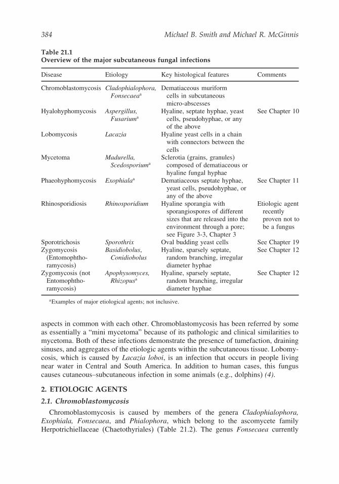
384 Michael B. Smith and Michael R. McGinnis
Table 21.1Overview of the major subcutaneous fungal infections
Disease Etiology Key histological features Comments
Chromoblastomycosis Cladophialophora,Fonsecaeaa
Dematiaceous muriformcells in subcutaneousmicro-abscesses
Hyalohyphomycosis Aspergillus,Fusariuma
Hyaline, septate hyphae, yeastcells, pseudohyphae, or anyof the above
See Chapter 10
Lobomycosis Lacazia Hyaline yeast cells in a chainwith connectors between thecells
Mycetoma Madurella,Scedosporiuma
Sclerotia (grains, granules)composed of dematiaceous orhyaline fungal hyphae
Phaeohyphomycosis Exophialaa Dematiaceous septate hyphae,yeast cells, pseudohyphae, orany of the above
See Chapter 11
Rhinosporidiosis Rhinosporidium Hyaline sporangia withsporangiospores of differentsizes that are released into theenvironment through a pore;see Figure 3-3, Chapter 3
Etiologic agentrecentlyproven not tobe a fungus
Sporotrichosis Sporothrix Oval budding yeast cells See Chapter 19Zygomycosis
(Entomophtho-ramycosis)
Basidiobolus,Conidiobolus
Hyaline, sparsely septate,random branching, irregulardiameter hyphae
See Chapter 12
Zygomycosis (notEntomophtho-ramycosis)
Apophysomyces,Rhizopusa
Hyaline, sparsely septate,random branching, irregulardiameter hyphae
See Chapter 12
aExamples of major etiological agents; not inclusive.
aspects in common with each other. Chromoblastomycosis has been referred by someas essentially a “mini mycetoma” because of its pathologic and clinical similarities tomycetoma. Both of these infections demonstrate the presence of tumefaction, drainingsinuses, and aggregates of the etiologic agents within the subcutaneous tissue. Lobomy-cosis, which is caused by Lacazia loboi, is an infection that occurs in people livingnear water in Central and South America. In addition to human cases, this funguscauses cutaneous–subcutaneous infection in some animals (e.g., dolphins) (4).
2. ETIOLOGIC AGENTS
2.1. Chromoblastomycosis
Chromoblastomycosis is caused by members of the genera Cladophialophora,Exophiala, Fonsecaea, and Phialophora, which belong to the ascomycete familyHerpotrichiellaceae (Chaetothyriales) (Table 21.2). The genus Fonsecaea currently

21. Subcutaneous Fungal Infections 385
Table 21.2Etiologic agents of human chromoblastomycosis, mycetoma, and lobomycosis
ChromoblastomycosisCladophialophora carrioniiExophiala dermatitidisExophiala jeanselmeiExophiala spiniferaFonsecaea compactaFonsecaea pedrosoiPhialophora verrucosaRhinocladiella aquaspersa
MycetomaAcremonium kilienseAcremonium recifeiAspergillus flavusAspergillus hollandicusEmericella nidulansFusarium falciformeCorynespora cassicolaCurvularia lunataCylindrocarpon cyanescensCylindrocarpon destructansExophiala jeanselmeiFusarium falciformeFusarium solaniFusarium verticillioidesLeptosphaeria senegalensisLeptosphaeria tompkinsiiMadurella griseaMadurella mycetomatisNeotestudina rosatiiPhaeoacremonium inflatipesPhialophora verrucosaPolycytella hominisPseudallescheria boydiiPseudochaetosphaeronema larensePyrenochaeta mackinnoniiPyrenochaeta romeroiRhinocladiella atrovirens
LobomycosisLacazia loboi
contains three species, two of which are primary agents of chromoblastomycosis,F. compacta and F. pedrosoi. Fonsecaea pedrosoi is the primary etiologic agent ofchromoblastomycosis throughout the world. On the basis of molecular analysis, anew species called F. monophora has been proposed for isolates that were previouslyidentified as F. pedrosoi which cause CNS infections (5). This latter infection is aphaeohyphomycosis because the fungus grows as dematiaceous hyphae in tissue.
386 Michael B. Smith and Michael R. McGinnis
The genus Cladophialophora contains a number of species, including C. bantiana,the etiologic agent of CNS phaeohyphomycosis. Recently, Cladosporium carrionii,an important agent of chromoblastomycosis, was reclassified as Cladophialophoracarrionii. This species is geographically more localized than F. pedrosoi.
2.2. Mycetoma
Mycetoma is caused by a variety of genera, the frequency of which vary geograph-ically. The genus Pseudallescheria contains a number of species. Pseudallescheriaboydii (anamorph Scedosporium apiospermum) is the major species of medical interest.It is unknown if the other species of Pseudallescheria can cause mycetoma. At thistime, it is thought that P. boydii is the only etiologic agent of this genus that causesmycetoma. Pseudallescheria is classified in the order Microascales.
The genus Madurella was originally established for sterile dematiaceous fungicausing mycetoma. Based on rDNA small subunit and internal transcribed spacer (ITS)sequence data, Madurella has been determined to be a heterogeneous group of fungi(6). Madurella mycetomatis, which worldwide is the most common cause of mycetoma,is in the Sordariales, whereas M. grisea is in the Pleosporales. The Ascomycetes ordersHypocreales, Eurotiales, and Dothideales contain several etiologic agents of mycetoma.
2.3. Lobomycosis
Lacazia loboi (previously named Loboa loboi) is a noncultured fungus that resemblesParacoccidioides brasiliensis in vivo. It is the sole etiologic agent of lobomycosis,regardless of human or animal origin. Based on its morphology in tissue and itsphylogenetic distinctness, the fungus is classified with the dimorphic Ascomycetes inthe Onygenales.
3. EPIDEMIOLOGY
Chromoblastomycosis, lobomycosis, and mycetoma have a number of features incommon. Infected patients typically live in tropical and subtropical regions. Theetiologic agents are introduced by localized trauma to the skin by woody plant materialor other injuries such as insect bites. They are more commonly seen in individualsworking in agriculture, mining, fishing, farming, and similar occupations where theyare in constant contact with the environment. Infections occur more commonly inmales, which is likely a reflection of occupational exposure. Mycetoma tends to occurin arid areas with short rainy seasons and a low relative humidity.
Lobomycosis, unlike the other two infections, is not worldwide in occurrence. Itis restricted to South and Central America, especially in communities located alongrivers. Lobomycosis tends to develop on exposed and cooler areas of the body such asthe extremities and the ears. This contrasts with chromoblastomycosis and mycetoma,which most frequently occur on the legs and feet.
4. PATHOGENESIS AND IMMUNOLOGY
The three infections are characterized by chronic, granulomatous, slowlyprogressing, cutaneous–subcutaneous inflammatory processes resulting in disfiguringlesions. The duration of infection prior to diagnosis can be years. Many patients recall

21. Subcutaneous Fungal Infections 387
splinters or similar wounds at the infection site prior to the development of the infection.Chromoblastomycosis does not involve bones and it does not usually spread throughthe lymphatic system. Mycetoma, in contrast, often extends to bone where it causesextensive destruction. Lobomycosis remains localized to the dermis and subcutaneoustissue and does not cause disseminated infections.
5. CLINICAL MANIFESTATIONS
5.1. Chromoblastomycosis
Chromoblastomycosis may exhibit a range of different types of lesions that varydepending on the duration and body site of the infection (7). After local injury, smallverrucous papules develop. Localized spread or extension of the lesions can occur byautoinoculation. The slow growing lesions can develop into clusters of large hyperker-atotic verrucous plaques that produce ulceration, cystic areas, and eventually, scarring(Fig. 21.1). Scarring can become so extensive that the flexibility of limbs can becompromised. Muriform cells occur in subcutaneous areas of epitheliomatous hyper-plasia and microabscesses, and are expelled to the surface by transepithelial elimination.This results in “black dots” on the lesion surface which are accumulations of necrotictissue and fungal cells. This process is similar to mycetoma, which is characterizedby “sclerotia” being transported to the surface by draining sinuses associated withedema (tumefaction). For this reason, chromoblastomycosis has been referred to as amini-mycetoma.
Fig. 21.1. Chromoblastomycosis caused by Fonsecaea pedrosoi. (Courtesy of Dr.C. Halde.) [Figure in color on CD-ROM].

388 Michael B. Smith and Michael R. McGinnis
Fig. 21.2. Mycetoma caused by Madurella mycetomatis. (Courtesy of Dr. C. Halde.) [Figurein color on CD-ROM].
5.2. Mycetoma
Fungal mycetoma develops more slowly than mycetoma caused by bacterial agents(actinomycotic mycetoma) (8). Encapsulated lesions with clearly defined marginsslowly develop after introduction of the fungus on a splinter or thorn. A subcutaneouspainless swelling develops that is usually soft, firm, and lobate. Additional foci often
Fig. 21.3. Lobomycosis caused by Lacazia loboi. Courtesy of Dr. P. Taborda. [Figure incolor on CD-ROM].

21. Subcutaneous Fungal Infections 389
develop, suppurate, and drain through multiple sinus tracts. Additional sinuses developthat are interconnected with each other, sterile deep abscesses, and the skin. Sclerotiaof different colors (e.g., white grain, black grain) that are formed by specific fungi findtheir way to the surface. Even though mycetoma is painless owing to anesthetic effector nerve damage, pain may occur when bone expands due to the fungal sclerotial mass,or when secondary infections develop. Mycetoma can produce functional disability,distortion, and deformity of limbs (Fig. 21.2).
5.3. Lobomycosis
Lesions of lobomycosis develop slowing and become cutaneous nodules or plaquesthat may be smooth, verrucous, or ulcerative, and eventually can become surroundedby keloidal scar tissue (4). The lesions may be solitary or multiple; painless or slightlypruritic; with the fungus spreading contiguously or via lymphatic channels (Fig. 21.3).The lesions contain granulomatous inflammatory tissue and the yeast cells. The fungusdoes not cause disseminated infection.
6. DIAGNOSIS
The diagnosis of these infections is based primarily on clinical presentation,histopathology, and culture. Culture is not useful in the diagnosis of lobomycosis asL. loboi has not yet been cultured. The tissue forms for these infections are distinctive,and can be used to a limited degree for presumptive identification purposes (see Fig.3.2, Chapter 3). Chains of yeast cells with a narrow connection between each yeastcell are characteristic of L. loboi (9). The architecture of the sclerotia in mycetomapatients can be associated with particular known agents (Fig. 21.4). The structure isespecially useful for distinguishing the granules of aerobic actinomycetes composedof 0.5 to 1.0 μm gram-positive filaments from sclerotia of true fungi having hyphalcells. The muriform cells and hyphae in the dermis of chromoblastomycosis cases do
Fig. 21.4. Sclerotia of Madurella mycetomatis in a foot. Gridley Fungus Stain. Courtesyof Dr. L. Ajello. [Figure in color on CD-ROM].

390 Michael B. Smith and Michael R. McGinnis
not allow for the differentiation of the different etiologic agents, although they arevirtually pathognomonic of the disease.
In culture, the agents of chromoblastomycosis and mycetoma are easily identified.Madurella mycetomatis represents an interesting challenge because it is defined on thebasis of being a typically sterile dematiaceous, slow growing mould isolated from amycetoma. As mentioned, the fungus appears to be a complex of different phylogeneticmembers.
7. TREATMENT
Chromoblastomycosis, lobomycosis, and mycetoma are chronic infections withouta single treatment of choice. Currently used treatments often have low cure rates andhigh relapse rates, although these rates vary based on the etiologic agent, affected bodyarea, and health status of the patient (Table 21.3). The dense fibrosis and lymphostasisthat occurs in these infections can impede drug penetration into the infected area.Courses of therapy of 6 to 12 months (or more) are not uncommon.
7.1. Chromoblastomycosis
Chromoblastomycosis is often treated with the triazole itraconazole, or terbinafine(3). These antifungal agents are used at high dosage concentrations, typically for 6 to12 months. Of the azoles, itraconazole has been the most successful antifungal drug,
Table 21.3Management of chromoblastomycosis, mycetoma, and lobomycosis
Infection Management
Chromoblastomycosis Surgery, electrodessication, and cryosurgery are effective in earlystages. Local heat can reduce extension of lesions. Disabled ordeformed limbs may require amputation. Combination therapy withterbinafine (500 mg daily) plus itraconazole (50–100 mg daily) orterbinafine alone has been successful.
Mycetoma Historically, surgery has been used for treatment of mycetoma.Mycetomas are well encapsulated, and care must be exercised toavoid rupturing. There are some reports of successful treatmentwith ketoconazole and itraconazole. The dose for both is 400 mgdaily with treatment continuing for a few to many years. A goodclinical response has been seen in some patients with ketoconazole,itraconazole, voriconazole, and terbinafine.
Lobomycosis Wide surgical excision of the affected area. Relapse is common.Electrodessication is useful in early stages of the disease.Clofazimine at 300 mg/d has been used with good results in somepatients. Antifungal drugs such as ketoconazole, itraconazole,amphotericin B, and 5-fluorocytosine are generally ineffective,although there is one report of successful treatment withitraconazole.
Modified from Lupi et al.(4)
.

21. Subcutaneous Fungal Infections 391
given as long-term continuous or pulse therapy. In one large study, the cure rate was42% with a mean treatment period of 7.2 months (2). In contrast, a large clinical studywith terbinafine had a cure rate of 74.2% after 12 months therapy (10). Overall curerates for both drugs range from 40% to 70% (3). These two drugs can be used aloneto treat small to medium-sized lesions, or they can be combined with localized heatand cryosurgery in one to several sessions. Combined antifungal drug therapy withitraconazole and terbinafine may be a useful approach to consider.
Superficial forms of chromoblastomycosis tend to respond well to antifungal drugs,whereas verrucous or tumorlike forms often do not respond well. These patientsoften require both chemotherapy and thermotherapy. Because of the high possibilityof secondary bacterial infections, prophylactic antibacterial therapy is often given. Ifsurgery is to be used, it is recommended that antifungal agents be given systematicallyboth before the procedure and afterwards.
7.2. Mycetoma
It is important to distinguish mycetoma caused by aerobic actinomycetes from casescaused by fungi because their treatment is different. An actinomycetoma is amenable totherapy with antibacterial agents. A fungal mycetoma is more difficult to manage andmay require aggressive surgery up to and including amputation in advanced disease.Surgical management may be appropriate after antifungal agents have been used. Agood clinical response has been seen in some patients treated with ketoconazole,itraconazole, voriconazole, and terbinafine.
The Mycetoma Research Center (Khartoum, Sudan; www.mycetoma.org) recom-mends both ketoconazole and itraconazole for first line use. Treatment duration dependsupon the severity of the infection and the general health status of the patient. Therapymay be needed for extended periods (years). Itraconazole treatment has been shownto have a high degree of success with a low recurrence rate. Surgery is used tocompletely excise small, well-encapsulated lesions, or to reduce of the amount ofinfected tissue. Effective surgery may require aggressive excision, which may lead topermanent disability. Surgery without effective chemotherapeutic treatment has a highrate of recurrence; up to 90% in advanced cases. Surgery is the treatment of choice forearly cases, typically well-encapsulated lesions without bone involvement.
7.3. Lobomycosis
Lobomycosis is an infection that involves the dermis and subcutaneous tissue thatis frequently managed with surgical excision (with wide margins). Surgical excisionis curative in most instances (4). Itraconazole may have value in treating this mycosis,but the use of this antifungal agent has been reported in only one patient.
REFERENCES
1. Pang KR, Wu JJ, Huang DB, Tyring SK. Subcutaneous fungal infections. Dermatol Ther2004;17:523–531.
2. Queiroz-Telles F, McGinnis MR, Salkin I, Graybill JR. Subcutaneous mycoses. Infect DisClin North Am 2003;17:59–85.
3. Bonifaz A, Saul A, Paredes-Solis V, Araiza J, Fierro-Arias L. Treatment of chromoblasto-mycosis with terbinafine: experience with four cases. J Dermatol Treat 2005;16:47–51.

392 Michael B. Smith and Michael R. McGinnis
4. Lupi O, Tyring SK, McGinnis MR. Tropical dermatology: fungal tropical diseases. J AmAcad Dermatol 2005;53:931–951.
5. Surash S, Tyagi A, De Hoog GS, Zeng JS, Barton RC, Hobson RP. Cerebral phaeohy-phomycosis caused by Fonsecaea monophora. Med Mycol 2005;43:465–472.
6. de Hoog GS, Adelmann D, Ahmed AO, van Belkum A. Phylogeny and typification ofMadurella mycetomatis, with a comparison of other agents of eumycetoma. Mycoses2004;47:121–130.
7. McGinnis MR. Chromoblastomycosis and phaeohyphomycosis: new concepts, diagnosis,and mycology. J Am Acad Dermatol 1983;8:1–16.
8. McGinnis MR. Mycetoma. Dermatol Clin 1996; 14:97–104.9. Haubold EM, Cooper CR, Wen JW, McGinnis MR, Cowan DF. Comparative morphology
of Lacazia loboi (syn. Loboa loboi) in dolphins and humans. Med Mycol 2000;38:9–14.10. Esterre P, Inzan CK, Ramarcel ER, et al. Treatment of chromomycosis with terbinafine:
preliminary results of an open pilot study. Br J Dermatol 1996;134 (Suppl 46):33–36.

Instructive Cases
INTRODUCTION
The instructive cases that follow are provided to allow the reader a review of manyof the important concepts presented in this book. They may also provide the clinicianpreparing for subspecialty boards practice cases and questions. Each case is presentedas an unknown to test knowledge obtained from reading this book or to emphasize animportant or complex topic in the field of medical mycology.
INSTRUCTIVE CASE 1
A 57-year-old patient with acute myelogenous leukemia (AML) and a 3-week historyof neutropenia develops fever that is unresponsive to broad-spectrum antimicrobialtherapy (piperacillin–tazobactam and levofloxacin). High-resolution computed tomog-raphy (HRCT) of the lungs revealed several pleural-based nodular lesions. The patientwas empirically started on amphotericin B deoxycholate 1 mg/kg every 24 hours.After 5 days of therapy, the patient’s serum potassium begins to drop but the serumcreatinine and blood urea nitrogen (BUN) remain stable.
Questions1. The decrease in serum potassium is most likely due to what amphotericin B toxicity?
A. Afferent arteriole constriction in the kidneyB. Distal tubular damage in the kidneyC. Suppression of erythropoietin synthesisD. Damage of the pancreatic islet cells
2. Which of the following approaches should be used if the patient’s serum creatininedoubles and he begins to develop azotemia?
A. Change amphotericin B dosing to every other day.B. Change amphotericin B to continuous infusion dosing.C. Change amphotericin B to a lipid amphotericin B formulation.D. Begin administering 500 ml of normal saline before and after infusion.
INSTRUCTIVE CASE 2
While riding his dirt bike on a vacant lot in southeastern Michigan, a 22-year-oldman abraded his right leg on the dirt while rounding a corner. He was taken to theemergency room, where the large abrasion was cleaned and débrided. Because therewas extensive loss of skin, a skin graft was placed several weeks later. About 2 weeksafter the graft was placed, he noted that a few “bumps” had developed in the graftedarea. These “bumps” became larger, broke open, and began to discharge what he
393

394 Instructive Cases
Fig. IC.1. Poor healing and nodular disease about lower extremity scar prior to antifungaltherapy (patient later proven to have sporotrichosis). [Figure in color on CD-ROM].
described as a thin fluid with some pink discoloration. Similar lesions then developedin the thigh proximal to the original injury and graft. Several different antimicrobialagents were prescribed (including those with Staphylococcus aureus coverage), but thelesions did not respond, and in fact new lesions appeared.
When the patient was seen by an infectious diseases consultant, the graft was beginningto break down and multiple nodules of various sizes were noted in the original abradedarea that had been grafted and proximal to the graft into the upper thigh (Fig. IC.1). Somewere ulcerated and weeping serosanguinous fluid, and others were crusted. The noduleswere not tender to palpation. The patient felt well and specifically denied having chillsor fever. Biopsies were taken of several nodules for culture and histopathological exami-nation. The tissue sections showed granulomas, but no organisms were seen. Within 1week, the cultures held at 25 �C showed growth of an off-white mould that on micro-scopic examination showed tiny conidia arranged “bouquet-like” on thin hyphae. Thediagnosis of sporotrichosis was made and the patient was begun on an experimentalprotocol using fluconazole, 400 mg daily. The lesions began to resolve by 2 months (Fig.IC.2), and were finally all resolved by 5 months. Therapy was continued for a total of 6months. No recurrences were noted. The patient was admonished to change hobbies fromdirt bike riding to something less dangerous, but he chose to ignore that medical advice.
INSTRUCTIVE CASE 3
A 29-year-old African American man was in his usual state of good health untilSeptember, at which time he presented with a nonproductive cough, shortness of breath,back pain, chills, fever, headache, and body aches. Evaluation led to a diagnosis ofpneumonia with right hilar and peritracheal adenopathy. He was originally treated

Instructive Cases 395
Fig. IC.2. Improvement of sporotrichosis after 2 months of fluconazole therapy. [Figurein color on CD-ROM].
with antimicrobial agents for community-acquired pneumonia without resolution ofsymptoms. Approximately 2 weeks later, three skin lesions consistent with granulomaswere noted. A punch biopsy performed approximately 1 month after his originalpresentation revealed granulomatous inflammation with endosporulating spherules,thus confirming the diagnosis of cutaneous coccidioidomycosis. Since the patient hadcomplained of intermittent headache, and there was now a diagnosis of pulmonary andcutaneous coccidioidomycosis, it was essential to rule out coccidioidal meningitis. Alumbar puncture was done, which was normal. At this time, the patient was also notedto have subcutaneous nodules in the left preauricular and right submandibular areas aswell as over the right humerus and left knee.
The patient was started on oral fluconazole, 800 mg daily. Subsequently the patientdeveloped multiple draining lesions over the anterior chest. A bone scan was positivefor disease of multiple ribs and the manubrium as well as the left parietal skull, rightfrontal skull, right superior orbit, and left hind foot. The positive areas on bone scanwere confirmed with plain radiographs, as osteomyelitis. Culture of fluid from thedraining sternal lesion grew Coccidioides immitis. Given the extensive disseminationto bone and skin in this patient, therapy was intensified to a lipid preparation ofamphotericin B at a dose of 5 mg/kg three times per week for approximately 3 months.At the same time, aggressive surgical débridement of the draining thoracic lesionswas undertaken, in an attempt to reduce the fungal burden. The patient’s originalcomplement fixation titer at presentation was less than 1:2. It subsequently rose to1:16 and eventually to ≥1:512, and stayed at that level despite therapy. The patientcontinues to have draining lesions of the anterior chest despite repeat surgeries and

396 Instructive Cases
several courses of amphotericin, and is now being treated with voriconazole 4 mg/kgtwice daily. Lifelong therapy is anticipated.
INSTRUCTIVE CASE 4
A-27 year-old male agricultural worker seen in consultation to evaluate an illnessof 2 months duration characterized by the presence of rapidly enlarging lymph nodesin both cervical chains, accompanied by pain, and high fever, especially at nights. Oneof the nodes had drained spontaneously, producing yellow-tinged purulent material.The patient also experienced productive cough without dyspnea. On examination,he was pale, looked frail, and had enlarged cervical, axillary, and inguinal lymphnodes (Fig. IC.3). His spleen and liver were normal by physical examination. Lungauscultation did not reveal altered breath sounds and chest radiographs were normal.
A human immunodeficiency (HIV) test was negative, hemoglobin was 8.8 g/dl, andwhite blood cell count was 25,700 cells/�l. A direct KOH examination of the dischargefrom the ruptured nodule showed abundant yeast cells, some with the characteristicmultiple budding of Paracoccidioides brasiliensis; this fungus was also isolated inculture. Serology with paracoccidioidin proved reactive with one band of precipitateand a complement fixation (CF) titer of 1:1024.
INSTRUCTIVE CASE 5
A 70-year-old man with hypereosinophilic syndrome, non-insulin dependent diabetesmellitus (NIDDM), and long-term prednisone therapy (25 mg/day × 25 years) wasadmitted with fatigue, weakness, and shortness of breath. Physician examination was
Fig. IC.3. Paracoccidioidomycosis. Hypertrophied cervical lymph node about to rupture.Scarring lesions of a similar process can be seen above. [Figure in color on CD-ROM].

Instructive Cases 397
remarkable for a temperature of 99˚F, an area of erythema over the left thigh thatwas tender to palpation and an effusion in the right knee. On admission, he had ahemoglobin of 5.9 g/dl, WBC 8600/mm3, a serum creatinine 2.7 mg/dl, and a glucose382 mg/dl. Aspiration of the right knee revealed a WBC of 40,500/mm3 with 96%neutrophils, Gram stain revealed gram-negative bacilli and budding yeasts. The patientwas initially started on imipenem, vancomycin, tobramycin, and fluconazole (FLZ)400 mg/day.
Within 72 hours, the blood cultures drawn on admission were found to be positive foryeast, later identified as Trichosporon asahii (beigelii). In addition, joint fluid cultureswere also positive forTrichosporon asahii and Pseudomonas aeruginosa. Biopsy andculture of the left thigh area of erythema also grew P. aeruginosa and T. asahii.
After 5 days of fluconazole treatment, the blood cultures were still positive andcaspofungin (Cancidas) 50 mg IV qd was added to the fluconazole. He also underwentarthroscopic flushing of the right knee. Ten days after admission, the patient hadnegative blood cultures and had responded well to antimicrobial therapy. He wassubsequently transferred to a rehabilitation unit in good condition.
In vitro susceptibilities (MICs) of T. asahii revealed fluconazole 8 �g/mg, itraconazole0.5 �g/ml, and caspofungin 2 �g/ml.
INSTRUCTIVE CASE 6
A 42-year-old black man presented to the University Hospital emergency departmentwith a 1-week history of fever, chills, cough productive of white sputum, night sweats,and malaise. He denied hemoptysis. Risk factors for HIV infection included a bloodtransfusion in 1985 and heterosexual promiscuity. He denied intravenous drug abuse orhomosexual activity. Over the last 8 months he had lost 80 pounds. Past medical historywas significant for a prior appendectomy, a perirectal abscess that was surgicallydrained, and a chronic perirectal fistula for 5 years. There was also a history ofvenereal disease including primary syphilis associated with a chancre and a positiverapid plasma reagin (RPR) of 1:32. There was no documented therapy and the patientwas lost to follow-up.
On admission, the patient was afebrile but cachectic. He was in no acute respiratorydistress. On physical examination, he was noted to have dry crusting lesions on thetip of his nose and both cheeks of the face (Fig. IC.4). Similar lesions were notedover the shins and a small draining abscess was noted over the medial aspect of theleft foot. Oral hairy leukoplakia was present on the tongue and he was noted to haveprominent generalized lymphadenopathy. No rales, rhonchi, or wheezes were heardon auscultation of chest. A fistula-in-ano was also noted. Chest radiograph revealedbilateral reticulonodular infiltrates, which were more prominent in the upper lobes.The patient was lymphopenic with an absolute lymphocyte count of 410 cells/�l. Theserum RPR of 1:32 was positive. Skin scrapings of the lesions were performed and wetpreparations of the specimens revealed characteristic thick-walled multinucleated yeastforms compatible with Blastomyces dermatitidis (Fig. IC.5). Subsequently, the patientunderwent bronchoscopy with bronchoalveolar lavage (BAL), and cytology prepara-tions of the washings also revealed B. dermatitidis. Cultures of skin scrapings and BALwashings grew this organism. An enzyme-linked immunoassay (ELISA) and Western

398 Instructive Cases
Fig. IC.4. Multiple cutaneous lesions in a patient with end-stage AIDS. Similar lesionswere noted on both cheeks of the face and both shins. [Figure in color on CD-ROM].
Blot confirmed HIV infection and total CD4+ T lymphocyte count was 8 cells/�l. Acomputed tomography (CT) scan of the head was performed because of complaintsof headache. This study revealed multiple enhancing brain lesions (Fig. IC.6). Owingto concerns of a second opportunistic pathogen, i.e., Toxoplasma gondii, the brainlesions were aspirated. Wet preparations again revealed characteristic yeast forms ofB. dermatitidis.
The patient was begun on highly active antiretroviral therapy (HAART) and ampho-tericin B deoxycholate therapy at an initial dose of 1 mg/kg per day. Intravenouspenicillin, 18 million units per day in divided doses, was administered for 21 daysbecause of concerns of neurosyphilis. During the remainder of the hospitalization,the patient was carefully monitored for evidence of renal insufficiency and 500 mlof normal saline were infused prior to each amphotericin B infusion. During this

Instructive Cases 399
Fig. IC.5. Wet preparation of skin scrapings. This figure shows the characteristic yeastforms in a wet preparation of skin scrapings. Scraping of the edges of the verrucous andulcerative lesions yield the best diagnostic results. [Figure in color on CD-ROM].
Fig. IC.6. CNS blastomycosis in an AIDS patient. Diagnosis may require aspiration of theabscesses if no active pulmonary or cutaneous disease is present.

400 Instructive Cases
hospitalization, he received a total of 1600 milligrams of amphotericin B. Serial CTscans documented progressive improvement of brain abscesses and oral fluconazolewas substituted after the full course of amphotericin B and the patient was dischargedto home. He completed 8 more weeks of fluconazole therapy at a dose of 800 mg/day.Although the brain abscesses had resolved on CT scan, a maintenance suppressive doseof fluconazole of 400 mg per day was initiated. HAART was continued and a clinicalresponse was documented by a falling HIV viral load and rising CD4+ T lymphocytecount.
INSTRUCTIVE CASE 7
A 45-year-old construction worker presented with a 3-week history of fever, chills,myalgias, headache, and dyspnea. Examination was unremarkable and laboratory testsrevealed a white blood cell count of 3300 cells/�l and a platelet count of 94,000cells/�l. Aspartate transaminase (AST) was 87 units/l, alkaline phosphatase 337 units/l.Angiotensin converting enzyme was elevated at 213 units/l. Chest and abdomenCT showed small nodules in the lungs, enlarged mediastinal lymph nodes, smallpleural effusions, and splenomegaly. Transbronchial biopsies of the subcarinal lymphnode and right lower lobe and bone marrow biopsy showed noncaseating granuloma.Histopathology was negative for fungi and cultures were negative after 1 week ofincubation. Prednisone 60 mg daily was prescribed for presumed sarcoidosis, whichresulted in resolution of fever and improvement of dyspnea. The prednisone dosagewas tapered over 4 weeks to 10 mg daily, which was maintained.
Two months later the patient complained of recurrent fever, 10-pound weight loss,and worsening dyspnea. Chest CT showed more extensive diffuse interstitial infil-trates and increasing splenomegaly. Hemoglobin was 9.3 g/dl, white blood cell count2500 cells/�l, and a platelet count of 89,000 cells/�l. Alkaline phosphatase was 987units/l. Cultures from the lung tissue, lymph nodes, and bone marrow performed duringthe earlier admission were negative after 4 weeks of incubation. Bronchoscopy wasperformed and cytology revealed small yeastlike structures resembling Histoplasmacapsulatum, which was later confirmed by culture. Treatment was started with ampho-tericin B, and the patient noted progressive improvement.
INSTRUCTIVE CASE 8
A 52-year-old man who underwent renal transplantation 2 years earlier presented witha slightly painful mass above his right knee. This mass began as a small lesion 4 weeks agoand has been slowly enlarging since first noted by the patient. He denies fever, chills, nightsweats, or trauma to the area. He is taking tacrolimus and prednisone as his immunosup-pressive regimen. The patient was afebrile with stable vital signs, but his physical exami-nation was significant for a 1.5 cm firm nodule above his right knee, which was slightlytender, but without erythema or drainage. Routine laboratory studies are unremarkable.
Surgery was consulted for excision of the nodule, which was performed withoutcomplications. Pathology showed chronic inflammation and pigmented fungal elementssuggestive of phaeohyphomycosis; margins were clear of infection (Fig. IC.7). Cultureof the specimen grew Exophiala jeanselmei. He was given itraconazole for 3 months,and no further lesions appeared.

Instructive Cases 401
Fig. IC.7. Histopathological examination of subcutaneous nodule showing granulomatouschanges and dark-walled fungal elements. Phaeohyphomycosis. [Figure in color on CD-ROM].
INSTRUCTIVE CASE 9
A 34-year-old African American man, who has been followed for approximately 10years, originally presented with back pain and tenderness over the right sternoclavicularjoint. Coccidioidomycosis was suspected and a serology was obtained with an initialcomplement fixation (CF) titer of 1:256. A whole body bone scan showed focal areasof increased uptake involving the left frontal bone, the right sternoclavicular joint, theleft sacroiliac joint, and the cervicothoracic region. These findings were confirmedwith plain radiograph and CT scans.
Approximately 1 month after original presentation, an incision and drainage of theproximal clavicle and the posterior superior spine of the iliac crest was performed.Cultures were positive for Coccidioides immitis. Three months after presentation, acervical corpectomy of C5 and C6 with autograft fusion and anterior cervical platingwas performed. Five months after presentation, decompression of the spinal cord withcorpectomy at T3 and T4 with autologous rib grafting was performed.
Medical therapy initially consisted of amphotericin B, which was infused over aperiod of approximately 4 months, three times per week. Follow-up therapy with high-dose oral fluconazole (1000 mg daily as a single dose) was administered. As the patientimproved clinically, CF titers steadily came down, and the dose was deescalated to400 mg of fluconazole daily. With approximately 10 years of follow-up, no furtherevidence of disease has been discerned. Complement fixation titers in the recent pasthave remained positive at 1:2. It has been recommended that he remain on therapyfor life.

402 Instructive Cases
INSTRUCTIVE CASE 10
A 68-year-old man with a long standing history of hairy cell leukemia wasadmitted to receive treatment with the experimental immunotoxin BL-22. He had beenpancytopenic for several months and had been receiving antifungal prophylaxis withitraconazole. On admission, antifungal prophylaxis was changed to fluconazole. Onday 2 the patient developed fever. At that time, his absolute neutrophil count was 76cells/�l. Blood cultures recovered a highly susceptible E. coli and the patient defer-vesced promptly on ceftazidime. On day 7 a new fever developed and meropenem,tobramycin, and caspofungin were started. A chest CT was obtained which showed a3 cm right upper lobe (RUL) nodule (Fig. IC.8). A bronchoalveolar lavage (BAL) wasperformed, and voriconazole and levofloxacin were added. Bacterial, fungal, and viralcultures, as well as Gram, calcofluor white, acid-fast, modified-acid-fast, and Gomorimethenamine silver (GMS) stains were negative. Polymerase chain reaction (PCR) forPneumocystis, Chlamydophila, Mycoplasma, and Legionella was also negative. Thepatient continued to have fever up to 40oC without new symptoms or hypotension.On day 11 he complained of chest pain and dry cough. A repeat CT showed markedenlargement of the nodule with development of a halo sign and abutting of the fissure(Fig. IC.9). A fine-needle aspirate of the mass showed broad, ribbonlike, nonseptatehyphae (Fig. IC-10). Voriconazole was discontinued and liposomal amphotericin B7.5 mg/kg per day was started. The patient developed hemoptysis and a repeat CTshowed further progression of the mass, but with apparent localization in the RUL
Fig. IC.8. CT scan of chest revealing small pulmonary nodule in the right upper lung.

Instructive Cases 403
Fig. IC.9. CT scan of chest revealing progression of pulmonary nodule in right upper lungfield to involve the right pleural surface.
Fig. IC.10. Calcofluor staining of fine-needle aspiration demonstrates broad nonseptatehyphae consistent with zygomycosis. [Figure in color on CD-ROM].

404 Instructive Cases
Fig. IC.11. CT scan of chest with severe advancement of locally progressive disease in theright upper lung fields.
(Fig. IC.11). An emergent right upper lobectomy was performed on day 18 (Fig. IC.12).An angioinvasive mould was readily seen on the GMS stain (Fig. IC.13), and was lateridentified as Rhizomucor pusillus. On the day of the surgery, granulocyte transfusionswere initiated. Hemoptysis and fever resolved 2 days after the surgery. Despite localcontrol of the fungal disease, the patient never recovered his white blood cell countsand over the next 6 weeks developed several complications in the ICU, includingherpes simplex pneumonia. At his own request, care was withdrawn.
Fig. IC.12. Gross pathology of right upper lobectomy. [Figure in color on CD-ROM].

Instructive Cases 405
Fig. IC.13. Zygomycosis with hyphae invading blood vessel, infiltrating vascular wall, andcausing subsequent thrombosis. GMS. [Figure in color on CD-ROM].
INSTRUCTIVE CASE 11
A 48-year-old male rural worker was referred for consultation to evaluate thepresence of painful, ulcerated lesions in the external region of his right foot (Fig. IC.14).This process had gone on for 18 months, and multiple local and systemic treatmentshad been given without success. The patient looked well and had no other symptoms.Physical examination also revealed the presence of an oral mucosal ulceration and ofhypertrophied cervical lymph nodes. Lung auscultation found fine rales and the chestradiograph showed interstitial infiltrates in the central fields with fibrous zones andbasal bullae (Fig. IC.15).
Direct KOH examination of ulcer exudate revealed multiple budding yeastsconsistent with Paracoccidioides brasiliensis; cultures later grew the fungus. Thepatient’s serum gave a band of precipitate and a CF titer of 1:32 with paracoccidioidin.
INSTRUCTIVE CASE 12
A 40-year-female with acute myelogenous leukemia now 115 days followingmatched allogeneic donor hematopoietic stem cell transplantation complicated by asuspected Aspergillus pneumonia presents to the clinic with increasing complaintsof nausea, stomach cramping, and rash on the hands spreading up her arms. Bylaboratory examination, she is noted to have an alanine transaminase (ALT) of85 units/l, AST 75 units/l, and total bilirubin of 2.1 mg/dl. Her current medica-tions include tacrolimus 5 mg twice daily (recent level 8 ng/ml), voriconazole200 mg twice daily, levofloxacin 500 mg daily, valacyclovir 500 mg twicedaily, metoprolol 25 mg twice daily and benzonatate (tesselon) pearls. She is

406 Instructive Cases
Fig. IC.14. Paracoccidioidomycosis. Multiple ulcerated lesions in the right foot withcrusting, exudation, and hemorrhagic dots. The borders are granulomatous and show somescarring. [Figure in color on CD-ROM].
admitted to the hospital for suspected graft versus host disease exacerbation.The primary service wishes to continue voriconazole therapy as this patienthas a history of poorly tolerating lipid amphotericin B formulations, but theyare concerned about the possibility of drug-induced hepatitis caused by voriconazole.
Questions1. Which of the following approaches should be recommended to the primary team to
manage the suspected drug-induced hepatotoxicity?
A. Discontinue voriconazole and switch to itraconazole.B. Discontinue voriconazole and switch to lipid amphotericin B with premedications.C. Continue voriconazole and lower dose by 50%.D. Continue voriconazole and closely monitor the patient.
INSTRUCTIVE CASE 13
A 44-year-old male field worker was in his usual state of health until 2 weeks priorto admission, when he developed headache, low-grade fever, nausea, and vomiting.His family noted decreased mentation and brought him to the emergency room. Initialexamination revealed a disheveled gentleman with lethargy and disorientation. A CTscan of the head was performed without contrast and was normal. A lumbar puncturerevealed 450 white cells/�l, of which 76% were lymphocytes, 10% monocytes, 10%neutrophils, and 4% eosinophils. The protein was 176 mg/dl and the glucose was 27

Instructive Cases 407
Fig. IC.15. Paracoccidioidomycosis. Bilateral interstitial infiltrates in central fields, basalbullae, and fibrous areas. The apices appear free of disease.
mg/dl. Studies were sent to rule out cryptococcosis, tuberculosis, and coccidioidomy-cosis. Coccidioidal antibody in the serum was 1:256 and in the cerebrospinal fluid(CSF) was 1:2. Other fungal and mycobacterial laboratory evalutions were negative.Magnetic resonance imaging (MRI) of the brain was done to evaluate for meningealinvolvement or vasculitic complications of coccidioidomycosis. This study revealedbasilar cisternal enhancement. A bone scan was ordered and was negative for bonyinvolvement. An HIV test was performed and confirmed as positive. The HIV RNAPCR was 330,000 copies/ml with an absolute CD4+ T lymphocyte count of 61 cells/�l.The patient was placed on high-dose oral fluconazole (1000 mg daily as a singledose). Highly active antiretroviral therapy (HAART) was also initiated and patientwas discharged to be followed in clinic. Patient and family were counseled regardingstrict adherence to fluconazole and HAART therapy, both of which needed to belife-long.
He also underwent lumbar punctures monthly, CSF was sent for cell count, glucose,protein, complement fixation (CF) titers, and fungal culture. Serum for fluconazolelevel was sent when the possibility of noncompliance was considered. The patientfollowed up with appointments inconsistently. Four months after admission, he wasbrought to the Emergency Room with confusion, lethargy, nausea, vomiting, andataxic gait. Evaluation again showed encephalopathy. CT scan now demonstrated

408 Instructive Cases
ventriculomegaly. Lumbar puncture revealed an elevated opening pressure of 250mmH2O, white cell count 50 cells/�l (85% lymphocytes, 11% neutrophils), protein153 mg/dl, and glucose of 10 mg/dl. HAART therapy and high dose fluconazole werereinitiated. Neurosurgical consultation was obtained and the patient was taken to theoperating room for ventriculoperitoneal shunting. The patient’s symptoms improvedsignificantly after shunting, but his clinical course was complicated by hyponatremiasecondary to syndrome of inappropriate antidiuretic hormone secretion (SIADH), whichwas managed with fluid restriction. He gradually improved and was discharged withclose follow-up at clinic.
INSTRUCTIVE CASE 14
A 22-year-old female with a failing 4-year-old renal allograft received several dosesof OKT3 and high doses of corticosteroids in an attempt to reverse the acute rejection ofthe transplanted kidney. Three months after this increased immunosuppressive trial andstill receiving her normal immunosuppressive regimen of tacrolimus, mycophenolate,and prednisone, she presented with a several week course of headaches, nausea, andvomiting. Her temperature was 37.2oC, and although mental status was normal, shehad bilateral clonus and papilledema on physical examination. Her laboratory resultsshowed a normal complete blood count and a serum creatinine of 4 mg/dl. An MRIof her brain demonstrated basilar inflammation and lumbar puncture (LP) revealeda white blood cell count of 100 cells/�l with 80% lymphocytes. CSF glucose was43 mg/dl and protein 79 mg/dl. India ink was positive for encapsulated yeasts, CSFcryptococcal polysaccharide antigen test was �1:256, and culture grew Cryptococcusneoformans. Her opening pressure was 400 mm H2O. She was started on 5 mg/kg perday of AmBisome for 20 days and flucytosine at 25 mg/kg per day for 14 days, andthen placed on 200 mg/day of fluconazole. The patient’s symptoms did not worsen andshe was reevaluated at 2 weeks with a repeat LP. That LP found an opening pressureof 140 mm H2O, and India ink and culture were negative. CSF antigen was 1:256 andCSF white cell count was 28 cells/�l.
Case Continued
Patient did relatively well on her suppressive fluconazole therapy at 200 mg/day for4 months (dosed for reduced renal function), when she developed severe headaches andan MRI scan showed diffuse supra- and infratentorial leptomeningeal enhancement.At that time CSF cryptococcal polysaccharide antigen was 1:16, white blood cellcount was 100 cells/�l, and cultures negative. After 2 weeks of AmBisome at 5mg/kg per day and no improvement, the patient was continued on fluconazole and a6 week dexamethasone taper was begun with immediate improvement in symptoms.Tacrolimus and mycophenolate were stopped and the patient was started on dialysis.One week after stopping the 6-week taper of corticosteroids, her headaches returnedand a 4-month steroid taper was begun. She improved and was eventually weaned offcorticosteroids and now has received suppressive fluconazole for 1 to 2 years and isdoing well awaiting a new transplant.
Instructive Cases 409
INSTRUCTIVE CASE 15
A 47-year-old black man presented to the University Hospital emergency departmentcomplaining of pleuritic chest pain for 2 weeks before admission. He subsequentlydeveloped severe dyspnea on exertion, fever and chills, and productive cough withhemoptysis. Over the 24 hours preceding admission, pleuritic chest pain, which wasinitially only on the left side, became bilateral and he presented to the EmergencyDepartment for evaluation. Past medical history was pertinent for hyperthyroidism andcigarette smoking. He denied any risk factors for HIV infection.
In the emergency department, he was in moderate respiratory distress but was afebrile.Chest radiograph revealed diffuse bilateral miliary infiltrates with a masslike lesion inthe lung field. White blood count was 14,400 cells/�l with a left shift. Arterial bloodgases on room air revealed a pH of 7.41, po2 of 33 mm Hg, and pco2 of 46 mm Hg.
The patient was admitted to the hospital and placed in respiratory isolation. Differ-ential diagnosis included severe community-acquired pneumonia, atypical pneumonia,miliary tuberculosis, and fungal disease. He was placed on supplemental oxygen,intravenous azithromycin and ceftriaxone, and a four-drug antituberculous treatmentregimen. Multiple sputum samples were remarkable only for many polymorphonuclearleukocytes. Direct fluorescent antibody (DFA) and urinary antigen tests were negativefor Legionella. Likewise, fungal and acid-fast stains of sputum samples were negative.An ELISA for HIV was also negative.
On hospital day 3 the patient became febrile, a chest radiograph revealed worseningbilateral pulmonary infiltrates, and intravenous trimethoprim–sulfamethoxazole wasadded to the existing antibacterial regimen as empiric therapy for possible Pneumocystispneumonia. On the fifth hospital day he complained of increasing dyspnea and wasnoted as having a respiratory rate of 60 breaths per minute. Arterial blood gases on100% oxygen by face mask revealed a pH of 7.45, po2 of 102 mm Hg, and pco2 of42 mm Hg. The patient was transferred to intensive care unit, where he was intubatedand placed on mechanical ventilation. Chest radiographs revealed bilateral pulmonaryinfiltrates (Fig. IC.16) with acute lung injury. Cytology samples obtained via theendotracheal tube at the time of intubation revealed numerous broad-based buddingyeast forms compatible with Blastomyces species (Fig. IC.17).
The patient received intravenous amphotericin B immediately at a dose of 0.7 mg/kgper day, but was rapidly increased to 1 mg/kg per day. During the remainder ofhis hospitalization, the patient became increasingly difficult to oxygenate, developedhypotension requiring pressers, progressed to multiorgan failure, and died withpulseless electrical activity.
INSTRUCTIVE CASE 16
An 18-year-old female presented with chest discomfort. Dilated veins were notedover the chest and upper abdomen. A chest radiograph that showed right hilarenlargement, which was calcified on CT. The right pulmonary artery was narrowedand the superior vena cava was occluded. Ventilation–perfusion lung scan that showedreduced blood flow to the right lung. Pulmonary function tests showed normal air flowand lung capacity. Tuberculin skin test was negative. Histoplasma immunodiffusion

410 Instructive Cases
Fig. IC.16. Diffuse pulmonary infiltrates in a patient with ARDS. Patients presenting withthis syndrome have a mortality rate greater than 50%.
tests showed an M band and the Histoplasma complement fixation test showed titers of1:32 to the yeast and 1:16 to the mycelial antigen. Mediastinal biopsy showed chronicinflammatory cells, granuloma, and fibrosis. Culture of the mediastinal biopsy tissuewas negative for fungus.
Fig. IC.17. Cytology preparation of endotracheal tube specimen revealing large, thick-walled yeasts consistent with Blastomyces dermatitidis. [Figure in color on CD-ROM].

Instructive Cases 411
Question
Is surgery indicated to correct the obstruction of the pulmonary artery or of thesuperior vena cava, and should the patient receive a course of antifungal therapy?
INSTRUCTIVE CASE 17
A 41-year-old male rural worker and heavy smoker consulted because of 3 months ofdry cough, severe progressive dyspnea, weight loss, asthenia, adynamia, and anorexia.He looked emaciated and experienced difficulties in breathing even at rest. Respiratoryrate was noted to be 36 breaths per minute with accessory muscles utilization. Onauscultation rales, rhonchi and hypoventilation were noticed. The chest radiograph(Fig. IC.18) revealed the presence of a diffuse reticulonodular infiltrates predominatingin both central fields with fibrous lines. Follow-up CT of chest documented widespreadfibrosis (Fig. IC.19). Arterial blood gas analysis revealed pH 7.44, po2 37 mm Hg,pco2 23 mm Hg, O2 saturation 81%, and bicarbonate 16 mEq/l.
A bronchoalveolar lavage fluid sample was examined for acid-fast bacilliwith negative results, but multiple budding cells corresponding toParacoccidioidesbrasiliensis were seen on direct examination and recovered in culture later on. Serologictests with paracoccidioidin were reactive with one band of precipitate and a titer of1:1024 in the complement fixation (CF) test.
Fig. IC.18. Paracoccidioidomycosis. Lung fibrosis involving specially the central field withapices appearing free. Bilateral basal bullae and pleural adhesions are seen in both lower fields.

412 Instructive Cases
Fig. IC.19. Par acoccidioidomycosis. High-resolution CT showing widespread fibrosis withhoneybee aspect, bullae formation. and pleural thickening in both lower lung fields.
INSTRUCTIVE CASE 18
A 51-year-old man was newly assigned to a construction project in Kern County.Two weeks after he began his assignment, he developed cough, shortness of breath,fever, chills, and night sweats. His physician obtained a chest radiograph, which showedconsolidation in the anterior left upper lobe and lingula, associated with adenopathy ofthe lateral aortic, left hilar, and pericarinal nodes. He was diagnosed with community-acquired pneumonia and prescribed antibiotics. One week later, he had not improvedand went to the local emergency room, where he was found to be febrile but nothypoxic. His white blood count was 9400 cells/�l, with an absolute eosinophil countof 959 cells/�l. He was prescribed oral levofloxacin 750 mg daily and discharged tohome. Serum for Coccidioides serology was collected at this visit and reported 5 dayslater: EIA IgM positive; immunodiffusion (ID) IgG negative, ID IgM positive; andCF titer (IgG) � 1:2. These results were suggestive of early disease, but were notcommunicated to the patient and possibly misinterpreted as negative.
Almost 3 weeks after initial presentation, the patient developed increasing shortnessof breath, became hypoxic, somewhat confused, with high fevers, and drenching sweats.He presented to our emergency room. His subsequent Coccidioides CF titer was 1:64.Since he was hypoxic with a po2 of less than 70 mm Hg, he was started on a lipidamphotericin preparation at 5 mg/kg daily as well as oral prednisone at a dose of 40mg twice daily for 5 days, followed by 40 mg once daily for 5 days, then 20 mgonce daily for 11 days. He improved and was discharged home with continuation ofamphotericin for 6 weeks, three times per week, and then switched to oral fluconazoleat 800 mg per day.
He was followed monthly. He developed a painful left shoulder while on therapy.Bone scan was negative and the shoulder pain was considered to be fluconazole

Instructive Cases 413
shoulder syndrome. Therapy was changed to Itraconazole, 200 mg twice daily. Hecomplained of headache, which eventuated in a lumbar puncture which proved tobe unremarkable. He gradually improved clinically and radiologically. His CF titersdecreased to 1:2, and remained at this level for three readings, at which point theitraconazole was discontinued almost exactly 1 year from initial presentation. He wasfollowed every 3 months for 1 year with serology after completing treatment with norise in CF titer or clinical evidence of recurrence.

Instructive Cases Discussion
INSTRUCTIVE CASE 1
Answers: 1. B, 2. C
Discussion
Question 1
Amphotericin B-induced nephrotoxicity occurs primarily through two mechanisms:(1) constriction of afferent arterioles leading to direct decreases in glomerular filtrationrate (GFR; glomerular toxicity) and (2) direct damage to the distal tubules (tubulartoxicity; answer B), which in turn can lead to glomerular feedback that further constrictsthe afferent arterioles. Tubular toxicity of amphotericin B is essentially limited to thedistal tubules and most commonly evident as hypokalemia (answer B). It occurs in themajority of patients receiving amphotericin B and may require up to 15 mmol of supple-mental potassium per hour. Amphotericin B-induced hypokalemia is not associated withincreased plasma aldosterone or renin levels and appears to result from increased perme-ability of the distal tubular cells due to direct toxic effects of amphotericin B. Althoughlipid amphotericin B formulations reduce distal tubular toxicity, they do not eliminatethis side effect. Distal tubular toxicity (decreases in serum potassium and magnesium)frequently precede decreases in glomerular filtration rate (increases in serum creatinine,BUN) during amphotericin B therapy, particularly in patients receiving lipid ampho-tericin B formulations. Afferent arteriole constriction (answer A) would be more specif-ically associated with decreases in glomerular filtration (decrease in serum creatinine).Suppression of erythropoietin synthesis (answer C) is a more chronic effect of ampho-tericin B and manifests primarily as normochromic, normocytic anemia. AmphotericinB has not been shown to directly damage pancreatic islet cells (answer D).
Question 2
Nephrotoxicity is the dose-limiting toxicity of amphotericin B therapy. Althoughall of the answers are potential approaches that have been applied to prevent thedevelopment of nephrotoxicity during amphotericin B therapy, switching to a lipidamphotericin B formulation (answer C) is advocated by most infectious diseases expertsonce glomerular toxicity has developed during amphotericin B therapy. Alternativedaily dosing of amphotericin B (answer A) is no longer recommended and there isno evidence that this method is less nephrotoxic than daily dosing. Administeringamphotericin B by continuous infusion (answer B) has been shown in two smallprospective trials to reduce infusion-related and nephrotoxic side effects; however, thisdosing method is not practical and the efficacy of amphotericin B administered bycontinuous infusion has not been well explored. Saline loading (answer D) can reduce
415

416 Instructive Cases Discussion
tubular-glomerular feedback and delay the onset of glomerular toxicity; however, itwill not reverse glomerular toxicity once it has developed.
Instructive case 1 contributed by R. E. Lewis and A. W. Fothergill
INSTRUCTIVE CASE 2
Discussion
This case is somewhat unusual in that the site of inoculation of Sporothrix schenckiihad received a skin graft, and thus the lesions were atypical. They arose in the graftedarea and contributed to loss of portions of the graft. The lack of response to antistaphy-lococcal antibiotics was a clue that this was not a typical bacterial infection. Thepossibility of sporotrichosis was raised by the infectious diseases consultant becauseof the proximal spread of nodules and the exposure to soil during the original accident.Biopsy confirmed this suspicion when the cultures yielded a mould. The fact that noorganisms were seen on the biopsy is not unusual.
Fluconazole was used because an experimental protocol was available at thetime, and the patient had no insurance and was unable to purchase other antifungalagents. The response to fluconazole was adequate, but slower than usually noted withitraconazole, which is the treatment of choice for sporotrichosis.
Instructive case 2 contributed by C.A. Kauffman
INSTRUCTIVE CASE 3
Discussion
This case demonstrates how an apparently uncomplicated coccidioidal pneumoniacan evolve into complex multifocal disseminated disease. Such cases frequently neces-sitate multiple therapy modalities.
Instructive case 3 contributed by R. H. Johnson and S. Baqi
INSTRUCTIVE CASE 4
Discussion
This is an example of the juvenile type paracoccidioidomycosis.
Instructive case 4 contributed by A. Restrepo, A. M. Tobón, and C. A. Agudelo
INSTRUCTIVE CASE 5
Discussion
This case is instructive for several reasons. The patient’s past medical history issignificant for a long history of immunosuppression due to long-term corticosteroid useand his diabetes mellitus. Both of these conditions predispose the patient to an increasedrisk of fungal infections because of alterations in cell-mediated immunity. The casedemonstrates the increasing incidence and the capacity of previously nonpathogenicyeast to produce invasive infection. In addition, the presentation of the patient with

Instructive Cases Discussion 417
nonspecific signs and symptoms is not uncommonly seen in many invasive fungalinfections. In fact, the problem with the diagnosis of invasive fungal infections isthat there are no “classic or pathognomonic manifestations.” This makes the diagnosisdifficult to establish and creates a delay in the initiation of appropriate antifungaltherapy. In this case, the aspiration of the infected knee demonstrated several organisms(gram-negative bacilli and yeast), thus assisting with the diagnosis. However, Candida,not Trichosporon would have been the more common cause of infection in this patient.It is not until the laboratory identifies the organisms that the true diagnosis is estab-lished. Although there are no clinical trials establishing the best antifungal agent forTrichosporon species, the azoles (fluconazole, voriconazole) have been shown to havein vitro activity. In this case, in vitro susceptibility results demonstrated that bothfluconazole and itraconazole had good activity. Although there are no establishedbreakpoints for the echinocandins, the minimum inhibitory concentration (MIC) of2.0 �g/ml for caspofungin appears to be within the “standard” ranges described forclinical activity against Candida species. Further, although there are some in vitroand animal studies demonstrating additive or synergistic activity with the combinationof echinocandins and azoles, the use of combination antifungal therapy has not beendemonstrated to be any better than monotherapy.
Instructive case 5 contributed by J. A. Vazquez
INSTRUCTIVE CASE 6
Discussion
Blastomycosis in AIDS1. Blastomycosis in acquired immunodeficiency syndrome (AIDS) patients is more likely
to be multiorgan disease, with CNS involvement being noted in up to 40% of patients.Routine magnetic resonance imaging (MRI) or CT scans should definitely be performedin any patient with severe end-stage AIDS whether or not the patient has neurologicsigns or symptoms.
2. Wet preparations of skin lesions allowed a presumptive clinical diagnosis and earlyinitiation of amphotericin B therapy in this patient.
3. Central nervous system disease in end-stage AIDS patients should be treated with afull course of amphotericin B, e.g., 1.5 to 2.5 grams.
4. Fluconazole at high doses (800 mg/day) may be a reasonable substitute in patientsintolerant to amphotericin B or as step down therapy in patients who have respondedto initial treatment with amphotericin B.
5. In immunosuppressed patients, chronic suppressive therapy with fluconazole shouldbe considered.
Instructive case 6 contributed by S. W. Chapman and D. C. Sullivan
INSTRUCTIVE CASE 7
Discussion
This case represents acute pulmonary histoplasmosis misdiagnosed as sarcoidosis.It illustrates the importance of thorough testing to exclude histoplasmosis before

418 Instructive Cases Discussion
beginning immunosuppressive treatment for sarcoidosis. While there are clinicalfeatures that help to distinguish sarcoidosis from histoplasmosis, differentiationrequires laboratory testing to exclude histoplasmosis. Administration of corticos-teroids resulted in transient clinical improvement, followed by progression withworsening pulmonary disease and progressive dissemination. Failure to demonstrateyeast resembling H. capsulatum on the initial bronchoscopy resulted in a mistakendiagnosis of sarcoidosis. Of note is that cytology and culture of respiratory secre-tions are often negative in acute histoplasmosis, and cannot be used to exclude thediagnosis.
Additional testing should include serology for antibodies to H. capsulatum, andtests for Histoplasma antigen in urine and respiratory secretions. Serology is oftennegative during the first month after exposure, but positive thereafter. Antigen maybe detected in the urine or bronchoscopy specimen of 75% of cases during the acuteillness and before antibodies have appeared. Corticosteroids for sarcoidosis should notbe given without thorough evaluation to exclude histoplasmosis. Of note is that fungalcultures require up to 4 weeks of incubation for isolation of H. capsulatum, duringwhich corticosteroids should be withheld except in severe cases. Tests for antigen andantibody, and cytology on bronchial washing or bronchoalveolar lavage, should beperformed and may provide early evidence for histoplasmosis. Of note, these tests donot exclude the diagnosis. If lung or other tissues are biopsied, they also should becultured and examined for fungus by histopathology.
If cytology, histopathology, antigen testing, and serology are negative and corticos-teroids are required for severe sarcoidosis, itraconazole may be given while waiting forculture results in selected patients in whom the diagnosis of histoplasmosis is suspectedbased on epidemiologic grounds.
Instructive case 7 contributed by L. J. Wheat and N. G. Conger
INSTRUCTIVE CASE 8
Discussion
Subcutaneous phaeohyphomycosis is among the most common manifestations ofdisease due to dematiaceous fungi. It is seen in both immunocompetent and immuno-compromised individuals and is not usually associated with dissemination, although therisk of dissemination is higher in immunosuppressed patients. Complete excision alonehas been reported as a successful therapy, particularly in immunocompetent patients. Inimmunocompromised patients, antifungal therapy is often given after surgical excisionto reduce the risk of dissemination. However, itraconazole and voriconazole bothhave significant interactions with immunosuppressive agents such as tacrolimus andsirolimus, and combined use of these drugs requires close monitoring and commonlyadjustment of the immunosuppressive agents.
Instructive case 8 contributed by S. G. Revankar

Instructive Cases Discussion 419
INSTRUCTIVE CASE 9
Discussion
This case illustrates the importance of surgery in the management of complex axialand nonaxial osteomyelitis secondary to coccidioidomycosis.
Instructive case 9 contributed by R. H. Johnson and S. Baqi
INSTRUCTIVE CASE 10
Discussion
Nodular infiltrates in the neutropenic patient are often caused by pathogenic fungi.Identification is critical for proper management. This case illustrates that pulmonaryzygomycosis in a neutropenic host may be associated with fever but a paucity of otherfindings on initial presentation. A BAL is often negative and more invasive procedures,such as a fine needle aspiration, may be necessary to establish a diagnosis. Duringneutropenia, pulmonary zygomycosis may progress rapidly, despite amphotericin Btherapy. Surgery has an important role in these patients, as it may be the only way ofcontrolling this angioinvasive infection. Granulocyte transfusions may have a role togain time until neutropenia resolves. Ultimately, however, recovery from these infec-tions is often contingent on recovery of bone marrow function. In the case presented,control of the pulmonary infection was achieved with combined medical and surgicalintervention. Unfortunately, the patient ultimately succumbed to complications of hishairy cell leukemia.
Instructive case 10 contributed by C. Antachopoulos, J. C. Gea-Banacloche,and T. J. Walsh
INSTRUCTIVE CASE 11
Discussion
This patient’s case is an example of chronic, adult multifocal paracoccidioidomy-cosis.
Instructive case 11 contributed by A. Restrepo, A. M. Tobón, and C. A. Agudelo
INSTRUCTIVE CASE 12
Answer: D
Discussion
Unpredictable, low-frequency idiosyncratic liver failure with azoles occurs on abackground of a much higher frequency of mild asymptomatic liver injury. Mild liverinjury is exacerbated by concomitant medications or dominated by other disease states(as in this case with the graft versus host disease that often affects the liver). Becausemild liver injury is generally reversible and transient, immediate discontinuation ofvoriconazole is not necessary (answer A or B). In clinical trials, voriconazole wascontinued in the majority of patients with elevated serum transaminases until they

420 Instructive Cases Discussion
reached greater than 3 times the upper limit of normal (which has not been reachedin this patient). Because the patient will likely receive corticosteroids for the graftversus host disease reactivation, continuation of voriconazole and monitoring of liverfunction tests with the initiation of steroid therapy (answer D) would be the mostreasonable approach. Although reduction of the voriconazole dose is an option (answerC), reducing antifungal intensity in an immunocompromised patient with active graftversus host disease and receiving steroids is undesirable. Many clinicians would poten-tially add a second antifungal agent in this patient if she had other signs of infection(i.e., pulmonary nodules in lung).
Instructive case 12 contributed by R. E. Lewis and A. W. Fothergill
INSTRUCTIVE CASE 13
Discussion
This case demonstrates the presentation and evaluation of coccidioidal meningitisand the management of two common complications, hydrocephalus, and SIADH.
Instructive case 13 contributed by R. H. Johnson and S. Baqi
INSTRUCTIVE CASE 14
Discussion
For induction therapy, she had received combination therapy with amphotericin Bin a lipid formulation and a reduced dose of flucytosine because of kidney dysfunction.Her clinical response did not require repeated lumbar punctures to control raisedintracranial pressure because symptoms did not worsen and actually improved. Herresponse to antifungal combination therapy was appropriate with a negative CSF cultureat the end of 2 weeks of induction therapy.
Discussion
In this case, the patient developed cryptococcal meningitis after receiving severetherapeutic immunosuppression in an attempt to save her renal transplant from rejection.She initially responded to potent antifungal combination therapy, which was adjustedto renal dysfunction. Initially she did well on this suppressive therapy, but as shecompletely lost kidney function and immunosuppressive therapy was reduced she againdeveloped meningeal symptoms and signs, but the work-up did not reveal evidence ofan ongoing viable yeast infection. It was then decided that this may represent immunereconstitution inflammatory syndrome (IRIS) and she was started on corticosteroids,which improved and eventually resolved her symptoms.
This case illustrates the dynamic relationship between the immune system andcryptococcosis. Although there are standardized, well-studied antifungal treatmentregimens, there are clearly times when clinical judgment must be used regardingmanagement. Currently management of increased intracranial pressure and IRIS isperformed without the luxury of guidance from robust evidence-based studies.
Instructive case 14 contributed by M. Chayakulkeeree and J. R. Perfect

Instructive Cases Discussion 421
INSTRUCTIVE CASE 15
Discussion
Blastomycosis Presenting with Acute Respiratory Distress Syndrome (ARDS)1. Patients presenting with severe pulmonary disease, whether miliary or ARDS, have
a high rate of mortality (> 50%). All patients presenting in this fashion should beinitially treated with amphotericin B.
2. Life-threatening pulmonary disease may be seen in nonimmunocompromised patients.Hence, blastomycosis must be considered in any patient living in or with recent travelto the endemic area and who presents with severe or over whelming pneumonia.
3. Most patients who die do so within the first week of therapy, emphasizing that ampho-tericin B therapy should be initiated as soon as possible after diagnosis.
4. Cytology may have a higher diagnostic yield than expectorated sputum examined underwet preparation.
Instructive case 15 contributed by S. W. Chapman and D. C. Sullivan
INSTRUCTIVE CASE 16
Discussion
Fibrosing mediastinitis (FM) results from excessive scarring around the hilar andmediastinal lymph nodes. This scar tissue extends from the lymph nodes to invadeimportant nearby structures, such as the pulmonary arteries or veins, bronchial arteries,vena cava, trachea, main stem and lobar or segmental bronchi, esophagus, pericardium,and even the heart. FM represents a scarring response to a prior episode of histoplas-mosis rather than an active and progressive infection.
The severity of the illness depends on the extent of the scarring, and the specificstructures that are involved. In many cases, the consequences are mild and nonpro-gressive, causing minimal or no limitation to function, and requiring no considerationfor therapy. In others, symptoms may be more severe, or even disabling, promptingconsideration of the treatment options. In patients with extensive involvement in bothlungs, the illness is progressive and eventually fatal in nearly half of cases.
Medical treatment with antifungal drugs that are used for treatment of other typesof histoplasmosis is not effective in patients with fibrosing mediastinitis, because themanifestations of FM are caused by the scar tissue, not by active infection, and scartissue is not affected by any medical treatments.
Minimally invasive procedures to open the blockages are useful in some cases.These procedures are relatively safe and sometimes effective, although the long-termresults are not fully understood. The largest experience has been with stenting of theoccluded blood vessels. Stenting of obstructed airways is not believed to be as useful,and there is little experience with this procedure. Stenting or dilatation of obstructedvessels is not always successful because the fibrotic tissue may be as hard as stone,preventing passage of a small wire past the blockage. Active bleeding may be stoppedby embolization of the involved blood vessel.
Surgical correction of the obstructed blood vessel or airway also is not often possible,and carries a high operative mortality. Thus, surgery should be reserved for severe cases

422 Instructive Cases Discussion
and only then after less risky procedures are tried. The operative mortality is at least20% overall, but can range from 50% to 75% in patients who undergo total removalof the involved lung (pneumonectomy). Of note is that the operative mortality may beeven higher if the surgeon is not experienced with treatment of fibrosing mediastinitis.The reason that the mortality is so high is that the scar tissue is like cement, encasingthe blood vessels and airways and obliterating tissue planes. Vital structures are oftendamaged while attempting to remove the scar tissue, causing bleeding or airway leaks.Death is caused by uncontrollable bleeding, respiratory failure, infection, and otherpost-operative complications. The indications for surgery are not well described, butat a minimum should include patient limitations severe enough to justify the risk ofthe known operative mortality. Surgical indications might include severe and recurrentbleeding not responsive to embolization, recurrent pneumonias that are not preventableby antibiotic prophylaxis, or respiratory failure. Surgery should be conducted only bysurgeons who are experienced with fibrosing mediastinitis.
Instructive case 16 contributed by L. J. Wheat and N. G. Conger
INSTRUCTIVE CASE 17
Discussion
This patient’s case is an example of the chronic unifocal pulmonary paracoccid-ioidomycosis.
Instructive case 17 contributed by A. Restrepo, A. M. Tobón, and C. A. Agudelo
INSTRUCTIVE CASE 18
Discussion
This case demonstrates the evolution of pneumonia from mild to severe over a timecourse characteristic of coccidioidomycosis (but not community-acquired pneumonia).The management of coccidioidomycosis with respiratory failure is also noted.
Instructive case 18 contributed by R. H. Johnson and S. Baqi

Index
Absidia, 25, 46, 117Absidia corymbifera, 228
Acervulus, 24Acremonium, 208–209
Acremonium falciforme, 208Acremonium kiliense, 208Acremonium recifei, 208Acremonium strictum, 208
Actinomycetes, 391Actinomycetoma, 391Actinomycoticmycetoma, 386Adiaspiromycosis, 48, 50Aflatoxin, 185Ajellomyces, 277, 333
Ajellomyces dermatitidis, 277Alcian blue stain, 266Aleurioconidia, 46Allylamine, 111, 370, 374Alternaria
Alternaria alternate, 217Amphotericin B, 109, 117–124,
190–192, 238Anamorph, 16, 22Ancylistaceae, 228Anidulafungin, 128, 129Annellides, 29Annelloconidia, 206Anthropophilic, 356Apophysomyces, 227, 228
Apophysomyces elegans, 228Appressed endospores, 44Arabinitol, 59Arthroconidia (sing., arthroconidium), 42, 44, 164,
296–298, 303, 370Asci (sing., ascus), 21–23Ascomata (sing., ascoma), 22, 23Ascomycete, 20–22, 384Ascomycota, 22–25Ascospore, 22, 169Aseptate (syn., nonseptate), 50, 228, 237Aspergilloma, 82, 86, 91, 185–186Aspergillosis, 48, 60–63, 181–194
Allergic bronchopulmonary aspergillosis (ABPA),89, 185
Aspergillus, 29Aspergillus flavus, 183Aspergillus fumigatus, 183
Aspergillus niger, 183Aspergillus terreus, 117, 183
Azole, 109–111, 124–128, 192–193
Basidiobolaceae, 228Basidiobolales, 228Basidiobolus, 29, 228, 234
Basidiobolus ranarum, 234Basidiocarps, 25Basidiomycetes, 25Basidiomycota, 16, 25Basidiospore, 22, 25, 255–258Basidium, 257Beauveria, 201, 210Bifonazole, 374, 378, 379Biofilms, 202Bipolaris, 216, 217, 219
Bipolaris hawaiiensis, 29Black piedra, 218, 356Blastic, 29Blastoconidia (sing., blastoconidium), 20, 42, 140,
164, 331Blastomyces, 19, 21, 277–279, 286–287
Blastomyces dermatitidis, 19, 21, 277–279,286–287
Blastomycoma, 287Blastomycosis, 70–72, 277–290Blastoschizomyces, 175–176
Blastoschizomyces capitatus, 175Butenafine, 374Butoconazole, 155
Calcofluor white, 18, 174, 317, 405Candida
Candida albicans, 137–139, 152–153Candida dubliniensis, 138Candida glabrata, 137–138, 152–153Candida guilliermondii, 129, 137–138Candida kefyr, 138Candida krusei, 138, 152–153Candida lusitaniae, 117, 137, 152–153Candida parapsilosis, 129, 138, 152–153Candida tropicalis, 138–139, 152–153
Candidemia, 148–149Candidiasis, 55–59, 137–159
Esophageal candidiasis, 142–144Hepatosplenic (chronic disseminated) candidiasis, 150
423

424 Index
Oropharyngeal candidiasis, 141–142Vulvovaginal candidiasis, 144–145
Candiduria, 145–147, 156Caspofungin, 128–128, 193Chaetomium, 216Chaetothyriales, 384Charcot-Leyden crystals, 217Cheilitis, 142Cheilosis, 142Chitinase, 72Chitosan, 111Chlamydoconidia, 20, 46Chlormidazole, 105Chorioretinitis, 149, 166Chromoblastomycosis, 47, 215, 383–391Chromogenicmedia, 19Chromomycosis, see chromoblastomycosisCiclopirox, 153, 374, 375, 377–379Cilofungin, 107Cladophialophora
Cladophialophora bantiana, 216, 219Cladophialophora carrionii, 386
CladosporiumCladosporium carrionii (currently Cladophialophora
carrionii)Cleistothecia (sing., cleistothecium), 22Clotrimazole, 153, 155, 162, 374, 378Coccidioides
Coccidioides immitis, 296–297Coccidioides posadasii, 296–297
Coccidioidin, 73, 333, 335Coccidioidomycosis, 72–76, 295–312Coelomycetes, 20, 29Cokeromyces
Cokeromyces recurvatus, 23Colletotrichum, 24Columellae, 28Conidiobolus
Conidiobolus coronatus, 234Conidiobolus incongruus, 23
Conidiogenesis, 21, 22, 29Conidioma, 32Conidiomata, 24Conidiophore, 206Cryptococcaceae, 171Cryptococcemia, 265Cryptococcoma, 82, 262Cryptococcosis, 63–67, 255–273Cryptococcuria, 265Cryptococcus, 256–257
Cryptococcus gattii, 4, 256–258Cryptococcus grubii, 256–258Cryptococcus neoformans, 18, 37, 256–258, 265–270
CunninghamellaCunninghamella bertholletiae, 228
Cunninghamellaceae, 228
Curvularia, 216–220, 385Curvularia lunata, 385
Cyclodextran, 126Cycloheximide, 18–20, 206, 256, 270
Dalmau, 20Deferoxamine, 7, 229–231, 237Dematiaceous, 29, 215Dermatomycoses, 17Dermatophyte, 355–356Dermatophytoma, 366, 378Dermatophytosis, 355–379Dikaryons, 25Disjunctor, 24Dothideales, 386
Echinocandin, 111, 128–129, 193Econazole, 153, 374Ecthyma gangrenosum, 204Endonyx, 366, 367Endophthalmitis, 149, 151, 166, 202, 206–208, 210,
264, 303Endospore, 37, 44, 296–297Endosporulation, 297Enolase, 55Entomophthorales, 25, 29, 227, 229, 230, 234Entomophthoramycosis, 234, 383Epidermophyton, 355, 356Epitheliomatoushyperplasia, 387Ergosterol. 108–111Erythema multiforme, 299Erythema nodosum, 298, 299Eschar, 235, 236Euascomycetes, 296Eumycetoma (syn., eumycotic mycetoma), see mycetomaEurotiales, 386Excysted, 246Exoantigen, 305, 324Exophiala
Exophiala dermatitidis, 385Exophiala spinifera, 385
Favic, 365Filobasidiella
Filobasidiella bacillospora (anamorph Cryptococcusgattii), 256
Filobasidiella neoformans, 256Fluconazole, 125–126Flucytosine (5-fluorocytosine, 5-FC), 112, 129–130Fluoropyrimidine, 129–130Folliculitis, 141, 153, 174Fonsecaea, 384, 385
Fonsecaea compacta, 385Fonsecaea monophora, 385Fonsecaea pedrosoi, 385, 386
Fumagillin, 185

Index 425
Fungemia, 140, 142, 158, 166, 170, 172, 175, 201–204,206, 207, 210
Fungi Imperfecti (mitosporic fungi), 23, 29Funguria, see candiduriaFusariosis, 202–204Fusarium, 202–204
Fusarium moniliforme, 202Fusarium oxysporum, 202Fusarium proliferatum, 202Fusarium solani, 202
Galactomannan, 60, 188, 194Geniculate, 24Geophilic, 356, 358, 364Geotrichum capitatum (currently Blastoschizomyces
captitatus)Glucan, 55, 111Glucuronoxylomannan, 259, 267, 268Gomori methenamine silver stain, 18Granuloma, 5, 44, 82, 86, 90, 94, 97, 99, 140, 141, 151,
259, 263, 285, 303, 305, 322, 323, 325, 326, 336,338, 350, 386, 389
Granulomatous, 5, 44, 86, 97, 99, 140, 151, 259, 305,320, 323, 325–327, 335, 350, 386
Graphium, 22Graphium eumorphum, 22
Griseofulvin, 355, 370, 374, 375, 377, 378Gymnothecia (sing., gymnothecium), 22
Hansenula (currently Pichia), 175Hemiascomycetes, 23Herpotrichiellaceae, 384Histoplasma
Histoplasma capsulatum, 16, 19, 21, 317–319, 324Histoplasma duboisii, 317, 327
Histoplasmin, 69Histoplasmosis, 67–70, 317–327Holomorph, 22Hortaea
Hortaea werneckii, 218Hyalohyphomycetes, 44, 201Hyalohyphomycosis (pl., hyalohyphomycoses), 201–210Hydroxyitraconazole, 126Hymenomycetes, 23Hyperendemic, 278, 319Hyperhidrosis, 357, 361Hyperkeratotic, 362, 366, 387Hypha (pl., hyphae), 42Hyphomycetes, 29, 44Hypocreales, 386Hyponychium, 366
Imidazole, 110, 176, 218Immunodiffusion, 54, 287, 305, 306, 325, 337Intertrigo, 141
Intracystic, 246Intrathecalamphotericin B, 311Itraconazole, 125–126, 192
Keratinocytes, 370Keratitis, 202–204, 206–208, 218, 222Kerion, 363–365Ketoacidosis, 7, 187, 229, 230, 237Ketoconazole, 287
LacaziaLacazia loboi (formerly Loboa loboi), 37, 386
Lasiodiplodia, 216Loboa loboi (currently Lacazia loboi)Lobomycosis, 383–391Lysis centrifugation, 16, 174
Madura foot, see Mycetoma, 383–391Madurella
Madurella grisea, 386Madurella mycetomatis, 386, 390
Malassezia, 173–175, 358, 378Malassezia equi, 356Malassezia furfur, 89, 173–175, 356Malassezia globosa, 173, 356Malassezia japonica, 356Malassezia nana, 356Malassezia obtusa, 356Malassezia pachydermatis, 173, 174, 356Malassezia restricta, 173, 356Malassezia slooffiae, 173, 356Malassezia sympodialis, 173, 356Malassezia yamatoensis, 356
Malbranchea, 29Mannan, 55, 59Mannitol, 60Mediastinal lymphadenopathy, 93, 94, 319, 323Mediastinitis, fibrosing, 94, 322, 323, 325Meningoencephalitis, 206, 262, 264Mesomycetozoa, 383Micafungin, 107, 128, 129, 167, 190, 193, 289Miconazole, 124, 153, 171, 175, 374, 375, 379Microabscesses, 82, 149, 150, 283, 383, 387Microascaceae, 22Microascales, 386Microascus
Microascus cinereus, 21, 22Microconidia, 202, 318Microfoci, 278, 318Microniche, 333Microsporum
Microsporum audouinii, 357, 364, 370Microsporum canis, 356, 357, 364, 365, 370Microsporum ferrugineum, 365, 370Microsporum gypseum, 364
Miliary, 88, 91, 282, 299, 308, 319

426 Index
Mitosporic fungi, 16, 22, 29Moniliaceous, 209, 263Mortierella, 228Mortierellaceae, 228Mucicarmine stain, 37, 266Mucor
Mucor circinelloide, 226Mucor hiemalis, 226Mucor racemosus, 226Mucor ramosissimus, 226Mucor rouxianus, 226
Mucoraceae, 226Mucorales, 25, 225, 227, 228, 232, 235Mucormycosis, see zygomycosisMultiseptate, 200Muriform, 47, 385, 387Mycetoma, 47, 99, 176, 202, 206, 208, 215, 383–391Mycobioticmedium, 19Mycotoxicoses (sing., mycotoxicosis), 15Myospherulosis, 44Myxotrichum
Myxotrichum deflexum, 26
Naftifine, 372Nonseptate, see aseptateNystatin, 153–155
Ochratoxin, 185Ochroconis, 216Olamine, 374Onychocola, 218Onycholysis, 365, 367Onychomycosis, 355, 365–367Onygenaceae, 333Onygenales, 333, 386Osteolytic, 99, 265, 285Ostiole, 32Otomycosis, 165, 186
Paecilomyces, 207–208Paecilomyces javanicus, 207Paecilomyces lilacinus, 117, 207, 208Paecilomyces marquandii, 207Paecilomyes varioti, 207, 208
ParacoccidioidesParacoccidioides brasiliensis, 331–334, 337
Paracoccidioidin, 333, 335, 398, 409Paracoccidioidomycosis, 76–77, 331–352Paronychia, 141Penicilliosis, 209–210Penicillium
Penicillium marneffei, 209–210Perithecia (sing., perithecium), 22Perlèche, 142Phaeohyphomycosis, 215–222
Phaeoid, 20, 22, 29, 215Phialides, 29Phialoconidia, 23Phialophora
Phialophora Americana, 23Phialophora verrucosa, 385
Phoma, 23Pichia, 175
PiedraBlack piedra, 218, 356White piedra, 164, 165, 356
PiedraiaPiedraia hortae, 218
Pityriasis versicolor, 357–358Pityrosporum, 356Pleosporales, 386Pneumocystis, 245–253
Pneumocystis carinii, 246, 247Pneumocystis jirovecii, 246
Pneumocystosis, 245–253Polyene, 109Posaconazole, 125, 127, 192–193, 239Prostatitis, 303Protoctista, 15, 383Protoctistan, 383Prototheca, 44Protothecosis, 50Pseudallescheria
Pseudallescheria boydii, 22, 46, 83, 99, 204, 386Pseudallescheriasis, 93Pseudohyphae, 20, 42, 47, 137, 151, 164, 383Pseudomembranes, 142Pycnidium, 32Pyrimidine, 112, 129–130Pythiosis, 50
RamichloridiumRamichloridium mackenzei, 216
Ravuconazole, 189, 192, 207Rhinocerebralzygomycosis, 230Rhinocladiella aquaspersa, 385Rhinosporidiosis, 383Rhinosporidium
Rhinosporidium seeberi, 37, 40Rhizomucor
Rhizomucor pusillus, 228Rhizopus
Rhizopus arrhizus, 46Rhizopus arrhizus (currently Rhizopus oryzae), 46Rhizopus microsporus var rhizopodiformis, 23Rhizopus oryzae, 227, 230, 238
Rhodotorula, 171–173Rhodotorula aurantiaca, 171Rhodotorula glutinis, 171Rhodotorula minuta, 171Rhodotorula mucilaginosa, 171

Index 427
Rhodotorula pallida, 171Rhodotorula pilimanae, 171Rhodotorula rubra (currently Rhodotorula
mucilaginosa)Sabouraud dextrose agar, 20Saccharomyces, 167–171
Saccharomyces boulardii, 167Saccharomyces carlsbergensis, 167Saccharomyces cerevisiae, 111, 167, 169–171Saccharomyces fragilis, 167
Saksenaceae, 228Saksenaea
Saksenaea vasiformis, 228saprobic (saprophytic), 18Scedosporium, 204–207
Scedosporium apiospermum, 22, 117, 125,204–207, 386
Scedosporium inflatum (currently Scedosporiumprolificans)
Scedosporium prolificans, 117, 125, 204–207Schizophyllum
Schizophyllum commune, 25Sclerotia (sing., sclerotium), 383, 384, 387, 389Scopulariopsis, 210
Scopulariopsis acremonium, 210Scopulariopsis brevicaulis, 210Scopulariopsis brumptii, 210Scopulariopsis cinereus, 21, 22Scopulariopsis cirrosus, 29Scopulariopsis fusca, 210Scopulariopsis koningii, 210
ScytalidiumScytalidium dimidiatum, 32
Seborrheic dermatitis, 358, 368–369, 378–379Septation, 42Sordariales, 386Spherulin, 73Spicules, 25Splendore-Hoeppli phenomenon, 350Sporangiole, 28, 29Sporangiophore, 228Sporangiospore, 228–230, 233, 240Sporobolomyces, 176
Sporobolomyces holsaticus, 176Sporobolomyces roseus, 176Sporobolomyces salmonicolor, 176
Sporobolomycetaceae, 176Sporomiella, 23Sporothrix
Sporothrix schenckii, 21, 44, 343–346, 350Sporotrichosis, 343–352Sporozoites, 246, 250Stenella
Stenella araguata, 218Straminipila, 15
Subungual onychomycosis, 365, 366, 369
Sulconazole, 374Synanamorph, 20, 22Syncephalastraceae, 228Syncephalastrum
Syncephalastrum racemosum, 228
Teleomorph, 16, 22Terbinafine, 111, 155, 207, 221, 222, 351, 364, 374,
375, 377–379, 390–391Thallic, 22, 29Thamnidaceae, 228Tinea
Tinea barbae, 355Tinea capitis, 357Tinea corporis, 355–358, 363–364, 374Tinea cruris, 357, 358, 363Tinea faciei, 355, 357, 363, 375Tinea imbricata, 363, 375Tinea manuum, 355, 357, 362, 374Tinea nigra, 218, 356Tinea pedis, 355, 357, 361, 362, 374, 378Tinea unguium, 355, 357Tinea versicolor (archaic term for pityriasis versicolor)
Tokelau (tinea imbricata), 363Transepithelial elimination, 386Triazole, see Azole (triazoles include fluconazole,
itraconazole, posaconazole, ravuconazole, andvoriconazole)
TrichophytonTrichophyton concentricum, 363Trichophyton mentagrophytes, 356–358Trichophyton rubrum, 356, 361, 362Trichophyton schoenleinii, 364, 365, 370Trichophyton soudanense, 367Trichophyton tonsurans, 356, 357, 364, 365, 370Trichophyton verrucosum, 363, 364Trichophyton violaceum, 357, 365, 367
Trichosporon, 163–167Trichosporon asahii, 163, 398Trichosporon asteroides, 164Trichosporon beigelii, 163, 268, 398Trichosporon capitatum (currently Blastoschizomyces
capitatus)Trichosporon cutaneum, 164Trichosporon inkin, 163, 164Trichosporon mucoides, 163Trichosporon ovoides, 164
Trichosporonosis, 64, 165–167Trophic forms, 246Trophocytes, 39Trophozoites, 246, 250
Urease, 20, 257, 259Urediniomycetes, 23Ustilaginomycetes, 23

428 Index
Verrucous lesions, 283, 301, 346, 387, 389, 391Voriconazole, 125–127, 192
Wangiella, 216
Zoophilic, 356, 358, 363, 364Zygomycetes, 21, 23, 25, 42, 46, 227–230, 234–237Zygomycosis, 227–240Zygomycota, 16, 21, 25–29Zygospore, 228