the folding mechanism of a two-domain protein: folding kinetics and domain docking of the gene-3...
TRANSCRIPT
The Folding Mechanism of a Two-domain Protein:Folding Kinetics and Domain Docking of the Gene-3Protein of Phage fd
Andreas Martin and Franz X. Schmid*
Laboratorium fur Biochemieund Bayreuther Zentrum furMolekulare BiowissenschaftenUniversitat Bayreuth, D-95440Bayreuth, Germany
The gene-3 protein (G3P) of filamentous phages is essential for the infec-tion of Escherichia coli. The carboxy-terminal domain anchors this proteinin the phage coat, whereas the two amino-terminal domains N1 and N2protrude from the phage surface. We analyzed the folding mechanism ofthe two-domain fragment N1-N2 of G3P (G3Pp) and the interplay betweenfolding and domain assembly. For this analysis, a variant of G3Pp wasused that contained four stabilizing mutations (IIHY-G3Pp). The observedrefolding kinetics extend from 10 ms to several hours. Domain N1 refoldsvery rapidly (with a time constant of 9.4 ms at 0.5 M guanidiniumchloride, 25 8C) both as a part of IIHY-G3Pp and as an isolated proteinfragment. The refolding of domain N2 is slower and involves two reac-tions with time constants of seven seconds and 42 seconds. These foldingreactions of the individual domains are followed by a very slow, spectro-scopically silent docking process, which shows a time constant of 6200seconds. This reaction was detected by a kinetic unfolding assay for nativemolecules. Before docking, N1 and N2 unfold fast and independently,after docking they unfold slowly in a correlated fashion. A high energybarrier is thus created by domain docking, which protects G3P kineticallyagainst unfolding. The slow domain docking is possibly important for theinfection of E. coli by the phage. Upon binding to the F pilus, the N2domain separates from N1 and the binding site for TolA on domain N1is exposed. Since domain reassembly is so slow, this binding site remainsaccessible until pilus retraction has brought N1 close to TolA on thebacterial surface.
q 2003 Elsevier Science Ltd. All rights reserved
Keywords: protein folding; folding kinetics; gene-3 protein; domaindocking; pilus*Corresponding author
Introduction
Infection of Escherichia coli by Ff filamentousphages (such as fd or M13) is initiated by two suc-cessive binding steps. The phage attaches first tothe F pilus as the primary receptor, and then tothe carboxy-terminal domain of the periplasmicprotein TolA, the coreceptor for phage infection.1 – 3
Both these early events in infection are mediatedby the phage gene-3 protein (G3P), which exists inthree to five copies at the tip of the phage particle.G3P consists of three domains, the two amino-terminal domains N1 (68 amino acid residues) andN2 (131 residues), and the carboxy-terminaldomain CT (150 residues). The domains are con-nected by glycine-rich linkers.3 – 6
The CT domain of G3P is partly embedded inthe phage coat,4,7 but the amino-terminal domains
0022-2836/03/$ - see front matter q 2003 Elsevier Science Ltd. All rights reserved
E-mail address of the corresponding author:[email protected]
Abbreviations used: G3P, gene-3 protein of phage fd;N1, N2 and CT, the two N-terminal and the C-terminaldomains of G3P, respectively; G3Pp, a fragment of G3Pthat consists of domains N1 and N2; IIHY-G3Pp, G3Pp
containing the four stabilizing mutations T13I, T101I,Q129H, and D209Y; GdmCl, guanidinium chloride;[GdmCl]M, midpoint of a GdmCl-induced unfoldingtransition; tM, midpoint of a thermal equilibriumunfolding transition; DGD, Gibbs free energy ofdenaturation; m, cooperativity value of a denaturant-induced equilibrium unfolding transition; mNU, and mUN,kinetic m-value of unfolding and refolding, respectively;t, and l, apparent time constant and rate constant,respectively, of a folding reaction.
doi:10.1016/S0022-2836(03)00433-9 J. Mol. Biol. (2003) 329, 599–610

N1 and N2 protrude from the phage surface.Together, these two domains form a bilobal, horse-shoe-like structure (Figure 1), in which the largerN2 domain wraps around the smaller N1domain.6,8,9 We denote the N1–N2 fragment asG3Pp.
N2 is, in fact, composed of two subdomains: aglobular part, which resembles N1 in size and intertiary structure, and a hinge subdomain, whichcomprises two b-strands and provides most of thecontacts with N1. This hinge subdomain wasdefined on the basis of two different crystal struc-tures of G3Pp, which suggest a rigid-body rotationof N2 relative to N1 about a “hinge” centeredaround residues G99 and S208.9 The two domainsare thus connected by the glycine-rich covalentlinker (which is mobile and not resolved in thecrystal structures), and by extensive non-covalentinteractions that involve a contact area of about1000 A2.6
Most proteins are, in fact, composed of domains,and these domains are regarded as structural, func-tional, or folding units.10 –12 The folding of proteinswith several domains is more complex than thefolding of single-domain proteins, because, inaddition to the folding of the individual domains,it includes their assembly. Domain folding andassembly can be linked kinetically, in particularwhen individual domains are unstable in isolation.The crystal structure of G3Pp reveals both a clearseparation into two structural domains and anextensive domain interface. Therefore, we useG3Pp as a model for investigating the foldingmechanism of two-domain proteins and the inter-relationship between domain folding and domaindocking. In these experiments the three intra-domain disulfide bonds of G3Pp are left intact.
The domain architecture of G3Pp provides aninteresting opportunity for studying the mechan-isms of protein folding, and it is essential for the
infection of E. coli by the phage. Binding of the N2domain to the tip of the F pilus initiatesinfection,4,13 then the pilus retracts by an unknownmechanism14,15 and brings the bound phage intoclose proximity of the cell surface. After pilus bind-ing, the non-covalent interactions between N1 andN2 are broken, the two domains separate andexpose the TolA binding site, which is located ondomain N1. This binding site for TolA involves asurface region of N1 that is used for its interactionwith N2 and thus it is inaccessible in the closedform of G3Pp.6 The domains must therefore separatebefore the infection can continue by the binding ofN1 to the coreceptor TolA,2 and this might dependon a local unfolding of the hinge subdomain of N2.
Here, we have analyzed how the domains ofG3Pp fold, how the interdomain interactions areformed and broken, and how domain folding anddocking are coupled kinetically. The results revealfirst insights into the domain folding and assemblyreactions of G3Pp, and suggest how these reactionsmight contribute to the mechanism of infection byfilamentous phages.
Results
Guanidinium chloride (GdmCl)-inducedunfolding transitions
For measuring the folding kinetics of the N1–N2fragment of G3P, we employed a variant with fourstabilizing mutations (T13I, T101I, Q129H andD209Y) that originated from an in vitro-selectionfor G3P mutants with increased proteaseresistance.16 We denote it as IIHY-G3Pp. The phagewith these four mutations in its G3P is fully func-tional during the infection of E. coli. IIHY-G3Pp
was used to investigate the folding mechanism,because the unfolding transition of this stabilizedvariant is shifted to higher concentrations ofdenaturant, which improves the solubility ofpartially folded intermediates.
IIHY-G3Pp contains four Trp and 15 Tyr residues,which are distributed unevenly among its twodomains. Twelve Tyr residues are located withindomain N2, and the unfolding of this domain canbe followed by Tyr fluorescence. In the nativestate, H129 binds into a pocket that is lined bythree Tyr residues and thus quenches theirfluorescence.16 Upon unfolding of N2, the quench-ing is relieved, and the corresponding increase inemission at 310 nm after excitation at 280 nmprobes selectively the unfolding of this domain(Figure 2(a)).
Three Trp residues are in the N1 domain, andonly one, W181, is in N2 at a solvent-exposedposition (Figure 1). A previous analysis indicatedthat W181 shows the same fluorescence in nativeand unfolded IIHY-G3Pp when excited at 295 nm(to abolish energy transfer from Tyr residues).16
Thus, Trp emission at 360 nm after excitation at
Figure 1. Tertiary structure of G3Pp (coordinates fromHolliger et al.9). Domain N1 is shown in red, the globularsubdomain of N2 in blue, and the hinge subdomain ofN2 in green. The 15 Tyr residues (yellow) as well as thefour Trp residues (pink) are shown in stick represen-tation. His129 and the single tryptophan residue Trp181in N2 are labelled. The Figure was prepared by usingMOLMOL.34
600 Folding Mechanism of the Gene-3 Protein
295 nm monitors the folding processes in the N1domain (Figure 2(b)).
In folded domain N2, the single exposed W181 isvery close to three Tyr residues (Figure 1). Theytransfer energy to this Trp, and the efficiency ofthis transfer is reduced upon unfolding. Therefore,when excited at 280 nm, the fluorescence of G3Pp
at 340 nm changes in two stages in the course ofunfolding. It decreases when, upon unfolding ofN2, the energy transfer to W181 becomes weaker,and it increases when the three Trp residues withinN1 become exposed. This change in two stages iswell resolved when the domains N2 and N1 differstrongly in stability, as in wild-type G3Pp.16 InIIHY-G3Pp the difference in stability between N1and N2 is smaller, and this two-stage behavior isless obvious in the equilibrium transition. Still, the
Trp fluorescence at 340 nm (after excitation at280 nm) provides a valuable probe for measuringthe kinetics of folding of domain N2 under con-ditions where N1 is folded.
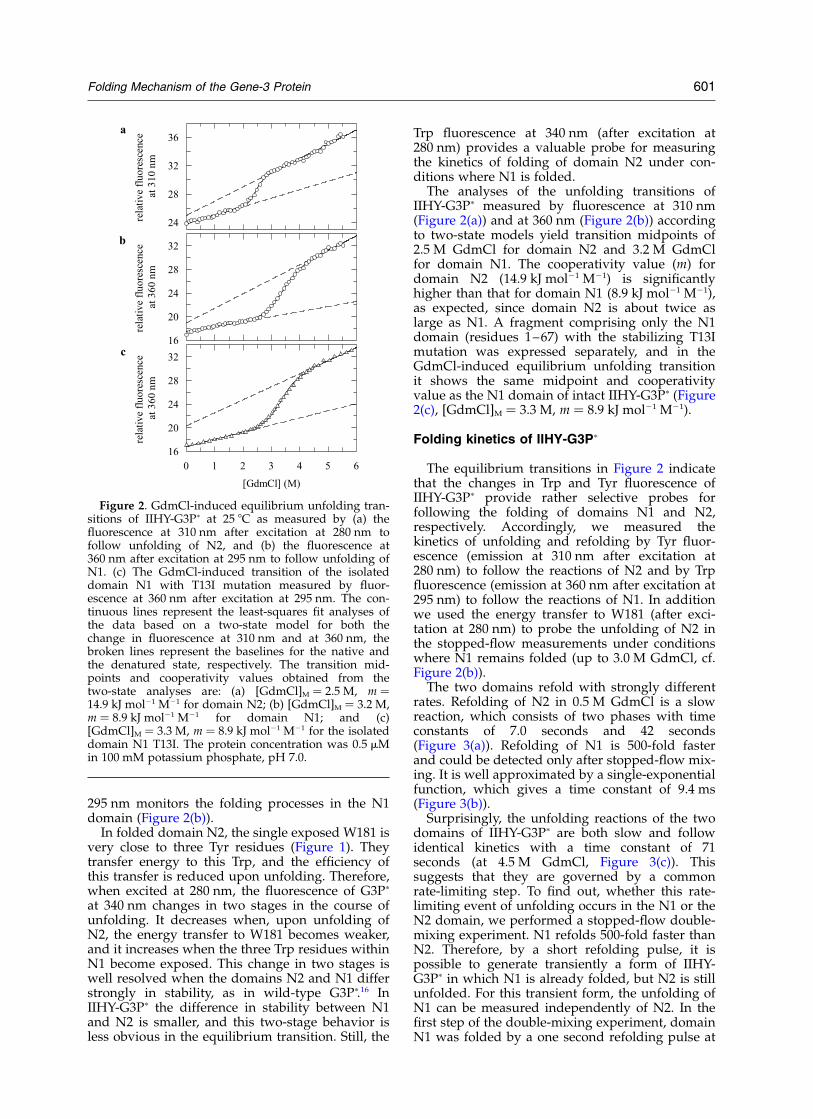
The analyses of the unfolding transitions ofIIHY-G3Pp measured by fluorescence at 310 nm(Figure 2(a)) and at 360 nm (Figure 2(b)) accordingto two-state models yield transition midpoints of2.5 M GdmCl for domain N2 and 3.2 M GdmClfor domain N1. The cooperativity value (m) fordomain N2 (14.9 kJ mol21 M21) is significantlyhigher than that for domain N1 (8.9 kJ mol21 M21),as expected, since domain N2 is about twice aslarge as N1. A fragment comprising only the N1domain (residues 1–67) with the stabilizing T13Imutation was expressed separately, and in theGdmCl-induced equilibrium unfolding transitionit shows the same midpoint and cooperativityvalue as the N1 domain of intact IIHY-G3Pp (Figure2(c), [GdmCl]M ¼ 3.3 M, m ¼ 8.9 kJ mol21 M21).
Folding kinetics of IIHY-G3Pp
The equilibrium transitions in Figure 2 indicatethat the changes in Trp and Tyr fluorescence ofIIHY-G3Pp provide rather selective probes forfollowing the folding of domains N1 and N2,respectively. Accordingly, we measured thekinetics of unfolding and refolding by Tyr fluor-escence (emission at 310 nm after excitation at280 nm) to follow the reactions of N2 and by Trpfluorescence (emission at 360 nm after excitation at295 nm) to follow the reactions of N1. In additionwe used the energy transfer to W181 (after exci-tation at 280 nm) to probe the unfolding of N2 inthe stopped-flow measurements under conditionswhere N1 remains folded (up to 3.0 M GdmCl, cf.Figure 2(b)).
The two domains refold with strongly differentrates. Refolding of N2 in 0.5 M GdmCl is a slowreaction, which consists of two phases with timeconstants of 7.0 seconds and 42 seconds(Figure 3(a)). Refolding of N1 is 500-fold fasterand could be detected only after stopped-flow mix-ing. It is well approximated by a single-exponentialfunction, which gives a time constant of 9.4 ms(Figure 3(b)).
Surprisingly, the unfolding reactions of the twodomains of IIHY-G3Pp are both slow and followidentical kinetics with a time constant of 71seconds (at 4.5 M GdmCl, Figure 3(c)). Thissuggests that they are governed by a commonrate-limiting step. To find out, whether this rate-limiting event of unfolding occurs in the N1 or theN2 domain, we performed a stopped-flow double-mixing experiment. N1 refolds 500-fold faster thanN2. Therefore, by a short refolding pulse, it ispossible to generate transiently a form of IIHY-G3Pp in which N1 is already folded, but N2 is stillunfolded. For this transient form, the unfolding ofN1 can be measured independently of N2. In thefirst step of the double-mixing experiment, domainN1 was folded by a one second refolding pulse at
Figure 2. GdmCl-induced equilibrium unfolding tran-sitions of IIHY-G3Pp at 25 8C as measured by (a) thefluorescence at 310 nm after excitation at 280 nm tofollow unfolding of N2, and (b) the fluorescence at360 nm after excitation at 295 nm to follow unfolding ofN1. (c) The GdmCl-induced transition of the isolateddomain N1 with T13I mutation measured by fluor-escence at 360 nm after excitation at 295 nm. The con-tinuous lines represent the least-squares fit analyses ofthe data based on a two-state model for both thechange in fluorescence at 310 nm and at 360 nm, thebroken lines represent the baselines for the native andthe denatured state, respectively. The transition mid-points and cooperativity values obtained from thetwo-state analyses are: (a) [GdmCl]M ¼ 2.5 M, m ¼14.9 kJ mol21 M21 for domain N2; (b) [GdmCl]M ¼ 3.2 M,m ¼ 8.9 kJ mol21 M21 for domain N1; and (c)[GdmCl]M ¼ 3.3 M, m ¼ 8.9 kJ mol21 M21 for the isolateddomain N1 T13I. The protein concentration was 0.5 mMin 100 mM potassium phosphate, pH 7.0.
Folding Mechanism of the Gene-3 Protein 601
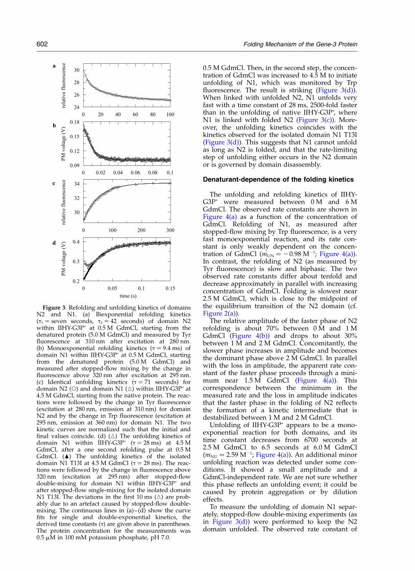
0.5 M GdmCl. Then, in the second step, the concen-tration of GdmCl was increased to 4.5 M to initiateunfolding of N1, which was monitored by Trpfluorescence. The result is striking (Figure 3(d)).When linked with unfolded N2, N1 unfolds veryfast with a time constant of 28 ms, 2500-fold fasterthan in the unfolding of native IIHY-G3Pp, whereN1 is linked with folded N2 (Figure 3(c)). More-over, the unfolding kinetics coincides with thekinetics observed for the isolated domain N1 T13I(Figure 3(d)). This suggests that N1 cannot unfoldas long as N2 is folded, and that the rate-limitingstep of unfolding either occurs in the N2 domainor is governed by domain disassembly.
Denaturant-dependence of the folding kinetics
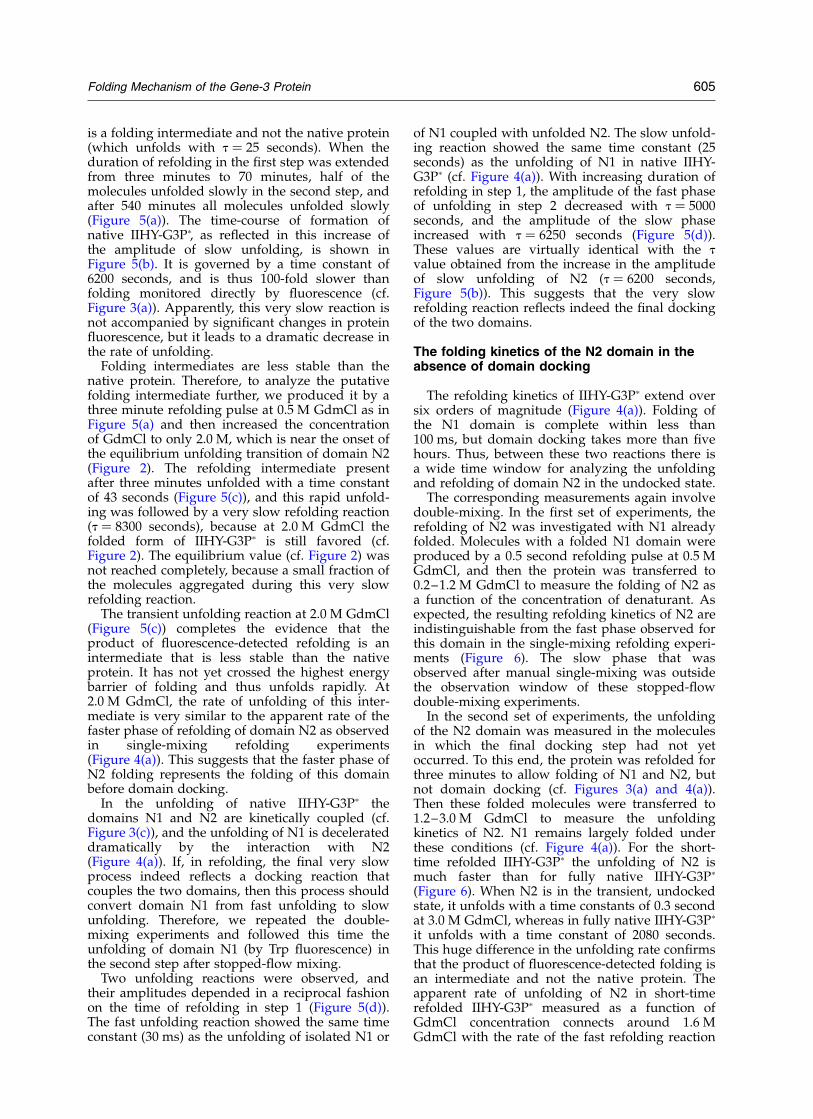
The unfolding and refolding kinetics of IIHY-G3Pp were measured between 0 M and 6 MGdmCl. The observed rate constants are shown inFigure 4(a) as a function of the concentration ofGdmCl. Refolding of N1, as measured afterstopped-flow mixing by Trp fluorescence, is a veryfast monoexponential reaction, and its rate con-stant is only weakly dependent on the concen-tration of GdmCl (mUN ¼ 20.98 M21; Figure 4(a)).In contrast, the refolding of N2 (as measured byTyr fluorescence) is slow and biphasic. The twoobserved rate constants differ about tenfold anddecrease approximately in parallel with increasingconcentration of GdmCl. Folding is slowest near2.5 M GdmCl, which is close to the midpoint ofthe equilibrium transition of the N2 domain (cf.Figure 2(a)).
The relative amplitude of the faster phase of N2refolding is about 70% between 0 M and 1 MGdmCl (Figure 4(b)) and drops to about 30%between 1 M and 2 M GdmCl. Concomitantly, theslower phase increases in amplitude and becomesthe dominant phase above 2 M GdmCl. In parallelwith the loss in amplitude, the apparent rate con-stant of the faster phase proceeds through a mini-mum near 1.5 M GdmCl (Figure 4(a)). Thiscorrespondence between the minimum in themeasured rate and the loss in amplitude indicatesthat the faster phase in the folding of N2 reflectsthe formation of a kinetic intermediate that isdestabilized between 1 M and 2 M GdmCl.
Unfolding of IIHY-G3Pp appears to be a mono-exponential reaction for both domains, and itstime constant decreases from 6700 seconds at2.5 M GdmCl to 6.5 seconds at 6.0 M GdmCl(mNU ¼ 2.59 M21; Figure 4(a)). An additional minorunfolding reaction was detected under some con-ditions. It showed a small amplitude and aGdmCl-independent rate. We are not sure whetherthis phase reflects an unfolding event; it could becaused by protein aggregation or by dilutioneffects.
To measure the unfolding of domain N1 separ-ately, stopped-flow double-mixing experiments (asin Figure 3(d)) were performed to keep the N2domain unfolded. The observed rate constant of
Figure 3. Refolding and unfolding kinetics of domainsN2 and N1. (a) Biexponential refolding kinetics(t1 ¼ seven seconds, t2 ¼ 42 seconds) of domain N2within IIHY-G3Pp at 0.5 M GdmCl, starting from thedenatured protein (5.0 M GdmCl) and measured by Tyrfluorescence at 310 nm after excitation at 280 nm.(b) Monoexponential refolding kinetics (t ¼ 9.4 ms) ofdomain N1 within IIHY-G3Pp at 0.5 M GdmCl, startingfrom the denatured protein (5.0 M GdmCl) andmeasured after stopped-flow mixing by the change influorescence above 320 nm after excitation at 295 nm.(c) Identical unfolding kinetics (t ¼ 71 seconds) fordomain N2 (W) and domain N1 (K) within IIHY-G3Pp at4.5 M GdmCl, starting from the native protein. The reac-tions were followed by the change in Tyr fluorescence(excitation at 280 nm, emission at 310 nm) for domainN2 and by the change in Trp fluorescence (excitation at295 nm, emission at 360 nm) for domain N1. The twokinetic curves are normalized such that the initial andfinal values coincide. (d) (K) The unfolding kinetics ofdomain N1 within IIHY-G3Pp (t ¼ 28 ms) at 4.5 MGdmCl, after a one second refolding pulse at 0.5 MGdmCl. (O) The unfolding kinetics of the isolateddomain N1 T13I at 4.5 M GdmCl (t ¼ 28 ms). The reac-tions were followed by the change in fluorescence above320 nm (excitation at 295 nm) after stopped-flowdouble-mixing for domain N1 within IIHY-G3Pp andafter stopped-flow single-mixing for the isolated domainN1 T13I. The deviations in the first 10 ms (K) are prob-ably due to an artefact caused by stopped-flow double-mixing. The continuous lines in (a)–(d) show the curvefits for single and double-exponential kinetics, thederived time constants (t) are given above in parentheses.The protein concentration for the measurements was0.5 mM in 100 mM potassium phosphate, pH 7.0.
602 Folding Mechanism of the Gene-3 Protein
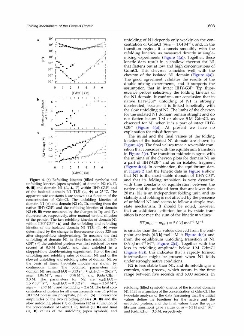
unfolding of N1 depends only weakly on the con-centration of GdmCl (mNU ¼ 1.04 M21), and, in thetransition region, it connects smoothly with therefolding kinetics, as measured directly in single-mixing experiments (Figure 4(a)). Together, thesekinetic data result in a shallow chevron for N1that flattens out at low and high concentrations ofGdmCl. This chevron coincides well with thechevron of the isolated N1 domain (Figure 4(a)).The good agreement validates the results of thedouble-mixing experiments, and it supports theassumption that in intact IIHY-G3Pp Trp fluor-escence probes selectively the folding kinetics ofthe N1 domain. It confirms our conclusion that innative IIHY-G3Pp unfolding of N1 is stronglydecelerated, because it is linked kinetically withthe slow unfolding of N2. The limbs of the chevronfor the isolated N1 domain remain straight and donot flatten below 1 M or above 5 M GdmCl, asobserved for N1 when it is a part of intact IIHY-G3Pp (Figure 4(a)). At present we have noexplanation for this difference.
The initial and the final values of the foldingkinetics of the isolated N1 domain are shown inFigure 4(c). The final values trace a reversible tran-sition that coincides with the equilibrium transitionin Figure 2(c). The transition midpoints agree withthe minima of the chevron plots for domain N1 asa part of IIHY-G3Pp and as an isolated fragment(Figure 4(a)). In combination, the equilibrium datain Figure 2 and the kinetic data in Figure 4 showthat N1 is the most stable domain of IIHY-G3Pp,and that its folding transition is very dynamic,with time constants of equilibration between thenative and the unfolded form that are lower than20 ms. N1 is an independent folding unit, and itsstability and folding is not affected by the presenceof unfolded N2 and seems to follow a simple two-state mechanism. It should be noted, however,that an additional criterion for a two-state tran-sition is not met: the sum of the kinetic m values:
RTðmNU 2 mUNÞ ¼ 5:0 kJ mol21 M21
is smaller than the m values derived from the end-point analysis (6.3 kJ mol21 M21; Figure 4(c)) andfrom the equilibrium unfolding transition of N1(8.9 kJ mol21 M21, Figure 2(c)). Together with theloss in refolding amplitude below 1 M GdmCl(Figure 4(c)), this indicates that a rapidly formedintermediate might be present when N1 foldsunder strongly native conditions.
N2 is less stable than N1, and its refolding is acomplex, slow process, which occurs in the timerange between five seconds and 6000 seconds. In
Figure 4. (a) Refolding kinetics (filled symbols) andunfolding kinetics (open symbols) of domain N2 (W, A,X, B) and domain N1 (K, O, L) within IIHY-G3Pp, andof the isolated domain N1 T13I (S, V) at 25 8C. Theapparent rate constants l are shown as a function of theconcentration of GdmCl. The unfolding kinetics ofdomain N1 (K) and domain N2 (W, A), starting from thenative IIHY-G3Pp, and the refolding kinetics of domainN2 (X, B) were measured by the changes in Trp and Tyrfluorescence, respectively, after manual tenfold dilutionof the protein. The fast refolding kinetics of domain N1within IIHY-G3Pp (O) and the unfolding and refoldingkinetics of the isolated domain N1 T13I (S, V) weredetermined by the change in fluorescence above 320 nmafter stopped-flow single-mixing. To measure the fastunfolding of domain N1 in short-time refolded IIHY-G3Pp (L) the unfolded protein was first refolded for onesecond at 0.5 M GdmCl and then unfolded in astopped-flow double-mixing experiment. Fits of the fastunfolding and refolding rates of domain N1 and of theslowest unfolding and refolding rates of domain N2 onthe basis of linear two-state models are shown bycontinuous lines. The obtained parameters fordomain N1 are kNU(H2O) ¼ 0.33 s21, kUN(H2O) ¼ 262 s21,mNU ¼ 1.04 M21, mUN ¼ 20.98 M21, and [GdmCl]M ¼3.3 M. The parameters for N2 are kNU(H2O) ¼1.5 £ 1027 s21, kUN(H2O) ¼ 0.052 s21, mNU ¼ 2.59 M21,mUN ¼ 22.77 M21, and [GdmCl]M ¼ 2.4 M. The final con-centration of protein for all measurements was 0.5 mM in100 mM potassium phosphate, pH 7.0. (b) Fluorescenceamplitudes of the two refolding phases (X, B) and theslow unfolding phase (W) of domain N2 as a function ofthe concentration of GdmCl. (c) Initial ([, ) and final(S, V) values of the unfolding (open symbols) and
refolding (filled symbols) kinetics of the isolated domainN1 T13I as a function of the concentration of GdmCl. Thetwo-state analysis of the data (assuming that the initialvalues define the baselines for the native and theunfolded protein, and the final values trace the equi-librium transition) gave values of m ¼ 6.3 kJ mol21 M21
and [GdmCl]M ¼ 3.5 M, respectively.
Folding Mechanism of the Gene-3 Protein 603
their unfolding, the two domains are kineticallycoupled, and N1 cannot unfold as long as it islocked in its native conformation by the folded N2domain. Thus, although N2 is less stable than N1,it provides a kinetic protection for N1 anddecelerates its unfolding up to 150,000-fold (at2.5 M GdmCl).
Kinetics of the formation of fully foldedIIHY-G3Pp molecules
G3P has no catalytic activity, and its N1 and N2domains do not exert their biological functionsuntil the phage infects an E. coli cell. This occurs along time after phage coat assembly. Therefore,functional assays cannot be used to follow thekinetics of renaturation in real time or to examinewhether the product of fluorescence-detected fold-ing is indeed the native protein. Previously, wedeveloped a generic two-step assay for measuringthe kinetics of the formation of fully folded proteinmolecules during a refolding reaction.17,18 Thisassay is based on the fact that the proteinmolecules that have passed beyond the highestactivation barrier in their refolding, and thus havereached the native state, are separated from theunfolded state by this energy barrier. They unfoldslowly when the conditions are switched to unfold-ing, because they must cross the high barrier again,now in the reverse direction. Partially foldedmolecules, however, that have not yet passed thefinal transition state, unfold rapidly. This translatesinto a double-mixing procedure. First, unfoldedprotein is mixed with refolding buffer to initiaterefolding. Then, after variable time intervals,samples are withdrawn, transferred to standardunfolding conditions, and the amplitude of thesubsequent slow unfolding reaction is determined.It is a direct measure for the amount of native mol-ecules that had been present at the time whenrefolding was interrupted.
This procedure was used to follow the formationof fully folded IIHY-G3Pp molecules during refold-ing in 0.5 M GdmCl. The unfolding assays wereperformed in 5.0 M GdmCl at 25 8C, conditionsunder which native IIHY-G3Pp unfolds in a mono-exponential reaction with a time constant of25 seconds (cf. Figure 4(a)).
In 0.5 M GdmCl, fluorescence-detected refoldingof IIHY-G3Pp is complete within three minutes(Figure 3(a)). An unfolding assay at 5.0 M GdmClperformed after three minutes of refolding andfollowed by Tyr fluorescence (unfolding of domainN2) revealed, however, that virtually all moleculeshad unfolded already within the dead-time ofmanual mixing (Figure 5(a)). This suggests thatthe product of the fluorescence-detected refolding
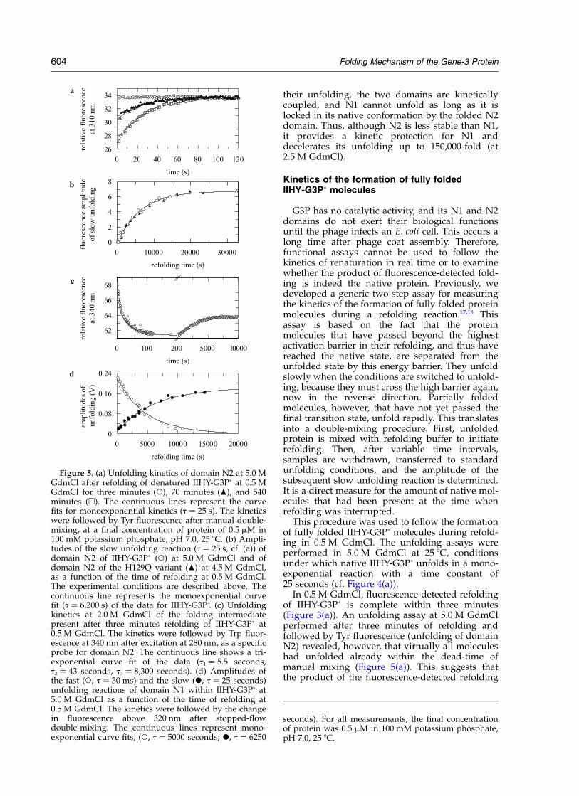
Figure 5. (a) Unfolding kinetics of domain N2 at 5.0 MGdmCl after refolding of denatured IIHY-G3Pp at 0.5 MGdmCl for three minutes (W), 70 minutes (O), and 540minutes (A). The continuous lines represent the curvefits for monoexponential kinetics (t ¼ 25 s). The kineticswere followed by Tyr fluorescence after manual double-mixing, at a final concentration of protein of 0.5 mM in100 mM potassium phosphate, pH 7.0, 25 8C. (b) Ampli-tudes of the slow unfolding reaction (t ¼ 25 s, cf. (a)) ofdomain N2 of IIHY-G3Pp (W) at 5.0 M GdmCl and ofdomain N2 of the H129Q variant (O) at 4.5 M GdmCl,as a function of the time of refolding at 0.5 M GdmCl.The experimental conditions are described above. Thecontinuous line represents the monoexponential curvefit (t ¼ 6,200 s) of the data for IIHY-G3Pp. (c) Unfoldingkinetics at 2.0 M GdmCl of the folding intermediatepresent after three minutes refolding of IIHY-G3Pp at0.5 M GdmCl. The kinetics were followed by Trp fluor-escence at 340 nm after excitation at 280 nm, as a specificprobe for domain N2. The continuous line shows a tri-exponential curve fit of the data (t1 ¼ 5.5 seconds,t2 ¼ 43 seconds, t3 ¼ 8,300 seconds). (d) Amplitudes ofthe fast (W, t ¼ 30 ms) and the slow (X, t ¼ 25 seconds)unfolding reactions of domain N1 within IIHY-G3Pp at5.0 M GdmCl as a function of the time of refolding at0.5 M GdmCl. The kinetics were followed by the changein fluorescence above 320 nm after stopped-flowdouble-mixing. The continuous lines represent mono-exponential curve fits, (W, t ¼ 5000 seconds; X, t ¼ 6250
seconds). For all measuremants, the final concentrationof protein was 0.5 mM in 100 mM potassium phosphate,pH 7.0, 25 8C.
604 Folding Mechanism of the Gene-3 Protein
is a folding intermediate and not the native protein(which unfolds with t ¼ 25 seconds). When theduration of refolding in the first step was extendedfrom three minutes to 70 minutes, half of themolecules unfolded slowly in the second step, andafter 540 minutes all molecules unfolded slowly(Figure 5(a)). The time-course of formation ofnative IIHY-G3Pp, as reflected in this increase ofthe amplitude of slow unfolding, is shown inFigure 5(b). It is governed by a time constant of6200 seconds, and is thus 100-fold slower thanfolding monitored directly by fluorescence (cf.Figure 3(a)). Apparently, this very slow reaction isnot accompanied by significant changes in proteinfluorescence, but it leads to a dramatic decrease inthe rate of unfolding.
Folding intermediates are less stable than thenative protein. Therefore, to analyze the putativefolding intermediate further, we produced it by athree minute refolding pulse at 0.5 M GdmCl as inFigure 5(a) and then increased the concentrationof GdmCl to only 2.0 M, which is near the onset ofthe equilibrium unfolding transition of domain N2(Figure 2). The refolding intermediate presentafter three minutes unfolded with a time constantof 43 seconds (Figure 5(c)), and this rapid unfold-ing was followed by a very slow refolding reaction(t ¼ 8300 seconds), because at 2.0 M GdmCl thefolded form of IIHY-G3Pp is still favored (cf.Figure 2). The equilibrium value (cf. Figure 2) wasnot reached completely, because a small fraction ofthe molecules aggregated during this very slowrefolding reaction.
The transient unfolding reaction at 2.0 M GdmCl(Figure 5(c)) completes the evidence that theproduct of fluorescence-detected refolding is anintermediate that is less stable than the nativeprotein. It has not yet crossed the highest energybarrier of folding and thus unfolds rapidly. At2.0 M GdmCl, the rate of unfolding of this inter-mediate is very similar to the apparent rate of thefaster phase of refolding of domain N2 as observedin single-mixing refolding experiments(Figure 4(a)). This suggests that the faster phase ofN2 folding represents the folding of this domainbefore domain docking.
In the unfolding of native IIHY-G3Pp thedomains N1 and N2 are kinetically coupled (cf.Figure 3(c)), and the unfolding of N1 is decelerateddramatically by the interaction with N2(Figure 4(a)). If, in refolding, the final very slowprocess indeed reflects a docking reaction thatcouples the two domains, then this process shouldconvert domain N1 from fast unfolding to slowunfolding. Therefore, we repeated the double-mixing experiments and followed this time theunfolding of domain N1 (by Trp fluorescence) inthe second step after stopped-flow mixing.
Two unfolding reactions were observed, andtheir amplitudes depended in a reciprocal fashionon the time of refolding in step 1 (Figure 5(d)).The fast unfolding reaction showed the same timeconstant (30 ms) as the unfolding of isolated N1 or
of N1 coupled with unfolded N2. The slow unfold-ing reaction showed the same time constant (25seconds) as the unfolding of N1 in native IIHY-G3Pp (cf. Figure 4(a)). With increasing duration ofrefolding in step 1, the amplitude of the fast phaseof unfolding in step 2 decreased with t ¼ 5000seconds, and the amplitude of the slow phaseincreased with t ¼ 6250 seconds (Figure 5(d)).These values are virtually identical with the tvalue obtained from the increase in the amplitudeof slow unfolding of N2 (t ¼ 6200 seconds,Figure 5(b)). This suggests that the very slowrefolding reaction reflects indeed the final dockingof the two domains.
The folding kinetics of the N2 domain in theabsence of domain docking
The refolding kinetics of IIHY-G3Pp extend oversix orders of magnitude (Figure 4(a)). Folding ofthe N1 domain is complete within less than100 ms, but domain docking takes more than fivehours. Thus, between these two reactions there isa wide time window for analyzing the unfoldingand refolding of domain N2 in the undocked state.
The corresponding measurements again involvedouble-mixing. In the first set of experiments, therefolding of N2 was investigated with N1 alreadyfolded. Molecules with a folded N1 domain wereproduced by a 0.5 second refolding pulse at 0.5 MGdmCl, and then the protein was transferred to0.2–1.2 M GdmCl to measure the folding of N2 asa function of the concentration of denaturant. Asexpected, the resulting refolding kinetics of N2 areindistinguishable from the fast phase observed forthis domain in the single-mixing refolding experi-ments (Figure 6). The slow phase that wasobserved after manual single-mixing was outsidethe observation window of these stopped-flowdouble-mixing experiments.
In the second set of experiments, the unfoldingof the N2 domain was measured in the moleculesin which the final docking step had not yetoccurred. To this end, the protein was refolded forthree minutes to allow folding of N1 and N2, butnot domain docking (cf. Figures 3(a) and 4(a)).Then these folded molecules were transferred to1.2–3.0 M GdmCl to measure the unfoldingkinetics of N2. N1 remains largely folded underthese conditions (cf. Figure 4(a)). For the short-time refolded IIHY-G3Pp the unfolding of N2 ismuch faster than for fully native IIHY-G3Pp
(Figure 6). When N2 is in the transient, undockedstate, it unfolds with a time constants of 0.3 secondat 3.0 M GdmCl, whereas in fully native IIHY-G3Pp
it unfolds with a time constant of 2080 seconds.This huge difference in the unfolding rate confirmsthat the product of fluorescence-detected folding isan intermediate and not the native protein. Theapparent rate of unfolding of N2 in short-timerefolded IIHY-G3Pp measured as a function ofGdmCl concentration connects around 1.6 MGdmCl with the rate of the fast refolding reaction
Folding Mechanism of the Gene-3 Protein 605
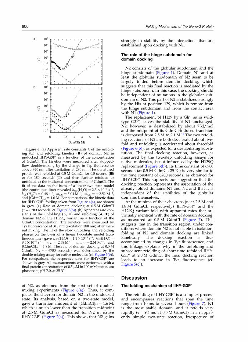
of N2, as obtained from the first set of double-mixing experiments (Figure 6(a)). Thus, it com-pletes the chevron for domain N2 in the undockedstate. Its analysis, based on a two-state model,gave a transition midpoint of [GdmCl]M ¼ 1.6 M,which is much lower than the transition midpointof 2.5 M GdmCl as measured for N2 in nativeIIHY-G3Pp (Figure 2(a)). This shows that N2 gains
strongly in stability by the interactions that areestablished upon docking with N1.
The role of the hinge subdomain fordomain docking
N2 consists of the globular subdomain and thehinge subdomain (Figure 1). Domain N1 and atleast the globular subdomain of N2 seem to belargely folded before domain docking, whichsuggests that this final reaction is mediated by thehinge subdomain. In this case, the docking shouldbe independent of mutations in the globular sub-domain of N2. This part of N2 is stabilized stronglyby the His at position 129, which is remote fromthe hinge subdomain and from the contact areawith N1 (Figure 1).
The replacement of H129 by a Gln, as in wild-type G3Pp, leaves the stability of N1 unchanged.N2, however, is destabilized by about 7 kJ/moland the midpoint of its GdmCl-induced transitionis decreased from 2.5 M to 2.1 M.16 The two refold-ing reactions of N2 are both decelerated about five-fold and unfolding is accelerated about threefold(Figure 6(b)), as expected for a destabilizing substi-tution. The final docking reaction, however, asmeasured by the two-step unfolding assays fornative molecules, is not influenced by the H129Qreplacement (Figure 5(b)). Its time constant of 6700seconds (at 0.5 M GdmCl, 25 8C) is very similar tothe time constant of 6200 seconds, as obtained forIIHY-G3Pp. This supports our suggestion that thedocking reaction represents the association of thealready folded domains N1 and N2 and that it isindependent of the stabilities of the globulardomains themselves.
At the minima of their chevrons (near 2.5 M and1.8 M GdmCl, respectively) IIHY-G3Pp and theH129Q variant fold with apparent rates that arevirtually identical with the rate of domain docking,as measured at 0.5 M GdmCl (Figure 7). Thissuggests that in the transition region, under con-ditions where domain N2 is not stable in isolation,folding of N2 and domain docking are linkedkinetically. The docking reaction is thusaccompanied by changes in Tyr fluorescence, andthis linkage explains why in the unfolding andsubsequent refolding of short-time refolded IIHY-G3Pp at 2.0 M GdmCl the final docking reactionleads to an increase in Tyr fluorescence (cf.Figure 5(c)).
Discussion
The folding mechanism of IIHY-G3Pp
The refolding of IIHY-G3Pp is a complex processand encompasses reactions that span the timerange from 10 ms to several hours (Figure 7). N1is the most stable domain, and it refolds veryrapidly (t ¼ 9.4 ms at 0.5 M GdmCl) in an appar-ently simple two-state reaction, irrespective of
Figure 6. (a) Apparent rate constants l of the unfold-ing (A) and refolding kinetics (B) of domain N2 inundocked IIHY-G3Pp as a function of the concentrationof GdmCl. The kinetics were measured after stopped-flow double-mixing by the change in Trp fluorescenceabove 320 nm after excitation at 280 nm. The denaturedprotein was refolded at 0.5 M GdmCl for 0.5 second (B)or for 180 seconds (A) and then further refolded orunfolded at the indicated concentrations of GdmCl. Thefit of the data on the basis of a linear two-state model(the continuous line) revealed kNU(H2O) ¼ 2.3 £ 1026 s21,kUN(H2O) ¼ 0.48 s21, mNU ¼ 5.04 M21, mUN ¼ 22.52 M21,and [GdmCl]M ¼ 1.6 M. For comparison, the kinetic datafor IIHY-G3Pp folding taken from Figure 4(a), are shownin grey. (p ) Rate of domain docking at 0.5 M GdmCl(t ¼ 6200 seconds, cf. Figure 5(b)). (b) Apparent rate con-stants of the unfolding (K, S) and refolding (O, V) ofdomain N2 of the H129Q variant as a function of theGdmCl concentration. The kinetics were measured byTyr fluorescence at 310 nm (excitation 280 nm) after man-ual mixing. The fit of the slow unfolding and refoldingphases on the basis of a linear two-state model (con-tinuous line) gave kNU(H2O) ¼ 1.1 £ 1026 s21, kUN(H2O) ¼8.5 £ 1023 s21, mNU ¼ 2.38 M21, mUN ¼ 22.61 M21, and[GdmCl]M ¼ 1.8 M. The rate of domain docking at 0.5 MGdmCl (p , t ¼ 6700 seconds) was determined by thedouble-mixing assay for native molecules (cf. Figure 5(b)).For comparison, the respective data for IIHY-G3Pp areshown in grey. All measurements were performed with afinal protein concentration of 0.5 mM in 100 mM potassiumphosphate, pH 7.0, at 25 8C.
606 Folding Mechanism of the Gene-3 Protein
whether it is present as an isolated protein frag-ment or as a part of IIHY-G3Pp. For unfolding, theresults are different. In isolation, N1 unfolds veryfast (t ¼ 28 ms at 4.5 M GdmCl), but as a part ofnative IIHY-G3Pp it unfolds 2500-fold more slowly(t ¼ 71 seconds).
The N2 domain is less stable than N1 and, infact, a major part of the Gibbs free energy of stabil-ization of N2 originates from its interaction withN1 in the fully folded protein. This interaction isprovided primarily by the hinge subdomain ofN2, which partially wraps around N1 in foldedG3Pp.6,8,9 Our previous analysis of the equilibriumstability of IIHY-G3Pp showed that the loss of thedomain interactions and the unfolding of N2indeed occur in a single cooperative transition.16
N2 refolds much more slowly than N1, and itsfolding is a complex process, which involves two,probably sequential reactions with time constants
of seven seconds and 42 seconds (at 0.5 M GdmCl,Figure 7). Both are strongly denaturant-dependent.Probably, they reflect the folding of at least theglobular part of domain N2 and the formation ofloose contacts with domain N1.
The folding reactions of the N1 and N2 domainsare followed by a very slow, spectroscopicallysilent reaction with a time constant of 6200 seconds(Figure 7). This reaction would have been over-looked in conventional single-mixing experiments,because it starts from a folding intermediate thatappeared native-like in its fluorescence properties.However, this intermediate unfolded more than8000-fold faster (at 3.0 M GdmCl, 25 8C) than fullyfolded protein. Its final conversion to the nativestate could therefore be measured by a kineticdouble-mixing assay for native molecules that isbased on this vast difference in the unfolding rates.
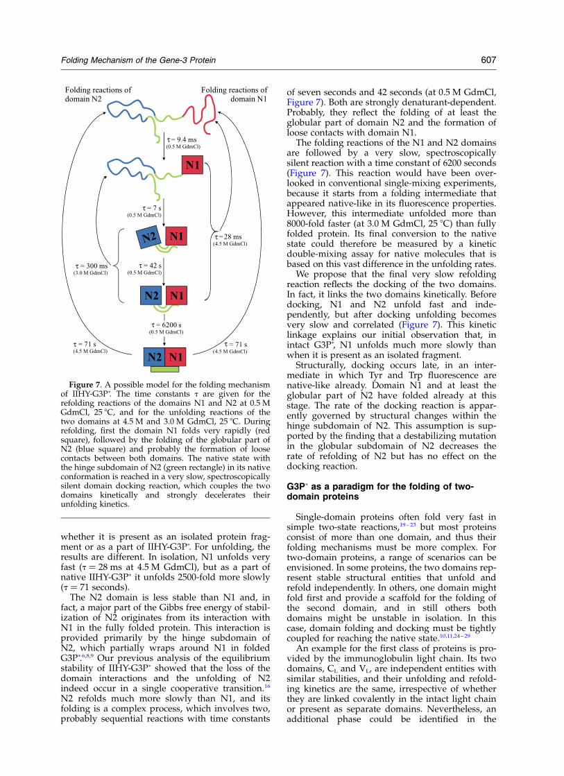
We propose that the final very slow refoldingreaction reflects the docking of the two domains.In fact, it links the two domains kinetically. Beforedocking, N1 and N2 unfold fast and inde-pendently, but after docking unfolding becomesvery slow and correlated (Figure 7). This kineticlinkage explains our initial observation that, inintact G3Pp, N1 unfolds much more slowly thanwhen it is present as an isolated fragment.
Structurally, docking occurs late, in an inter-mediate in which Tyr and Trp fluorescence arenative-like already. Domain N1 and at least theglobular part of N2 have folded already at thisstage. The rate of the docking reaction is appar-ently governed by structural changes within thehinge subdomain of N2. This assumption is sup-ported by the finding that a destabilizing mutationin the globular subdomain of N2 decreases therate of refolding of N2 but has no effect on thedocking reaction.
G3Pp as a paradigm for the folding of two-domain proteins
Single-domain proteins often fold very fast insimple two-state reactions,19 – 23 but most proteinsconsist of more than one domain, and thus theirfolding mechanisms must be more complex. Fortwo-domain proteins, a range of scenarios can beenvisioned. In some proteins, the two domains rep-resent stable structural entities that unfold andrefold independently. In others, one domain mightfold first and provide a scaffold for the folding ofthe second domain, and in still others bothdomains might be unstable in isolation. In thiscase, domain folding and docking must be tightlycoupled for reaching the native state.10,11,24 – 29
An example for the first class of proteins is pro-vided by the immunoglobulin light chain. Its twodomains, CL and VL, are independent entities withsimilar stabilities, and their unfolding and refold-ing kinetics are the same, irrespective of whetherthey are linked covalently in the intact light chainor present as separate domains. Nevertheless, anadditional phase could be identified in the
Figure 7. A possible model for the folding mechanismof IIHY-G3Pp. The time constants t are given for therefolding reactions of the domains N1 and N2 at 0.5 MGdmCl, 25 8C, and for the unfolding reactions of thetwo domains at 4.5 M and 3.0 M GdmCl, 25 8C. Duringrefolding, first the domain N1 folds very rapidly (redsquare), followed by the folding of the globular part ofN2 (blue square) and probably the formation of loosecontacts between both domains. The native state withthe hinge subdomain of N2 (green rectangle) in its nativeconformation is reached in a very slow, spectroscopicallysilent domain docking reaction, which couples the twodomains kinetically and strongly decelerates theirunfolding kinetics.
Folding Mechanism of the Gene-3 Protein 607
refolding of the intact light chain, which wasascribed to domain docking.25 In gII crystallin ofthe eye lens, the two domains differ in stability,and the less stable C-terminal domain is stabilizedby its interactions with the N-terminal domain.The folding kinetics of gII crystallin are welldescribed by two interdigitated chevrons thatreflect the folding of the two domains, a separatedomain docking step could not be identified in thekinetic analysis.24
In the case of G3Pp, the folding kinetics of thetwo domains and the kinetics of domain dockingare well separated. Figure 7 shows our model forthe folding mechanism of G3Pp. N1 is the moststable domain and folds within a few milliseconds.It provides a framework for the folding of N2,which proceeds in the time range of a few minutes,probably in loose association with N1. Finally, thetwo domains become docked in a very slow reac-tion with a time constant of about 6000 seconds.Such a very slow domain docking reaction has notbeen observed before. This docking occurs afterconformational folding, but nevertheless it leadsto the highest energy barrier along the foldingpath and thus to a strong kinetic protection againstunfolding for the two domains. Unfolding of N1 isdecelerated up to 150,000-fold, as compared withthe undocked state.
Folding and function of G3P
The very slow domain assembly and dis-assembly reactions are the last step in refoldingand the first step in unfolding of G3Pp, respectively,and they might be important for the function ofthis protein. G3P is located at the tip of the phageand mediates the infection of E. coli. The N1–N2part protrudes from the surface of the phagevirions, and the two domains must remain stablyassociated to protect them from inactivation indiverse environments. Such a protection is accom-plished by the very high activation barrier fordomain disassembly.
As soon as infection is initiated by the inter-action of N2 with the tip of an F pilus, the twodomains must come apart to expose the bindingsite for TolA on the N1 domain. The open stateshould persist until N1 and TolA are close enoughto interact with each other. Riechmann & Holliger3
found by ELISA measurements that isolated N1binds to TolA about 100-fold more tightly than todomain N2, but still, within G3P binding of N1 toN2 is entropically favored because the twodomains are covalently linked. The very slowdomain re-assembly reaction might ensure that,once N1 and N2 became separated, domain closureis blocked long enough that the pilus can retract tobring N1 into close proximity of Tol A and allowthe two to interact.
Apparently, the rates of the individual steps inthe folding of G3Pp seem to be determined byfunction rather than by the physical principles offolding. The fast folding of the N1 domain is
important to rapidly present a platform for thesubsequent steps in folding, and the final dockingstep provides a strong kinetic stabilization of G3P.Protection by a high activation barrier has itsprice: folding to the docked state over this barrieris very slow. Other extracellular proteins, such assecreted proteases, are protected by a high barrierof activation energy against unfolding. They avoidvery slow folding by coupling barrier formationwith the proteolytic removal of a pro-domain afterfolding. This final reaction is important for func-tion: it generates the proteolytic activity. Such acoupling between folding and barrier formationhas been well characterized for a-lytic protease.30,31
The phage virions and thus G3P mature in thebacterial periplasm, and it remains to be seenwhether they can use the host folding machineryfor accelerating their own folding.
Materials and Methods
Expression and purification of G3Pp and of theisolated domain N1
For the expression of IIHY-G3Pp (residues 1–217 ofmature G3P, plus ProSerGly(His)6) and its H129Q variantthe gene fragments coding for the two N-terminaldomains of G3P with the stabilizing mutations T13I,T101I, Q129H, D209Y and T13I, T101I, D209Y, respect-ively, were PCR-amplified from the correspondingsingle-stranded phage DNA. The fragments were clonedinto the expression plasmid pET11a (Novagen, Madison,Wisconsin, USA) via its Nde I and Bam HI restriction sites,the proteins were overproduced in E. coli BL21(DE3)-pLysS (Stratagene, La Jolla, USA) and purified asdescribed.16
For the expression of the isolated domain N1 T13I(residues 1–67 of mature G3P, plus Ala(His)6) the G3P-N1 fragment with signal sequence was amplified fromthe phage DNA, cloned into pET11a, and the proteinwas overproduced in E. coli BL21(DE3)pLysS. It waspurified as described.16
GdmCl-induced unfolding transitions
The samples of IIHY-G3Pp were prepared asdescribed,16 their fluorescence was measured in 10 mmcells at 310 nm after excitation at 280 nm and at 360 nmafter excitation at 295 nm. The experimental data wereanalyzed according to two-state models by non-linearleast-squares fit with proportional weighting to obtainthe Gibbs free energy of denaturation DGD as a functionof the concentration of GdmCl.32 The band-widths were5 nm and 10 nm for excitation and emission, respectively.Hitachi F-4010 and F-4500 fluorescence spectrometerswere used.
Kinetic single and double-mixing experiments
All GdmCl-induced unfolding and refolding experi-ments were performed in 100 mM potassium phosphate(pH 7.0), at 25 8C with a final protein concentration of0.5 mM. Slow kinetics were measured after a manual ten-fold dilution of the native or denatured (in 5.0 M GdmCl)protein with GdmCl solutions of varying concentrations
608 Folding Mechanism of the Gene-3 Protein
to give final concentrations of GdmCl between 1.5 M and6.0 M for unfolding and 0.25–2.5 M for refolding. Thechanges in Tyr fluorescence (emission at 310 nm afterexcitation at 280 nm) and Trp fluorescence (emission at360 nm after excitation at 295 nm) were monitored in a10 mm cell with a Hitachi F-4010 fluorescence spec-trometer at a response of 0.5 second and band-widths of5 nm and 10 nm for excitation and emission, respectively.
Fast refolding and unfolding reactions were measuredafter stopped-flow mixing in a DX.17MV spectrometerfrom Applied Photophysics (Leatherhead, UK). Thenative or unfolded (in 6.6 M GdmCl) protein was diluted11-fold with GdmCl solutions of various concentrations.The kinetics were followed by the change in fluorescenceabove 320 nm after excitation at 295 nm (10 nm band-width) in an observation chamber with 2 mm path-length. A 0.5 cm cell with acetone (transparent above320 nm) was placed between the observation chamberand the photomultiplier to absorb scattered light fromthe excitation beam. The kinetics were measured at leastfive times under identical conditions and averaged.
The fast unfolding kinetics of domain N1 linked withdenatured domain N2 were measured in stopped-flowdouble-mixing experiments. In the first step, 27.5 mMunfolded protein in 6.0 M GdmCl was diluted 11-foldwith buffer to initiate refolding at 0.55 M GdmCl, and inthe second step after one second of refolding it wasdiluted sixfold with GdmCl solutions of varying concen-trations to unfold domain N1 again. The changes influorescence above 320 nm after excitation at 295 nmwere measured as described above.
Stopped-flow double-mixing was also used to obtainthe refolding and unfolding kinetics of domain N2 inthe presence of the already folded domain N1 but beforethe final domain docking reaction. For the measurementof refolding the unfolded protein (66 mM in 5.5 MGdmCl) was first diluted 11-fold with buffer to allowrefolding of domain N1 for 0.5 second at 0.5 M GdmCl,and then diluted sixfold with GdmCl solutions ofvarying concentrations to obtain final concentrations of0.2–1.2 M GdmCl and 1 mM protein. To analyze theunfolding of domain N2 before domain docking withN1 the unfolded protein (66 mM in 5.5 M GdmCl) wasrefolded for three minutes by an 11-fold dilution withbuffer and unfolded again by a sixfold dilution withGdmCl solutions of varying concentrations to give finalconcentrations of GdmCl between 1.5 M and 3.0 M. Thefolding reactions of N2 were monitored by the changesin the fluorescence of W181 above 320 nm after excitationat 280 nm (10 nm band-width).
The dependences on GdmCl of the apparent rate con-stants for domains N1 and N2 were analyzed accordingto two-state models to obtain the microscopic rate con-stants and kinetic m-values of unfolding and refolding.33
Kinetics of domain docking measured by the two-step assay for native molecules
To follow the formation of native IIHY-G3Pp and theH129Q variant, unfolded protein (50 mM in 5.0 MGdmCl) was first manually diluted tenfold with bufferto initiate refolding at 0.5 M GdmCl and then, aftertimes of refolding between one minute and 540 minutes,it was unfolded again at 5.0 M GdmCl either by manualdilution tenfold with a GdmCl solution of 5.5 M or by11-fold stopped-flow mixing with 5.45 M GdmCl. Themanual mixing in the Hitachi F-4010 fluorescence spec-trometer was used to follow the slow unfolding reaction
(t ¼ 25 seconds) of domain N2 by the change in Tyrfluorescence at 310 nm (10 nm band-width) after exci-tation at 280 nm (5 nm band-width). In the stopped-flowspectrometer, the fast (t ¼ 30 ms) and the slow (t ¼ 25seconds) unfolding phases of domain N1 were detectedby the fluorescence above 320 nm after excitation at295 nm (10 nm band-width).
The unfolding of the short-time refolded intermediateafter three minutes at 0.5 M GdmCl was initiated by amanual dilution tenfold to 2.0 M GdmCl and monitoredby the change in the fluorescence of W181 at 340 nmafter excitation at 280 nm.
Acknowledgements
We thank the members of our laboratory formany discussions. This work was supported bygrants from the Deutsche Forschungsgemeinschaftand the Fonds der Chemischen Industrie.
References
1. Levengood, S. K., Beyer, W. F., Jr & Webster, R. E.(1991). TolA: a membrane protein involved in colicinuptake contains an extended helical region. Proc.Natl Acad. Sci. USA, 88, 5939–5943.
2. Click, E. M. & Webster, R. E. (1997). Filamentousphage infection: required interactions with the TolAprotein. J. Bacteriol. 179, 6464–6471.
3. Riechmann, L. & Holliger, P. (1997). The C-terminaldomain of TolA is the coreceptor for filamentousphage infection of E. coli. Cell, 90, 351–360.
4. Stengele, I., Bross, P., Garces, X., Giray, J. & Rasched,I. (1990). Dissection of functional domains in phagefd adsorption protein. Discrimination betweenattachment and penetration sites. J. Mol. Biol. 212,143–149.
5. Marvin, D. A. (1998). Filamentous phage structure,infection and assembly. Curr. Opin. Struct. Biol. 8,150–158.
6. Lubkowski, J., Hennecke, F., Pluckthun, A. &Wlodawer, A. (1999). Filamentous phage infection:crystal structure of g3p in complex with its corecep-tor, the C-terminal domain of TolA. Structure, 7,711–722.
7. Boeke, J. D. & Model, P. (1982). A prokaryoticmembrane anchor sequence: carboxyl terminus ofbacteriophage f1 gene III protein retains it in themembrane. Proc. Natl Acad. Sci. USA, 79, 5200–5204.
8. Lubkowski, J., Hennecke, F., Pluckthun, A. &Wlodawer, A. (1998). The structural basis of phagedisplay elucidated by the crystal structure of theN-terminal domains of G3p. Nature Struct. Biol. 5,140–147.
9. Holliger, P., Riechmann, L. & Williams, R. L. (1999).Crystal structure of the two N-terminal domains ofg3p from filamentous phage fd at 1.9 A: evidencefor conformational lability. J. Mol. Biol. 288, 649–657.
10. Garel, J.-R. (1992). Folding of large proteins:multi-domain and multisubunit proteins. In Protein Folding(Creighton, T. E., ed.), pp. 405–454, Freeman, NewYork.
11. Jaenicke, R. (1999). Stability and folding of domainproteins. Prog. Biophys. Mol. Biol. 71, 155–241.
Folding Mechanism of the Gene-3 Protein 609
12. Netzer, W. J. & Hartl, F. U. (1997). Recombination ofprotein domains facilitated by co-translational fold-ing in eukaryotes. Nature, 388, 343–349.
13. Deng, L. W. & Perham, R. N. (2002). Delineating thesite of interaction on the pIII protein of filamentousbacteriophage fd with the F-pilus of Escherichia coli.J. Mol. Biol. 319, 603–614.
14. Jacobson, A. (1972). Role of F pili in the penetrationof bacteriophage fl. J. Virol. 10, 835–843.
15. Frost, L. S. (1993). Conjugative pili and pilus-specificphages. In Bacterial Conjugation (Clewell, D. B., ed.),pp. 189–221, Plenum Press, New York.
16. Martin, A. & Schmid, F. X. (2003). Evolutionarystabilization of the gene-3-protein of phage fd revealsthe principles that govern the thermodynamic stab-ility of two-domain proteins. J. Mol. Biol. 328,863–875.
17. Schmid, F. X. (1983). Mechanism of folding of ribo-nuclease A. Slow refolding is a sequential reactionvia structural intermediates. Biochemistry, 22,4690–4696.
18. Schmid, F. X. (1986). Fast-folding and slow-foldingforms of unfolded proteins. In Enzyme Structure PartL (Hirs, C. H. W. & Timasheff, S. N., eds), 1st edit.,vol. 131, pp. 71–82, Academic Press, New York.
19. Jackson, S. E. & Fersht, A. R. (1991). Folding ofchymotrypsin inhibitor 2. 1. Evidence for a two-statetransition. Biochemistry, 30, 10428–10435.
20. Schindler, T., Herrler, M., Marahiel, M. A. & Schmid,F. X. (1995). Extremely rapid folding in the absenceof intermediates: the cold-shock protein from Bacillussubtilis. Nature Struct. Biol. 2, 663–673.
21. Huang, G. S. & Oas, T. G. (1995). Submillisecondfolding of monomeric lambda repressor. Proc. NatlAcad. Sci. USA, 92, 6878–6882.
22. Jackson, S. E. (1998). How do small single-domainproteins fold? Fold. Des. 3, R81–R91.
23. Alm, E. & Baker, D. (1999). Matching theory andexperiment in protein folding. Curr. Opin. Struct.Biol. 9, 189–196.
24. Rudolph, R., Siebendritt, R., Neslauer, G., Sharma,A. K. & Jaenicke, R. (1990). Folding of an all-betaprotein: independent domain folding in gamma-II-
crystallin from calf eye lens. Proc. Natl Acad. Sci.USA, 87, 4625–4629.
25. Tsunenaga, M., Goto, Y., Kawata, Y. & Hamaguchi,K. (1987). Unfolding and refolding of a type kappaimmunoglobulin light chain and its variable andconstant fragments. Biochemistry, 26, 6044–6051.
26. Teschner, W., Rudolph, R. & Garel, J.-R. (1987).Intermediates on the folding pathway of octopinedehydrogenase from Pecten jacobaeus. Biochemistry,26, 2791–2796.
27. Dautry-Varsat, A. & Garel, J. R. (1981). Independentfolding regions in aspartokinase-homoserinedehydrogenase. Biochemistry, 20, 1396–1401.
28. Murry-Brelier, A. & Goldberg, M. E. (1989). Alternatesuccession of steps can lead to the folding of a multi-domain oligomeric protein. Proteins: Struct. Funct.Genet. 6, 395–404.
29. Zitzewitz, J. A. & Matthews, C. R. (1999). Moleculardissection of the folding mechanism of the alphasubunit of tryptophan synthase: an amino-terminalautonomous folding unit controls several rate-limit-ing steps in the folding of a single domain protein.Biochemistry, 38, 10205–10214.
30. Cunningham, E. L., Mau, T., Truhlar, S. M. E. &Agard, D. A. (2002). The pro region N-terminaldomain provides specific interactions required forcatalysis of alpha-lytic protease folding. Biochemistry,41, 8860–8867.
31. Cunningham, E. L., Jaswal, S. S., Sohl, J. L. & Agard,D. A. (1999). Kinetic stability as a mechanism forprotease longevity. Proc. Natl Acad. Sci. USA, 96,11008–11014.
32. Santoro, M. M. & Bolen, D. W. (1988). Unfolding freeenergy changes determined by the linear extrapol-ation method. 1. Unfolding of phenylmethanesulfo-nyl a-chymotrypsin using different denaturants.Biochemistry 27, 8063–8068.
33. Kiefhaber, T. (1995). Protein folding kinetics. MethodsMol. Biol. 40, 313–341.
34. Koradi, R., Billeter, M. & Wuthrich, K. (1996).MOLMOL: a program for display and analysis ofmacromolecular structures. J. Mol. Graph. 14, 51–55.see also pp. 29–32.
Edited by C. R. Matthews
(Received 24 January 2003; received in revised form 24 March 2003; accepted 24 March 2003)
610 Folding Mechanism of the Gene-3 Protein