the forensics of blood - pbworks
TRANSCRIPT

Is it blood?Until 1967, police investigators assumed
that what looked like blood on a crime scene was probably blood. But a 1967 U.s. supreme court case called Miller v. Pate, in which a criminal used red paint on clothes, prompted the courts to reconsider that assumption. sev-eral tests have since been developed to con-firm that a red liquid or stain is actually blood.
fter a homicide or an assault has been committed, police investi-gators usually find blood at the
scene of the crime, giving them clues as to what happened. The blood’s texture and shape and how it is distributed around the victim often help investigators determine when the crime was committed, whether the crime was preceded by a fight between individuals, and which weapon was used—say, a knife, a gun, or an object used to hit a person.
But criminals have tried many ways to hide, clean up, and remove blood evidence. For example, what looks like blood may be another substance placed there by the crimi-nal to mislead police investigators. Also, some criminals clean up the blood from the crime scene or move the victim’s body somewhere else, making it harder to reconstruct what really happened.
To take these potential scenarios into account, forensic scientists—who apply the latest scientific discoveries to law—have developed techniques that can tell whether the red liquid seen around the victim is actually blood, determine whether it is human blood, and establish whether the blood comes from the victim or the criminal.
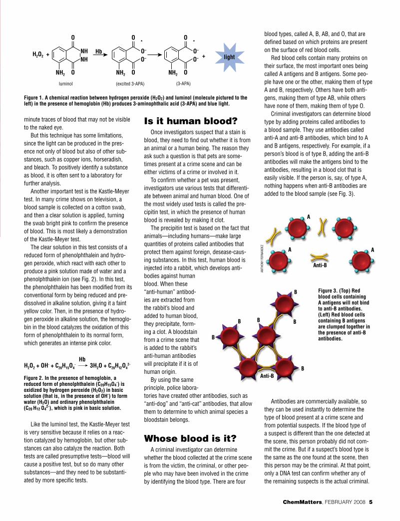
One of these tests consists of spraying a suspected sample with a solution of luminol (c8H7N3O2), a chemical popularized by the TV series “csI” (short for “crime scene Inves-tigation”), and hydrogen peroxide (H2O2). If blood is present, the sample glows with a blu-ish color in the dark.
The luminol is first activated with an oxi-dant, usually a solution of hydrogen peroxide and a hydroxide salt in water. Then, in the presence of a protein present in blood called hemoglobin, the hydrogen peroxide is decom-posed to form oxygen and water.
When luminol reacts with the hydroxide salt, a dianion is formed. The oxygen pro-duced from the hydrogen peroxide then reacts with the luminol dianion. The product of this reaction, an organic peroxide, is very unstable and immediately decomposes with loss of nitrogen to produce 3-aminophthalic acid (3-ApA) in an excited state. As 3-ApA relaxes, it releases a visible blue light (see Fig. 1).
Luminol is sensitive to the presence of extremely small amounts of blood. It can detect bloodstains that have been diluted up to 300,000 times! since it is nearly impossible to clean up every trace of blood at a crime scene, luminol is especially effective at detecting
� Chemmatters, february 2008 www.acs.org/chemmatters
GeTTy ImAGes.cOm
mIk
e cI
esIe
LskI
The FOreNsIcs of BLOOd
By Brian Rohrig
minute traces of blood that may not be visible to the naked eye.
But this technique has some limitations, since the light can be produced in the pres-ence not only of blood but also of other sub-stances, such as copper ions, horseradish, and bleach. To positively identify a substance as blood, it is often sent to a laboratory for further analysis.
Another important test is the kastle-meyer test. In many crime shows on television, a blood sample is collected on a cotton swab, and then a clear solution is applied, turning the swab bright pink to confirm the presence of blood. This is most likely a demonstration of the kastle-meyer test.
The clear solution in this test consists of a reduced form of phenolphthalein and hydro-gen peroxide, which react with each other to produce a pink solution made of water and a phenolphthalein ion (see Fig. 2). In this test, the phenolphthalein has been modified from its conventional form by being reduced and pre-dissolved in alkaline solution, giving it a faint yellow color. Then, in the presence of hydro-gen peroxide in alkaline solution, the hemoglo-bin in the blood catalyzes the oxidation of this form of phenolphthalein to its normal form, which generates an intense pink color.
Like the luminol test, the kastle-meyer test is very sensitive because it relies on a reac-tion catalyzed by hemoglobin, but other sub-stances can also catalyze the reaction. Both tests are called presumptive tests—blood will cause a positive test, but so do many other substances—and they need to be substanti-ated by more specific tests.
Is it human blood?Once investigators suspect that a stain is
blood, they need to find out whether it is from an animal or a human being. The reason they ask such a question is that pets are some-times present at a crime scene and can be either victims of a crime or involved in it.
To confirm whether a pet was present, investigators use various tests that differenti-ate between animal and human blood. One of the most widely used tests is called the pre-cipitin test, in which the presence of human blood is revealed by making it clot.
The precipitin test is based on the fact that animals—including humans—make large quantities of proteins called antibodies that protect them against foreign, desease-caus-ing substances. In this test, human blood is injected into a rabbit, which develops anti-bodies against human blood. When these “anti-human” antibod-ies are extracted from the rabbit’s blood and added to human blood, they precipitate, form-ing a clot. A bloodstain from a crime scene that is added to the rabbit’s anti-human antibodies will precipitate if it is of human origin.
By using the same principle, police labora-tories have created other antibodies, such as “anti-dog” and “anti-cat” antibodies, that allow them to determine to which animal species a bloodstain belongs.
Whose blood is it?A criminal investigator can determine
whether the blood collected at the crime scene is from the victim, the criminal, or other peo-ple who may have been involved in the crime by identifying the blood type. There are four
blood types, called A, B, AB, and O, that are defined based on which proteins are present on the surface of red blood cells.
red blood cells contain many proteins on their surface, the most important ones being called A antigens and B antigens. some peo-ple have one or the other, making them of type A and B, respectively. Others have both anti-gens, making them of type AB, while others have none of them, making them of type O.
criminal investigators can determine blood type by adding proteins called antibodies to a blood sample. They use antibodies called anti-A and anti-B antibodies, which bind to A and B antigens, respectively. For example, if a person’s blood is of type B, adding the anti-B antibodies will make the antigens bind to the antibodies, resulting in a blood clot that is easily visible. If the person is, say, of type A, nothing happens when anti-B antibodies are added to the blood sample (see Fig. 3).
Antibodies are commercially available, so they can be used instantly to determine the type of blood present at a crime scene and from potential suspects. If the blood type of a suspect is different than the one detected at the scene, this person probably did not com-mit the crime. But if a suspect’s blood type is the same as the one found at the scene, then this person may be the criminal. At that point, only a dNA test can confirm whether any of the remaining suspects is the actual criminal.
Chemmatters, february 2008 �
ANTH
ONy
FerN
ANde
z
Figure 1. A chemical reaction between hydrogen peroxide (h2o2) and luminol (molecule pictured to the left) in the presence of hemoglobin (hb) produces 3-aminophthalic acid (3-APA) and blue light.
Figure 2. in the presence of hemoglobin, a reduced form of phenolphthalein (c20h15o4
-) is oxidized by hydrogen peroxide (h2o2) in basic solution (that is, in the presence of oh-) to form water (h2o) and ordinary phenolphthalein (c20 h12 o4
2-), which is pink in basic solution.
Figure 3. (top) red blood cells containing A antigens will not bind to anti-B antibodies. (Left) red blood cells containing B antigens are clumped together in the presence of anti-B antibodies.

Bloodstains can provide important clues too
The physical properties of blood may also play a key role in allowing investigators to reconstruct events at a crime scene.
The size and shape of blood droplets can provide useful information. Blood that falls straight down will tend to form more symmet-rical droplets, while blood falling at an angle will be less symmetrical (see Fig. 4).
The roughness and porosity of the sur-face on which blood has fallen also plays a key role—blood dropped onto concrete, for
example, will tend to have a more jagged shape than blood dropped onto a softer or less porous surface.
Blood is an example of a colloid—a fluid substance where very small particles of another substance are dispersed. colloids are often very sticky because the suspended sol-ids can attract other chemicals. Glue, paint,
and jelly are other examples of sticky colloids.
Not only is blood sticky, but it is also viscous. Viscous sub-stances flow slowly, like molas-
ses and honey. since blood is both sticky and viscous, it forms readily
discernible patterns on walls and other surfaces.
One type of pattern, called a transfer stain, results from blood that is transferred from one object to another, either when an object moves through a preexisting bloodstain onto an unstained surface—such as a foot sliding through blood—or when a blood-stained object transfers blood to another area—such as when a bloody knife is thrown down. Another type of bloodstain, called the pro-jected stain, is produced by blood that flies off a surface under pressure.
Analyzing blood is a gruesome business, but it is necessary in order to reconstruct what happened at the scene of an accident or crime. even in death, blood can tell a story.
Blood composition
Blood contains a liquid por-tion called plasma, which contains mostly water along
with dissolved nutrients, minerals, and oxygen. Plasma, which makes up about 55% of the blood’s total volume, is a pale yellow liquid. Sus-pended within the plasma are two types of cells called red and white blood cells and cell fragments called platelets.
Red blood cells make up 40% of the total blood’s volume, typically live for about 120 days, and are replen-ished at a rate of 2 to 3 million per second in the bone marrow. Red blood cells contain hemoglobin, a complex molecule that carries oxygen and removes carbon dioxide and gives blood its red color. Its molecular formula is C2952H4664N812O832S8Fe4.
White blood cells make up 5% of blood’s total volume. They are a key compo-nent of the body’s immune system and play a crucial role in fighting off infection, attacking bacteria, viruses, and other microbes. The number of white blood cells present in the blood is a key indicator of health: A person suffering from leuke-mia has too many white blood cells, while a depressed immune system may reflect a deficiency of white blood cells.
The platelets are cell fragments involved in clotting blood, which helps stop bleeding.
www.acs.org/chemmatters � Chemmatters, february 2008
ANTH
ONy
FerN
ANde
z
pHOT
OdIs
c
red blood cell
PlateletPlatelet
red blood cell
White blood cellWhite blood cell
Figure 4. the shape of a blood droplet depends on the angle at which it fell.
A scanning electron microscope image from human blood.
cOUr
Tesy
OF
NATI
ONAL
cAN
cer
INsT
ITUT
e

Antibodies and antigens
On the surface of every cell in our body are proteins called antigens. The two main antigens present on the surface of red blood cells are called A and
B antigens. Some people have only A antigens on their red blood cells, some have only B antigens, some others have both A and B antigens, and others have none, making these people’s blood of type A, B, AB, and O, respectively.
Blood also contains proteins called antibodies that attach to these antigens, but antibodies attach only to foreign anti-gens. For example, type A blood contains antibodies called anti-B antibodies that bind only to B antigens, and type B blood contains antibodies called anti-A antibodies that bind only to A antigens. Table 1 summarizes the various antigen and antibody components of blood.
Crime scene investigators determine the blood type of a specimen by injecting either anti-A or anti-B antibodies and by seeing whether antibodies and antigens bind with one another. For example, if anti-A antibodies are used, they bind only to A antigens, so the blood type is either A or AB. By further adding anti-B antibodies to blood from the same stain would then determine whether it is of type A or AB. If it is of type A, its antigens won’t bind to the anti-B antibodies; if it is of type AB, its antigens will.
Table 2 summarizes how the blood type is established (+ shows binding between the antibodies and antigens and – shows no such binding).
Determining blood type can also be done by identifying antibodies instead of antigens. A criminal investigator can use commercially available A and B cells to test for the pres-ence of anti-A and anti-B antibodies as follows. When A cells are added to a blood specimen, the cells bind with one another only in the presence of anti-A antibodies; hence, the cells have B antigens on their surface and are of the B type. When B cells are added, the cells bind with one another when anti-B antibodies are present; hence, the cells are of the A type. All four blood types can be identified this way, as summarized in Table 3.
REfERENcESBevel, Tom and Gardner, Ross M. Bloodstain
Pattern Analysis, 2nd ed. CRC Press: Boca Raton, FL, 2002.
James, Stuart H. and Nordby, Jon J. Forensic Science: An Introduction to Scientific and Investigative Techniques, 2nd ed. CRC Press: Boca Raton, FL, 2005.
Saferstein, Richard. Criminalistics: An Introduction to Forensic Science. Pearson Prentice Hall: Saddle River, NJ, 2004.
INTERNET REfERENcESBlood typehttp://en.wikipedia.org/wiki/Blood_typeBlood plasmahttp://en.wikipedia.org/wiki/Blood_plasmaForensic Investigations—Blood Spatter: All
About Bloodwww.clt.uwa.edu.au/__data/page/112508/
fsb04.pdf
Brian Rohrig teaches chemistry at Jonathan Alder High school, in plain city (near columbus), OH. His most recent ChemMatters article, “The captivating chemistry of candles,” appeared in the december 2007 issue.
Chemmatters, february 2008 �
Anti-A antibody
+ bloodstain
Anti-B antibody
+ bloodstain
Antigen present
Blood type
+ - A A
- + B B
+ + A and B AB
- - neither A nor B o
table 2. identification of blood types with known antibodies.
Type A cells+ bloodstain
Type B cells+ bloodstain
Antibody present
Blood type
+ - Anti-A B
- + Anti-B A
+ + Both anti-A and anti-B
o
- - neither anti-A nor anti-B
AB
table 3. identification of blood types with type A and type B cells.
Blood type Antigens Antibodies
A A Anti-B
B B Anti-A
AB AB neither anti-A nor anti-B
o neither A or B Both anti-A and anti-Btable 1. Antigens and antibodies associated with the four blood types.
pHOT
OdIs
c