the formation and propertie osf the organic matrix of...
TRANSCRIPT

349
The Formation and Properties of the Organic Matrix ofReptilian Tooth Enamel
By D. F. G. POOLE(From the Department of Zoology, Makerere College, University College of
East Africa, Kampala, Uganda)
With two plates (figs, i and 2)
SUMMARY
A number of features of enamel formation in the lizard Agama atricollis are described.The behaviour and properties of the ameloblasts indicate that the process of enamelformation is similar to the corresponding process in mammals; the fibrous enamelmatrix appears to be formed from outgrowths of the cytoplasm of these cells. Inter-prismatic material, as it is known in mammals, is not produced, so that reptilianmatrix tends to be uniformly fibrous. Nevertheless, the fibres are initially arrangedin groups corresponding to the ameloblasts. There is no distinct pre-enamel stagebecause matrix production is immediately followed by a limited influx of mineral inan elementary state, converting the matrix into an basiphil form. Striae of Retziusmay be due to periodic pauses in the normal process of matrix production enabling theameloblasts to assimilate and secrete mineral. Before the onset of final calcification,the matrix seems to undergo a modification rendering it capable of influencing the sizeand orientation of mineral crystallites.
The organic matrix has a refractive index of 1-57 and has no intrinsic birefringence.However, in suitable liquids the parallel fibres produce a positive form birefringence.If paraffin wax is allowed to crystallize on the matrix, optically negative streaks areformed parallel with the fibres, perhaps as the result of crystal overgrowth.
Evidence obtained indicates that this reptilian type of ectodermal enamel is a likelyprecursor of the mammalian prismatic type. The evolution from one to the othercould have been achieved in a comparatively simple step.
INTRODUCTION
THE nature of reptilian enamel is of interest in that it has now beenestablished that two quite different types of enamel may be found cover-
ing the teeth of different vertebrates. Mammalian enamel is prismatic in struc-ture and is produced by the ectodermal cells of the enamel organ, whereasthe enamel covering the teeth and scales of fish (Kvam, 1946, 1950; Levi,1939, 1940a; Kerr, 1955; Poole, 1956a) and that covering the teeth of am-phibia (Kvam, 1946; Levi, 19406) is not prismatic and its matrix is formedby the mesodermal odontoblast cells of the dentine papilla. Although the teethof a wide range of reptiles have been examined (Erler, 1935; Schmidt, 1947;Poole, 19566), as yet no prisms have been found in the enamel; however,there is some evidence that the enamel matrix of reptiles has certain stainingproperties in common with mammalian matrix (Kvam, 1946).
Very many accounts of mammalian enamel formation are to be found indental and zoological literature; summaries of the most important details are
[Quarterly Journal of Microscopical Science, Vol. 98, part 3, pp. 349-367, Sept. 1957.]

350 Poole—The Formation and Properties of the
given by Kvam (1946) and Marsland (1951, 1952)- It may be briefly statedhere that two main stages are recognizable in enamel formation—the produc-tion of an organic matrix with only a slight mineral content, followed by amaturation process by means of which this matrix becomes highly calcified.
The organic matrix is formed by the ameloblasts of the enamel organ. Theso-called Tomes's processes seem to be intimately connected with the forma-tion of the matrix. The two most popular views are that either these processessecrete the matrix, or that they are converted into the matrix as they grow outfrom the basal ends of the ameloblasts. During its development, the propertiesof the matrix change a number of times. Soon after deposition it is acidophiland is known as pre-enamel; an influx of calcium salts, probably organic orcolloidal in form, produces a change to intense basiphil properties. Theenamel matrix remains in this condition, with a mineral content not greaterthan 35% (Weinmann, Wessinger, and Reed, 1942), until it has reached thefinal thickness of the future enamel. Immediately before the final heavy influxof mineral a return to acidophil properties is shown, but, as calcification pro-ceeds, there is a withdrawal of organic material and water, the residual matrixbecoming soluble in acids. As enamel production occurs, certain changes alsotake place in the ameloblasts including alterations in the size, shape, and con-tents of the cells, and in the position of the nucleus (Marsland, 1951).
The finished enamel shows incremental lines, the striae of Retzius, whichmay also be seen in the matrix before its final, heavy calcification. Theyappear to be related in some way to matrix production (Marsland, 1951) andthe direction of calcification is almost at right angles to these lines. Alkalinephosphatase distribution follows a regular pattern in developing mammaliantooth-germs, although different authors have reached slightly different con-clusions about distribution in ameloblasts and odontoblasts. A survey of theseresults is given by Symons (1955) who concludes that alkaline phosphatasemay be initially concerned with cell differentiation and growth rather thanwith calcification.
In a fully calcified reptilian tooth the enamel is made up of incrementallayers which follow approximately the shape of the tooth crown. As inmammals, this pattern is due to the presence of striae of Retzius. Mineralcrystallites tend to be arranged at right angles to the layers, and whilst thereis an irregular variation of crystallite direction in crocodile enamel (Erler,1935), an extremely regular variation occurs in the enamel of placodonts(Schmidt, 1947&), gorgonopsids, and cynodonts (Poole, 1956). A regular varia-tion produces a prismatic appearance under certain conditions of examination,but no true prisms are found.
Fibres have been described in sections of fully formed crocodile enamel(Kvam, 1946) which are said to cross one another in three planes (Marcus,1931; Schulte, 1930). Erler (1935) was unable to confirm a three-dimensionalfibre network, but found air-filled spaces in the enamel impermeable toliquids. Such air spaces occur in the enamel of many reptiles, fossil and recent(Schmidt, 1947a; Poole, 1956ft). It is possible that some of these structural

Organic Matrix of Reptilian Tooth Enamel 351
features have been confused by different authors; Kvam (1946), for example,states, 'longitudinally to the tooth there are undulating fibres'; but neither inthe text nor in the illustrations does he distinguish between these and thestriae of Retzius.
During development the organic matrix of the enamel of lizards andmammals stains red with Heidenhain's Azan, contrasting with mesodermalenamel of lower vertebrates, which stains blue in the same way as the col-lagenous dentine (Kvam, 1946). Because of this it is suggested that reptilianenamel has a keratinous nature rather than collagenous and, therefore, isectodermal in origin rather than mesodermal.
The object of the work to be described here has been to make a moredetailed examination of the formation and properties of reptilian enamelmatrix, and to see how far such properties compare with the enamel matrixof mammals.
MATERIAL AND METHODS
The results given here were obtained from the examination of embryonicjaws of the lizard Agama atricollis. This lizard was chosen because of therelative ease with which eggs are obtained at certain times of the year, butsome other lizards including the local gecko (Hemidactylus sp.) and skink(Mabuia varia) were also studied briefly, as well as a few embryos of thecrocodile-(Crocodilus niloticus). In all these cases the properties of the enamelmatrix were the same. As there appears to be no complete account of thestructure of a fully formed lizard-tooth, sections of the teeth of the localmonitor lizard {Vor anus niloticus) were prepared by grinding and examinedunder the polarizing microscope. The structure of the enamel was so similarto crocodile enamel that a description is not necessary, but the range of reptilespossessing a similar enamel structure (Poole, 19566) must now include modernLacertilia.
One or more agamid eggs from a clutch were opened at weekly intervalsand the embryos fixed in either Bouin or Susa. Unfortunately, the series ofembryos produced was not regular since the rate of development varied fromegg to egg. Nevertheless, enough stages were found to provide the mainlandmarks in tooth formation. After fixation, no further decalcificationwas required.
Under the prevailing local climatic conditions, difficulties were experiencedwith the normal technique of dehydrating and clearing, particularly withbulk tissue about to be embedded. To overcome this, dehydration was carriedout with mixtures of ethyl and w-butyl alcohol. The tissue to be embeddedwas transferred from absolute butyl alcohol into a mixture of butyl alcoholand paraffin wax (m.p. 54° C) and finally into pure paraffin. The techniqueproved to be very successful, since no appreciable hardening occurred evenwhen the material was left to embed overnight. Sections on the slide werecleared by passing them from 95% alcohol through terpineol into benzene.
Standard sections of each embryo were prepared and stained with

352 Poole—The Formation and Properties of the
haematoxylin and eosin. Harris's haematoxylin was preferred to others sinceit gave more delicate differentiation of tissues than Heidenhain's, and yetwas more stable than Delafield's. Some sections were also stained with Azan,which, as previously mentioned, is said to give a colour differentiation betweenkeratin and collagen.
Finally, alkaline phosphatase tests were made by the technique of Gomori(1939) as modified by Danielli (1946). Again the dehydration and embeddingtechnique was modified by using butyl alcohol. To reduce the time requiredfor embedding, a vacuum oven was used and the pressure lowered with awater pump. An embedding time of i\ h gave satisfactory results.
GENERAL FEATURES OF THE TEETH AND TOOTH-GERMS
Agamid lizards have an acrodont dentition, each tooth being ankylosed tothe upper edge of the jaw. The tooth tips are flattened laterally and triangularin shape so that each jaw has the appearance of a continuously serrated cuttingedge. The embryonic jaws of Agama atricollis possess nine teeth at hatchingcompared with about twice this number in the adult jaw. During the post-hatching growth period new teeth are produced one by one in a pocket at theback of the jaw and, as the jaw increases in length, new teeth are added at theend of the row. This is also found in acrodont chameleons (Rose, 1892). Noevidence has arisen from the study of Agama to contradict the general beliefthat teeth are not replaced in acrodont forms. Nevertheless, although theteeth are all similar in size at hatching, the second tooth in each adult jaw isconsiderably enlarged. Presumably, in this particular case, post-hatchinggrowth is maintained for some time.
The first signs of tooth formation were found in an embryo 28 days old.Here the oral epithelium had invaginated along the length of the jaw and soonafterwards this lamina gives rise to the nine germs of the teeth which will bepresent at hatching. The appearance of an early tooth-germ is shown in figs.1, A, B. At this stage it is possible to distinguish an enamel organ consisting ofan inner and outer enamel epithelium separated by stellate cells, and a small
FIG. 1 (plate). A series of developing tooth germs of Agama atricollis as seen in verticalsections.
A and B, very young germ stained with Heidenhain's Azan. The enamel organ consists ofinner and outer enamel epithelia separated by stellate cells. Nuclei are situated centrally in theameloblasts; between these cells and the odontoblasts is a layer of pre-dentine.
c, enlarging germ stained with haematoxylin and eosin. Enamel formation extending down-wards as the bone of the jaw differentiates below.
D, stage similar to C, stained with Azan. Ameloblasts are elongated with nuclei at theirouter ends; enamel matrix shows vertical refractive stripes caused by the bunching of fibres.
E, later germ stained with haematoxylin and eosin. Considerable enlargement has occurredand the base of the tooth-germ is approaching the bony platform to which it will eventuallybecome ankylosed. A blood-vessel passes through the bone into the pulp.
F, enlarged base of germ shown in E, where enamel formation is in progress. The inner endsof the ameloblasts show Tomes's processes, which stain in the same way as the cytoplasm ofthe ameloblasts and project through the terminal cell membrane.
c, enlarged tip of same in which the enamel has here reached its final thickness. No Tomes'sprocesses are present, the inner border of the ameloblasts being smooth.
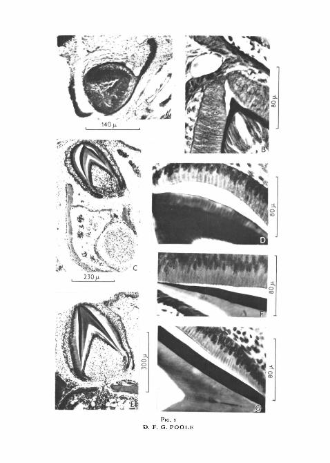
Fie. i
D. F. G. POOLE

Organic Matrix of Reptilian Tooth Enamel 353
number of odontoblasts differentiated from pulp cells. The organization ofthe enamel organ is rather simpler than that of crocodile, which, according toRose (1893), is the only reptile in which the condition of the enamel organapproaches that of mammals. Although the structure of this lizard enamelorgan is clearly seen over the tip of young germs, only the inner enamelepithelium is clearly defined at later stages.
The tissue separating the inner enamel epithelium from the odontoblastsin fig. 1, B is pre-dentine. It gives a faint pink colour with haematoxylin andeosin, whilst its collagenous nature is shown by the fact that it stains bluewith Azan. Thus, the first tissue produced is dentine and in this respect thereptilian tooth-germ resembles that of mammals and differs from fish, wherethe mesodermal enamel matrix is completed before dentine formation begins.
The tooth-germ increases in size by the downward extension of the enamelorgan; new ameloblasts are continually produced at the base where inner andouter enamel epithelia can be distinguished until later stages. Inside theameloblasts new odontoblasts appear and lay down pre-dentine. Enamel islaid down over the pre-dentine at the tip and eventually, as the downwardextension of the enamel organ occurs (fig. i, c, E), it is also laid down over thepre-dentine at the sides of the tooth. As the tooth-germ enlarges, a bony plat-form develops below to which the completed tooth will eventually becomeankylosed. Occasionally the bony platform is not continuous (fig. 1, E) andblood-vessels pass from the mesodermal tissue surrounded by the bone,through a gap and into the pulp cavity. One large blood-vessel is always seento enter the pulp cavity, but the origin of the vessel varies from germ to germ.
Finally, some weeks before hatching, a pocket is formed at the back of thejaw. Into this extends the dental lamina and a new tooth-germ begins toform. This is the first of the teeth which will be added to the series afterhatching. Ankylosis of the nine teeth in each jaw also occurs after hatching.
HISTOLOGY OF THE AMELOBLASTS AND ENAMEL MATRIX
Fig. 1, B is an enlargement of the tip of a young germ; a thin layer of pre-dentine is present but, as yet, no enamel has been formed. The ameloblastsare elongated with centrally placed, granular nuclei; the latter are typicallybasiphil and the cytoplasm acidophil. The odontoblasts producing the pre-dentine are also long, but in this case the nuclei are at the inner ends of thecells adjacent to the pulp.
As soon as the enamel appears it has very precise staining properties. Theappearance of a germ where enamel formation is under way is illustrated infig. 1, c. The staining properties of the ameloblasts are unchanged but theyare considerably longer and their nuclei are at the outer ends of the cells(fig. 1, D). At the base of the germ no enamel is present and the ameloblasts,with centrally placed nuclei, are shorter than those at the tip. A thin layer ofpre-dentine is present beneath them so that the condition at the base of thisgerm is identical with that over the tip of the young germ described above.Enamel formation is thus preceded by the elongation of ameloblasts, the

354 Pooh—The Formation and Properties of the
migration of the nuclei to the outer part of the cell, and the deposition of alayer of pre-dentine. These changes proceed gradually as enamel formationextends downwards from the tip of a germ.
The enamel matrix which is present at this stage stains so intensely withhaematoxylin that little or no structure may be seen in it. With Azan, enamelbecomes a deep red, the same colour as the keratinous scales which developon the surface of the embryo; this contrasts sharply with the bright bluedentine and bone. These properties do indeed suggest that the matrix is morelike keratin than collagen, as was suggested by Kvam (1946).
In sections which have been stained with Azan or lightly with haematoxylin,the matrix is not of a uniform appearance (fig. i, D). Fibres are seen through-out and there are alternating light and dark stripes running vertically to thetooth surface and parallel with the general fibre direction. The striped effectresults from refraction, for it is seen even in unstained preparations and,moreover, the light and dark areas change when the microscope tube is rackedup and down. Opposite each ameloblast a slight bunching of fibres occurs andthe refractive index along the axis of a group of fibres is probably slightlydifferent from that between groups, where the density of fibres is somewhatless. Thus, alternating zones of slightly different refractive index are foundwhich produce the refractive stripes. Although the cytoplasm is acidophil, theameloblasts present rather a similar pattern of darker intracellular contentsseparated by lighter intercellular zones, as may be seen in fig. 1, D. Fibresrunning in directions other than that described above have not been observed.
The appearance of the inner ends of the ameloblasts is also important.Because of distortion during the various treatments, the ameloblasts arefrequently pulled away from the enamel, and in such places the inner surfaceof this cell-layer is seen to be very irregular (figs. I , F ; 2, B, D). This irregularityis due to the fact that the end of each ameloblast projects through a terminalmembrane into the space caused by the distortion. However, where theameloblasts and enamel are closer together it is possible to see that theseprojections pass across and are continuous with the fibrous matrix, and it isevident that rupture of the connexion is only produced by severe distortion(fig. 2, B). Similar cell projections are well known in mammalian enamel for-mation as Tomes's processes.
If an unstained section is examined with phase contrast, the fibrousproperties of the matrix are again apparent and the continuity of the amelo-blasts with the matrix in an undistorted area is especially clear. From thebase of each ameloblast a zone of the matrix runs out towards the amelo-dentinal junction, gradually fanning out and becoming rather more fibrousas it extends deeper into the zone of enamel.
It is important to note here that in areas where the enamel and ameloblastshave not been pulled apart, Tomes's processes are not visible and the deeplystaining, basiphil matrix extends right up to the bases of the ameloblasts.Where Tomes's processes do occur, they stain in exactly the same way as thecytoplasm of the ameloblasts (figs. 1, F; 2, B, D). Therefore it seems very

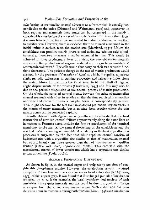
FIG. 2D. F. G. POOLE

Organic Matrix of Reptilian Tooth Enamel 355
likely that Tomes's processes are artifacts caused by distortion, and reallyrepresent the basal parts of the ameloblast cytoplasm which have been pulledout beyond the margins of the cells. However, Tomes's processes are onlyapparent during the production of the matrix; in fig. 1, c, E, they are presentover the developing enamel along the sides of the tooth, but are absent overthe tip where the matrix has reached its final width. This is illustrated in fig.1, F, G. Thus, when matrix production has ceased, there is no longer continuitybetween the contents of the ameloblasts and the enamel matrix, and Tomes'sprocesses are no longer produced.
Fig. 1, E is of an advanced stage of tooth formation. The staining propertiesof the matrix remain unchanged but it is now uniformly fibrous in appearance,the bunching having disappeared. This has probably arisen from the un-twisting of the fibres. Although they cannot be seen in the illustration, thematrix now shows incremental zones which are evident in the fully formedenamel of all reptiles. Wide zones with the typical staining properties of thematrix are separated by lines which do not stain so readily. These lines alsohave a slightly different refractive index from the rest of the matrix, for evenin unstained sections they can be made to appear light or dark by movingthe focus of the microscope. The matrix fibres pass uninterruptedly acrossthe striae and, as shown in fig. 2, A, these properties are seen most clearly in thematrix immediately before the final calcification. Fig. 2, A illustrates anotherimportant feature for, at this stage, there is a very rigid attachment betweenthe ameloblasts and enamel matrix, with the result that distortion now tearsthe matrix away from the dentine. Therefore, although continuity between thematrix and the contents of the ameloblasts disappears at the end of matrixformation, there is a later re-attachment of the ameloblastic terminal mem-brane to the surface of the matrix. Similar features have been described in theformation of mammalian enamel (Marsland, 1951).
At the hatching stage, no matrix can be seen over the tip of the tooth,although remains of the matrix are found along the sides. Fibres and incre-mental layer lines may still be recognized in these remains, whose stainingproperties are somewhat less basiphil than in younger stages. It is suggested
FIG. 2 (plate). Vertical sections of developing tooth-germs of Agama atricollis.A, ameloblasts and enamel shortly before the onset of final calcification, stained with Azan.
The enamel matrix, which is now uniformly fibrous and shows striae of Retzius, is so firmlyattached to the ameloblasts that distortion causes a fracture at the amelodentinal junction.
B, part of a germ where matrix formation is still in progress, stained with haematoxylinand eosin. The shape of Tomes's processes is seen to depend upon the degree of distortionof the germ; a number are still connected to both ameloblasts and matrix, but where distortionis absent the basiphil matrix extends right up to the bases of the ameloblasts.
c and D, side of a germ over the mid-line of which enamel formation has ceased and theresidual matrix has become acid-soluble. Here the tooth is triangular in section; enamel anddentine formation are still in progress, the ameloblasts showing clearly a basal membrane andTomes's processes. Processes from the odontoblasts pass into the light coloured pre-dentine.Stained with haematoxylin and eosin.
E and F, alkaline phosphatase distribution. Most of the germ shows intense activity butneither the ameloblasts nor odontoblasts stain throughout. Pre-dentine is unstained, enamelis black and calcified dentine, the inner border of which is globular in outline, appears grey.

356 Poole—The Formation and Properties of the
that, as calcification proceeds, a large part of the organic matrix is resorbed,but it is unlikely that resorption is complete and, in fact, a slight organicresidue can be obtained by extremely slow and careful decalcification ofcrocodile enamel. This residual organic material becomes soluble in all butthe most dilute acids. Finally, the ameloblasts have again altered in shape,being now much shortened with each nucleus appearing to occupy most ofthe cell. However, at the side of such a tooth as this, development is at anearlier stage and shows all the features of a young germ (fig. 2, c, D); lateralexpansion thus continues until the base of a tooth reaches its final size.
From the above description it is evident that developing reptile enamel hasa number of features in common with that of mammals. The staining reactionwith Azan is similar in the two cases and there is little doubt that the repti-lian matrix, like mammalian, is produced by the ameloblasts. The Tomes'sprocesses of the reptilian ameloblasts are suspected to be artifacts; a similarsuggestion has been made in the case of mammals (Kvam, 1946, and others),where the size and shape of the processes is also related to the degree of dis-tortion of the germ. However, the evidence for this is not quite conclusive,with the result that the Tomes's processes are still regarded by some as anatural feature of the ameloblasts, intimately related to matrix production(Marsland, 1951). In Agatna the staining properties of these processes arethe same as those of the cytoplasm of the ameloblasts and, therefore, it seemsprobable that as distortion takes place, the cell contents are pulled out beyondthe cell margins. Yet the inner border of the ameloblasts is marked by adistinct membrane which remains in position even after distortion, so that,if Tomes's processes are produced as suggested here, a means must existfor the cytoplasm of the ameloblasts to pass through the membrane withoutrupturing it. The terminal membrane in mammals is perforated, spacesalternating with condensations of intercellular material known as the terminalbar apparatus. A similar feature has not yet been recognized in Agama,although the existence of small perforations of some sort might be postulatedto account for the production of Tomes's processes.
Although they differ in staining properties, there is no distinct structuraldemarcation between the reptilian matrix and Tomes's processes; thefibres are less distinct in the outermost matrix layers, and gradually mergeinto the granular processes. The fact that only severe distortion will causecomplete rupture illustrates the intimacy of the connexion between fibres andprocesses. For these reasons it is suggested that the matrix is produced by thebasal regions of the ameloblasts which tend to grow out through the terminalmembrane and gradually become converted into fibres. Mammalian matrixmay also be produced in the same way (Kvam, 1946; Marsland, 1951). Inreptiles this process results in an initial bunching of fibres opposite eachameloblast, but eventually the fibres become more uniformly distributed.
Reptilian matrix shows no pre-enamel stage and only a doubtful 'transi-tional* phase, both of which possess acidophil properties in mammals. Thelayer tentatively described as pre-enamel in the lizard Lacerta vivipara

Organic Matrix of Reptilian Tooth Enamel 357
(Kvam, 1946) could be the same as the zone of Tomes's processes describedhere in Agatna, although, perhaps, there is no reason for making such a distinc-tion. Even in mammals there appears to be no clear-cut distinction betweenpre-enamel and Tomes's processes, except that the latter exist as separatedunits, and it is also possible for the pre-enamel stage to be missing, as in themouse (Kvam, 1946). In an attempt to account for these varying details, thefollowing developmental plan is proposed.
In both reptiles and mammals, enamel formation begins with the acidophilcontents of the ameloblasts growing out through the terminal membrane and
FIG. 3. Diagram of the structure of the matrix and arrangement of the ameloblasts during theformation of reptilian enamel, a, granular nuclei of ameloblasts; b, cytoplasmic region; c,terminal cell membrane, possibly perforated; d, Tomes's processes formed in regions ofdistortion and passing through terminal membrane; e, zone where outgrowing contents ofameloblasts are converted into matrix;/, fibres of the matrix arranged, initially, in groups
corresponding to the ameloblasts; g, amelodentinal junction.
becoming transformed into the matrix protein. In reptiles and some mammals,this transformation is rapid and is quickly followed by an influx of mineralconverting the matrix into a basiphil form. If, however, the transformationand mineral deposition are delayed, the matrix retains the form and acidophilproperties of the cytoplasm and is known as pre-enamel. In either case thematrix remains continuous with the cell contents, so that distortion pullsthe cell-walls and terminal membrane away from the matrix, leaving theextenuated cell contents behind as Tomes's processes. The existence of pre-enamel will then depend upon the rate of transformation and initial calcifica-tion of the matrix, and the length and shape of Tomes's processes uponthe extent to which the ameloblasts are pulled away from the matrix. Thus,until conversion occurs, there will be little structural difference between thepre-enamel, Tomes's processes, and cytoplasm of the ameloblasts. An attemptto illustrate some of these features has been made in fig. 3.
Striae of Retzius are common to both mammalian and reptilian enamels,yet their significance is uncertain. It has been shown that the final heavy
358 Poole—The Formation and Properties of the
calcification of mammalian enamel advances as a front which is actually per-pendicular to the striae (Diamond and Weinmann, 1940) and, moreover, inboth reptiles and mammals these zones can be recognized in the matrix aconsiderable time before the onset of final calcification. In view of these facts,it is now believed that the striae are related to matrix production rather thanto calcification. However, there is evidence that the mineral concerned in theinitial influx is derived from the ameloblasts (Marsland, 1951). Unless theameloblasts can produce matrix proteins and assimilate calcium salts simul-taneously, these two processes must be separated in time. This would beachieved if, after producing a layer of matrix, the ameloblasts temporarilysuspended the production of organic material and began to assimilate andsecrete mineral instead. The cells would thus carry on their two separate func-tions alternately. The periodic change in the rate of matrix production wouldaccount for the presence of the striae of Retzius, which, in reptiles, appear asslight periodic differences in staining properties and refractive index alongthe matrix fibres. In mammals the striae seem to be the result of regular,slight displacements of the prisms (Gustafson, 1945) and this may also bedue to the periodic suspension of the normal process of matrix production.On the whole, the zones of normal matrix between the striae of mammalianenamel are much wider than in reptiles, so that the time required to mineralizeone zone and convert it into a basiphil form is correspondingly greater.This might account for the fact that an acidophil pre-enamel region exists inthe matrix of many mammals, but is missing from reptiles where the thinmatrix zones can be converted rapidly.
Results obtained with Agama are only sufficient to indicate that the finalmaturation of reptilian enamel follows approximately along the same lines asin mammals. Features noted include the firm re-attachment of the terminalmembrane to the matrix, the general shortening of the ameloblasts and theresidual matrix becoming acid-soluble. A similarity in the final crystallizationprocesses is suggested by the fact that adult reptilian enamel consists ofhydroxyapatite with a crystallite size similar to that of mammalian enamelbut approximately ten times greater than that of mammalian or reptiliandentine (Little and Poole, unpublished results). This contrasts with themesodermal enamel of lower vertebrates which has a crystallite size similarto that of dentine (Poole, 1956a).
ALKALINE PHOSPHATASE DISTRIBUTION
As shown in fig. 2, E, the enamel organ and pulp cavity are sites of con-siderable phosphatase activity. However, the ameloblasts remain unstainedexcept for the nucleus and the supranuclear or basal cytoplasm (see Symons,1955), which appear grey. It was found that if prolonged periods of incubationare used, up to 24 h for example, the basal cytoplasm and nucleus of eachameloblast stains quite intensely and this could be due to a gradual diffusionof enzyme from the surrounding enamel organ. Such a diffusion has beenshown to occur in mammals during both fixation (Lison, 1948) and incubation
Organic Matrix of Reptilian Tooth Enamel 359
(Martin and Jacoby, 1949), but it is also possible that the nucleus is behavingas a slowly acting precipitation centre (Johansen and Linderstrom-Lang,1953), the amount of phosphate produced thereby depending upon the timeavailable for the precipitation to occur. Whatever the correct explanation maybe, it seems unlikely that the ameloblasts possess intrinsic alkaline phos-phatase at this point in their history.
The inner enamel epithelium at the base of a germ always stains intensely.Here, new epithelial cells are being formed by division and these will even-tually differentiate into ameloblasts as the formation of enamel and dentineextends downwards from the tip. Exactly the same condition is found inmammalian germs; because of this and the absence of phosphatase from thefully formed ameloblast, it has been suggested that, at this stage, phosphatasemay be related more to cell differentiation and growth, or to the manufactureof nucleic acids and other materials, rather than to calcification (Symons,
1955)-Nevertheless, in mammals, phosphatase activity is shown throughout the
cytoplasm of the ameloblasts during maturation (Symons, 1955) and, further-more, phosphatase can be detected in the ameloblasts of fish during the calci-fication of mesodermal enamel (Kerr, 1955). This evidence has been taken toindicate that alkaline phosphatase is concerned with the process of calcifica-tion. However, histological evidence suggests that mammalian ameloblastsare not responsible for the final calcification and that their function is to re-move organic material and water from the calcifying matrix (Marsland, 1952).Perhaps alkaline phosphatase is related to the latter function, but furtherevidence is clearly needed. Unfortunately, because the hardness of the den-tine makes section cutting impossible without prior decalcification, it has notyet been possible to discover whether phosphatase is present in the amelo-blasts during the maturation of reptilian enamel.
In Agama no general staining of the odontoblasts was ever observed; theinner ends of these cells always stained black, so that it is tempting to suggestthat diffusion has also occurred here, in this case from the pulp. However,recent results using azo-dye coupling techniques indicate that mammalianodontoblasts possess intrinsic phosphatase of their own (Symons, 1955) andthe significance of the results obtained here is therefore difficult to assess. Afeature of interest in the reptilian germ is that the parts of the dentinaltubules passing through calcifying dentine nearly always stain black, whereasthe parts of the same tubules passing through the uncalcified pre-dentineremain unstained. Clearly, then, during the calcification of the dentine thecontents of the dentinal tubules have the ability to precipitate phosphateeither by phosphatase enzyme or by some other means.
Finally, mention must be made of the appearance of enamel and dentineafter the phosphatase tests have been made. As shown in fig. 2, F, enamel isintensely black whilst the dentine is only light grey. In controls which havebeen subjected to the last part of the test only—the precipitation of phosphateas cobalt sulphide—exactly the same pattern of black enamel and grey dentine

360 Poole—The Formation and Properties of the
is produced, indicating that such phosphate is that laid down by the germnaturally. Despite the fact that it stains only lightly, the dentine must possessa considerable amount of mineral, for it is already very hard and brittle, andreadily cracks on sectioning. Moreover, it can be made to turn black by pro-longed exposure to a cobalt solution. Probably, therefore, the dentine mineralis in the fully formed hydroxyapatite condition and since the exchange ofCa + + ions and C o + + ions in this mineral is sluggish (Johansen and Linder-stram-Lang, 1953), complete ionic exchange can only occur after a relativelylong period of exposure to a cobalt solution. On the other hand, enamel be-comes black after only a few minutes' exposure, suggesting that the mineralhere is in a simpler form and is possibly related to the mineral producedunder the influence of enzyme in the phosphatase technique. These resultsconfirm the suggestion made earlier that the young basiphil enamel matrixpossesses calcium phosphate in some elementary form.
Thus, the behaviour of the mineral involved in the first influx is differentfrom that involved in the final calcification. In the latter process, apparentlyunder the influence of the matrix, mineral crystallizes into orientated hydroxy-apatite units of a characteristic size; yet the mineral concerned in the initialinflux remains in an elementary condition. Presumably, therefore, during itsbasiphil stage the matrix undergoes some sort of modification, as a resultof which the matrix fibres are rendered capable of exerting an influence overthe final calcification of mineral. The striae of Retzius have been shown hereto become increasingly apparent as the age of the matrix advances; this mightalso be taken to indicate the occurrence of some sort of progressive change inthe matrix before final calcification begins.
OPTICAL PROPERTIES OF THE MATRIX
The properties described below were found in enamel matrix at all stages,irrespective of the fixative used. When a tooth section is examined in waterbetween crossed nicols, both enamel and dentine light up as the surface of thetooth is rotated into the 45 ° position. If a sensitive tint (first order red) quartzplate is introduced with its slow axis parallel with the tooth surface, enamelappears yellow and dentine blue-green. The birefringence of the enamel is,therefore, negative with respect to the tooth surface, or positive with respectto the axis enamel fibres which are perpendicular to the surface. The positivebirefringence of the dentine with respect to the surface indicates that, as inmany vertebrates (Poole, 1956a, b) the positive collagen fibres are orientatedparallel with the surface of the tooth.
A significant difference in the behaviour of the enamel and dentine matriceswas found when sections were mounted in aqueous phenol solution and re-examined as above. Under these conditions both the enamel and dentine areyellow, indicating that the sign of birefringence of the dentine has beenreversed. This reversal in phenol solution is a property of collagen not sharedby other skeletal proteins (Frey-Wyssling, 1953), and is to be expected indentine but not in enamel if the latter is keratinous. However, the optical

Organic Matrix of Reptilian Tooth Enamel 361
properties of the enamel matrix were found to depend very much upon therefractive index of the solution in which sections were mounted. A summaryof the properties in various mounting liquids is given in table 1.
TABLE I
Liquid
Water . . • .Ethyl alcoholButyl alcoholBenzeneClove oilMixture of clove oil and
bromonaphthalene
BromonaphthaleneMethylene iodide .
Refractiveindex
i-331 3 61-401-501-53
i-57
1 6 61 7 6
Birefringence ofenamel matrix relative
to fibre axis
positive and strongpositivepositivepositive but faintpositive but very faint
isotropic
positivepositive and strong
Becke linetest
R.I. enamel > 1-33R.I. enamel > 1-36R.I. enamel > 1-40R.I. enamel > 1-50R.I. enamel > 1-53
R.I. enamel = 1-57enamel invisible
R.I. enamel < i-66R.I. enamel < 1-76
By means of the Becke line test the refractive index of the enamel matrixwas found to be about 1-57; young matrix was very slightly less than this,and mature matrix slightly greater. It may be seen from the table that thefibrous matrix has no birefringent properties when it is mounted in a liquidwith a similar refractive index of 1-57. Birefringence is only produced bymounting in liquids whose refractive indices are either significantly greater orsmaller than 1-57, with maximum birefringence produced at the extremes(1-33 and 1-76). All this points to the fact that the matrix fibres have nointrinsic birefringence; but since they are parallel with each other they form,together with the mounting liquid, a Weiner mixed body. Such a systemproduces a form-birefringence positive with respect to the direction oforientation of the micelles, the magnitude depending upon the differencebetween the refractive indices of the two components. If these two indicesare the same, the system is optically isotropic.
The behaviour of the enamel matrix in paraffin wax is very interesting.During the examination of newly prepared sections before the wax had beenremoved, it was found, even under these conditions, that the matrix showednot only distinct activity but also, rather surprisingly, a birefringence whichwas negative with respect to the fibre axis; that is, opposite in sign to theform-birefringence described above. As the wax is dissolved away in a solvent,the negative birefringence is replaced by the positive form-birefringence.These properties were investigated further by taking wax sections and heatingthe slide until the wax became molten. In the molten medium the fibrousmatrix showed, as in any other liquid, the typical positive form-birefringence;but as soon as the wax solidified the negative birefringence reappeared. It waspossible to repeat these reversals indefinitely by alternately warming andcooling the slide.
The explanation of this phenomenon is probably to be found in the2421.3 B b
362 Poole—The Formation and Properties of the
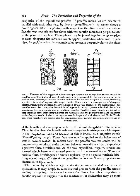
properties of the crystallized paraffin. If paraffin molecules are orientatedparallel with each other (e.g. by flow or crystallization), the system shows abirefringence which is positive with respect to the direction of orientation.Paraffin wax crystals are flat plates with the paraffin molecules perpendicularto the plane of the plate. These plates may be joined together, edge to edge,to form elongated flat lamellae which appear needle-like when seen in sideview. In such lamellae the wax molecules are again perpendicular to the plane
;-—ij !
S"7
1 1| j
1 !
i i! !
FIG. 4. Diagram of the suggested submicroscopic appearance of reptilian enamel matrix inparaffin wax. The index ellipse of each system is represented by the axes «e and n0. A, inmolten wax, randomly scattered paraffin molecules He between the parallel fibres producinga positive form birefringence with respect to the fibre axes. B, the arrangement of elongatedparaffin streaks resulting from the crystallization of the wax. Because of the orientation of theparaffin molecules, a negative intrinsic birefringence is set up. C, a more detailed plan of theassociation between matrix and crystallized paraffin. Parallel enamel fibrils are joined byperpendicular cross members; the latter are responsible for the primary alignment of paraffinmolecules, as a result of which the negative streaks lie parallel with the vertical fibrils. Fibrilsand cross members are represented by continuous lines, paraffin molecules and streaks by
broken lines.
of the lamella and also perpendicular to the axis of elongation of the lamella.Thus, in side view, the lamella exhibits a negative birefringence with respectto the longitudinal axis and because of this is known as a 'negative streak'(Frey-Wyssling, 1953). These facts can now be applied to the behaviour ofwax in enamel matrix. In molten form the paraffin wax molecules will berandomly scattered and so the medium behaves normally as a liquid to producea positive form-birefringence. As the wax crystallizes, negative streaks areformed which become orientated parallel with the enamel fibres. Thus thepositive form-birefringence becomes replaced by the negative intrinsic bire-fringence of the paraffin streaks as crystallization occurs. These properties areillustrated in fig. 4, A, B.
The method by which the negative streaks become orientated is a matter ofspeculation. It may simply be a mechanical process with the paraffin streakstending to slip into the spaces between the fibres, but other properties ofparaffin crystallites suggest that the mechanism of orientation may be more

Organic Matrix of Reptilian Tooth Enamel 363
complex than this. In strips of cold-drawn polyethylene it is found that themolecules become orientated approximately parallel with the direction ofdrawing. If paraffin wax is allowed to crystallize on the surface of such stripsthe paraffin molecules become aligned parallel with the polyethylene mole-cules with the result that the crystal plates of paraffin are arranged with theplane of the plate perpendicular to the direction in which the polyethylenestrips are drawn (Richards, 1951). Similarly, in spherulites of polyethylenewhere molecules are arranged tangentially, molecules of crystallizing paraffinagain become parallel with those of the polyethylene with the result that theparaffin streaks are arranged radially across the concentric layers (Wilemsand Wilems, 1956). From this it is evident that the arrangement of paraffinstreaks is secondary since it depends upon the initial direction in which theconstituent molecules become orientated.
A similar mechanism may determine the arrangement of paraffin streaksin enamel matrix. If so, the matrix must possess some sort of cross structuresrunning at right angles to the fibres. Such structures would determine theprimary orientation of the paraffin molecules perpendicular to the fibres axesand would result in the secondary arrangement of negative streaks parallelwith the fibres, as suggested in fig. 4, c. The coarse fibrous appearance of theenamel matrix is, perhaps, only the visible manifestation of a more delicatesubmicroscopic pattern. The latter would need to possess two main com-ponents; first, fibrils lying parallel with each other and at right angles to thetooth surface which set up the positive form-birefringence; secondly, sidegroups lying perpendicular to and possibly connecting the fibrils together.The whole system thus forms a lattice grid, the cross members of whichdetermine the orientation of paraffin molecules. It has been shown that thecrystal structure of polyethylene is very closely similar to that of paraffin wax(Bunn, 1939), and is therefore ideal for the orientated overgrowth of crystals(Wilems and Wilems, 1956). If the above suggestions are correct, the sidegroups running between the fibrils in the organic matrix may also possess acrystal structure related in some way to that of paraffin wax.
The orientation of crystals by organic fibres is of special interest in thestudy of calcified tissues, for in bone, dentine, and mesodermal enamel(Schmidt, 1938, 1940; Poole, 1956a), and in reptilian enamel as describedhere, crystallites of hydroxyapatite are arranged with their crystal c axesparallel with the organic fibres axes. Recent electron microscope studies(Jackson and Randal, 1956; Fernandez-Moran and Engstrom, 1956) havemade it possible to 'see' the intimate relationship between mineral crystallitesand organic fibres, but the precise mechanism of mineral orientation, whetheror not it involves crystal overgrowth, is still obscure.
DISCUSSION
The evidence presented here indicates that the process of enamel formationin reptiles is essentially the same as the corresponding process in mammals.The main difference is that whereas developing reptilian matrix is continuously
364 Poole—The Formation and Properties of the
fibrous, mammalian matrix is broken up into units, the prisms, each onehaving been produced by one ameloblast and isolated from its neighboursby interprismatic material. Nevertheless, the fibres of young reptilian matrixhave been shown to be arranged in groups which correspond with the amelo-blasts; the production of new material to separate the groups from each otherwould result in a condition very similar to that found in mammals and, pre-sumably, this is the most important change which took place in the evolutionof the mammalian type of enamel from reptilian.
It has been suggested that the terminal bar apparatus in mammals may beassociated with the production of interprismatic material (Marsland, 1951), sothat it is of special interest to note that these heavy intercellular condensationsat the bases of the ameloblasts have not been found in the reptiles where theenamel lacks prisms. The transition of prismatic enamel from the homo-geneous reptilian type, therefore, may have been achieved by the develop-ment of the terminal bar apparatus, which, by the production of a materialdiffering somewhat from the matrix proper, separates the products of theameloblasts into prisms.
As regards structural properties, it must also be noted here that althoughmammalian matrix shows no fibres even when examined with the electronmicroscope (Little, 1956), it has, nevertheless, a faint but definite positivebirefringence with respect to the axes of the prisms (Schmidt, 1934; Keil,X937)- This property has been taken as an indication of an organized finestructure within the matrix which might be responsible for the orientation ofmineral crystallites parallel with the prisms. However, it is clear that theorientated, possibly molecular, units producing the birefringence must be ofvery narrow width if they are beyond the resolution of the electron micro-scope. Consequently, the second important change in the evolution of pris-matic enamel was the production of a more refined and delicate matrix insteadof the coarse fibrous type characteristic of reptiles. As has already beenpointed out, the fibrous appearance of reptilian matrix may only be the out-ward manifestation of a more intricate submicroscopic structure.
The evolution of prismatic enamel from reptilian seems, therefore, to havebeen a comparatively simple step, and tubular enamel, found in most mar-supials and a few placentals, must be considered to be a later specializationof the simpler prismatic type. Tubular enamel possesses both prisms andtubules, the latter arising at the amelodentinal junction in the so-called'clumsy' joint. The origin of these tubules is still not clear, but they are knownto be interprismatic and may be occluded by the deposition of calcium salts inolder enamel (Sprawson, 1930). Although X-ray analysis shows the presenceof hydroxyapatite crystallites orientated approximately parallel with theprism axes, as in normal prismatic enamel, marsupial enamel exhibits mostunusual optical properties when treated with dehydrating and clearing agents(Poole, 1952). These properties may be in some part due to the high organiccontent of the enamel. The possession of prisms shows that tubular enamelmust be related to the more normal mammalian type, but the tubules, the

Organic Matrix of Reptilian Tooth Enamel 365
peculiar optical properties, and the relatively high organic content all pointto a specialized type of prismatic enamel.
In higher vertebrates, therefore, three distinct types of enamel matrixexist, all of which result from the calcification of a matrix derived from theectodermal ameloblasts. Similarly, in lower vertebrates, different types ofenamel are to be found with the common feature of being formed in a matrixderived from the mesodermal odontoblasts (Poole, 1956a). The phylogeneticrelationships between mesodermal and ectodermal enamels have been dis-cussed by Kvam (1946, 1953), who considers the first step in the transitionfrom one to the other to have been the production of a tissue jointly by meso-derm and ectoderm. In general, it has been tacitly assumed that both theproduction and the calcification of mammalian matrix are performed by theameloblasts, and the suggestion has repeatedly been made that even in fishthe ameloblasts may furnish the mineral required for the calcification of themesodermal matrix (Tomes, 1898; Kvam, 1946, 1953; Kerr, 1955). If this isso, the only further requirement for the evolution of an ectodermal enamel isthe ability of the ameloblasts to produce an organic matrix.
However, evidence that has gradually accumulated over a number of yearssuggests strongly that much of the mineral involved in the calcification ofmammalian enamel is derived from the dental pulp (see Marsland, 1952).Marsland himself came to the conclusion that, although the initial mineralinflux originates from the ameloblasts, none of the calcium salts required forthe final calcification comes from these cells and their function during matura-tion is to withdraw organic material and water from the calcifying matrix.Thus, there is a double source of mineral salts for the calcification of mam-malian enamel and the phylogenetic transition suggested above is not quiteso easy to accept. In fact, no hypothesis can be regarded as satisfactory untilthe source of the mineral used in the calcification of the mesodermal enamelhas been clearly established.
Finally, brief reference may be made to the problem of the organizingactivities of tooth-tissues. In mammals it is generally believed that as soon asthe enamel organ and dentine papilla are completed, the ameloblasts producean organizer which induces the odontoblasts to begin dentine formation;later, the ameloblasts themselves are induced by the odontoblasts to produceenamel. In lower vertebrates also it is possible that the ameloblasts organizethe activities of the odontoblasts, but the induction of the ameloblasts by theodontoblasts is lacking and, presumably, this is a necessary condition forthe production of a wholly ectodermal enamel. There appears to be no directevidence for such an organizer from the dentine papilla even in mammals;nevertheless, in the somewhat similar process of feather production, wheremesodermal and ectodermal tissues also work in close association, there isevidence that the mesodermal papilla induces activity in the ectodermalcap, culminating in the formation of a feather (Wang, 1943). This is regardedas an example of a 'secondary' organizer acting at a late stage in the lifehistory of an animal (Waddington, 1956), and it is tempting to suggest that the

366 Poole—The Formation and Properties of the
induction of ameloblastic activity by the dentine papilla, which also occurs atvarious stages in life, may be a parallel process.
It is hoped that, as a result of this account of the formation and propertiesof reptilian tooth-tissues, some of the relationships between the various typesof vertebrate enamel are a little clearer. Much of the discussion has necessarilybeen speculative, but further research may solve many of the outstandingproblems, such as the points of origin of the first type of ectodermal enameland of the later prismatic type. The study of the teeth of primitive mammalsand their possible reptilian ancestors would be of great value, and it is hopedthat such material may sometime become available.
In conclusion, the author would like to express his gratitude to the ResearchGrants Committee, Makerere College, for providing funds for the purchaseof a polarizing microscope; to Professor A. I. Darling, University of BristolDental School, for a number of helpful comments on the work undertaken;and to Professor L. C. Beadle, Zoology Department, Makerere College, forreading and criticizing the manuscript.
REFERENCES
BUNN, C. W., 1939. Trans. Faraday Soc, 35, 182.DANIELLI, J. F., 1946. J. exp. Biol., 22, no .DIAMOND, M., and WEINMANN, J. P., 1940. The enamel of human teeth. New York (Columbia
University Press).ERLER, G., 1935. Z. Zellforsch., 23, 589.FERNANDEZ-MORAN, H., and ENGSTROM, A., 1956. Nature, 178, 495.FREY-WYSSLING, A., 1953. Submicroscopic morphology of protoplasm. Amsterdam (Elsevier).GOMORI, G., 1939. Proc. Soc. exp. Biol. and Med., 42, 23.GUSTAFSON, G., 1945. Odontol. Tidskr., 53 (suppl.).JACKSON, S. F., and RANDAL, J. T., 1956. Nature, 178, 798.JOHANSEN, G., and LINDERSTROM-LANG, K., 1953. J. Histochem. .and Cytochem., 1, 442.KEIL, A., 1937. Dtsch. Zahn-, Nund-, v. Kieferheilk, 4, 60.KERR, T., 1955. Proc. Zool. Soc. Lond., 125, 95.KVAM, T., 1946. Norsk Tannlaegefor Tidende, 56 (suppl.).
1950. Trondheim, Kgl. Vid. Selsk.1953- Det Kangelige Norske Videnskabers Selskabs Forhandlinger, 26, Nr. 19.
LEVI, G., 1939. Arch. d'Anat. micr., 35, 101.1940a. Ibid., 35, 201.19406. Ibid., 35, 415.
LISON, L., 1948. Bull. Histol. Tech. micr., 25, 23.LITTLE, K., 1956. Private communication.MARCUS, H., 1931. Z. Zellforsch., 12, 395.MARSLAND, E. A., 1951. Brit. dent. J., 91, 251.
1952. Ibid., 92, 109.MARTIN, B. F., and JACOBY, F., 1949. J. Anat., 83, 351.POOLE, D. F. G., 1952. Ph.D. Thesis, Bristol.
1956a. Quart. J. micr. Sci., 97, 99.19566. Ibid., 97, 303.
RICHARDS, R. B., 195 I. J. Polymer Sci., 6, 397.ROSE, C, 1892. Dtsch. Monatsschr. f. Zahnhlk., 10, 126.
1893. Schwalbes morph. Arb., 3, 195.SCHMIDT, W. J., 1934. Abderhalden's Handb. d. biol. Arbeitsmeth. Abt. 5, T. 10, 435.
1938. Z. Zellforsch., 28, 761.1940. Ibid., 30, 615.

Organic Matrix of Reptilian Tooth Enamel 367SCHMIDT, W. J., 1947a. Ibid., 34, 55.
1947*. Ibid., 34, 78.SCHULTE, H., 1930. Z. Zellforsch., 10, 456.SPRAWSON, E., 1930. Proc. Roy. Soc, 106, 376.SYMONS, N. B. B., 1955. J. Anat., 89, 238.TOMES, C. S., 1898. Phil. Trans. B, 190, 443.WADDINCTON, C. H., 1956. Principles of embryology. London (Allen & Unwin).WANG, H., 1943. Physiol. Zool., 16, 325.WEINMANN, J. P., WESSINGER, G. D., and REED, G., 1942. J. dent. Res., 21, 171.WILEMS, J., and WILEMS, I., 1956. Nature, 178, 429.