the genus taira, with notes on tibial apophyses and descriptions
TRANSCRIPT

The genus Taira, with notes on tibial apophyses and descriptions of three new species(Araneae: Amaurobiidae)
Xin-Ping Wang: College of Life Sciences, Hebei University, Baoding, Hebei, 071002, China. E-mail:[email protected]
Peter Jager: Arachnology, Research Institute Senckenberg, Senckenberganlage 25, D-60325 Frankfurt am Main,
Germany
Zhi-Sheng Zhang: College of Life Sciences, Southwest University, Chongqing, 400715, China
Abstract. Homologies of the tibial apophyses of Taira with those of other members of the subfamily Amaurobiinae areevaluated. The monophyly of the genus Taira and the phylogenetic relationships of its species are analyzed usingparsimony. The genus Taira is supported by two putative synapomorphies: the presence of broad epigynal teeth and thedistally originating tegular sclerite apophysis. A diagnosis and description of Taira and a key to its species are provided.Three new species are described from China: Taira qiuae new species (LK), T. sichuanensis new species (LK), and T. zhui newspecies (LK).
Keywords: Identification key, taxonomy, homology, phylogenetic relationships, China
Lehtinen (1967) created the new genus Taira for thespecies Amaurobius flavidorsalis Yaginuma 1964, describedfrom Japan, compared it with other genera of Amaurobiidae,and placed Taira with the genus Tamgrinia Lehtinen 1967 inthe tribe Tairini within the Holarctic subfamily Amaurobii-nae Thorell 1870. Only four papers have been published onTaira since its establishment in 1967. Wang (2000) ques-tioned the sister group relationship between Taira andTamgrinia by comparing spinnerets, tracheae, and tricho-bothria of the family Amaurobiidae. Zhu et al. (2004) andWang & Ran (2004) described two new species based onspecimens from two closely situated localities in LiboCounty, Guizhou, China. A study by Zhang et al. (2008)concluded that T. lunaris Wang & Ran 2004 is a juniorsynonym of T. liboensis Zhu, Chen & Zhang 2004. In recentyears, field work by Zhang et al. (2008) in southern Chinahas yielded five more new species: T. cangshan Zhang, Zhu &Song 2008, T. latilabiata Zhang, Zhu & Song 2008, T. obtusaZhang, Zhu & Song 2008, T. concava Zhang, Zhu & Song2008, and T. sulciformis Zhang, Zhu & Song 2008, and a newcombination, T. decorata (Yin & Bao 2001). As a result, atotal of eleven species from Japan and southern China areincluded in Taira, including the three new species describedhere. In all previous studies, the diagnosis of the genus Taira,and therefore the distinction between Taira and otheramaurobiids, was only vaguely defined. Zhang et al. (2008)provided a diagnosis for Taira that we argue was based on amisinterpretation of non-homologous features (i.e., tibialapophyses, see below). This study is focused on diagnosingand describing the genus Taira, discussing its monophyly,estimating the species relationships, and providing a key tothe Taira species described so far. The three new speciesdescribed here belong to a distinct group, which differs fromother Taira by the widely separated, elongated spermathecaein females and the long RTA (except T. sichuanensis),presence of an intermediate apophysis between the RTA anddorsal tibial apophysis, and the uniquely modified conductorin males.
METHODS
All measurements are in mm. Scale lines are 0.2 mm longexcept where indicated otherwise. Eye diameters are taken atthe widest point. The length of body, prosoma, andopisthosoma do not include the length of the chelicerae orspinnerets. The distribution map was generated using GISArcView software, and the text files of the studied species aredownloadable from Wang (2009). More type specimen photosof the species included in this paper can be viewed in Li &Wang (2009).
Anatomical abbreviations used in the text and figures: Eyes:AME–anterior median eyes; ALE–anterior lateral eyes; PLE–posterior lateral eyes; PME–posterior median eyes. FemaleGenitalia: CD–copulatory duct; EL–epigynal median lobe;ET–epigynal tooth; FD–fertilization duct; S–spermatheca.Male palp: C–conductor hyaline apophysis; C1–conductorapophysis I (from which hyaline apophysis arises); C2–conductor apophysis II, which is an extension of theconductor apophysis I; C3–conductor apophysis III; CYM–cymbium; dDTA–distal/subdistal tooth of DTA; DTA–dorsaltibial apophysis; E–embolus; eDTA–ectal branch of DTA;ITA–Intermediate tibial apophysis; MA–median apophysis;mDTA–mesal branch of DTA; RTA–retrolateral tibialapophysis; ST–subtegulum; T–tegulum; TB– tibia; TS–tegularsclerite; TSA–tegular sclerite apophysis.
Specimens studied in the current paper are deposited in theCalifornia Academy of Sciences, San Francisco (CAS); HunanNormal University, Changsha, Hunan (HNU); Institute ofZoology, Chinese Academy of Sciences, Beijing (IZCAS);Museum of Hebei University, Baoding, Hebei (MHBU);Southwest University, Chongqing (SWUC); and SenckenbergMuseum, Frankfurt (SMF).
AMAUROBIINAE TIBIAL APOPHYSES
Tibial apophyses of the subfamily Amaurobiinae areimportant characters in species identification, generic limita-tion, and, therefore, valuable tools for phylogenetic analyses.In previous publications, however, different arachnologists
2010. The Journal of Arachnology 38:57–72
57
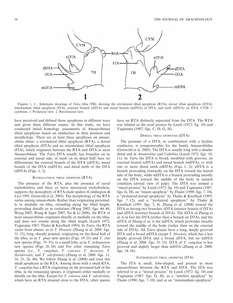
have perceived and defined those apophyses in different waysand given them different names. In this study, we haveconducted initial homology assessments of Amaurobiinaetibial apophyses based on similarities in their position andmorphology. There are at least three apophyses on amaur-obiine tibiae: a retrolateral tibial apophysis (RTA), a dorsaltibial apophysis (DTA) and an intermediate tibial apophysis(ITA), which originates between the RTA and DTA in mostAmaurobiinae. The Taira DTA usually has branches on itsexternal and mesal side, or teeth on its distal half; here wedifferentiate the external branch of the DTA (eDTA), mesalbranch of the DTA (mDTA), and distal teeth of the DTA(dDTA) (Figs. 1, 2).
RETROLATERAL TIBIAL APOPHYSIS (RTA)
The presence of the RTA, plus the presence of tarsaltrichobothria and three or more metatarsal trichobothria,supports the monophyly of RTA-clade spiders (Coddington &Levi 1991; Griswold et al. 2005). The morphology of the RTAvaries among amaurobiids. Rather than originating proximal-ly or medially on tibia, extending along the tibial length,protruding distally as in coelotines (Wang 2002: figs. 44–46;Wang 2003; Wang & Jager 2007; Xu & Li 2008), the RTA ofmost amaurobiines originates distally or medially on the tibia,and does not extend along the tibial length (Leech 1972;Yaginuma 1987; Thaler & Knoflach 1993). In Taira, the RTAvaries from absent, as in T. liboensis (Zhang et al. 2008: figs.31–33), long, sharply pointed, originating on the distal half ofthe tibia, as in T. qiuae new species (Figs. 19–21) and T. zhuinew species (Figs. 33–35), to a small lobe, as in T. sichuanensisnew species (Figs. 26–28) and five other remaining Tairaspecies (i.e., T. cangshan, T. concava, T. decorata, T.flavidorsalis, and T. sulciformis) (Zhang et al. 2008: figs. 11,16, 21, 26, 40). We follow Zhang et al. (2008) and treat thissmall apophysis as the RTA. Among those with a small RTA,T. cangshan has a RTA originating on the proximal half of thetibia; in the remaining species, it originates either medially ordistally on the tibia. Except for T. convava and T. sulciformis,which have an RTA situated close to the DTA, other species
have an RTA distinctly separated from the DTA. The RTAwas labeled as the ectal process by Leech (1972: fig. 10) andYaginuma (1987: figs. C, D, G, H).
DORSAL TIBIAL APOPHYSIS (DTA)
The presence of a DTA, in combination with a hyalineconductor, is synapomorphic for the family Amaurobiidae(Griswold et al. 2005). The DTA is usually long with a slenderdistal end in Amaurobius and Callobius (Leech 1972: figs. 10,11). In Taira the DTA is broad, modified with grooves, anexternal branch (eDTA) and mesal branch (mDTA), or withone or more distal teeth (dDTA) (Figs. 1, 2). eDTA is abranch protruding externally on the DTA toward the lateralside of the body, while mDTA is a branch protruding mesallyon the DTA toward the middle of the body in naturalcondition (dorsal view of palp). The DTA was termed a‘‘mesal process’’ by Leech (1972: fig. 10) and Yaginuma (1987.figs. G, H), an ‘‘innere apophyse’’ by Thaler (1990: figs. 7–10),a ‘‘prolateral-dorsal apophysis’’ by Thaler & Knoflach (1993:figs. 7–12), and a ‘‘prolateral apophysis’’ by Thaler &Knoflach (1991: figs. 3, 4). Zhang et al. (2008) treated theDTA as having two branches, iDTA (interior branch of DTA)and eDTA (exterior branch of DTA). The iDTA of Zhang etal. is in fact the DTA (rather than a branch on DTA), and theeDTA of Zhang et al. is the mDTA, which protrudes mesallytoward the middle of the body (rather than on the externalside of DTA). All Taira species have a long, deeply groovedDTA and a broad mDTA except T. liboensis, which has a lessdeeply grooved DTA and a broad eDTA, but no mDTA(Zhang et al. 2008: figs. 31–33). DTA of T. cangshan is lessgrooved and slightly larger than mDTA (Zhang et al. 2008:figs. 14–16).
INTERMEDIATE TIBIAL APOPHYSIS (ITA)
The ITA is small, lobe-shaped, and present in mostamaurobiines between the RTA and DTA. The ITA wasreferred to as a ‘‘dorsal process’’ by Leech (1972: fig. 10) andYaginuma (1987: figs. G, H), as a ‘‘mittlere apophyse’’ byThaler (1990: figs. 7–10), and as an ‘‘intermediate apophysis’’
Figures 1, 2.—Schematic drawings of Taira tibia (TB), showing the retrolateral tibial apophysis (RTA), dorsal tibial apophysis (DTA),intermediate tibial apophysis (ITA), external branch (eDTA) and mesal branch (mDTA) of DTA, and teeth (dDTA) on DTA. CYM 5
cymbium. 1. Prolateral view. 2. Retrolateral view.
58 THE JOURNAL OF ARACHNOLOGY

by Thaler & Knoflach (1993: figs. 7–12). The lateral tibialapophysis found in coelotines (Wang 2002: figs. 89, 107) alsoarises from the dorsal side of the RTA and its possiblehomology to the Amaurobiinae ITA needs further investiga-tion. Most Taira species lack an ITA except for the three newspecies described in this study: T. qiuae, T. sichuanensis, and T.zhui (Figs. 20, 28, 35). We followed Zhang et al. (2008) andtreated the small tibial apophysis of previous described Tairaspecies as RTA, rather than ITA.
RELATIONSHIPS
TAXA
Representatives of other Amaurobiinae and Coelotinae areincluded as outgroup taxa to test the monophyly of Taira andto help root the tree: Tamgrinia laticeps (Schenkel 1936),Coelotes atropos (Walckenaer 1830), Draconarius wudangensis(Chen & Zhao 1997), Callobius bennetti (Blackwall 1846) andAmaurobius fenestralis (Strom 1768). We chose one Tamgriniaspecies to root the tree because it is cribellate and was treatedas a member of the tribe Tairini together with Taira byLehtinen (1967). Tamgrinia could be more closely related toAgeleninae and Coelotinae than to Amaurobiinae (Wang2000; Wang & Zhu 2008; Wu et al. 2002). Two coelotinespecies were also chosen as outgroup taxa because they werepreviously placed in the same family with Amaurobiinae, theyare well studied relative to other amaurobiids, and theirrelationship with Amaurobiinae has been explored in otherstudies (Spagna & Gillespie 2008; Wu et al. 2002). Toreconstruct the relationships among the species, we compileda data matrix that includes nine Taira species with both maleand female described.
CHARACTERS
Twenty characters, mostly from genitalic structures, areused in the matrix. The female genitalia contributed ninecharacters, the male palp contributed nine, and the last twocharacters signify whether or not they are the cribellatespiders and whether the small hood of trichobothria isridged:
Character 0: Epigynal teeth (0 5 absent; 1 5
present).
Character 1: Epigynal teeth, shape (0 5 slender,
longer than wide; 1 5 broad, wider than long or
with subequal length and width).
Character 2: Epigynal teeth, position (0 5
situated anteriorly on anterior part of epigynum,
with bases distinctly separated from epigastric
furrow; 1 5 situated posteriorly on posterior part
of epigynum, with bases close to epigastric
furrow). Taira species share the synapomorphy
of having broad epigynal teeth. Similar to other
amaurobiines, the epigynal teeth of Taira origi-
nate posteriorly near the epigastric furrow.
Character 3: Epigynal lobe (0 5 absent; 1 5
present).
Character 4: Epigynal lobe, shape (0 5 epigynum
with a small median lobe and two large lateral
lobes; 1 5 epigynum with a single, large, medially
situated lobe).
Character 5: The single, large, medially situated
epigynal lobe (0 5 extending longitudinally,
longer than wide; 1 5 extending transversely,
wider than long). An epigynal lobe is present in
all Taira species, as well as in other Amaurobii-
nae. In Callobius, there are two broad lateral
lobes and a small, anteriorly originated median
lobe, but in Amaurobius and Taira, there is only
one large lobe, which varies in size and shape. The
size and shape of epigynal lobe could be species-
specific and important in species diagnosis. The
slightly elongated epigynal lobe is shared by T.
flavidorsalis and T. sulciformis.
Character 6: Spermathecae, length (0 5 long,
with distinct heads arising distally; 1 5 short,
without heads). Another feature shared by Taira
and other Amaurobiinae is the presence of short
spermathecae, compared to the long ducts in
most Coelotinae.
Character 7: Short spermathecae, separation (0 5
separated by their width or less; 1 5 separated by
at least twice their width). Widely separated
spermathecae apparently evolved in parallel in
three species of the clade D and T. cangshan
(Fig. 3).
Character 8: Short spermathecae, shape (0 5
round; 1 5 elongated, with the length twice the
width). The short spermathecae in Taira are
usually round or slightly elongated, but in three
species of clade D (Fig. 3), (i.e., T. qiuae, T.
sichuanensis, and T. zhui) they are elongated with
the length at least twice the width.
Character 9: RTA (0 5 extending along tibial
length, protruding distally; 1 5 not extending
along tibial length). In Amaurobiinae, the RTA
arises medially or distally on the tibia, rather than
extending along the tibial length as in Coelotinae.
The RTA is absent in T. liboensis, more or less
long, with a sharply pointed distal end in T. qiuae
and T. zhui, but small in other Taira species.
Character 10: DTA (0 5 absent; 1 5 present).
Character 11: ITA (0 5 absent; 1 5 present). The
ITA arises between the RTA and DTA in
Amaurobiinae, but only the three new species of
the clade D (Fig. 3) have this apophysis in Taira.
Character 12: DTA, the broad, mesally protrud-
ing branch (mDTA) (0 5 absent; 1 5 present).
Most Taira species have a broad, mesally
protruding branch on the DTA. One species T.
liboensis has a broad, externally protruding
branch (eDTA) but lacks a mDTA. The three
new species of clade D have small teeth on the
DTA, but has neither an eDTA nor a mDTA.
WANG ET AL.—GENUS TAIRA (AMAUROBIINAE) 59

Figure 3.—The preferred cladogram of hypothesized Taira species relationships (L 5 23, Ci 5 90, Ri 5 93); see text for discussion.
60 THE JOURNAL OF ARACHNOLOGY

Character 13: DTA, distal teeth (0 5 absent; 1 5
present). DTA distal teeth are present in the three
closely related new species of clade D.
Character 14: Tegular sclerite apophysis (0 5
absent; 1 5 present).
Character 15: Tegular sclerite apophysis, position
(0 5 proximal; 1 5 distal).
Character 16: Distally originating tegular sclerite
apophysis (0 5 normal; 1 5 strongly expanded
anteriorly and covering most part of embolus).
The presence of a tegular sclerite apophysis is
another synapomorphy of Amaurobiinae, al-
though in Taira it arises more distally and as a
result is distinctly separated from the median
apophysis by about the length of median apoph-
ysis. In three closely related D-clade Taira species,
the tegular sclerite apophysis is strongly expanded
anteriorly and leaves only the apex of the
embolus visible in ventral view.
Character 17: Conductor modification (0 5
absent; 1 5 present). Unique to the three new
Taira species at node D, the conductor is
modified to have 2–3 strongly sclerotized apoph-
yses (Figs 19–21: C1, C2, C3).
Character 18: Cribellum (0 5 absent; 1 5
present).
Character 19: Trichobothria, small hood (0 5
smooth; 1 5 ridged).
RESULTS
We generated a data matrix and optimized characters usingWinClada version 1.00.08 (Nixon, 1999) (Fig. 3). To performthe parsimony analyses, we used Hennig86 version 1.5 (Farris,1988), with all characters treated as non-additive. The exactsearch algorithm (ie*;) was used, resulting in the eight mostparsimonious trees. In the analysis, we arbitrarily used slow(Deltran) optimization, which favors parallelism over reversal.The preferred tree is shown in Fig. 3 (L 5 23, Ci 5 90, Ri 5
93). Four of the eight resulting trees were excluded becausethey indicate the sister group relationship between T. cangshanand clade D, based on the widely separated spermathecae. Theround-shaped spermathecae in T. changshan, which are similarto other Taira, differ from the elongated spermathecae ofclade D taxa. We excluded two more trees because they show asister group relationship between T. liboensis and clade B,supported by the absence of an ITA. Although having similarspermathecae and lacking an ITA, T. liboensis differs fromothers by the absence of an RTA, the absence of an mDTA,and the presence of an eDTA. Another tree shows the sameTaira species relationship as the preferred tree but wasexcluded because it supports sister group relationship betweenAmaurobius and Taira. Our study focuses on Taira speciesrelationships, so the characters indicating generic-level rela-tionships are understudied.
The species of the genus Taira are united at node A by thepresence of broad epigynal teeth (character 1, state 1) and thedistally originating tegular sclerite apophysis (character 15,
state 1). Three distinct species groups are found, and theirrelationships remain unresolved: T. liboensis, clade B species,and clade D species. Clade B includes five species: T. cangshan,T. concava, T. decorata, T. flavidorsalis, and T. sulciformis,based on the absence of an ITA (character 11, state 0, which isparallel in T. liboensis) and the presence of a broad, mesallyprotruding branch on the DTA (mDTA) (Character 12, state1). The elongated epigynal median lobe (character 5, state 0)supports a sister group relationship between T. flavidorsalisand T. sulciformis at node C. Three new species (T. qiuae, T.sichuanensis, and T. zhui) are united at node D by the widelyseparated spermathecal bases (character 7, state 1), the longspermathecae, which can be twice as long as wide (character 8,state 1), the presence of teeth on distal and subdistal DTA(character 13, state 1), the anteriorly expanded tegular scleriteapophysis that hides most of the embolus (character 16, state1), and the presence of 2–3 broad, strongly sclerotizedconductor apophyses (character 17, state 1).
SYSTEMATICS
Family Amaurobiidae Thorell 1870
Subfamily Amaurobiinae Thorell 1870
Taira Lehtinen 1967
Taira Lehtinen 1967:266.
Type species: Amaurobius flavidorsalis Yaginuma 1964
Diagnosis.—The genus Taira can be distinguished fromAmaurobius and related genera by two putative synapomor-phies: the presence of broad epigynal teeth in females(Figs. 22, 29, 36) and the distally originating tegular scleriteapophysis in males (Figs. 19, 27, 34). An additional diagnosticcharacter includes the branched or toothed dorsal tibialapophysis (Figs. 21, 26, 33).
Description.—Small to medium-sized cribellate spiders(Figs. 13, 14, 18, 24, 25, 31, 32, 38, 39), total length 4.18–6.43 (males) and 5.30–10.7 (females). Carapace elongate, darkbrown, with distinct wide, light-colored, longitudinal medianband and two wide, dark-colored, longitudinal lateral bands;cephalic area slightly narrowed, with cover of gray setae andsparsely distributed black setae; fovea longitudinal, deep.Anterior eye row straight or slightly recurved, posterior eyerow strongly recurved, with anterior margin of PME distinctlyposterior to posterior margin of PLE (Fig. 16); AME smallest,PME subequal to or slightly larger than AME, ALE largest,PLE subequal to or slightly smaller than ALE; AMEseparated from each other by less than their diameter,separated from ALE by 1–1.5 times AME diameter, PMEseparated from each other by 1.5–2 times PME diameter,widely separated from PLE by at least 2 times PME diameter;eyes with median ocular quadrangle wider in back than infront, longer than wide (Fig. 16). Clypeus high, approximatelytwo times AME diameter, curved downward. Sternum longerthan wide (width/length 5 0.75–0.80), sparsely covered withblack setae, anterior margin straight, lateral margins withoutextensions between coxae, posterior margin pointed, slightlyseparating coxae IV (Fig. 17). Chilum undivided, hairless.Chelicerae with 4 promarginal and 3 retromarginal teeth.Labium subequal or slightly longer than wide. Enditesrectangular, anteriorly slightly pointed, laterally slightlydepressed, with promarginal scopula, without serrula
WANG ET AL.—GENUS TAIRA (AMAUROBIINAE) 61

(Fig. 15). Tibiae with two rows of trichobothria; metatarsi andtarsi with one row of trichobothria; trichobothria with largehood transversely striated, small hood longitudinally ridged(Fig. 5), tarsal organ with simple opening. Tarsi with threeclaws, paired superior claws with 8–10 teeth on each; scopulaeabsent; leg spination often varying among individuals, typicalleg spination pattern (only surfaces bearing the spines listed,each leg segment was divided into four surfaces, dorsal,prolateral, ventral, retrolateral, then indicating the number ofspines in the proximal, middle, and distal one-thirds of eachsegment): femur: I p0–0–2; II p0–0–2, r0–0–1; III p0–0–1, r0–0–1; IV p0–0–1; tibia: I p1–1–1, v2–2–2, r0-1-1; II p0–1–1, v0–2–2, r0-1-1; III p1–1–0, v1–1–2, r1-1-0; IV p0–0–1; v1–0–2;metatarsus: I p0-1-0, v2–2–2, r0-1-1; II p0-1-1, v2–2–2, r0-1-1;III p1–1–1, v2–2–2, r1–1–1; IV v0–0–2. Tracheal tubes simple,limited to opisthosoma, spiracle situated close to spinneretsand connected to atrium, from which two lateral and twomedian tubes arise (Fig. 4). Cribellum divided (Figs. 6, 7).ALS short, conical, two-segmented, apex of ALS with 2 majorampullate gland spigots and approximately 28–55 piriformgland spigots (Fig. 8); PMS short, one-segmented, with 1minor ampullate gland spigot, 1 aciniform gland spigot, 1cylindrical gland spigot, about 5–8 paracribellar spigots(Fig. 9); PLS with approximately 8–22 aciniform glandspigots, 2–3 cylindrical gland spigots, 1 ‘‘amaurobiid PLSspigot’’ on distal end, 2 paracribellar spigots beside ‘‘amaur-obiid PLS spigot’’ (Fig. 10).
Female epigynum simple; atrium small or indistinct, withopenings on anterior margin of epigynal median lobe; epigynalmedian lobe distinct; epigynal teeth short, broad, situatedlateral of median lobe, close to but slightly separated fromepigastric furrow; copulatory ducts small, originating medial-ly, or indistinct in some species; spermathecae small, widelyseparated, round in most species (elongated only in T. qiuae,T. sichuanensis and T. zhui); fertilization ducts long, can be aslong as spermathecae.
Male palp without patellar apophysis; retrolateral tibialapophysis (RTA) small, situated distally or medially, widelyseparated or close to dorsal tibial apophysis (DTA) (but longand bent dorsally in T. qiuae and T. zhui, and absent in T.liboensis); dorsal tibial apophysis large, with distinct groove,branched on its external and mesal surfaces, with mesalbranch usually broad and large; intermediate tibial apoph-ysis only observed in three clade D species; cymbium short,with distal end extending slightly beyond bulb, withoutdistinct spines; proximal cymbium strongly constricted andconcave to narrow base; conductor broad, arising from distalbulb, hyaline with sclerotized base (in T. qiuae, T.sichuanensis, and T. zhui, the conductor is modified to small,less sclerotized apophysis and broad, highly sclerotized,branched, beak-shaped apophyses); median apophysis long,with slender apex and broad base, arising from lesssclerotized tegulum area; tegulum with distally originatingtegular sclerite apophysis, which is widely separated frombase of median apophysis; embolus broad, short, arisingdistally on prolateral tegulum.
Natural history.—Species of Taira build small cribellatewebs (Figs. 40, 41), which are similar to Amaurobius webs. The
spiders can be found on buildings, cliffs, trees, in caves, and onother substrates and favor shady, humid conditions. Individ-uals usually live together in high density, particularly in thelate spring and early summer during which the adults areactive. Although the adult female can also be found from Julyto August (personal observation), we collected specimens of T.liboensis from caves where adults are active in the summer(Wang & Ran 2004; Zhu et al. 2004).
Composition.—Eleven species: T. cangshan Zhang, Zhu &Song 2008, T. concava Zhang, Zhu & Song 2008, T. decorata(Yin & Bao 2001), T. flavidorsalis (Yaginuma 1964), T.latilabiata Zhang, Zhu & Song 2008, T. liboensis Zhu, Chen &Zhang 2004, T. obtusa Zhang, Zhu & Song 2008, T. qiuae newspecies, T. sichuanensis new species, and T. sulciformis Zhang,Zhu & Song 2008, and T. zhui new species
Distribution.—China, Japan (Fig. 42).
Figure 4.—Taira decorata (Yin & Bao 2001), female from WuyiMt., Fujian, China, trachea (dashed line refers to the position ofepigastric furrow).
62 THE JOURNAL OF ARACHNOLOGY

KEY TO SPECIES OF TAIRA
1. Male (those of T. obtusa, T. latilabiata unknown) . . . . . . . . . . . . . . . . . . . . . . . . . . . . . . . . . . . . . . . . . . . . . . . . . . . . . 2
Female . . . . . . . . . . . . . . . . . . . . . . . . . . . . . . . . . . . . . . . . . . . . . . . . . . . . . . . . . . . . . . . . . . . . . . . . . . . . . . . . . . . 10
2. Conductor with additional, highly sclerotized apophyses (C1, C2 and C3 in Figs. 19–21); tegular sclerite apophysis (TSA)
broad, anteriorly expanding, covering most of embolus from ventral view (Figs. 19–21, 26–28, 33–35) . . . . . . . . . . . . . . . . 3
Conductor with single broad, hyaline apophysis; tegular sclerite apophysis small, embolus visible from ventral view (Zhang et
al. 2008: figs. 9–11) . . . . . . . . . . . . . . . . . . . . . . . . . . . . . . . . . . . . . . . . . . . . . . . . . . . . . . . . . . . . . . . . . . . . . . . . . . . 5
3. RTA long, bent distally, with a sharp distal end (Figs. 20, 35) . . . . . . . . . . . . . . . . . . . . . . . . . . . . . . . . . . . . . . . . . . . . 4
RTA short, exhibiting only a small lobe (Figs. 27, 28) . . . . . . . . . . . . . . . . . . . . . . . . . . . . . . . . . . . . . . . . . . . . sichuanensis
4. Median apophysis with sharp, long basal process (Fig. 34) . . . . . . . . . . . . . . . . . . . . . . . . . . . . . . . . . . . . . . . . . . . . . . . zhui
Median apophysis with blunt, short basal process (Fig. 19) . . . . . . . . . . . . . . . . . . . . . . . . . . . . . . . . . . . . . . . . . . . . . qiuae
Figures 5–10.—Taira decorata (Yin & Bao 2001), female from Wuyi Mt., Fujian, China, SEM pictures. 5. Trichobothria. 6. Cribellum. 7.Cribellum, enlarged, showing the spigots. 8. ALS. 9. PMS. 10. PLS.
WANG ET AL.—GENUS TAIRA (AMAUROBIINAE) 63

5. RTA present; DTA with distinct branch on its mesal side; prolateral tegular lobe absent or indistinct (Zhang et al. 2008: fig. 10) . . . 6
RTA absent; DTA with distinct branch on its ectal side; tegulum with distinct prolateral lobe (Zhang et al. 2008: figs. 32, 33)
. . . . . . . . . . . . . . . . . . . . . . . . . . . . . . . . . . . . . . . . . . . . . . . . . . . . . . . . . . . . . . . . . . . . . . . . . . . . . . . . . . . . . . liboensis
6. DTA distinctly longer than its mesal branch; RTA arising from distal half of tibia (Zhang et al. 2008: figs. 9–11) . . . . . . . . 7
DTA and its mesal branch about the same length; RTA arising from proximal half of tibia (Zhang et al. 2008: figs. 14–16) cangshan
7. RTA distinctly separated from DTA (Zhang et al. 2008: figs. 16, 26) . . . . . . . . . . . . . . . . . . . . . . . . . . . . . . . . . . . . . . . 8
RTA close to DTA (Zhang et al. 2008: figs. 21, 40) . . . . . . . . . . . . . . . . . . . . . . . . . . . . . . . . . . . . . . . . . . . . . . . . . . . . 9
8. Distal embolus abruptly narrowed; tegular sclerite apophysis with slightly notched apex (Zhang et al. 2008: figs. 9, 10) flavidorsalis
Distal embolus as broad as its base; tegular sclerite apophysis with rounded apex (Zhang et al. 2008: fig. 25) . . . . . . . . decorata
9. DTA slender; tegular sclerite apophysis round (Zhang et al. 2008: fig. 20) . . . . . . . . . . . . . . . . . . . . . . . . . . . . . . . . . concava
DTA broad; tegular sclerite apophysis blunt (Zhang et al. 2008: fig. 39) . . . . . . . . . . . . . . . . . . . . . . . . . . . . . . . . sulciformis
10. Epigynal lobe with length and width subequal, or longer than wide (Zhang et al. 2008: figs. 7, 36) . . . . . . . . . . . . . . . . . 11
Epigynal lobe wider than long (Figs. 11, 22) . . . . . . . . . . . . . . . . . . . . . . . . . . . . . . . . . . . . . . . . . . . . . . . . . . . . . . . . 12
11. Epigynal lobe widest anteriorly (Zhang et al. 2008: fig. 7) . . . . . . . . . . . . . . . . . . . . . . . . . . . . . . . . . . . . . . . . . . flavidorsalis
Epigynal lobe widest medially (Zhang et al. 2008: fig. 36) . . . . . . . . . . . . . . . . . . . . . . . . . . . . . . . . . . . . . . . . . . . sulciformis
12. Spermathecal bases widely separated by at least two times their width (Figs. 12, 23, 37) . . . . . . . . . . . . . . . . . . . . . . . . . 13
Spermathecal bases slightly separated by less then their width (Zhang et al. 2008: figs. 18, 30) . . . . . . . . . . . . . . . . . . . . . 17
13. Spermathecae elongated, with length at least two times their width (Figs. 23, 37) . . . . . . . . . . . . . . . . . . . . . . . . . . . . . . 14
Spermathecae round (Fig. 12) . . . . . . . . . . . . . . . . . . . . . . . . . . . . . . . . . . . . . . . . . . . . . . . . . . . . . . . . . . . . . . . . . . 16
14. Spermathecae strongly converging anteriorly, with distal ends separated by only J of proximal separation (Fig. 30) sichuanensis
Spermathecae slightly converging anteriorly, with distal ends separated by more than K of proximal separation (Figs. 23, 37) 15
15. Spermathecal bases distinctly folded (Fig. 37) . . . . . . . . . . . . . . . . . . . . . . . . . . . . . . . . . . . . . . . . . . . . . . . . . . . . . . . . zhui
Spermathecal bases smooth, not folded (Fig. 23) . . . . . . . . . . . . . . . . . . . . . . . . . . . . . . . . . . . . . . . . . . . . . . . . . . . . . qiuae
16. Epigynal lobe distinctly curved; spermathecae separated by about 2 times their width (Fig. 12) . . . . . . . . . . . . . . . . . cangshan
Epigynal lobe not curved; spermathecae separated by about 3 times their width (Zhang et al. 2008: fig. 27) . . . . . . . latilabiata
17. Epigynal teeth and lateral margins of epigynal lobe widely separated by the width of epigynal teeth (Zhang et al. 2008: fig. 17)
. . . . . . . . . . . . . . . . . . . . . . . . . . . . . . . . . . . . . . . . . . . . . . . . . . . . . . . . . . . . . . . . . . . . . . . . . . . . . . . . . . . . . . concava
Epigynal teeth and lateral margins of epigynal lobe close together (Zhang et al. 2008: figs. 22, 29) . . . . . . . . . . . . . . . . . . 18
18. Spermathecae separated by less than half of their width (Zhang et al. 2008: fig. 23) . . . . . . . . . . . . . . . . . . . . . . . . . . decorata
Spermathecae separated by more than half of their width (Zhang et al. 2008: figs. 30, 35) . . . . . . . . . . . . . . . . . . . . . . . . 19
19. Spermathecae with distinct, anterior extensions (Zhang et al. 2008: fig. 35) . . . . . . . . . . . . . . . . . . . . . . . . . . . . . . . . . obtusa
Spermathecae round, without anterior extensions (Zhang et al. 2008: fig. 30) . . . . . . . . . . . . . . . . . . . . . . . . . . . . . . . liboensis
Taira decorata (Yin & Bao 2001)Figs. 4–10, 42
Titanoeca decorata Yin & Bao 2001:60, figs. 2a–e.Taira decorata Zhang, Zhu & Song 2008:507, figs. 22–26.
Remarks.—In addition to the genitalic illustrations by Zhanget al. (2008), in this study we also examined its tracheae (Fig. 4),trichobothria (Fig. 5), and spinnerets (Figs. 6–10), which aresimilar to T. liboensis of Wang (2000: figs. 16–18, 35).
Taira cangshan Zhang, Zhu & Song 2008Figs. 11–17, 42
Taira cangshan Zhang et al. 2008:505, figs. 12–16.
Remarks.—In addition to the material examined by Zhanget al. (2008), more specimens were collected from other partsof Yunnan, China (1K, Lushui County, Yaojiaping He atPianma Road, 44.7 km, elev. 2516 m, 25.97479uN,098.71027uE, disturbed forest, night collecting in forest and
Figures 11, 12.—Taira cangshan Zhang, Zhu & Song 2008, female (CASENT9021382) from Lushui, Yunnan, China, epigynum (ventral anddorsal view).
64 THE JOURNAL OF ARACHNOLOGY

along roadcuts, 20 May 2005, C. Griswold & D. Kavanaugh,CAS, CASENT9021382; 1K, same data, CAS, ENT9022279);1K, same data, HNU, ENT9022278; 1K, same data, HNU,ENT9022357).
In this study, we have re-illustrated the epigynum and vulva.Our illustration of the epigynum shows a much narrower lobethan that of Zhang et al. (2008) because it is viewed from aslightly different angle (Figs. 11, 12). We also took photos ofhabitus, eyes, sternum, labium, and endites to display thegeneral somatic structures of Taira (Figs. 13–17).
Taira liboensis Zhu, Chen & Zhang 2004Figs. 18, 42
Taira liboensis Zhu et al. 2004:61, figs. 1A–F (female holotypeand male paratype, in MHBU, examined). Zhang et al.,2008:509, figs. 29–33.
Taira lunaris Wang & Ran 2004:31, figs. 1–4 (female holotypeand paratype, in IZCAS, examined). First synonymized byZhang et al. 2008.
Remarks.—In addition to the material examined by Zhanget al. (2008) and Wang & Ran (2004), we collected morespecimens from Guizhou, China (4K, Guiyang City, QianlinPark, 8 August 2007, Z.S. Zhang, SWUC; 1K, GuidingCounty, Yanxia Town, Jingangdong Cave, 10 August 2007,Z.S. Zhang, SWUC; 1L2K, Guiding County, Yanxia Town,Dayandong Cave, 9 August 2007, Z.S. Zhang, SWUC).
Figures 13–17.—Taira cangshan Zhang, Zhu & Song 2008, female (CASENT9021382) from Lushui, Yunnan, China. 13. Habitus, dorsal view.14. Habitus, ventral view. 15. Labium and endites. 16. Eyes, view between dorsal and front. 17. Sternum.
Figure 18.—Taira liboensis Zhu, Chen & Zhang 2004, femaleholotype of T. lunaris Wang & Ran, 2004 from Libo, Guizhou, China,habitus, dorsal view.
WANG ET AL.—GENUS TAIRA (AMAUROBIINAE) 65

Habitus photos of the female holotype of Taira lunaris (5T.liboensis) were taken for the purpose of comparison with otherTaira species (Fig. 18).
Taira qiuae new species
Figs. 19–25, 42
Types.—CHINA: Shaanxi, L holotype, 4K paratypes fromTaibai Shan, S. flanks, above Houzhenzi, secondary broad-leaf forest, 1300–1700 m, 8 June 1997, P. Jager and B.Martens, deposited in SMF; 1L paratype from Taibai Shan, S.flanks, above Houzhenzi, secondary broad-leaf forest,33u52932.420N, 107u48934.730E, 1700 m, sieving leaf litter, 7
June 1997, P. Jager and B. Martens, deposited in SMF; 30L32K
paratypes from Taibai Shan, above Houzhenzi, 1250–1800 m,23–25 May 2009, Z.S. Zhang, deposited in SWUC.
Etymology.—The specific name is in honor and memory ofProfessor Qiong-Hua Qiu (deceased), who advised andsupported Xin-Ping Wang’s work; noun (name) in genitivecase.
Diagnosis.—This new species is similar to T. zhui newspecies in having a free standing, long RTA and the long, moreor less anteriorly converging spermathecae but can bedistinguished by the longer spermathecae, the smoothspermathecal bases (not folded) in females, the less extending
Figures 19–23.—Taira qiuae new species, male holotype and female paratype from Taibaishan, Shaanxi, China, drawings. 19. Palp, ventralview. 20. Palp, retrolateral view. 21. Palp, prolateral view. 22. Epigynum, ventral view (arrow points to copulatory opening). 23. Epigynum,dorsal view.
66 THE JOURNAL OF ARACHNOLOGY

base of the median apophysis and the different shapes of theconductor apophyses (much larger C1 and strongly prolat-erally curved C3) in male (Figs. 19–23).
Description.—Male (holotype): Medium-sized spider, totallength 5.40 (Fig. 24). Carapace 3.00 long, 2.16 wide;opisthosoma 2.40 long, 1.65 wide. AME smallest, ALElargest, PME and PLE subequal in size (AME 0.07, ALE0.14, PME 0.11, PLE 0.12); AME separated from each otherby less than their diameter, widely separated from ALE byabout 1.5 times AME diameter; PME separated from eachother by slightly more than their diameter, from PLE byalmost 2 times PME diameter; AME and PME widelyseparated by about 2 times AME diameter (AME–AME0.05, AME–ALE 0.10, ALE–PLE 0.05, PME–PME 0.13,PME–PLE 0.18, AME–PME 0.15). Palpal RTA long, bentdorsally, with sharply pointed distal end (Fig. 20); dorsaltibial apophysis (DTA) large, toothed proximally anddistally (Fig. 21); intermediate tibial apophysis (ITA) small,arising between the RTA and DTA (Fig. 20); conductorreduced to a slender, hyaline apophysis, which arises andhides behind the well-developed, strongly branched, beak-shaped conductor apophyses (C1, C2, C3 in Figs. 19–21);median apophysis long, with broad base and slender apex(Figs. 19, 20); tegular sclerite extending anteriorly andforming a broad apophysis on distal bulb, the latter coveringmost of embolus (Fig. 19); embolus broad, only the distalend visible from ventral view (Fig. 19).
Female (paratype): Medium-sized spider, total length 6.62(Fig. 25). Carapace 2.77 long, 1.87 wide; opisthosoma 3.85long, 2.82 wide. AME smallest, ALE largest, PME and PLEsubequal (AME 0.07, ALE 0.13, PME 0.10, PLE 0.10); AMEclose together, separated from each other by less than theirdiameter, widely separated from ALE by about 1.5 timesAME diameter; PME separated from each other by 1.5 times
their diameter, from PLE by almost 2 times PME diameter;AME and PME widely separated by about 2 times AMEdiameter (AME–AME 0.05, AME–ALE 0.11, ALE–PLE 0.04,PME–PME 0.16, PME–PLE 0.18, AME–PME 0.15). Epigy-num with a triangular plate anterior of median lobe; medianlobe two times wider than long; epigynal teeth broad,separated from median lobe by approximately their width;copulatory ducts small but distinct, arising medially andextending laterally to spermathecae; spermathecae long,kidney-shaped, widely separated from each other anteriorlyby at least their width and posteriorly by at least two timestheir width; fertilization ducts long, approximately half thelength of spermathecae (Figs. 22, 23).
Distribution.—China (Shaanxi) (Fig. 42).
Taira sichuanensis new species
Figs. 26–32, 42
Types.—CHINA: Sichuan, L holotype, 6L12K paratypesfrom Changning County, Meidong Town, Gaojian Village,28u1895.00N, 104u58956.80E, 700 m, 1 May, 2008, Z.S. Zhang& R.Y. Zuo, deposited in SWUC; 7K paratypes from the samelocality, 5 June 2008, Z.S. Zhang, deposited in SWUC.
Etymology.—The specific name refers to the type locality,Sichuan Province, China; adjective.
Diagnosis.—This new species is similar to T. qiuae newspecies and T. zhui new species in having the distinctconductor apophyses, the anteriorly expanding tegular scleriteapophysis, and the elongated spermathecae but can bedistinguished by the small RTA, the presence of a broadconductor, and the different shapes of conductor apophyses(much smaller C1 and C2) in males, and by the small epigynalteeth, the presence of a distinct atrial opening, and therelatively transversely extending spermathecae in females(Figs. 26–30).
Figures 24, 25.—Taira qiuae new species, male holotype (24) and female paratype (25) from Taibaishan, Shaanxi, China, photos, habitus,dorsal view.
WANG ET AL.—GENUS TAIRA (AMAUROBIINAE) 67

Description.—Male (holotype): Medium-sized spider, totallength 4.90 (Fig. 31). Carapace 2.10 long, 1.50 wide; opistho-soma 2.80 long, 1.60 wide. Median eyes subequal in size, ALElargest, PLE slightly smaller than ALE (AME 0.08, ALE 0.13,PME 0.08, PLE 0.10); AME separated from each other by lessthan their diameter, from ALE by about AME diameter; PMEseparated from each other by slightly more than 1.5 times theirdiameter, from PLE by almost 2 times PME diameter (AME–AME 0.05, AME–ALE 0.08, PME–PME 0.13, PME–PLE0.15, ALE–PLE 0.05). Palpal RTA short, forming a smalllobe; dorsal tibial apophysis (DTA) large, toothed distally;intermediate tibial apophysis (ITA) arising between the RTAand DTA; conductor broad, distally hyaline; conductorapophyses broad, strongly branched; median apophysis long,with broad base and slender apex; tegular sclerite extending
anteriorly and forming a broad apophysis on distal bulb, thelatter covering most of embolus; embolus broad, only thedistal end visible from ventral view (Figs. 26–28).
Female (paratype): Medium-sized spider, total length 3.80(Fig. 32). Carapace 1.50 long, 1.00 wide; opisthosoma 2.30long, 1.50 wide. AME smallest, ALE largest, posterior eyessubequal in size (AME 0.05, ALE 0.10, PME 0.08, PLE 0.08);AME separated from each other by about their diameter, fromALE by about 1.5 times AME diameter; PME separated fromeach other by slightly more than their diameter, from PLE byalmost 2 times PME diameter (AME–AME 0.05, AME–ALE0.08, PME–PME 0.10, PME–PLE 0.15, ALE–PLE 0.05).Epigynum with a small median lobe; epigynal teeth small,originating on posterior margin of epigynum and close tomedian lobe; copulatory ducts small, arising medially between
Figures 26–30.—Taira sichuanensis new species, male holotype and female paratype from Changning, Sichuan, China. 26. Palp, prolateralview. 27. Palp, ventral view. 28. Palp, retrolateral view. 29. Epigynum, ventral view (arrow points to copulatory opening). 30. Epigynum,dorsal view.
68 THE JOURNAL OF ARACHNOLOGY

spermathecae; spermathecae long, more or less extendingtransversely, separated from each other anteriorly by approx-imately their width and posteriorly by at least three times theirwidth; fertilization ducts long, at least half the length ofspermathecae (Figs. 29, 30).
Distribution.—China (Sichuan) (Fig. 42).
Taira zhui new species
Figs. 33–39, 42
Types.—CHINA: Chongqing, L holotype, 2L28K paratypesfrom Jinyunshan Natural Nature Reserve, 29u419080–29u529030N, 106u179430–106u249500E, 350–951 m, BeipeiMay 2008, R.Y. Zue & Z.H. Liu, deposited in SWUC; 1L
paratype from same locality as above, 26 April 2008, Z. S.Zhang, deposited in SWUC. 6L10K paratypes from samelocality as above, 17 May 2009, Z.S. Zhang, deposited inSWUC.
Etymology.—The specific name is in honor of ProfessorMing-Sheng Zhu, who advised and supported Zhi-ZhengZhang’s work; noun (name) in genitive case.
Diagnosis.—This new species is similar to T. qiuae newspecies in having a long RTA and long, anteriorlyconverging spermathecae but can be distinguished by thedistinctly folded spermathecal bases in females, thedistinctly extending base of the median apophysis and
the different shapes of the conductor apophyses (muchsmaller C1 and slightly prolaterally curved C3) in males(Figs. 33–37).
Description.—Male (holotype): Medium-sized spiders, totallength 4.60 (Fig. 38). Carapace 2.50 long, 1.70 wide; opistho-soma 2.30 long, 1.50 wide. AME smallest, 2/3 size of ALE,ALE largest; posterior eyes subequal in size, slightly smallerthan ALE (AME 0.10, ALE 0.15, PME 0.13, PLE 0.13); AMEclose together, slightly separated from each other by 1/3 oftheir diameter, from ALE by slightly less than AME diameter;PME separated from each other by about their diameter, fromPLE by almost 1.5 times PME diameter (AME–AME 0.03,AME–ALE 0.08, PME–PME 0.13, PME–PLE 0.18). PalpalRTA long, bent dorsally, with sharply pointed distal end;dorsal tibial apophysis (DTA) large, toothed proximally anddistally; intermediate tibial apophysis (ITA) small, arisingbetween the RTA and DTA; conductor reduced to a slender,hyaline apophysis, the latter arising from the distal end of thewell-developed, strongly branched, beak-shaped conductorapophyses (C1, C2, C3); median apophysis long, with slenderapex and broad base, the latter with a long, slender apophysis;tegular sclerite extending anteriorly and forming a broadapophysis on distal bulb, the latter covering most of embolus;embolus broad, only distal end visible from ventral view(Figs. 33–35).
Figures 31, 32.—Taira sichuanensis new species, male holotype (31) and female paratype (32) from Changning, Sichuan, China, photos,habitus, dorsal view.
WANG ET AL.—GENUS TAIRA (AMAUROBIINAE) 69

Female (paratype): Medium-sized spider, total length 6.50(Fig. 39). Carapace 2.60 long, 1.80 wide; opisthosoma 4.00long, 3.00 wide. AME smallest, 2/3 size of ALE, ALE largest;posterior eyes subequal in size (AME 0.10, ALE 0.15, PME0.13, PLE 0.13); AME close together, slightly separated fromeach other by 1/3 of their diameter, from ALE by slightly morethan AME diameter; PME separated from each other by morethan their diameter, from PLE by almost 2 times PMEdiameter (AME–AME 0.03, AME–ALE 0.13, PME–PME0.18, PME–PLE 0.23, AME–PME 0.15. Epigynum with atriangular plate anterior of median lobe; median lobe twotimes wider than long; epigynal teeth broad, separated frommedian lobe by approximately their width; copulatory ducts
small but distinct, arising medially and extending laterally tospermathecae; spermathecae long, kidney-shaped, widelyseparated from each other anteriorly by at least their widthand posteriorly by at least two times their width; spermathecalbases with distinct folders looping around; fertilization ductslong, slightly longer than half the length of spermathecae(Figs. 36, 37).
Distribution.—China (Sichuan) (Fig. 42).
ACKNOWLEDGMENTS
The first author thanks Charles E. Griswold (CAS, SanFrancisco) and Norman I. Platnick (American Museum ofNatural History, New York) for their advice and comments
Figures 33–37.—Taira zhui new species, male holotype and female paratype from Jinyunshan, Beipei, Chongqing, China, drawings. 33. Palp,prolateral view. 34. Palp, ventral view. 35. Palp, retrolateral view. 36. Epigynum, ventral view (arrow points to copulatory opening). 37.Epigynum, dorsal view.
70 THE JOURNAL OF ARACHNOLOGY

Figures 38, 39.—Taira zhui new species, male holotype (38) and female paratype (39) from Jinyunshan, Beipei, Chongqing, China, photos,habitus, dorsal view.
Figure 40, 41.—Habitat and web of Taira. 40. Male T. sichuanensis new species from Changning, Sichuan, China on the female web. 41.Female T. obtusa Zhang, Zhu & Song 2008 from Shennongjia, Hubei, China on the entrance of the web hole.
Figure 42.—Records of Taira species in China and Japan.
WANG ET AL.—GENUS TAIRA (AMAUROBIINAE) 71

on the paper. Charles E. Griswold loaned the YunnanGaoligongshan material studied, which are based upon worksupported by the China Natural History Project of theCalifornia Academy of Sciences and the US National ScienceFoundation grant DEB 0103795 (Peter Fritsch, PrincipalInvestigator; David Kavanaugh, Nina Jablonski, and the lateJoe Slowinski, co-Principal Investigators). Jeremy Miller(CAS) provided T. cangshan habitus photos and Ming-ShengZhu (MHBU) loaned the type specimens and gave permissionto use the SEM photos taken in his laboratory. The expeditionof P. Jager to Shaanxi, China in 1997 was supportedfinancially by the Stifterverband fur die Deutsche Wis-senschaft, Essen, Germany. Zhi-Sheng Zhang’s work wassupported by the Ministry of Science and Technology of thePeople’s Republic of China (MOST grant nos. 2006FY120100and 2005DKA21404), the Chinese post-doctoral Science Fund(20080431265), the Natural Science Foundation Project ofChongqing (CSTC 2008BB7088) and the post-doctoralprogram of Southwest University of China. We particularlythank the following individuals for reading and providingcritical comments to the manuscript: Shu-Qiang Li (IZCAS,Beijing), Xiang Xu (HNU, Changsha), and Ming-Sheng Zhu(Hebei University, Baoding).
LITERATURE CITED
Coddington, J.A. & H.W. Levi. 1991. Systematics and evolution ofspiders (Araneae). Annual Review of Ecology and Systematics22:565–592.
Farris, J.S. 1988. Hennig86, Version 1.5. Published by the author.Griswold, C.E., M.J. Ramırez, J.A. Coddington & N.I. Platnick.
2005. Atlas of phylogenetic data for entelegyne spiders (Araneae:Araneomorphae: Entelegynae) with comments on their phylogeny.Proceedings of the California Academy of Sciences 56(SupplementII):1–324.
Leech, R.E. 1972. A revision of the Nearctic Amaurobiidae(Arachnida: Araneida). Memoirs of the Entomological Society ofCanada 84:1–182.
Lehtinen, P.T. 1967. Classification of the cribellate spiders and someallied families, with notes on the evolution of the suborderAraneomorpha. Annales Zoologici Fennici 4:199–468.
Li, S.Q. & X.P. Wang. 2009. Endemic spiders in China, Version 1.0.Online at: http://www.ChineseSpecies.com (accessed January 13,2009).
Nixon, K.C. 1999. WinClada Version 1.0000. Published by theauthor, Ithaca, New York.
Spagna, J. & R.G. Gillespie. 2008. More loci, fewer shifts: improvingunderstanding of evolution of the spinning apparatus in non-orb-weaving spiders. Molecular Phylogenetics and Evolution 46:347–368.
Thaler, K. 1990. Amaurobius ruffoi n. sp., eine weitere Reliktart derSudalpen-mit Bemerkungen uber die Amaurobiidae der Alpen(Arachnida: Aranei). Zoologischer Anzeiger 225:241–252.
Thaler, K. & B. Knoflach. 1991. Eine neue Amaurobius-Art ausGriechenland (Arachnida: Araneae, Amaurobiidae). Mitteilungender Schweizerischen Entomologischen Gesellschaft 64:265–268.
Thaler, K. & B. Knoflach. 1993. Two new Amaurobius species(Araneae: Amaurobiidae) from Greece. Bulletin of the BritishArachnological Society 9:132–136.
Wang, X.P. 2000. A revision of the genus Tamgrinia (Araneae:Amaurobiidae), with notes on amaurobiid spinnerets, tracheae andtrichobothria. Invertebrate Taxonomy 14:449–464.
Wang, X.P. 2002. A generic-level revision of the spider subfamilyCoelotinae (Araneae, Amaurobiidae). Bulletin of the AmericanMuseum of Natural History 269:1–150.
Wang, X.P. 2003. Species revision of the coelotine spider generaBifidocoelotes, Coronilla, Draconarius, Femoracoelotes, Leptocoe-
lotes, Longicoelotes, Platocoelotes, Spiricoelotes, Tegecoelotes, andTonsilla (Araneae: Amaurobiidae). Proceedings of the CaliforniaAcademy of Sciences 54:499–662.
Wang, X.P. 2009. Online Coelotinae, Version 2.0. Online at http://www.amaurobiidae.com (accessed January 13, 2009).
Wang, X.P. & P. Jager. 2007. A revision of some spiders of thesubfamily Coelotinae F.O. Pickard-Cambridge 1898 from China:transfers, synonymies, and new species (Arachnida, Araneae,Amaurobiidae). Senckenbergiana Biologica 87:23–49.
Wang, X.P. & J.C. Ran. 2004. On the spider genus Taira (Araneae,Amaurobiidae). Bulletin of the British Arachnological Society13:31–32.
Wang, X.P. & M.S. Zhu. 2008. Himalmartensus, a new genus of thespider family Amaurobiidae from Nepal (Araneae). Acta Arach-nologica Sinica 36:241–250.
Wu, C., D.X. Song & M.S. Zhu. 2002. On the phylogeny of someimportant groups of spiders by using the third domain of 12SrRNA gene sequence analyses. Acta Arachnologica Sinica11:65–73.
Xu, X. & S.Q. Li. 2008. Ten new species of the genus Draconarius
(Araneae: Amaurobiidae) from China. Zootaxa 1786:19–34.
Yaginuma, T. 1987. On amaurobiid spiders of Japan. Pp. 451–465. InEssays and studies published in commemoration of the twentiethanniversary of Otemon-Gakuin University. Otemon-Gakuin Uni-versity, Ibaraki, Osaka, Japan.
Zhang, Z.S., M.S. Zhu & D.X. Song. 2008. Revision of the spidergenus Taira (Araneae, Amaurobiidae, Amaurobiinae). Journal ofArachnology 36:502–512.
Zhu, M.S., H.M. Chen & Z.S. Zhang. 2004. A new species of thegenus Taira from China (Araneae: Amaurobiidae: Amaurobiinae).Journal of Hebei University (Natural Science Edition) 24:61–64.
Manuscript received 16 March 2009, revised 20 August 2009.
72 THE JOURNAL OF ARACHNOLOGY