the growth cone cytoskeleton in axon outgrowth and guidance
TRANSCRIPT

The Growth Cone Cytoskeleton in AxonOutgrowth and Guidance
Erik W. Dent1, Stephanie L. Gupton2,3, and Frank B. Gertler2
1Department of Anatomy, University of Wisconsin-Madison, Madison, Wisconsin 537062The Koch Institute for Integrative Cancer Research at MIT, Massachusetts Institute of Technology, Cambridge,Massachusetts 02139
3Department of Cell and Developmental Biology, University of North Carolina-Chapel Hill, Chapel Hill,North Carolina 27599
Correspondence: [email protected]
Axon outgrowth and guidance to the proper target requires the coordination of filamentous(F)-actin and microtubules (MTs), the dynamic cytoskeletal polymers that promote shapechange and locomotion. Over the past two decades, our knowledge of the many guidancecues, receptors, and downstream signaling cascades involved in neuronal outgrowth andguidance has increased dramatically. Less is known, however, about how those cascadesof information converge and direct appropriate remodeling and interaction of cytoskeletalpolymers, the ultimate effectors of movement and guidance. During development, muchof the communication that occurs between environmental guidance cues and the cytoskele-ton takes place at the growing tip of the axon, the neuronal growth cone. Several articles onthis topic focus on the “input” to the growth cone, the myriad of receptor types, and their cor-responding cognate ligands. Others investigate the signaling cascades initiated by receptorsand propagated by second messenger pathways (i.e., kinases, phosphatases, GTPases).Ultimately, this plethora of information converges on proteins that associate directly withthe actin and microtubule cytoskeletons. The role of these cytoskeletal-associated proteins,as well as the cytoskeleton itself in axon outgrowth and guidance, is the subject of this article.
As evidenced by other articles on this topic,our understanding of the cues, receptors,
and signaling events underlying axon out-growth and guidance has grown dramaticallyin recent years. Here, we focus on recent re-search involving cytoskeletal dynamics down-stream of guidance receptor signaling that hasbegun to unravel the mechanisms underlyingguided growth cone movement during develop-ment. We begin by covering how growth conemorphology changes during outgrowth and
guidance. Since cytoskeletal dynamics underliechanges in growth cone morphology, we discusswhere different cytoskeletal polymers reside inthe growth cone and what forms of dynamicreorganization they undergo. We then presenta description of selected actin- and micro-tubule-associated proteins that have been iden-tified in developing neurons and discuss howthey may function in axon outgrowth and guid-ance. Finally, we present a working model ofhow growth cones integrate multiple signaling
Editors: Marc Tessier-Lavigne and Alex L. Kolodkin
Additional Perspectives on Neuronal Guidance available at www.cshperspectives.org
Copyright # 2010 Cold Spring Harbor Laboratory Press; all rights reserved.
Advanced Online Article. Cite this article as Cold Spring Harb Perspect Biol doi: 10.1101/cshperspect.a001800
1
on January 29, 2019 - Published by Cold Spring Harbor Laboratory Press http://cshperspectives.cshlp.org/Downloaded from
cascades to produce functionally useful output,and highlight some of the outstanding ques-tions and challenges that face the field of growthcone cytoskeletal biology.
GROWTH CONE FORM AND FUNCTION
The axonal growth cone is the highly dynamic“fan-shaped” distal tip of the axon. Growthcones assume many shapes and sizes and appearto probe their environment constantly by ex-tending and retracting membrane protrusions(Dent and Gertler 2003; Lowery and Van Vactor2009). These protrusions take the form of
tapered finger-like projections, called filopodia,and flat sheet-like protrusions called lamellipo-dia or veils (Fig. 1). When viewed in time-lapsemicroscopy, filopodia and lamellipodia are oftenextremely dynamic: They can form, extend, orwithdraw within seconds to minutes. Filopodiaand lamellipodia comprise the peripheral re-gions of the growth cone. This dynamic periph-ery transitions to a more stable central region ofthe growth cone. Although the central regionexhibits less plasma membrane dynamics thanthe periphery, there is substantial molecularmotion within this region, including the con-stant shuttling of organelles and vesicles. The
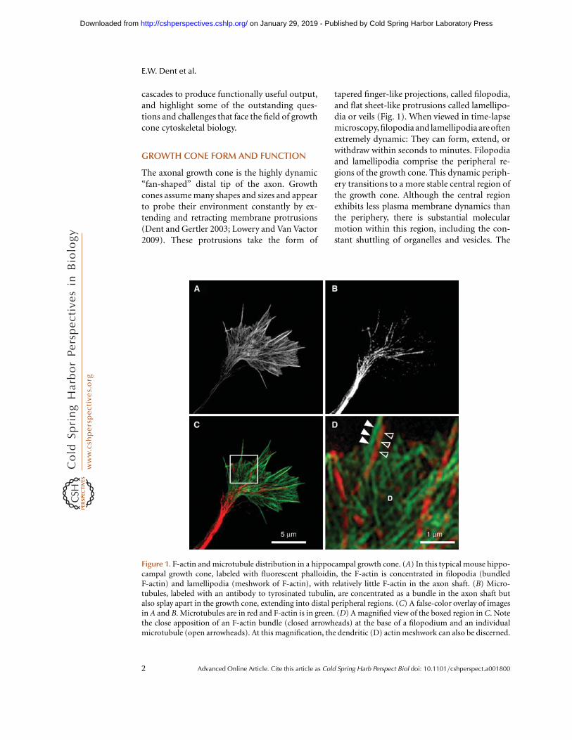
Figure 1. F-actin and microtubule distribution in a hippocampal growth cone. (A) In this typical mouse hippo-campal growth cone, labeled with fluorescent phalloidin, the F-actin is concentrated in filopodia (bundledF-actin) and lamellipodia (meshwork of F-actin), with relatively little F-actin in the axon shaft. (B) Micro-tubules, labeled with an antibody to tyrosinated tubulin, are concentrated as a bundle in the axon shaft butalso splay apart in the growth cone, extending into distal peripheral regions. (C) A false-color overlay of imagesin A and B. Microtubules are in red and F-actin is in green. (D) A magnified view of the boxed region in C. Notethe close apposition of an F-actin bundle (closed arrowheads) at the base of a filopodium and an individualmicrotubule (open arrowheads). At this magnification, the dendritic (D) actin meshwork can also be discerned.
E.W. Dent et al.
2 Advanced Online Article. Cite this article as Cold Spring Harb Perspect Biol doi: 10.1101/cshperspect.a001800
on January 29, 2019 - Published by Cold Spring Harbor Laboratory Press http://cshperspectives.cshlp.org/Downloaded from

central region of the growth cone then transi-tions into the cylindrical axon shaft.
In a still micrograph, these regions of thedistal axon can be identified, but it is importantto realize that these domains are transient. Asa consequence of outgrowth, the growth coneconstantly undergoes dynamic changes in itsstructure, allowing it to lay down new regionsof axon along the path to its target as it moves.Axon outgrowth results from progress throughthree stages: protrusion, engorgement, and con-solidation. Protrusion is the extension of newmembrane at the edges of the growth cone, driv-en by filamentous actin (F-actin) polymeriza-tion. Engorgement results from microtubule(MT)-driven transport of membranous organ-elles and vesicles into the otherwise actin-domi-nated peripheral regions. Consolidation resultsfrom the contraction and stabilization of theproximal growth cone into a cylindrical axonshaft, accompanied by the bidirectional move-ment of organelles and vesicles. These stages
were first described at the morphological levelusing differential interference contrast (DIC)microscopy (Goldberg and Burmeister 1986).However, as we describe below, these stagesresult from specific cytoskeletal changes thatoccur in discrete locations within the growthcone. In addition to these stages of outgrowth,growth cones also undergo cycles of pausingand retraction that engage other cytoskeletalchanges. Throughout this article, we discusshow these changes in output are triggered byguidance cues received at the growth cone oralong the axon shaft.
ACTIN DYNAMICS IN GROWTH CONEPROTRUSION
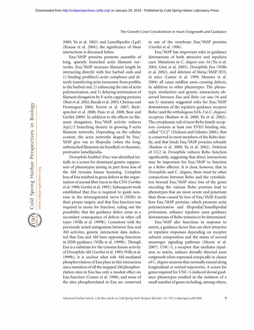
Growth cone protrusion and motility is drivenprimarily by the cyclical polymerization anddepolymerization of actin filaments (Fig. 2).Actin dynamics are necessary for directed axo-nal outgrowth, but not necessarily for growth
Actin/Profilin complex
LamellipodiumFilopodium
Actinfilament
A B
Barbed end protector proteins(Ena/VASP, Formins)
Capping proteins
Actin bundling proteins
Actin motor proteins
Actin severing proteins
Dendritic nucleator proteins
(CapZ, Eps8)
(Fascin, Filamin)Retrogradeactin flow
ATP-actin
ADP-pi-actin
ADP-actin
Pointed end(monomer dissociation)
Barbed end(monomer addition)
(Arp2/3 complex)
(Myosin V, Myosin X)
(Cofilin, Gelsolin)
Figure 2. Structural characteristics of F-actin and actin-associated proteins. (A) Actin filaments are polar poly-mers composed of a barbed end, where the bulk of actin monomer addition occurs, and a pointed end, wheredissociation of actin monomers occur. The nucleotide state of the actin changes as the filaments age(ATP!ADPpi!ADP). (B) F-actin in a filopodium forms bundles due to the action of bundling proteins. Actinmonomers add onto existing filaments at the tip of the filopodium through the action of barbed-end bindingproteins. Actin filaments are constantly undergoing retrograde flow (large vertical arrows) and are disassemblednear their pointed ends by severing proteins. Motor proteins use the bundled F-actin to transport cargo bothanterogradely and retrogradely. In contrast, F-actin in the lamellipodium forms a dendritic network throughthe action of dendritic nucleator proteins and capping proteins. Addition of actin monomers also occursnear the membrane, and disassembly occurs more proximally in the growth cone.
The Growth Cone Cytoskeleton in Axon Outgrowth and Guidance
Advanced Online Article. Cite this article as Cold Spring Harb Perspect Biol doi: 10.1101/cshperspect.a001800 3
on January 29, 2019 - Published by Cold Spring Harbor Laboratory Press http://cshperspectives.cshlp.org/Downloaded from

cone translocation, per se. Many studies, both inculture (Marsh and Letourneau 1984; Lafontet al. 1993; Dent and Kalil 2001) and with-in model organisms (Bentley and Toroian-Raymond 1986; Chien et al. 1993; Kaufmannet al. 1998), have shown that neurons treatedwith agents that depolymerize F-actin are stillcapable of elongation. Such neurons, however,generally cannot respond to guidance cuesand become misrouted in vivo or form axonalloops in culture, presumably because they can-not change direction once they start turning.Importantly, F-actin dynamics appear to beparticularly important for growth cone explora-tion of the environment.
As in other cell types, G-actin is added ontothe free barbed ends of filaments located at thetips of filopodia (Mallavarapu and Mitchison1999) and lamellipodia (Symons and Mitchison1991). By labeling actin filaments within livinggrowth cones, either with fluorescent mono-mer (EGFP-actin or by microinjecting G-actincoupled to a dye) or fluorescently labeled phal-loidin, F-actin is found to be in a steady retro-grade flow from the leading edge to the centerof the growth cone. Elegant studies from theForscher lab have shown that this retrograde F-actin flow is a result of both myosin-II-drivenactin transport and a pushing force that actinexerts on the peripheral membrane as it is poly-merizing (Lin et al. 1996; Zhang et al. 2003;Medeiros et al. 2006). The balance betweenthe rate of polymerization and retrograde flowdetermines if the growth cone extends orwithdraws protrusions: If the polymerizationrate exceeds retrograde flow (which averages3–6mm/min), then the growth cone protrudes.If the polymerization rate is balanced with thevelocity of retrograde flow, then the membraneremains stationary. Thus, it would be expectedthat if myosin activity is decreased, retrogradeflow would decrease and protrusion wouldincrease. To a certain extent this does happen,but the protrusions tend to be unstable andrandom (Medeiros et al. 2006). Importantly,when connection to the underlying substratumis increased by tighter coupling between theactin cytoskeleton and transmembrane adhe-sion receptors, retrograde flow slows and the
balance shifts to polymerization-driven protru-sion (Suter and Forscher 2001). Thus, retro-grade actin flow tends to act as a backgroundactivity upon which other factors can act.
Interestingly, the motor protein myosin-IIhas also been shown to be important for sever-ing actin filaments in the proximal “transitionzone” of the growth cone that demarcates theborder between the growth cone’s actin-richperiphery and the MT-rich central region(Medeiros et al. 2006). Myosin-II does not severactin filaments directly, but rather exerts con-tractile forces on anti-parallel F-actin that con-tracts the actin meshwork to a point that itbreaks the filaments into smaller pieces; theseundergo rapid depolymerization in the transi-tion zone. There are also a number of knownactin-severing proteins that likely have impor-tant roles in severing actin filaments in thetransition zone (Fig. 2), but the potential re-quirements for two prominent severing pro-teins found in this region, gelsolin (Lu et al.1997) and cofilin (Pak et al. 2008), have yet tobe demonstrated.
Driven by dynamic remodeling of the actincytoskeleton, lamellipodial and filopodial pro-trusions underlie growth cone motility andguidance. It is widely assumed that repulsivegrowth cone turning arises from disruption andloss of F-actin superstructures and actomyosincontraction, while attractive growth cone turn-ing entails asymmetrical incorporation of actinon the side of the growth cone closest to the che-moattractant. In support of the aforementionedmodel for growth cone chemoattraction, Mar-sick and colleagues recently reported that appli-cation of chemoattractive guidance cues NGF ornetrin1 to dorsal root ganglia (DRG) inducedincreased protrusion, F-actin accumulation,and increased barbed end density on the sideof the growth cone nearest the source of thegradient (Marsick et al. 2010). It is important,however, to note that there are relatively fewdetailed, high-resolution studies of cytoskeletaland morphological dynamics of growth conesturning toward or away from guidance cues(e.g., Dent et al. 2004; Suter et al. 2004; Brownand Bridgman 2009; Brown et al. 2009; Marsicket al. 2010). Therefore, it remains unclear
E.W. Dent et al.
4 Advanced Online Article. Cite this article as Cold Spring Harb Perspect Biol doi: 10.1101/cshperspect.a001800
on January 29, 2019 - Published by Cold Spring Harbor Laboratory Press http://cshperspectives.cshlp.org/Downloaded from
whether all attractive and repulsive events in-volve analogous cytoskeletal and shape changes.
An additional level of complexity in under-standing the function of cytoskeletal machineryduring guidance is that individual componentslikely act in a context-dependent fashion, influ-enced by the signaling status in the growth cone,the repertoire of cytoskeletal proteins presentin the growth cone, and adhesive interactions.For example, the actin severing/depolymerizingprotein cofilin is required for axon extension inhippocampal neurons (Garvalov et al. 2007)and dorsal root ganglia neurons (Endo et al.2003), but likely not in cerebellar granular neu-rons (Tahirovic et al. 2010). In addition, cofilinactivity has been implicated in both attractive(Marsick et al. 2010) and repulsive (Wen et al.2007) guidance responses. Moreover, in Xeno-pus spinal neurons, the requirements for cofilinfunction in response to BMP7 are thought tochange as neurons mature: In 4–8 hour neu-rons, axons are attracted to BMP7, and cofilinmust be inactive for this response; after over-night culture, BMP7 repels axons, and this re-sponse requires asymmetric cofilin activity inthe portion of the growth cone exposed to thehigher BMP7 concentration (Wen et al. 2007).Despite its role in BMP7 repulsion, growthcone collapse induced by Sema3A requires inac-tivation of cofilin (Aizawa et al. 2001). There-fore, it may not be possible to extrapolate themechanism of turning or collapse from one cueto another, from one neuron type to another,or the role of particular cytoskeletal proteinsin different contexts. There may also be species-specific differences that affect how certain cy-toskeletal proteins are utilized (Marsick et al.2010). Such context-dependent functions arereminiscent of the ways in which guidance cuesand their cognate receptors can elicit distinctresponses.
In this article, we focus on how actin dynam-ics affect membrane protrusions and generatestructures that interact with the MT cytoskele-ton. It is clear, however, that actin-dependentor actin-influenced processes, such as adhesion,membrane trafficking, and endo/exocytosis,play equally important roles in growth cone for-mation, motility, and guidance responses (e.g.,
Jurney et al. 2002; Suh et al. 2004; Tojima et al.2007; Kolpak et al. 2009; Hines et al. 2010;Tojima et al. 2010). While space constraintspreclude coverage of these topics, the readershould bear in mind that some of the proteinsdiscussed below might exert their effects onguidance by influencing adhesion dynamicsand membrane/vesicle transport.
MICROTUBULE DYNAMICS IN AXONGUIDANCE
Although actin and actin-associated proteinsare usually the first cytoskeletal proteins thatcome to mind when one considers axon guid-ance, a number of studies implicate micro-tubules (MTs) directly in growth cone steering.The idea that MTs are involved in guidancedecisions was first suggested by pioneeringstudies from the Kirschner laboratory, show-ing in living neurons that fluorescently labeledMTs were capable of exploring the growthcone periphery, and that the orientation ofMTs often predicted the direction of outgrowth(Sabry et al. 1991; Tanaka and Kirschner 1995).Further work from several laboratories con-firmed and extended these findings by showingthat the dynamic pool of MTs (identified bya post-translational modification that adds ac-terminal tyrosine residue—referred to as ty-rosinated MTs) was key for either directed out-growth toward a target or chemorepulsion attwo substrate boundaries (Lin and Forscher1993; Lin and Forscher 1995; Williamson et al.1996; Challacombe et al. 1997). Nevertheless,the general perception remained that actin fila-ments initiated that change in the direction ofgrowth and MTs followed their lead.
However, a more recent study showed thatsimply changing MT dynamics by locally apply-ing, or photo-uncaging, MT-specific drugs onone side of a growth cone was sufficient toinduce growth cone turning (Buck and Zheng2002). These results led the authors to proposethat MTs were indeed playing an instructiverole in growth cone guidance, since simplychanging MT dynamics asymmetrically wassufficient to induce growth cone turning. Nev-ertheless, these results do not mitigate the role
The Growth Cone Cytoskeleton in Axon Outgrowth and Guidance
Advanced Online Article. Cite this article as Cold Spring Harb Perspect Biol doi: 10.1101/cshperspect.a001800 5
on January 29, 2019 - Published by Cold Spring Harbor Laboratory Press http://cshperspectives.cshlp.org/Downloaded from

that actin plays in growth cone turning. Indeed,growth cone turning induced by taxol applica-tion can be abrogated by inclusion of nano-molar concentrations of cytochalasin D, a drugthat caps actin filaments (Buck and Zheng2002). These results indicate that even if extracel-lular cues signal directly to MTs, the output oftheresponse,growthconeturning, is likely topro-ceed through changes in the actin cytoskeleton.
These studies raise an intriguing question:Could spatially and temporally restricted pre-sentation of guidance cues regulate MT dynam-ics and stability directly to give rise to growthcone turning? To answer this question, we firsthave to understand what effect guidance cueshave on microtubule dynamics. However, quan-tifying MT dynamics in neuronal growth conesis difficult. MTs constantly polymerize and de-polymerize throughout the growth cone peri-pheral domain due to a behavior inherent inthe structure of these polymers termed “dy-namic instability.” Growth cones occupy rela-tively small areas and expand and contract,making imaging MT dynamics challenging. Itis a daunting task to image these dynamics forextended periods of time while locally applyingguidance cues. Thus, there have been no studiesto date that have imaged MT dynamics ingrowth cones during local application of a guid-ance cue. However, MT dynamics have beenimaged after bath application of several guid-ance cues. In one study, Sema3a was bathapplied to large, paused growth cones from cul-tured cortical neurons (Dent et al. 2004). MTsrapidly lost their dynamic behavior and col-lapsed back onto the central region of thegrowth cone. In this study, netrin was shownto induce opposite changes in MTs, causingincreased splaying of MTs in the growth coneand axon shaft. However, the effect of netrinwas only documented in fixed growth cones.A more recent study has shown that bath appli-cation of Wnt3a induces MTs to lose direc-tionality and polymerize perpendicular to thedirection of growth cone translocation (Purroet al. 2008). Over time, this behavior resultsin MT looping, growth cone enlargement, andpausing. These studies indicate that MTs aresensitive to guidance cues, but further research
is necessary to understand how MT dynamicsare directly or indirectly regulated by such cues.
Additionally, there have been a number ofstudies of MT dynamics in large Aplysia growthcones that provide insight into how MT dynam-ics may be regulated in growth cones duringguidance decisions (Suter and Forscher 2000).These studies by the Forscher and Suter labora-tories include imaging of MTand actin dynam-ics at high temporal and spatial resolution whileconducting a localized adhesion-based assay.In this assay, a polystyrene bead is coated witha cell adhesion molecule (apCAM) and posi-tioned on top of an adherent and well-spreadgrowth cone. By restraining this bead witha microneedle, these researchers were able toshow an ensuing stereotyped series of events(Suter et al. 1998; Suter et al. 2004; Lee andSuter 2008; Schaefer et al. 2008). Normally,the actin in the growth cone undergoes constit-utive retrograde flow. As the apCAM bead en-gages receptors on the growth cone, an actinscaffold begins to accumulate near the plasmamembrane underlying the bead. This actinmatrix couples to the cell adhesion complex,engaging a “clutch” type mechanism. Duringthis time, termed the latency phase, exploratoryMTs polymerize and are transported anterog-radely toward the actin matrix under the bead.Progressive coupling of the bead to the growthcone, forming a mature adhesion site, instigatesa loss of actin retrograde flow behind the beadand possibly an increase in severing and/orunbundling of the actin. Clearing of the actinbehind the bead allows for more microtubuleinvasion toward the bead and potential captureat the adhesion sight. This traction period (Leeand Suter 2008) or growth phase (Schaefer et al.2008) is accompanied by protrusion of lamelli-podia and filopodia beyond the bead. Presum-ably, this sequence of stereotyped cytoskeletalbehaviors would repeat as soon as the growthcone made another productive adhesion in amore distal region of the growth cone.
Using this methodology, one group stressesthat actin arcs, the actomyosin contractile net-work in the transition region of the growthcone, are important for guiding MTs to thebead adhesion site (Schaefer et al. 2008). The
E.W. Dent et al.
6 Advanced Online Article. Cite this article as Cold Spring Harb Perspect Biol doi: 10.1101/cshperspect.a001800
on January 29, 2019 - Published by Cold Spring Harbor Laboratory Press http://cshperspectives.cshlp.org/Downloaded from

other group indicates that a critical aspectunderlying the ability of MTs to target thebead, and presumably induce a new axis of out-growth, is the uncoupling of putative MT-actinlinker molecules (Lee and Suter 2008). Thisrestricted bead assay provides essential knowl-edge about cytoskeletal interactions underlyingadhesion-based growth. However, many guid-ance cues are not cell-adhesion molecules. Thus,this assay may be better defined as a neuritegrowth model (Schaefer et al. 2008), rather thana model for guidance (Lee and Suter 2008), perse. An important question for future experi-ments will be to determine if local applicationof guidance cues to the growth cone inducesthe same types of cytoskeletal behaviors in ac-tively translocating growth cones, as are seenin these large, paused, Aplysia growth cones.
ACTIN-ASSOCIATED PROTEINS IN AXONGUIDANCE
The exquisite control of actin nucleation, elon-gation, depolymerization, bundling, and con-traction necessary to shape the growth coneand enable dynamic responses to a plethoraof extracellular cues is mediated by a complexrepertoire of actin accessory proteins found inmany cell types (see Table 1 and Fig. 2). Morethan 100 such accessory proteins are used byeukaryotic cells to nucleate filaments, controlfilament length, bundle or cross-link filaments,disassemble filament networks, and maintain apool of actin monomers (Pollard and Cooper2009). While an increasing number of theseactin accessory proteins have been identifiedin neurons, it is likely that the abundance of var-ious classes of actin regulators differs accordingto cell type, and it is clear that the relative levelsof some key types of actin regulatory proteinsin the growth cone differ from the stoichiome-tries found in systems that are commonly usedto analyze the regulation of actin dynamics(Strasser et al. 2004). Thus, an exquisite balanceof actin accessory proteins likely contributes tothe distinctive morphology of growth cones.Many actin binding proteins in the growth coneregulate lamellipodia and filopodia dynamics,axon guidance, or both, but how guidance cues
orchestrate cytoskeletal remodeling by themany proteins within the growth cone to elicitthe proper response remains largely unknown.While the list of known actin-associated proteinsis expansive and many are expressed in the devel-oping nervous system, relatively few have beenimplicated in axon guidance. Here, we consideractin-associated proteins that have been specifi-cally implicated in growth cone guidance.
Barbed-end Binding Proteins
This group of proteins associates with actin fil-ament barbed ends. Some cap barbed ends, ter-minating filament elongation. Other barbed-end binding proteins protect the filamentend from polymerization-terminating cappingproteins, and in some cases alter the rate of G-actin incorporation at the filament end. As inother cells, most filaments within growth conesare oriented with their barbed ends toward thefront edge of lamellipodial veils and filopodialtips, sites where rapid responses to guidancecues are likely essential for proper navigation(Fig. 2).
Ena/VASP Proteins
The Ena/VASP proteins were the first examplesof barbed-end binding proteins implicated inaxon guidance (Drees and Gertler 2008). Thereare three vertebrate Ena/VASP paralogs (Mena,VASP, and EVL), while Drosophila and C. eleganseach contain a single ortholog, Enabled (Ena)and UNC-34, respectively. While Ena/VASPproteins are found in many cell types, they arehighly expressed in the developing nervoussystem, where they concentrate in the filopodialtips of growth cones as well as the leading edge oflamellipodia (Lanier et al. 1999), two structuresrich in elongating barbed ends. The localiza-tion of Ena/VASP proteins to these structures,as well as interactions with signaling proteins,is controlled in part by protein–protein inter-actions between the conserved EVH1 (Ena/VASP homology 1) domain and proteins thatcontain EVH1-binding motifs (Niebuhr et al.1997; Ball et al. 2002). At least two moleculesinvolved in axon guidance contain functionalEVH1-binding sites: Robo/Sax3 (Bashaw et al.
The Growth Cone Cytoskeleton in Axon Outgrowth and Guidance
Advanced Online Article. Cite this article as Cold Spring Harb Perspect Biol doi: 10.1101/cshperspect.a001800 7
on January 29, 2019 - Published by Cold Spring Harbor Laboratory Press http://cshperspectives.cshlp.org/Downloaded from
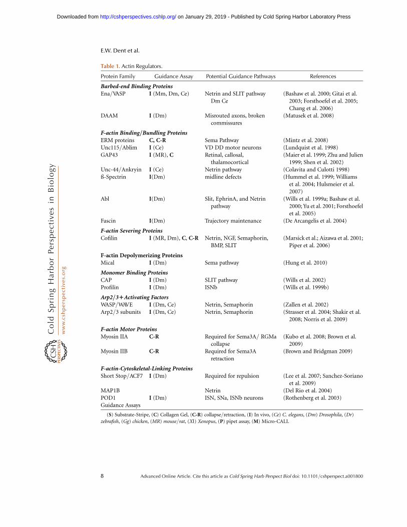
Table 1. Actin Regulators.
Protein Family Guidance Assay Potential Guidance Pathways References
Barbed-end Binding ProteinsEna/VASP I (Mm, Dm, Ce) Netrin and SLIT pathway
Dm Ce(Bashaw et al. 2000; Gitai et al.
2003; Forsthoefel et al. 2005;Chang et al. 2006)
DAAM I (Dm) Misrouted axons, brokencommissures
(Matusek et al. 2008)
F-actin Binding/Bundling ProteinsERM proteins C, C-R Sema Pathway (Mintz et al. 2008)Unc115/Ablim I (Ce) VD DD motor neurons (Lundquist et al. 1998)GAP43 I (MR), C Retinal, callosal,
thalamocortical(Maier et al. 1999; Zhu and Julien
1999; Shen et al. 2002)Unc-44/Ankryin I (Ce) Netrin pathway (Colavita and Culotti 1998)ß-Spectrin I(Dm) midline defects (Hummel et al. 1999; Williams
et al. 2004; Hulsmeier et al.2007)
Abl I(Dm) Slit, EphrinA, and Netrinpathway
(Wills et al. 1999a; Bashaw et al.2000; Yu et al. 2001; Forsthoefelet al. 2005)
Fascin I(Dm) Trajectory maintenance (De Arcangelis et al. 2004)
F-actin Severing ProteinsCofilin I (MR, Dm), C, C-R Netrin, NGF, Semaphorin,
BMP, SLIT(Marsick et al.; Aizawa et al. 2001;
Piper et al. 2006)
F-actin Depolymerizing ProteinsMical I (Dm) Sema pathway (Hung et al. 2010)
Monomer Binding ProteinsCAP I (Dm) SLIT pathway (Wills et al. 2002)Profilin I (Dm) ISNb (Wills et al. 1999b)
Arp2/31Activating FactorsWASP/WAVE I (Dm, Ce) Netrin, Semaphorin (Zallen et al. 2002)Arp2/3 subunits I (Dm, Ce) Netrin, Semaphorin (Strasser et al. 2004; Shakir et al.
2008; Norris et al. 2009)
F-actin Motor ProteinsMyosin IIA C-R Required for Sema3A/ RGMa
collapse(Kubo et al. 2008; Brown et al.
2009)Myosin IIB C-R Required for Sema3A
retraction(Brown and Bridgman 2009)
F-actin-Cytoskeletal-Linking ProteinsShort Stop/ACF7 I (Dm) Required for repulsion (Lee et al. 2007; Sanchez-Soriano
et al. 2009)MAP1B Netrin (Del Rio et al. 2004)POD1 I (Dm) ISN, SNa, ISNb neurons (Rothenberg et al. 2003)Guidance Assays
(S) Substrate-Stripe, (C) Collagen Gel, (C-R) collapse/retraction, (I) In vivo, (Ce) C. elegans, (Dm) Drosophila, (Dr)
zebrafish, (Gg) chicken, (MR) mouse/rat, (Xl) Xenopus, (P) pipet assay, (M) Micro-CALI.
E.W. Dent et al.
8 Advanced Online Article. Cite this article as Cold Spring Harb Perspect Biol doi: 10.1101/cshperspect.a001800
on January 29, 2019 - Published by Cold Spring Harbor Laboratory Press http://cshperspectives.cshlp.org/Downloaded from
2000; Yu et al. 2002) and Lamellipodin (Lpd)(Krause et al. 2004); the significance of theseinteractions is discussed below.
Ena/VASP proteins promote assembly oflong, sparsely branched actin filament net-works. Ena/VASP increases filament length byinteracting directly with free barbed ends and1) binding profilin:G-actin complexes and di-rectly transferring actin monomer from profilinto the barbed end, 2) enhancing the rate of actinpolymerization, and 3) delaying termination offilament elongation by F-actin capping proteins(Bear et al. 2002; Barzik et al. 2005; Chereau andDominguez 2006; Ferron et al. 2007; Breit-sprecher et al. 2008; Pasic et al. 2008; Bear andGertler 2009). In addition to the effects on fila-ment elongation, Ena/VASP activity reducesArp2/3 branching density in growing F-actinfilament networks. Depending on the cellularcontext, the actin networks shaped by Ena/VASP give rise to filopodia (when the long,unbranched filaments are bundled) or dynamic,protrusive lamellipodia.
Drosophila Enabled (Ena) was identified ini-tially in a screen for dominant genetic suppres-sors of phenotypes arising in part from loss ofthe Abl tyrosine kinase homolog. Completeloss of Ena resulted in gross defects in the organ-ization of axonal fiber tracts in the CNS (Gertleret al. 1990; Gertler et al. 1995). Subsequent workestablished that Ena is required to guide neu-rons in the intersegmental nerve b (ISNb) totheir proper targets, and that Ena function wasrequired in axons for function, ruling out thepossibility that the guidance defect arose as asecondary consequence of defects in other celltypes (Wills et al. 1999b). Consistent with thepreviously noted antagonism between Ena andAbl activites, genetic interaction data indica-ted that Ena and Abl have opposing functionsin ISNb guidance (Wills et al. 1999b). ThoughEna is a substrate for the tyrosine kinase activityof Drosophila Abl (Gertler et al. 1995; Wills et al.1999b), it is unclear what role Abl-mediatedphosphorylation of Ena plays in this interactionsince mutation of all the mapped Abl phosphor-ylation sites in Ena has only a modest effect onEna function (Comer et al. 1998), and none ofthe sites phosphorylated in Ena are conserved
in any of the vertebrate Ena/VASP proteins(Gertler et al. 1996).
Ena/VASP has important roles in guidancedownstream of both attractive and repulsivecues. Mutations in C. elegans unc-34 (Yu et al.2002; Gitai et al. 2003), Drosophila Ena (Willset al. 2002), and deletion of Mena/VASP/EVLin mice (Lanier et al. 1999; Menzies et al.2004) all cause midline axon crossing defects,in addition to other phenotypes. The pheno-typic similarities and genetic interactions ob-served between Ena and Robo (or unc-34 andsax-3) mutants suggested roles for Ena/VASPdownstream of the repulsive guidance receptorRobo (and the orthologous SAX-3 in C. elegans)receptors (Bashaw et al. 2000; Yu et al. 2002).The cytoplasmic tail of most Robo family recep-tors contains at least one EVH1-binding site,called “CC2” (Dickson and Gilestro 2006), thatis conserved in most members of the Robo fam-ily, and that binds Ena/VASP proteins robustly(Bashaw et al. 2000; Yu et al. 2002). Deletionof CC2 in Drosophila reduces Robo functionsignificantly, suggesting that direct interactionsmay be important for Ena/VASP to functionas a Robo effector. It is clear, however, that inDrosophila and C. elegans, there must be otherconnections between Robo and the cytoskele-ton beyond Ena/VASP since loss of the genesencoding the various Robo proteins lead tophenotypes that are more severe and penetrantthan those caused by loss of Ena/VASP. Exactlyhow Ena/VASP proteins, which promote actinpolymerization and filopodial/lamellipodialprotrusion, enhance repulsive axon guidancedownstream of Robo remains to be determined.
Ena/VASP also functions in response tonetrin, a guidance factor that can elicit attractiveor repulsive responses depending on receptorsubunit composition and the status of secondmessenger signaling pathways (Moore et al.2007). UNC-5, a receptor that mediates repul-sion to netrin, induces dorsally directed axonoutgrowth when expressed ectopically in classesof C. elegans neurons that normally extend alonglongitudinal or ventral trajectories. A screen forgenes required for UNC-5-induced dorsal guid-ance phenotypes resulted in the isolation of asmall number of genes including, among others,
The Growth Cone Cytoskeleton in Axon Outgrowth and Guidance
Advanced Online Article. Cite this article as Cold Spring Harb Perspect Biol doi: 10.1101/cshperspect.a001800 9
on January 29, 2019 - Published by Cold Spring Harbor Laboratory Press http://cshperspectives.cshlp.org/Downloaded from

unc-6 (netrin), unc-40 (DCC), unc-44 (Ankyrin,an F-actin binding protein), and unc-34 (Ena/VASP) (Colavita and Culotti 1998). Therefore,it is likely that Ena/VASP functions in normalUNC-5-dependent repulsion to netrin.
In addition to their function downstream ofUNC-5, experiments in both Drosophila andC. elegans indicate that Ena/VASP contributesto phenotypes arising from chimeric or consti-tutively active forms of the netrin receptor DCC(Gitai et al. 2003; Forsthoefel et al. 2005) (DCCorthologs are: Frazzled in Drosophila and UNC-40 in C. elegans). Further analysis verified thatEna/VASP mediates part of the normal chemo-attractive response to netrin during axon guid-ance. Understanding the nature of Ena/VASPmutant axon guidance phenotypes in all modelsystems is complicated by the dual roles ofEna/VASP in both attraction and repulsion inresponse to both netrin and Robo-mediatedrepulsion. Examination of unc-34 phenotypesin C. elegans lacking SLIT-Robo guidance pro-vided unambiguous confirmation that Ena/VASP is indeed required for netrin attraction(Gitai et al. 2003).
Mutational analysis of an activated formof UNC-40 revealed that UNC-34 functionsdownstream of a conserved cytoplasmic motifknown as P1 (Gitai et al. 2003). The same studyrevealed that the Rac and ablim orthologs(mig-10 and unc-115, respectively) functiondownstream of the UNC-40 P2 motif and actin parallel to UNC-34. Subsequent genetic anal-ysis suggested that UNC-115 function affectsfilopodia formation, perhaps in parallel toUNC-34 (Norris et al. 2009). While the role ofthe conserved P1 and P2 motifs in expressingthe activated UNC-40 phenotype is clear, theirrole in normal axon guidance is less clear asthe fra mutant phenotype in Drosophila can berescued using fra transgenes that lack P1 andP2 (Garbe et al. 2007b). Whether UNC-34 in-teracts directly with UNC-40, or whether adap-tor proteins connect them via a downstreamsignaling pathway, is presently unknown. Re-gardless of how Ena/VASP proteins are linkedto UNC-40/DCC signaling, however, they arerequired to elicit filopodia formation and elon-gation after netrin stimulation in vivo (Chang
et al. 2006) and in cultured cortical neurons(Lebrand et al. 2004).
One mechanism that may coordinate Ena/VASP function in filopodia formation withnetrin-mediated guidance responses involvesPKA signaling (PKA; cAMP-dependent kinase).Second messenger signaling by cAMP andcGMP plays significant roles in axon guidanceby modulating the intensity of responses tocues and perhaps by inducing conversionbetween attractive and repulsive responses tocertain guidance cues (Bashaw and Klein2010). Cultured neurons treated with netrinexhibit a robust increase in filopodia formation(Shekarabi and Kennedy 2002) that requiresEna/VASP, among other cytoskeletal regulatoryproteins, and PKA activity (Lebrand et al.2004); neurons treated with PKA activatorselaborate filopodia (Chen et al. 2003; Argawet al. 2008) in an Ena/VASP-dependent man-ner (Lebrand et al. 2004). At present, it isunknown whether PKA phosphorylation ofEna/VASP is essential for netrin-elicited filopo-dia induction.
While it seems likely that netrin elicits filo-podia formation through a process that requiresPKA and Ena/VASP in cultured neurons, otherdata preclude a simple netrin.DCC.cAMP.
PKA.Ena/VASP pathway. Treatment with net-rin does not increase global cAMP levels or PKAactivation (Bouchard et al. 2004; Bouchard et al.2008; Bashaw and Klein 2010), so it remainsunclear how Ena/VASP phosphorylation levelsrise upon netrin stimulation. Furthermore, PKAis not required for axon extension towards net-rin in vivo, but enhances chemoattraction tonetrin, at least in part, by increasing surfacelevels of DCC (Bouchard et al. 2004).
The role of both Ena/VASP and filopodiaformation in axon guidance has been ques-tioned based on the ability of retinal axons tonavigate properly while expressing an Ena/VASP function-blocking construct (Dwivedyet al. 2007) that depletes and sequesters Ena/VASP (Bear et al. 2000). This construct wastransfected into the retina of developing Xeno-pus, and the axons expressing the construct weremonitored for filopodia formation, axon navi-gation, and terminal arborization (Dwivedy
E.W. Dent et al.
10 Advanced Online Article. Cite this article as Cold Spring Harb Perspect Biol doi: 10.1101/cshperspect.a001800
on January 29, 2019 - Published by Cold Spring Harbor Laboratory Press http://cshperspectives.cshlp.org/Downloaded from

et al. 2007). Despite a significant reduction infilopodia in the axons that expressed the inhib-itory construct, these axons made their wayto their target region but exhibited defects interminal branching and final target selection.This result has been interpreted as evidencethat filopodia and Ena/VASP are not requiredfor accurate axon guidance (Dwivedy et al. 2007;Lowery and Van Vactor 2009). If true, this con-clusion would be surprising given the extensivegenetic evidence in Drosophila, C. elegans, andmice demonstrating roles for Ena/VASP in mul-tiple guidance pathways. Another interpretationof these data is that a major guidance decisionmade by retinal axons is whether and where tobranch in the tectum. It is important to notethat the transfection approach used is limitedin that it is not possible to be certain when theinhibitory construct was expressed at levels suf-ficient to block Ena/VASP. Furthermore, only asmall subset of neurons in the retina were trans-fected, therefore it is likely that pioneeringaxons were not affected by the inhibitoryconstruct. Compensation for Ena/VASP couldoccur during outgrowth to the target via fasci-culation; however, branch formation in the tec-tum occurs by interstitial axon branching afterthe primary growth cone has extended beyondthe target (McLaughlin et al. 2003). Thus, topo-graphically specific interstitial branching is amajor form of guidance in the tectum, and itis particularly sensitive to Ena/VASP disruption(Dwivedy et al. 2007). This hypothesis is consis-tent with the ability of Ena/VASP to affectbranching in murine hippocampal (Lebrandet al. 2004) and cortical (Dent and Gertler, un-publ.) neurons, two neuronal types that branchprimarily by interstitial branching (Halloranand Kalil 1994).
MRL Proteins
Ena/VASP are also linked to both netrin andSlit-dependent guidance by their direct interac-tion with MRL proteins: MIG-10 in C. elegans,and Lamellipodin (Lpd) and RIAM in verte-brates (Krause et al. 2004; Chang et al. 2006;Quinn et al. 2006; Quinn et al. 2008; Michaelet al. 2010; Smith et al. 2010). In addition to
acting as downstream signaling componentsof netrin and Slit guidance pathways, MIG-10 plays a role in the initial establishmentof asymmetry in the HSN neuron. Prior toHSN axon formation, MIG-10 accumulates onthe side of the cell oriented toward a netrinsource, the earliest known molecule to exhibitsuch Netrin-dependent asymmetric localiza-tion. MRL proteins are linked to second mes-senger signaling by tyrosine kinases and bybinding to phosphinositides such as PI (3,4)P2 and to Ras superfamily proteins. Consistentwith a role for PI (3,4)P2 in spatiotemporalregulation of MRL localization, mutations inage-1, a PI3K, impair netrin-dependent local-ization of MIG-10, most likely because PI3Kactivity is required to produce PI(3,4) P2 amongother phosphoinositides. MRL proteins pro-mote actin polymerization, membrane protru-sion, and can regulate activation of adhesionreceptors. Genetic and cell biological evidenceindicate that while Ena/VASP is required forfull MRL function, other actin regulators actdownstream of MRL signaling. Clearly, imple-mentation of fluorescent biosensors and devel-opment of phospho-specific antibodies that canbe used for immunolocalization will be requiredto examine spatiotemporal signaling to thecytoskeleton downstream of netrin and otherguidance cues.
DAAM1
Recent work implicates another barbed-endbinding protein, disheveled-associated activa-tor of morphogenesis (DAAM), as a regulatorof filopodia formation and axonal morphogen-esis in Drosophila (Matusek et al. 2008). DAAMis a member of the formin protein superfamilythat nucleates linear actin filaments, regulatesfilament growth rate through processive elon-gation (continuous attachment to the growingend), and can block filament termination bybarbed-end capping proteins. Most forminproteins contain three conserved domains: theGTPase binding domain (GBD), Formin homo-logy 1 (FH1) domain, and Formin homology2 (FH2) domain (Higgs 2005). Formins areautoinhibited by head-to-tail intramolecular
The Growth Cone Cytoskeleton in Axon Outgrowth and Guidance
Advanced Online Article. Cite this article as Cold Spring Harb Perspect Biol doi: 10.1101/cshperspect.a001800 11
on January 29, 2019 - Published by Cold Spring Harbor Laboratory Press http://cshperspectives.cshlp.org/Downloaded from

interactions that are typically relieved by bind-ing to Rho GTPases, thereby allowing the FH1and FH2 to bind effectors and drive actin poly-merization (Goode and Eck 2007). The FH1 do-main binds to profilin, while the FH2 domaincontains the core nucleating activity. Unlikeother formins, DAAM1 is activated by bindingto Dvl, a component of noncanonical Wnt sig-naling pathways (Liu et al. 2008), raising theinteresting possibility that this molecule couldbe involved in Wnt-mediated guidance.
The Drosophila DAAM1 ortholog “DAAM”is expressed highly in the nervous system(Matusek et al. 2008). Loss-of-function analysisand targeted expression of activated constructsrevealed that DAAM plays significant roles infilopodia formation on axonal growth cones:Loss of DAAM reduces the frequency of filopo-dia formation in primary cultures from mu-tants, while expression of activated DAAMincreases filopodia formation. Most eukaryotespossess multiple formin genes (Higgs 2005;Higgs and Peterson 2005), and the cellular rolesof these proteins have recently been explored.Mutant embryos lacking functional DAAMexhibit a range of CNS phenotypes that areobserved in about a third of the embryos lackingboth the maternally and zygotically producedprotein. The DAAM mutants show occasion-al gaps in longitudinal connectives and moreseverely affected embryos show malformedcommissures, misrouted axons, or failures incommissure separation (Matusek et al. 2008).Genetic interaction studies indicate that re-duced levels of DAAM exacerbate phenotypescaused by loss of Rac and vice versa, showingthat these two proteins function in interde-pendent, or perhaps parallel, pathways im-portant for axon outgrowth and guidance.Similarly, reduction in Ena or profilin levels exa-cerbated the DAAM loss-of-function pheno-type. Expression of a constitutively active formof DAAM induced a variety of defects, includ-ing a partial collapse of the longitudinal tractsthat could be ameliorated significantly by re-ducing Ena levels. Therefore, it appears thatthere is likely significant interplay betweenDAAM and Ena function during axon out-growth and guidance.
Coimmunoprecipitation of various forminsin different systems revealed that there are smallpools of Ena/VASP in complex with formins(Grosse et al. 2003; Schirenbeck et al. 2006;Homem and Peifer 2009). The distribution ofDAAM and Ena overlaps partially in growthcone filopodia (Matusek et al. 2008), so thesetwo molecules may interact as well. While vari-ous models have been proposed to explain thefunction of a formin:Ena/VASP complex, nonehave been tested rigorously. Regardless, it seemslikely that complexes between multiple mole-cules that can each independently drive filo-podia formation may exist. This offers thegrowth cone exquisite flexibility to respondrapidly to distinct guidance cues that triggeractivation of a subset of such filopodia-promot-ing proteins. In the future, it will be interestingto see if DAAM acts in the same guidance path-ways as Ena. In this respect, it is curious thatmutations in genes encoding DAAM or otherformin superfamily proteins have not beenrecovered in genetic screens conducted in Dro-sophila and C. elegans for axon guidance defectsor for modifiers of guidance pathways, whilemutations in Ena/VASP genes have been iden-tified repeatedly. Given the large number offormin family members (Higgs 2005; Higgsand Peterson 2005), it is likely that future stud-ies will reveal that other formins are involved ingrowth cone morphology and guidance. It willbe interesting to further define how forminfamily proteins and Ena/VASP proteins cooper-ate in filopodia formation and axon guidance.
Capping Proteins
Not all barbed-end-binding proteins facilitateactin polymerization. Capping proteins bindto the barbed ends of actin filaments and blockaccess of monomers to the barbed end, thushalting polymerization and reducing the lengthof F-actin (Xu et al. 1999). Antagonism bet-ween filament elongation/protection factorsand capping proteins contributes to filopodialdynamics. In fact, while filopodial formationhas been the subject of great interest, relativelylittle is understood about the mechanismsthat govern filopodial withdrawal, including
E.W. Dent et al.
12 Advanced Online Article. Cite this article as Cold Spring Harb Perspect Biol doi: 10.1101/cshperspect.a001800
on January 29, 2019 - Published by Cold Spring Harbor Laboratory Press http://cshperspectives.cshlp.org/Downloaded from

determining which of the many filament cap-ping proteins play predominant roles in nega-tively regulating filopodial dynamics in growthcones. Hippocampal neurons from mice lackinggelsolin, a protein that severs actin filaments in aCa2þ-dependent manner and caps the free bar-bed ends of the severed filaments (Sun et al.1999), have increased numbers of filopodiathat arise as a consequence of reduced retraction(Lu et al. 1997). A role for gelsolin in axon guid-ance has not been reported. Similarly, deletionof the multifunctional capping protein EPS8,which interacts with a number of signaling pro-teins involved in Rac and Ras signaling, as wellas having actin filament capping activity, resultsin increased filopodia number after treatmentof hippocampal neurons with BDNF (Mennaet al. 2009). EPS8 knockout mice do not showany obvious axon guidance defects; however,the expression of two EPS8-related moleculesduring development may mask a possiblerequirement for EPS8.
The most widely studied capping protein,heterodimeric capping protein (CP), does in-deed mediate growth cone morphology, axonelongation, and possibly axon guidance. InDrosophila, reduction in filament elongation/anti-capping activity of Ena ameliorates ablphenotypes, including defects in axon guidance(Gertler et al. 1990; Gertler et al. 1995; Willset al. 1999b; Drees and Gertler 2008). Based onthis finding, Grevengoed and colleagues exam-ined the effects of reducing CP levels in ablmutants and found that such animals exhibitmore severe phenotypes than those observedin abl mutants alone with respect to morphol-ogy of the nervous system (the opposite of theeffect observed upon reduction of Ena levels)(Grevengoed et al. 2003). Therefore, CP mayplay a role in axon guidance through its well-characterized ability to cap growing actin fila-ments. Careful analysis of cp loss-of-functionmutants is required to assess the role of CP inaxon outgrowth and guidance.
Unexpectedly, CP also appears to have a rolein controlling growth cone morphology andaxon elongation independent of its cappingactivity. Loss of a CP subunit, CapzB2, resultedin aberrant growth cone morphology and
neurite outgrowth, and also mis-localized mi-crotubules that extended into the peripheraldomain of growth cones. Surprisingly, this phe-notype is not a result of reduced capping, butinstead arises due to loss of a newly identifiedinteraction between CapzB2 and bIII tubulin.Mutants that maintained capping activity butlacked the ability to bind bIII tubulin couldnot rescue the phenotype, while capping defi-cient mutants that bound bIII tubulin rescued(Davis et al. 2009). This study supports theidea that proper localization of microtubulesin the central region of the growth cone is nec-essary for normal growth cone morphologyand translocation. It will be challenging todetermine the relative contributions of tubulinbinding and filament capping by CP in axonguidance. The control of filament capping dur-ing guidance, and its functional significance tonavigation, remain outstanding questions inthe field.
F-actin Binding Proteins
ERM Proteins
Proteins that bind to the sides of actin filamentsaffect the architecture of cellular F-actin net-works and can tether the cytoskeleton to dif-ferent locations or functional partners. Ezrin,radixin, and moesin comprise the ERM familyof highly homologous, multifunctional F-actinbinding proteins. ERMs contain an N-termi-nal membrane binding domain linked by ana-helical region to a C-terminal actin-bindingdomain, allowing them to tether the actin cyto-skeleton to the plasma membrane through in-teractions with PIP2 and L1CAM (Algrainet al. 1993; Turunen et al. 1994; Chishti et al.1998). They are expressed in the developing ner-vous system and are concentrated specificallyin growth cones (Goslin et al. 1989; Gonzalez-Agosti and Solomon 1996; Paglini et al. 1998;Takahashi et al. 1999; Dickson et al. 2002; Mintzet al. 2008), making them well positioned tocoordinate growth cone responses to guidancecues. Suppression of radixin and moesin re-sults in reduced grow cone area, disorganized F-actin, and lamellipodial veil retraction (Pagliniet al. 1998); while these growth cones display
The Growth Cone Cytoskeleton in Axon Outgrowth and Guidance
Advanced Online Article. Cite this article as Cold Spring Harb Perspect Biol doi: 10.1101/cshperspect.a001800 13
on January 29, 2019 - Published by Cold Spring Harbor Laboratory Press http://cshperspectives.cshlp.org/Downloaded from

increased filopodial activity, their rate of ad-vance is significantly impaired and axonal mat-uration is aberrant (Paglini et al. 1998). Since theL1CAM adhesion molecule has been reported toform a complex with neuropilin1, the Sema3Aobligate co-receptor, a role for ERMs in theresponse to Sema3A was investigated (Mintzet al. 2008). Activated ERMs (as detected witha phospho-specific antibody that recognizesa phosphorylation site that induces ERMs tochange conformation into an active state) werelocalized asymmetrically within growth conesand are rapidly and transiently inactivatedby Sema3A. Expression of F-actin bindingdefective ERM mutants induces defects inaxonal orientation in a slice overlay assay andimpairs Sema3A-induced growth cone collapse(Mintz et al. 2008). The multifunctional proteinand phosphoinositide binding FERM domain(Tepass 2009) within ERMs regulates neuropi-lin1 endocytosis when ERMs are activated, whilethe C-terminal ERM domain binds and capsF-actin, suggesting that ERMs coordinate mem-brane and actin dynamics (Mintz et al. 2008).The potential role of ERMs in Sema3A-elicitedendocytosis is especially intriguing in light ofevidence suggesting that asymmetric endocyto-sis is required for Sema3a-mediated repulsiveguidance (Tojima et al. 2010). Other studieshave implicated ERMs in the control of adhe-sion and axon branching in addition to Sema3Acollapse (Schlatter et al. 2008). Consistent withthe proposed role for ERMs in axon outgrowthand guidance, RNAi depletion of the sole ERMortholog in C. elegans causes mild defects inaxon outgrowth and morphology. It will beinteresting to see if loss-of-function analysis ofthe ERM proteins in other systems yields resultssimilar to those observed by the expression ofmutant proteins.
UNC-115/Ablim
The C. elegans F-actin binding protein UNC-115 is similar to the human protein ablim/limatin (Struckhoff and Lundquist 2003); ithas a villin headpiece domain, found in severalother actin binding proteins, and three LIMdomains. UNC-115 function is likely involved
as an effector of multiple guidance signalingpathways (Lundquist et al. 1998) and is knownto function downstream of activated unc-40/DCC (Gitai et al. 2003). Genetic interaction ex-periments place unc-115 downstream of theGEF unc-73/TRIO. Further experiments placeUNC-115 downstream of two Rac proteins,CED-10, a component of the UNC-40/DCCpathway, and RAC-2/3 (Struckhoff and Lund-quist 2003). Exactly how Rac signals to UNC-115 remains to be determined.
Analysis of animals overexpressing unc-115and of unc-115 loss-of-function mutants re-vealed significant roles for UNC-115 in modu-lating filopodia formation and growth conesize (Yang and Lundquist 2005; Norris et al.2009). Mutations in the Drosophila ablim/Unc-115 homolog, Dunc-115, revealed defectsin retinal axon projection to the lamina (Garciaet al. 2007). Mammals have three highly relatedUNC-115 paralogs. Deletion of a single spliceisoform of ablim1 revealed no apparent defectsin neuronal structures that express this ablimisoform most robustly, including photorecep-tors, inner retinal neurons, and the retinofugalprojections, perhaps because the other ablim1splice isoforms were still expressed in theknockout animal (Lu et al. 2003). Ablim2 andablim3 are also expressed in the nervous systemand may compensate for loss of ablim1 (Bar-rientos et al. 2007). Consistent with this idea,when a putative dominant–negative form ofablim lacking the F-actin binding region wasintroduced into the developing chick retina, ret-inal axons exhibited strong pathfinding and fas-ciculation defects (Erkman et al. 2000).
How does UNC-115 function to link guid-ance to F-actin and the regulation of filopodiaand lamellipodia? The molecular mechanismsby which UNC-115 functions beyond bindingto F-actin and localizing to bundled F-actinin cells are not fully understood. High levelsof UNC-115 expression in cultured cells caninduce aberrant F-actin bundles (Yang andLundquist 2005), consistent with a potentialbundling activity of UNC-115. UNC-115 mayalso recruit additional proteins to the cytoskele-ton. Interestingly, ablim2 and ablim3 (and mostlikely ablim1) bind directly to the evolutionarily
E.W. Dent et al.
14 Advanced Online Article. Cite this article as Cold Spring Harb Perspect Biol doi: 10.1101/cshperspect.a001800
on January 29, 2019 - Published by Cold Spring Harbor Laboratory Press http://cshperspectives.cshlp.org/Downloaded from

conserved F-actin binding protein “STARS.” Likethe ablims, STARS localizes to F-actin bundlesand F-actin-rich regions in cultured cells (Araiet al. 2002). Interestingly, when overexpressedSTARS stimulates Rho GTPase-dependent F-actin assembly leads to activation of the SRFtranscription factor (Arai et al. 2002).
SRF-dependent transcription is required forproper neuronal morphology and axon guid-ance (Knoll et al. 2006; Stern et al. 2009). ManySRF target genes are cytoskeletal proteins orsignaling proteins that regulate cytoskeletal dy-namics and, interestingly, SRF activity itself isregulated by the status of the actin cytoskeleton(Posern and Treisman 2006; Knoll and Nord-heim 2009). Monomeric actin binds to MAL,an SRF co-factor, and inhibits the ability ofMAL to activate SRF. Under conditions wherethe F-actin:G-actin levels are high, MAL canbind SRF, leading to transcription. Conversely,when the amount of F-actin decreases andG-actin levels rise, MAL is inhibited andthereby blocks SRF activity. The ablimsenhance STARS-driven SRF activation, andRNAi depletion of the ablims impairs STARSactivation of SRF (Barrientos et al. 2007). Thisleads to an interesting question: Could alteredF-actin:G-actin ratios arising during guidanceresponses mediated by UNC-115/ablim orother cytoskeletal regulators in the growthcone influence gene expression during guid-ance? MAL-dependent SRF-transcription canbe modulated by synaptic activity, indicatingthat F-actin:G-actin ratios can affect SRF bylong-range signaling. NGF-dependent axongrowth, branching, and target innervationrequires that SRF and NGF-stimulated DRGneurons exhibit MAL-dependent SRF activa-tion (Knoll and Nordheim 2009). Therefore,the MAL-SRF pathway may be capable ofresponding dynamically to outgrowth/guid-ance factors, though this intriguing hypothesisawaits formal proof.
Consistent with a role in signaling forF-actin:G-actin dynamics during axon guid-ance, conditional deletion of SRF in the hippo-campus results in guidance defects in the mossyfiber tracts (Knoll and Nordheim 2009). SRF-deficient hippocampal neurons have abnormal
growth cone morphology, exhibit few or nofilopodia, do not collapse in response to ephrinA, and develop abnormal thick looped F-actinand MT bundles in the growth cone peri-phery. Of course, SRF deletion and inhibitionexperiments could lead to chronic changesin gene expression that in turn might affectguidance. Whether the ablim-STARS path-way, or other guidance molecules that can shiftthe F-actin:G-actin ratio, do actually connectthe dynamic changes in the status of thegrowth cone cytoskeleton during guidance toalteration of gene expression remains to bedetermined.
Fascin
Fascin, a potent F-actin bundling protein foundin filopodia, is highly expressed in the develop-ing nervous system of mouse (De Arcangeliset al. 2004). Fascin cross-links and stabilizesactin filaments, giving rise to parallel/unipolarF-actin bundles (Sasaki et al. 1996; Aratynet al. 2007), and it is involved in filopodia for-mation in multiple cell types (Vignjevic et al.2006). In the large growth cones of the helisomasnail, inhibition of fascin with TPA, a PKC ago-nist, resulted in a marked loss of actin bundleswithin growth cones, suggesting that fascin iscrucial for the maintenance of filopodia andnormal growth cone morphology (Cohanet al. 2001). Loss of fascin function in Droso-phila results in impaired neurite morphologyand a failure in proper trajectory maintenance(De Arcangelis et al. 2004), suggesting that fas-cin may be involved in motility or guidance.Consistent with this idea, semaphorin-inducedgrowth cone collapse is associated with an in-crease in fascin localization, which the authorssuggest increases the F-actin bundles requiredfor myosin-based contraction/retraction (Brownand Bridgman 2009).
Loss-of-function analysis in Drosophila andmice, however, has yet to reveal a significant rolefor fascin in axon guidance. Gene targetedfascin1 mutant mice revealed that loss of fas-cin1 had only subtle effects on nervous systemdevelopment; the only potential guidance-dependent phenotype involved a failure of the
The Growth Cone Cytoskeleton in Axon Outgrowth and Guidance
Advanced Online Article. Cite this article as Cold Spring Harb Perspect Biol doi: 10.1101/cshperspect.a001800 15
on January 29, 2019 - Published by Cold Spring Harbor Laboratory Press http://cshperspectives.cshlp.org/Downloaded from

posterior portion of the anterior commissure toform (Yamakita et al. 2009). Dorsal root gangliacultured from the fascin1 knockouts exhi-bit fewer and shorter filopodia, though theyare able to extend axons normally (Yamakitaet al. 2009). The subtle phenotype observed infascin1 mutants is unlikely due to overlappingexpression with fascin paralogs, as neither ofthe two other fascin genes in mice are expressedin the developing nervous system (outsideof the retina). Other actin bundling proteinsmight substitute for fascin, or alternatively,homeostatic regulatory mechanisms mightpartially compensate for fascin and allow fornormal nervous system development. It seemslikely that genetic interaction screens inDrosophila or C. elegans may be necessary toidentify conditions that sensitize animals to aloss of fascin function during nervous systemdevelopment.
Abl
The Ableson (Abl) nonreceptor tyrosine kinasemediates transduction of signals from growth-factor and adhesion receptors and regulates adiverse array of signaling pathways, includingmany involved in cytoskeletal regulation (for acomprehensive Abl-targets and ligands review,see Bradley and Koleske 2009). Abl and Arg(an Abl-related gene) are the only known non-receptor tyrosine kinases that bind the actincytoskeleton directly; in addition, Arg (but notAbl) can bind MTs. Drosophila Abl contains theactin binding, but not MT, interacting motifs;therefore, we focus on Abl as an actin-bindingkinase. Direct interactions with actin supportF-actin bundling by Abl and Arg. Cytoskeletalinteractions may also help concentrate Ablfamily proteins in close proximity to key targetsthat can regulate the dynamics of membraneprotrusion. Abl proteins can also bind and/orphosphorylate other cytoskeletal regulatoryproteins such as WAVE family proteins, Cortac-tin, and Ena/VASP. Abl can also regulate cytos-keletal dynamics by modulating the activity ofGEFs and GAPs for Rho family proteins. Ablis therefore of great interest with respect to itsability to interact directly with F-actin and for
the many genetic and biochemical interactionsthat link it to cytoskeletal regulatory proteinsduring axon guidance.
The evidence that implicates Abl in axonguidance in vivo stems from work in Drosophila,where functional Abl is required in concertwith various interacting genes to form axonalconnections in the embryonic nervous system(Gertler et al. 1989); Elkins et al. 1990; Willset al. 1999a; Bashaw et al. 2000; Liebl et al.2000; Wills et al. 2002; Liebl et al. 2003; Leeet al. 2004; Forsthoefel et al. 2005; Loweryet al. 2010). The ladder-like embryonic CNSaxon scaffold of Drosophila is severely disruptedif both maternal and zygotic contributions ofAbl protein are eliminated (Grevengoed et al.2001). While the CNS is fairly normal in zygoticAbl mutants, widespread disruption in CNSaxonal connections is observed in Abl mutantembryos also harboring a mutation in the adhe-sion molecule fasciclin I (Elkins et al. 1990). Inaddition, CNS defects are observed in Abl mu-tant embryos that have reduced levels of theadhesion receptor Neurotactin (Nrt) (Lieblet al. 2003), and also in Abl mutants that lackfunctional Armadillo, a Beta-catenin homologthat is a component of the N-Cadherin cell:celladhesion machinery (Loureiro and Peifer 1998).These interactions suggest that one aspect ofAbl function in the formation of the embryonicCNS involves modulation of cell:cell interac-tions. Beyond CNS midline crossing, Abl alsoregulates guidance of the ISNb motor axons.In Abl mutants, ISNb motor neurons stop shortand fail to reach their targets, while overexpres-sion results in these motor axons proceedingpast their targets (Wills et al. 1999a). Abl func-tion in ISNb guidance serves to antagonizethe functions of Ena and the receptor tyrosinephosphatase DLAR in the establishment ofthese axon trajectories.
The role of Abl in regulating midline crossinghas been investigated in several studies. Ablappears to act in multiple repulsive and attractivemidline guidance systems. Abl binds, and canphosphorylate, the Robo receptor (Bashaw et al.2000), potentially activating Robo and prevent-ing axons from crossing the midline. Consis-tent with this model, a Robo-like phenotype is
E.W. Dent et al.
16 Advanced Online Article. Cite this article as Cold Spring Harb Perspect Biol doi: 10.1101/cshperspect.a001800
on January 29, 2019 - Published by Cold Spring Harbor Laboratory Press http://cshperspectives.cshlp.org/Downloaded from

caused by overexpression of Abl in the nervecord (Bashaw et al. 2000). Other genetic inter-action analyses indicate that Abl collaborateswith different sets of interacting moleculesthat either enhance, or dampen, midline cross-ing (Wills et al. 2002). Consistent with thisidea, either too much or too little Abl activitycauses axons to cross the midline inappropri-ately (Hsouna et al. 2003). Therefore, a precisebalance between Abl function and Abl interac-tion partners is essential for proper midlineguidance.
Genetic evidence in Drosophila also impli-cates Abl in netrin-mediated guidance. Abl mu-tations exacerbate Fra(DCC) and netrin CNSmutant phenotypes; Fra/Abl double mutantsshow a marked reduction in commissural axons(Forsthoefel et al. 2005). Consistent with an invivo role for Abl as an effector of Fra signaling,Abl heterozygosity reduces the number of axonsthat inappropriately cross the midline in embryosexpressing a chimeric Robo-Fra receptor. Fra canbe coimmunoprecipitated with Abl and maybe an Abl substrate (Forsthoefel et al. 2005).Another guidance receptor, Dscam, which actsin both netrin-dependent and netrin-independ-ent guidance, exhibits a potent synthetic interac-tion with Abl mutations that give rise to severemidline crossing defects (Andrews et al. 2008).
Abl has been linked functionally to cytoske-letal regulation by genetic data, indicating thataccurate axon guidance requires a precise bal-ance between Abl signaling pathways andcytoskeletal-binding proteins that regulate actindynamics. As noted previously, reduction inEna levels can suppress many Abl-dependentphenotypes (Gertler et al. 1990; Gertler et al.1995; Wills et al. 1999a). Other cytoskeletal pro-teins have been implicated in Abl-mediatedguidance. These include: trio (a rac GEF thatfunctions with Abl in netrin responses [Lieblet al. 2000; Forsthoefel et al. 2005]); Abi (anadaptor protein that functions antagonisticallywith Abl and can also bind various actin regula-tory and signaling proteins [Lin et al. 2009]);and cap (an actin monomer binding protein[Wills et al. 2002]). Interestingly, Abl is alsoconnected to MT dynamics through potentinteractions with two proteins, orbit/CLASP
and Msps (mini spindles), both of which areþTIPs protein (Lee et al. 2004; Lowery et al.2010). Therefore, Abl appears poised to coordi-nate actin and MT dynamics during guidanceresponses.
In vertebrates, the role of Abl and Arg inaxon guidance is less clear than it is for Droso-phila Abl. Abl can interact with the Ephrin-Areceptors, EphB2 and EphA4 (Yu et al. 2001),and is necessary for proper ephrinA-inducedrepulsion in cultured retinal axons (Harbottand Nobes 2005). Surprisingly, despite awealth of evidence implicating Abl in multipleguidance pathways in Drosophila, clear rolesfor Abl and the related Arg kinases have yetto be demonstrated in vivo in vertebrates.Mice in which both Abl and Arg were deletedspecifically in the nervous system exhibit noobvious axon guidance defects (Moresco et al.2005; Bradley and Koleske 2009). Furthermore,axon guidance phenotypes have not been re-ported for mutations in the C. elegans Ablortholog.
There is no simple explanation for whyDrosophila Abl is involved in so many guidanceprocesses while similar roles for Abl in othersystems have not been evident. One possibleexplanation involves the relatively poorly con-served carboxy terminus of Abl. While the ac-tivity of the highly conserved mammalianAbl kinase domain can substitute for fly Ablduring this process (Henkemeyer et al. 1990;Wills et al. 1999a), the vertebrate c-terminus isnot functional in the fly. The large c-terminaldomain of Abl family proteins is unique amongreceptor tyrosine kinases and contains, amongother features, the actin-binding sites found inall Abl proteins. Transgenic rescue experimentsrevealed that while the vertebrate amino termi-nal portion of Abl (including the SH3, SH2, andtyrosine kinase domains) can functionallysubstitute for the Drosophila Abl protein, thec-terminus cannot (Henkemeyer et al. 1990).Interestingly, Drosophila Abl has two EVH-1(Ena/VASP Homology) binding motifs in itscarboxy terminus that are not conserved inAbl family proteins found in other species; itwould be interesting to determine whetherdirect binding of Ena and Abl at these sites plays
The Growth Cone Cytoskeleton in Axon Outgrowth and Guidance
Advanced Online Article. Cite this article as Cold Spring Harb Perspect Biol doi: 10.1101/cshperspect.a001800 17
on January 29, 2019 - Published by Cold Spring Harbor Laboratory Press http://cshperspectives.cshlp.org/Downloaded from

a significant role in their genetic interactions inDrosophila.
UNC-44
Mutations in unc-44 were identified in a screenfor suppressors of the guidance defects inducedby ectopic expression of unc-5 (Colavita andCulotti 1998). The unc-44 gene encodes anankyrin-related protein (Otsuka et al. 1995)that is likely a component of the cortical cyto-skeleton and contributes to the ankyrin-spectrinmeshwork underlying the plasma membrane.Ankyrin is composed of three domains: 1) amembrane protein-binding domain containing23 approximately 33-amino-acid repeats (anky-rin repeats); 2) a spectrin-binding domain;and 3) a regulatory domain. unc-44 mutantsin C. elegans exhibit aberrant axon guidanceand fasciculation with inappropriate partners(Hedgecock et al. 1985; Zallen et al. 1999).While UNC-44 was among the first cytoskeletalproteins implicated in axon guidance, relativelylittle is known about how it participates in axo-nal guidance.
Spectrin
Spectrins form a multiunit integral structuralelement that links molecules in the cell mem-brane to the F-actin cytoskeleton to form thesubmembranous cortical spectrin network. TheSpectrin family of F-actin binding proteins isexpressed ubiquitously during developmentand organizes the protein network underlyingthe plasma membrane. Spectrins are comprisedof a- and b-subunits. The a-Spectrin subunitcontains an SH3 domain, allowing it to connectthe actin cytoskeleton to numerous other pro-teins (Nedrelow et al. 2003; Bialkowska et al.2005), while b-Spectrin contains a PH do-main, allowing direct binding to lipids inthe membrane (Williams et al. 2004) and anAnkyrin-binding domain. Drosophila harbor-ing mutations in the b-Spectrin gene exhibitaxon guidance defects that include inappropri-ate midline crossing, while a-Spectrin does notseem to be required for axon guidance (Garbeet al. 2007a; Hulsmeier et al. 2007). Boththe Ankyrin-binding and PH domains are
dispensable for b-Spectrin function in guid-ance. Genetic interactions implicate b-Spectrinin SLIT/ROBO-mediated midline repulsion aswell (Garbe et al. 2007a).
GAP-43
GAP-43, an abundant F-actin binding proteinthat localizes to the membrane when it is pal-mitoylated, modulates the stabilization of actinfilaments in a phospho-dependent fashion(He et al. 1997). GAP-43 knockout mice exhibitdefects in the formation and guidance of theRGC axons, though different groups have re-ported varied phenotypes including “axon stall-ing” that is later corrected (Strittmatter et al.1995), and enlargement of the optic chiasm sub-sequent to passage of the RGC axons (Krugeret al. 1998). Interestingly, GAP-43-deficient ani-mals exhibit major defects in forebrain commis-sures, including the corpus callosum, anteriorcommissure, and hippocampal commissure(Shen et al. 2002). Thalamocortical targetingis also disrupted, resulting in aberrant topo-graphical maps in cortex (Maier et al. 1999).However, GAP-43-deficient neurons still col-lapse in response to the midline repellentSlit-2 (Shen et al. 2002). This may be due tocompensation by other family members suchas MARCKS and CAP-23. Surprisingly, giventhat a GAP-43 deficiency results in decreasedgrowth cone size and F-actin concentration inthe growth cone (Aigner et al. 1995; Shen et al.2002), GAP-43 function in other guidance cuepathways is presently lacking.
Myosin II
Myosin II is a motor protein that causes anti-parallel filaments to contract, driving actin ret-rograde flow in growth cones (Lin et al. 1996).Myosin II is likely to play a major role in forceproduction necessary for growth cone motilityas well as aid in actin depolymerization bydisruption of F-actin networks (Medeiroset al. 2006). Both roles are likely important foraxon guidance. Myosin-II-based retrogradeflow also affects microtubule dynamics and
E.W. Dent et al.
18 Advanced Online Article. Cite this article as Cold Spring Harb Perspect Biol doi: 10.1101/cshperspect.a001800
on January 29, 2019 - Published by Cold Spring Harbor Laboratory Press http://cshperspectives.cshlp.org/Downloaded from

restricts the localization of the majority of mi-crotubules to within the central domain of thegrowth cone (Burnette et al. 2008; Schaeferet al. 2008). To achieve directional motility,however, myosin II activity is likely regulatedspatially by signaling from guidance cues. InDrosophila CNS neurons, expression of a con-stitutively active Myosin Light Chain Kinase(MLCK) causes incorrect midline crossing (Kimet al. 2002), confirming the importance of pro-per regulation of myosin II activity duringaxon guidance. Expression of active MLCK inconjunction with Slit or Robo mutants inducedfurther defects in axon guidance, while acti-vated MLCK expression decreased aberrantguidance in the context of Comm mutants. Sim-ilarly, the effects of active MLCK expressionwere exacerbated by increased Frazzled levelsand suppressed by loss of Frazzled function(Kim et al. 2002). Therefore, attractive and re-pulsive guidance cues utilize myosin II activity.Signaling from Rho family proteins can re-gulate myosin II, and there is evidence thatRho family proteins link myosin II regulationto guidance receptors (Fritz and VanBerkum2002; Gallo 2006; Loudon et al. 2006; Murrayet al. 2010).
There are three distinct isoforms of non-muscle myosin II (A, B, and C), and these haveoverlapping and distinct functions in variousaspects of motility and adhesion (Vicente-Man-zanares et al. 2009). A high-resolution studyinvestigated the role of myosin II in repulsion(Brown et al. 2009). Application of the repellentSema3A to mouse DRG neurons caused a redis-tribution of myosin IIB within the growth conethat coincided with a partial depolymerizationof the F-actin meshwork but left large F-actinbundles intact. These bundles were proposedto act as the substrate for myosin-II-based con-traction, driving growth cone collapse (Brownet al. 2009). Myosin IIA normally redistributesto the neurite neck during retraction. MyosinIIA overexpression blocks collapse and re-traction of mouse DRGs (Brown et al. 2009),leading the authors to conclude that myosinIIA stabilizes the actin cytoskeleton to preventretraction. In contrast, genetic deletion ofmyosin IIB or global inhibition of myosin II
activity by blebbistatin treatment blockedretraction (Brown et al. 2009). During Sema3Atreatment, Myosin IIB redistributes from a broadpattern over the entire growth cone to the rear ofthe growth cone and neck of the connectingneurite. To put these findings together, amodel has been proposed in which Sema3Atreatment leads to myosin IIA removal fromthe growth cone, reducing the stability of theactin network and inducing collapse. Subse-quently, myosin IIB is thought to associatewith the remaining actin bundles at the growthcone neck region to drive retraction (Brownet al. 2009).
F-actin Severing Proteins
Cofilin
Turnover and depolymerization of the actincytoskeleton is an integral step in growth conecollapse and retraction, often triggering chemo-repuslive turning. It is, however, important tobear in mind that efficient turnover and recy-cling of the actin cytoskeleton is essential forefficient locomotion (Sarmiere and Bamburg2004). The most well-studied actin severingproteins are ADF/cofilin and gelsolin. Sincecofilin is the predominant protein of this familyin neurons, we refer to them collectively as“cofilin.” Cofilin binds both monomeric andF-actin, but it prefers ADP-bound forms ofactin (Maciver et al. 1998). When bound toF-actin, cofilin alters the twisting conformationof actin filaments, leading to filament severing(McGough et al. 1997). Cofilin also increasesthe rate constant of dissociation of monomersfrom the pointed end of actin filaments, leadingto their depolymerization (Carlier et al. 1997).These activities promote filament assembly ascofilin reveals new polymerization-competentbarbed ends, and increases the available poolof actin monomers (Chan et al. 2000; Ichetov-kin et al. 2002; Ghosh et al. 2004). Cofilinactivity is negatively regulated through phos-phorylation by LIM kinases, whereas dephos-phorylation by the slingshot phosphataseactivates cofilin. Cofilin can also be regulatedby binding to the phosphoinositide PI(4,5)P2,
The Growth Cone Cytoskeleton in Axon Outgrowth and Guidance
Advanced Online Article. Cite this article as Cold Spring Harb Perspect Biol doi: 10.1101/cshperspect.a001800 19
on January 29, 2019 - Published by Cold Spring Harbor Laboratory Press http://cshperspectives.cshlp.org/Downloaded from

which sequesters the protein to the plasmamembrane (Mouneimne et al. 2004).
Cofilin localizes to the growth cone andhas been implicated in various axon guidancepathways (Aizawa et al. 2001; Piper et al. 2006;Marsick et al. 2010). A recent study utilizingboth DRG and temporal retinal neurons fromchick supports a role for cofilin in chemoattrac-tion toward NGF and netrin-1, respectively.Following treatment with NGF or netrin-1,the inactive, phosphorylated, form of cofilinwas decreased, indicating that cofilin is acti-vated downstream of these attractive guidancecues (Marsick et al. 2010). Strikingly, asymm-etric application of NGF or netrin-1 gradientsinduced increased levels of F-actin and free fila-ment barbed ends oriented toward the gradient.The NGF and netrin gradients also inducedformation of membrane protrusions directedtoward the pipet. Using a system to deliver activecofilin directly into growth cones across theplasma membrane, the authors demonstratedthat asymmetric cofilin activity could elicitdirected actin assembly and protrusion, similarto what is observed following application ofNGF or netrin (Marsick et al. 2010). Global acti-vation of cofilin within the growth cone damp-ened actin assembly and growth cone turningtoward guidance cues. These data suggest thatasymmetric cofilin activity within a growthcone, induced by netrin-1 or NGF, can elicitattractive turning (Marsick et al. 2010). It islikely that asymmetric stimulation with a guid-ance cue induces cofilin activation adjacent tothe plasma membrane. The active cofilin canthen cleave capped actin filaments to liberatefree barbed ends for polymerization; this issimilar in principle to the role cofilin playsin chemotactic responses in carcinoma cells(Wang et al. 2007).
What about cofilin function during repul-sion and growth cone collapse? Intuitively, itwould be logical to enlist cofilin activity todepolymerize the actin cytoskeleton duringrepulsion and growth cone collapse. In mostsystems where cofilin has been investigated,however, the evidence generally suggests thatinactivation of cofilin is required for collapse/repulsion. For example, Lim-Kinase-mediated
inhibition of cofilin is required for Sema3A-elicited growth cone collapse of DRG neurons(Aizawa et al. 2001). In contrast, treatment ofchick DRGs with BDNF, a chemoattractive fac-tor, induces an increase in filopodia numberthrough a cofilin-dependent mechanism (Chenet al. 2006). Cofilin activity appears to be re-quired for outgrowth, chemoattraction, andprotrusion formation, and in most instancescofilin must be inhibited for growth cone col-lapse or repulsion.
Only one study reports a different role forcofilin in growth cone guidance and suggestsa biphasic role in axon guidance (Wen et al.2007). BMP7 elicits turning responses fromXenopus spinal axons by altering the activationstate of cofilin through LIM kinase and Sling-shot phosphatase. Within a few hours of plating,spinal axons were attracted to BMP7 gradientsby a LIMK-dependent mechanism. However,after overnight culture, the axons were repelledby BMP7 by an SSH-dependent mechanism(Wen et al. 2007). Therefore, in this system,BMP7 chemoattraction requires inhibition ofcofilin, while BMP7 repulsion requires activecofilin. These findings are opposite to the afore-mentioned studies in which cofilin activityappears associated with chemoattraction, andrepulsion/collapse requires cofilin inhibition.There are at least two ways the apparent differ-ences may be reconciled: BMP7 may activate aset of actin regulatory proteins that alter theoutput of cofilin activity and are not activatedby Sema3A treatment, or Xenopus spinal axongrowth cones may differ in some fundamentalway from the types of growth cones employedfor other studies that assayed cofilin in guid-ance responses. A cofilin biosensor that couldbe used in living neurons would help clarifythe contexts in which cofilin might play posi-tive or negative roles in growth cone guidanceresponses.
Actin Depolymerizing Proteins
Mical
One of the most exciting and important contri-butions to our understanding of how guidancecues affect actin remodeling has been the
E.W. Dent et al.
20 Advanced Online Article. Cite this article as Cold Spring Harb Perspect Biol doi: 10.1101/cshperspect.a001800
on January 29, 2019 - Published by Cold Spring Harbor Laboratory Press http://cshperspectives.cshlp.org/Downloaded from

identification of Mical, an F-actin depolymeriz-ing protein, as a component of the pathway thatmediates the response to Semaphorin1a by thePlexin A receptor (Terman et al. 2002; Hunget al. 2010). Mical was identified as a ligandfor the conserved C2 cytoplasmic domain ofPlexin A and could be coimmunoprecipitatedwith Plexin A. The Mical protein has multipleconserved domains and motifs including: a cal-ponin homology “CH” domain (implicated inprotein:protein interactions including F-actinbinding; a Lim domain; a proline-rich region;a coiled-coil region similar to the one found inERM proteins; and an amino terminal mono-oxygenase domain (Terman et al. 2002). Micalbinds to Plexin A through its c-terminal ERM-like coiled-coil domain (Hung et al. 2010).
Genetic analysis in Drosophila establishedthat Mical functions in the Plexin A pathwayduring motor axon guidance (Terman et al.2002). Interestingly, some of the milder Micalmutants survive and were found to exhibitsevere defects in bristle morphology (Hunget al. 2010). Bristles are comprised of preciselyarranged actin filament bundles and are formedby highly regulated actin assembly and cross-linking. The identification of dramatic bristlephenotypes in Mical mutants was an importantclue that Mical likely regulated actin dynamics.Biochemical analyses indicated that Mical bindsF-actin and F-actin bundles and that Mical dis-assembled both individual and bundled fila-ments (Hung et al. 2010). Surprisingly, thisdepolymerization activity required the redoxenzymatic activity contained within the Micalmonooxygenase domain as well as the coenzymeNADPH. Kinetic actin polymerization assaysindicated that activated Mical reduced actinpolymerization and drove F-actin depolymeri-zation. Activated Mical also decreases the lengthand width of F-actin bundles. Mical function ineither axon guidance or in controlling bristlemorphology in vivo required the F-actin depo-lymerizing redox domain, the CH domain, andthe c-terminal Plexin A interaction domain.
Put together, the Semaphorin-Plexin A-Mical pathway represents one of the clearestexamples to date of how the spatiotemporaldynamics of F-actin are coupled to an axon
guidance pathway. Mical colocalizes with F-actinin growth cones (Hung et al. 2010) and expres-sion of activated Mical causes growth cones toacquire a more complex, highly branched mor-phology. Such complex morphologies are associ-ated with growth cones pausing at choice points.Thus, it will be interesting to determine if thethree vertebrate Mical orthologs also functionin axon guidance and whether the Mical proteinsfunction in other guidance responses.
Monomer Binding Proteins
Profilin
Unlike previously described actin-associatedproteins, profilin and cyclase associated pro-tein (CAP) bind to actin monomers (G-actin).Profilin is one of the most abundant proteinswithin cells (Kaiser et al. 1999) and serves multi-ple functions. First, profilin suppresses sponta-neous actin nucleation in cells (Pollard andBorisy 2003; Pollard and Cooper 2009). Second,through interactions with other monomer-binding proteins, profilin promotes the ex-change of ADP for ATP on monomers. Finally,profilin-bound ATP-actin provides a pool ofpolymerization-competent monomer that canbe readily added to free barbed ends to supportpolymerization (Dominguez 2009). Profilin:actin complexes can bind through a distinctinterface to polyproline-rich ligands, includingN-WASP, WAVE, DAAM (and other forminsuperfamily proteins), and Ena. When com-plexed with these actin regulatory proteins,profilin:actin supports direct monomertransfer onto filament barbed ends to stimulatefilament nucleation and elongation (Domi-nguez 2009).
Mutations in chickadee (chick), a Drosophilaprofilin homolog, were identified in a screenfor mutants affecting motor axon outgrowthand navigation (Vactor et al. 1993; Wills et al.1999a). Mutations in chick result in prematurearrest of ISNb motor axons before they reachtheir targets, a phenotype similar to Abl mu-tants (Wills et al. 1999b) and the oppositeto what is observed in Ena mutants: ISNb ax-ons bypassing their targets. A dosage-sensitivegenetic interaction between chick and Abl was
The Growth Cone Cytoskeleton in Axon Outgrowth and Guidance
Advanced Online Article. Cite this article as Cold Spring Harb Perspect Biol doi: 10.1101/cshperspect.a001800 21
on January 29, 2019 - Published by Cold Spring Harbor Laboratory Press http://cshperspectives.cshlp.org/Downloaded from
identified, indicating that they cooperate inguidance. However, it is somewhat unclearhow chick cooperates with Abl since the majorknown profilin-binding component of thepathway is Ena, which acts antagonisticallyto Abl. Perhaps the interaction between Enaand profilin and their regulation by Abl ismore complex than predicted by in vitro bio-chemical analyses. Alternatively, other profilin-binding proteins, such as DAAM (Matuseket al. 2008), may function in ISNb axonguidance.
CAP
First identified in yeast, CAP proteins containan n-terminal domain that binds and co-activates adenylyl cyclase (only in the yeast pro-teins), a proline-rich region that mediates inter-actions with SH3 domains, and a c-terminaldomain that binds G-actin. In Drosophila, CAPis expressed in the developing nervous system(Wills et al. 2002). Mutations in CAP exhibit apotent transheterozygous interaction with Ablto cause a specific axon guidance defect at theCNS midline, one not seen in either single het-erozygous mutant (Wills et al. 2002). CAP alsointeracts genetically with SLIT during midlinecrossing. CAP appears to be in a biochemicalcomplex with both Abl and profilin (Willset al. 2002), consistent with earlier findingsindicating that mammalian CAP can bind theAbl SH3 domain (Hubberstey and Mottillo2002). The exact role of CAP in axon guidanceand how it responds to signaling downstreamof Robo and Abl remains unclear. CAP can actas a monomer sequestering protein (Hubber-stey and Mottillo 2002), so it is possible that itfunctions to limit actin polymerization duringsome steps in axon navigation.
Actin-nucleating Proteins
In the last few years, several actin-nucleatingproteins have been identified, and a subset ofthese is found in the developing brain of differ-ent model organisms. These proteins includethe Arp2/3 complex (Shakir et al. 2008; Norriset al. 2009), spire (Schumacher et al. 2004),
formin family proteins such as DAAM (Schu-macher et al. 2004; Matusek et al. 2008; Hotulai-nen et al. 2009), and cordon-bleu (Ahuja et al.2007).
Activation of the Arp2/3 complex, whichnucleates new filaments off the sides of existingF-actin, is accomplished primarily by WASP/WAVE (also called “SCAR”) proteins, whichthemselves are regulated by Rho GTPases andother signaling cascades. In Drosophila, loss offunction of components in the Arp2/3 complexor its activator SCAR result in aberrant axonalmorphology of commissural and longitudinalaxons (Zallen et al. 2002). However, thesedefects may arise as a secondary consequenceof other extensive defects in actin-driven devel-opmental processes. To overcome phenotypesobserved in the absence of Arp2/3 function,Ng and Luo performed analysis of single cellclones in developing mushroom body (MB)neurons of Drosophila and found that removalof an essential component of Arp2/3, or ofWAVE, WASP, or both, resulted in MB neuronswith no obvious axonal growth or morpholog-ical defects (Ng and Luo 2004). In C. elegans,removal of both WAVE and WASP, or compo-nents of the Arp2/3 complex, result in clearaxon guidance defects, but do not affect theability of axons to elongate (Shakir et al. 2008;Norris et al. 2009). Therefore, it appears thatthe Arp2/3 complex is dispensable for axonformation and outgrowth, but is required inspecific guidance pathways.
The role of the Arp2/3 complex in axonoutgrowth in mammals has been more difficultto define. In one study, expression of an Arp2/3inhibitory construct in cultured hippocampalneurons caused no obvious changes in growthcone morphology and filopodia formation,but yielded significant increases in axon lengthand impaired responses to Sema3A (Strasseret al. 2004). A second study using both theArp2/3 inhibitory strategy and siRNA againstArp2/3 subunits to remove Arp2/3 activity inprimary hippocampal neurons also found thatreduction in Arp2/3 activity caused increasedaxon elongation (Pinyol et al. 2007). A thirdstudy, however, found that RNAi-mediateddepletion of Arp2/3 in neurons resulted in
E.W. Dent et al.
22 Advanced Online Article. Cite this article as Cold Spring Harb Perspect Biol doi: 10.1101/cshperspect.a001800
on January 29, 2019 - Published by Cold Spring Harbor Laboratory Press http://cshperspectives.cshlp.org/Downloaded from

reduced filopodial and lamellipodial protrusiondynamics. It should be noted that while somekey experiments (e.g., growth cone protrusiondynamics) were performed in primary hippo-campal neurons, most of the data presented inthis study were derived from the use of differen-tiated neuronal-like B35 cells that may or maynot reflect the properties of primary neurons(Korobova and Svitkina 2008). It is possiblethat the inhibitory strategy used by Strasseret al. and Pinyol et al. was not fully effective inblocking Arp2/3 activity; however, the siRNAknockdown strategy used by Korobova andSvitkina depleted Arp2/3 only to about 40%of normal levels. Furthermore, Pinyol et al.used an Arp2/3 inhibitory construct andRNAi approaches that both yielded results con-sistent with those of Strasser and colleagues. It isalso possible that other differences in experi-mental design such as the length of time inculture before and after transfection, prior toanalysis, may be responsible for these differentfindings. Regardless of these differences, itis possible to conclude that, as in Drosophilaand C. elegans, Arp2/3 is not essential for neu-rite and subsequent axon formation and elon-gation based on these various loss-of-functionstudies. Also, as is the case from the analysisof C. elegans Arp2/3 mutants (Norris et al.2009), a reduction in Arp2/3 may compromisefilopodial dynamics. Ultimately, it will be nec-essary to perform true loss-of-function anal-ysis on Arp2/3 and its activators to evaluatethe exact role of Arp2/3 in growth cone mor-phology and axon elongation in mammalianneurons.
While it is unclear whether all guidanceresponses involve Arp2/3-mediated nucleation,Sema3A and netrin likely utilize this pathway ofactin assembly. Sema3A-induced growth conerepulsion is correlated with a decrease in inten-sity of the Arp2/3 complex and cortactin, andan associated increase in the intensity of fascinand myosin II (Brown and Bridgman 2009).Furthermore, as described above, neurons ex-pressing an Arp2/3 inhibitory construct fail torespond properly to Sema3A (Strasser et al.2004). Inhibition of N-WASP, an Arp2/3 activa-tor, via expression of a dominant–negative
construct in commissural neurons blocks theincrease in filopodia and growth cone area ob-served normally after netrin stimulation (Shek-arabi et al. 2005), suggesting that Arp2/3 maybe required for netrin response.
It will be important to determine whichnucleating activities are required for growthcone locomotion, axon guidance, or both. Fur-thermore, it is unclear whether there is func-tional overlap between the different families ofactin nucleators (Arp2/3, spire, DAAM1, andcordon-blue). Finally, it will be interesting todefine the roles of recently discovered nuclea-tors, such as the neuronal-specific cordon-blue(Ahuja et al. 2007), in axon navigation.
Actin/MT Cross-linking Proteins
The coordination of actin and microtubuledynamics is of key importance during axonguidance. Several actin and microtubule cross-linking proteins, such as Drosophila pod-1(dpod1), have been implicated in axon guid-ance. In cultured S2 cells, Dpod1 colocalizeswith lamellar actin and MTs, while in neuronsDpod1 localizes to the tips of growing axons(Rothenberg et al. 2003). Both loss- and over-expression of Dpod1 cause defects in axontargeting (Rothenberg et al. 2003). It will beinteresting to determine which guidance path-ways utilize pod-1. MAP1B, discussed in thefollowing section, is also an actin-microtubulecross-linking protein. MAP1B binds to F-actin(Togel et al. 1998; Noiges et al. 2002) and is im-plicated in netrin-1-mediated guidance (Del Rioet al. 2004). Spectraplakins are another actin-microtubule linker family implicated in devel-opment. Expression data for the mammalianspectraplakin, ACF7, and genetic analyses ofthe Drosophila spectraplakin Shortstop (Shot),suggest that spectraplakin plays an importantrole in nervous system development (Bernieret al. 2000; Sanchez-Soriano et al. 2009). ACF7and Shot regulate both microtubule organiza-tion and filopodia formation in the developingnervous system, suggesting they may functionto coordinate the organization of both cytoske-letal networks during axonal growth (Sanchez-Soriano et al. 2009).
The Growth Cone Cytoskeleton in Axon Outgrowth and Guidance
Advanced Online Article. Cite this article as Cold Spring Harb Perspect Biol doi: 10.1101/cshperspect.a001800 23
on January 29, 2019 - Published by Cold Spring Harbor Laboratory Press http://cshperspectives.cshlp.org/Downloaded from

MICROTUBULE-ASSOCIATED PROTEINSIN AXON GUIDANCE
Another promising avenue of research, in addi-tion to studying the intrinsic dynamics anddistribution of MTs in growth cones, is under-standing how microtubule-associated proteins(MAPs) are involved in axon guidance decisions(Fig. 3). There are many types of MAPs (seeTable 2) that regulate distinct aspects of MTfunction, and many have been studied in greatdetail. However, there is relatively little workon how MAPs function specifically in axonguidance. We now touch upon recent studiesthat have implicated MAPs in growth coneturning and axon guidance decisions.
Plus-End-Tracking Proteins (þTIPs)
This set of proteins has the defining feature thatthey bind to the rapidly growing (þ) ends of
MTs (Akhmanova and Steinmetz 2008). WhenMTs convert from polymerizing to a depolyme-rizing state, that is, when they undergo “catas-trophe,” þTIP proteins dissociate from theMT. These proteins are particularly importantbecause they are concentrated on the dynamicfraction of MTs that extend into the distal re-gions of the growth cone, and they are thereforein close proximity to guidance cue receptors.Thus, they may be some of the first MAPs tointegrate signaling cascades induced by guid-ance cue receptor activation.
One of the first þTIP proteins implicatedin axon guidance is CLASP (Lee et al. 2004),a CLIP (Cytoplasmic Linker Protein)-associ-ated protein that is known to enhance MTrescue (the switch from depolymerization topolymerization) and stabilization (Galjart2005). In a screen for interactors with Abl tyro-sine kinase, it was discovered that Orbit/MAST
+TIP proteins(APC/CLASP/EB3)
Motors(Kinesin/Dynein)
Destabilizingproteins
(Kif 2A, C/SCG10)Stabilizingproteins
(MAP1b/Tau/DCX)
Severing proteins(Spastin/Katanin)
Minus end(dimer dissociation)
Catastrophe
RescueGDP-tubulin
GTP-tubulin
β
β
α
α
Plus end(dimer addition and dissociation)
MicrotubuleA B
Figure 3. Structural characteristics of microtubules and microtubule-associated proteins. (A) Microtubules arepolar polymers composed of a plus end, where dimer addition and dissociation occur, and a minus end wheredimer dissociation can occur. In neurons the minus end of microtubules is often stabilized. Microtubule dynam-ics occur primarily through polymerization and depolymerization at the plus end. The conversion of microtu-bule growth to shrinkage is termed “catastrophe” and the conversion from shrinkage to growth is termed“rescue” in this figure. The nucleotide state of tubulin also changes soon after dimer addition (GTP!GDP).(B) As microtubules polymerize, they bind þTIP proteins at their plus ends. There are many structural micro-tubule-associated proteins (MAPs) that usually act to stabilize the microtubule. Motor proteins, such as thekinesin family of proteins and cytoplasmic dynein, also transport cargos along microtubules. Several proteinsaid in the depolymerization of microtubules, while others can sever microtubules.
E.W. Dent et al.
24 Advanced Online Article. Cite this article as Cold Spring Harb Perspect Biol doi: 10.1101/cshperspect.a001800
on January 29, 2019 - Published by Cold Spring Harbor Laboratory Press http://cshperspectives.cshlp.org/Downloaded from

Table 2. Microtubule Regulators.
Protein Family Guidance Assay
Result/Potential
Guidance Pathways References P/O/B References
Plus-End-Tracking Proteins (1TIPs)APC M (MR) Active on side of turn (Koester et al. 2007) þO/Pþ (Shi et al. 2004; Zhou et al.
2004; Purro et al. 2008)APC2 I (Gg), S (Gg) Active on side of turn/
ephrin-A2(Shintani et al. 2009) þO (Shintani et al. 2009)
CLASP I (Dm) Inactive on side of turn (Lee et al. 2004) 2O (Lee et al. 2004)EB1-3 — ??? — EB1 - comp. MAP1B (Jimenez-Mateos et al.
2005)EB3 - neuritogenesis (Geraldo et al. 2008)
Lis1 — ??? — þO (Tsai et al. 2005; Grabhamet al. 2007)
Neuron Navigator C KD shows nopreference to netrin
(Martinez-Lopez et al. 2005) ??? —
Short Stop/ACF7 I (Dm) Required for repulsion (Lee et al. 2007) þO (Sanchez-Soriano et al.2009)
Microtubule-stabilizing ProteinsCRMP family I (Ce) Disrupted axon tracts (Hedgecock et al. 1985) þP/þO (Quinn et al. 2003;
Yoshimura et al. 2005)Dishevelled-1 — ??? — þO (Ciani et al. 2004; Purro
et al. 2008)Doublecortin
(anti-catastrophe)I (MR) Disrupted axon tracts (Deuel et al. 2006) þO/2B (Koizumi et al. 2006; Bielas
et al. 2007)mDia1/2/3 — ??? — þO (Arakawa et al. 2003)MAP1B I (MR), M,S,C Active on side of turn/
netrin(Mack et al. 2000) þO/2B (Gonzalez-Billault et al.
2001)(Meixner et al. 2000) (Bouquet et al. 2004)(Del Rio et al. 2004)
MAP2c — ??? — þO (Dehmelt et al. 2003)
Continued
The
Gro
wth
Co
ne
Cyto
skeleton
inA
xon
Ou
tgrow
than
dG
uid
ance
Advan
cedO
nlin
eA
rticle.C
iteth
isarticle
asC
old
Sprin
gH
arbPersp
ectB
ioldoi:
10.1
101
/cshpersp
ect.a0
01800
25
on January 29, 2019 - Published by C
old Spring H
arbor Laboratory Press
http://cshperspectives.cshlp.org/D
ownloaded from

Table 2. Continued.
Protein Family Guidance Assay
Result/Potential
Guidance Pathways References P/O/B References
Tau — ??? — þO/2B (Takei et al. 2000; Dawsonet al. 2001; Qiang et al.2009)
Microtubule-destabilizing ProteinsSCG10 — ??? — þO (Suh et al. 2004; Morii et al.
2006; Tararuk et al.2006; Li et al. 2009)
SCLIP — ??? — 2B (Poulain and Sobel 2007)Kinesin 13 (Kif2A, C) — ??? — 2B (Homma et al. 2003)
Microtubule-severing ProteinsKatanin — ??? — þ/2O (Karabay et al. 2004; Yu
et al. 2008)Spastin — ??? — þO/þB (Wood et al. 2006; Yu et al.
2008; Riano et al. 2009)Microtubule Motor ProteinsDynein/Dynactin� I (Dm), S (MR) CNS axon guidance
defects(Phillis et al. 1996) þO (Grabham et al. 2007;
Nadar et al. 2008)S (MR) Substrate border
turning defects(Myers et al. 2006)
Kinesin-5 S (MR) Inactive on side of turn (Nadar et al. 2008) 2O (Nadar et al. 2008)Guidance Assays
(S) Substrate-Stripe, (C) Collagen Gel, (C-R) collapse/retraction, (I) In vivo, (Ce) C. elegans, (Dm) Drosophila, (Dr) zebrafish, (Gg) chicken, (MR) mouse/rat, (Xl) Xenopus, (P) pipet
assay, (M) Micro-CALI.
(�) also considered a þTIP protein.
(P/O/B) Polarity/Outgrowth/Branching.
E.W.D
ent
etal.
26A
dvan
cedO
nlin
eA
rticle.C
iteth
isarticle
asC
old
Sprin
gH
arbPersp
ectB
ioldoi:
10.1
10
1/csh
persp
ect.a001800
on January 29, 2019 - Published by C
old Spring H
arbor Laboratory Press
http://cshperspectives.cshlp.org/D
ownloaded from

(the Drosophila homolog of CLASP) mutantshad ectopic CNS midline crossing defects (Leeet al. 2004). Since midline crossing is tightlyinfluenced by Slit/Robo signaling throughAbl, this study placed Orbit/MAST down-stream of this guidance cue. These Orbit/MAST loss-of-function mutants also have de-fects in ISNb motor neurons, causing them tostop short and innervate incorrect muscles.The vertebrate homolog of Orbit, CLASP, wasexpressed in Xenopus spinal neurons, where itwas found to be highly enriched on dynamicMTs within the growth cone. These MTs werecapable of extending into the peripheral regionsof the growth cone and into filopodia. Interest-ingly, when CLASP was overexpressed, it causedMTs to form loops within the growth cone,resulting in growth cone pausing. This is anintriguing study that implicates a þTIP proteinin axon guidance, but questions remain. It isunclear why a protein implicated in MT rescueand stabilization would be activated on theside of the growth cone exposed to the highestconcentration of Slit, since Slit has been shownto cause growth cone collapse and turningwhen applied as a gradient (Piper et al. 2006).However, another þTIP protein, Shot/ACF7,is also required for proper repulsion at theDrosophila CNS midline, indicating that þTIPsmay function in a heretofore unknown way toallow growth cone repulsion (Lee et al. 2007).
In contrast, recent work implicates anotherþTIP protein, APC (adenomatous polyposiscoli), as a positive regulator of growth coneturning (Koester et al. 2007). This study usedmicro-CALI (chromaphore-assisted laser inac-tivation) with two different antibodies to showthat inactivating a region of APC known toassociate with extending MTs induced turningaway from the laser-treated side. In contrast,inactivation of another region of APC knownto be involved in b-catenin binding, inducedturning toward the laser treated side, presum-ably by enhancing APC binding to MTs. Theauthors argue that these results show thatwhen APC is actively associated with MTs, thegrowth cone turns toward that side of thegrowth cone (Koester et al. 2007). Thus, itwould be predicted that local guidance signals
change the association of APC with MTs, result-ing in turning either toward or away from thesignal, depending on whether the guidancecue is attractive or repulsive. Recently, an intri-guing study implicated APC2, the second APCfamily member, in the guidance of retinotectalneurons. Knockdown of APC2 results in guid-ance defects in response to ephrin-A2, both ina stripe assay and in vivo (Shintani et al.2009). Furthermore, APC2 overexpression sta-bilizes MTs, and guidance defects followingshRNA treatment could be rescued by stabilizingMTs with taxol. This work suggests that APC2plays a role in axon guidance through the sta-bilization of MTs. Since APC2 was shown to bepreferentially expressed in the nervous system,it may be the major form of APC that regulatesaxon outgrowth and guidance (Shintani et al.2009). However, APC2 does not contain theregion of APC implicated in MTor þTIP bind-ing, suggesting it functions in a complex withother MAPs.
For the rest of the þTIP protein family,including CLIP proteins, EB proteins, Lis1,and Neuronal Navigator 1 (mNav1), less isknown about their function in axon guidance.However, mNav1 has been implicated in axonguidance in response to netrin-1 (Martinez-Lopez et al. 2005). Knocking down mNav1with RNAi in rhombic lip explants decreasedtheir preferential growth toward netrin-secret-ing explants. Thus, mNav1 may be importantfor neurons to turn toward a positive guidancecue. Generally speaking, since many þTIP pro-teins are important for MT stabilization, eitherthrough promotion of MT growth, anti-catas-trophe activity, rescue activity, stabilization, orlinking MTs to the actin cytoskeleton or cortex,their activity will likely be associated with theattractive guidance cues, and their inactivationwith repulsive cues. However, this hypothesisremains to be tested.
Microtubule-stabilizing Proteins(Structural MAPs)
Given the hypothesis that axon guidance andgrowth cone turning depend upon regulatingMT dynamics, it is not surprising that the few
The Growth Cone Cytoskeleton in Axon Outgrowth and Guidance
Advanced Online Article. Cite this article as Cold Spring Harb Perspect Biol doi: 10.1101/cshperspect.a001800 27
on January 29, 2019 - Published by Cold Spring Harbor Laboratory Press http://cshperspectives.cshlp.org/Downloaded from

microtubule stabilizing proteins (MAPs) thathave been implicated in guidance decisions arelikely to be active on the side of the growthcone toward which it turns. Indeed, in a numberof studies, MAP1B has been shown to play arole in axon guidance. An earlier study of thefirst MAP1B null mouse demonstrated thatMAP1B is essential for corpus callosum devel-opment (Meixner et al. 2000). This suggeststhat there is an axon guidance defect but doesnot rule out that MAP1B deficiency couldslow callosal axon outgrowth, resulting in agen-esis of the callosum. However, another studyusing micro-CALI implicated MAP1B directlyin growth cone turning (Mack et al. 2000). Byusing an antibody to a specific MAP1B phos-pho-epitope, it was shown that inactivation ofphospho-MAP1B on one side of the growthcone caused turning in the opposite direction,presumably through destabilization of MTs onthe laser-exposed side. A more recent studyplaced MAP1B in the netrin signaling cascadeby showing that MAP1B null axons were defi-cient in their outgrowth response to netrin 1,and that netrin addition resulted in phosphory-lation of MAP1B through GSK3 and cdk5 (DelRio et al. 2004). Thus, it is likely that MAP1Bplays a prominent role in stabilizing MTs pre-ferentially on one side of the growth cone dur-ing chemoattractive turning. Future studieswill have to determine if it is downstream ofother guidance pathways.
In addition to MAP1B, there are other pro-teins that specifically stabilize MTs in neurons.Although they have been shown to function inoutgrowth, branching, and polarity of neurons,very few studies have been undertaken to deter-mine if these proteins function in axon guidance.For example, the CRMP family of proteinsappears to be an important downstream com-ponent of the Sema3A signaling pathway and isessential for the growth cone collapse responseto bath-applied Sema3A. However, there havebeen no studies in mammalian neurons thatshow it is directly involved in growth cone turn-ing, although it is known to be important foraxon outgrowth and polarity (Quinn et al.2003; Yoshimura et al. 2005). Nevertheless, afew early studies on Unc-33, the C. elegans
homolog of CRMP, implicated that its lossresulted in axon guidance errors in several path-ways (Hedgecock et al. 1985; Li et al. 1992).Other classic MAPs, such as MAP2c and Tau,also play important roles in axon outgrowthbut have yet to be shown to function directly inaxon guidance pathways. Doublecortin is aMT-stabilizing protein that when knocked outresults in disrupted axon tracts (Deuel et al.2006). However, no direct growth cone turningor guidance assays have tested doublecortinfunction downstream of specific guidance cues.
Microtubule-destabilizing and SeveringProteins
Based on the work mentioned above that seemsto favor MT stabilization on the side of thegrowth cone toward which it turns, one wouldpredict that MT destabilizing proteins wouldbe active on the opposite side of the growthcone, or inactivated on the turning side. Un-fortunately, there are to date no data arguingfor or against this hypothesis. Nevertheless, thetwo families of MT destabilizing proteins inneurons, the stathmin and kinesin 13 families,have been studied in some detail, and althoughtheir function in axon guidance has not beendefined, they do affect axon outgrowth andbranching (Homma et al. 2003; Suh et al. 2004;Morii et al. 2006; Tararuk et al. 2006; Poulainand Sobel 2007; Li et al. 2009). Interestingly, itappears that their destabilizing activity must betightly regulated: too much and MT levels inthe neurons fall, inhibiting outgrowth; too littleand MTs grow to the tips of growth cones andform looped structures, also resulting in less out-growth due to growth cone pausing.
Overexpression or knockdown of MTsever-ing proteins produces similar phenotypes tooverexpression or knockdown of MT destabiliz-ing proteins. If the two known MTsevering pro-teins in neurons, spastin and katanin, are highlyoverexpressed, they result in diminished axon/neurite outgrowth due to increased severing ofMTs in axons (Karabay et al. 2004; Riano et al.2009). Knocking them down in neurons canresult in stunted axons as well. Interestingly,spastin appears to play a major role not in
E.W. Dent et al.
28 Advanced Online Article. Cite this article as Cold Spring Harb Perspect Biol doi: 10.1101/cshperspect.a001800
on January 29, 2019 - Published by Cold Spring Harbor Laboratory Press http://cshperspectives.cshlp.org/Downloaded from

axon outgrowth, but in axon branching. Over-expression of spastin results in a prominentincrease in axonal branches (Yu et al. 2008). Itwill be interesting to determine if either of theseproteins is required for axon guidance decisionssince they are present in growth cones as well asaxon shafts.
Microtubule Motor Proteins
MTmotor proteins are best known for shuttlingmaterial either anterogradely, toward the growthcone (kinesins), or retrogradely, toward the cellbody (dynein). However, as noted above forthe kinesin 13 family, they can also destabilizeMTs at their þends. Conversely, cytoplasmicdynein can also act as a þTIP protein, associat-ing with the rapidly growing ends of MTs.Although these proteins have not been exten-sively studied in the context of axon guidanceand growth cone turning, there are hints thatthey may function in these processes. The firstobservations implicating dynein in axon guid-ance were from a study in Drosophila showingthat a dynein light chain mutant had disruptedsensory axon projections (Phillis et al. 1996).
More recent data indicate that dynein maybe functioning antagonistically with myosin IIand kinesin 5 in growth cones (Myers et al.2006; Nadar et al. 2008). Dynein is importantfor MTs to extend into the distal-most regionsof the growth cone, including filopodia (Myerset al. 2006; Grabham et al. 2007). To do this,however, MTs must beat the constant myosin-II-dependent retrograde flow of actin. Further-more, kinesin 5 appears to be concentrated inthe transition zone of the growth cone andwhen active appears to inhibit MT entry intodistal regions of the growth cone (Nadar et al.2008). How would disruption of either dyneinor kinesin 5 activity affect growth cone turning?Interestingly, when kinesin 5 is inhibited withan allosteric inhibitor (monastrol), growthcones ignore borders of laminin/poly-lysine,which they normally respond to by turning(Nadar et al. 2008). Dynein inhibition, viasiRNA directed against dynein heavy chain,resulted in the decreased ability of growth conesto turn at such borders (Myers et al. 2006).
Although both of these studies implicate dyneinand kinesin 5 in growth cone turning, it isstill unclear how dynein- or kinesin-depletedneurons respond to attractive or repulsive guid-ance cues presented in soluble form. Based onthese studies, it is likely that dynein, kinesin 5,and probably other kinesins would serve impor-tant functions in growth cone guidance.
A WORKING MODEL FOR CYTOSKELETALFUNCTION IN AXON GUIDANCE
Great strides have been made in recent yearselucidating the roles that the cytoskeleton playsin axon outgrowth and guidance. Although pre-senting a model necessarily oversimplifies theresults of the numerous studies we havedescribed in this review, a model provides atemplate for guiding future experiments.Thus, we present the cytoskeleton in a turninggrowth cone and outline three regions whereactin and microtubule functions may be partic-ularly important for integrating signals down-stream of guidance cues (Fig. 4). This includescytoskeletal events that may be occurring inresponse to a gradient of a positive guidancecue (in gray). Such cytoskeletal events may bereversed if the growth cone is exposed to a gra-dient of a negative guidance cue; however, com-plicating matters is the possibility that attractiveand repulsive guidance may involve separatesets of cytoskeletal-associated proteins. Thus,we postulate events that may occur duringboth protrusion of the growth cone toward apositive guidance cue, and retraction of thegrowth cone on the side away from the cue.
During protrusion of the growth conetoward the positive guidance cue (Fig. 4B), itis likely that both actin-nucleating proteinsand barbed-end binding proteins (tip complexproteins) are activated to coordinate actin-based protrusion of the growth cone. As men-tioned above, actin severing may also beup-regulated to provide more actin filamentfree ends for protrusive growth. Further backin the growth cone, potentially in the transi-tion zone, integration of the actin and MTnetworks likely occurs (Fig. 4C). This stageof cytoskeletal integration may be critical for
The Growth Cone Cytoskeleton in Axon Outgrowth and Guidance
Advanced Online Article. Cite this article as Cold Spring Harb Perspect Biol doi: 10.1101/cshperspect.a001800 29
on January 29, 2019 - Published by Cold Spring Harbor Laboratory Press http://cshperspectives.cshlp.org/Downloaded from
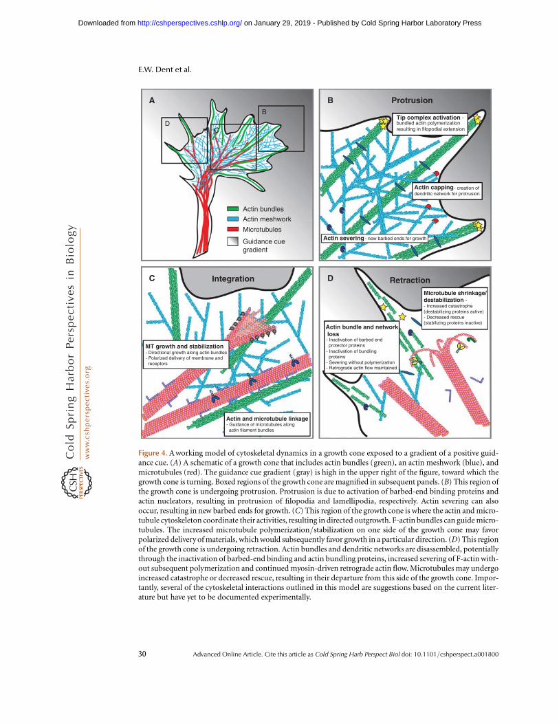
Actin bundles
ProtrusionA B
C D RetractionIntegration
Tip complex activation -bundled actin polymerizationresulting in filopodial extension
MT growth and stabilization- Directional growth along actin bundles- Polarized delivery of membrane and receptors
Actin and microtubule linkage
Actin bundle and network loss
- Guidance of microtubules along actin filament bundles
- Inactivation of barbed end protector proteins- Inactivation of bundling proteins- Severing without polymerization- Retrograde actin flow maintained
Microtubule shrinkage/destabilization -- Increased catastrophe(destabilizing proteins active)- Decreased rescue(stabilizing proteins inactive)
Actin capping
Actin severing
- creation ofdendritic network for protrusion
- new barbed ends for growth
DC
B
Actin meshwork
Microtubules
Guidance cuegradient
Figure 4. A working model of cytoskeletal dynamics in a growth cone exposed to a gradient of a positive guid-ance cue. (A) A schematic of a growth cone that includes actin bundles (green), an actin meshwork (blue), andmicrotubules (red). The guidance cue gradient (gray) is high in the upper right of the figure, toward which thegrowth cone is turning. Boxed regions of the growth cone are magnified in subsequent panels. (B) This region ofthe growth cone is undergoing protrusion. Protrusion is due to activation of barbed-end binding proteins andactin nucleators, resulting in protrusion of filopodia and lamellipodia, respectively. Actin severing can alsooccur, resulting in new barbed ends for growth. (C) This region of the growth cone is where the actin and micro-tubule cytoskeleton coordinate their activities, resulting in directed outgrowth. F-actin bundles can guide micro-tubules. The increased microtubule polymerization/stabilization on one side of the growth cone may favorpolarized delivery of materials, which would subsequently favor growth in a particular direction. (D) This regionof the growth cone is undergoing retraction. Actin bundles and dendritic networks are disassembled, potentiallythrough the inactivation of barbed-end binding and actin bundling proteins, increased severing of F-actin with-out subsequent polymerization and continued myosin-driven retrograde actin flow. Microtubules may undergoincreased catastrophe or decreased rescue, resulting in their departure from this side of the growth cone. Impor-tantly, several of the cytoskeletal interactions outlined in this model are suggestions based on the current liter-ature but have yet to be documented experimentally.
E.W. Dent et al.
30 Advanced Online Article. Cite this article as Cold Spring Harb Perspect Biol doi: 10.1101/cshperspect.a001800
on January 29, 2019 - Published by Cold Spring Harbor Laboratory Press http://cshperspectives.cshlp.org/Downloaded from

directed outgrowth because several studiesshow that disrupting either MT stabilizationor interactions between MTs and F-actin canbe disruptive to growth cone turning, but notnecessarily to axon outgrowth. Disassembly ofthe actin and MT cytoskeleton is also conti-nuously taking place in an actively transloca-ting growth cone and may be favored on theside of the growth cone opposite a chemoat-tractive gradient (Fig. 4D), or on the side of agrowth cone facing a chemorepulsive gradient.Such retraction is likely to involve the in-activation of actin barbed-end protector andactin bundling proteins, accompanied by actinsevering without subsequent polymerization.Furthermore, MT destabilization and cata-strophe (and/or decreased rescue) are likely tobe favored in retracting regions of the growthcone.
The coordination of all of these events isrequired to properly guide neurons to their cor-rect targets during development. However,because there have been relatively few high res-olution live-cell imaging studies of the cytoske-leton during growth cone turning, it is stillunclear if these postulated events are necessaryor sufficient to induce turning. Furthermore,we must remember that although direct manip-ulation of the cytoskeleton shows us how thegrowth cone “can” respond, it does not necessa-rily show how the growth cone “does” respondto guidance cues under natural conditions.Therefore, it will be critical to determine howgrowth cone turning occurs in response toguidance cues known to be responsible for spe-cific axon guidance decisions in the organism,and ultimately to observe these events in vivoas they unfold during neural development.
ACKNOWLEDGMENTS
The authors would like to apologize for anyomissions or misinterpretations that mayhave occurred in the article. The authors weresupported by the following sources: NIHNS064014 (EWD), Dana Foundation (EWD),Whitehall Foundation (EWD), NIHGM068678 (FBG).
REFERENCES
Ahuja R, Pinyol R, Reichenbach N, Custer L, Klingensmith J,Kessels MM, Qualmann B. 2007. Cordon-bleu is an actinnucleation factor and controls neuronal morphology.Cell 131: 337–350.
Aigner L, Arber S, Kapfhammer JP, Laux T, Schneider C,Botteri F, Brenner HR, Caroni P. 1995. Overexpressionof the neural growth-associated protein GAP-43 inducesnerve sprouting in the adult nervous system of transgenicmice. Cell 83: 269–278.
Aizawa H, Wakatsuki S, Ishii A, Moriyama K, Sasaki Y,Ohashi K, Sekine-Aizawa Y, Sehara-Fujisawa A, MizunoK, Goshima Y, et al. 2001. Phosphorylation of cofilin byLIM-kinase is necessary for semaphorin 3A-inducedgrowth cone collapse. Nat Neurosci 4: 367–373.
Akhmanova A, Steinmetz MO. 2008. Tracking the ends: adynamic protein network controls the fate of microtu-bule tips. Nat Rev Mol Cell Biol 9: 309–322.
Andrews GL, Tanglao S, Farmer WT, Morin S, Brotman S,Berberoglu MA, Price H, Fernandez GC, Mastick GS,Charron F, et al. 2008. Dscam guides embryonic axonsby Netrin-dependent and -independent functions.Development 135: 3839–3848.
Arai A, Spencer JA, Olson EN. 2002. STARS, a striated mus-cle activator of Rho signaling and serum response factor-dependent transcription. J Biol Chem 277: 24453–24459.
Arakawa Y, Bito H, Furuyashiki T, Tsuji T, Takemoto-KimuraS, Kimura K, Nozaki K, Hashimoto N, Narumiya S. 2003.Control of axon elongation via an SDF-1alpha/Rho/mDia pathway in cultured cerebellar granule neurons.J Cell Biol 161: 381–391.
Aratyn YS, Schaus TE, Taylor EW, Borisy GG. 2007. Intrinsicdynamic behavior of fascin in filopodia. Mol Biol Cell 18:3928–3940.
Argaw A, Duff G, Boire D, Ptito M, Bouchard JF. 2008.Protein kinase A modulates retinal ganglion cell growthduring development. Exp Neurol 211: 494–502.
Ball LJ, Jarchau T, Oschkinat H, Walter U. 2002. EVH1domains: structure, function and interactions. FEBSLett 513: 45–52.
Barrientos T, Frank D, Kuwahara K, Bezprozvannaya S,Pipes GC, Bassel-Duby R, Richardson JA, Katus HA,Olson EN, Frey N. 2007. Two novel members of theABLIM protein family, ABLIM-2 and -3, associate withSTARS and directly bind F-actin. J Biol Chem 282:8393–8403.
Barzik M, Kotova TI, Higgs HN, Hazelwood L, Hanein D,Gertler FB, Schafer DA. 2005. Ena/VASP proteinsenhance actin polymerization in the presence of barbedend capping proteins. J Biol Chem 280: 28653–28662.
Bashaw GJ, Kidd T, Murray D, Pawson T, Goodman CS.2000. Repulsive axon guidance: Abelson and Enabledplay opposing roles downstream of the roundaboutreceptor. Cell 101: 703–715.
Bashaw GJ, Klein R. 2010. Signaling from axon guidancereceptors. Cold Spring Harb Perspect Biol 2: a001941.
Bear JE, Gertler FB. 2009. Ena/VASP: towards resolving apointed controversy at the barbed end. J Cell Sci 122:1947–1953.
The Growth Cone Cytoskeleton in Axon Outgrowth and Guidance
Advanced Online Article. Cite this article as Cold Spring Harb Perspect Biol doi: 10.1101/cshperspect.a001800 31
on January 29, 2019 - Published by Cold Spring Harbor Laboratory Press http://cshperspectives.cshlp.org/Downloaded from

Bear JE, Loureiro JJ, Libova I, Fassler R, Wehland J, GertlerFB. 2000. Negative regulation of fibroblast motility byEna/VASP proteins. Cell 101: 717–728.
Bear JE, Svitkina TM, Krause M, Schafer DA, Loureiro JJ,Strasser GA, Maly IV, Chaga OY, Cooper JA, Borisy GG,et al. 2002. Antagonism between Ena/VASP proteinsand actin filament capping regulates fibroblast motility.Cell 109: 509–521.
Bentley D, Toroian-Raymond A. 1986. Disoriented path-finding by pioneer neurone growth cones deprived offilopodia by cytochalasin treatment. Nature 323: 712–715.
Bernier G, Pool M, Kilcup M, Alfoldi J, De Repentigny Y,Kothary R. 2000. Acf7 (MACF) is an actin and microtu-bule linker protein whose expression predominates inneural, muscle, and lung development. Dev Dyn 219:216–225.
Bialkowska K, Saido TC, Fox JE. 2005. SH3 domain of spec-trin participates in the activation of Rac in specializedcalpain-induced integrin signaling complexes. J Cell Sci118: 381–395.
Bielas SL, Serneo FF, Chechlacz M, Deerinck TJ, Perkins GA,Allen PB, Ellisman MH, Gleeson JG. 2007. Spinophilinfacilitates dephosphorylation of doublecortin by PP1 tomediate microtubule bundling at the axonal wrist. Cell129: 579–591.
Bouquet C, Soares S, von Boxberg Y, Ravaille-VeronM, Propst F, Nothias F. 2004. Microtubule-associatedprotein 1B controls directionality of growth cone migra-tion and axonal branching in regeneration of adult dorsalroot ganglia neurons. J Neurosci 24: 7204–7213.
Bouchard JF, Horn KE, Stroh T, Kennedy TE. 2008. Depola-rization recruits DCC to the plasma membrane of embry-onic cortical neurons and enhances axon extension inresponse to netrin-1. J Neurochem 107: 398–417.
Bouchard JF, Moore SW, Tritsch NX, Roux PP, Shekarabi M,Barker PA, Kennedy TE. 2004. Protein kinase A activationpromotes plasma membrane insertion of DCC from anintracellular pool: A novel mechanism regulating com-missural axon extension. J Neurosci 24: 3040–3050.
Bradley WD, Koleske AJ. 2009. Regulation of cell migrationand morphogenesis by Abl-family kinases: emergingmechanisms and physiological contexts. J Cell Sci 122:3441–3454.
Breitsprecher D, Kiesewetter AK, Linkner J, Urbanke C,Resch GP, Small JV, Faix J. 2008. Clustering of VASPactively drives processive, WH2 domain-mediated actinfilament elongation. EMBO J 27: 2943–2954.
Brown JA, Bridgman PC. 2009. Disruption of the cytoskele-ton during Semaphorin 3A induced growth cone collapsecorrelates with differences in actin organization and asso-ciated binding proteins. Devel Neurobio 69: 633–646.
Brown JA, Wysolmerski RB, Bridgman PC. 2009. Dorsalroot ganglion neurons react to semaphorin 3A applica-tion through a biphasic response that requires multiplemyosin II isoforms. Mol Biol Cell 20: 1167–1179.
Buck KB, Zheng JQ. 2002. Growth cone turning induced bydirect local modification of microtubule dynamics.J Neurosci 22: 9358–9367.
Burnette DT, Ji L, Schaefer AW, Medeiros NA, Danuser G,Forscher P. 2008. Myosin II activity facilitates
microtubule bundling in the neuronal growth coneneck. Dev Cell 15: 163–169.
Carlier MF, Laurent V, Santolini J, Melki R, Didry D, Xia GX,Hong Y, Chua NH, Pantaloni D. 1997. Actin depolyme-rizing factor (ADF/cofilin) enhances the rate of filamentturnover: implication in actin-based motility. J Cell Biol136: 1307–1322.
Challacombe JF, Snow DM, Letourneau PC. 1997. Dyna-mic microtubule ends are required for growth cone turn-ing to avoid an inhibitory guidance cue. J Neurosci 17:3085–3095.
Chan AY, Bailly M, Zebda N, Segall JE, Condeelis JS. 2000.Role of cofilin in epidermal growth factor-stimulatedactin polymerization and lamellipod protrusion. J CellBiol 148: 531–542.
Chang C, Adler CE, Krause M, Clark SG, Gertler FB, Tessier-Lavigne M, Bargmann CI. 2006. MIG-10/Lamellipodinand AGE-1/PI3K Promote Axon Guidance and Out-growth in Response to Slit and Netrin. Curr Biol 16:854–862.
Chen N, Furuya S, Shinoda Y, Yumoto M, Ohtake A, Sato K,Doi H, Hashimoto Y, Kudo Y, Higashi H. 2003. Extra-cellular carbohydrate-signal triggering cAMP-dependentprotein kinase-dependent neuronal actin-reorganiza-tion. Neuroscience 122: 985–995.
Chen TJ, Gehler S, Shaw AE, Bamburg JR, Letourneau PC.2006. Cdc42 participates in the regulation of ADF/cofilinand retinal growth cone filopodia by brain derived neuro-trophic factor. J Neurobiol 66: 103–114.
Chereau D, Dominguez R. 2006. Understanding the roleof the G-actin-binding domain of Ena/VASP in actinassembly. J Struct Biol 155: 195–201.
Chien CB, Rosenthal DE, Harris WA, Holt CE. 1993. Naviga-tional errors made by growth cones without filopodia inthe embryonic Xenopus brain. Neuron 11: 237–251.
Ciani L, Krylova O, Smalley MJ, Dale TC, Salinas PC. 2004.A divergent canonical WNT-signaling pathway regulatesmicrotubule dynamics: dishevelled signals locally to sta-bilize microtubules. J Cell Biol 164: 243–253.
Cohan CS, Welnhofer EA, Zhao L, Matsumura F, YamashiroS. 2001. Role of the actin bundling protein fascin ingrowth cone morphogenesis: localization in filopodiaand lamellipodia. Cell Motil Cytoskeleton 48: 109–120.
Colavita A, Culotti JG. 1998. Suppressors of ectopic UNC-5growth cone steering identify eight genes involved inaxon guidance in Caenorhabditis elegans. Dev Biol 194:72–85.
Comer AR, Ahern-Djamali SM, Juang JL, Jackson PD, Hoff-mann FM. 1998. Phosphorylation of Enabled by the Dro-sophila Abelson tyrosine kinase regulates the in vivofunction and protein-protein interactions of Enabled.Mol Cell Biol 18: 152–160.
Davis DA, Wilson MH, Giraud J, Xie Z, Tseng HC, EnglandC, Herscovitz H, Tsai LH, Delalle I. 2009. Capzb2 inter-acts with beta-tubulin to regulate growth cone morphol-ogy and neurite outgrowth. PLoS Biol 7: e1000208.
Dawson HN, Ferreira A, Eyster MV, Ghoshal N, Binder LI,Vitek MP. 2001. Inhibition of neuronal maturation inprimary hippocampal neurons from tau deficient mice.J Cell Sci 114: 1179–1187.
E.W. Dent et al.
32 Advanced Online Article. Cite this article as Cold Spring Harb Perspect Biol doi: 10.1101/cshperspect.a001800
on January 29, 2019 - Published by Cold Spring Harbor Laboratory Press http://cshperspectives.cshlp.org/Downloaded from

De Arcangelis A, Georges-Labouesse E, Adams JC. 2004.Expression of fascin-1, the gene encoding the actin-bundling protein fascin-1, during mouse embryogenesis.Gene Expr Patterns 4: 637–643.
Dehmelt L, Smart FM, Ozer RS, Halpain S. 2003. The role ofmicrotubule-associated protein 2c in the reorganizationof microtubules and lamellipodia during neurite initia-tion. J Neurosci 23: 9479–9490.
Del Rio JA, Gonzalez-Billault C, Urena JM, Jimenez EM,Barallobre MJ, Pascual M, Pujadas L, Simo S, La Torre A,Wandosell F, et al. 2004. MAP1B is required for Netrin 1signaling in neuronal migration and axonal guidance.Curr Biol 14: 840–850.
Dent EW, Barnes AM, Tang F, Kalil K. 2004. Netrin-1 andsemaphorin 3A promote or inhibit cortical axon branch-ing, respectively, by reorganization of the cytoskeleton.J Neurosci 24: 3002–3012.
Dent EW, Gertler FB. 2003. Cytoskeletal dynamics andtransport in growth cone motility and axon guidance.Neuron 40: 209–227.
Dent EW, Kalil K. 2001. Axon branching requires interac-tions between dynamic microtubules and actin filaments.J Neurosci 21: 9757–9769.
Deuel TA, Liu JS, Corbo JC, Yoo SY, Rorke-Adams LB, WalshCA. 2006. Genetic interactions between doublecortinand doublecortin-like kinase in neuronal migration andaxon outgrowth. Neuron 49: 41–53.
Dickson TC, Mintz CD, Benson DL, Salton SR. 2002. Func-tional binding interaction identified between the axonalCAM L1 and members of the ERM family. J Cell Biol157: 1105–1112.
Dickson BJ, Gilestro GF. 2006. Regulation of commissuralaxon pathfinding by slit and its Robo receptors. AnnuRev Cell Dev Biol 22: 651–675.
Dominguez R. 2009. Actin filament nucleation and elonga-tion factors–structure-function relationships. Crit RevBiochem Mol Biol 44: 351–366.
Drees F, Gertler F. 2008. Ena/VASP: proteins at the tip of thenervous system. Curr Opin Neurobiol 18: 53–59.
Dwivedy A, Gertler FB, Miller J, Holt CE, Lebrand C. 2007.Ena/VASP function in retinal axons is required forterminal arborization but not pathway navigation.Development 134: 2137–2146.
Elkins T, Zinn K, McAllister L, Hoffmann FM, GoodmanCS. 1990. Genetic analysis of a Drosophila neural celladhesion molecule: interaction of fasciclin I and Abelsontyrosine kinase mutations. Cell 60: 565–575.
Endo M, Ohashi K, Sasaki Y, Goshima Y, Niwa R, UemuraT, Mizuno K. 2003. Control of growth cone motilityand morphology by LIM kinase and Slingshot via phos-phorylation and dephosphorylation of cofilin. J Neurosci23: 2527–2537.
Erkman L, Yates PA, McLaughlin T, McEvilly RJ,Whisenhunt T, O’Connell SM, Krones AI, Kirby MA,Rapaport DH, Bermingham JR, et al. 2000. A POUdomain transcription factor-dependent program regu-lates axon pathfinding in the vertebrate visual system.Neuron 28: 779–792.
Ferron F, Rebowski G, Lee SH, Dominguez R. 2007. Struc-tural basis for the recruitment of profilin-actin complexes
during filament elongation by Ena/VASP. EMBO J 26:4597–4606.
Forsthoefel DJ, Liebl EC, Kolodziej PA, Seeger MA. 2005.The Abelson tyrosine kinase, the Trio GEF and Enabledinteract with the Netrin receptor Frazzled in Drosophila.Development 132: 1983–1994.
Fritz JL, VanBerkum MFA. 2002. Regulation of rho familyGTPases is required to prevent axons from crossing themidline. Dev Biol 252: 46–58.
Galjart N. 2005. CLIPs and CLASPs and cellular dynamics.Nat Rev Mol Cell Biol 6: 487–498.
Gallo G. 2006. RhoA-kinase coordinates F-actin organiza-tion and myosin II activity during semaphorin-3A-induced axon retraction. J Cell Sci 119: 3413–3423.
Garbe DS, Das A, Dubreuil RR, Bashaw GJ. 2007a. beta-Spectrin functions independently of Ankyrin to regulatethe establishment and maintenance of axon connectionsin the Drosophila embryonic CNS. Development 134:273–284.
Garbe DS, O’Donnell M, Bashaw GJ. 2007b. Cytoplasmicdomain requirements for Frazzled-mediated attractiveaxon turning at the Drosophila midline. Development134: 4325–4334.
Garcia MC, Abbasi M, Singh S, He Q. 2007. Role of Droso-phila gene dunc-115 in nervous system. Invert Neurosci7: 119–128.
Garvalov BK, Flynn KC, Neukirchen D, Meyn L, Teusch N,Wu X, Brakebusch C, Bamburg JR, Bradke F. 2007.Cdc42 regulates cofilin during the establishment of neu-ronal polarity. J Neurosci 27: 13117–13129.
Geraldo S, Khanzada UK, Parsons M, Chilton JK, Gordon-Weeks PR. 2008. Targeting of the F-actin-binding proteindrebrin by the microtubule plus-tip protein EB3 isrequired for neuritogenesis. Nat Cell Biol 10: 1181–1189.
Gertler FB, Comer AR, Juang JL, Ahern SM, Clark MJ, LieblEC, Hoffmann FM. 1995. enabled, a dosage-sensitivesuppressor of mutations in the Drosophila Abl tyrosinekinase, encodes an Abl substrate with SH3 domain-binding properties. Genes Dev 9: 521–533.
Gertler FB, Bennett RL, Clark MJ, Hoffmann FM. 1989.Drosophila abl tyrosine kinase in embryonic CNS axons:a role in axonogenesis is revealed through dosage-sensitive interactions with disabled. Cell 58: 103–113.
Gertler FB, Doctor JS, Hoffmann FM. 1990. Genetic sup-pression of mutations in the Drosophila abl proto-oncogene homolog. Science 248: 857–860.
Gertler FB, Niebuhr K, Reinhard M, Wehland J, Soriano P.1996. Mena, a relative of VASP and Drosophila Enabled,is implicated in the control of microfilament dynamics.Cell 87: 227–239.
Ghosh M, Song X, Mouneimne G, Sidani M, Lawrence DS,Condeelis JS. 2004. Cofilin promotes actin polymeriza-tion and defines the direction of cell motility. Science304: 743–746.
Gitai Z, Yu TW, Lundquist EA, Tessier-Lavigne M,Bargmann CI. 2003. The netrin receptor UNC-40/DCCstimulates axon attraction and outgrowth throughenabled and, in parallel, Rac and UNC-115/AbLIM.Neuron 37: 53–65.
Goldberg DJ, Burmeister DW. 1986. Stages in axon forma-tion: observations of growth of Aplysia axons in culture
The Growth Cone Cytoskeleton in Axon Outgrowth and Guidance
Advanced Online Article. Cite this article as Cold Spring Harb Perspect Biol doi: 10.1101/cshperspect.a001800 33
on January 29, 2019 - Published by Cold Spring Harbor Laboratory Press http://cshperspectives.cshlp.org/Downloaded from
using video-enhanced contrast-differential interferencecontrast microscopy. J Cell Biol 103: 1921–1931.
Gonzalez-Agosti C, Solomon F. 1996. Response of radixin toperturbations of growth cone morphology and motilityin chick sympathetic neurons in vitro. Cell Motil Cyto-skeleton 34: 122–136.
Gonzalez-Billault C, Avila J, Caceres A. 2001. Evidence forthe role of MAP1B in axon formation. Mol Biol Cell 12:2087–2098.
Goode BL, Eck MJ. 2007. Mechanism and function of for-mins in the control of actin assembly. Annu Rev Biochem76: 593–627.
Goslin K, Birgbauer E, Banker G, Solomon F. 1989. Therole of cytoskeleton in organizing growth cones: a micro-filament-associated growth cone component dependsupon microtubules for its localization. J Cell Biol 109:1621–1631.
Grabham PW, Seale GE, Bennecib M, Goldberg DJ, ValleeRB. 2007. Cytoplasmic dynein and LIS1 are requiredfor microtubule advance during growth cone remodelingand fast axonal outgrowth. J Neurosci 27: 5823–5834.
Grevengoed EE, Loureiro JJ, Jesse TL, Peifer M. 2001. Abel-son kinase regulates epithelial morphogenesis in Droso-phila. J Cell Biol 155: 1185–1198.
Grevengoed EE, Fox DT, Gates J, Peifer M. 2003. Balancingdifferent types of actin polymerization at distinct sites:roles for Abelson kinase and Enabled. J Cell Biol 163:1267–1279.
Grosse R, Copeland JW, Newsome TP, Way M, Treisman R.2003. A role for VASP in RhoA-Diaphanous signalling toactin dynamics and SRF activity. Embo J 22: 3050–3061.
Halloran MC, Kalil K. 1994. Dynamic behaviors of growthcones extending in the corpus callosum of living corticalbrain slices observed with video microscopy. J Neurosci14: 2161–2177.
Harbott LK, Nobes CD. 2005. A key role for Abl family kin-ases in EphA receptor-mediated growth cone collapse.Mol Cell Neurosci 30: 1–11.
He Q, Dent EW, Meiri KF. 1997. Modulation of actin fila-ment behavior by GAP-43 (neuromodulin) is dependenton the phosphorylation status of serine 41, the proteinkinase C site. J Neurosci 17: 3515–3524.
Hedgecock EM, Culotti JG, Thomson JN, Perkins LA. 1985.Axonal guidance mutants of Caenorhabditis elegansidentified by filling sensory neurons with fluoresceindyes. Dev Biol 111: 158–170.
Henkemeyer M, West S, Gertler F. 1990. A novel tyrosinekinase-independent function of Drosophila abl corre-lates with proper subcellular localization. Cell 63: 949–960.
Higgs HN. 2005. Formin proteins: a domain-based ap-proach. Trends Biochem Sci 30: 342–353.
Higgs HN, Peterson KJ. 2005. Phylogenetic analysis of theformin homology 2 domain. Mol Biol Cell 16: 1–13.
Hines JH, Abu-Rub M, Henley JR. 2010. Asymmetric endo-cytosis and remodeling of beta(1)-integrin adhesionsduring growth cone chemorepulsion by MAG. Nat Neu-rosci 13: 829–837.
Homem CC, Peifer M. 2009. Exploring the roles of diapha-nous and enabled activity in shaping the balance betweenfilopodia and lamellipodia. Mol Biol Cell 20: 5138–5155.
Homma N, Takei Y, Tanaka Y, Nakata T, Terada S, KikkawaM, Noda Y, Hirokawa N. 2003. Kinesin superfamily pro-tein 2A (KIF2A) functions in suppression of collateralbranch extension. Cell 114: 229–239.
Hotulainen P, Llano O, Smirnov S, Tanhuanpaa K, Faix J,Rivera C, Lappalainen P. 2009. Defining mechanisms ofactin polymerization and depolymerization during den-dritic spine morphogenesis. J Cell Biol 185: 323–339.
Hsouna A, Kim YS, VanBerkum MF. 2003. Abelson tyrosinekinase is required to transduce midline repulsive cues.J Neurobiol 57: 15–30.
Hummel T, Schimmelpfeng K, Klambt C. 1999. Commis-sure formation in the embryonic CNS of Drosophila.Development 126: 771–779.
Hubberstey AV, Mottillo EP. 2002. Cyclase-associated pro-teins: CAPacity for linking signal transduction and actinpolymerization. FASEB J 16: 487–499.
Hulsmeier J, Pielage J, Rickert C, Technau GM, Klambt C,Stork T. 2007. Distinct functions of alpha-Spectrin andbeta-Spectrin during axonal pathfinding. Development134: 713–722.
Hung RJ, Yazdani U, Yoon J, Wu H, Yang T, Gupta N, HuangZ, van Berkel WJ, Terman JR. 2010. Mical links sema-phorins to F-actin disassembly. Nature 463: 823–827.
Ichetovkin I, Grant W, Condeelis J. 2002. Cofilin producesnewly polymerized actin filaments that are preferred fordendritic nucleation by the Arp2/3 complex. Curr Biol12: 79–84.
Jimenez-Mateos EM, Paglini G, Gonzalez-Billault C,Caceres A, Avila J. 2005. End binding protein-1 (EB1)complements microtubule-associated protein-1B duringaxonogenesis. J Neurosci Res 80: 350–359.
Jurney WM, Gallo G, Letourneau PC, McLoon SC. 2002.Rac1-mediated endocytosis during ephrin-A2- and sem-aphorin 3A-induced growth cone collapse. J Neurosci 22:6019–6028.
Kaiser DA, Vinson VK, Murphy DB, Pollard TD. 1999. Pro-filin is predominantly associated with monomeric actinin Acanthamoeba. J Cell Sci 112: 3779–3790.
Karabay A, Yu W, Solowska JM, Baird DH, Baas PW. 2004.Axonal growth is sensitive to the levels of katanin, a pro-tein that severs microtubules. J Neurosci 24: 5778–5788.
Kaufmann N, Wills ZP, Van Vactor D. 1998. DrosophilaRac1 controls motor axon guidance. Development 125:453–461.
Kim YS, Fritz JL, Seneviratne AK, VanBerkum MF. 2002.Constitutively active myosin light chain kinase altersaxon guidance decisions in Drosophila embryos. DevBiol 249: 367–381.
Knoll B, Kretz O, Fiedler C, Alberti S, Schutz G, Frotscher M,Nordheim A. 2006. Serum response factor controls neu-ronal circuit assembly in the hippocampus. Nat Neurosci9: 195–204.
Knoll B, Nordheim A. 2009. Functional versatility of tran-scription factors in the nervous system: the SRF para-digm. Trends Neurosci 32: 432–442.
Koester MP, Muller O, Pollerberg GE. 2007. Adenomatouspolyposis coli is differentially distributed in growth conesand modulates their steering. J Neurosci 27: 12590–12600.
E.W. Dent et al.
34 Advanced Online Article. Cite this article as Cold Spring Harb Perspect Biol doi: 10.1101/cshperspect.a001800
on January 29, 2019 - Published by Cold Spring Harbor Laboratory Press http://cshperspectives.cshlp.org/Downloaded from

Kolpak AL, Jiang J, Guo D, Standley C, Bellve K, Fogarty K,Bao Z-Z. 2009. Negative Guidance Factor-Induced Mac-ropinocytosis in the Growth Cone Plays a Critical Role inRepulsive Axon Turning. J Neurosci 29: 10488–10498.
Koizumi H, Tanaka T, Gleeson JG. 2006. Doublecortin-likekinase functions with doublecortin to mediate fiber tractdecussation and neuronal migration. Neuron 49: 55–66.
Korobova F, Svitkina T. 2008. Arp2/3 complex is importantfor filopodia formation, growth cone motility, and neuri-togenesis in neuronal cells. Mol Biol Cell 19: 1561–1574.
Krause M, Leslie JD, Stewart M, Lafuente EM, Valderrama F,Jagannathan R, Strasser GA, Rubinson DA, Liu H, WayM, et al. 2004. Lamellipodin, an Ena/VASP ligand, isimplicated in the regulation of lamellipodial dynamics.Dev Cell 7: 571–583.
Kruger K, Tam AS, Lu C, Sretavan DW. 1998. Retinal gan-glion cell axon progression from the optic chiasm to ini-tiate optic tract development requires cell autonomousfunction of GAP-43. J Neurosci 18: 5692–5705.
Kubo T, Endo M, Hata K, Taniguchi J, Kitajo K, Tomura S,Yamaguchi A, Mueller BK, Yamashita T. 2008. MyosinIIA is required for neurite outgrowth inhibition pro-duced by repulsive guidance molecule. J Neurochem105: 113–126.
Lafont F, Rouget M, Rousselet A, Valenza C, Prochiantz A.1993. Specific responses of axons and dendrites to cytos-keleton perturbations: an in vitro study. J Cell Sci 104:433–443.
Lanier LM, Gates MA, Witke W, Menzies AS, Wehman AM,Macklis JD, Kwiatkowski D, Soriano P, Gertler FB. 1999.Mena is required for neurulation and commissure forma-tion. Neuron 22: 313–325.
Lebrand C, Dent EW, Strasser GA, Lanier LM, Krause M,Svitkina TM, Borisy GG, Gertler FB. 2004. Critical roleof Ena/VASP proteins for filopodia formation in neuronsand in function downstream of netrin-1. Neuron 42:37–49.
Lee AC, Suter DM. 2008. Quantitative analysis of microtu-bule dynamics during adhesion-mediated growth coneguidance. Dev Neurobiol 68: 1363–1377.
Lee H, Engel U, Rusch J, Scherrer S, Sheard K, Van Vactor D.2004. The microtubule plus end tracking protein Orbit/MAST/CLASP acts downstream of the tyrosine kinaseAbl in mediating axon guidance. Neuron 42: 913–926.
Lee S, Nahm M, Lee M, Kwon M, Kim E, Zadeh AD, Cao H,Kim HJ, Lee ZH, Oh SB, et al. 2007. The F-actin-microtubule crosslinker Shot is a platform for Krasavietz-mediated translational regulation of midline axon repul-sion. Development 134: 1767–1777.
Li W, Herman RK, Shaw JE. 1992. Analysis of the Caeno-rhabditis elegans axonal guidance and outgrowth geneunc-33. Genetics 132: 675–689.
Li YH, Ghavampur S, Bondallaz P, Will L, Grenningloh G,Puschel AW. 2009. Rnd1 regulates axon extension byenhancing the microtubule destabilizing activity ofSCG10. J Biol Chem 284: 363–371.
Liebl EC, Forsthoefel DJ, Franco LS, Sample SH, Hess JE,Cowger JA, Chandler MP, Shupert AM, Seeger MA.2000. Dosage-sensitive, reciprocal genetic interactionsbetween the Abl tyrosine kinase and the putative GEFtrio reveal trio’s role in axon pathfinding. Neuron 26:107–118.
Liebl EC, Rowe RG, Forsthoefel DJ, Stammler AL, SchmidtER, Turski M, Seeger MA. 2003. Interactions between thesecreted protein Amalgam, its transmembrane receptorNeurotactin and the Abelson tyrosine kinase affectaxon pathfinding. Development 130: 3217–3226.
Lin CH, Espreafico EM, Mooseker MS, Forscher P. 1996.Myosin drives retrograde F-actin flow in neuronal growthcones. Neuron 16: 769–782.
Lin CH, Forscher P. 1993. Cytoskeletal remodeling duringgrowth cone-target interactions. J Cell Biol 121: 1369–1383.
Lin CH, Forscher P. 1995. Growth cone advance is inverselyproportional to retrograde F-actin flow. Neuron 14:763–771.
Lin TY, Huang CH, Kao HH, Liou GG, Yeh SR, Cheng CM,Chen MH, Pan RL, Juang JL. 2009. Abi plays an opposingrole to Abl in Drosophila axonogenesis and synaptogen-esis. Development 136: 3099–3107.
Liu W, Sato A, Khadka D, Bharti R, Diaz H, Runnels LW,Habas R. 2008. Mechanism of activation of the Forminprotein Daam1. Proc Natl Acad Sci U S A 105: 210–215.
Loudon RP, Silver LD, Yee HF, Gallo G. 2006. RhoA-kinaseand myosin II are required for the maintenance of growthcone polarity and guidance by nerve growth factor.J Neurobiol 66: 847–867.
Loureiro J, Peifer M. 1998. Roles of Armadillo, a Drosophilacatenin, during central nervous system development.Curr Biol 8: 622–632.
Lowery LA, Lee H, Lu C, Murphy R, Obar RA, Zhai B,Schedl M, Van Vactor D, Zhan Y. 2010. Parallel Geneticand Proteomic Screens Identify Msps as a CLASP-AblPathway Interactor in Drosophila. Genetics: 101534/genetics1110115626.
Lowery LA, Van Vactor D. 2009. The trip of the tip: under-standing the growth cone machinery. Nat Rev Mol CellBiol 10: 332–343.
Lu C, Huang X, Ma HF, Gooley JJ, Aparacio J, Roof DJ, ChenC, Chen DF, Li T. 2003. Normal retinal development andretinofugal projections in mice lacking the retina-specificvariant of actin-binding LIM domain protein.Neuroscience 120: 121–131.
Lu M, Witke W, Kwiatkowski DJ, Kosik KS. 1997. Delayedretraction of filopodia in gelsolin null mice. J Cell Biol138: 1279–1287.
Lundquist EA, Herman RK, Shaw JE, Bargmann CI. 1998.UNC-115, a conserved protein with predicted LIM andactin-binding domains, mediates axon guidance inC. elegans. Neuron 21: 385–392.
Maciver SK, Pope BJ, Whytock S, Weeds AG. 1998. The effectof two actin depolymerizing factors (ADF/cofilins) onactin filament turnover: pH sensitivity of F-actin bindingby human ADF, but not of Acanthamoeba actophorin.Eur J Biochem 256: 388–397.
Mack TG, Koester MP, Pollerberg GE. 2000. The microtu-bule-associated protein MAP1B is involved in local stabi-lization of turning growth cones. Mol Cell Neurosci 15:51–65.
Maier DL, Mani S, Donovan SL, Soppet D, Tessarollo L,McCasland JS, Meiri KF. 1999. Disrupted cortical mapand absence of cortical barrels in growth-associated
The Growth Cone Cytoskeleton in Axon Outgrowth and Guidance
Advanced Online Article. Cite this article as Cold Spring Harb Perspect Biol doi: 10.1101/cshperspect.a001800 35
on January 29, 2019 - Published by Cold Spring Harbor Laboratory Press http://cshperspectives.cshlp.org/Downloaded from

protein (GAP)-43 knockout mice. Proc Natl Acad Sci U SA 96: 9397–9402.
Mallavarapu A, Mitchison T. 1999. Regulated actin cytoske-leton assembly at filopodium tips controls their extensionand retraction. J Cell Biol 146: 1097–1106.
Marsh L, Letourneau PC. 1984. Growth of neurites withoutfilopodial or lamellipodial activity in the presence ofcytochalasin B. J Cell Biol 99: 2041–2047.
Marsick BM, Flynn KC, Santiago-Medina M, Bamburg JR,Letourneau PC. 2010. Activation of ADF/cofilin medi-ates attractive growth cone turning toward nerve growthfactor and netrin-1. Dev Neurobiol 70: 565–588.
Martinez-Lopez MJ, Alcantara S, Mascaro C, Perez-BranguliF, Ruiz-Lozano P, Maes T, Soriano E, Buesa C. 2005.Mouse neuron navigator 1, a novel microtubule-associ-ated protein involved in neuronal migration. Mol CellNeurosci 28: 599–612.
Matusek T, Gombos R, Szecsenyi A, Sanchez-Soriano N,Czibula A, Pataki C, Gedai A, Prokop A, Rasko I, MihalyJ. 2008. Formin proteins of the DAAM subfamily play arole during axon growth. J Neurosci 28: 13310–13319.
McGough A, Pope B, Chiu W, Weeds A. 1997. Cofilinchanges the twist of F-actin: implications for actin fila-ment dynamics and cellular function. J Cell Biol 138:771–781.
McLaughlin T, Hindges R, O’Leary DD. 2003. Regulation ofaxial patterning of the retina and its topographic map-ping in the brain. Curr Opin Neurobiol 13: 57–69.
Medeiros NA, Burnette DT, Forscher P. 2006. Myosin IIfunctions in actin-bundle turnover in neuronal growthcones. Nat Cell Biol 8: 215–226.
Meixner A, Haverkamp S, Wassle H, Fuhrer S, ThalhammerJ, Kropf N, Bittner RE, Lassmann H, Wiche G, Propst F.2000. MAP1B is required for axon guidance and Isinvolved in the development of the central and peripheralnervous system. J Cell Biol 151: 1169–1178.
Menna E, Disanza A, Cagnoli C, Schenk U, Gelsomino G,Frittoli E, Hertzog M, Offenhauser N, Sawallisch C,Kreienkamp H-J, et al. 2009. Eps8 regulates axonal filopo-dia in hippocampal neurons in response to brain-derivedneurotrophic factor (BDNF). PLoS Biol 7: e1000138.
Menzies AS, Aszodi A, Williams SE, Pfeifer A, Wehman AM,Goh KL, Mason CA, Fassler R, Gertler FB. 2004. Menaand vasodilator-stimulated phosphoprotein are requiredfor multiple actin-dependent processes that shape thevertebrate nervous system. J Neurosci 24: 8029–8038.
Michael M, Vehlow A, Navarro C, Krause M. 2010. c-Abl,Lamellipodin, and Ena/VASP proteins cooperate in dor-sal ruffling of fibroblasts and axonal morphogenesis.Curr Biol 20: 783–791.
Mintz CD, Carcea I, McNickle DG, Dickson TC, Ge Y, SaltonSR, Benson DL. 2008. ERM proteins regulate growth coneresponses to Sema3A. J Comp Neurol 510: 351–366.
Moore SW, Tessier-Lavigne M, Kennedy TE. 2007. Netrinsand their receptors. Adv Exp Med Biol 621: 17–31.
Moresco EM, Donaldson S, Williamson A, Koleske AJ.2005. Integrin-mediated dendrite branch maintenancerequires Abelson (Abl) family kinases. J Neurosci 25:6105–6118.
Morii H, Shiraishi-Yamaguchi Y, Mori N. 2006. SCG10, amicrotubule destabilizing factor, stimulates the neurite
outgrowth by modulating microtubule dynamics in rathippocampal primary cultured neurons. J Neurobiol 66:1101–1114.
Mouneimne G, Soon L, DesMarais V, Sidani M, Song X, YipS-C, Ghosh M, Eddy R, Backer JM, Condeelis J. 2004.Phospholipase C and cofilin are required for carcinomacell directionality in response to EGF stimulation. J CellBiol 166: 697–708.
Murray A, Naeem A, Barnes SH, Drescher U, Guthrie S.2010. Slit and Netrin-1 guide cranial motor axon path-finding via Rho-kinase, myosin light chain kinase andmyosin II. Neural Dev 5: 16.
Myers KA, Tint I, Nadar CV, He Y, Black MM, Baas PW.2006. Antagonistic forces generated by cytoplasmicdynein and myosin-II during growth cone turning andaxonal retraction. Traffic 7: 1333–1351.
Nadar VC, Ketschek A, Myers KA, Gallo G, Baas PW. 2008.Kinesin-5 is essential for growth-cone turning. Curr Biol18: 1972–1977.
Nedrelow JH, Cianci CD, Morrow JS. 2003. c-Src bindsalpha II spectrin’s Src homology 3 (SH3) domainand blocks calpain susceptibility by phosphorylatingTyr1176. J Biol Chem 278: 7735–7741.
Ng J, Luo L. 2004. Rho GTPases regulate axon growththrough convergent and divergent signaling pathways.Neuron 44: 779–793.
Niebuhr K, Ebel F, Frank R, Reinhard M, Domann E, CarlUD, Walter U, Gertler FB, Wehland J, Chakraborty T.1997. A novel proline-rich motif present in ActA of Liste-ria monocytogenes and cytoskeletal proteins is the ligandfor the EVH1 domain, a protein module present in theEna/VASP family. EMBO J 16: 5433–5444.
Noiges R, Eichinger R, Kutschera W, Fischer I, Nemeth Z,Wiche G, Propst F. 2002. Microtubule-associated protein1A (MAP1A) and MAP1B: light chains determine dis-tinct functional properties. J Neurosci 22: 2106–2114.
Norris AD, Dyer JO, Lundquist EA. 2009. The Arp2/3 com-plex, UNC-115/abLIM, and UNC-34/Enabled regulateaxon guidance and growth cone filopodia formation inCaenorhabditis elegans. Neural Dev 4: 38.
Otsuka AJ, Franco R, Yang B, Shim KH, Tang LZ, Zhang YY,Boontrakulpoontawee P, Jeyaprakash A, Hedgecock E,Wheaton VI, et al. 1995. An ankyrin-related gene(unc-44) is necessary for proper axonal guidance inCaenorhabditis elegans. J Cell Biol 129: 1081–1092.
Paglini G, Kunda P, Quiroga S, Kosik K, Caceres A. 1998.Suppression of radixin and moesin alters growth conemorphology, motility, and process formation in primarycultured neurons. J Cell Biol 143: 443–455.
Pak CW, Flynn KC, Bamburg JR. 2008. Actin-binding pro-teins take the reins in growth cones. Nat Rev Neurosci 9:136–147.
Pasic L, Kotova TI, Schafer DA. 2008. Ena/VASP proteinscapture actin filament barbed ends. J Biol Chem 283:9814–9819.
Phillis R, Statton D, Caruccio P, Murphey RK. 1996. Muta-tions in the 8 kDa dynein light chain gene disrupt sensoryaxon projections in the Drosophila imaginal CNS.Development 122: 2955–2963.
Pinyol R, Haeckel A, Ritter A, Qualmann B, Kessels MM.2007. Regulation of N-WASP and the Arp2/3 complex
E.W. Dent et al.
36 Advanced Online Article. Cite this article as Cold Spring Harb Perspect Biol doi: 10.1101/cshperspect.a001800
on January 29, 2019 - Published by Cold Spring Harbor Laboratory Press http://cshperspectives.cshlp.org/Downloaded from

by Abp1 controls neuronal morphology. PLoS ONE2: e400.
Piper M, Anderson R, Dwivedy A, Weinl C, van Horck F,Leung KM, Cogill E, Holt C. 2006. Signaling mechanismsunderlying Slit2-induced collapse of Xenopus retinalgrowth cones. Neuron 49: 215–228.
Pollard TD, Borisy GG. 2003. Cellular motility driven byassembly and disassembly of actin filaments. Cell 112:453–465.
Pollard TD, Cooper JA. 2009. Actin, a central player in cellshape and movement. Science 326: 1208–1212.
Posern G, Treisman R. 2006. Actin’ together: serum responsefactor, its cofactors and the link to signal transduction.Trends Cell Biol 16: 588–596.
Poulain FE, Sobel A. 2007. The "SCG10-LIke Protein" SCLIPis a novel regulator of axonal branching in hippocampalneurons, unlike SCG10. Mol Cell Neurosci 34: 137–146.
Purro SA, Ciani L, Hoyos-Flight M, Stamatakou E, SiomouE, Salinas PC. 2008. Wnt regulates axon behaviorthrough changes in microtubule growth directionality:a new role for adenomatous polyposis coli. J Neurosci28: 8644–8654.
Qiang L, Yu W, Liu M, Solowska JM, Baas PW. 2009. Basicfibroblast growth factor elicits formation of interstitialaxonal branches via enhanced severing of microtubules.Mol Biol Cell 21: 334–344.
Quinn CC, Chen E, Kinjo TG, Kelly G, Bell AW, Elliott RC,McPherson PS, Hockfield S. 2003. TUC-4b, a novel TUCfamily variant, regulates neurite outgrowth and associ-ates with vesicles in the growth cone. J Neurosci 23:2815–2823.
Quinn CC, Pfeil DS, Chen E, Stovall EL, Harden MV, GavinMK, Forrester WC, Ryder EF, Soto MC, Wadsworth WG.2006. UNC-6/netrin and SLT-1/slit guidance cues orientaxon outgrowth mediated by MIG-10/RIAM/lamellipo-din. Curr Biol 16: 845–853.
Quinn CC, Pfeil DS, Wadsworth WG. 2008. CED-10/Rac1mediates axon guidance by regulating the asymmetricdistribution of MIG-10/lamellipodin. Curr Biol 18:808–813.
Riano E, Martignoni M, Mancuso G, Cartelli D, Crippa F,Toldo I, Siciliano G, Di Bella D, Taroni F, Bassi MT, etal. 2009. Pleiotropic effects of spastin on neurite growthdepending on expression levels. J Neurochem 108:1277–1288.
Rothenberg ME, Rogers SL, Vale RD, Jan LY, Jan YN. 2003.Drosophila pod-1 crosslinks both actin and microtubulesand controls the targeting of axons. Neuron 39: 779–791.
Sabry JH, O’Connor TP, Evans L, Toroian-Raymond A,Kirschner M, Bentley D. 1991. Microtubule behaviorduring guidance of pioneer neuron growth cones insitu. J Cell Biol 115: 381–395.
Sanchez-Soriano N, Travis M, Dajas-Bailador F, Goncalves-Pimentel C, Whitmarsh AJ, Prokop A. 2009. Mouse ACF7and drosophila short stop modulate filopodia formationand microtubule organisation during neuronal growth.J Cell Sci 122: 2534–2542.
Sarmiere PD, Bamburg JR. 2004. Regulation of the neuronalactin cytoskeleton by ADF/cofilin. J Neurobiol 58:103–117.
Sasaki Y, Hayashi K, Shirao T, Ishikawa R, Kohama K. 1996.Inhibition by drebrin of the actin-bundling activity ofbrain fascin, a protein localized in filopodia of growthcones. J Neurochem 66: 980–988.
Schaefer AW, Schoonderwoert VT, Ji L, Mederios N,Danuser G, Forscher P. 2008. Coordination of actin fila-ment and microtubule dynamics during neurite out-growth. Dev Cell 15: 146–162.
Schirenbeck A, Arasada R, Bretschneider T, Stradal TE,Schleicher M, Faix J. 2006. The bundling activity ofvasodilator-stimulated phosphoprotein is required forfilopodium formation. Proc Natl Acad Sci U S A 103:7694–7699.
Schlatter MC, Buhusi M, Wright AG, Maness PF. 2008.CHL1 promotes Sema3A-induced growth cone collapseand neurite elaboration through a motif required forrecruitment of ERM proteins to the plasma membrane.J Neurochem 104: 731–744.
Schumacher N, Borawski JM, Leberfinger CB, Gessler M,Kerkhoff E. 2004. Overlapping expression pattern of theactin organizers Spir-1 and formin-2 in the developingmouse nervous system and the adult brain. Gene ExprPatterns 4: 249–255.
Shakir MA, Jiang K, Struckhoff EC, Demarco RS, Patel FB,Soto MC, Lundquist EA. 2008. The Arp2/3 activatorsWAVE and WASP have distinct genetic interactions withRac GTPases in Caenorhabditis elegans axon guidance.Genetics 179: 1957–1971.
Shekarabi M, Kennedy TE. 2002. The netrin-1 receptorDCC promotes filopodia formation and cell spreadingby activating Cdc42 and Rac1. Mol Cell Neurosci 19:1–17.
Shekarabi M, Moore SW, Tritsch NX, Morris SJ, BouchardJF, Kennedy TE. 2005. Deleted in colorectal cancer bind-ing netrin-1 mediates cell substrate adhesion and recruitsCdc42, Rac1, Pak1, and N-WASP into an intracellular sig-naling complex that promotes growth cone expansion.J Neurosci 25: 3132–3141.
Shen Y, Mani S, Donovan SL, Schwob JE, Meiri KF. 2002.Growth-associated protein-43 is required for commissu-ral axon guidance in the developing vertebrate nervoussystem. J Neurosci 22: 239–247.
Shi SH, Cheng T, Jan LY, Jan YN. 2004. APC and GSK-3betaare involved in mPar3 targeting to the nascent axon andestablishment of neuronal polarity. Curr Biol 14:2025–2032.
Shintani T, Ihara M, Tani S, Sakuraba J, Sakuta H, NodaM. 2009. APC2 plays an essential role in axonal projec-tions through the regulation of microtubule stability.J Neurosci 29: 11628–11640.
Smith K, Humphreys D, Hume PJ, Koronakis V. 2010. Enter-opathogenic Escherichia coli recruits the cellular inositolphosphatase SHIP2 to regulate actin-pedestal formation.Cell Host Microbe 7: 13–24.
Stern S, Debre E, Stritt C, Berger J, Posern G, Knoll B. 2009.A nuclear actin function regulates neuronal motility byserum response factor-dependent gene transcription.J Neurosci 29: 4512–4518.
Strasser G, Rahim N, VanderWaal K, Gertler F, Lanier L.2004. Arp2/3 is a negative regulator of growth cone trans-location. Neuron 43: 81–94.
The Growth Cone Cytoskeleton in Axon Outgrowth and Guidance
Advanced Online Article. Cite this article as Cold Spring Harb Perspect Biol doi: 10.1101/cshperspect.a001800 37
on January 29, 2019 - Published by Cold Spring Harbor Laboratory Press http://cshperspectives.cshlp.org/Downloaded from
Strittmatter SM, Fankhauser C, Huang PL, Mashimo H,Fishman MC. 1995. Neuronal pathfinding is abnormalin mice lacking the neuronal growth cone proteinGAP-43. Cell 80: 445–452.
Struckhoff EC, Lundquist EA. 2003. The actin-binding pro-tein UNC-115 is an effector of Rac signaling during axonpathfinding in C. elegans. Development 130: 693–704.
Suh LH, Oster SF, Soehrman SS, Grenningloh G, SretavanDW. 2004. L1/Laminin modulation of growth coneresponse to EphB triggers growth pauses and regulatesthe microtubule destabilizing protein SCG10. J Neurosci24: 1976–1986.
Sun HQ, Yamamoto M, Mejillano M, Yin HL. 1999. Gelso-lin, a multifunctional actin regulatory protein. J BiolChem 274: 33179–33182.
Suter DM, Errante LD, Belotserkovsky V, Forscher P. 1998.The Ig superfamily cell adhesion molecule, apCAM,mediates growth cone steering by substrate-cytoskeletalcoupling. J Cell Biol 141: 227–240.
Suter DM, Forscher P. 2000. Substrate-cytoskeletal couplingas a mechanism for the regulation of growth cone motil-ity and guidance. J Neurobiol 44: 97–113.
Suter DM, Forscher P. 2001. Transmission of growth conetraction force through apCAM-cytoskeletal linkages isregulated by Src family tyrosine kinase activity. J CellBiol 155: 427–438.
Suter DM, Schaefer AW, Forscher P. 2004. Microtubuledynamics are necessary for SRC family kinase-dependentgrowth cone steering. Curr Biol 14: 1194–1199.
Symons MH, Mitchison TJ. 1991. Control of actin polymer-ization in live and permeabilized fibroblasts. J Cell Biol114: 503–513.
Tahirovic S, Hellal F, Neukirchen D, Hindges R, GarvalovBK, Flynn KC, Stradal TE, Chrostek-Grashoff A,Brakebusch C, Bradke F. 2010. Rac1 regulates neuronalpolarization through the WAVE complex. J Neurosci 30:6930–6943.
Takahashi M, Yamagata M, Noda M. 1999. Specific expres-sion of ezrin, a cytoskeletal-membrane linker protein,in a subset of chick retinotectal and sensory projections.Eur J Neurosci 11: 545–558.
Takei Y, Teng J, Harada A, Hirokawa N. 2000. Defects in axo-nal elongation and neuronal migration in mice with dis-rupted tau and map1b genes. J Cell Biol 150: 989–1000.
Tanaka E, Kirschner MW. 1995. The role of microtubules ingrowth cone turning at substrate boundaries. J Cell Biol128: 127–137.
Tararuk T, Ostman N, Li W, Bjorkblom B, Padzik A,Zdrojewska J, Hongisto V, Herdegen T, Konopka W,Courtney MJ, et al. 2006. JNK1 phosphorylation ofSCG10 determines microtubule dynamics and axoden-dritic length. J Cell Biol 173: 265–277.
Tepass U. 2009. FERM proteins in animal morphogenesis.Curr Opin Genet Dev 19: 357–367.
Terman JR, Mao T, Pasterkamp RJ, Yu HH, Kolodkin AL.2002. MICALs, a family of conserved flavoprotein oxi-doreductases, function in plexin-mediated axonal repul-sion. Cell 109: 887–900.
Togel M, Wiche G, Propst F. 1998. Novel features of the lightchain of microtubule-associated protein MAP1B: micro-tubule stabilization, self interaction, actin filament
binding, and regulation by the heavy chain. J Cell Biol143: 695–707.
Tojima T, Akiyama H, Itofusa R, Li Y, Katayama H, MiyawakiA, Kamiguchi H. 2007. Attractive axon guidance involvesasymmetric membrane transport and exocytosis in thegrowth cone. Nat Neurosci 10: 58–66.
Tojima T, Itofusa R, Kamiguchi H. 2010. Asymmetric cla-thrin-mediated endocytosis drives repulsive growthcone guidance. Neuron 66: 370–377.
Tsai JW, Chen Y, Kriegstein AR, Vallee RB. 2005. LIS1RNA interference blocks neural stem cell division, mor-phogenesis, and motility at multiple stages. J Cell Biol170: 935–945.
Vactor DV, Sink H, Fambrough D, Tsoo R, Goodman CS.1993. Genes that control neuromuscular specificity inDrosophila. Cell 73: 1137–1153.
Vicente-Manzanares M, Ma X, Adelstein RS, Horwitz AR.2009. Non-muscle myosin II takes centre stage in celladhesion and migration. Nat Rev Mol Cell Biol 10:778–790.
Vignjevic D, Kojima S, Aratyn Y, Danciu O, Svitkina T,Borisy GG. 2006. Role of fascin in filopodial protrusion.J Cell Biol 174: 863–875.
Wang W, Eddy R, Condeelis J. 2007. The cofilin pathway inbreast cancer invasion and metastasis. Nat Rev Cancer 7:429–440.
Wen Z, Han L, Bamburg JR, Shim S, Ming GL, Zheng JQ.2007. BMP gradients steer nerve growth cones by a bal-ancing act of LIM kinase and Slingshot phosphatase onADF/cofilin. J Cell Biol 178: 107–119.
Williams JA, MacIver B, Klipfell EA, Thomas GH. 2004. TheC-terminal domain of Drosophila (beta) heavy-spectrinexhibits autonomous membrane association and modu-lates membrane area. J Cell Sci 117: 771–782.
Williamson T, Gordon-Weeks PR, Schachner M, Taylor J.1996. Microtubule reorganization is obligatory forgrowth cone turning. Proc Natl Acad Sci U S A 93:15221–15226.
Wills Z, Bateman J, Korey CA, Comer A, Van Vactor D.1999a. The tyrosine kinase Abl and its substrate enabledcollaborate with the receptor phosphatase Dlar to controlmotor axon guidance. Neuron 22: 301–312.
Wills Z, Emerson M, Rusch J, Bikoff J, Baum B, Perrimon N,Van Vactor D. 2002. A Drosophila homolog of cyclase-associated proteins collaborates with the Abl tyrosinekinase to control midline axon pathfinding. Neuron 36:611–622.
Wills Z, Marr L, Zinn K, Goodman CS, Van Vactor D. 1999b.Profilin and the Abl tyrosine kinase are required formotor axon outgrowth in the Drosophila embryo.Neuron 22: 291–299.
Wood JD, Landers JA, Bingley M, McDermott CJ, Thomas-McArthur V, Gleadall LJ, Shaw PJ, Cunliffe VT. 2006. Themicrotubule-severing protein Spastin is essential for axonoutgrowth in the zebrafish embryo. Hum Mol Genet 15:2763–2771.
Xu J, Casella JF, Pollard TD. 1999. Effect of capping protein,CapZ, on the length of actin filaments and mechanicalproperties of actin filament networks. Cell MotilCytoskeleton 42: 73–81.
E.W. Dent et al.
38 Advanced Online Article. Cite this article as Cold Spring Harb Perspect Biol doi: 10.1101/cshperspect.a001800
on January 29, 2019 - Published by Cold Spring Harbor Laboratory Press http://cshperspectives.cshlp.org/Downloaded from

Yamakita Y, Matsumura F, Yamashiro S. 2009. Fascin1 isdispensable for mouse development but is favorable forneonatal survival. Cell Motil Cytoskeleton 66: 524–534.
Yang Y, Lundquist EA. 2005. The actin-binding proteinUNC-115/abLIM controls formation of lamellipodiaand filopodia and neuronal morphogenesis in Caeno-rhabditis elegans. Mol Cell Biol 25: 5158–5170.
Yoshimura T, Kawano Y, Arimura N, Kawabata S, Kikuchi A,Kaibuchi K. 2005. GSK-3beta regulates phosphorylationof CRMP-2 and neuronal polarity. Cell 120: 137–149.
Yu HH, Zisch AH, Dodelet VC, Pasquale EB. 2001. Multiplesignaling interactions of Abl and Arg kinases with theEphB2 receptor. Oncogene 20: 3995–4006.
Yu TW, Hao JC, Lim W, Tessier-Lavigne M, Bargmann CI.2002. Shared receptors in axon guidance: SAX-3/Robosignals via UNC-34/Enabled and a Netrin-independentUNC-40/DCC function. Nat Neurosci 5: 1147–1154.
Yu W, Qiang L, Solowska JM, Karabay A, Korulu S, Baas PW.2008. The microtubule-severing proteins spastin andkatanin participate differently in the formation of axonalbranches. Mol Biol Cell 19: 1485–1498.
Zallen JA, Cohen Y, Hudson AM, Cooley L, Wieschaus E,Schejter ED. 2002. SCAR is a primary regulator ofArp2/3-dependent morphological events in Drosophila.J Cell Biol 156: 689–701.
Zallen JA, Kirch SA, Bargmann CI. 1999. Genes required foraxon pathfinding and extension in the C. elegans nervering. Development 126: 3679–3692.
Zhang XF, Schaefer AW, Burnette DT, SchoonderwoertVT, Forscher P. 2003. Rho-dependent contractile re-sponses in the neuronal growth cone are independent ofclassical peripheral retrograde actin flow. Neuron 40:931–944.
Zhou FQ, Zhou J, Dedhar S, Wu YH, Snider WD. 2004.NGF-induced axon growth is mediated by localizedinactivation of GSK-3beta and functions of the micro-tubule plus end binding protein APC. Neuron 42:897–912.
Zhu Q, Julien JP. 1999. A key role for GAP-43 in the retino-tectal topographic organization. Exp Neurol 155:228–242.
The Growth Cone Cytoskeleton in Axon Outgrowth and Guidance
Advanced Online Article. Cite this article as Cold Spring Harb Perspect Biol doi: 10.1101/cshperspect.a001800 39
on January 29, 2019 - Published by Cold Spring Harbor Laboratory Press http://cshperspectives.cshlp.org/Downloaded from

published online November 24, 2010Cold Spring Harb Perspect Biol Erik W. Dent, Stephanie L. Gupton and Frank B. Gertler The Growth Cone Cytoskeleton in Axon Outgrowth and Guidance
Subject Collection Neuronal Guidance
PrimerMechanisms and Molecules of Neuronal Wiring: A
Alex L. Kolodkin and Marc Tessier-LavigneGuidanceWiring the Brain: The Biology of Neuronal
Alain Chédotal and Linda J. Richards
DeathGuidance Molecules in Axon Pruning and Cell
Pierre Vanderhaeghen and Hwai-Jong ChengPlasticityGuidance Molecules in Synapse Formation and
Kang Shen and Christopher W. CowanInitiating and Growing an Axon
F. Polleux and William Snider Outgrowth and GuidanceThe Growth Cone Cytoskeleton in Axon
GertlerErik W. Dent, Stephanie L. Gupton and Frank B.
System MidlineNavigating Intermediate Targets: The Nervous
Barry J. Dickson and Yimin Zou
The Olfactory System−−Topographic Mapping
VosshallTakeshi Imai, Hitoshi Sakano and Leslie B.
Cellular Strategies of Axonal PathfindingJonathan Raper and Carol Mason Dendrite and Axon Spacing
Self-avoidance and Tiling: Mechanisms of
Wesley B. Grueber and Alvaro SagastiGuidance Molecules in Axon Regeneration
TuszynskiRoman J. Giger, Edmund R. Hollis II and Mark H.
Trafficking Guidance ReceptorsBettina Winckler and Ira Mellman
Signaling from Axon Guidance ReceptorsGreg J. Bashaw and Rüdiger Klein
Axon Guidance Molecules in Vascular PatterningRalf H. Adams and Anne Eichmann
CompetitionBifunctional Guidance Molecules, and Visual Map Development: Bidirectional Signaling,
David A. Feldheim and Dennis D. M. O'Leary
Human Genetic Disorders of Axon GuidanceElizabeth C. Engle
http://cshperspectives.cshlp.org/cgi/collection/ For additional articles in this collection, see
Copyright © 2010 Cold Spring Harbor Laboratory Press; all rights reserved
on January 29, 2019 - Published by Cold Spring Harbor Laboratory Press http://cshperspectives.cshlp.org/Downloaded from