the hand dr idara c. eshiet. carpal bones the skeleton of the hand is composed of the carpal bones...
TRANSCRIPT

The Hand
Dr Idara C. Eshiet

Carpal bones
The skeleton of the hand is composed of the carpal bones in the wrist and the metacarpals in the hand proper.
The carpal bones are arranged in two rows from medial to lateral;
1. Proximal row- Pisiform, Triquetrum, Lunate , Scaphoid
2. Distal row- Hamate, capitate, Trapeziod, Trapezium


• The carpal tunnel is formed anteriorly at the wrist by a deep arch formed by the carpal bones and the flexor retinaculum.
Carpal tunnel and structures at the wrist

• The base of the carpal arch is formed medially by the pisiform and the hook of the hamate and laterally by the tubercles of the scaphoid and trapezium.
Carpal arch

• flexor retinaculum is a thick connective tissue ligament that bridges the space between the medial and lateral sides of the base of the arch and converts the carpal arch into the carpal tunnel.
Flexor retinaculum

• Four tendons of the flexor digitorum profundus
• Four tendons of the flexor digitorum superficialis
• One tendon of the flexor pollicis longus
• Median nerve
Structure and relations


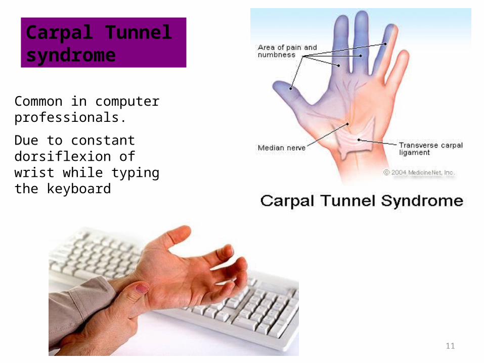
• Carpal tunnel syndrome is an entrapment syndrome caused by pressure on the median nerve within the carpal tunnel.
Carpal tunnel syndrome

Signs and Symptoms of Carpal Tunnel Syndrome
• Patients complain of pain, tingling and numbness in the distribution of the median nerve
• Weakness and atrophy of the thenar eminence could result
Tinel’s Sign- Tapping the median nerve in the carpal tunnel elicits pain in the distribution of the median nerve
11
Carpal Tunnel syndrome
Common in computer professionals.
Due to constant dorsiflexion of wrist while typing the keyboard

• The palmar aponeurosis is a triangular-shaped condensation of deep fascia that covers the palm and is anchored to the skin in distal regions.
• The apex of the triangle is continuous with the palmaris longus tendon.
Palmar aponeurosis

• Dupuytren contracture is a disease of the palmar fascia resulting in progressive shortening, thickening, and fibrosis of the palmar fascia and aponeurosis.
Dupuytren Contracture of Palmar Fascia

• 1. Hypothenar compartment
• 2. Thenar compartment • 3. Central compartment • 4. Adductor
compartment • 5.Interosseous
compartment
Compartments of palm

• The thenar compartment contains the thenar muscles while the hypothenar compartment contains the hypothenar muscles.
• The central compartment contains the flexor tendons and their sheaths, the lumbricals, the superficial palmar arterial arch and the digital vessels and nerves.
• The adductor compartment is the deepest and contains adductor pollicis.
• The interossei lie in the interosseous compartment

• The intrinsic muscles of the hand are located in five compartments
• All of the intrinsic muscles of the hand are innervated by the deep branch of the ulnar nerve except for the three thenar and two lateral lumbrical muscles, which are innervated by the median nerve.
Muscles

Muscles of the HandThenar Muscles
Abductor Pollicis Brevis Abducts thumb
Opponens Pollicis To oppose thumb
Flexor Pollicis Brevis Flexes thumb
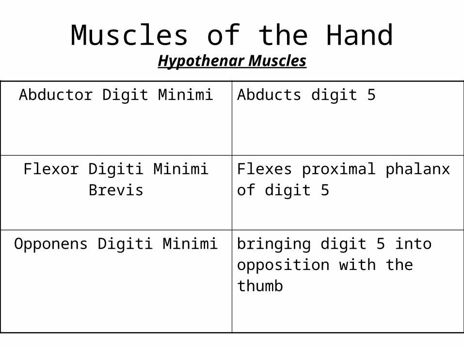
Muscles of the HandHypothenar Muscles
Abductor Digit Minimi Abducts digit 5
Flexor Digiti Minimi Brevis Flexes proximal phalanx of digit 5
Opponens Digiti Minimi bringing digit 5 into opposition with the thumb

Thenar and hypothenar Muscles

Palmaris brevis
Action:Improves palmar grip

Adductor Compartment
Adductor pollicis
Adducts thumb towards middle digit

Movements of thumb

• Extension: extensor pollicis longus, extensor pollicis brevis
• Flexion: flexor pollicis longus and flexor pollicis brevis
• Abduction: abductor pollicis longus and abductor pollicis brevis.
• Adduction: adductor pollicis • Opposition: opponens pollicis.
Movements of thumb

Lumbrical muscles

Muscles of the Hand Short Muscles Lumbricals
1 and 2Flex digits at metacarpo-phalangeal joints and extends interphalangeal joints
Lumbricals3 and 4
Flex digits at metacarpo-phalangeal joints and extends interphalangeal joints
Dorsal interossei1-4
Abducts digits from axial line and act with lumbricals to flex metacarpo-phalangeal joints and extends interphalangeal joints
Palmar interossei1-3
Adducts toward axial line & assist lumbriaclas in flexing the same joints as above

Dorsal interosseiAction of Dorsal Interossei :
DAB : Abduction of 2nd – 4th digits
Little finger and thumb have no Dorsal interossei muscle

Palmar interossei
Action of Palmar interossei :
PAD :Adduction of 2nd , 4th and 5th digit
Middle finger and thumb have no palmar interossei muscle

•Ulnar artery & Superficial Palmar arch•Radial artery & Deep Palmar arch•Dorsal Venous arch
BLOOD SUPPLY TO THE HAND

Arteries & Veins
• The blood supply to the hand is by the radial & ulnar arteries, which form 2 interconnected vascular arches (superficial & deep) in the palm.
• Vessels to the digits, muscles, & joints originate from the 2 arches & the parents arteries:
The radial artery contributes substantially to the supply of the thumb & the lateral side of the index finger;
The remaining digits & the medial side of the index finger are supplied mainly by the ulnar artery.

Radial artery VS. Ulnar artery in the Hand
The radial artery contributes substantially to the supply of the thumb & the lateral side of the index finger;
• The remaining digits & the medial side of the index finger are supplied mainly by the ulnar artery.

Ulnar artery & superficial palmar arch
• A palmar digital artery to medial side of little finger
• 3 large, common palmar digital arteries, which are joined by the palmar metacarpal arteries from the deep palmar arch before bifurcating into the proper palmar digital arteries, which enter the fingers.

Radial artery & Deep Palmar arch
• 2 vessels, the princeps pollicis artery & the radialis indicis artery, arise from the radial artery.
• The princeps pollicis artery is the major blood supply to the thumb.
• The radialis indicis artery supplies the lateral side of the index finger.

Deep Palmar arch
• The deep palmar arch gives rise to :
• 3 palmar metacarpal arteries, which join the common palmar digital arteries from the superficial palmar arch;

Dorsal venous arch of the hand
• The deep veins follow the arteries;
• the superficial veins drain into a dorsal venous network on the back of the hand over the metacarpal bones.

Dorsal venous arch
• The cephalic vein originates from the lateral side of the dorsal venous network & passes over the anatomical snuffbox into the forearm.
• The basilic vein originates from the medial side of the dorsal venous network & passes into the dorsomedial aspect of the forearm.

• To test for adequate anastomoses between the radial and ulnar arteries, compress both the radial and ulnar arteries at the wrist, then release pressure from one or the other, and determine the filling pattern of the hand.
Allen's test

Positive allen test

• The hand is supplied by the ulnar, median, and radial nerves.
Nerves

• Immediately distal to the pisiform, ulnar nerve divides into a deep branch, which is mainly motor and a superficial branch, which is mainly sensory.
Ulnar nerve
Deep branch: supplies the hypothenar muscles ,interossei, adductor pollicis, and the two medial lumbricals(4 and 5).
Superficial branch: supply skin on the palmar and dorsal surface of the little finger and the medial half of the ring finger.


• The ulnar nerve is most commonly injured at two sites:
• 1. Elbow • 2. wrist• Clawing of the hand:• Metacarpophalangeal joints of the fingers are
hyperextended and the interphalangeal joints are flexed.
Ulnar nerve injury

Clawing of the hand

• Compression of the ulnar nerve may occur at the wrist where it passes between the pisiform and the hook of the hamate.
Ulnar Canal Syndrome (Guyon Tunnel Syndrome)

• The median nerve is the most important sensory nerve in the hand because it innervates skin on the thumb, index and middle fingers, and lateral side of the ring finger.
• Branches:• 1. Recurrent branch: innervates the three thenar
muscles• 2. Palmar digital nerves: supplies the palmar skin
of the lateral 31/2 digits and dorsal nail beds. It also supplies the lateral two lumbrical muscles( 1 and 2).
Median nerve


• Refers to a deformity in which thumb movements are limited to flexion and extension of the thumb in the plane of the palm.
• Severance of median nerve paralyzes the thenar muscles and the thumb loses much of its usefulness.
Ape hand

• The only part of the radial nerve that enters the hand is the superficial branch.
• Innervates skin over the dorsolateral aspect of the palm and the dorsal aspects of the lateral three and one-half digits distally to approximately the terminal interphalangeal joints.
Superficial branch of the radial nerve

Superficial branch of the radial nerve

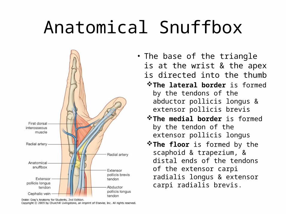
Anatomical Snuffbox• The base of the triangle is at the
wrist & the apex is directed into the thumbThe lateral border is formed by the
tendons of the abductor pollicis longus & extensor pollicis brevis
The medial border is formed by the tendon of the extensor pollicis longus
The floor is formed by the scaphoid & trapezium, & distal ends of the tendons of the extensor carpi radialis longus & extensor carpi radialis brevis.
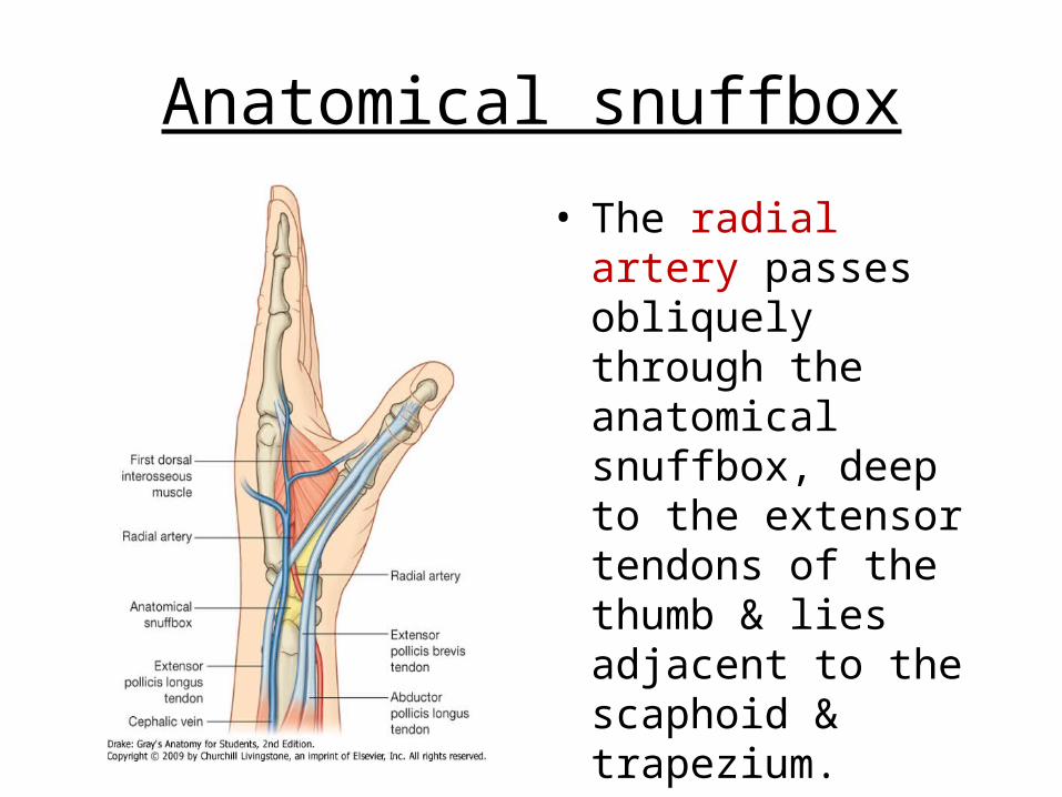
Anatomical snuffbox
• The radial artery passes obliquely through the anatomical snuffbox, deep to the extensor tendons of the thumb & lies adjacent to the scaphoid & trapezium.