the hippo signaling pathway restricts the oncogenic...
TRANSCRIPT

RESEARCH COMMUNICATION
The Hippo signaling pathwayrestricts the oncogenicpotential of an intestinalregeneration programJing Cai,1 Nailing Zhang,1 Yonggang Zheng,1
Roeland F. de Wilde,2 Anirban Maitra,2
and Duojia Pan1,3
1Department of Molecular Biology and Genetics, HowardHughes Medical Institute, Johns Hopkins University Schoolof Medicine, Baltimore, Maryland 21205, USA; 2Departmentof Pathology, The Sol Goldman Pancreatic Cancer ResearchCenter, Johns Hopkins University School of Medicine,Baltimore, Maryland 21205, USA
Although a developmental role for Hippo signaling inorgan size control is well appreciated, how this pathwayfunctions in tissue regeneration is largely unknown.Here we address this issue using a dextran sodium sulfate(DSS)-induced colonic regeneration model. We find thatregenerating crypts express elevated Yes-associated pro-tein (YAP) levels. Inactivation of YAP causes no obviousintestinal defects under normal homeostasis, but se-verely impairs DSS-induced intestinal regeneration. Con-versely, hyperactivation of YAP results in widespreadearly-onset polyp formation following DSS treatment.Thus, the YAP oncoprotein must be exquisitely con-trolled in tissue regeneration to allow compensatory pro-liferation and prevent the intrinsic oncogenic potentialof a tissue regeneration program.
Supplemental material is available at http://www.genesdev.org.
Received August 5, 2010; revised version accepted September 14,2010.
Injury to many mammalian tissues triggers a sophisti-cated repair process that ultimately replenishes the lostcells and restores the structural and functional integrityof the affected tissues (Schafer and Werner 2008; Oviedoand Beane 2009). Such injury-induced cell proliferationmust be tightly controlled to ensure that an appropriatenumber of cells are produced. Insufficient regenerativeproliferation underlies pathological conditions suchas age-related tissue atrophy and deterioration. Con-versely, excessive and persistent regenerative prolifer-ation may lead to tumorigenesis (Beachy et al. 2004;Kluwe et al. 2009). While a number of signaling mole-cules have been implicated in driving cell proliferationduring tissue regeneration, little is known about mecha-nisms that normally restrict and/or terminate regenerativeproliferation.
We reasoned that developmental pathways that nor-mally terminate organ growth during animal developmentmay also play a critical role in restricting regenerativegrowth in adult tissues. A prominent candidate is theHippo tumor suppressor pathway (Badouel et al. 2009;Zhao et al. 2010). First identified in Drosophila, the Hippopathway has emerged recently as a conserved mechanismthat restricts organ size in diverse species, includingmammals. This pathway comprises several tumor sup-pressors acting in a kinase cascade that culminates in thephosphorylation and inactivation of the transcriptionalcoactivator Yorkie (Yki) in Drosophila, or its mammalianhomolog, YAP (Yes-associated protein) (Pan 2007; Zhaoet al. 2010). In Drosophila and mice, inactivation of thetumor suppressors of the Hippo pathway or activation ofthe oncogene Yki or YAP results in massive tissue over-growth characterized by increased cell proliferation anddiminished cell death (Dong et al. 2007; Camargo et al.2007; Zhou et al. 2009; Lee et al. 2010; Lu et al. 2010;Song et al. 2010). Conversely, inactivation of Yki or YAPleads to tissue and/or cellular atrophy (Huang et al. 2005;Zhang et al. 2010). While these findings demonstratea critical role for Hippo signaling in controlling organ sizeduring animal development and normal homeostasis,whether and how the Hippo pathway functions in thecontext of tissue regeneration are largely unknown.
Results and Discussion
To examine the role of Hippo signaling during tissue re-generation, we took advantage of the well-establisheddextran sodium sulfate (DSS)-induced colitis and regen-eration model (Okayasu et al. 1990). A 5-d DSS treatmentresulted in damages in the colonic crypt base and reducedthe number of proliferating cells in the lower portion ofthe crypt (Fig. 1A,B). Two days after the withdrawal ofDSS, the crypts were composed of tightly compactedproliferating cells that extended to the entire crypt, witha concomitant loss of differentiated goblet and entero-endocrine cells. Four days after DSS withdrawal, crypthistology and cell differentiation were largely restored,and cell proliferation was again restricted to the lowerportion of the crypt (Fig. 1A,B,E). As shown previously,DSS-induced injury and regeneration was accompaniedby induction of Stat3 phosphorylation (Bollrath et al.2009; Grivennikov et al. 2009; Pickert et al. 2009), whichwas detected specifically in the damaged and regeneratingcrypts (Fig. 1D). We examined the temporal and spatialregulation of YAP in the DSS model. In normal adultmice, YAP protein is expressed in the entire crypt, includ-ing both proliferating and post-mitotic cells (Fig. 1C). TheYAP protein level was slightly decreased in the cryptsafter a 5-d DSS treatment. Strikingly, a dramatic increaseof YAP protein level was detected in the crypts 2 d afterDSS withdrawal (Fig. 1C,D). This increase of YAP proteinlevel was not due to increased transcription, since YapmRNA was slightly decreased in the regenerating crypts(Fig. 1E). Despite the dramatic increase in YAP proteinlevels, the relative phosphorylation of YAP at its Hippo-responsive S112 site (as measured by P-S112-YAP/YAPratio) was only slightly decreased in regenerating crypts(Fig. 1D), suggesting that Hippo signaling remainedlargely unperturbed in regenerating crypts. Consistent
[Keywords: Hippo signaling; cancer; growth control; mouse; regeneration]3Corresponding author.E-MAIL [email protected]; FAX (410) 502-3177.Article is online at http://www.genesdev.org/cgi/doi/10.1101/gad.1978810.
GENES & DEVELOPMENT 24:2383–2388 � 2010 by Cold Spring Harbor Laboratory Press ISSN 0890-9369/10; www.genesdev.org 2383
Cold Spring Harbor Laboratory Press on April 7, 2019 - Published by genesdev.cshlp.orgDownloaded from

with this finding, immunostaining showed that YAP wasdistributed nondiscriminatively in the cytoplasm and thenucleus in the regenerating crypts (Fig. 1C).
The dramatic increase in YAP protein levels in regen-erating crypts suggests that YAP may play a potential rolein intestinal regeneration. To investigate this possibility,we crossed a conditional knockout allele of Yap with
Villin-Cre (VilCre), which results in gene deletion in theepithelium of the small intestine and colon startingat embryonic day 12.5 (E12.5) (Madison et al. 2002).VilCre;Yapflox/flox mice developed normally, and histo-logical analysis of adult mice revealed no visible defectsin cell differentiation, cell death, cell proliferation, or cellmigration along the crypt–villus axis (Fig. 3; Supplemen-tal Figs. S1, S3D,E), suggesting that YAP is dispensable fornormal intestinal homeostasis. Following DSS treatment,however, VilCre;Yapflox/flox mice showed a dramatic in-crease in mortality rate and a rapid decrease in bodyweight compared with the control littermates (Fig. 2A).Histological analysis revealed substantial damage withsignificant loss of crypts and scattered colonic epithe-lial cells in the VilCre;Yapflox/flox mice compared withthe control littermates (Fig. 2B,C). Furthermore, theVilCre;Yapflox/flox colon contained fewer proliferatingcells and more apoptotic cells (Fig. 2D,E). Thus, whileYAP is largely dispensable for intestinal homeostasisunder normal conditions, it is required for DSS-inducedcrypt regeneration.
The inconsequentiality of YAP loss for normal intestinalhomeostasis can be compatible with at least two possibil-ities: the Hippo pathway may be simply dispensable, or,alternatively, Hippo signaling may be constitutively acti-vated such that YAP is kept largely inactive under normal
Figure 1. Increased YAP protein levels in regenerating crypts. (A)H&E staining of colon sections from 8-wk-old wild-type mice before(0 d) and after a 5-d DSS treatment (5 d), followed by normal drinkingwater for 2 d (5 + 2 d) or 4 d (5 + 4 d). (Top row) Low magnification.(Bottom row) High magnification. (B) Ki67 staining. Note therestriction of Ki67-positive proliferating cells to the crypt base ina normal colon, the loss of Ki67-positive cells in 5-d crypts, theexpansion of Ki67-positive cells from the crypt base to the wholeregenerating crypt in 5 + 2-d colons, and the restoration of Ki67-positive cells to the crypt base in 5 + 4-d colons. (C) YAP staining. Inwild-type crypts, YAP was detected in the entire crypt epithelium.Note the dramatic increase of YAP staining in 5 + 2-d crypts. Alsonote the absence of appreciable YAP nuclear accumulation incontrol and regenerating crypts. (D) Western blotting analysis. Pro-tein extracts from control and regenerating crypts were probed withthe indicated antibodies. Note the increase of YAP, P-YAP, andP-Stat3 in 5 + 2-d and 5 + 4-d crypts. The YAP protein level andP-YAP/YAP ratio were quantified in the graphs to the right. Data aremean 6 SD. n = 3. (*) P < 0.01, t-test. (E) Real-time PCR analysis.mRNAs from control and regenerating crypts were analyzed for theexpression of the indicated genes. Note the decrease of Yap mRNAin 5 + 2-d and 5 + 4-d crypts. Also note the decreased expression ofthe goblet cell marker Mucin2 and the enteroendocrine cell markerChromogranin A (ChgA) in 5 + 2-d crypts, and the recovery of theirexpression levels in 5 + 4-d crypts. Data are mean 6 SD. n = 3. (*) P <0.01, t-test. Bars, 100 mm.
Figure 2. Impaired regeneration of Yap-deficient colonic crypts. (A)Increased mortality and loss of body weight in Yap-deficient mice afterDSS treatment. Mice were treated with 2.5% DSS for 7 d and suppliedwith normal drinking water thereafter. Thirteen wild-type and nineYap-deficient mice were treated for mortality rate analysis. Nine wild-type and eight Yap-deficient mice were treated for body weightanalysis. (*) P < 0.05; (**) P < 0.01, t-test. (B) H&E staining of colonsections after a 7-d DSS treatment. (Top row) Low magnification.(Bottom row) High magnification. Note the absence of crypt structureand the expansion of stromal cells in Yap-deficient colons. (C) Histo-logical score of colons after a 7-d DSS treatment. Tissue damages werequantified as described in the Materials and Methods. Eight wild-typeand nine Yap-deficient mice were treated for this analysis. Data aremean 6 SEM. (*) P < 0.05, t-test. (D,E) Ki67 and cleaved caspase-3staining showing fewer proliferating and more apoptotic cells inYap-deficient crypts after DSS treatment. Bars, 100 mm.
Cai et al.
2384 GENES & DEVELOPMENT
Cold Spring Harbor Laboratory Press on April 7, 2019 - Published by genesdev.cshlp.orgDownloaded from

homeostasis. The latter, but not the former, model pre-dicts that inactivation of tumor suppressors that normallyrestrict YAP activity may lead to tumor formation and/orintestinal overgrowth due to hyperactivation of the YAPoncoprotein. To investigate this possibility, we generatedconditional knockout mice for the Hippo pathway com-ponent Sav1 (Supplemental Fig. S2). Consistent with thelatter model, 4-wk-old VilCre;Sav1flox/flox mice showedan enlargement of crypts in both the colon and smallintestine, which contained more and faster proliferatingcells (Fig. 3A–C; Supplemental Fig. S3). Interestingly, thehyperplasia of Sav1-deficient crypts was completely re-versed by loss of YAP, even though the latter by itself hadno visible effect on normal crypt morphology or cellproliferation (Fig. 3A–C; Supplemental Fig. S3). Consistentwith the genetic suppression of the Sav1-deficient pheno-type by loss of Yap, Sav1-deficient crypts showed de-creased YAP S112 phosphorylation (Fig. 3D) as well as
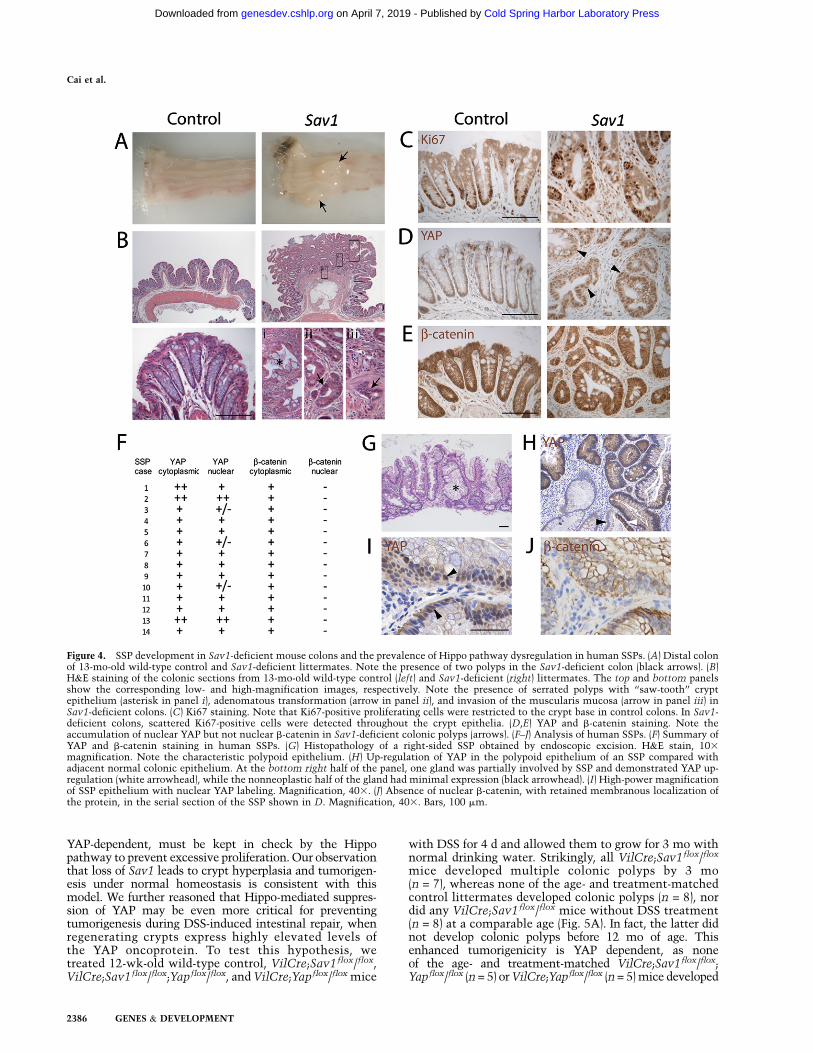
nuclear accumulation of YAP (Supplemental Fig. S2E), incontrast to its nondiscriminative subcellular localizationin normal or regenerating crypts (Fig. 1C). By 13 mo of age,all male VilCre;Sav1flox/flox mice had developed colonicpolyps (average number 1.71 6 0.76, n = 7), whereas noneof the control male littermates developed any polyps ata comparable age (n = 12) (Fig. 4A). Histological analy-sis revealed that the polyps did not resemble the pro-totypal intestinal adenomas observed in the ApcMin modelof tumorigenesis (Moser et al. 1990; Su et al. 1992). Incontrast, there were prominent invaginations of the hy-perplastic epithelium leading to a ‘‘saw-tooth’’ appearanceof the glands, a feature characteristic of sessile serratedpolyps (SSPs), which comprise a recently cataloguedalternative pathway to colorectal neoplasia in humans(Fig. 4B, panel i; Montgomery 2004; Snover et al. 2005).Over time, SSPs left in situ are prone to adenomatoustransformation and the development of invasive adeno-carcinomas (Montgomery 2004; Snover et al. 2005). In-deed, such an adenomatous transformation (Fig. 4B, panelii) and invasion of the muscularis mucosa by neoplasticglands (Fig. 4B, panel iii) were observed. The Sav1-de-ficient SSPs showed nuclear accumulation of YAP, but notnuclear accumulation of b-catenin or elevated overalllevels of b-catenin (Fig. 4D,E), further underscoring theirdistinction from the conventional tubular adenomas.Consistent with the complete suppression of Sav1 loss-induced crypt hyperplasia by loss of YAP in younger mice,none of the aged VilCre;Sav1flox/flox;Yapflox/flox (n = 6males) and VilCre;Yapflox/flox mice (n = 6 males) developedany polyps or histological abnormalities in the colon. Toour knowledge, the VilCre;Sav1flox/flox mice describedhere represent the first mouse model for SSPs.
The development of SSPs in Sav1-deficient mousecolons prompted us to investigate whether aberrant Hipposignaling may be a general hallmark of SSPs in humans.Therefore, we examined the expression and subcellularlocalization of YAP and b-catenin proteins in an archivalcollection of 14 histologically documented SSPs that hadbeen endoscopically removed (Fig. 4F–J). All of the 14 SSPs(100%) demonstrated up-regulation of YAP protein, com-pared with the adjacent normal colonic mucosa (Fig. 4F,H).Eleven of 14 (78%) SSPs demonstrated unequivocal nu-clear YAP accumulation within the polypoid epithelium,while three of 14 (22%) demonstrated only infrequentnuclear YAP labeling, although cytoplasmic protein wasstill up-regulated compared with the colonic epithelium.Of note, in the 11 SSPs with nuclear YAP localization, weconfirmed the absence of nuclear b-catenin in serial sectionsof the polypoid epithelium, and its restriction to themembranous and cytoplasmic compartments (Fig. 4I,J).Thus, as in Sav1-deficient mouse colons (Fig. 4D,E), nuclearlocalization of YAP occurs in the absence of nuclear oroverall b-catenin accumulation in human SSPs.
The dispensable role for Yap but not Sav1 in normalintestinal growth is consistent with the view that, undernormal homeostasis, the Hippo pathway keeps the YAPoncoprotein in a relatively inactive state. This raises thecritical question of why intestinal cells invest the energyto synthesize a growth regulator like YAP, but meanwhileemploy an elaborate signaling cascade to turn off itsactivity. An attractive model is that the intestinal epithe-lia, which are constantly exposed to environmental in-sults, must be poised for regeneration in anticipation ofsevere tissue damage. This regenerative capacity, which is
Figure 3. Loss of Sav1 results in Yap-dependent crypt hyperplasia.(A) Isolated colonic crypts from 4-wk-old wild-type, Sav1, Yap, orSav1 Yap double-mutant colons. Note the enlarged width of Sav1mutant crypts. Quantification of crypt width is shown in C, graph i.(B) H&E staining of the colonic sections from 4-wk-old wild-type,Sav1, Yap, or Sav1 Yap double-mutant colons. Note the enlargedwidth of Sav1 mutant crypts. (C, graph i) Quantification of cryptwidth from A. Eighty crypts from three mice of each genotype wereused. Data are mean 6 SEM. (*) P < 0.01, t-test. (Graph ii)Quantification of cell proliferation. Four-week-old wild-type, Sav1,Yap, or Sav1 Yap double-mutant mice were analyzed for BrdU andKi67 staining 2 h after a single i.p. injection of BrdU. The number ofBrdU+ or Ki67+ cells and the ratio of BrdU+/Ki67+ in each crypt werequantified with 200 crypts from three mice of each genotype. Dataare mean 6 SEM. (*) P < 0.01, t-test. (D) Quantification of YAPprotein and mRNA levels. Note the increased YAP protein andmRNA levels in Sav1-deficient crypts. Also note that, while theabsolute amount of P-YAP was similar in wild-type and Sav1-deficient crypts, the P-YAP/YAP ratio was decreased in Sav1-deficientcrypts. Data used in the graph are mean 6 SD. n = 3. (*) P < 0.01,t-test. Bars, 100 mm.
Hippo function in intestinal homeostasis
GENES & DEVELOPMENT 2385
Cold Spring Harbor Laboratory Press on April 7, 2019 - Published by genesdev.cshlp.orgDownloaded from
YAP-dependent, must be kept in check by the Hippopathway to prevent excessive proliferation. Our observationthat loss of Sav1 leads to crypt hyperplasia and tumorigen-esis under normal homeostasis is consistent with thismodel. We further reasoned that Hippo-mediated suppres-sion of YAP may be even more critical for preventingtumorigenesis during DSS-induced intestinal repair, whenregenerating crypts express highly elevated levels ofthe YAP oncoprotein. To test this hypothesis, wetreated 12-wk-old wild-type control, VilCre;Sav1flox/flox,VilCre;Sav1flox/flox;Yapflox/flox, and VilCre;Yapflox/flox mice
with DSS for 4 d and allowed them to grow for 3 mo withnormal drinking water. Strikingly, all VilCre;Sav1flox/flox
mice developed multiple colonic polyps by 3 mo(n = 7), whereas none of the age- and treatment-matchedcontrol littermates developed colonic polyps (n = 8), nordid any VilCre;Sav1flox/flox mice without DSS treatment(n = 8) at a comparable age (Fig. 5A). In fact, the latter didnot develop colonic polyps before 12 mo of age. Thisenhanced tumorigenicity is YAP dependent, as noneof the age- and treatment-matched VilCre;Sav1flox/flox;Yapflox/flox (n = 5) or VilCre;Yapflox/flox (n = 5) mice developed
Figure 4. SSP development in Sav1-deficient mouse colons and the prevalence of Hippo pathway dysregulation in human SSPs. (A) Distal colonof 13-mo-old wild-type control and Sav1-deficient littermates. Note the presence of two polyps in the Sav1-deficient colon (black arrows). (B)H&E staining of the colonic sections from 13-mo-old wild-type control (left) and Sav1-deficient (right) littermates. The top and bottom panelsshow the corresponding low- and high-magnification images, respectively. Note the presence of serrated polyps with ‘‘saw-tooth’’ cryptepithelium (asterisk in panel i), adenomatous transformation (arrow in panel ii), and invasion of the muscularis mucosa (arrow in panel iii) inSav1-deficient colons. (C) Ki67 staining. Note that Ki67-positive proliferating cells were restricted to the crypt base in control colons. In Sav1-deficient colons, scattered Ki67-positive cells were detected throughout the crypt epithelia. (D,E) YAP and b-catenin staining. Note theaccumulation of nuclear YAP but not nuclear b-catenin in Sav1-deficient colonic polyps (arrows). (F–J) Analysis of human SSPs. (F) Summary ofYAP and b-catenin staining in human SSPs. (G) Histopathology of a right-sided SSP obtained by endoscopic excision. H&E stain, 103
magnification. Note the characteristic polypoid epithelium. (H) Up-regulation of YAP in the polypoid epithelium of an SSP compared withadjacent normal colonic epithelium. At the bottom right half of the panel, one gland was partially involved by SSP and demonstrated YAP up-regulation (white arrowhead), while the nonneoplastic half of the gland had minimal expression (black arrowhead). (I) High-power magnificationof SSP epithelium with nuclear YAP labeling. Magnification, 403. (J) Absence of nuclear b-catenin, with retained membranous localization ofthe protein, in the serial section of the SSP shown in D. Magnification, 403. Bars, 100 mm.
Cai et al.
2386 GENES & DEVELOPMENT
Cold Spring Harbor Laboratory Press on April 7, 2019 - Published by genesdev.cshlp.orgDownloaded from

colonic polyps. Histological analysis and Ki67 stainingrevealed the presence of SSPs in DSS-induced Sav1-de-ficient colons (Fig. 5B,C). Like the late-onset polypsobserved in Sav1-deficient colons without DSS treatment,the DSS-induced Sav1-deficient polyps showed an accu-mulation of nuclear YAP (Fig. 5D). Taken together, weconclude that DSS-induced injury and repair greatly exac-erbated the tumorigenicity of the Sav1-deficient crypts.
The prevailing view that the Hippo pathway func-tions as a critical regulator of mammalian tissue ho-meostasis is based largely on the premise that YAP gainof function leads to tissue overgrowth and cell transfor-mation (Overholtzer et al. 2006; Zender et al. 2006;Camargo et al. 2007; Dong et al. 2007; Zhou et al. 2009;Lee et al. 2010; Lu et al. 2010; Song et al. 2010). WhetherYAP is normally required for mammalian tissue homeo-stasis is less clear. Our present study demonstrates thatYAP is dispensable for normal intestinal homeostasis, butis required for intestinal regeneration following tissueinjury. We note that the requirement for YAP in DSS-induced regeneration resembles that of Stat3 (Bollrathet al. 2009; Grivennikov et al. 2009; Pickert et al. 2009),suggesting a potential cross-talk between the Hippo andthe JAK/STAT pathways. Since Stat3 phosphorylation wasnot affected in Yap- or Sav1-deficient crypts after DSStreatment (data not shown), it is unlikely that Stat3functions downstream from the Hippo pathway. Whetherthe Hippo pathway resides downstream from Stat3 orfunctions in parallel to the JAK/Stat3 pathway requiresfurther investigation.
Our current study also offers an example ofa molecular explanation for the long-standingassociation between tissue regeneration and can-cer (Beachy et al. 2004; Kluwe et al. 2009). Fol-lowing DSS-induced injury, regenerating cryptsexpress highly elevated levels of the YAP onco-protein. Given YAP’s well-established transform-ing activity, this regenerative response is poten-tially oncogenic, and therefore must be kept incheck via Hippo-mediated YAP phosphorylation.Loss of Hippo signaling, as in the Sav1-deficientcrypts, can expose the intrinsic oncogenic poten-tial of this regeneration program and result inaccelerated malignant transformation. It is tempt-ing to speculate that other regenerative responsesmay share a similar oncogenic potential, and thatthe Hippo pathway may play a more widespreadrole in restraining the oncogenic potential intrin-sic to these regenerative processes.
Materials and methods
Generation of Sav1 conditional knockout mice
A targeting vector containing the third exon of the Sav1 gene was
generated by recombineering as described previously (Liu et al.
2003). Transformed embryonic stem cell colonies were
screened by long-template PCR with the following primer sets:
PS5F (59-GAAAAACACCTCTCACCTCTGAATATCATTCCC
TCCTCCAGCTCC-39) and PS5R (59-TTAAGGGTTATTGAA
TATGATCGGAATTGGGCTGCAGGAA-39) to generate a 4.4-kb
band for positive clones; and PS3F (59-GCTCTATGGCTTCT
GAGGCGGAAAGAACCAGCTGGGGCTCGAC-39) and PS3R
(59-CTCAAGGAAACACAAGGTAGCTCATGAACTATCTCG
TATTCAGGG-39) to generate a 4.8-kb band for positive clones.
Successfully targeted embryonic stem cell clones (confirmed
by both 59 PCR and 39 PCR) were microinjected into C57BL/6 blastocysts.
Germline transmission from generated chimeric offspring was confirmed
by long-template PCR (Supplemental Fig. S2A,B). Mice carrying the
targeted allele were bred to Flp recombinase transgenic mice (kindly
provided by Dr. Jeremy Nathans, Johns Hopkins University School of
Medicine) to remove the FRT-flanked Neo cassette and to generate the
Sav1flox mice.
Genomic DNAs extracted from tail biopsies were genotyped with a PCR
primer set (PS1, 59-AGGGGATTCTGACATTTCAGTCAGTT-39; PS2, 59-
AGTCACATGCTGACCACAAGCAGAA-39; PS3; 59-TGCCATTAAGTG
TAATCACTGG-39) that generated a 231-base-pair (bp) band from the wild-
type allele, a 335-bp band from the Flox allele, and a 460-bp band from the
knockout allele (Supplemental Fig. S2C).
Yapflox mice were generated as described (Zhang et al. 2010). Villin-Cre
mice were purchased from the Jackson Laboratory. Mice with Sav1 or Yap
specifically deleted in the intestinal epithelium were generated by breeding
Sav1 flox or Yap flox mice with VilCre mice. The Cre, Yap knockout, and Yap
Flox alleles were genotyped as described (Zhang et al. 2010). Animal
protocols were approved by the Institutional Animal Care and Use
Committee of Johns Hopkins University.
DSS treatment of mice
For induction of colitis, mice received 2.5% DSS (MP Biomedical,
molecular weight 36,000–50,000 kDa) in drinking water for several days
as indicated in the text. After DSS treatment, mice were supplied with
normal drinking water and euthanized, and colons were harvested at the
time points indicated in the text.
Isolation of intestinal epithelial cells
Colons at ;1 cm away from the anus were cut longitudinally and rinsed in
PBS to remove feces. Pieces (2–3 mm) of colons were incubated in PBS
Figure 5. DSS-induced regeneration accelerated polyp development in Sav1-deficient colons in a Yap-dependent manner. (A) Distal colon of 12-wk-old wild-type, Sav1, Yap, or Sav1 Yap double-mutant mice treated with 2.5% DSS for 4 d,followed by normal drinking water for 3 mo. Note the presence of multiple largecolonic polyps in the Sav1-deficient colon (arrows). (B) H&E staining of colonicsections from animals in A. The top and bottom panels show the correspondinglow- and high-magnification images, respectively. Note the presence of serratedcrypt epithelium in Sav1-deficient polyps (asterisk). (C) Ki67 staining of colonsections from control and Sav1-deficient littermates. Note the presence ofscattered Ki67-positive cells throughout the serrated crypt epithelium. (D) YAPstaining of colon sections from control and Sav1-deficient littermates. Note theaccumulation of nuclear YAP in Sav1-deficient colonic polyps (arrowheads). Bars,100 mm.
Hippo function in intestinal homeostasis
GENES & DEVELOPMENT 2387
Cold Spring Harbor Laboratory Press on April 7, 2019 - Published by genesdev.cshlp.orgDownloaded from

with 5 mM EDTA for 15 min at 37°C. Vigorous shaking released crypts.
Crypts were photographed and the width was measured in AxioVision
release 4.7. For RNA and protein analysis, the supernatant containing free
crypts was collected and centrifuged. The pellet was washed in ice-cold
PBS and subjected to RNA extraction or snap-frozen in liquid nitrogen for
Western blot analysis.
Analysis of YAP protein expression in human SSPs
Formalin-fixed, paraffin-embedded sections were obtained from 14 archi-
val SSPs that had been removed endoscopically at Johns Hopkins Hospital.
The diagnosis of SSP was rendered based on established criteria by a
specialized gastrointestinal pathologist (A.M.) (Montgomery 2004; Snover
et al. 2005). Heat-induced antigen retrieval was performed in a steamer
using citrate buffer (pH 6.0) (Vector Laboratories) for 25 min followed by
30 min of cooling. Nonspecific binding was blocked for 10 min with 5%
bovine serum albumin (BSA) in TBST. Serial sections were then incubated
with two primary antibodies—anti-YAP (1:150 dilution; catalog no. 2060-1,
Epitomics Inc.) and anti-b-catenin (1:1000 dilution; catalog no. DS9800,
BD Transduction)—for 1 h at room temperature. The sections were then
incubated for 30 min with secondary antibody (Leica Microsystems) fol-
lowed by detection with 3,39-Diaminobenzide (Sigma-Adrich Co.) for 1 min.
Sections were washed three times after each step in Tris-buffered saline with
0.1% Tween-20. Finally, sections were counterstained with Harris hema-
toxylin, subsequently rehydrated in distilled H2O and graded series of
ethanol (70%, 95%, and 100%), and mounted.
Acknowledgments
We thank Drs. Qian Chen, Karen K. David, and Jianzhong Yu for
discussion. A.M. is supported by P01CA134292, R01CA113669, and
R01CA134767. D.P. is an investigator of the Howard Hughes Medical
Institute.
References
Badouel C, Garg A, McNeill H. 2009. Herding Hippos: Regulating growth
in flies and man. Curr Opin Cell Biol 21: 837–843.
Beachy PA, Karhadkar SS, Berman DM. 2004. Tissue repair and stem cell
renewal in carcinogenesis. Nature 432: 324–331.
Bollrath J, Phesse TJ, von Burstin VA, Putoczki T, Bennecke M, Bateman
T, Nebelsiek T, Lundgren-May T, Canli O, Schwitalla S, et al. 2009.
gp130-mediated Stat3 activation in enterocytes regulates cell survival
and cell-cycle progression during colitis-associated tumorigenesis.
Cancer Cell 15: 91–102.
Camargo FD, Gokhale S, Johnnidis JB, Fu D, Bell GW, Jaenisch R,
Brummelkamp TR. 2007. YAP1 increases organ size and expands
undifferentiated progenitor cells. Curr Biol 17: 2054–2060.
Dong J, Feldmann G, Huang J, Wu S, Zhang N, Comerford SA, Gayyed MF,
Anders RA, Maitra A, Pan D. 2007. Elucidation of a universal size-
control mechanism in Drosophila and mammals. Cell 130: 1120–1133.
Grivennikov S, Karin E, Terzic J, Mucida D, Yu GY, Vallabhapurapu S,
Scheller J, Rose-John S, Cheroutre H, Eckmann L, et al. 2009. IL-6 and
Stat3 are required for survival of intestinal epithelial cells and
development of colitis-associated cancer. Cancer Cell 15: 103–113.
Huang J, Wu S, Barrera J, Matthews K, Pan D. 2005. The Hippo signaling
pathway coordinately regulates cell proliferation and apoptosis by
inactivating Yorkie, the Drosophila homolog of YAP. Cell 122:
421–434.
Kluwe J, Mencin A, Schwabe RF. 2009. Toll-like receptors, wound
healing, and carcinogenesis. J Mol Med 87: 125–138.
Lee KP, Lee JH, Kim TS, Kim TH, Park HD, Byun JS, Kim MC, Jeong WI,
Calvisi DF, Kim JM, et al. 2010. The Hippo–Salvador pathway
restrains hepatic oval cell proliferation, liver size, and liver tumori-
genesis. Proc Natl Acad Sci 107: 8248–8253.
Liu P, Jenkins NA, Copeland NG. 2003. A highly efficient recombineer-
ing-based method for generating conditional knockout mutations.
Genome Res 13: 476–484.
Lu L, Li Y, Kim SM, Bossuyt W, Liu P, Qiu Q, Wang Y, Halder G, Finegold
MJ, Lee JS, et al. 2010. Hippo signaling is a potent in vivo growth and
tumor suppressor pathway in the mammalian liver. Proc Natl Acad
Sci 107: 1437–1442.
Madison BB, Dunbar L, Qiao XT, Braunstein K, Braunstein E, Gumucio
DL. 2002. Cis elements of the villin gene control expression in
restricted domains of the vertical (crypt) and horizontal (duodenum,
cecum) axes of the intestine. J Biol Chem 277: 33275–33283.
Montgomery E. 2004. Serrated colorectal polyps: Emerging evidence
suggests the need for a reappraisal. Adv Anat Pathol 11: 143–149.
Moser AR, Pitot HC, Dove WF. 1990. A dominant mutation that
predisposes to multiple intestinal neoplasia in the mouse. Science
247: 322–324.
Okayasu I, Hatakeyama S, Yamada M, Ohkusa T, Inagaki Y, Nakaya R.
1990. A novel method in the induction of reliable experimental acute
and chronic ulcerative colitis in mice. Gastroenterology 98: 694–702.
Overholtzer M, Zhang J, Smolen GA, Muir B, Li W, Sgroi DC, Deng CX,
Brugge JS, Haber DA. 2006. Transforming properties of YAP, a candi-
date oncogene on the chromosome 11q22 amplicon. Proc Natl Acad
Sci 103: 12405–12410.
Oviedo NJ, Beane WS. 2009. Regeneration: The origin of cancer or
a possible cure? Semin Cell Dev Biol 20: 557–564.
Pan D. 2007. Hippo signaling in organ size control. Genes Dev 21: 886–
897.
Pickert G, Neufert C, Leppkes M, Zheng Y, Wittkopf N, Warntjen M,
Lehr HA, Hirth S, Weigmann B, Wirtz S, et al. 2009. STAT3 links IL-
22 signaling in intestinal epithelial cells to mucosal wound healing.
J Exp Med 206: 1465–1472.
Schafer M, Werner S. 2008. Cancer as an overhealing wound: An old
hypothesis revisited. Nat Rev Mol Cell Biol 9: 628–638.
Snover DC, Jass JR, Fenoglio-Preiser C, Batts KP. 2005. Serrated polyps of
the large intestine: A morphologic and molecular review of an
evolving concept. Am J Clin Pathol 124: 380–391.
Song H, Mak KK, Topol L, Yun K, Hu J, Garrett L, Chen Y, Park O, Chang
J, Simpson RM, et al. 2010. Mammalian Mst1 and Mst2 kinases play
essential roles in organ size control and tumor suppression. Proc Natl
Acad Sci 107: 1431–1436.
Su LK, Kinzler KW, Vogelstein B, Preisinger AC, Moser AR, Luongo C,
Gould KA, Dove WF. 1992. Multiple intestinal neoplasia caused by
a mutation in the murine homolog of the APC gene. Science 256:
668–670.
Zender L, Spector MS, Xue W, Flemming P, Cordon-Cardo C, Silke J, Fan
ST, Luk JM, Wigler M, Hannon GJ, et al. 2006. Identification and
validation of oncogenes in liver cancer using an integrative onco-
genomic approach. Cell 125: 1253–1267.
Zhang N, Bai H, David KK, Dong J, Zheng Y, Cai J, Giovannini M, Liu P,
Anders RA, Pan D. 2010. The Merlin/NF2 tumor suppressor func-
tions through the YAP oncoprotein to regulate tissue homeostasis in
mammals. Dev Cell 19: 27–38.
Zhao B, Li L, Lei Q, Guan KL. 2010. The Hippo–YAP pathway in organ
size control and tumorigenesis: An updated version. Genes Dev 24:
862–874.
Zhou D, Conrad C, Xia F, Park JS, Payer B, Yin Y, Lauwers GY, Thasler W,
Lee JT, Avruch J, et al. 2009. Mst1 and Mst2 maintain hepatocyte
quiescence and suppress hepatocellular carcinoma development
through inactivation of the Yap1 oncogene. Cancer Cell 16: 425–438.
Cai et al.
2388 GENES & DEVELOPMENT
Cold Spring Harbor Laboratory Press on April 7, 2019 - Published by genesdev.cshlp.orgDownloaded from

10.1101/gad.1978810Access the most recent version at doi: 24:2010, Genes Dev.
Jing Cai, Nailing Zhang, Yonggang Zheng, et al. intestinal regeneration programThe Hippo signaling pathway restricts the oncogenic potential of an
Material
Supplemental
http://genesdev.cshlp.org/content/suppl/2010/10/21/24.21.2383.DC1
Related Content
Sci. Signal. November , 2010 3: ec342
Annalisa M. VanHookHippo Checks Regeneration
References
http://genesdev.cshlp.org/content/24/21/2383.full.html#related-urls
Articles cited in:
http://genesdev.cshlp.org/content/24/21/2383.full.html#ref-list-1This article cites 27 articles, 11 of which can be accessed free at:
License
ServiceEmail Alerting
click here.right corner of the article or
Receive free email alerts when new articles cite this article - sign up in the box at the top
Copyright © 2010 by Cold Spring Harbor Laboratory Press
Cold Spring Harbor Laboratory Press on April 7, 2019 - Published by genesdev.cshlp.orgDownloaded from