the histone demethylase kdm5b collaborates with tfap2c and
TRANSCRIPT

Histone Demethylase KDM5B Collaborates with TFAP2C and Myc ToRepress the Cell Cycle Inhibitor p21cip (CDKN1A)
Ping-Pui Wong, Fabrizio Miranda, KaYi V. Chan, Chiara Berlato, Helen C. Hurst, and Angelo G. Scibetta
Centre for Tumour Biology, Bart’s Cancer Institute, Queen Mary University of London, London, United Kingdom
The TFAP2C transcription factor has been shown to downregulate transcription of the universal cell cycle inhibitor p21cip
(CDKN1A). In examining the mechanism of TFAP2C-mediated repression, we have identified a ternary complex at the proximalpromoter containing TFAP2C, the oncoprotein Myc, and the trimethylated lysine 4 of histone H3 (H3K4me3) demethylase,KDM5B. We demonstrated that while TFAP2C and Myc can downregulate the CDKN1A promoter independently, KDM5B actsas a corepressor dependent on the other two proteins. All three factors collaborate for optimal CDKN1A repression, which re-quires the AP-2 binding site at �111/�103 and KDM5B demethylase activity. Silencing of TFAP2C-KDM5B-Myc led to in-creased H3K4me3 at the endogenous promoter and full induction of CDKN1A expression. Coimmunoprecipitation assaysshowed that TFAP2C and Myc associate with distinct domains of KDM5B and the TFAP2C C-terminal 270 amino acids (aa) arerequired for Myc and KDM5B interaction. Overexpression of all three proteins resulted in forced S-phase entry and attenuationof checkpoint activation, even in the presence of chemotherapy drugs. Since each protein has been linked to poor prognosis inbreast cancer, our findings suggest that the TFAP2C-Myc-KDM5B complex promotes cell cycle progression via direct CDKN1Arepression, thereby contributing to tumorigenesis and therapy failure.
The activation factor 2 (TFAP2) family consists of five homol-ogous, developmentally regulated transcription factors,
TFAP2A to -E, each encoded by a separate gene. Structurally,TFAP2 proteins contain a highly conserved C-terminal helix-span-helix motif required for dimerization, a basic DNA bindingdomain, and a third, less-conserved region toward the N terminuswhich contains a proline and glutamine-rich activation domain.These factors have been shown to bind to palindromic GC-richDNA recognition sequences as either homo- or heterodimers andact as transcriptional activators or repressors in a promoter-spe-cific manner (9).
Although studies in knockout mice (reviewed in reference 9)and of phenotypically related inherited human traits (25, 32) haveshown that these factors have important functions during em-bryogenesis, they are minimally expressed in most adult tissues.However, expression of the TFAP2A and TFAP2C proteins hasbeen demonstrated in a variety of solid tumors, including breastcancer and melanoma (reviewed in reference 24). TFAP2A ex-pression in breast tumors has been associated with a favorableoutcome and shows a positive correlation with expression of es-trogen recepter � (ER�) and CDKN1A (14), while elevated ex-pression of TFAP2C has generally been correlated with an adversephenotype and resistance to hormone therapy (13, 16). Studies invitro have shown that direct AP-2 transcriptional targets includemany genes involved in tumor progression. Of particular interestis the regulation of the universal cell cycle inhibitor p21cip
(CDKN1A), which can be activated through p53-dependent and-independent pathways to mediate cell cycle arrest (1). AP-2 fac-tors bind a specific sequence in the proximal region of the pro-moter, and while TFAP2A positively regulates CDKN1A expres-sion (27, 41), TFAP2C has been shown to repress its expression inbreast cancer cells (36). These opposing activities at the CDKN1Alocus may contribute to the contrasting phenotypes associatedwith tumors expressing these two AP-2 factors.
When mediating transcriptional activation, it has been shownthat AP-2 factors associate with the CITED family of adapter pro-
teins (Cited 2 and 4), which in turn recruit the histone acetyltrans-ferases (HATs) CBP/p300 (2, 7). To date, no clear AP-2 corepres-sor proteins have been identified, although modification throughsumoylation may be required for repressor activity (4, 10).TFAP2A has also been reported to interact with other nuclearfactors, including Myc, pRB, and p53 (reviewed in reference 9), toregulate the transcription of target genes. The proto-oncogeneMYC controls cellular growth, differentiation, and apoptosis, andits deregulation contributes to the development of a variety ofcancers, including breast cancer. It encodes a basic region-helix-loop-helix-zipper (bHLHZ) transcription factor which forms aspecific heterodimer with the small bHLHZ protein, Max. Myc-Max heterodimers bind with high affinity to the palindromicDNA sequence CACGTG (E box) but can also bind several otherrelated sequences, leading to interaction at up to 15% of all pro-moters. While the genes activated by Myc include those requiredfor cell growth and cell cycle progression, most of the genes itdownregulates are involved in cell cycle arrest, and they notablyinclude p21cip (CDKN1A) (11, 17). As with other activators oftranscription, Myc is able to recruit HAT-containing coactivatorcomplexes to its target genes (reviewed in reference 8), but theprecise mechanism behind Myc-mediated gene repression is stilldisputed: Myc interaction occurs close to the transcriptional start
Received 30 September 2011 Returned for modification 25 October 2011Accepted 18 February 2012
Published ahead of print 27 February 2012
Address correspondence to Helen Hurst, [email protected], or AngeloScibetta, [email protected].
H.C.H. and A.G.S. contributed equally to this work.
Supplemental material for this article may be found at http://mcb.asm.org/.
Copyright © 2012, American Society for Microbiology. All Rights Reserved.
doi:10.1128/MCB.06373-11
0270-7306/12/$12.00 Molecular and Cellular Biology p. 1633–1644 mcb.asm.org 1633
Dow
nloa
ded
from
http
s://j
ourn
als.
asm
.org
/jour
nal/m
cb o
n 02
Dec
embe
r 20
21 b
y 89
.151
.186
.59.

site (TSS), but direct DNA binding, at least at the CDKN1A gene,is not required (12, 17).
The reported colocalization of TFAP2C and Myc at the proxi-mal promoter of p21cip (CDKN1A) prompted us to investigate thepossibility of a functional link between these two transcriptionfactors in the repression of this gene in breast tumor cells. More-over, recent studies have suggested that Myc forms a complex withthe transcriptional corepressor KDM5B (lysine-specific demeth-ylase 5B; also known as Jarid1B/PLU-1). Like TFAP2C, theKDM5B protein is overexpressed in breast cancer lines and tu-mors (22). Its proven ability to demethylate H3K4me3 (trimethy-lated lysine 4 of histone H3), an epigenetic marker particularlyassociated with the TSS of active promoters, is consistent with thetranscriptional repressor role associated with this protein, and ithas been proposed that KDM5B may promote tumorigenesis bydownregulating expression of oncosuppressor genes, such asBRCA1 and HOXA5 (26, 39). In this work, we have investigatedwhether KDM5B regulates the expression of CDKN1A in cooper-ation with the TFAP2C and Myc proteins. Our data show thatTFAP2C, Myc, and KDM5B form a functional protein complex inthe vicinity of the TSS of the CDKN1A gene and corepress itsexpression in proliferating and drug-treated breast cancer cells,thus promoting cell cycle progression.
MATERIALS AND METHODSCell culture, transient transfection, and antibodies. MCF-7, HepG2,and H1299 cells (ATCC) were grown in Dulbecco’s modified Eagle me-dium (DMEM) supplemented with 10% fetal bovine serum in 10% CO2at 37°C. The shTFAP2C-MCF-7 line (36) was induced using 1 �g/mldoxycycline (Sigma). Transfection reagents were Genejuice (Merck) forplasmid DNA and Interferin (Polyplus) for small interfering RNA(siRNA), used according to the manufacturer’s directions. SubconfluentMCF-7 or shTFAP2C-MCF-7 cells were transiently transfected with 20nM nonsilencing control siRNA (no. 1022076; Qiagen) or with validatedsiRNA targeting KDM5B (J-009899-06; Darmacon) or Myc (6341; CellSignaling). The following antibodies for Western blotting (Santa Cruzunless stated otherwise) were used: KDM5B (3273; Cell Signaling), Myctag (2276; Cell Signaling), p53 (p53-DO-1, sc-126), TFAP2C (6E4, sc-53162), p21cip (2946; Cell Signaling), Myc (sc-788), �-actin (C-2, sc-8432), lamin A/C (4477; Cell Signaling), green fluorescent protein (GFP)(sc-8334), and Hsc70 (sc7298). Horseradish peroxidase-conjugated sec-ondary antibodies were from DakoCytomation.
Plasmids and reporter assays. The pcDNA3.1-based TFAP2C andKDM5B expression plasmids, the pGL-3-CDKN1A luciferase reporter,and pGL-3-Mut4/Mut5 constructs have been previously described (26,27). The fusion protein GFP-TFAP2C was generated by cloning theTFAP2C cDNA sequence in frame into the pEGFP-C1 vector (Clontech).The C-terminal deletion mutant (�C) was truncated at the internal SalIrestriction site, retaining amino acids 1 to 180. The N-terminal deletionmutant (�N) fused sequences C-terminal of the SalI site (amino acids 181to 450) to EGFP and retains the basic region and the helix-span-helixdimerization domains (5). To ensure efficient nuclear translocation of allfusion proteins, an oligonucleotide (5=-GA TCT CCA AAA AAG AAGAGA AAG GTT CA-3=), encoding a recognized nuclear localization signal(15), was ligated in frame between the GFP and wild-type (wt) or trun-cated TFAP2C sequences. The KDM5B deletion mutant constructs wereas described previously (38). The pcDNA3-Myc expression plasmid wasfrom Addgene. Reporter assays were controlled by cotransfection with thepSV-�-galactosidase control vector (Promega). Cells were harvested after48 h and processed for Western blotting or assayed for luciferase and�-galactosidase activity (Promega).
RNA analysis. Total RNA was extracted from subconfluent cells (RNeasyminikit; Qiagen), and 500 ng was reverse transcribed using the high-capacity
reverse transcription kit (Applied Biosystems). The cDNA was analyzed byquantitative real-time PCR (q-RT-PCR) using TaqMan reaction mix and theStepOne real-time PCR system (Applied Biosystems) according to the man-ufacturer’s instructions. TaqMan primer/probe sets were used to analyze theexpression of TFAP2C (Hs00231476_m1), CDKN1A (Hs00355782_m1),KDM5B (Hs00366783_m1) and Myc (Hs0095030_m1), while a glyceralde-hyde-3-phosphate dehydrogenase (GAPDH) TaqMan probe (no. 402869)was used as an internal control (Applied Biosystems). Analysis of q-RT-PCRdata was carried out using the comparative ��CT method. For each sample,the intensity of the amplimer was normalized against that of the GAPDHcontrol, and the data were depicted as fold changes (2���CT) with respect tothe RNA level observed in the control experiments.
ChIP assay. Subconfluent MCF-7 or shTFAP2C-MCF-7 cells werecross-linked by addition of formaldehyde directly to the culture mediumto a final concentration of 1% for 10 min and assayed using the ChIP-ITExpress chromatin immunoprecipitation (ChIP) kit (Active Motif).Chromatin was sheared to an average size of 500 bp using a Sonics Vibra-cell VCX500 sonicator and immunoprecipitated with ChIP-validated an-tibodies against KDM5B (3), TFAP2C (H77, sc-8977; Santa Cruz), Myc(sc-764), histone H3 (ab1791; Abcam), monomethylated lysine 4 of his-tone H3 (H3K4me1) (ab8895; Abcam), dimethylated lysine 4 of histoneH3 (H3K4me2) (ab7766; Abcam) or H3K4me3 (ab8580; Abcam), and 2�g purified rabbit IgG (sc2027). Quantification of immunoprecipitatedDNA was carried out in triplicate using the StepOne real-time PCR systemwith SYBR green PCR master mix (Applied Biosystems). Primer se-quences and calculation of values as fold enrichment compared with re-sults for the IgG control were as detailed previously (36).
Coimmunoprecipitation assays. Immunoprecipitation was carriedout according to the manufacturer’s protocol using the Universal Mag-netic coimmunoprecipitation kit (Active Motif). Cells were lysed in nu-clear extraction buffer supplemented with 5 �M trichostatin A, 1 �Mphenylmethylsulfonyl fluoride (Sigma), and protease inhibitor cocktail(Roche). Nuclear extract (0.5 mg) was incubated with 50 �l of proteinG-coated magnetic beads and 2 �g of either KDM5B (ab27689; Abcam),TFAP2C (6E4, H77 or AP25H01, sc-81181), Myc (sc-788), Myc tag (no.2276; Cell Signaling), or control IgG (sc-2027) antibody overnight at 4°C.Immunoprecipitates were washed three times with the supplied wash buf-fer for 5 min at 4°C and resuspended in 45 �l of 2� SDS Western loadingbuffer. For GFP-tagged proteins, immunoprecipitation was carried outusing the GFP-Trap kit (ChromoTek). Samples were resolved via SDS-PAGE and transferred onto polyvinylidene difluoride (PVDF) mem-branes for Western blot analysis. Additional Western blots establishedthat the �C mutant is recognized only by the H77 antibody while the �Nmutant interacts with all three TFAP2C antibodies.
Cell cycle analysis. Asynchronous HepG2 cells were transiently trans-fected with expression vectors and treated with 1 mM hydroxyurea, 6 nMvinblastine, or vehicle alone (control). After incubation at 37°C for 16 h,cells were trypsinized, washed in phosphate-buffered saline, and fixedusing ice-cold 70% ethanol. Fixed cells were incubated with 50 �l of 100�g/ml RNase and stained with 300 �l of 50-�g/ml propidium iodide(Invitrogen) for 30 min at room temperature. Flow cytometry and cellcycle analysis were performed as previously described (36).
RESULTSCDKN1A repression by TFAP2C, KDM5B, and Myc. To studythe relative activities of TFAP2C, KDM5B, and Myc in the repres-sion of CDKN1A, we used the estrogen receptor-positive (ER�)MCF-7 breast cancer line, which expresses all three factors. Wehave previously described an MCF-7 derivative line with doxycy-cline-inducible short hairpin RNA (shRNA) targeting TFAP2C(shTFAP2C cells); induction of TFAP2C silencing leads to up-regulation of CDKN1A and p53-independent G1/S arrest (36).Here, transient transfection of this line with validated specificsiRNA against Myc and KDM5B allowed us to examine the ex-pression level of the endogenous CDKN1A transcript and protein
Wong et al.
1634 mcb.asm.org Molecular and Cellular Biology
Dow
nloa
ded
from
http
s://j
ourn
als.
asm
.org
/jour
nal/m
cb o
n 02
Dec
embe
r 20
21 b
y 89
.151
.186
.59.

(Fig. 1A and B) after silencing each factor either individually or incombination. TFAP2C and Myc silencing, alone or together, inducedexpression of the CDKN1A transcript, while knockdown of KDM5Balone or with either TFAP2C or Myc had little effect. However, silenc-ing all three proteins significantly increased CDKN1A mRNA induc-tion (Fig. 1A, compare last 2 columns), indicating that KDM5B cancollaborate with the two transcription factors to repress CDKN1Atranscription in cycling cells. We observed a similar pattern of induc-tion at the protein level (Fig. 1B and C).
Reporter assays were used to confirm that CDKN1A repressionby these factors occurs at the promoter level. Initially, HepG2 cells(which do not express AP-2 factors or KDM5B) were transientlytransfected with a luciferase reporter construct containing theCDKN1A promoter (�2325/�8), and escalating amounts of anexpression vector for each test factor individually. As shown pre-viously, independent overexpression of either TFAP2C or Mycrepressed CDKN1A promoter activity in a dose-dependent man-ner; however, addition of KDM5B alone did not significantly alterreporter activity (Fig. 2A). In contrast, when a constant, subopti-mal dose of the TFAP2C construct was cotransfected with increas-ing amounts of the KDM5B expression construct, KDM5B core-pressor activity was observed, since reporter activity declined asthe dose of KDM5B plasmid increased (Fig. 2B). A similar resultwas also seen with a constant level of the Myc expression construct(data not shown). Moreover, by optimizing transfection inputlevels for all the constructs, we could demonstrate that coexpres-sion of all three proteins significantly further repressed CDKN1Apromoter activity (Fig. 2C), thus mirroring the induction of en-dogenous CDKN1A mRNA observed after cosilencing TFAP2C,KDM5B, and Myc in MCF-7 cells (Fig. 1A).
TFAP2C, Myc, and KDM5B form a nuclear complex inMCF-7 cells. To address if negative regulation of CDKN1A byTFAP2C, Myc, and KDM5B occurs through complex formationbetween these factors, we first used confocal microscopy to showthat all three proteins colocalize in the nuclear compartment ofMCF-7 cells, and TFAP2C silencing did not alter the localizationof KDM5B or Myc (see Fig. S1 in the supplemental material).
Next, we looked for physical interaction between these factors,examining endogenous proteins present in MCF-7 nuclear ex-tracts by coimmunoprecipitation (coIP) assays. As seen in Fig. 3A,Myc and TFAP2C were both immunoprecipitated using an anti-body specific for KDM5B (lane 5), and TFAP2C and KDM5B werealso both detected in anti-Myc immunoprecipitates (lane 6). Inorder to detect endogenous KDM5B or Myc in TFAP2C immu-noprecipitates, we tested antibodies specific for different regionsalong the 450-amino-acid (aa) protein. The H77 polyclonal anti-body was raised against a peptide corresponding to amino acids145 to 221; 6E4 recognizes an epitope within the C-terminal 20 aa(13), while AP25H01 was raised against a recombinant proteincorresponding to amino acids 50 to 200 of TFAP2C. Interestingly,Myc was coprecipitated with both H77 and 6E4, while KDM5Bwas precipitated only with 6E4 (Fig. 3A, compare lanes 3 and 4).Although AP25H01 efficiently precipitated TFAP2C, it did notbring down either KDM5B or Myc (lane 2), suggesting that theepitope recognized by this antibody is not accessible whenTFAP2C interacts with KDM5B and/or Myc. These assays confirmthat endogenous TFAP2C, Myc, and KDM5B interact physicallywith each other to form a complex.
Protein domains required for interaction and KDM5B core-pressor activity. KDM5B is a 1,544-aa protein which comprises
several conserved domains, including an ARID (A/T-rich inter-acting domain) domain that, in the case of KDM5 family mem-bers, binds GC-rich DNA sequences (26, 34), a C5HC2 zinc finger(Zf), three plant homeobox (PHD) domains, a Jumonji N (JmjN)domain, and the Jumonji C (JmjC) demethylase catalytic domain(Fig. 3B) (38). Previous studies of the KDM5B Drosophila mela-nogaster ortholog, Lid, and mammalian KDM5A/B establishedthat Myc can interact independently with both the JmjC and Zfdomains (20, 28). To determine which KDM5B domains mediateinteraction with TFAP2C, we expressed wild-type TFAP2C to-gether with a series of tagged KDM5B deletion mutants (Fig. 3B)(38) in H1299 cells, which do not express either factor. After 48 h,nuclear lysates were prepared for coIP with the TFAP2C antibody,6E4. Proteins lacking the JmjN, Arid, JmjC, Zf, or PHD1 domainstill bound TFAP2C, while removal of the two C-terminal PHDdomains abolished TFAP2C interaction (Fig. 3C, mutant �C).Thus, distinct KDM5B domains are required for its interactionwith TFAP2C and Myc.
For the reciprocal experiment, GFP fusion proteins with full-length or truncated TFAP2C sequences were generated (see Ma-terials and Methods): deletion of the C-terminal region (�C) re-tained the proline-rich activation domain, while �N retained just40 amino acids of N-terminal sequence plus the C-terminal basicDNA binding and helix-span-helix dimerization domains. Eachfusion protein was expressed in H1299 cells, together with full-length, tagged KDM5B, and nuclear extracts were assayed for in-teracting proteins using the GFP-trap system. As shown in Fig. 3D,only the full-length and �N fusion proteins bound KDM5B andendogenously expressed Myc. Assessing this finding together withthe coIP data with TFAP2C antibodies (Fig. 3A) suggests that thebasic-helix-span-helix domains and the 40 amino acids immedi-ately N-terminal of this region are necessary for TFAP2C interac-tion with both Myc and KDM5B.
To identify the KDM5B domains required for corepressorfunction, we transiently transfected HepG2 cells with the (�2325/�8) CDKN1A luciferase reporter construct and the series ofKDM5B deletion mutants (Fig. 3B) either with or without aTFAP2C expression vector. As before, expression of KDM5Balone had no effect on reporter activity but was able to cooperatewith TFAP2C to repress the CDKN1A promoter (Fig. 4A, com-pare first and second black columns). In this assay, the majority ofthe KDM5B mutants also showed significant, wt levels of core-pression activity (denoted by asterisks; Fig. 4A). The exceptionswere constructs lacking either the C-terminal PHD domains (�C)or the JmjC domain (�JmjC) (Fig. 4A; nonsignificant [n.s.] dif-ference from the control). Since it was unclear whether the de-methylase activity itself or the physical requirement of the JmjCdomain for protein-protein interactions was essential for core-pressor activity, we additionally tested a point mutation, H499A,known to abrogate demethylase activity (38). Compared to wtKDM5B, the H499A mutant and the JmjC mutant both showedsignificantly reduced corepressor activity when expressed with ei-ther TFAP2C or Myc or both factors (Fig. 4B). Together these datasuggest that KDM5B-mediated repression at the CDKN1A pro-moter requires its functional interaction with TFAP2C/Myc pro-teins plus an intact, functional JmjC domain.
AP-2 binding at the CDKN1A promoter is critical for repres-sion by the TFAP2C/Myc/KDM5B ternary complex. We havepreviously identified an AP-2 DNA binding site (GCCCCGGGG)at �111/�103 in the CDKN1A promoter (27). In contrast, there is
CDKN1A Repression by TFAP2C and Myc Requires KDM5B
May 2012 Volume 32 Number 9 mcb.asm.org 1635
Dow
nloa
ded
from
http
s://j
ourn
als.
asm
.org
/jour
nal/m
cb o
n 02
Dec
embe
r 20
21 b
y 89
.151
.186
.59.

no consensus Myc binding site within the proximal promoter, andit is thought that Myc is recruited to the locus by “piggy-backing”on other factors, such as Miz-1 or Sp1, that bind to sites within 100bp of the CDKN1A TSS (17, 29). To investigate the importance ofthe AP-2 binding site for repression by the TFAP2C/Myc/KDM5Bcomplex, we performed further reporter assays using additional
FIG 1 TFAP2C, Myc, and KDM5B collaborate to repress CDKN1A expres-sion. shTFAP2C-MCF-7 cells were transiently transfected with the indicatedsiRNA and/or treated with doxycycline (Dox) to induce TFAP2C silencing. Allsamples were additionally transfected with a nonsilencing control (nsRNAi);the amount was adjusted to keep the total level of siRNA constant across allsamples. Cells were harvested 72 h after addition of siRNA/Dox. (A) Total
RNA was analyzed by quantitative PCR (qPCR) and normalized to GAPDH.CDKN1A data are represented as fold change (2���CT) from results for con-trol cells (first column), averaged from 3 independent experiments � standarderror. �, P � 0.05; ��, P � 0.01 (Student’s t test). TFAP2C, Myc, and KDM5Bsilencing was confirmed by qPCR (not shown). Whole-cell lysates from eachexperiment were analyzed by Western blotting to monitor changes in proteinlevels; Hsc70 was used as a loading control. One experiment is shown in panelB; in panel C, the p21cip (CDKN1A) blots from all the experiments werescanned and normalized to the Hsc70 signal, and each condition is expressedas fold induction over control results (first column), as in panel A.
FIG 2 TFAP2C, Myc, and KDM5B corepress CDKN1A promoter activity. (A)HepG2 cells were transfected with a CDKN1A-luc reporter construct (contain-ing genomic sequences from �2325 to �8 relative to the TSS; 150 ng) plusincreasing amounts of TFAP2C, KDM5B, or Myc expression vectors as indi-cated. (B) As in panel A, with TFAP2C fixed (50 ng) and/or KDM5B (escalat-ing dose; ng) where indicated. (C) As in panel B, with fixed levels of TFAP2C(50 ng), KDM5B (300 ng), and/or Myc (50 ng) expression constructs as indi-cated. Luciferase values were corrected for transfection efficiency and are ex-pressed as fold activity compared to the vector-alone (VA) control (first col-umn), set at 1, using data from 3 independent experiments, each performed intriplicate; error bars indicate the standard errors. Student’s t test was used tocompare data from the indicated samples: �, P � 0.05; ��, P � 0.001. n.s., notsignificant.
Wong et al.
1636 mcb.asm.org Molecular and Cellular Biology
Dow
nloa
ded
from
http
s://j
ourn
als.
asm
.org
/jour
nal/m
cb o
n 02
Dec
embe
r 20
21 b
y 89
.151
.186
.59.

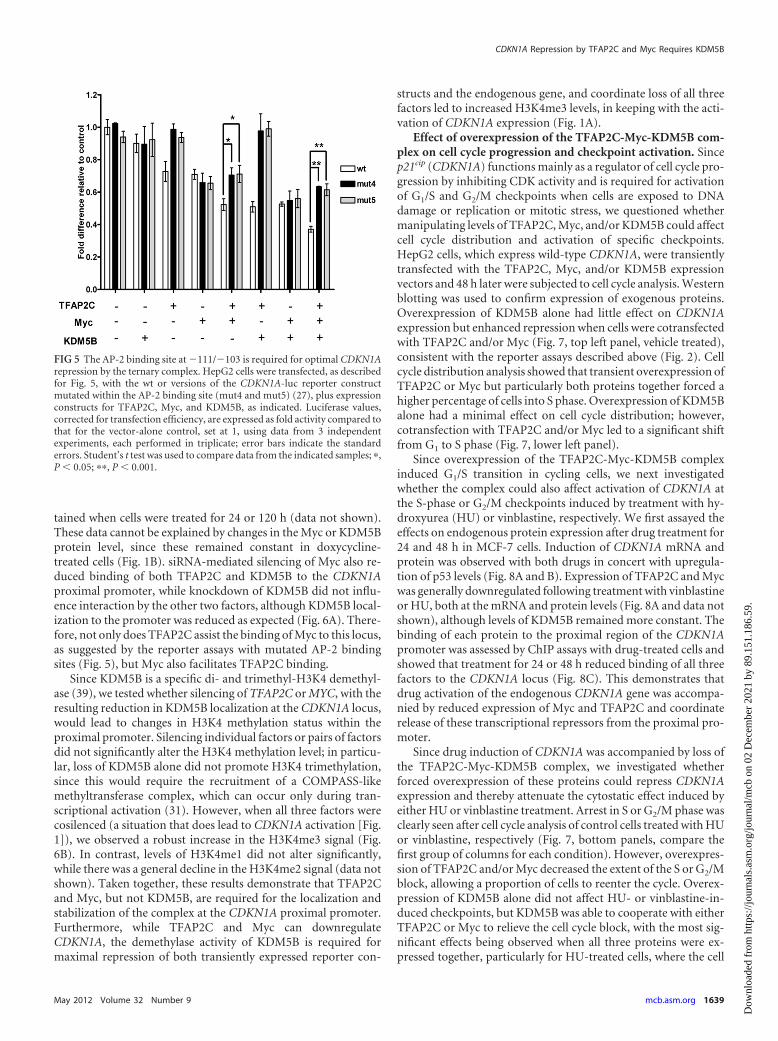
CDKN1A luciferase constructs mutated to abolish AP-2 binding(mut4 and mut5) (27). Mutation of the AP-2 binding site clearlyabrogated repression by TFAP2C alone and with KDM5B (Fig. 5,column groups 3 and 6). Myc-mediated repression was less af-
fected by mutation of the AP-2 binding site (Fig. 5, column groups4 and 7), but intriguingly, the degree of repression was signifi-cantly attenuated on mutant promoters when Myc was coex-pressed with TFAP2C, either alone or with KDM5B (Fig. 5, col-
FIG 3 KDM5B forms a complex with TFAP2C and Myc. (A) Cytoplasmic (C) and nuclear (N) extracts were prepared from MCF-7 cells (see Materials andMethods). The nuclear extracts were subjected to immunoprecipitation (IP) with antibodies against Myc, KDM5B, or TFAP2C (6E4, H77, or AP25H01, asmarked) prior to Western blotting (WB) for the indicated proteins (lanes 1 to 6). C and N proteins, equivalent to 5% of the amount used for IP, were also analyzedby WB to control for protein size and extract integrity. (B) Schematic representation of the domain structure of KDM5B and the series of peptide-tagged deletionmutants. (C) H1299 cells (lacking TFAP2C and KDM5B expression) were cotransfected with TFAP2C and tagged wt or mutant KDM5B proteins or vector alone(VA) as a control. Nuclear extracts were subjected to IP for TFAP2C (6E4) followed by WB for TFAP2C or the peptide Tag to reveal mutant KDM5B proteins,as indicated. (D) GFP-tagged TFAP2C wild-type (WT) or a �N or �C deletion mutant was cotransfected with KDM5B, and nuclear extracts were immunopre-cipitated using the GFP-trap system prior to WB to detect each protein plus endogenously expressed Myc. Additional blotting for actin and lamin A/C was usedto confirm the integrity of the cytoplasmic and nuclear extracts, respectively; the 6E4 antibody was used for all TFAP2C blots.
CDKN1A Repression by TFAP2C and Myc Requires KDM5B
May 2012 Volume 32 Number 9 mcb.asm.org 1637
Dow
nloa
ded
from
http
s://j
ourn
als.
asm
.org
/jour
nal/m
cb o
n 02
Dec
embe
r 20
21 b
y 89
.151
.186
.59.

umn groups 5 and 8). This suggests, therefore, that while Myc doesnot require an intact AP-2 site to interact with and repress theCDKN1A locus, when TFAP2C is present, its binding at �111/�103 allows optimal recruitment of Myc and KDM5B to theproximal promoter and more-efficient repression.
We next used ChIP assays to verify the colocalization ofTFAP2C, Myc, and KDM5B at the endogenous CDKN1A locus. Inagreement with results of the reporter assays described above, theproximal region of the CDKN1A promoter (primers at �21/
�44), but not regions further upstream (�2290/�2185 or �935/�864), was immunoprecipitated specifically using antibodies toTFAP2C, KDM5B, or Myc (Fig. 6A). Functional interrelation-ships between these factors at the CDKN1A locus were tested bycomparing ChIP assays with control cells and those silenced foreach factor in turn. Treatment with doxycycline for 72 h reducedTFAP2C binding, as expected; however, the localization of Mycand KDM5B at the promoter was also reduced to backgroundlevels in TFAP2C-silenced cells (Fig. 6A). Similar results were ob-
FIG 4 Identification of KDM5B domains required for corepressor activity. (A) HepG2 cells were cotransfected with 150 ng of CDKN1A-luc and 50 ng TFAP2Cexpression plasmid or vector-alone (VA) control, as indicated, plus 300 ng of wt or mutant KDM5B, as labeled. (B) As in panel A, with TFAP2C and/or Myc (50ng) in combination with wt or KDM5B JmjC domain mutants (see Fig. 3B), as indicated. Efficient expression of all KDM5B constructs was assessed by blotting(see Fig. S2A in the supplemental material). Luciferase values, corrected for transfection efficiency, are expressed as fold activity compared to that for thevector-alone control, set at 1, using data from 3 independent experiments, each performed in triplicate; error bars indicate the standard errors. Student’s t test wasused to compare data from the indicated samples: n.s., not significant; �, P � 0.05; ��, P � 0.001.
Wong et al.
1638 mcb.asm.org Molecular and Cellular Biology
Dow
nloa
ded
from
http
s://j
ourn
als.
asm
.org
/jour
nal/m
cb o
n 02
Dec
embe
r 20
21 b
y 89
.151
.186
.59.
tained when cells were treated for 24 or 120 h (data not shown).These data cannot be explained by changes in the Myc or KDM5Bprotein level, since these remained constant in doxycycline-treated cells (Fig. 1B). siRNA-mediated silencing of Myc also re-duced binding of both TFAP2C and KDM5B to the CDKN1Aproximal promoter, while knockdown of KDM5B did not influ-ence interaction by the other two factors, although KDM5B local-ization to the promoter was reduced as expected (Fig. 6A). There-fore, not only does TFAP2C assist the binding of Myc to this locus,as suggested by the reporter assays with mutated AP-2 bindingsites (Fig. 5), but Myc also facilitates TFAP2C binding.
Since KDM5B is a specific di- and trimethyl-H3K4 demethyl-ase (39), we tested whether silencing of TFAP2C or MYC, with theresulting reduction in KDM5B localization at the CDKN1A locus,would lead to changes in H3K4 methylation status within theproximal promoter. Silencing individual factors or pairs of factorsdid not significantly alter the H3K4 methylation level; in particu-lar, loss of KDM5B alone did not promote H3K4 trimethylation,since this would require the recruitment of a COMPASS-likemethyltransferase complex, which can occur only during tran-scriptional activation (31). However, when all three factors werecosilenced (a situation that does lead to CDKN1A activation [Fig.1]), we observed a robust increase in the H3K4me3 signal (Fig.6B). In contrast, levels of H3K4me1 did not alter significantly,while there was a general decline in the H3K4me2 signal (data notshown). Taken together, these results demonstrate that TFAP2Cand Myc, but not KDM5B, are required for the localization andstabilization of the complex at the CDKN1A proximal promoter.Furthermore, while TFAP2C and Myc can downregulateCDKN1A, the demethylase activity of KDM5B is required formaximal repression of both transiently expressed reporter con-
structs and the endogenous gene, and coordinate loss of all threefactors led to increased H3K4me3 levels, in keeping with the acti-vation of CDKN1A expression (Fig. 1A).
Effect of overexpression of the TFAP2C-Myc-KDM5B com-plex on cell cycle progression and checkpoint activation. Sincep21cip (CDKN1A) functions mainly as a regulator of cell cycle pro-gression by inhibiting CDK activity and is required for activationof G1/S and G2/M checkpoints when cells are exposed to DNAdamage or replication or mitotic stress, we questioned whethermanipulating levels of TFAP2C, Myc, and/or KDM5B could affectcell cycle distribution and activation of specific checkpoints.HepG2 cells, which express wild-type CDKN1A, were transientlytransfected with the TFAP2C, Myc, and/or KDM5B expressionvectors and 48 h later were subjected to cell cycle analysis. Westernblotting was used to confirm expression of exogenous proteins.Overexpression of KDM5B alone had little effect on CDKN1Aexpression but enhanced repression when cells were cotransfectedwith TFAP2C and/or Myc (Fig. 7, top left panel, vehicle treated),consistent with the reporter assays described above (Fig. 2). Cellcycle distribution analysis showed that transient overexpression ofTFAP2C or Myc but particularly both proteins together forced ahigher percentage of cells into S phase. Overexpression of KDM5Balone had a minimal effect on cell cycle distribution; however,cotransfection with TFAP2C and/or Myc led to a significant shiftfrom G1 to S phase (Fig. 7, lower left panel).
Since overexpression of the TFAP2C-Myc-KDM5B complexinduced G1/S transition in cycling cells, we next investigatedwhether the complex could also affect activation of CDKN1A atthe S-phase or G2/M checkpoints induced by treatment with hy-droxyurea (HU) or vinblastine, respectively. We first assayed theeffects on endogenous protein expression after drug treatment for24 and 48 h in MCF-7 cells. Induction of CDKN1A mRNA andprotein was observed with both drugs in concert with upregula-tion of p53 levels (Fig. 8A and B). Expression of TFAP2C and Mycwas generally downregulated following treatment with vinblastineor HU, both at the mRNA and protein levels (Fig. 8A and data notshown), although levels of KDM5B remained more constant. Thebinding of each protein to the proximal region of the CDKN1Apromoter was assessed by ChIP assays with drug-treated cells andshowed that treatment for 24 or 48 h reduced binding of all threefactors to the CDKN1A locus (Fig. 8C). This demonstrates thatdrug activation of the endogenous CDKN1A gene was accompa-nied by reduced expression of Myc and TFAP2C and coordinaterelease of these transcriptional repressors from the proximal pro-moter.
Since drug induction of CDKN1A was accompanied by loss ofthe TFAP2C-Myc-KDM5B complex, we investigated whetherforced overexpression of these proteins could repress CDKN1Aexpression and thereby attenuate the cytostatic effect induced byeither HU or vinblastine treatment. Arrest in S or G2/M phase wasclearly seen after cell cycle analysis of control cells treated with HUor vinblastine, respectively (Fig. 7, bottom panels, compare thefirst group of columns for each condition). However, overexpres-sion of TFAP2C and/or Myc decreased the extent of the S or G2/Mblock, allowing a proportion of cells to reenter the cycle. Overex-pression of KDM5B alone did not affect HU- or vinblastine-in-duced checkpoints, but KDM5B was able to cooperate with eitherTFAP2C or Myc to relieve the cell cycle block, with the most sig-nificant effects being observed when all three proteins were ex-pressed together, particularly for HU-treated cells, where the cell
FIG 5 The AP-2 binding site at �111/�103 is required for optimal CDKN1Arepression by the ternary complex. HepG2 cells were transfected, as describedfor Fig. 5, with the wt or versions of the CDKN1A-luc reporter constructmutated within the AP-2 binding site (mut4 and mut5) (27), plus expressionconstructs for TFAP2C, Myc, and KDM5B, as indicated. Luciferase values,corrected for transfection efficiency, are expressed as fold activity compared tothat for the vector-alone control, set at 1, using data from 3 independentexperiments, each performed in triplicate; error bars indicate the standarderrors. Student’s t test was used to compare data from the indicated samples; �,P � 0.05; ��, P � 0.001.
CDKN1A Repression by TFAP2C and Myc Requires KDM5B
May 2012 Volume 32 Number 9 mcb.asm.org 1639
Dow
nloa
ded
from
http
s://j
ourn
als.
asm
.org
/jour
nal/m
cb o
n 02
Dec
embe
r 20
21 b
y 89
.151
.186
.59.

cycle profiles resembled those of untreated cells (Fig. 7, comparethe last series of columns for the HU-treated condition with thefirst series of columns for the control condition). For each treat-ment, repression of p21cip expression was maximal when all threeproteins were coexpressed (Fig. 7, upper panels, last lane).
In summary, since its overexpression is sufficient to induceentry into S phase of the cell cycle and activate proliferation ofnormal cycling cells, TFAP2C resembles Myc in these assays andacts as an oncogene. KDM5B has little effect alone but can amplifyresponses to the other two factors. Activation of S or G2/M check-points is attenuated when TFAP2C or Myc is overexpressed, withand without KDM5B overexpression, suggesting that elevated lev-els of these proteins in cancer cells may collaborate to confer re-
sistance to genotoxic or mitotic stress via direct downregulation ofthe universal cell cycle inhibitor CDKN1A.
DISCUSSION
As with TFAP2C (13, 16) and Myc (37), KDM5B overexpressionhas been associated with a poorer outcome in breast cancer pa-tients (22), and all three proteins have been shown to promote cellproliferation by facilitating G1/S transition (36, 39), a checkpointparticularly sensitive to cellular levels of p21cip. Repression ofCDKN1A, mediated by Myc or TFAP2C, has been documentedpreviously (see the introduction), and the related KDM5A de-methylase was shown to regulate H3K4 methylation status at thislocus in gastric tumor cells, although its site and mode of binding
FIG 6 Colocalization and functional interrelation between endogenous TFAP2C, Myc, and KDM5B at the CDKN1A locus. (A) shTFAP2C-MCF-7 cells weretransfected with nonsilencing control siRNA (nsRNAi) or siRNA against Myc (siMyc) or KDM5B (siKDM5B) or treated with doxycycline (Dox) to induceshTFAP2C and harvested 72 h later for use in ChIP with antibodies against IgG (control), TFAP2C, (KDM5B, or Myc. Precipitates were analyzed by qPCR usingprimer pairs specific to 3 distinct regions (�2290/�2185, �935/�864, and �21/�44) across the CDKN1A 5= sequence, as indicated by the double-headedarrows. (B) ChIP was performed as described for panel A using antibodies specific for histone 3 and H3K4-me3. qPCRs were performed using the �21/�44CDKN1A proximal promoter-specific primers. Results are shown as fold enrichment above the IgG (background) control (A) or as an H3K4me3/H3 ratio (B).Data were averaged from three independent experiments, � standard errors.
Wong et al.
1640 mcb.asm.org Molecular and Cellular Biology
Dow
nloa
ded
from
http
s://j
ourn
als.
asm
.org
/jour
nal/m
cb o
n 02
Dec
embe
r 20
21 b
y 89
.151
.186
.59.

to the promoter were not defined (40). Our data suggest that inbreast tumor cells, where it is expressed predominantly (19),KDM5B is the key H3K4me3 demethylase which acts in a ternarycomplex with Myc and TFAP2C to regulate CDKN1A expression,thus uniting many separate observations and providing a mecha-nistic rationale for how deregulated expression of these factorsmay permit breast cancer cells to sustain their proliferation andsurvival.
Since Myc repression of CDKN1A does not involve het-erodimerization with Max or direct DNA binding (12), a numberof studies have looked at the requirement of Myc to access thepromoter through interaction with other DNA binding proteins,particularly Sp1 and Miz-1. The precise mechanism remains un-clear, possibly because more than one pathway is active, with celltype-specific factors undoubtedly playing a role (reviewed in ref-erence 17). Here we have explored the interdependence of Mycand TFAP2C. Investigating the binding of these proteins at theendogenous CDKN1A gene in silenced MCF-7 cells by ChIP (Fig.6) revealed that each factor appeared to stabilize and/or facilitatethe binding of the other to the proximal promoter. This was partlyrecreated using reporter assays where maximal repression by allthree factors could be achieved only on promoter constructs withan intact AP-2 binding site (Fig. 5). Taking the ChIP and reporterassay data together, therefore, this suggests that TFAP2C and Mycinteract with the CDKN1A locus at separate sites, with only theAP-2 site at �111/�103 for TFAP2C defined, and together theyrecruit the histone demethylase KDM5B to form a repressive ter-
nary complex required for optimal downregulation of CDKN1A(Fig. 9, diagram). Interestingly, a similar mutual interdependencehas recently been suggested to allow the stable binding of bothTFAP2C and FoxA1 to specific chromatin sites, leading to therecruitment of ER� and estrogen-responsive gene expression inbreast cell lines (33).
The majority of studies examining the activity of the KDM5family of histone demethylases have relied on the analysis of globalH3K4 methylation status, which suggested that these enzymes canact on a broad range (tri, di, mono) of methylation states at thissite. However, a small number of specifically regulated genes havebeen identified which, as shown here for CDKN1A, all exhibitedKDM5B binding close to their TSS. Moreover, modulation ofKDM5B expression led to consistent alterations in H3K4me3 lev-els at these loci, but dimethylation (me2) and monomethylation(me1) levels remained relatively constant (39). This again mirrorsour ChIP results at the CDKN1A locus, where cosilencing of theternary complex proteins led to induction of gene transcription(Fig. 1) and a concomitant increase in H3K4me3 but little altera-tion in me2 or me1 levels (Fig. 6B and data not shown). Thissuggests that the observations made on global H3K4 methylationstatus may not always reflect the activities of KDM5 demethylasesbound at the promoters of their directly regulated genes.
The C-terminal region of KDM5B contains two PHD fingers,one of which has been shown to bind H3K4me3 and contribute tobinding to active genes (reviewed in reference 18). This C-termi-nal domain was also found here to be required for interaction with
FIG 7 TFAP2C/KDM5B/MYC overexpression attenuates drug-activated checkpoints and forces cells to enter S phase. HepG2 cells were transfected withTFAP2C, KDM5B, or Myc expression vectors in combination, as shown. After 24 h, transfected cells were treated with normal medium (vehicle treated, LHpanels), hydroxyurea (middle panels), or vinblastine (RH panels) for a further 16 h. Cells were then either lysed and assayed by Western blotting for the proteinsindicated (upper panels) or ethanol fixed and stained with propidium iodide for cell cycle analysis (lower panels). Graphs show percentages of cells from eachcondition in sub-G1, G1, S, or G2/M phase. Representative experiments from 3 repeats (each done in triplicate) are shown; �, P � 0.05; ��, P � 0.01 (Student’st test; significant differences between control [1st group of columns] and 3-way-transfected cells [last group of columns] for cycling [vehicle treated] ordrug-treated cells).
CDKN1A Repression by TFAP2C and Myc Requires KDM5B
May 2012 Volume 32 Number 9 mcb.asm.org 1641
Dow
nloa
ded
from
http
s://j
ourn
als.
asm
.org
/jour
nal/m
cb o
n 02
Dec
embe
r 20
21 b
y 89
.151
.186
.59.

TFAP2C (Fig. 3). Studies primarily of the Drosophila KDM5 ho-mologue Lid have shown that the JmjC and Zf motifs were inde-pendently required for Myc interaction (20, 28), and thus thesetwo factors interact with distinct domains of KDM5B. In addition,analysis of TFAP2C deletion mutants plus data from coIP with a
range of TFAP2C antibodies (Fig. 3) defines amino acids 181 to430 as the region of TFAP2C able to bind KDM5B and Myc. Thisincludes the basic-helix-span-helix domain, suggesting thatTFAP2C dimerization may be required for factor interaction, al-though actual binding may be mediated by a 40-aa region imme-
FIG 8 HU and vinblastine induce CDKN1A and reduce TFAP2C/KDM5B/Myc occupancy at the proximal promoter. MCF-7 cells were treated with hy-droxyurea (HU) or vinblastine (Vin) or vehicle control (CTRL) for 24 and 48 h. (A) Whole-cell extracts from treated or control cells, blotted for the indicatedproteins. (B) q-RT-PCR analysis of CDKN1A expression in control and treated cells. Data represent the means and standard errors for 3 independent experi-ments. Significant differences (�, P � 0.05; ��, P,0.01) between induced and uninduced cells are given (Student’s t test). (C) ChIP assay for TFAP2C, KDM5B,and Myc factors was performed as for Fig. 6 using primers to the �21/�44 region of CDKN1A. Values are averages for three independent ChIP experiments; errorbars represent standard errors between the repeats.
Wong et al.
1642 mcb.asm.org Molecular and Cellular Biology
Dow
nloa
ded
from
http
s://j
ourn
als.
asm
.org
/jour
nal/m
cb o
n 02
Dec
embe
r 20
21 b
y 89
.151
.186
.59.

diately N-terminal of the basic domain. The sequences requiredfor Myc/KDM5B interaction therefore overlap those previouslyshown to mediate TFAP2C binding to CITED2/p300 (6) duringgene activation, suggesting that competition by distinct com-plexes for an overlapping interaction domain may determinewhether TFAP2C will activate or repress transcription.
Reporter assays confirmed the requirement for the C-terminalKDM5B PHD fingers for corepressor activity with TFAP2C. Anintact JmjC domain was also essential, with even a single aminoacid substitution (H499A), known to prevent demethylase activ-ity, neutralizing corepressor function (Fig. 4B). Superficially thissuggests that demethylase activity is required for corepression,which would agree with our ChIP data on histone methylationstatus at the CDKN1A locus (Fig. 6). However, previously re-ported domain structure analyses for KDM5B, again using theglobal H3K4 methylation level as the readout, concluded that dis-ruption of any of the domains within the N-terminal half of theprotein will ablate demethylase activity. This is thought to be dueto spatial constraints on the folding of the JmjN and JmjC do-mains to form a functional enzyme (38, 39). Since other domainswere not found to be required for corepressor activity withTFAP2C, it suggests either that there are differences between de-methylase activity monitored on total histone versus observationsat gene-specific sites or the intact JmjC domain alone is requiredfor optimal localization of KDM5B at the CDKN1A promoter andeven the H499A mutation is sufficient to disrupt this.
In Drosophila, the interaction between dMyc and the JmjC do-main of Lid was shown to negatively regulate demethylase activityand thereby activate gene expression (20, 28). The increased levelof H3K4me3 we observe at the CDKN1A locus upon silencing ofTFAP2C/Myc/KDM5B suggests, however, that here TFAP2C andMyc recruit active demethylase that contributes to repression byremoval of the H3K4me3 epigenetic mark. This agrees with manyother studies confirming a role in dynamic gene silencing for theKDM5 family; notably, the recruitment of KDM5A by the coretranscription factor RBP-J to silence target genes during Notchsignaling has several parallels with our findings here (21). Never-theless, repression by the ternary complex is likely to involve ad-ditional mechanisms, since we have previously reported that lossof histone deacetylases (HDACs) and increased histone acetyla-
tion also occur during induction of CDKN1A upon TFAP2C si-lencing in MCF-7 cells (36). However, KDM5B has also beenshown to interact with HDACs through its PHD fingers (3), andthe homologue KDM5A has been purified in HDAC corepressorcomplexes (35).
We have extended our examination of the interaction ofTFAP2C/Myc/KDM5B with each other and the CDKN1A locus byusing functional experiments to demonstrate that overexpressionof these factors can perturb normal cell cycle control, both incycling cells and in those incubated with drugs to activate p53-dependent checkpoints. In each case, the most efficient reversal ofdrug-induced CDKN1A activation occurred when all three ter-nary complex proteins were coexpressed, and this was sufficient toforce arrested cells back into cycle (Fig. 7). Moreover, bothTFAP2C and MYC are estrogen-responsive genes, and loss of thisregulation in breast cancer has been associated with the lack ofCDKN1A induction in response to antioestrogen therapy, thuscontributing to resistance (13, 23, 36). Significantly, the ubiqui-tously expressed demethylase homologue KDM5A was found tobe upregulated in drug-tolerant subpopulations of cells from sev-eral tumor origins treated with a range of anticancer agents (30).Thus, this family of histone demethylases may be particularly im-plicated in the epigenetic heterogeneity within cancer cell popu-lations that can contribute to the development of treatment resis-tance and tumor progression. This suggests, therefore, that theassociation of TFAP2C, KDM5B, and Myc allows these proteins tocooperate when overexpressed in breast tumors, leads to a poorpatient prognosis, and provides a rationale for the further inves-tigation of histone demethylase activity as a therapeutic target inthis disease.
ACKNOWLEDGMENTS
We thank Joyce Taylor-Papadimitriou and Charlie Degui Chen for kindlyproviding KDM5B antibodies and constructs and Richard Grose for hiscomments on the manuscript.
This work was supported by Cancer Research UK program grant no.C6775/A6250 (to H.C.H.) and Breast Cancer Campaign grant no.2009MayPR60 (to A.G.S.). K.V.C. was supported by a Cancer ResearchUK studentship. P.-P.W. was funded by MRC93277.
REFERENCES1. Abbas T, Dutta A. 2009. p21 in cancer: intricate networks and multiple
activities. Nat. Rev. Cancer 9:400 – 414.2. Bamforth SD, et al. 2001. Cardiac malformations, adrenal agenesis, neu-
ral crest defects and exencephaly in mice lacking cited2, a new tfap2 co-activator. Nat. Genet. 29(4):469 – 474.
3. Barrett A, et al. 2007. Breast cancer associated transcriptional repressorPLU-1/JARID1B interacts directly with histone deacetylases. Int. J. Cancer121:265–275.
4. Berlato C, et al. 2011. Alternative TFAP2A isoforms have distinct activi-ties in breast cancer. Breast Cancer Res. 13:R23.
5. Bosher JM, Totty NF, Hsuan JJ, Williams T, Hurst HC. 1996. A familyof AP-2 proteins regulates c-erbB-2 expression in mammary carcinoma.Oncogene 13:1701–1707.
6. Braganca J, et al. 2003. Physical and functional interactions among AP-2transcription factors, p300/CREB-binding protein, and CITED2. J. Biol.Chem. 278:16021–16029.
7. Braganca J, et al. 2002. Human CREB-binding protein/p300-interactingtransactivator with ED-rich tail (CITED) 4, a new member of the CITEDfamily, functions as a co-activator for transcription factor AP-2. J. Biol.Chem. 277:8559 – 8565.
8. Cowling VH, Cole MD. 2006. Mechanism of transcriptional activation bythe Myc oncoproteins. Semin. Cancer Biol. 16:242–252.
9. Eckert D, Buhl S, Weber S, Jager R, Schorle H. 2005. The AP-2 family oftranscription factors. Genome Biol. 6:246.
FIG 9 Schematic diagram of CDKN1A repression by TFAP2C/Myc/KDM5B.The TSS (�1) and proximal promoter region of CDKN1A are represented withthe AP-2 site at �111/�103, bound by a TFAP2C dimer (A). Myc (M) isshown binding adjacent regions through interaction with another undefinedDNA binding factor (shown as gray ovals). Myc and TFAP2C stabilize eachother’s binding to the promoter (double-headed arrow) and together recruitKDM5B, which has a negative effect on chromatin structure via demethylationof H3K4me3, leading to suppression of RNA polymerase II (Pol)-mediatedtranscription of the gene.
CDKN1A Repression by TFAP2C and Myc Requires KDM5B
May 2012 Volume 32 Number 9 mcb.asm.org 1643
Dow
nloa
ded
from
http
s://j
ourn
als.
asm
.org
/jour
nal/m
cb o
n 02
Dec
embe
r 20
21 b
y 89
.151
.186
.59.

10. Eloranta JJ, Hurst HC. 2002. Transcription factor ap-2 interacts with thesumo-conjugating enzyme ubc9 and is sumolated in vivo. J. Biol. Chem.277(34):30798 –30804.
11. Gartel AL, Radhakrishnan SK. 2005. Lost in transcription: p21 repres-sion, mechanisms, and consequences. Cancer Res. 65:3980 –3985.
12. Gartel AL, et al. 2001. Myc represses the p21(WAF1/CIP1) promoter andinteracts with Sp1/Sp3. Proc. Natl. Acad. Sci. U. S. A. 98:4510 – 4515.
13. Gee JM, et al. 2009. Overexpression of TFAP2C in invasive breast cancercorrelates with a poorer response to anti-hormone therapy and reducedpatient survival. J. Pathol. 217:32– 41.
14. Gee JM, Robertson JF, Ellis IO, Nicholson RI, Hurst HC. 1999. Immu-nohistochemical analysis reveals a tumour suppressor-like role for thetranscription factor AP-2 in invasive breast cancer. J. Pathol. 189:514 –520.
15. Goldfarb DS, Gariepy J, Schoolnik G, Kornberg RD. 1986. Syntheticpeptides as nuclear localization signals. Nature 322:641– 644.
16. Guler G, et al. 2007. Wwox and Ap2� expression levels predict tamoxifenresponse. Clin. Cancer Res. 13:6115– 6121.
17. Herkert B, Eilers M. 2010. Transcriptional repression: the dark side ofMyc. Genes Cancer 1:580 –586.
18. Islam AB, Richter WF, Lopez-Bigas N, Benevolenskaya EV. 2011. Se-lective targeting of histone methylation. Cell Cycle 10:413– 424.
19. Krishnakumar R, Kraus WL. 2010. PARP-1 regulates chromatin struc-ture and transcription through a KDM5B-dependent pathway. Mol. Cell39:736 –749.
20. Li L, Greer C, Eisenman RN, Secombe J. 2010. Essential functions of thehistone demethylase lid. PLoS Genet. 6:e1001221.
21. Liefke R, et al. 2010. Histone demethylase KDM5A is an integral part ofthe core Notch-RBP-J. repressor complex. Genes Dev. 24:590 – 601.
22. Lu PJ, et al. 1999. A novel gene (PLU-1) containing highly conservedputative DNA/chromatin binding motifs is specifically up-regulated inbreast cancer. J. Biol. Chem. 274:15633–15645.
23. Mukherjee S, Conrad SE. 2005. c-Myc suppresses p21WAF1/CIP1 ex-pression during estrogen signaling and antiestrogen resistance in humanbreast cancer cells. J. Biol. Chem. 280:17617–17625.
24. Pellikainen JM, Kosma VM. 2007. Activator protein-2 in carcinogenesiswith a special reference to breast cancer—a mini review. Int. J. Cancer120:2061–2067.
25. Satoda M, et al. 2000. Mutations in TFAP2B cause Char syndrome, afamilial form of patent ductus arteriosus. Nat. Genet. 25:42– 46.
26. Scibetta AG, et al. 2007. Functional analysis of the transcription repressorPLU-1/JARID1B. Mol. Cell. Biol. 27:7220 –7235.
27. Scibetta AG, Wong PP, Chan KV, Canosa M, Hurst HC. 2010. Dualassociation by TFAP2A during activation of the p21cip/CDKN1A pro-moter. Cell Cycle 9:4525– 4532.
28. Secombe J, Li L, Carlos L, Eisenman RN. 2007. The Trithorax groupprotein Lid is a trimethyl histone H3K4 demethylase required for dMyc-induced cell growth. Genes Dev. 21:537–551.
29. Seoane J, Le HV, Massague J. 2002. Myc suppression of the p21(Cip1)Cdk inhibitor influences the outcome of the p53 response to DNA dam-age. Nature 419:729 –734.
30. Sharma SV, et al. 2010. A chromatin-mediated reversible drug-tolerantstate in cancer cell subpopulations. Cell 141:69 – 80.
31. Shilatifard A. 2008. Molecular implementation and physiological rolesfor histone H3 lysine 4 (H3K4) methylation. Curr. Opin. Cell Biol. 20:341–348.
32. Stoetzel C, et al. 2009. Confirmation of TFAP2A gene involvement inbranchio-oculo-facial syndrome (BOFS) and report of temporal boneanomalies. Am. J. Med. Genet. A 149A:2141–2146.
33. Tan SK, et al. 2011. AP-2gamma regulates oestrogen receptor-mediatedlong-range chromatin interaction and gene transcription. EMBO J. 30:2569 –2581.
34. Tu S, et al. 2008. The ARID domain of the H3K4 demethylase RBP2 bindsto a DNA CCGCCC motif. Nat. Struct. Mol. Biol. 15:419 – 421.
35. Vermeulen M, et al. 2010. Quantitative interaction proteomics and ge-nome-wide profiling of epigenetic histone marks and their readers. Cell142:967–980.
36. Williams CM, et al. 2009. AP-2gamma promotes proliferation in breasttumour cells by direct repression of the CDKN1A gene. EMBO J. 28:3591–3601.
37. Wolfer A, et al. 2010. MYC regulation of a “poor-prognosis” metastaticcancer cell state. Proc. Natl. Acad. Sci. U. S. A. 107:3698 –3703.
38. Xiang Y, et al. 2007. JARID1B is a histone H3 lysine 4 demethylaseup-regulated in prostate cancer. Proc. Natl. Acad. Sci. U. S. A. 104:19226 –19231.
39. Yamane K, et al. 2007. PLU-1 is an H3K4 demethylase involved in tran-scriptional repression and breast cancer cell proliferation. Mol. Cell 25:801– 812.
40. Zeng J, et al. 2010. The histone demethylase RBP2 is overexpressed ingastric cancer and its inhibition triggers senescence of cancer cells. Gas-troenterology 138:981–992.
41. Zeng YX, Somasundaram K, el-Deiry WS. 1997. AP2 inhibits cancer cellgrowth and activates p21WAF1/CIP1 expression. Nat. Genet. 15:78 – 82.
Wong et al.
1644 mcb.asm.org Molecular and Cellular Biology
Dow
nloa
ded
from
http
s://j
ourn
als.
asm
.org
/jour
nal/m
cb o
n 02
Dec
embe
r 20
21 b
y 89
.151
.186
.59.