the host gene for intronic u17 small nucleolar rnas in mammals
TRANSCRIPT
MOLECULAR AND CELLULAR BIOLOGY,0270-7306/98/$04.0010
Aug. 1998, p. 4509–4518 Vol. 18, No. 8
Copyright © 1998, American Society for Microbiology. All Rights Reserved.
The Host Gene for Intronic U17 Small Nucleolar RNAs in MammalsHas No Protein-Coding Potential and Is a Member of the
59-Terminal Oligopyrimidine Gene FamilyPAWEL PELCZAR AND WITOLD FILIPOWICZ*
Friedrich Miescher-Institut, CH-4002 Basel, Switzerland
Received 18 February 1998/Returned for modification 30 March 1998/Accepted 11 May 1998
Intron-encoded U17a and U17b RNAs are members of the H/ACA-box class of small nucleolar RNAs(snoRNAs) participating in rRNA processing and modification. We have investigated the organization andexpression of the U17 locus in human cells and found that intronic U17a and U17b sequences are transcribedas part of the three-exon transcription unit, named U17HG, positioned approximately 9 kb upstream of theRCC1 locus. Comparison of the human and mouse U17HG genes has revealed that snoRNA-encoding intronsequences but not exon sequences are conserved between the two species and that neither human nor mousespliced U17HG poly(A)1 RNAs have the potential to code for proteins. Analyses of polysome profiles and effectsof translation inhibitors on the abundance of U17HG RNA in HeLa cells indicated that despite its cytoplasmiclocalization, little if any U17HG RNA is associated with polysomes. This distinguishes U17HG RNA fromanother non-protein-coding snoRNA host gene product, UHG RNA, described previously (K. T. Tycowski,M. D. Shu, and J. A. Steitz, Nature 379:464–466, 1996). Determination of the 5* terminus of the U17HG RNArevealed that transcription of the U17HG gene starts with a C residue followed by a polypyrimidine tract,making this gene a member of the 5*-terminal oligopyrimidine (5*TOP) family, which includes genes encodingribosomal proteins and some translation factors. Interestingly, other known snoRNA host genes, including theUHG gene (Tycowski et al., op. cit.), have features of the 5*TOP genes. Similar characteristics of the tran-scription start site regions in snoRNA host and ribosomal protein genes raise the possibility that expressionof components of ribosome biogenesis and translational machineries is coregulated.
Nucleoli of eukaryotic cells contain a large number of dis-tinct small nucleolar RNAs (snoRNAs) which are involved invarious aspects of rRNA processing and modification (re-viewed in references 45, 61, 63, and 66). These RNAs can besubdivided into two major classes. Members of one class con-tain short conserved sequence elements referred to as boxes Cand D/D9 and are associated with the phylogenetically con-served protein fibrillarin (45, 63). Most of them function asguide RNAs specifying sites of 29-O-methylation in rRNA (13,38, 69). Another class of snoRNAs contains conserved se-quence elements known as boxes H and ACA (5, 25). Membersof this group function as guide RNAs in site-specific pseudo-uridylation of rRNA (24, 52). For the yeast Saccharomycescerevisiae, it has been shown that the H/ACA snoRNAs areassociated with the nucleolar protein GAR1 (5, 7, 25).
Biogenesis of snoRNAs can follow many distinct pathways.Some of the snoRNAs, e.g., U3, U8, U13, and 7-2/MRP RNAs,are transcribed from independent transcription units and con-tain either a 59-terminal trimethylguanosine cap (polymerase[pol] II transcripts such as U3, U8, or U13) or a 59-triphos-phate (the pol III-transcribed 7-2/MRP RNA) (reviewed inreference 45). Most of the snoRNAs studied to date in verte-brates and some snoRNAs in yeast are, however, encodedwithin introns of the mosaic pol II-transcribed genes (43, 45).Generally, these snoRNAs are processed exonucleolytically fromexcised introns (12, 15, 36), although maturation of some in-tronic snoRNAs, such as U16 and U18, involves endonucleases(10, 11). Being posttranscriptionally processed from largertranscripts, all intron-encoded snoRNAs are not capped but
contain a monophosphate group at the 59 end (37, 68). Thebiogenesis of most snoRNAs in plants and some snoRNAs inyeast follows yet another pathway. In these organisms, snoR-NAs are clustered and transcribed as polycistronic pre-snoRNA transcripts that are subsequently cleaved into singlesnoRNAs in a process involving both endo- and exonucleases(19, 42, 45, 72).
Characterization of genes that act as hosts for intronicsnoRNAs has produced interesting findings. Most snoRNAhost genes analyzed to date encode proteins essential for ri-bosome biogenesis or function such as ribosomal and nucleolarproteins or translational factors (reviewed in reference 45).This observation has led to the speculation that cotranscriptionof snoRNAs with mRNAs for nucleolar proteins or transla-tional components may provide a regulatory mechanism tocoordinate the accumulation of different molecules requiredfor the assembly and function of ribosomes (45, 62). Morerecently, Tycowski et al. (67) discovered that the UHG gene,which harbors intronic snoRNAs U22 and U25 to U31, isdistinct from all other snoRNA hosts characterized to date.The spliced poly(A)1 RNAs produced from UHG genes inhumans, mice, and frogs are not conserved in sequence andhave no apparent protein-coding potential. The finding that inUHG genes snoRNA-encoding introns and not exons are evo-lutionarily conserved and express functional RNAs requires amodification of the current description of exons as the maininformation-carrying regions of a gene (67, 69).
We are interested in the function and biogenesis of theH/ACA class snoRNA U17 (also referred to as E1 [16, 37, 57]).This evolutionarily conserved RNA has been implicated in theearly processing event in the 59 external transcribed spacerupstream of the 18S rRNA region and also has been shown topsoralen cross-link to 18S rRNA and the spacer in vivo (19, 47,
* Corresponding author. Mailing address: Friedrich Miescher-Insti-tut, P.O. Box 2543, CH-4002 Basel, Switzerland. Phone: (61) 6976993or 6978234. Fax: (61) 6973976. E-mail: [email protected].
4509
on April 1, 2018 by guest
http://mcb.asm
.org/D
ownloaded from
56). The U17 RNA has all the features of guide RNAs whichspecify sites of pseudouridylation, but its potential target se-quence in rRNA is not readily apparent (16, 24, 54a). It ispossible that U17 RNA catalyzes modification of some other,as yet unidentified RNA or that its function in rRNA-process-ing reactions does not involve pseudouridylation. The obser-vation that in human cells the U17 RNA is more abundantthan other RNAs of the H/ACA-box family (reference 35a andthis work) is consistent with these possibilities. U17 RNA waspreviously characterized in many vertebrate species (14, 16, 37,48, 57, 60). In Xenopus laevis (16) and fugu fish (14), six U17sequence variants reside in introns of the ribosomal protein S7(formerly referred to as S8) gene. In humans, two U17 RNAs,U17a and U17b (23, 37, 48), were postulated to originate fromthe 59-proximal introns of the multiexon 59 untranslated region(59UTR) of the gene encoding the guanine nucleotide ex-change factor RCC1, which participates in control of nucleo-cytoplasmic transport (reviewed in reference 27).
In this study we demonstrate that introns containing U17aand U17b sequences in humans do not reside in the RCC1gene but are part of an independent transcription unit posi-tioned approximately 9 kb upstream of the RCC1 locus. Com-parisons of the human U17 host gene, named U17HG (U17host gene), with its mouse counterpart (mU17HG) have re-vealed that these genes have very similar exon/intron organi-zations but are unlikely to encode proteins. Hence, U17HG isanother example, in addition to UHG (67), of a snoRNA hostgene whose only apparent function is to act as a vehicle for theexpression of intron-located snoRNAs. Characterizations ofhuman and mouse U17HG genes have also revealed that theirtranscription starts with a C residue followed by an oligopy-rimidine tract. This feature makes these genes members of the59-terminal oligopyrimidine (59TOP) family, which includesgenes encoding ribosomal proteins and some other essentialhousekeeping proteins (reviewed in references 1 and 46). In-terestingly, other known snoRNA host genes, including thenon-protein-coding UHG gene characterized previously by Ty-
cowski et al. (67), also have features of the 59TOP family.59TOP sequences in mRNAs have been shown to play a role incoordinating their translation in response to cell growth con-ditions (1, 46). Identification of non-protein-coding snoRNAhost genes as new members of the 59TOP family is consistentwith the 11 oligopyrimidine tracts having an additional, mostlikely transcriptional function, in regulating gene expression.
MATERIALS AND METHODS
Unless stated otherwise, all procedures for manipulating DNA and RNA werecarried out according to Sambrook et al. (59) and Ausubel et al. (3).
Cloning of the human U17HG cDNAs. A 282-bp fragment corresponding tothe first four noncoding exons of the U17/RCC1 transcription unit (Fig. 1) wasobtained by PCR using cDNA prepared with a HeLa cell total RNA as atemplate and GATTCGCAGTGGTCGCTTCTTCTC and CTCTCCAAGTTTACCTCTGCCTCC oligonucleotides as upstream and downstream primers, re-spectively. This PCR fragment was used as a probe to screen a human Namalwa(Burkitt lymphoma) cell cDNA library in plasmid pRS314/UNVP16 (65) (kindgift of P. Matthias of this institute). The probe was labeled with [a-32P]dATP(3,000 Ci/mmol; Amersham) by the random priming method (21). Hybridizationwas carried out at 55°C in 63 SSC (13 SSC is 0.15 M NaCl plus 15 mM sodiumcitrate) containing 0.5% nonfat dry milk. The washes were at 55°C twice for 10min in 23 SSC–0.1% sodium dodecyl sulfate (SDS) and twice for 10 min in 0.23SSC–0.1% SDS. The isolated positive clones were analyzed by restriction map-ping and sequenced on both strands by the dideoxy method using appropriateprimers.
Cloning of the mU17HG cDNA and genomic sequences. Two oligonucleotides,GGGGAGACAAACCATGCAGG and CCAACGTTGTGGAAAGGGAC, spe-cific for conserved regions of the human and Xenopus U17 snoRNAs and cor-responding to positions 180 to 199 and 90 to 109 in the human U17 RNA,respectively (37), were used as primers for PCR performed with mouse genomicDNA. The amplified 413-bp fragment was cloned into the HincII site of pBlue-script KS1 and sequenced. A candidate 36-nucleotide (nt) exon sequence sep-arating the two U17 RNA-containing introns was identified by using theHEXON exon-finding program from the Baylor College of Medicine GeneFinder package (http://dot.imgen.bcm.tmc.edu:9331/gene-finder/gf.html). Threepartially overlapping oligonucleotides, TCCATCAACAAAGTACCTGAAATC,TCAACAAAGTACCTGAAATCATTG, and AAGTACCTGAAATCATTGACCGGG, complementary to the exon sequence, were used as gene-specificprimers for nested PCR in the 59RACE (rapid amplification of cDNA ends)protocol, and two partially overlapping oligonucleotides, CCCGGTCAATGATTTCAGGTACTTTG and GTCAATGATTTCAGGTACTTTGTTG, were usedas primers in the 39RACE reactions. Both the 39RACE and the 59RACE exper-
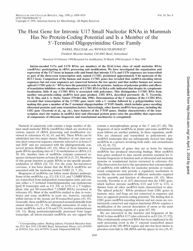
FIG. 1. Expression of the human U17/RCC1 locus. (a) Schematic structure of the upstream portion of the human locus. Numbering of exons, shown as open andgray boxes, is according to Furuno et al. (23). Extensions of exon 3 identified in class II and class III cDNAs are denoted by A and B. Only terminal RCC1 protein-codingexons are shown. U17a and U17b sequences (black arrows), Alu sequences (open arrows), transcription start sites, and polyadenylation sites (AAAA) are indicated.Major splicing patterns supported by results presented in this work are shown in thick lines above the gene structure; the minor splicing pathway is indicated by thinlines. (b) Structures of clones isolated from the human Namalwa B-cell cDNA library. Numbers in parentheses indicate the number of clones corresponding to eachclass. Clone sizes are indicated. Bars below the cDNA schemes show the fragments used as probes for the cDNA library screening (gray) and for Northern analysis(black and open boxes represent U17HG- and RCC1-specific probes, respectively, used for experiments shown in Fig. 2). p, cDNA library screen might have been biasedin favor of isolating class I clones. These clones hybridize to the probe over its entire length (296 bp, exons 1 through 4), while class II and III clones hybridize onlyto 153 bp (exons 1 to 3) of the probe.
4510 PELCZAR AND FILIPOWICZ MOL. CELL. BIOL.
on April 1, 2018 by guest
http://mcb.asm
.org/D
ownloaded from
iments were performed with mouse NIH 3T3 total RNA, by using a 59/39RACEkit (Boehringer Mannheim) as instructed by the manufacturer. The obtained59RACE and 39RACE PCR fragments were cloned into the HincII site of thepBluescript KS1, giving clones pmU17HG/59RACE and pmU17HG/39RACE,respectively. The genomic sequence of the mU17HG locus was obtained by PCRusing mouse genomic DNA as a template and oligonucleotides TGACATTCAAATGCTTTAATTCAG and TCTCTCTAGGCGTCGCTCTCTTGG, specificfor terminal sequences of the mU17HG cDNA, as primers. The obtained 1.6-kbPCR fragment was cloned into the HincII site of pBluescript KS1, resulting inpmU17HG/gene, and sequenced.
Cell culture. Human HeLa and mouse NIH 3T3 cells were grown in mono-layers at 5% CO2 in Dulbecco modified Eagle medium (Gibco) containing 10%fetal calf serum (Gibco) and 2 mM glutamine. Namalwa cells were grown at 5%CO2 in RPMI 1640 medium (Gibco) containing 10% fetal calf serum. Cyclohex-imide (20 mg/ml), pactamycin (560 ng/ml), or puromycin (50 mg/ml) was added tocells 4 h prior to harvesting where indicated. HeLa suspension culture was grownin spinner flasks in Joklik’s minimal essential medium (Gibco) containing 5%newborn calf serum (Amimed) as described elsewhere (22).
Cell fractionation and RNA isolation. HeLa cells grown in suspension werefractionated as described previously (17). Total RNA from human and mousecultured cells and from cytoplasmic and nuclear fractions was isolated by theguanidinium thiocyanate-phenol-chloroform method (26). Poly(A)1 RNA wasisolated directly from cell lysates by using an Oligotex oligo(dT) affinity matrix(Qiagen) according to the manufacturer’s protocol. Mouse (BALB/c) genomicDNA was a kind gift of R. Neve of this institute.
Analysis of HeLa cell extracts on sucrose gradients. HeLa cell suspensionculture (3 3 107 cells, 5 3 105 cells/ml) was treated for 5 min with cycloheximide(100 mg/ml), and then washed once with 20 ml of phosphate-buffered salinecontaining cycloheximide (100 mg/ml) and twice with 20 ml of buffer A (5 mMTris-HCl [pH 7.4], 1.5 mM KCl, 2.5 mM MgCl2, 100 mg of cycloheximide per ml).The cells were lysed in 400 ml of buffer A containing 3 mM dithiothreitol, 30 Uof RNasin (Promega), 0.5% Triton X-100, and 0.5% sodium deoxycholate. Thelysates were centrifuged for 8 min at 4°C at 3,000 3 g. The supernatant wascollected and loaded onto 17 to 51% linear sucrose density gradients prepared in20 mM Tris-HCl (pH 7.4)–80 mM NaCl–5 mM MgCl2. The lysates were centri-fuged at 4°C for 2 h at 36,000 rpm in an SW41 rotor (Beckman). RNA wasisolated from individual fractions by the proteinase K method.
Northern analysis. Total RNA and poly(A)1 RNA were separated on a 1.4%agarose-formaldehyde gel, blotted onto a Hybond-N nylon membrane (Amer-sham) by using 203 SSPE (13 SSPE is 0.18 M NaCl, 10 mM NaH2PO4, and 1mM Na2EDTA [pH 7.7]), and UV cross-linked to the membrane. A KpnI-HindIII fragment of phU17HG/123a, corresponding to nt 20 to 237 of theU17HG-A cDNA, was used as a U17HG-specific probe. phU17HG/123a wasconstructed by cloning a PCR fragment corresponding to nt 20 to 237 of thehuman U17HG cDNA into the HincII site of pBluescriptII KS1. A KpnI-HindIIIfragment of plasmid pRCC1, containing exons 6 and 7 of the human RCC1cDNA, was used as the RCC1-specific probe. pRCC1 was constructed by cloninga PCR fragment, extending from position 29 (relative to the initiator AUG) inexon 6 to position 1261 in exon 7 of the RCC1 protein-coding sequence, into theHincII site of pBluescriptII KS1. Blots were hybridized overnight at 42°C in 53SSPE containing 50% formamide, 10% dextran sulfate, 1% SDS, and 50 g ofsalmon sperm DNA per ml. They were washed in 23 SSC–0.1% SDS for 30 minat 42°C and subsequently in 0.23 SSC–0.1% SDS for 30 min at 55°C.
RNase A/T1 mapping. RNase A/T1 mapping was performed as described byGoodall et al. (26). The following plasmids were used to generate probes. Thehuman U17HG-specific probe was transcribed with T3 polymerase from plasmidphU17HG/123a linearized with EcoRI. The UHG-specific probe was a T7 RNApolymerase transcript from a plasmid containing the human UHG cDNA insert(gift of K. Tycowski, Yale University). This probe covers positions 932 to 1118 ofthe UHG cDNA (67). The human U17a-specific probe was transcribed from aplasmid containing the ClaI-XhoI fragment of plasmid pA (36), cloned in theClaI and XhoI sites of pBluescriptII KS1. The plasmid, referred to as pU17a(C-X), was cut with BamHI and transcribed with T3 RNA polymerase. The humanb-actin gene-specific probe was transcribed by T3 polymerase, by using plasmidATCC65129 (obtained from the American Type Culture Collection) cut withDraIII. The human U22 snoRNA-specific probe was transcribed by SP6 RNApolymerase from plasmid pU22 (obtained from K. Tycowski) linearized withMunI. This probe is complementary to the entire 125-nt-long U22 snoRNA. The18S rRNA-specific probe, complementary to nt 715 to 793 of the human 18SrRNA, was transcribed from plasmid pT7/RNA/18S (Ambion). The plasmid wascut with HindIII and transcribed with T7 RNA polymerase. The 18S rRNA-specific probe was labeled with [a-32P]UTP with a specific activity of 30 mCi/mmol; all other probes were labeled with [a-32P]UTP with a specific activity of30 Ci/mmol. An amount of probe corresponding to 105 cpm was used in eachmapping experiment. Protected fragments were separated on 6% polyacryl-amide–8 M urea gels. Where indicated, radioactivity of protected fragments wasquantitated with a Storm 860 PhosphorImager (Molecular Dynamics).
Primer extension and RACE analysis. For primer extension analysis, threeindependent partially overlapping primers complementary to exon 1 of the hu-man U17HG RNA (primer A [AGCGACCACTGCGAATCTGTCTCC], primerB [GAAGAAGCGACCACTGCGAATCTG], and primer C [CAAGGAGAAGAAGCGACCACTGCG] [see Fig. 3b]) were end labeled with [g-32P]ATP (3,000
Ci/mmol; Amersham) and T4 polynucleotide kinase and gel purified. Annealingreaction mixtures contained 20 pmol of each primer and 15 mg of RNA in 160mM HEPES-KOH (pH 7.7)–1 M NaCl–1 mM Na2EDTA. Following annealingovernight at 48°C, the mixture was ethanol precipitated and dissolved in 20 ml ofthe first-strand synthesis buffer for Superscript II reverse transcriptase (Gibco-BRL) containing 10 mM dithiothreitol, a 1 mM concentration of each de-oxynucleoside triphosphate, and 400 U of Superscript II reverse transcriptase.Samples were incubated at 50°C for 50 min and processed for the analysis on a20% acrylamide–8 M urea gel.
For the 59RACE analysis, three partially overlapping primers (RACE 1 [TAAATGTCAATGCCAAAATGCGAAG], RACE 2 [ATGCCAAAATGCGAAGTGCAGATCG], and RACE 3 [TGCGAAGTGCAGATCGTCTTCTCTC] [seeFig. 3b]), complementary to exon 3 of the human U17HG RNA, were used asgene-specific nested backward primers in successive 59RACE steps, using HeLatotal RNA and a 59/39RACE kit. The PCR/RACE product was sequenced di-rectly with an automated DNA sequencer or cloned into the HincII site ofpBluescript KS1 in order to sequence individual clones.
Nucleotide sequence accession numbers. The nucleotide sequences ofU17HG-A and U17HG-AB cDNAs have been submitted to the EMBL/Gen-Bank/DDBJ nucleotide sequence libraries under accession no. AJ006834 andAJ006835, respectively. The nucleotide sequences of the mU17HG cDNA andmU17HG gene have been submitted under accession no. AJ006837 andAJ006836, respectively.
RESULTS
Expression of the U17/RCC1 host transcription unit in hu-man cells. Previous studies on expression of the RCC1 gene inhuman cells have indicated that transcription of this gene isinitiated at two different promoters positioned approximately 9kb apart (23, 54). Initiation at the downstream promoter pro-duced a pre-mRNA in which a 59-terminal single noncodingexon (exon 5 in Fig. 1) is spliced to downstream exons encod-ing the RCC1 protein. Initiation at the upstream promoterwould yield a transcript containing four short noncoding exons(exons 1 to 4; in Fig. 1) spliced to the coding part of mRNA;U17a and U17b snoRNA sequences are located in introns 1and 2 of this transcript (23, 37, 48) (Fig. 1). As pre-mRNAscontaining so many noncoding exons are unusual and the splic-ing pattern of the RCC1 transcript initiating at the upstreampromoter was deduced from a single cDNA clone (54), wehave investigated the expression of the U17/RCC1 locus inhuman cells in more detail.
A human Namalwa B-cell cDNA library was screened with aprobe corresponding to the four 59-proximal noncoding exonsof the transcript initiated at the upstream RCC1 promoter.Nine identified positive clones can be grouped into threeclasses (Fig. 1B). Only class I, represented by two clones, isconsistent with the previously proposed expression pattern ofthe U17/RCC1 locus. Class II is represented by four clonescalled U17HG-A. In these clones, exons 1, 2, and 3 are splicedtogether as expected but exon 3 extends for an additional 600nt and terminates with poly(A). Class III, comprising twoclones called U17HG-AB, is similar to class II except that theclones represent RNAs that are polyadenylated another 1.26kb further downstream in the extended exon 3 (Fig. 1). Oneadditional isolated clone contained an insert corresponding toa fragment of unspliced U17HG transcript (data not shown). Asearch of sequence databases, with the 39-end portions ofU17HG-A and U17HG-AB RNAs, identified three indepen-dent expressed sequence tag (EST) entries representing RNApolyadenylated at the U17HG-A site (GenEMBL accession no.H54400, N25905, and AA522843) and three corresponding toRNA polyadenylated at the U17HG-AB site (GenEMBL ac-cession no. H19509, H46276, and AA563638), further confirm-ing the use of both upstream polyadenylation sites. These find-ings show that transcription from the upstream U17/RCC1gene promoter gives rise to three different classes of tran-scripts, all hosting the U17a and U17b RNAs.
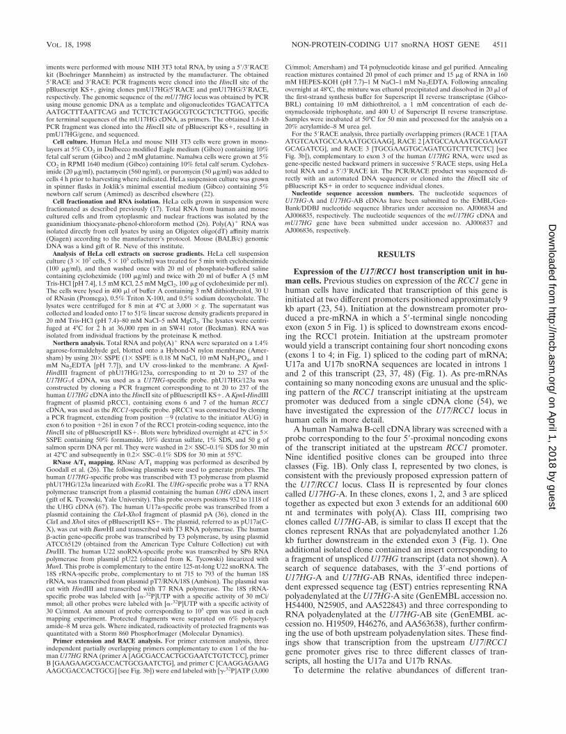
To determine the relative abundances of different tran-
VOL. 18, 1998 NON-PROTEIN-CODING U17 snoRNA HOST GENE 4511
on April 1, 2018 by guest
http://mcb.asm
.org/D
ownloaded from
scripts initiated at the upstream U17/RCC1 promoter, North-ern analysis was performed. Total and poly(A)1 RNAs fromHeLa cells and Namalwa B cells were probed with two differ-ent cDNA probes, one specific for the upstream exons 1, 2, and3 and another specific for the RCC1 protein-coding exons 6and 7 (Fig. 1B). Hybridization with the exon 1 to 3 proberevealed the presence of the 0.9-kb transcript corresponding inlength to the U17HG-A RNA (Fig. 2A). This probe detectedneither the 2.2-kb human U17HG-AB RNA nor the 2.6-kbRCC1 mRNA initiated at the upstream promoter (Fig. 2B),indicating that U17 host transcripts polyadenylated at theU17HG-AB site or spliced into the RCC1 coding region aremuch less abundant than the U17HG-A RNA. AdditionalRNase A/T1 mapping experiments carried out with a probewhich distinguishes U17HG/RCC1 spliced mRNA from thesum of U17HG-A and U17HG-AB RNAs have indicated thatthe former RNA constitutes less than 10% (10% correspondsto the detection limit) of the total spliced RNA initiated at theupstream promoter (data not shown). Hybridization of theNorthern blot shown in Fig. 2A with the probe specific for theRCC1 coding exons 6 and 7 revealed a 2.6-kb band, the sizeexpected for the RCC1 mRNA initiated at the downstreampromoter (Fig. 2B). Comparison of Northern blots shown inFig. 2A and B (72 and 15 h of autoradiography, respectively)indicates that the 2.6-kb U17HG/RCC1 mRNA constitutes atmost 5 to 10% of the total RCC1 mRNA present in HeLa orNamalwa B cells.
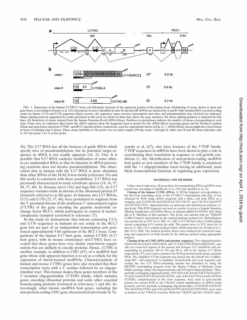
Characterization of the 5* end of transcripts initiated at theupstream promoter. Primer extension and RACE analysiswere performed to characterize the 59 terminus of the humanU17HG RNA. Three different oligonucleotides complemen-tary to the 59-proximal exon of the U17HG RNA were used asprimers for reverse transcription. Each of the oligonucleotidesyielded two major extended cDNA products, consistent withtranscription of the U17HG RNA being initiated at C residuespositioned 14 or 15 nt upstream of the 59 end of the longestcDNA clone characterized in this work (Fig. 3). To furtherverify the sequence of the 59 end of the U17HG RNA, RACE
analysis was performed. Direct sequencing of the RACE prod-uct placed the start site at the upstream C residue (C11 [Fig.3b]) identified by primer extension. Sequencing of individualclones resulting from cloning of the RACE product in pBlue-scriptII KS1 vector has identified two clones terminating atthe C11 and two clones terminating at the C12 (data notshown).
Based on the results presented in this study and the fact thatthe single previously characterized cDNA clone (54) (Fig. 3b)also starts at the second of the two C residues, the start site oftranscription from this promoter is assigned to C residues 11and 12. These C residues are preceded and followed by tractsof 10 and 5 pyrimidines, respectively (Fig. 3b), which makes thehuman U17HG gene similar to the 59TOP genes (see Discus-sion).
Human and murine U17 host poly(A)1 RNAs have no ap-parent coding potential. With the exception of several Alurepeats (Fig. 1A), U17HG-A and U17HG-AB RNAs show no
FIG. 2. Expression of the U17HG (A) and RCC1 (B) genes in human celllines determined by Northern analysis. An RNA blot containing 15 mg of total(lanes 1 and 2) and 2 mg of poly(A)1 (lanes 3 and 4) RNA from Namalwa B cells(lanes 2 and 4) and HeLa cells (lanes 1 and 3) was probed first with the cDNAfragments corresponding to upstream exons 1 to 3 (A) and then with the probespecific for the RCC1 protein-coding exons 6 and 7 (B). The probes, schemati-cally shown in Fig. 1b, were of comparable length and specific activity. Majortranscripts identified by each probe are indicated by arrows. Positions of RNAsize markers (in kilobases) are also shown. Autoradiography was for 72 h (A) and15 h (B).
FIG. 3. Characterization of the transcription start site of the U17HG RNA.(A) Determination of the 59 end of U17HG RNA by primer extension. Lanes 1to 3, primer extension reactions with primers A to C, respectively (for positionsof oligonucleotide primers, see panel b). Lanes 4 to 7, sequencing reactionsperformed with the primer C and pU17HG/PE1 as a template. The relevantportion of the sequence is shown on the right. Reverse transcription productsobtained with primer C, corresponding to cDNAs extended to the C residues atpositions 11 and 12, are indicated by arrows. (b) Sequence of the 250 to 1150region of the U17HG locus. Two adjacent C residues, 11 and 12, identified astranscription start sites are in bold. Regions corresponding to oligonucleotides A,B, and C used as primers in primer extension experiments, as well as oligonu-cleotides RACE 1, RACE 2, and RACE 3 used in successive steps of the RACEexperiment (see Materials and Methods), are underlined. 59 ends of cDNAclones isolated in this work are indicated by arrows; numbers of clones ending ata particular site are shown in parentheses. The 59 end of the cDNA clone pcD40,described by Ohtsubo et al. (54), is also indicated.
4512 PELCZAR AND FILIPOWICZ MOL. CELL. BIOL.
on April 1, 2018 by guest
http://mcb.asm
.org/D
ownloaded from
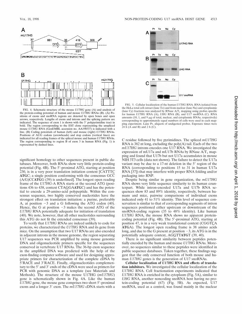
significant homology to other sequences present in public da-tabases. Moreover, both RNAs show very little protein-codingpotential (Fig. 4B). The 59-proximal ATG, starting at position230, is in a very poor translation initiation context [CATTTCATGC; a single position conforming with the consensus GCC(A/G)CCATGG (39) is underlined]. The longest open readingframe of the U17HG-A RNA starts at the second ATG (posi-tions 436 to 438, context CTAGGAATGC) and has the poten-tial to encode a 29-amino-acid polypeptide. Within the con-sensus sequence, two highly conserved nucleotides have thestrongest effect on translation initiation: a purine, preferablyA, at position 23 and a G following the ATG codon (40).Hence, the G at position 23 makes the second ATG of theU17HG RNA potentially adequate for initiation of translation(40). We note, however, that all other nucleotides surroundingthis ATG do not fit the extended consensus (39).
To verify that U17HG RNAs are indeed unlikely to code forproteins, we characterized the U17HG RNA and its gene frommice. On the assumption that two U17 RNAs are also encodedin adjacent introns in the mouse genome, the region separatingU17 sequences was PCR amplified by using mouse genomicDNA and oligonucleotide primers specific for the sequencesconserved in vertebrate U17 RNAs. The 36-bp exon sequencein the amplified DNA was predicted with the help of theexon-finding computer software and used for designing appro-priate primers for characterization of the complete cDNA by59RACE and 39RACE. Finally, oligonucleotides complemen-tary to the 59 and 39 ends of the cDNA were used as primers forPCR with genomic DNA as a template (see Materials andMethods). The structure of the mouse U17HG (mU17HG)gene is schematically shown in Fig. 4A. Like the humanU17HG gene, the mouse gene comprises two short 59-proximalexons and a longer 39 exon. The mU17HG cDNA starts with a
C residue followed by five pyrimidines. The spliced mU17HGRNA is 382 nt long, excluding the poly(A) tail. Each of the twomU17HG introns encodes one U17 RNA. We investigated theexpression of mU17a and mU17b RNAs by RNase A/T1 map-ping and found that U17b but not U17a accumulates in mouseNIH 3T3 cells (data not shown). The failure to detect the U17avariant may be due to a 17-nt deletion in the 59 region of theRNA (corresponding to positions 15 to 31 in human U17aRNA [37]) that may interfere with proper RNA folding and/orpackaging into RNP.
Despite being similar in gene organization, the mU17HGRNA shows very little sequence similarity to its human coun-terpart. While intron-encoded U17a and U17b RNA se-quences show 83 and 89% identity, respectively, between hu-mans and mice, pairwise comparisons of individual exonsindicated only 41 to 51% identity. This level of sequence con-servation is similar to that of corresponding segments of intronsequences positioned either upstream or downstream of thesnoRNA-coding regions (35 to 48% identity). Like humanU17HG RNA, the mouse RNA shows no apparent protein-coding potential (Fig. 4B). The 59-proximal ATG, starting atposition 47, is in a very weak translational context (CGGTCAATGA). The longest open reading frame is 38 amino acidslong, and due to the G present at position 23, its ATG is in thepotentially adequate context, AGGTTATGT (39, 40).
There is no significant similarity between peptides poten-tially encoded by the human and mouse U17HG RNAs. More-over, no sequences similar to these peptides were identified inpublic sequence databases. Taken together, these findings sug-gest that the only conserved function of both mouse and hu-man U17HG genes is the generation of U17 snoRNAs.
Cellular localization of U17HG RNA and effects of transla-tion inhibitors. We investigated the cellular localization of theU17HG RNA. Cell fractionation experiments indicated thatU17HG RNA is enriched in the cytoplasm (Fig. 5A), similar toUHG RNA, another noncoding snoRNA host having no pro-tein-coding potential (67) (Fig. 5B). As expected, U17snoRNA, used as a control, was found mainly in the nuclear
FIG. 4. Schematic structure of the mouse U17HG gene (A) and analysis ofthe protein-coding potential of human and mouse U17HG RNAs (B). (A) Po-sitions of exons and snoRNA regions are denoted by open boxes and openarrows, respectively. Lengths of exons and introns and the splicing pattern areindicated. The sequence of exon 1 is shown with the 59 polypyrimidine tract inbold. The region corresponding to the EST clone representing the unsplicedmouse U17HG RNA (GenEMBL accession no. AA198537) is indicated with aline. (B) Coding potentials of human (left) and mouse (right) U17HG RNAs.Positions of AUG codons (arrowheads) and stop codons (vertical lines) areindicated for all reading frames of the spliced mouse and human U17HG RNAs.The region corresponding to region B of exon 3 in human RNA (Fig. 1) isrepresented by dashed lines.
FIG. 5. Cellular localization of the human U17HG RNA. RNA isolated fromthe HeLa total cell extract (lane To) and from nuclear (lane Nu) and cytoplasmic(lane Cy) fractions was analyzed by RNase A/T1 mapping using probes specificfor human U17HG RNA (A), UHG RNA (B), and U17 snoRNA (C). RNAamounts (10, 1, and 9 mg of total, nuclear, and cytoplasmic RNAs, respectively)corresponding to approximately equal numbers of cells were used in each map-ping experiment. Lane Pr, aliquots of undigested probes. Exposure times were24 h (A and B) and 2 h (C).
VOL. 18, 1998 NON-PROTEIN-CODING U17 snoRNA HOST GENE 4513
on April 1, 2018 by guest
http://mcb.asm
.org/D
ownloaded from
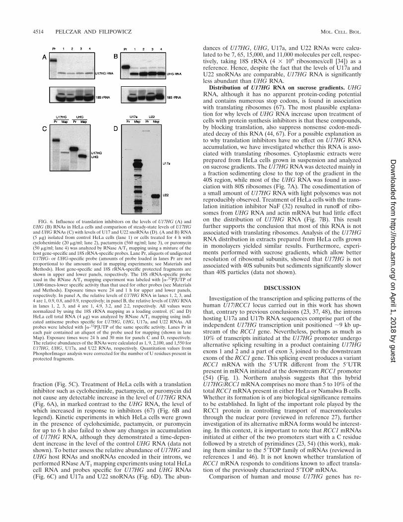
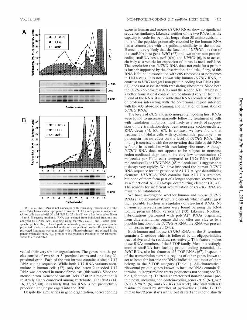
fraction (Fig. 5C). Treatment of HeLa cells with a translationinhibitor such as cycloheximide, pactamycin, or puromycin didnot cause any detectable increase in the level of U17HG RNA(Fig. 6A), in marked contrast to the UHG RNA, the level ofwhich increased in response to inhibitors (67) (Fig. 6B andlegend). Kinetic experiments in which HeLa cells were grownin the presence of cycloheximide, pactamycin, or puromycinfor up to 6 h also failed to show any changes in accumulationof U17HG RNA, although they demonstrated a time-depen-dent increase in the level of the control UHG RNA (data notshown). To better assess the relative abundance of U17HG andUHG host RNAs and snoRNAs encoded in their introns, weperformed RNase A/T1 mapping experiments using total HeLacell RNA and probes specific for U17HG and UHG RNAs(Fig. 6C) and U17a and U22 snoRNAs (Fig. 6D). The abun-
dances of U17HG, UHG, U17a, and U22 RNAs were calcu-lated to be 7, 65, 15,000, and 11,000 molecules per cell, respec-tively, taking 18S rRNA (4 3 106 ribosomes/cell [34]) as areference. Hence, despite the fact that the levels of U17a andU22 snoRNAs are comparable, U17HG RNA is significantlyless abundant than UHG RNA.
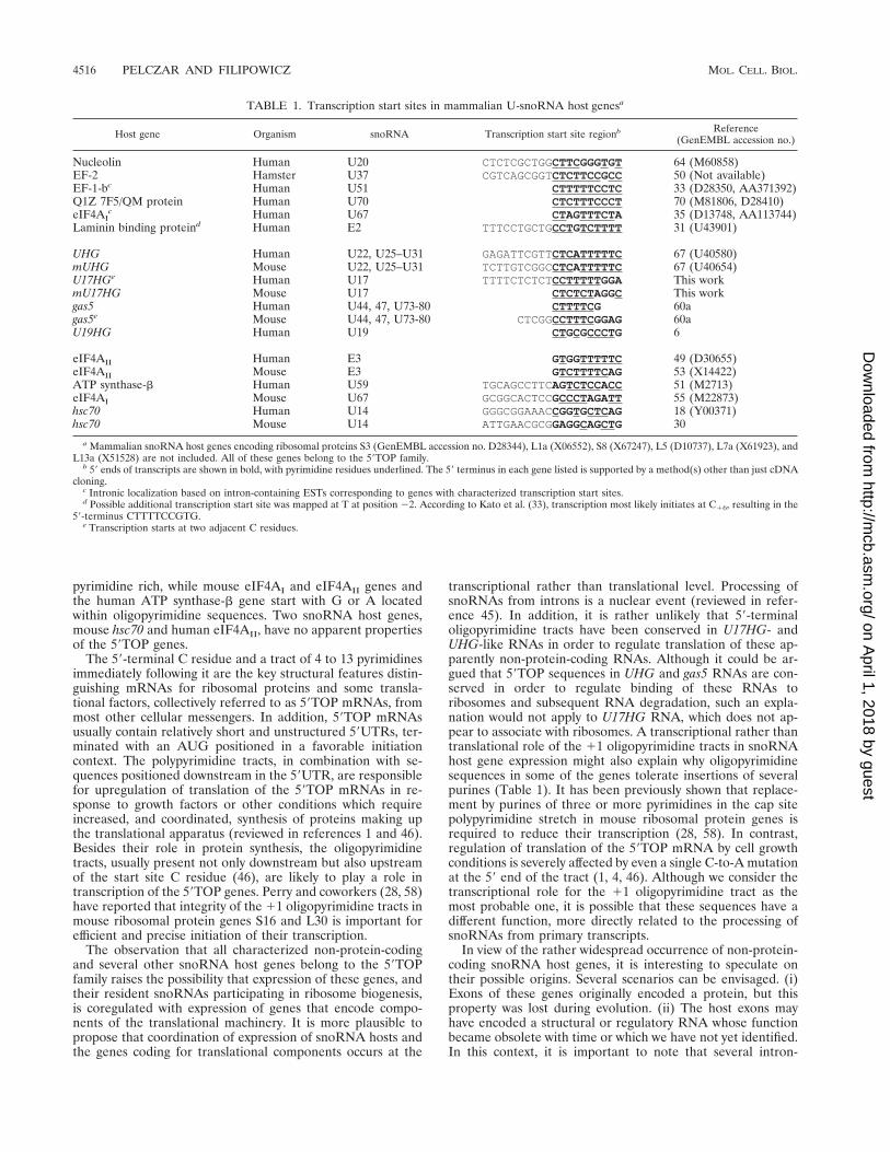
Distribution of U17HG RNA on sucrose gradients. UHGRNA, although it has no apparent protein-coding potentialand contains numerous stop codons, is found in associationwith translating ribosomes (67). The most plausible explana-tion for why levels of UHG RNA increase upon treatment ofcells with protein synthesis inhibitors is that these compounds,by blocking translation, also suppress nonsense codon-medi-ated decay of this RNA (44, 67). For a possible explanation asto why translation inhibitors have no effect on U17HG RNAaccumulation, we have investigated whether this RNA is asso-ciated with translating ribosomes. Cytoplasmic extracts wereprepared from HeLa cells grown in suspension and analyzedon sucrose gradients. The U17HG RNA was detected mainly ina fraction sedimenting close to the top of the gradient in the40S region, while most of the UHG RNA was found in asso-ciation with 80S ribosomes (Fig. 7A). The cosedimentation ofa small amount of U17HG RNA with light polysomes was notreproducibly observed. Treatment of HeLa cells with the trans-lation initiation inhibitor NaF (32) resulted in runoff of ribo-somes from UHG RNA and actin mRNA but had little effecton the distribution of U17HG RNA (Fig. 7B). This resultfurther supports the conclusion that most of this RNA is notassociated with translating ribosomes. Analysis of the U17HGRNA distribution in extracts prepared from HeLa cells grownin monolayers yielded similar results. Furthermore, experi-ments performed with sucrose gradients, which allow betterresolution of ribosomal subunits, showed that U17HG is notassociated with 40S subunits but sediments significantly slowerthan 40S particles (data not shown).
DISCUSSION
Investigation of the transcription and splicing patterns of thehuman U17/RCC1 locus carried out in this work has shownthat, contrary to previous conclusions (23, 37, 48), the intronshosting U17a and U17b RNA sequences comprise part of theindependent U17HG transcription unit positioned ;9 kb up-stream of the RCC1 gene. Nevertheless, perhaps as much as10% of transcripts initiated at the U17HG promoter undergoalternative splicing resulting in a product containing U17HGexons 1 and 2 and a part of exon 3, joined to the downstreamexons of the RCC1 gene. This splicing event produces a variantRCC1 mRNA with the 59UTR different from the 59UTRpresent in mRNA initiated at the downstream RCC1 promoter(54) (Fig. 1). Northern analysis suggests that this hybridU17HG/RCC1 mRNA comprises no more than 5 to 10% of thetotal RCC1 mRNA present in either HeLa or Namalwa B cells.Whether its formation is of any biological significance remainsto be established. In light of the important role played by theRCC1 protein in controlling transport of macromoleculesthrough the nuclear pore (reviewed in reference 27), furtherinvestigation of its alternative mRNA forms would be interest-ing. In this context, it is important to note that RCC1 mRNAsinitiated at either of the two promoters start with a C residuefollowed by a stretch of pyrimidines (23, 54) (this work), mak-ing them similar to the 59TOP family of mRNAs (reviewed inreferences 1 and 46). It is not known whether translation ofRCC1 mRNA responds to conditions known to affect transla-tion of the previously characterized 59TOP mRNAs.
Comparison of human and mouse U17HG genes has re-
FIG. 6. Influence of translation inhibitors on the levels of U17HG (A) andUHG (B) RNAs in HeLa cells and comparison of steady-state levels of U17HGand UHG RNAs (C) with levels of U17 and U22 snoRNAs (D). (A and B) RNA(5 mg) isolated from control HeLa cells (lane 1) or cells treated for 4 h withcycloheximide (20 mg/ml; lane 2), pactamycin (560 ng/ml; lane 3), or puromycin(50 mg/ml; lane 4) was analyzed by RNase A/T1 mapping using a mixture of thehost gene-specific and 18S rRNA-specific probes. Lane Pr, aliquots of undigestedU17HG- or UHG-specific probe (amounts of probe loaded in lanes Pr are notproportional to the amounts used in mapping experiments; see Materials andMethods). Host gene-specific and 18S rRNA-specific protected fragments areshown in upper and lower panels, respectively. The 18S rRNA-specific probeused in the RNase A/T1 mapping experiment was labeled with [a-32P]UTP of1,000-times-lower specific activity than that used for other probes (see Materialsand Methods). Exposure times were 24 and 1 h for upper and lower panels,respectively. In panel A, the relative levels of U17HG RNA in lanes 1, 2, 3, and4 are 1, 0.9, 0.8, and 0.9, respectively; in panel B, the relative levels of UHG RNAin lanes 1, 2, 3, and 4 are 1, 4.9, 3.2, and 2.2, respectively. All values werenormalized by using the 18S rRNA mapping as a loading control. (C and D)HeLa cell total RNA (4 mg) was analyzed by RNase A/T1 mapping using indi-cated antisense probes specific for U17HG, UHG, U17a, and U22 RNAs. Allprobes were labeled with [a-32P]UTP of the same specific activity. Lanes Pr ineach pair contained an aliquot of the probe used for mapping (shown in laneMap). Exposure times were 24 h and 30 min for panels C and D, respectively.The relative abundances of the RNAs were calculated as 1, 9, 2,100, and 1,550 forU17HG, UHG, U17a, and U22 RNAs, respectively. Quantitation values fromPhosphorImager analysis were corrected for the number of U residues present inprotected fragments.
4514 PELCZAR AND FILIPOWICZ MOL. CELL. BIOL.
on April 1, 2018 by guest
http://mcb.asm
.org/D
ownloaded from
vealed their very similar organizations. The genes in both spe-cies consist of two short 59-proximal exons and one long 39-proximal exon. Each of the two introns contains a single U17RNA coding sequence. While both U17 RNA variants accu-mulate in human cells (37), only the intron 2-encoded U17RNA was detected in mouse fibroblasts (this work). Since themouse intron 1-encoded variant lacks 17 nt in a region that isrelatively highly conserved among vertebrate U17 RNAs (14,16, 37, 57, 60), it is likely that this RNA is not productivelyprocessed and/or packaged into the RNP.
Despite the similarities in gene organization, corresponding
exons in human and mouse U17HG RNAs show no significantsequence similarity. Likewise, neither of the two RNAs has thecapacity to code for peptides longer than 38 amino acids, andnone of the peptides potentially encoded by the human RNAhas a counterpart with a significant similarity in the mouse.Hence, it is very likely that the function of U17HG, like that ofthe snoRNA host gene UHG (67) and two other non-protein-coding snoRNA hosts, gas5 (60a) and U19HG (6), is to act ex-clusively as a vehicle for expression of intron-located snoRNAs.The conclusion that U17HG RNA does not code for a proteinis further supported by the observation that little, if any, of thisRNA is found in association with 80S ribosomes or polysomesin HeLa cells. It is not known why human U17HG RNA, incontrast to UHG and gas5 non-protein-coding host RNAs (60a,67), does not associate with translating ribosomes. Since boththe U17HG 59-proximal ATG and the second ATG, which is ina better translational context, are positioned very far from the59 end of the RNA, it is possible that RNA secondary structureor proteins interacting with the 59-terminal region interferewith the 40S ribosome scanning and initiation of translation ofU17HG RNA.
The levels of UHG and gas5 non-protein-coding host RNAswere found to increase markedly following treatment of cellswith translation inhibitors, most likely as a result of suppres-sion of the translation-dependent nonsense codon-mediatedRNA decay (44, 60a, 67). In contrast, we have found thattreatment of HeLa cells with cycloheximide, pactamycin, orpuromycin has no effect on the level of U17HG RNA. Thisfinding is consistent with the observation that little of this RNAis found in association with translating ribosomes. AlthoughU17HG RNA does not appear to be subject to nonsensecodon-mediated degradation, its very low concentration (7molecules per HeLa cell) compared to U17a RNA (15,000molecules/cell) or UHG RNA (65 molecules/cell) suggests thatit decays very rapidly. We have inspected the human U17HGRNA sequence for the presence of AUUUA-type destabilizingelements. U17HG-A RNA contains four AUUUA stretches,but none of them form part of a longer sequence known to actas a functional AUUUA-type destabilizing element (20, 41).The reasons for inefficient accumulation of U17HG RNA re-main to be established.
We have investigated whether human and mouse U17HGRNAs share secondary structure elements which might suggesttheir possible function as regulatory or structural RNAs. Noobvious conserved structures were found by using the RNAfolding program Mfold version 2.3 (73). Likewise, Northernhybridizations performed with poly(A)1 RNAs originatingfrom different human organs did not offer any clue as to apossible function of the U17HG-A RNA; the RNA was presentin all tissues investigated (54a).
Both human and mouse U17HG RNAs at the 59 terminuscontain a C residue which is followed by an oligopyrimidinetract of five and six residues, respectively. This feature makesthese RNAs members of the 59TOP family. Most interestingly,another snoRNA host lacking protein-coding potential, theUHG RNA, also has features of 59TOP RNAs (67). Inspectionof the transcription start site regions of other genes known toact as hosts for intronic snoRNAs indicated that most of thembelong to the 59TOP category (Table 1). All characterizedribosomal protein genes known to host snoRNAs contain 59-terminal oligopyrimidine tracts (sequences not shown; see Ta-ble 1, footnote a). Thirteen characterized non-ribosomal pro-tein hosts, including non-protein-coding genes UHG (67), gas5(60a), U19HG (6), and U17HG (this work), also start with a Cresidue followed by stretches of pyrimidines (Table 1). Thehuman hsc70 gene starts with C but its start site is not distinctly
FIG. 7. U17HG RNA is not associated with translating ribosomes in HeLacells. Cytoplasmic extracts prepared from control HeLa cells grown in suspension(A) or cells treated with 30 mM NaF for 25 min (B) were fractionated on linear17 to 51% sucrose gradients. RNA was isolated from individual fractions andanalyzed by RNase A/T1 mapping using U17HG-, UHG-, and b-actin gene-specific probes. Only relevant parts of autoradiograms, containing gene-specificprotected bands, are shown below the sucrose gradient profiles. Radioactivity inprotected fragments was quantified with a PhosphorImager and plotted in thepanels which also show A260 profiles of the gradients. Positions of 40S ribosomalsubunits are indicated.
VOL. 18, 1998 NON-PROTEIN-CODING U17 snoRNA HOST GENE 4515
on April 1, 2018 by guest
http://mcb.asm
.org/D
ownloaded from
pyrimidine rich, while mouse eIF4AI and eIF4AII genes andthe human ATP synthase-b gene start with G or A locatedwithin oligopyrimidine sequences. Two snoRNA host genes,mouse hsc70 and human eIF4AII, have no apparent propertiesof the 59TOP genes.
The 59-terminal C residue and a tract of 4 to 13 pyrimidinesimmediately following it are the key structural features distin-guishing mRNAs for ribosomal proteins and some transla-tional factors, collectively referred to as 59TOP mRNAs, frommost other cellular messengers. In addition, 59TOP mRNAsusually contain relatively short and unstructured 59UTRs, ter-minated with an AUG positioned in a favorable initiationcontext. The polypyrimidine tracts, in combination with se-quences positioned downstream in the 59UTR, are responsiblefor upregulation of translation of the 59TOP mRNAs in re-sponse to growth factors or other conditions which requireincreased, and coordinated, synthesis of proteins making upthe translational apparatus (reviewed in references 1 and 46).Besides their role in protein synthesis, the oligopyrimidinetracts, usually present not only downstream but also upstreamof the start site C residue (46), are likely to play a role intranscription of the 59TOP genes. Perry and coworkers (28, 58)have reported that integrity of the 11 oligopyrimidine tracts inmouse ribosomal protein genes S16 and L30 is important forefficient and precise initiation of their transcription.
The observation that all characterized non-protein-codingand several other snoRNA host genes belong to the 59TOPfamily raises the possibility that expression of these genes, andtheir resident snoRNAs participating in ribosome biogenesis,is coregulated with expression of genes that encode compo-nents of the translational machinery. It is more plausible topropose that coordination of expression of snoRNA hosts andthe genes coding for translational components occurs at the
transcriptional rather than translational level. Processing ofsnoRNAs from introns is a nuclear event (reviewed in refer-ence 45). In addition, it is rather unlikely that 59-terminaloligopyrimidine tracts have been conserved in U17HG- andUHG-like RNAs in order to regulate translation of these ap-parently non-protein-coding RNAs. Although it could be ar-gued that 59TOP sequences in UHG and gas5 RNAs are con-served in order to regulate binding of these RNAs toribosomes and subsequent RNA degradation, such an expla-nation would not apply to U17HG RNA, which does not ap-pear to associate with ribosomes. A transcriptional rather thantranslational role of the 11 oligopyrimidine tracts in snoRNAhost gene expression might also explain why oligopyrimidinesequences in some of the genes tolerate insertions of severalpurines (Table 1). It has been previously shown that replace-ment by purines of three or more pyrimidines in the cap sitepolypyrimidine stretch in mouse ribosomal protein genes isrequired to reduce their transcription (28, 58). In contrast,regulation of translation of the 59TOP mRNA by cell growthconditions is severely affected by even a single C-to-A mutationat the 59 end of the tract (1, 4, 46). Although we consider thetranscriptional role for the 11 oligopyrimidine tract as themost probable one, it is possible that these sequences have adifferent function, more directly related to the processing ofsnoRNAs from primary transcripts.
In view of the rather widespread occurrence of non-protein-coding snoRNA host genes, it is interesting to speculate ontheir possible origins. Several scenarios can be envisaged. (i)Exons of these genes originally encoded a protein, but thisproperty was lost during evolution. (ii) The host exons mayhave encoded a structural or regulatory RNA whose functionbecame obsolete with time or which we have not yet identified.In this context, it is important to note that several intron-
TABLE 1. Transcription start sites in mammalian U-snoRNA host genesa
Host gene Organism snoRNA Transcription start site regionb Reference(GenEMBL accession no.)
Nucleolin Human U20 CTCTCGCTGGCTTCGGGTGT 64 (M60858)EF-2 Hamster U37 CGTCAGCGGTCTCTTCCGCC 50 (Not available)EF-1-bc Human U51 CTTTTTCCTC 33 (D28350, AA371392)Q1Z 7F5/QM protein Human U70 CTCTTTCCCT 70 (M81806, D28410)eIF4AI
c Human U67 CTAGTTTCTA 35 (D13748, AA113744)Laminin binding proteind Human E2 TTTCCTGCTGCCTGTCTTTT 31 (U43901)
UHG Human U22, U25–U31 GAGATTCGTTCTCATTTTTC 67 (U40580)mUHG Mouse U22, U25–U31 TCTTGTCGGCCTCATTTTTC 67 (U40654)U17HGe Human U17 TTTTCTCTCTCCTTTTTGGA This workmU17HG Mouse U17 CTCTCTAGGC This workgas5 Human U44, 47, U73-80 CTTTTCG 60agas5e Mouse U44, 47, U73-80 CTCGGCCTTTCGGAG 60aU19HG Human U19 CTGCGCCCTG 6
eIF4AII Human E3 GTGGTTTTTC 49 (D30655)eIF4AII Mouse E3 GTCTTTTCAG 53 (X14422)ATP synthase-b Human U59 TGCAGCCTTCAGTCTCCACC 51 (M2713)eIF4AI Mouse U67 GCGGCACTCCGCCCTAGATT 55 (M22873)hsc70 Human U14 GGGCGGAAACCGGTGCTCAG 18 (Y00371)hsc70 Mouse U14 ATTGAACGCGGAGGCAGCTG 30
a Mammalian snoRNA host genes encoding ribosomal proteins S3 (GenEMBL accession no. D28344), L1a (X06552), S8 (X67247), L5 (D10737), L7a (X61923), andL13a (X51528) are not included. All of these genes belong to the 59TOP family.
b 59 ends of transcripts are shown in bold, with pyrimidine residues underlined. The 59 terminus in each gene listed is supported by a method(s) other than just cDNAcloning.
c Intronic localization based on intron-containing ESTs corresponding to genes with characterized transcription start sites.d Possible additional transcription start site was mapped at T at position 22. According to Kato et al. (33), transcription most likely initiates at C16, resulting in the
59-terminus CTTTTCCGTG.e Transcription starts at two adjacent C residues.
4516 PELCZAR AND FILIPOWICZ MOL. CELL. BIOL.
on April 1, 2018 by guest
http://mcb.asm
.org/D
ownloaded from
containing genes, the spliced products of which code for aknown or putative regulatory RNA rather than an mRNA,have been characterized (2, 8, 9, 29, 71). There is no evidencethat introns in these genes encode any functional RNAs. (iii)The UHG- and U17HG-like genes originated from polycis-tronic snoRNA genes, similar to genes expressed in plants andyeast (42, 54b, 72), by conversion of the inter-snoRNA spacersinto spliceable exons. Loss of an endonuclease activity requiredfor processing poly-snoRNAs could be a factor triggering suchan evolutionary event. The last scenario would imply that tran-scriptional units encoding poly-snoRNAs are evolutionarily oldprototype genes. Such genes could have originally arisen byduplication of a single-unit DNA (or RNA) segment at a timewhen the complexity of rRNA modification was increasing.Characterization of snoRNA transcription units in additionaldistantly related organisms might shed more light on the pos-sible origins of the non-protein-coding snoRNA host genes.
ACKNOWLEDGMENTS
We thank P. Matthias for the cDNA library, K. Tycowski for thehuman UHG cDNA clone, C. Smith and J. A. Steitz for sharingunpublished results, F. Dragon, B. Hohn, Z. Lorkovic, and G. Thomasfor critical reading of the manuscript, and H. Angliker, F. Fisher, andP. Muller for oligonucleotide synthesis and DNA sequencing.
REFERENCES
1. Amaldi, F., and P. Pierandrei-Amaldi. 1997. TOP genes: a translationallycontrolled class of genes including those coding for ribosomal proteins. Prog.Mol. Subcell. Biol 18:1–17.
2. Amrein, H., and R. Axel. 1997. Genes expressed in neurons of adult maleDrosophila. Cell 88:459–469.
3. Ausubel, F. M., R. Brent, R. E. Kingston, D. D. Moore, J. G. Seidman, J. A.Smith, and K. Struhl (ed.). 1994. Current protocols in molecular biology.John Wiley & Sons, New York, N.Y.
4. Avni, D., S. Shama, F. Loreni, and O. Meyuhas. 1994. Vertebrate mRNAswith a 59-terminal pyrimidine tract are candidates for translational repres-sion in quiescent cells: characterization of the translational cis-regulatoryelement. Mol. Cell. Biol. 14:3822–3833.
5. Balakin, A. G., L. Smith, and M. J. Fournier. 1996. The RNA world of thenucleolus: two major families of small RNAs defined by different box ele-ments with related functions. Cell 86:823–834.
6. Bortolin, M., and T. Kiss. 1998. Human U19 intron-encoded snRNA isprocessed from a long primary transcript that possesses little potential forprotein coding. RNA 4:445–454.
7. Bousquet-Antonelli, C., Y. Henry, J. P. G’elugne, M. Caizergues-Ferrer, andT. Kiss. 1997. A small nucleolar RNP protein is required for pseudouridy-lation of eukaryotic ribosomal RNAs. EMBO J. 16:4770–4776.
8. Brannan, C. I., E. C. Dees, R. S. Ingram, and S. M. Tilghman. 1990. Theproduct of the H19 gene may function as an RNA. Mol. Cell. Biol. 10:28–36.
9. Brockdorff, N., A. Ashworth, G. F. Kay, V. M. McCabe, D. P. Norris, P. J.Cooper, S. Swift, and S. Rastan. 1992. The product of the mouse Xist geneis a 15 kb inactive X- specific transcript containing no conserved ORF andlocated in the nucleus. Cell 71:515–526.
10. Caffarelli, E., M. Arese, B. Santoro, P. Fragapane, and I. Bozzoni. 1994. Invitro study of processing of the intron-encoded U16 small nucleolar RNA inXenopus laevis. Mol. Cell. Biol. 14:2966–2974.
11. Caffarelli, E., A. Fatica, S. Prislei, E. De Gregorio, P. Fragapane, and I.Bozzoni. 1996. Processing of the intron-encoded U16 and U18 snoRNAs: theconserved C and D boxes control both the processing reaction and thestability of the mature snoRNA. EMBO J. 15:1121–1131.
12. Cavaille, J., and J. P. Bachellerie. 1996. Processing of fibrillarin-associatedsnoRNAs from pre-mRNA introns: an exonucleolytic process exclusivelydirected by the common stem-box terminal structure. Biochimie 78:443–456.
13. Cavaille, J., M. Nicoloso, and J. P. Bachellerie. 1996. Targeted ribose meth-ylation of RNA in vivo directed by tailored antisense RNA guides. Nature383:732–735.
14. Cecconi, F., C. Crosio, P. Mariottini, G. Cesareni, M. Giorgi, S. Brenner,and F. Amaldi. 1996. A functional role for some Fugu introns larger than thetypical short ones: the example of the gene coding for ribosomal protein S7and snoRNA U17. Nucleic Acids Res. 24:3167–3172.
15. Cecconi, F., P. Mariottini, and F. Amaldi. 1995. The Xenopus intron-en-coded U17 snoRNA is produced by exonucleolytic processing of its precur-sor in oocytes. Nucleic Acids Res. 23:4670–4676.
16. Cecconi, F., P. Mariottini, F. Loreni, P. Pierandrei-Amaldi, N. Campioni,and F. Amaldi. 1994. U17XS8, a small nucleolar RNA with a 12 nt comple-mentarity to 18S rRNA and coded by a sequence repeated in the six introns
of Xenopus laevis ribosomal protein S8 gene. Nucleic Acids Res. 22:732–741.17. Dignam, J. D., R. M. Lebovitz, and R. G. Roeder. 1983. Accurate transcrip-
tion initiation by RNA polymerase II in a soluble extract from isolatedmammalian nuclei. Nucleic Acids Res. 11:1475–1489.
18. Dworniczak, B., and M. E. Mirault. 1987. Structure and expression of ahuman gene coding for a 71 kd heat shock ‘cognate’ protein. Nucleic AcidsRes. 15:5181–5197.
19. Enright, C. A., E. S. Maxwell, G. L. Eliceiri, and B. Sollner-Webb. 1996.59ETS rRNA processing facilitated by four small RNAs: U14, E3, U17, andU3. RNA 2:1094–1099.
20. Fan, X. C., V. E. Myer, and J. A. Steitz. 1997. AU-rich elements target smallnuclear RNAs as well as mRNAs for rapid degradation. Genes Dev. 11:2557–2568.
21. Feinberg, A. P., and B. Vogelstein. 1984. A technique for radiolabeling DNArestriction endonuclease fragments to high specific activity. Anal. Biochem.137:266–267.
22. Filipowicz, W., and O. Vicente. 1990. RNA 39-terminal phosphate cyclasefrom HeLa cells. Methods Enzymol. 181:499–510.
23. Furuno, N., K. Nakagawa, U. Eguchi, M. Ohtsubo, T. Nishimoto, E. Soeda,and M. Ohtubo. 1991. Complete nucleotide sequence of the human RCC1gene involved in coupling between DNA replication and mitosis. Genomics11:459–461.
24. Ganot, P., M. L. Bortolin, and T. Kiss. 1997. Site-specific pseudouridineformation in preribosomal RNA is guided by small nucleolar RNAs. Cell89:799–809.
25. Ganot, P., M. Caizergues-Ferrer, and T. Kiss. 1997. The family of box ACAsmall nucleolar RNAs is defined by an evolutionarily conserved secondarystructure and ubiquitous sequence elements essential for RNA accumula-tion. Genes Dev. 11:941–956.
26. Goodall, G. J., K. Wiebauer, and W. Filipowicz. 1990. Analysis of pre-mRNAprocessing in transfected plant protoplasts. Methods Enzymol. 181:148–161.
27. Gorlich, D., and I. W. Mattaj. 1996. Nucleocytoplasmic transport. Science271:1513–1518.
28. Hariharan, N., and R. P. Perry. 1990. Functional dissection of a mouseribosomal protein promoter: significance of the polypyrimidine initiator andan element in the TATA-box region. Proc. Natl. Acad. Sci. USA 87:1526–1530.
29. Hogan, N. C., K. L. Traverse, D. E. Sullivan, and M. L. Pardue. 1994. Thenucleus-limited Hsr-omega-n transcript is a polyadenylated RNA with aregulated intranuclear turnover. J. Cell Biol. 125:21–30.
30. Hunt, C. R. 1996. GenBank accession no. U73744 (direct submission).31. Jackers, P., F. Minoletti, D. Belotti, N. Clausse, G. Sozzi, M. E. Sobel, and
V. Castronovo. 1996. Isolation from a multigene family of the active humangene of the metastasis-associated multifunctional protein 37LRP/p40 atchromosome 3p21.3. Oncogene 13:495–503.
32. Jefferies, H. B., and G. Thomas. 1994. Elongation factor-1 alpha mRNA isselectively translated following mitogenic stimulation. J. Biol. Chem. 269:4367–4372.
33. Kato, S., S. Sekine, S. W. Oh, N. S. Kim, Y. Umezawa, N. Abe, M. Yokoyama-Kobayashi, and T. Aoki. 1994. Construction of a human full-length cDNAbank. Gene 150:243–250.
34. Kiledian, M., C. G. Burd, M. Gorlach, D. S. Portman, and G. Dreyfuss. 1994.Structure and function of hnRNP proteins, p. 125–149. In K. Nagai, and I. W.Mattaj (ed.), RNA-protein interactions. Oxford University Press, Oxford,England.
35. Kim, N. S., T. Kato, N. Abe, and S. Kato. 1993. Nucleotide sequence ofhuman cDNA encoding eukaryotic initiation factor 4AI. Nucleic Acids Res.21:2012.
35a.Kiss, T. Personal communication.36. Kiss, T., and W. Filipowicz. 1995. Exonucleolytic processing of small nucle-
olar RNAs from pre-mRNA introns. Genes Dev. 9:1411–1424.37. Kiss, T., and W. Filipowicz. 1993. Small nucleolar RNAs encoded by introns
of the human cell cycle regulatory gene RCC1. EMBO J. 12:2913–2920.38. Kiss-Laszlo, Z., Y. Henry, J. P. Bachellerie, M. Caizergues-Ferrer, and T.
Kiss. 1996. Site-specific ribose methylation of preribosomal RNA: a novelfunction for small nucleolar RNAs. Cell 85:1077–1088.
39. Kozak, M. 1987. An analysis of 59-noncoding sequences from 699 vertebratemessenger RNAs. Nucleic Acids Res. 15:8125–8148.
40. Kozak, M. 1996. Interpreting cDNA sequences: some insights from studieson translation. Mamm. Genome 7:563–574.
41. Lagnado, C. A., C. Y. Brown, and G. J. Goodall. 1994. AUUUA is notsufficient to promote poly(A) shortening and degradation of an mRNA: thefunctional sequence within AU-rich elements may be UUAUUUA(U/A)(U/A). Mol. Cell. Biol. 14:7984–7995.
42. Leader, D. J., G. P. Clark, J. Watters, A. F. Beven, P. J. Shaw, and J. W. S.Brown. 1997. Clusters of multiple different small nucleolar RNA genes inplants are expressed as and processed from polycistronic pre-snoRNAs.EMBO J. 16:5742–5751.
43. Leverette, R. D., M. T. Andrews, and E. S. Maxwell. 1992. Mouse U14snRNA is a processed intron of the cognate hsc70 heat shock pre-messengerRNA. Cell 71:1215–1221.
44. Maquat, L. E. 1995. When cells stop making sense: effects of nonsense
VOL. 18, 1998 NON-PROTEIN-CODING U17 snoRNA HOST GENE 4517
on April 1, 2018 by guest
http://mcb.asm
.org/D
ownloaded from
codons on RNA metabolism in vertebrate cells. RNA 1:453–465.45. Maxwell, E. S., and M. J. Fournier. 1995. The small nucleolar RNAs. Annu.
Rev. Biochem. 64:897–934.46. Meyuhas, O., D. Avni, and S. Shama. 1996. Translational control of ribo-
somal protein mRNA in eukaryotes, translational control. Cold Spring Har-bor Laboratory Press, Cold Spring Harbor, N.Y.
47. Mishra, R. K., and G. L. Eliceiri. 1997. Three small nucleolar RNAs that areinvolved in ribosomal RNA precursor processing. Proc. Natl. Acad. Sci. USA94:4972–4977.
48. Nag, M. K., T. T. Thai, E. A. Ruff, N. Selvamurugan, M. Kunnimalaiyaan,and G. L. Eliceiri. 1993. Genes for E1, E2, and E3 small nucleolar RNAs.Proc. Natl. Acad. Sci. USA 90:9001–9005.
49. Nakamura, A., R. Amikura, M. Mukai, S. Kobayashi, and P. F. Lasko. 1996.Requirement for a noncoding RNA in Drosophila polar granules for germcell establishment. Science 274:2075–2079.
50. Nakanishi, T., K. Kohno, M. Ishiura, H. Ohashi, and T. Uchida. 1988.Complete nucleotide sequence and characterization of the 59- flanking re-gion of mammalian elongation factor 2 gene. J. Biol. Chem. 263:6384–6391.
51. Neckelmann, N., C. K. Warner, A. Chung, J. Kudoh, S. Minoshima, R.Fukuyama, M. Maekawa, Y. Shimizu, N. Shimizu, J. D. Liu, et al. 1989. Thehuman ATP synthase beta subunit gene: sequence analysis, chromosomeassignment, and differential expression. Genomics 5:829–843.
52. Ni, J., A. L. Tien, and M. J. Fournier. 1997. Small nucleolar RNAs directsite-specific synthesis of pseudouridine in ribosomal RNA. Cell 89:565–573.
53. Nielsen, P. J., and H. Trachsel. 1988. The mouse protein synthesis initiationfactor 4A gene family includes two related functional genes which are dif-ferentially expressed. EMBO J. 7:2097–2105.
54. Ohtsubo, M., R. Kai, N. Furuno, T. Sekiguchi, M. Sekiguchi, H. Hayashida,K. Kuma, T. Miyata, S. Fukushige, T. Murotsu, et al. 1987. Isolation andcharacterization of the active cDNA of the human cell cycle gene (RCC1)involved in the regulation of onset of chromosome condensation. GenesDev. 1:585–593.
54a.Pelczar, P., and W. Filipowicz. Unpublished data.54b.Petfalski, E., T. Dandekar, Y. Henry, and D. Tollervey. 1998. Processing of
the precursors to small nucleolar RNAs and rRNAs requires common com-ponents. Mol. Cell. Biol. 18:1181–1189.
55. Reddy, N. S., W. W. Roth, P. W. Bragg, and A. J. Wahba. 1988. Isolation andmapping of a gene for protein synthesis initiation factor 4A and its expres-sion during differentiation of murine erythroleukemia cells. Gene 70:231–243.
56. Rimoldi, O. J., B. Raghu, M. K. Nag, and G. L. Eliceiri. 1993. Three newsmall nucleolar RNAs that are psoralen cross-linked in vivo to unique re-gions of pre-rRNA. Mol. Cell. Biol. 13:4382–4390.
57. Ruff, E. A., O. J. Rimoldi, B. Raghu, and G. L. Eliceiri. 1993. Three smallnucleolar RNAs of unique nucleotide sequences. Proc. Natl. Acad. Sci. USA90:635–638.
58. Safrany, G., and R. P. Perry. 1995. The relative contributions of varioustranscription factors to the overall promoter strength of the mouse ribosomalprotein L30 gene. Eur. J. Biochem. 230:1066–1072.
59. Sambrook, J., F. Fritsch, and T. Maniatis. 1989. Molecular cloning: a lab-oratory manual, 2nd ed. Cold Spring Harbor Laboratory Press, Cold SpringHarbor, N.Y.
60. Selvamurugan, N., O. H. Joost, E. S. Haas, J. W. Brown, N. J. Galvin, andG. L. Eliceiri. 1997. Intracellular localization and unique conserved se-quences of three small nucleolar RNAs. Nucleic Acids Res. 25:1591–1596.
60a.Smith, C., and J. A. Steitz. Submitted for publication.61. Smith, C. M., and J. A. Steitz. 1997. Sno storm in the nucleolus: new roles for
myriad small RNPs. Cell 89:669–672.62. Sollner-Webb, B. 1993. Novel intron-encoded small nucleolar RNAs. Cell
75:403–405.63. Sollner-Webb, B., K. T. Tycowski, and J. A. Steitz. 1996. Ribosomal RNA.
Structure, evolution, processing and function in protein biosynthesis, p. 469–490. In R. A. Zimmerman, and A. E. Dahlberg (ed.), Ribosomal processingin eukaryotes. CRC Press, Boca Raton, Fla.
64. Srivastava, M., O. W. McBride, P. J. Fleming, H. B. Pollard, and A. L.Burns. 1990. Genomic organization and chromosomal localization of thehuman nucleolin gene. J. Biol. Chem. 265:14922–14931.
65. Strubin, M., J. W. Newell, and P. Matthias. 1995. OBF-1, a novel B cell-specific coactivator that stimulates immunoglobulin promoter activitythrough association with octamer-binding proteins. Cell 80:497–506.
66. Tollervey, D., and T. Kiss. 1997. Function and synthesis of small nucleolarRNAs. Curr. Opin. Cell Biol. 9:337–342.
67. Tycowski, K. T., M. D. Shu, and J. A. Steitz. 1996. A mammalian gene withintrons instead of exons generating stable RNA products. Nature 379:464–466.
68. Tycowski, K. T., M. D. Shu, and J. A. Steitz. 1993. A small nucleolar RNAis processed from an intron of the human gene encoding ribosomal proteinS3. Genes Dev. 7:1176–1190.
69. Tycowski, K. T., C. M. Smith, M. D. Shu, and J. A. Steitz. 1996. A smallnucleolar RNA requirement for site-specific ribose methylation of rRNA inXenopus. Proc. Natl. Acad. Sci. USA 93:14480–14485.
70. van den Ouweland, A. M., M. Verdijk, M. M. Mannens, and B. A. van Oost.1992. The QM gene is X-linked and therefore not involved in suppression oftumorigenesis in Wilms’ tumor. Hum. Genet. 90:144–146.
71. Wevrick, R., and U. Francke. 1997. An imprinted mouse transcript homol-ogous to the human imprinted in Prader-Willi syndrome (IPW) gene. Hum.Mol. Genet. 6:325–332.
72. Zagorski, J., D. Tollervey, and M. J. Fournier. 1988. Characterization of anSNR gene locus in Saccharomyces cerevisiae that specifies both dispensableand essential small nuclear RNAs. Mol. Cell. Biol. 8:3282–3290.
73. Zucker, M., and D. H. Turner. http://www.ibc.wustl.edu.
4518 PELCZAR AND FILIPOWICZ MOL. CELL. BIOL.
on April 1, 2018 by guest
http://mcb.asm
.org/D
ownloaded from