the human cochlea and cochlear...
TRANSCRIPT

ACTAUNIVERSITATIS
UPSALIENSISUPPSALA
2018
Digital Comprehensive Summaries of Uppsala Dissertationsfrom the Faculty of Medicine 1537
The Human Cochlea and CochlearImplantation
Morphological Characteristics and ClinicalCorrelations
NADINE SCHART-MORÉN
ISSN 1651-6206ISBN 978-91-513-0566-0urn:nbn:se:uu:diva-373643

Dissertation presented at Uppsala University to be publicly examined in Enghoffsalen,Akademiska sjukhuset, ingång 50, Uppsala, Friday, 15 March 2019 at 09:00 for the degree ofDoctor of Philosophy (Faculty of Medicine). The examination will be conducted in Swedish.Faculty examiner: Elena Mäki-Torkko (Örebro Universitetet).
AbstractSchart-Morén, N. 2018. The Human Cochlea and Cochlear Implantation. MorphologicalCharacteristics and Clinical Correlations. Digital Comprehensive Summaries of UppsalaDissertations from the Faculty of Medicine 1537. 45 pp. Uppsala: Acta UniversitatisUpsaliensis. ISBN 978-91-513-0566-0.
The most common sensory deficit in the world is sensorineural hearing loss. Cochlearimplantation (CI) can majorly contribute to restore hearing, not only in patients with severeto profound hearing loss, but also in hearing-impaired patients with residual low-frequencyhearing. The overall aims of the present thesis were to study human cochlear anatomy in orderto improve structural preservation during CI surgery. An archival collection of temporal bonesunderwent micro-computer tomography and synchrotron radiation phase-contrast imaging (SR-PCI) with 3D reconstructions, new techniques to digitally image and reproduce the humaninner ear. Studying the anatomy of the facial nerve and its interaction with the cochlea revealedthat a fusion of the two was found in 1.4 % of the specimens (cochlear-facial dehiscence).This may cause facial nerve excitation after CI. CT-scans and intraoperative electricallyauditory brainstem response (e-ABR) measurements were analyzed in patients with cochlear-facial dehiscence. A large evoked late myogenic potential at low stimulation levels duringintraoperative e-ABR measurements, can foresee excitation at CI activation. The 3D anatomy ofthe fundus of the inner acoustic canal was also studied, helping to interpret preoperative imagingof the VIIIth nerve before CI. In a subsequent study, SR-PCI reproduced the soft tissue anatomyat the round window region. Results indicated a high risk for trauma at cochleostomy. Foroptimal preservation, the round window approach was recommended. In a long-term follow-upthe first 21 consecutively operated patients in Uppsala, that underwent hearing preservation CI-surgery, data could be retrieved in 15 patients. Pure tone audiometry was assessed preoperativelyand at one, three and >5 years following surgery. Insertion angle, number of electrodes insidethe cochlea, user-time of the processor, and stimulation strategies were documented. Resultsshowed that long-term preservation of hearing is possible in most cases. There was a highcorrelation between insertion depth and preservation of residual hearing. Also, patients withcomplete hearing loss experienced good performance in speech discrimination and user time.
Keywords: cochlear implantation, micro-CT, synchrotron, facial nerve stimulation, hearingpreservation, round window
Nadine Schart-Morén, Department of Surgical Sciences, Otolaryngology and Head and NeckSurgery, Akademiska sjukhuset, Uppsala University, SE-75185 Uppsala, Sweden.
© Nadine Schart-Morén 2018
ISSN 1651-6206ISBN 978-91-513-0566-0urn:nbn:se:uu:diva-373643 (http://urn.kb.se/resolve?urn=urn:nbn:se:uu:diva-373643)

Panta rei. Alles fliesst.


List of Papers
This thesis is based on the following papers, which are referred to in the text by their Roman numerals.
I Schart-Morén N, Sune L, Rask-Andersen H, Li H. Anatomical
Characteristics of Facial Nerve and Cochlea Interaction. Audiol Neurootol. 2017;22(1):41-49.
II Schart-Morén N, Hallin K, Agrawal SK, Ladak HM, Eriksson PO, Li H, Rask-Andersen H. Peri-Operative Electrically Evoked Auditory Brainstem Response Assessment of Facial Nerve/Cochlea Interaction at Cochlear Implantation. Cochlear Implants Int. 2018; 19(6): 324-329.
III Schart-Morén N, Sune L, Rask-Andersen H, Li H. Three-Di-mensional Analysis of the Fundus of the Human Internal Acoustic Canal. Ear Hear. 2018;39(3):563-572.
IV Schart-Morén N, Agrawal SK, Ladak HM, Li H, Rask-Ander-sen H. Effects of Various Trajectories on Tissue Preservation in Cochlear Implant Surgery: A Micro-Computed Tomography and Synchrotron Radiation Phase-Contrast Imaging Study. Ear Hear. 2018 Jun26. Epub ahead of print.
V Schart-Morén N, Erixon E, Li H, Rask-Andersen H. Cochlear Implantation and Residual Hearing Preservation. Long-term Follow-up of the First Consecutively Operated Patients Using the Round Window Approach in Uppsala. Submitted manu-script.
Reprints were made with permission from the respective publishers.


Contents
Introduction ................................................................................................... 11
Background ................................................................................................... 12 Anatomy of the cochlea and VIIIth nerve ................................................ 12 History ...................................................................................................... 12 Cochlear implantation and hearing preservation ...................................... 13 Cochlear-facial dehiscence ....................................................................... 14 Imaging techniques .................................................................................. 15
Aims of Present Studies ................................................................................ 17
Materials and Methods .................................................................................. 18 Papers I, III, and IV .................................................................................. 18 Papers I, III, and IV .................................................................................. 19 Paper II ..................................................................................................... 19 Paper IV ................................................................................................... 20 Paper V ..................................................................................................... 22
Results ........................................................................................................... 24 Paper I ...................................................................................................... 24 Paper II ..................................................................................................... 25 Paper III .................................................................................................... 26 Paper IV ................................................................................................... 28 Paper V ..................................................................................................... 31
Discussion ..................................................................................................... 33 The cochlea and the facial nerve .............................................................. 33 The internal acoustic canal ....................................................................... 34 The round window region ........................................................................ 35 Clinical perspectives on hearing preservation .......................................... 36
Conclusions ................................................................................................... 37
Future perspectives ....................................................................................... 38
Sammanfattning på svenska .......................................................................... 39

Acknowledgements ....................................................................................... 41
References ..................................................................................................... 43

Abbreviations
ACO Anterior cochleostomy AICO Anterior-inferior cochleostomy BM Basilar membrane CD Distance between the cochlea and the
facial nerve CFD Cochlear-facial dehiscence CI Cochlear implant(ation) CO Cochleostomy CT Computed tomography EAS Electro-acoustic stimulation e-ABR Electric auditory brainstem response FNS Facial nerve stimulation HP Hearing preservation IAC Internal acoustic canal ICO Inferior cochleostomy LLFP Lateral wall/ligament fusion point LP Labyrinthine portion MRI Magnetic resonance imaging OSL Osseous spiral lamina PCI Phase-contrast imaging PTA Pure tone average RW Round window SL Spiral ligament SNC Saccular nerve canal SR-PCI Synchrotron radiation phase-contrast
imaging


11
Introduction
The sense of hearing is essential for the performance of our daily life. World-wide, approximately half a billion people suffer from disabling hearing loss. According to a survey performed between 2014–2016 by the government agency Statistics Sweden, 18.5 % of the Swedish population (> 16 years) suf-fer from some kind of hearing loss. Thirty to fifty children are born each year in Sweden with deafness, of which approximately 70 % have a genetic predis-position; however, the most common cause of hearing loss is age-related, which can, in moderate cases, be treated with hearing aids. In more severe cases surgery with cochlear implantation may be needed. More than 250 coch-lear implant (CI) surgeries are performed each year in Sweden, with a total of more than 4,000 surgeries nationwide since the beginning of the 1980s, and approximately half a million individuals have been treated worldwide. Due to the size of multielectrode devices, a so-called cochleostomy (CO) is often used to insert the electrode. This trajectory pathway makes it possible to insert the electrode array beyond the narrow, curved portion of the basal turn of the cochlea.
A new era in CI surgery began over 10 years ago. The concept was intro-duced to combine electric and acoustic stimulation (EAS) of the same ear in patients with residual low-frequency hearing (von Ilberg et al. 1999). This type of surgery initiated a new soft atraumatic approach and technique. Elec-trodes are now inserted through the round window (RW) instead. This tech-nique was combined with softer implants to limit inner ear trauma during sur-gical implantation. The vision to create more compliant and softer electrode arrays placed within the scala tympani without interfering with any associated structures is still on-going. An important obstacle is the many anatomic vari-ations of the human cochlea. It requires a more individualized design of the electric array in the future to avoid trauma and induced inflammation (Nadol et al. 2014). A broad knowledge, based on the structure and function of the human inner ear, is therefore key for further enhancement of modern hearing implants for a wide range of patients. This includes patients with residual hear-ing and those with profound sensorineural hearing loss or total deafness.
This thesis aims to further examine and broaden our understanding of hu-man cochlear anatomy, associated cranial nerves and their relevant variations for CI. For this, we took advantage of novel imaging techniques, such as mi-cro-computerized tomography (micro-CT) and synchrotron radiation phase-contrast imaging (SR-PCI), as well as computer-based 3D reconstructions.

12
Background
Anatomy of the cochlea and VIIIth nerve A thorough knowledge of the anatomy of the human cochlea and the VIIIth cranial nerve is essential for CI surgeons, as a correct placement of the elec-trode inside the scala tympani may yield better functional results (Aschendorff et al. 2007). Moreover, it is important to verify the existence of a patent coch-lear nerve in the internal acoustic canal (IAC) and cerebellar-pontine angle before CI surgery. To preserve hearing, it is necessary to perform CI surgery with meticulous care. In addition, the facial nerve lies close to the cochlea and can be a challenge as in most types of ear surgery. Its position near the coch-lear turn can lead to the spread of electrical currents of the CI and unwanted side effects. In this thesis, anatomic variations and the topography of the facial nerve were analyzed in more detail. Its visualization via CT scans, possible use of 3D reconstructions, and clinical decisions on alternate electrodes are discussed together with possibilities for avoiding and alleviating these com-plications.
The present study focused on the variable cochlear anatomy using new im-aging techniques such as micro-CT and synchrotron radiation. Particular in-terest was paid to the structure of the basal turn of the cochlea and the RW. This portion is often named the “hook” due to its fishhook-like structure and is trajectory pathway during most CIs. Since imaging can reproduce soft tis-sues as well, it may offer unique possibilities in viewing the effects of several types of surgical procedures. The research may also provide more knowledge about optimal trajectory pathways for electrode insertion.
The thesis also included analyses of the intricate anatomy of the fundus of the IAC using micro-CT. These results culminated later in an investigation of the cranial nerve anatomy using synchrotron imaging (Mei et al. 2018).
History In an overview, Van De Water 2012 described the historical aspects of inner ear anatomy and biology that underlie the design of hearing and balance pros-thetic devices. He stated that Vesalius 1543 and Eustachi 1564 were the first to describe the anatomy of the human cochlea. At first, it was thought that the inner ear was filled with some type of gas (Galen 1542) until Cotugno 1761

13
found that the cochlea and vestibular organs contain liquid, later called peri-lymph. Antonio Scarpa published his studies in 1789 on the inner ear mem-branous labyrinth and endolymph space (Scarpa 1789). He described the in-nervation of the three ampullae, and the maculae of the utricle and the saccule via fibers that originate from the acoustic nerve. He also published anatomical observations of the RW in 1772 and showed that Fallopia was the first to de-scribe both the oval and the RWs (Scarpa 1772).
Thanks to the development of compound microscopes and improved histo-logical techniques, the organ of Corti was discovered together with several other inner ear cell types (Claudius 1856, Deiters 1860, Hensen 1863).
Retzius and Brödel provided aesthetic illustrations of the inner ear anatomy in the 19th century, that are still valid and valuable today giving us important insight into the structure of the inner ear.
Electron microscopy made it possible to analyze the microstructures of the inner ear in more detail. Lim 1969 and Engstrom and Engstrom 1972 are some of the pioneers in this field, using both transmission and scanning electron microscopy. Over the last several decades, new imaging techniques like mi-cro-CT and SR-PCI have been introduced that provide clinicians with new insights in temporal bone anatomy and pathology, increasing diagnostic and surgical precision.
Cochlear implantation and hearing preservation A CI is a hearing implant that is used when conventional hearing aids can no longer amplify acoustic hearing in patients with severe or profound hearing loss. It consists of a sound processor that is worn behind the outer ear and captures sound, transforms it into a digital code, then sends it through the coil to the internal receiver. The implant converts this digitally coded information into electrical impulses conveyed to an electrode array placed in the cochlea. The electrodes electrically stimulate the neurons within the spiral ganglion in the cochlea. It is important that the oto-surgeon is well acquainted with the inner ear anatomy to avoid harming the fine structures of the cochlea. Re-cently, patients with more residual hearing have been operated upon with shorter and softer electrodes. The aim of these implantations is to preserve the natural residual low-frequency hearing and compensate the high frequency loss by electric stimulation.

14
Figure 1. Cochlear implant. Reprinted with kind permission from MED-EL GmbH Innsbruck, Austria.
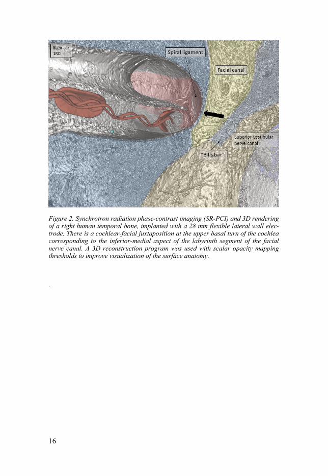
Cochlear-facial dehiscence Cochlear-facial dehiscence (CFD), a fusion between the basal part of the coch-lea and the labyrinthine portion (LP) of the facial nerve canal, is a rare condi-tion. The LP of the facial nerve and the otic capsule of the cochlea are ana-tomical neighbours (see Figure 2). This is why, in rare cases, electric currents can spread from the cochlear electrodes and lead to adverse stimulation of the facial nerve. This phenomenon occurs typically in otosclerosis, malformations of the inner ear and after temporal bone fractures. An opening, or dehiscence, between the facial nerve and the cochlea occured in 0.6 % of samples in the temporal bones collection, as previously described by Fang et al. 2016. Blake et al. 2014 were the first to describe this condition in a case report. They re-ported two cases with hearing loss, tinnitus and autophony. A combination of family history, physical examination, and high resolution computer tomogra-phy supported their diagnosis.

15
Imaging techniques Our knowledge of the micro-anatomy of the inner ear is mainly based on con-ventional histology on two-dimensional sections, imaged by optical micros-copy. However, this source has several major deficits and limitations com-pared to 3D imaging. Apart from the risk of slicing or staining artifacts, it is extremely time consuming to record the entire organ.
Conventional clinical computed tomography and magnetic resonance im-aging (MRI) typically have resolution capabilities in the 0.5 mm range. For resolving microanatomy of the cochlear structures, a resolution is necessary that is at least two orders of magnitude higher. Micro-CT and SR-PCI are ca-pable of assessing the native 3D structure of tissues in the range of several microns.
In micro-CT, a micro-focused X-ray source illuminates the object and a planar X-ray detector collects magnified projection images. Based on hun-dreds of angular views acquired while the object rotates, a computer synthe-sizes a stack of virtual cross section slices of the object. Due to the size of the machine and the high intensity of radiation over a long period, it is only suit-able in experimental conditions. While CT imaging is absorption-contrast based, phase-contrast imaging (PCI) can be combined with synchrotron radi-ation to give an improved contrast of soft tissue while maintaining precise visualization of bone.
Micro-CT and SR-PCI, therefore, have the potential to replace histology as the gold standard for evaluating intra-cochlear structures in human specimens, as well as motivate further optimization for translation into the clinic setting.
16
Figure 2. Synchrotron radiation phase-contrast imaging (SR-PCI) and 3D rendering of a right human temporal bone, implanted with a 28 mm flexible lateral wall elec-trode. There is a cochlear-facial juxtaposition at the upper basal turn of the cochlea corresponding to the inferior-medial aspect of the labyrinth segment of the facial nerve canal. A 3D reconstruction program was used with scalar opacity mapping thresholds to improve visualization of the surface anatomy.
.

17
Aims of Present Studies
The overall aims of the present studies were to analyze the human cochlear anatomy that is relevant for cochlear implantation, as well as to broaden our understanding of the anatomic variations using new imaging techniques such as micro-CT and SR-PCI. The specific aim of each paper was: (I): To study the relationship between the labyrinthine portion of the facial canal and the cochlea in human inner ear molds and temporal bones using micro-CT and 3D rendering. (II): To describe how electrically evoked auditory brainstem response (e-ABR) and radiological investigations may assist in identifying patients with cochlear-facial dehiscence, a rare condition that causes abnormal facial nerve stimulation (FNS) after activation of the cochlear implant. (III): To describe the anatomical variations of the IAC and their clinical im-plications using micro-CT and 3D rendering. (IV): To describe the architecture of the basilar membrane (BM) and RW in the basal part of the human cochlea and to see how it relates to different CI trajectory pathways by using micro-CT and SR-PCI. (V): To perform long-term follow-up of patients operated with CIs and hear-ing preservation.

18
Materials and Methods
Papers I, III, and IV Uppsala archival temporal bone collection
The Uppsala archival temporal bone collection consists of 113 unselected hu-man temporal bones from autopsies and 334 plastic and silicone molds made from the labyrinth. The collection of temporal bones was established by Her-man Wilbrand, Jan Stahle, Karin Wadin, and Helge Rask-Andersen during the 1970s and 1980s at the Department of Diagnostic Radiology and Otolaryngol-ogy at the Uppsala University Hospital, Sweden. Seventy-eight bones were micro-dissected in order to expose the vestibular and cochlear aqueducts. Af-ter maceration, the roof of the internal acoustic canal was drilled away and the fundus was exposed. Thirty-five bones were left un-dissected. Data from this collection was previously published by Wilbrand 1975, Rask-Andersen et al. 1977, and Wadin and Wilbrand 1987. Recently, this unique collection has formed the basis for new publications on the anatomy of the cochlea, RW, and vestibular aqueduct (Erixon et al. 2009, Atturo et al. 2014, Nordstrom et al. 2016).
The plastic and silicone molds in the collection were prepared in the 1970s. The temporal bones were soaked and cleaned in potassium hydroxide (KOH), boiled and then treated with 2 % hydrogen peroxide (H2O2) and trypsinized. The bones were positioned in a wax form with the cavities of the inner ear canals left open. Next, a polyester resin or silicone rubber material was poured into the wax form. The forms were placed in a low pressure chamber to im-prove the dispersion of the molding material into the tiny, bony canals of the temporal bones. The bone was suspended with hydrochloric acid after hard-ening.
In Paper I, 282 out of 334 molds could be analyzed for cochlear-facial de-hiscence (CFD). The distance between the cochlea and the facial nerve (CD) was measured in 48 out of 59 randomly chosen silicone molds and in 49 out of 51 resin molds. All 113 temporal bones underwent micro-CT. Eighty bones could be analyzed for superior semicircular canal dehiscence (SSCD) (see Ta-ble 1).

19
Table 1.
Uppsala archival bone collection
334 molds 282 were analyzed for CFD CD was measured in 48 out of 59 silicone molds CD was measured in 49 out of 51 resin molds 113 temporal bones 113 underwent micro-CT 80 bones could be analyzed for SSCD
In Papers III, which is more descriptive, we analyzed 113 temporal bones and 334 plastic and silicone molds.
In Paper IV, we drilled out different conventional COs using a 1mm dia-mond burr in 17 out of the 78 micro-dissected bones. Afterwards, the bones underwent micro-CT.
Papers I, III, and IV Micro-computed tomography
The type of clinical CT used today suffers from a fairly low image contrast resolution. In experimental situations, micro-CT allows high-intensity irradi-ation and offers a resolution down to the micrometer level with greatly im-proved image contrast. X-rays are emitted from a generator and travel through a sample, in our cases temporal bones or corrosion casts, which are detected on the opposite side to produce radiograph imaging. The X-ray tube and the detector rotate around the sample by a fraction of a degree, and another pro-jection image is taken at the new position. This procedure is repeated until a 360-degree rotation is achieved. The projection images are processed using a computer software (Slicer 4.6) to visualize the internal structures of the sam-ples. The reconstructed images are transferred and modelled into a 3D volu-metric object for quantitative analysis or visualization. However, one disad-vantage is the low reproduction of soft tissues, such as the BM and spiral lig-ament (SL). Our micro-CT reproduction allowed for edge enhancement by accentuating the contrast between the boundaries of structures.
Paper II Patients In 2017, a short time after publishing our first micro-CT study on the relation-ship between the cochlea and the facial nerve, two patients with the precise clinical condition of a CFD were diagnosed in our clinic. Two surgeons had operated on them at different times, and there was no other medical condition,

20
like otosclerosis or skull base fracture to explain the facial excitation during the devices’ activation. Both patients were operated with lateral wall elec-trodes. In the first patient a MedEL flexsoft 28 mm was used. In the second patient a MedEl flexsoft 31 mm long electrode was inserted via the RW. Clinical scan procedures All CI-patients in Uppsala routinely undergo preoperative CT scans and MRI to confirm normal inner ear anatomy. Perioperative conventional X-rays con-firm the correct position of the electrode after insertion. In both patients, a postoperative cone beam CT scan and 3D reconstructions were analyzed and compared to preoperative images for description of the labyrinthine portion of the facial nerve and its relationship to the cochlea. Electrical evoked auditory brainstem responses Auditory brainstem response (ABR) measurements during CI surgery allows us to test the auditory nerve function objectively. We use it routinely in our clinic to better predict hearing outcomes with CI. Normally, the ear is stimu-lated acoustically, but during CI, it is possible to electrically stimulate the cochlear nerve via the electrode. The principle of ABR recording is stimuli presented to the ear and registry of the voltage changes from the skin elec-trodes on the scalp surface. A computer program filters and removes the back-ground noise. Normally, waves I-V are analyzed. However, wave I is missing in e-ABR because the electrode stimulates the cochlea directly where wave I is normally generated. Wave I is often shadowed by a stimulation artifact (Ab-bas and Brown 1988, Firszt et al. 2002).
Two separate systems were used, one for stimulation and one for recording. In this study, we used the Med El programming device and the recording sys-tem was an evoked potential, triggered from the stimulating system to initiate recording. The system for the evoked potential was the Otometrics Chartr 200 (GN Otometrics, Taastrup, Denmark). We recorded e-ABRs through stimula-tion channels 1, 7, and 11 on the electrode.
Paper IV Temporal bone preparation
The technique used in our studies was recently described by Elfarnawany et al. 2017 and Koch et al. 2017. Sixteen freshly frozen and then fixed adult ca-daveric temporal bones were used in this study. All specimens were obtained with approval from the body bequeathal program at Western University, Lon-don, Ontario, Canada, in accordance with the Anatomy Act of Ontario and

21
Western’s Committee for Cadaveric Use in Research. Following thawing, a cylindrical cutter was used to core a sample (40 mm in diameter and 60 mm in length) of the middle ear from each temporal bone. The samples were fixed in a 4F1G (3.7 % formaldehyde and 1 % glutaraldehyde in phosphate buffer) bath for 5 days. The samples were rinsed twice and dehydrated using an etha-nol series (50 %, 60 %, 70 %, 80 %, 90 %, 95 %, and 100 %). No additional processing (i.e., staining, sectioning or decalcification) was performed on the samples. Sample fixation eliminated the risk of degradation during the two-month time difference between imaging sessions and scanning. Samples were transported to the synchrotron facilities in motion-proof boxes to prevent the risk of damage during shipping. Synchrotron radiation phase-contrast imaging
The SR-PCI on the 16 temporal bones was performed by our collaborating research team at Western University, Ontario, Canada. The setup is compara-ble to conventional radiography. It comprises an X-ray source, a sample, and a detector with no extra optical elements. The detector stands at a distance from the sample. This permits the phase shifted beam to interfere with the original beam and produce measurable fringes.
Compared to a conventional radiogram, the fringes correspond to surfaces and structural boundaries of the sample (edge enhancement). Each sample was scanned using the Bio-Medical Imaging and Therapy 05ID-2 beamline at Ca-nadian Light Source Inc. in Saskatoon, Saskatchewan, Canada. It provides an SR beam produced by a superconducting wiggler source (Wysokinski et al. 2015). A monochromator, yielding an energy bandwidth of ΔE/E = 10-3 over an energy range of 25-150 keV, was used to filter the beam. The imaging setup is installed at the beamline length of 55 m from the source. It is made up of a sample stage and a charge-coupled device-based detector system. Both are positioned on a vibration isolation table. The distance between the sample and detector was 2 m, and the photon energy was 47 keV. Motorized alignment stages were used to align the sample and detector for high-resolution tomog-raphy. The detector (an AA-60 beam monitor coupled with a C9300-124 cam-era, Hamamatsu Photonics, Shizuoka, Japan) has a 12-bit resolution and an effective pixel size of 9 x 9 μm². The imaging field of view was set to 4000 x 950 pixels, corresponding to 36.0 x 8.6 mm, and 3000 projections over 180° rotations were acquired per view. The 3D image volume had an isotropic voxel size of 9 μm. The acquisition time to capture all projections per view was approximately 30 minutes. While CT imaging is absorption-contrast based, phase-contrast imaging (PCI) can potentially be combined with syn-chrotron radiation to improve soft-tissue contrast while maintaining an accu-rate visualization of bone. Conventional absorption-contrast based CT de-pends on the attenuation of X-rays, whereas in PCI, the phase shift caused by the sample is transformed into detectable variations in X-ray intensity. PCI

22
can provide edge enhancement by emphasizing the contrast between the boundaries of different structures in the image.
Paper V Patients Erixon et al. 2012 presented our results on hearing preservation (HP) CI sur-gery in the first 21 consecutively operated on patients using the RW approach in Uppsala. One patient died during the follow-up period and was therefore excluded. Data from 15 of these patients was available for our long term fol-low-up, showing how hearing is affected over a period of more than 5 years. The mean age at operation was 59 years (range: 19¬87 years), nine women, eleven men. Surgery All 21 patients were operated on by the same surgeon between September 2008 and October 2010. In 20 cases, a MedEl flex EAS electrode (24 mm long) with an insertion depth of 17.5-23.5 mm was used in 20 cases and in one case a MedEl flex soft electrode (31 mm long) with an insertion depth of 28.5 mm. The surgical approach was via the RW after gentle drilling of the bony overhang and insertion of the electrode after a vertical incision of the mem-brane. Corticosteroids (Triamcinolon solution 40 mg/ml) were installed in the middle ear while inserting the electrode slowly by free hand. The correct elec-trode position was checked and confirmed by postoperative radiology. Audiometry Hearing thresholds were evaluated at frequencies of 250, 500, 1000, 2000, 3000, 4000, 6000 and 8000 Hz preoperatively, as well as at 1, and 3 years postoperatively with one long-term follow-up after more than 5 years (mean 86 months, range: 61-103 months) in both ears of each patient.
Additionally, the percent of HP over the whole frequency range (125- 8000 Hz) was calculated as stated by the HEARRING group consensus in 2013, according the following formula:
23
In this equation, PTApre is pure tone average measured preoperatively, PTApost is pure tone average measured postoperatively, and PTAmax is the maximum sound intensity generated by a standard audiometer, usually 120 dB hearing level. In accordance with this groups’ suggestion, >75 % of the preserved residual hearing can be classified as complete HP, >25-75 % as partial HP and 0-25 % as minimal HP (Skarzynski et al. 2013).
Fitting of speech processor
One month after surgery, the patients were fitted with their speech processor. Different stimulation strategies were carefully evaluated by the patient and the engineers. We documented user-time of the processor, and whether the pa-tients were fitted with full-frequency stimulation (e-only), cut-off frequency stimulation strategy with acoustic stimulation in the lower frequencies (a+e), or natural hearing (n+e), both during the first year and at the long-term follow-up. The user-time of the processor was recorded (full-time user > 8 hours per day, part-time user < 8 hours per day, and non-user).

24
Results
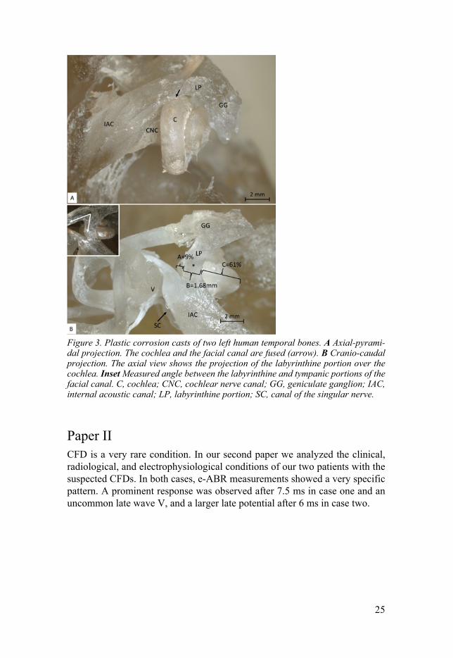
Paper I In the first paper we studied the relationship between the labyrinthine portion (LP) of the facial canal and the cochlea in human inner ear molds and temporal bones. We measured the cochlea-facial distance in silicon and polyester resin molds in our archival collection of human temporal bones. The association between the labyrinthine portion and the upper basal turn of the cochlea was analyzed (see Figure 3). The mean distance in the axial-pyramidal direction (vertical plane) was 0.20 mm (range 0-0.46 mm) in the plastic resin group and 0.22 mm (range 0.06-0.45 mm) in the silicone group. The relative portion of the cochlea exposed on either side of the nerve canal was calculated. The mean percentage located laterally from the nerve was 8 % (range 0 %-26 %) and that located medial to the nerve was 62 % (range 39 %-79 %). Local thinning of the otic capsule and local anatomy may explain the development of CFD. It was found in 1.4 % of the molds. A reduced cochlea-facial distance was noted in one bone with a superior semicircular canal dehiscence but not in bones with superior semicircular canal “blue line”. The otic capsule often im-pinged upon the LP of the facial nerve canal and caused narrowing. We used micro-CT and 3D rendering with multi-planar sectioning for the first time in our temporal bone collection to add and visualize information about the inter-action between the LP and cochlea.
25
Figure 3. Plastic corrosion casts of two left human temporal bones. A Axial-pyrami-dal projection. The cochlea and the facial canal are fused (arrow). B Cranio-caudal projection. The axial view shows the projection of the labyrinthine portion over the cochlea. Inset Measured angle between the labyrinthine and tympanic portions of the facial canal. C, cochlea; CNC, cochlear nerve canal; GG, geniculate ganglion; IAC, internal acoustic canal; LP, labyrinthine portion; SC, canal of the singular nerve.
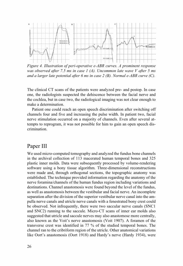
Paper II CFD is a very rare condition. In our second paper we analyzed the clinical, radiological, and electrophysiological conditions of our two patients with the suspected CFDs. In both cases, e-ABR measurements showed a very specific pattern. A prominent response was observed after 7.5 ms in case one and an uncommon late wave V, and a larger late potential after 6 ms in case two.
26
Figure 4. Illustration of peri-operative e-ABR curves. A prominent response was observed after 7.5 ms in case 1 (A). Uncommon late wave V after 5 ms and a larger late potential after 6 ms in case 2 (B). Normal e-ABR curve (C).
The clinical CT scans of the patients were analyzed pre- and postop. In case one, the radiologists suspected the dehiscence between the facial nerve and the cochlea, but in case two, the radiological imaging was not clear enough to make a determination.
Patient one could reach an open speech discrimination after switching off channels four and five and increasing the pulse width. In patient two, facial nerve stimulation occurred on a majority of channels. Even after several at-tempts to reprogram, it was not possible for him to gain an open speech dis-crimination.
Paper III We used micro-computed tomography and analyzed the fundus bone channels in the archival collection of 113 macerated human temporal bones and 325 plastic inner molds. Data were subsequently processed by volume-rendering software using a bony tissue algorithm. Three-dimensional reconstructions were made and, through orthogonal sections, the topographic anatomy was established. The technique provided information regarding the anatomy of the nerve foramina/channels of the human fundus region including variations and destinations. Channel anastomosis were found beyond the level of the fundus, as well as anastomosis between the vestibular and facial nerve. An incomplete separation after the division of the superior vestibular nerve canal into the am-pulla nerve canals and utricle nerve canals with a fenestrated bony crest could be observed. Not infrequently, there were two saccular nerve canals (SNC1 and SNC2) running to the saccule. Micro-CT scans of inner ear molds also suggested that utricle and saccule nerves may also anastomose more centrally, also known as the Voit’s nerve anastomosis (Voit 1907). A foramen of the transverse crest was identified in 77 % of the studied temporal bones. The channel ran to the cribriform region of the utricle. Other anatomical variations like Oort’s anastomosis (Oort 1918) and Hardy’s nerve (Hardy 1934), were

27
also described together with their clinical implications. Three-dimensional re-constructions and cropping outlined the bone canals and demonstrated the highly variable VIIIth nerve anatomy at the fundus of the human inner acous-tic canal (see Figure 5). A myriad of channel interconnections suggested an intricate system of neural interactive pathways in humans.
Figure 5. Varied anatomy of the human fundus (left ears). Each channel was followed to the labyrinth with the help of cropping. Foramen of the transverse crest (*) ran to the utricule (U) while those situated beneath ran to the saccule (S). Two or even three cibriform plates of the saccule are seen. The location of the singular nerve channel (SiNC) varies. The wall between the facial nerve canal (FC) and superior vestibular nerve canal (SVNC) is fenestrated in (A). The vertical crest (VC) is poorly defined in (D). C indicates cochlea.
28
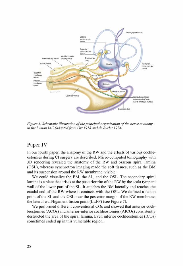
Figure 6. Schematic illustration of the principal organization of the nerve anatomy in the human IAC (adapted from Ort 1918 and de Burlet 1924).
Paper IV In our fourth paper, the anatomy of the RW and the effects of various cochle-ostomies during CI surgery are described. Micro-computed tomography with 3D rendering revealed the anatomy of the RW and osseous spiral lamina (OSL), whereas synchrotron imaging made the soft tissues, such as the BM and its suspension around the RW membrane, visible.
We could visualize the BM, the SL, and the OSL. The secondary spiral lamina is a plate that arises at the posterior rim of the RW by the scala tympani wall of the lower part of the SL. It attaches the BM laterally and reaches the caudal end of the RW where it contacts with the OSL. We defined a fusion point of the SL and the OSL near the posterior margin of the RW membrane, the lateral wall/ligament fusion point (LLFP) (see Figure 7).
We performed different conventional COs and showed that anterior coch-leostomies (ACOs) and anterior-inferior cochleostomies (AICOs) consistently destructed the area of the spiral lamina. Even inferior cochleostomies (ICOs) sometimes ended up in this vulnerable region.

29
Figure 7. Magnification of hook region of the cochlea. The twisted osseous spiral lamina (brown) is shown after cropping. The secondary spiral lamina is viewed (red) through the RW. LLFP defines the beginning of the BM. The cochlear aque-duct and inferior cochlear vein channels can be seen. PSSC; posterior semicircular canal. SN; singular nerve. OC; otic capsule. PP; postis posterior.
Figure 8. SR-PCI and 3D rendering of a right human cochlea. The RW membrane is viewed from the scala tympani (inset). The spiral ligament/secondary spiral lamina (red) suspends the BM laterally. The opening of the cochlear aqueduct is located in the floor of the scala tympani at the interior surface of the crista fenestrae (red inter-rupted line).

30
Figure 9. SR-PCI of different surgical approaches (facial recess view) and how they wind up inside the cochlea. The soft tissues of the basal part of the cochlea were traced and color-labeled on serial sections for reconstruction. The data were fed into the 3D software program and models were made using the threshold paint tool in the editor module. A. Cropping of the bony overhang was made to define the mar-gins of the round window membrane (RWM) and different COs as defined by the au-thors. ACO; anterior cochleostomy, AICO; anterior-inferior cochleostomy, ICO; in-ferior cochleostomy. B and D. Deeper sections expose the spiral ligament which is crossed by ACO and AICO trajectories. D. Lateral view. E. Anterior view after cropping of the cochlea (inset).

31
Paper V Nineteen of the 20 evaluated patients had preserved residual hearing after one and three years post-operatively, as previously described by Erixon et al. 2012. We could collect data for long-term follow-up from more than five years post-operatively in 15 patients. Out of these 15 patients, 12 still had residual hear-ing after a follow-up period of over five years (mean: 86 months, range: 61- 103 months). Another 4 out of 15 patients had complete HP (>75 %), 8 had partial HP (25-75 %), and 3 patients had complete hearing loss. There was a high correlation between insertion angle and HP rate (Pearson test, p< 0.002). The patients with complete HP had a relatively smaller insertion angle com-pared to the patients with partial or complete hearing loss. Most patients that could be followed-up with in the long-term used their implants full time (13 out of 15 patients). The patients experienced noticeable benefits from the de-vices. One patient was a non-user (probably due to dementia) and one patient a part-time user (three hours/day).

32
T
able
2. T
here
wer
e 15
pat
ient
s w
ith
long
-ter
m fo
llow
-up
data
. The
dat
a in
clud
ed:
Deg
rees
= e
lect
rode
inse
rtio
n an
-gl
e ve
rifi
ed b
y ra
diol
ogy;
No
of e
lect
rode
s in
side
the
coch
lea
veri
fied
by
radi
olog
y; S
tim
ulat
ion
a+e
= a
cous
tic
and
elec
tric
al s
tim
ulat
ion,
e o
nly
= e
lect
ric
stim
ulat
ion
only
, n+
e =
nat
ural
hea
ring
in lo
w fr
eque
ncie
s, a
nd e
lect
ric
stim
u-la
tion
in th
e hi
gher
freq
uenc
ies;
MS
= m
onos
ylla
bles
; B
S= b
isyl
labl
es.

33
Discussion
Cochlear implantation today is routinely performed worldwide to restore hear-ing in patients with severe sensorineural hearing loss. In the last decade, elec-trodes have become thinner and more flexible so that even patients with quite usable residual hearing in the low frequencies and losses in high frequency regions can be operated upon and benefit from this technology.
Hearing preservation by inserting the cochlear implant electrode through the RW is now a widely-used surgical technique, but we do not know exactly how this method preserves residual hearing from a long-term perspective. What is the optimal length of an electrode for preserve hearing? How deep do we need to stimulate the cochlea to gain ideal hearing results? Does the size of the cochlea matters? My thesis does not provide answers to all these ques-tions, but it is an attempt to clarify some gaps in our knowledge. First, I fo-cused on the anatomy of the inner ear with new imaging techniques, such as micro-CT and SR-PCI. For this purpose, the Uppsala collection of human tem-poral bones from the 1970s was an invaluable resource and fortunately we were able to start a collaboration with the Canadian research group. This team had already produced high-quality work in reproducing the human temporal bone and middle ear by synchrotron imaging, and they sought collaborations in order to further interpret the complex inner ear data. It is an exciting devel-opment that corrosion anatomy, which took months and even years to execute, can now be done in a few days with the help of these data using 3D recon-structions.
The cochlea and the facial nerve In Paper I, the close interface between the first segment of the facial nerve canal and upper basal turn of the cochlea is illustrated in man. Micro-CT with 3D rendering offered new possibilities to study this topographic anatomy of the human temporal bone.
A reduced cochlea-facial distance may lead to spread of electric currents from the implant array to the LP of the facial nerve and cause facial nerve stimulation. Influencing factors may be the topographic anatomy and otic cap-sule properties. In our clinic, just a couple of months after publishing this first paper, two cases with exactly this clinical condition occured. Both patients were operated on with lateral wall electrodes and could initially not reach any

34
open speech discrimination due to an irritating facial stimulation when acti-vating the implant. Smullen et al. 2005 noted 6.5 % FNS in 600 patients where the most common causes were malformations and otosclerosis. The incidence was much lower when using perimodiolar electrodes. This is reasonable since perimodiolar electrodes follow along the medial wall of the cochlea at a slightly longer distance from the facial nerve canal. The second of our patients was recently operated on again, and we altered to a perimodiolar electrode. He is now a full-time user and has a speech understanding rate for monosyl-lables with 38 % at a 65 dB level (six months after activation). Occasionally he experiences eye twitching, which he is seldom aware of in his daily life.
In both cases, intraoperative e-ABR measurements at low stimulation lev-els showed a late prominent response due to myogenic activity from the facial nerve. It is debatable whether this will influence our decision to replace an already implanted lateral wall electrode with a perimodiolar one or not the next time this occurs.
The internal acoustic canal Documentation of the nerve components in the internal acoustic canal is es-sential before cochlear implantation surgery. Interpretations may be chal-lenged by wide anatomical variations of the VIIIth nerve and their ramifica-tions. Malformations may further distort proper nerve identification. The goal of this study was to describe anatomical variations of the inner acoustic canal and their clinical implications. We used micro-CT and analyzed the fundus bone channels in our archival collection of human temporal bones. Three-di-mensional reconstructions were made to visualize the anatomy of the nerve channels of the fundus, including variations and their true destinations using the serial cropping technique. An incomplete separation after the division of the superior vestibular nerve canal into the ampulla nerve canals and utricle nerve canals with a fenestrated bony crest could be observed. Sometimes there were two saccular nerve canals (SNC1 and SNC2) running to the saccule. This could readily be verified using cropping and surface enhancement algorithms. This type of variation may challenge proper identification of the cochlear nerve at MRI before CI, since one of the nerves may be mistakenly interpreted as a cochlear nerve. We found a remarkable variation of the position and size of the singular nerve channel that acts as an important landmark for the pre-vention of fenestration of the labyrinth in middle fossa surgery, retro-sigmoid vestibular schwannoma surgery, and cochleo-vestibular neurectomy (Gacek 1984, Kos et al. 2006).
A foramen of the transverse crest was identified in 77 % of our temporal bones. The channel ran to the cribriform region of the utricle. In a follow-up study using synchrotron imaging this canal was found to contain a vessel, pre-sumably an artery (Mei et al, 2008).

35
Micro-CT imaging also reproduced anastomoses between the vestibular and facial nerve. These connections may have clinical importance, for exam-ple in Bell’s palsy, Ramsey-Hunt syndrome and after treatment of hemi-facial spasms, conditions where patients have a higher incidence of vestibular symp-toms (Fisch and Esslen 1972, Wakasugi 1972). We could also see anastomo-ses between the vestibular and cochlear nerve, such as Oort’s anastomosis and Hardy’s nerve. These fibers belong to the olivo-cochlear bundle and convey efferent fibers from the brain to the cochlear, as well as some afferent fibers (Rasmussen 1946).
The round window region There is still much discussion in the literature on how to perform the best CO and preserve hearing. A literature survey on HP surgery shows that there is no general agreement on the optimal trajectory pathway into the cochlea. Both RW, CO and enlarged RW approaches are used. In addition, the COs are placed differently on the promontory with the most accepted placement de-fined as either ACO, ICO or in between AICO (Atturo et al. 2014). A clear definition of each of these approaches has not been realized, and in a survey investigation by Adunka and Buchman 2007, North American surgeons placed different COs in different places. Their review documented the clear variations for scala tympani at CI. The study seemed to show that there is no clear definition for the surgical anatomy of this region and highlights the need for further investigations of optimal trajectory pathways.
One reason for the different approaches is the highly variable anatomy of the hook region. This part is defined by the curved basal part of the cochlear tube. Near the RW, it dilates and curves laterally at the promontory and turns postero-medial. Another distinct feature is the variable morphology of the RW niche and the bony overhang. It is, therefore, not possible to define the margin of the RW unless the overhang is surgically removed. When performing COs, there is no direct visualization of the spiral lamina, and the surgeon’s view does not have a clear indication if the route also passes across the lamina from the scala vestibuli. It was, therefore, pertinent to perform a thorough analysis of the hook anatomy to see the influence of different trajectories for CI on cochlear soft tissue anatomy. Synchrotron imaging and cropping techniques with 3D visualization helped this study to describe the relationship between various COs and cochlear soft tissues, such as OSL, BM, and SL. The results showed that soft tissue in the basal part of the cochlea is seldom preserved after COs. Depending on the size and shape of the cochlea, the soft tissues can, however, sometimes be preserved after ICO. The RW approach seems to be the least traumatic and is recommended in HP surgery.

36
Clinical perspectives on hearing preservation Documentation of long-term hearing outcomes in cochlear implantation is im-portant. Especially nowadays when companies develop new electrodes that are even more flexible, thinner and are promoted as structure preservative. It is not possible to perform randomized, controlled double-blinded studies in this kind of population. Causon et al. 2015 made a retrospective analysis of the contribution of factors in HP outcomes in CI. Only 12 of 284 papers were approved for evaluation including our original study. Seven factors had a sig-nificant effect on HP, namely insertion site, progressive versus stable hearing loss, insertion angle, use of steroids, hearing etiology and electrode array type.
The HEARRING group (Rajan et al. 2018) concludes that, irrespective of the degree of residual hearing present, the concepts of hearing and structure preservation should be applied in every child undergoing cochlear implanta-tion and that HP cochlear implantation is a secure and trustworthy therapy option. Today, even children with residual hearing in the low frequencies are operated on. Wilson et al. 2016 pointed out that children perform better with CIs than with hearing aids, even if their hearing is not fully preserved. They have also found that children need early access to high frequency sound in order to reach their full potential. For the counselling of this pediatric group, it is therefore even more important to follow patients after HP CI surgery over a longer time period.
The results of my study demonstrated that HP is possible when using flex-ible lateral wall electrode with RW insertion even over a long-term perspec-tive. These outcomes were consistent with results in the literature that demon-strated the preservation of functional hearing after implantation (Moteki et al. 2017, Roland et al. 2018).
A total of 12 out of 15 patients had the same stimulation strategies at long-term follow-up compared to the initial ones at activation. Three patients changed their stimulation strategy from a+e stimulation to e-only over the whole frequency range. Interestingly, two of these patient had quite acceptable low-frequency hearing left even in the long-term follow-up. Long-term fol-low-up is necessary, particularly for patients using EAS, as changes in hearing may necessitate changes to amplification settings, cross-over frequencies, and their general EAS setup in the future.
In summary, the basic anatomical works performed in the present investiga-tion were highly helpful in the clinical assessment of patients suitable for HP- surgery. In particular, the synchrotron reproduction of soft tissue offered new insights into the intricate anatomy inside the RW membrane. To visualize the micro-structures behind this “curtain” may give surgeons a different view of its great delicacy and the challenges in performing non-traumatizing CI sur-gery.

37
Conclusions
Micro-CT with 3D rendering offers new possibilities in studying the topo-graphic anatomy of the human temporal bone. The varied shape of the cross-section of the LP of the facial nerve could often be explained by an “intruding” cochlea. In 1.4 % of our specimens we found a fusion of the LP of the facial nerve and the cochlea, which is a rare condition, called cochlear-facial dehis-cence. Predicting facial nerve stimulation by assessing the distance between the LP of the facial nerve and the cochlea is difficult when using conventional CT-scans. A large evoked late myogenic potential at low stimulation levels during intraoperative e-ABR measurements may foresee FNS at CI activation. We used micro-CT with 3D rendering to display the anatomy of the fundus of the human inner acoustic canal. We could gain additional information regard-ing the anatomy of the nerve channels and their destinations from this method. A foramen of the transverse crest was also identified. These results may assist in the interpretation of preoperative imaging of the VIIIth nerve. SR-PCI reproduced the soft tissues of the human inner ear, including the RW membrane, OSL and SL. The microanatomy of this region should be ac-counted for when considering cochlear implant surgery. Results suggested that CO approaches often traumatize the soft tissues at the hook region. For optimal structure preservation, the RW approach is recommended. For our 21 first consecutive patients at our clinic who underwent HP CI sur-gery, the data of 15 patients was available for a long-term follow-up. Long-term HP was possible in most of these cases. There was a high correlation between insertion depth and HP outcome. Even patients with complete hear-ing loss experienced good performance in both speech discrimination results and user time.

38
Future perspectives
In most patients receiving CIs it is possible to preserve residual hearing. A major question, however, is how to preserve these results over a longer period of time. Consequently, further investigations are necessary to better compre-hend the essential factors involved in hearing deterioration such as genetic background, surgery techniques, and possible inflammatory intra-cochlear re-sponses. Another possibility is to find more direct medical treatments, such as drug delivery to the inner ear using corticosteroid-diluted and surface-coated electrodes also carrying stem cells or neurotrophins (Li et al. 2017). Other current research focuses on designing an array that brings the electrodes closer to the neurons or other principle stimulation modes such as optical stimulation. Light can be more conveniently defined compared to wide current spread from conventional electrode contacts (Richardson et al. 2017). Eventually, future technologies could be integrated in order to allow individual tailoring of the array design.

39
Sammanfattning på svenska
Målet med detta avhandlingsarbete var att studera människans komplexa in-neröreanatomi med avseende på cochleaimplantat (CI). Detta har ytterligare aktualiserats av s.k. hörselbevarande CI-kirurgi. Patienter med grav hörsel-nedsättning men bevarad hörsel i de lägre frekvensområdena är idag en viktig målgrupp för CI-behandling. Uppsala har tidigare bidragit till kunskapsut-vecklingen inom detta område. I denna avhandling studerades bland annat det unika material av tidigare utförda exakta temporalsbensavgjutningar samt mikrodissektioner.
Med hjälp av Mikro-CT undersöktes varje preparat i detalj. De tomo-grafiska snitten bearbetades digitalt i ett datorprogram varefter benen kunde framställas tre-dimensionellt för analys av anatomiska relationer.
Dessutom användes en ny teknik, s.k. synkrotron avbildning, utförd i Lon-don, Canada. Därigenom kunde vi jämföra olika tekniker på vårt material i Uppsala. Synkrotron strålning med faskontrast bildåtergivning (synchrotron radiation with phase contrast imaging, SR-PCI) kan användas för att visuali-sera intra-cochleära mikrostrukturer och har dessutom hög mjukdelskontrast.
Ansiktsnervens topografiska anatomi studerades med fokus på dess första del (den s.k. intralabyrintära portionen) och förhållandet till hörselsnäckan. CI-elektroder belägna i detta område kan ge upphov till oönskad elektrisk sti-mulering av ansiktsnerven vid inkopplingen av den yttre processorn. Anato-min studerades i detalj för att öka förståelsen för hur denna komplikation kan uppstå. En avsaknad av den beniga skiljeväggen mellan ansiktsnervens in-tralabyrintära portion och hörselsnäckan fann vi i 1,4 % av temporalbenen. Detta är ett sällsynt sjukdomstillstånd med namnet ”cochlear-facial de-hicence” (CFD). Två patienter med kraftig stimulering av ansiktsnerven efter operation med CI upptäcktes på vår klinik och CFD diagnostiserades. Ett för-höjt sent svar vid de intraoperativa e-ABR mätningarna kan vara en metod att förutse oönskad stimulering av ansiktsnerven.
Dessutom studerades den topografiska anatomin av inre hörselgången med micro-CT och 3D rekonstruktioner. Den 7:e och 8:e kranialnervernas benka-naler analyserades och variationer beskrvs. Studien syftade till att förbättra tolkningen av den preoperativa MR undersökningen vid CI utredning.
I arbetet studerades även hörselsnäckans anatomi i syfte att bevara kvarstå-ende hörsel vid CI-kirurgi. De komplexa anatomiska relationerna i basen av snäckan där elektroden införs kunde analyseras tre-dimensionellt i detalj för första gången med hjälp av synkrotronundersökningar. Vår rekommendation

40
är att elektroden inläggs via runda fönstret för att inte skada känsliga strukturer i hörselsnäckan.
En långtidsuppföljning utfördes av de första opererade patienterna med hörselbevarande CI kirurgi. Patienterna följdes över 5 år och vi fann att 12 av 15 patienter hade bevarad hörsel i det opererade örat. Det förelåg en hög kor-relation mellan hur djupt elektroden förts in i hörselsnäckan och kvarvarande hörsel. Även patienter som förlorade resthörsel var heltidsanvändare med goda resultat i taluppfattningstesterna.

41
Acknowledgements
I wish to express my deep gratitude to the many people who have helped me throughout the course of this work:
Special thanks to all the patients who took part in the studies!
My supervisor, Professor Helge Rask-Andersen, for your incredible energy in both research projects and clinical work and for always being optimistic and having millions of new ideas. Your enthusiasm kept me going throughout my scientific ups and downs. Thank you for sharing your unlimited knowledge in the field of otology!
My co-supervisor Per Olof Eriksson, for your support, friendly discussions and being a fantastic mentor!
Hao Li, co-author and collaborator, for your hard work with the micro-CT, always willing to help out with my computer and Endnotes problems, your kindness and patience.
Göran Laurell, professor of the department, for sharing your great experience, practical hints, and help with financial support.
Manochehr Amani, for giving me the time to pursue research and courses.
My collaborators, Sumit Agrawal and Hanif Ladak, co-authors, for sharing their synchrotron images with us and all their vital and professional support. Sune Larsson, co-author, for sharing the micro-CT. Karin Lodin, for help with illustrations. Karin Wadin, for her work with the temporal bones. Karin Hallin, co-author and skilled CI-engineer, for all your valuable advices and help.
My former supervisor Magnus von Unge who introduced me to the world of science and did not get annoyed when I changed my focus from eardrums to the inner ear.
My present and former clinical supervisors and colleagues in ENT in Stuttgart, Stockholm, Västerås and Uppsala! It was and is a privilege to work with you!

42
Elsa, my colleague and co-author, for good advices, sharing your former ma-terial for Paper V and weakness for sushi. You are making audiology great! Lotta, my colleague and roommate, for always being there, having vibrant discussions on audiological cases, and much more.
Karin, Lennart, and Niklas for being fantastic colleagues and sharing your surgical skills with me. You are my idols in otosurgery! The amazing Cochlear Implant Team who takes care of all our CI-patients in a fantastic way. You make Thursday mornings the highlight of my week! A special thanks to Jonas who helped me with statistics and audiograms in Paper V. All nurses, audiologists and other staff at the ENT department in Uppsala for their professionalism and good support to the patients. My friends, close and far away! I really enjoy being with you, hanging out, having dinners or dancing. The family, close and extended: Lena, Stellan, Elina, Staffan, Aura, and Artur for all the good times together. Håkan, for your support and all the time we spent together. You are the reason for all this and why I ended up in Sweden! André, my brother, and my parents Waltraud and Ralph for always believing in me. And my fantastic daughters, Louise and Mathilda! I am so proud of being your mother!

43
References
Abbas, P. J. and C. J. Brown (1988). "Electrically evoked brainstem potentials in cochlear implant patients with multi-electrode stimulation." Hear Res 36(2-3): 153-162.
Adunka, O. F. and C. A. Buchman (2007). "Scala tympani cochleostomy I: results of a survey." Laryngoscope 117(12): 2187-2194.
Aschendorff, A., J. Kromeier, T. Klenzner and R. Laszig (2007). "Quality control after insertion of the nucleus contour and contour advance electrode in adults." Ear Hear 28(2 Suppl): 75s-79s.
Atturo, F., M. Barbara and H. Rask-Andersen (2014). "Is the human round window really round? An anatomic study with surgical implications." Otol Neurotol 35(8): 1354-1360.
Atturo, F., M. Barbara and H. Rask-Andersen (2014). "On the anatomy of the 'hook' region of the human cochlea and how it relates to cochlear implantation." Audiol Neurootol 19(6): 378-385.
Blake, D. M., S. Tomovic, A. Vazquez, H. J. Lee and R. W. Jyung (2014). "Cochlear-facial dehiscence--a newly described entity." Laryngoscope 124(1): 283-289.
Causon, A., C. Verschuur and T. A. Newman (2015). "A Retrospective Analysis of the Contribution of Reported Factors in Cochlear Implantation on Hearing Preser-vation Outcomes." Otol Neurotol 36(7): 1137-1145.
Claudius, M. (1856). "Bemerkungen über den Bau der häutigen Spiralleiste der Schnecke." Z Wiss Zool 7: 154-161.
Cotugno, D. (1761). "De aquaeductibus auris humanae internae anatomica disserta-tio." Neapoli et Bononiae: Typographia Sanctae Thomae Aquinatis. .
Deiters, O. (1860). "Beiträge zur Kenntnis der Lamina spiralis menbranacea der Schnecke." Z Wiss Zool.
Elfarnawany, M., S. R. Alam, S. A. Rohani, N. Zhu, S. K. Agrawal and H. M. Ladak (2017). "Micro-CT versus synchrotron radiation phase contrast imaging of human cochlea." J Microsc 265(3): 349-357.
Engstrom, B. and H. Engstrom (1972). "Structural and physiological features of the organ of Corti." Audiology 11(1): 6-28.
Erixon, E., H. Hogstorp, K. Wadin and H. Rask-Andersen (2009). "Variational anat-omy of the human cochlea: implications for cochlear implantation." Otol Neurotol 30(1): 14-22.
Erixon, E., S. Kobler and H. Rask-Andersen (2012). "Cochlear implantation and hear-ing preservation: Results in 21 consecutively operated patients using the round window approach." Acta Otolaryngol 132(9): 923-931.
Eustachi, B. (1564). "Epistola de auditus organis." Opuscula anatomica, Venetijs: Vincentius Luchinus.
Fang, C. H., S. Y. Chung, D. M. Blake, A. Vazquez, C. Li, J. P. Carey, H. W. Francis and R. W. Jyung (2016). "Prevalence of Cochlear-Facial Dehiscence in a Study of 1,020 Temporal Bone Specimens." Otol Neurotol 37(7): 967-972.

44
Firszt, J. B., R. D. Chambers, Kraus and R. M. Reeder (2002). "Neurophysiology of cochlear implant users I: effects of stimulus current level and electrode site on the electrical ABR, MLR, and N1-P2 response." Ear Hear 23(6): 502-515.
Fisch, U. and E. Esslen (1972). "Total intratemporal exposure of the facial nerve. Pathologic findings in Bell's palsy." Arch Otolaryngol 95(4): 335-341.
Gacek, R. R. (1984). "Cupulolithiasis and posterior ampullary nerve transection." Ann Otol Rhinol Laryngol Suppl 112: 25-30.
Galen, C. (1542). "De usu partium corporis humani." Froben Basileae: Omnia Cl. Ga-leni opera.
Hardy, M. (1934). "Observations on the innervation of the macula sacculi in man." The Anatomical Record 59(4): 403-418.
Hensen, V. (1863). "Zur Morphologie der Schnecke des Menschen und der Säuge-tiere." Z Wiss Zool 13: 481-512.
Koch, R. W., M. Elfarnawany, N. Zhu, H. M. Ladak and S. K. Agrawal (2017). "Eval-uation of Cochlear Duct Length Computations Using Synchrotron Radiation Phase-Contrast Imaging." Otol Neurotol 38(6): e92-e99.
Kos, M. I., G. Feigl, F. Anderhuber, C. Wall, J. H. Fasel and J. P. Guyot (2006). "Transcanal approach to the singular nerve." Otol Neurotol 27(4): 542-546.
Li, H., F. Edin, H. Hayashi, O. Gudjonsson, N. Danckwardt-Lilliestrom, H. Engqvist, H. Rask-Andersen and W. Xia (2017). "Guided growth of auditory neurons: Bio-active particles towards gapless neural - electrode interface." Biomaterials 122: 1-9.
Lim, D. J. (1969). "Three dimensional observation of the inner ear with the scanning electron microscope." Acta Otolaryngol Suppl 255: 1-38.
Mei, X., N. Schart-Moren, H. Li, H. M. Ladak, S. Agrawal, R. Behr and H. Rask-Andersen (2018). "Three-dimensional imaging of the human internal acoustic ca-nal and arachnoid cistern: a synchrotron study with clinical implications." J Anat.
Moteki, H., S. Y. Nishio, M. Miyagawa, K. Tsukada, S. Iwasaki and S. I. Usami (2017). "Long-term results of hearing preservation cochlear implant surgery in patients with residual low frequency hearing." Acta Otolaryngol 137(5): 516-521.
Nadol, J. B., Jr., J. T. O'Malley, B. J. Burgess and D. Galler (2014). "Cellular immu-nologic responses to cochlear implantation in the human." Hear Res 318: 11-17.
Nordstrom, C. K., G. Laurell and H. Rask-Andersen (2016). "The Human Vestibular Aqueduct: Anatomical Characteristics and Enlargement Criteria." Otol Neurotol 37(10): 1637-1645.
Oort (1918). "Über der Verästelung des Nervus octavus bei Säugetieren." Anat. Anz Bd 51: 272-280.
Rajan, G., D. Tavora-Vieira, W. D. Baumgartner, B. Godey, J. Muller, M. O'Driscoll, H. Skarzynski, P. Skarzynski, S. I. Usami, O. Adunka, S. Agrawal, I. Bruce, M. De Bodt, M. Caversaccio, H. Pilsbury, J. Gavilan, R. Hagen, A. Hagr, M. Ka-meswaran, E. Karltorp, M. Kompis, V. Kuzovkov, L. Lassaletta, L. Yongxin, A. Lorens, M. Manoj, J. Martin, G. Mertens, R. Mlynski, L. Parnes, S. Pulibalathin-gal, A. Radeloff, C. H. Raine, R. Rajeswaran, J. Schmutzhard, G. Sprinzl, H. Staecker, K. Stephan, S. Sugarova, M. Zernotti, P. Zorowka and P. Van de Hey-ning (2018). "Hearing preservation cochlear implantation in children: The HEARRING Group consensus and practice guide." Cochlear Implants Int 19(1): 1-13.
Rask-Andersen, H., J. Stahle and H. Wilbrand (1977). "Human cochlear aqueduct and its accessory canals." Ann Otol Rhinol Laryngol Suppl 86(5 Pt 2 Suppl 42): 1-16.
Rasmussen, G. L. (1946). "The olivary peduncle and other fiber projections of the superior olivary complex." J Comp Neurol 84: 141-219.

45
Richardson, R. T., A. C. Thompson, A. K. Wise and K. Needham (2017). "Challenges for the application of optical stimulation in the cochlea for the study and treatment of hearing loss." Expert Opin Biol Ther 17(2): 213-223.
Roland, J. T., Jr., B. J. Gantz, S. B. Waltzman and A. J. Parkinson (2018). "Long-term outcomes of cochlear implantation in patients with high-frequency hearing loss." Laryngoscope.
Scarpa, A. (1772). "De structura fenestrae rotundae auris et de tympanos secundario anatomicae observationes." Mutinae. Societas Typogr. .
Scarpa, A. (1789). "Disquisitiones anatomicae de audit et olfactu." Ticini et Medi-olani.
Skarzynski, H., P. van de Heyning, S. Agrawal, S. L. Arauz, M. Atlas, W. Baumgart-ner, M. Caversaccio, M. de Bodt, J. Gavilan, B. Godey, K. Green, W. Gstoettner, R. Hagen, D. M. Han, M. Kameswaran, E. Karltorp, M. Kompis, V. Kuzovkov, L. Lassaletta, F. Levevre, Y. Li, M. Manikoth, J. Martin, R. Mlynski, J. Mueller, M. O'Driscoll, L. Parnes, S. Prentiss, S. Pulibalathingal, C. H. Raine, G. Rajan, R. Rajeswaran, J. A. Rivas, A. Rivas, P. H. Skarzynski, G. Sprinzl, H. Staecker, K. Stephan, S. Usami, Y. Yanov, M. E. Zernotti, K. Zimmermann, A. Lorens and G. Mertens (2013). "Towards a consensus on a hearing preservation classification system." Acta Otolaryngol Suppl(564): 3-13.
Smullen, J. L., M. Polak, A. V. Hodges, S. B. Payne, J. E. King, 3rd, F. F. Telischi and T. J. Balkany (2005). "Facial nerve stimulation after cochlear implantation." Laryngoscope 115(6): 977-982.
Wadin, K. and H. Wilbrand (1987). "The labyrinthine portion of the facial canal. A comparative radioanatomic investigation." Acta Radiol 28(1): 17-23.
Wakasugi, B. (1972). "Facial nerve block in the treatment of facial spasm." Arch Oto-laryngol 95(4): 356-359.
Van De Water, T. R. (2012). "Historical Aspects of Inner Ear Anatomy and Biology that Underlie the Design of Hearing and Balance Prosthetic Devices." Anat Rec (Hoboken) 295(11): 1741-1759.
Vesalius, A. (1543). "De humani corporis fabrica." Basileae: I. Oporinus. Wilbrand, H. F. (1975). "Multidirectional tomography of the facial canal." Acta Ra-
diol Diagn (Stockh) 16(6): 654-672. Wilson, K., M. Ambler, K. Hanvey, M. Jenkins, D. Jiang, J. Maggs and K. Tzifa
(2016). "Cochlear implant assessment and candidacy for children with partial hearing." Cochlear Implants Int 17 Suppl 1: 66-69.
Voit, M. (1907). "Zur Frage der Verästelung des Nervus acusticus bei den Säugetie-ren." Anat Anz Bd XXXI: 635-640.
von Ilberg, C., J. Kiefer, J. Tillein, T. Pfenningdorff, R. Hartmann, E. Sturzebecher and R. Klinke (1999). "Electric-acoustic stimulation of the auditory system. New technology for severe hearing loss." ORL J Otorhinolaryngol Relat Spec 61(6): 334-340.

Acta Universitatis UpsaliensisDigital Comprehensive Summaries of Uppsala Dissertationsfrom the Faculty of Medicine 1537
Editor: The Dean of the Faculty of Medicine
A doctoral dissertation from the Faculty of Medicine, UppsalaUniversity, is usually a summary of a number of papers. A fewcopies of the complete dissertation are kept at major Swedishresearch libraries, while the summary alone is distributedinternationally through the series Digital ComprehensiveSummaries of Uppsala Dissertations from the Faculty ofMedicine. (Prior to January, 2005, the series was publishedunder the title “Comprehensive Summaries of UppsalaDissertations from the Faculty of Medicine”.)
Distribution: publications.uu.seurn:nbn:se:uu:diva-373643
ACTAUNIVERSITATIS
UPSALIENSISUPPSALA
2018