the infant with stridor - europe pubmed central
TRANSCRIPT

THE INFANT WITH STRIDORA FOLLOW-lTP SURVEY OF 80 CASES
BY
JOHN APLEYFrom the Department of Child Health. Bristol University, and Bristol Royal Hospital for Sick Children
(RECEIVED FOR PUBLICATION JUNE 19, 1953)
It is a tendency of specialists in any field toexamine all phenomena exclusively with the instru-ments of their own speciality. Infantile stridor hasbeen a perquisite of laryngologists for so long thatit is not surprising to find the many descriptions of itdevoted largely to laryngeal factors. A great deal ofattention has been paid to the larynx but very littleto the infant. As a result, information regardingassociated conditions, complications and sequelae,prognosis and mortality has been neglected. Thispaper records follow-up observations on 80 infantswho had stridor. The sole criterion for inclusion wasthe occurrence of sustained stridor in early infancy.The data include observations on stridor itself, onthe infants who presented with it, and on themechanism and aetiology of its production.
StridorThe Oxford English Dictionary defines stridor as:
*A harsh vibratory sound produced by a bronchial,tracheal or laryngeal obstruction'. Wilson (1952a)rightly affirms that stridor 'may be caused byalmost any lesion of the respiratory system'. Theprimary abnormality may, however, occur in theneck or thorax or in the nervous system with onlysecondarv involvement of the respiratory tract. It isevident, therefore, that the comprehensive term'congenital laryngeal stridor' can be misleading,since the stridor may be neither laryngeal in sitenor congenital in origin. I prefer the less ambitiousterm 'infantile stridor', which avoids implicationsand, by inference, draws attention to the need fordiagnosis of the cause.Thackeray wrote of the baby who would 'crow
with delight', and it is a common experience to heara healthy infant make an occasional stridorousinspiration with pleasure or excitement, or, still morecommonly, at the onset of a bout of crying. Differen-tiation between the normal and abnormal is, how-ever, generally obvious: in true stridor the sound isrepeated or sustained, though it ranges from a mild
purr, easily audible only with deep breathing, to aharsh croak which may be heard outside the roomor, indeed, the house in which the child is lying.
Charcter and Intensitv. The most vivid descrip-tions of stridor in the present series were provided bymothers. They varied from 'crowing like a cock' to'cooing like a dove'. Between these extremes fellthe wheezers and the graters, the creakers and thecroakers, and many more, including the child whowas thought to have swallowed a whistle. Gruntingwas a common descriptive term, 'asthma' notunusual, and 'hissing' or 'bubbling in the throat',or even 'a sieving noise', have been applied todescribe what was heard. In one case the paradoxicalcombination of aphonia with stridor occurred.The character and intensity of the stridor were
evidently related to the degree of obstruction of theairway, but bore no apparent relation to the type ofunderlying anomaly.
Changes in Character. In one patient a 'crowchanged to a 'whistle' at 21 months, and to a'squeak' a few months later. Less dramatic changesin the sound were usual. Quite frequently thestridor was extremely soft; it could readily bemissed by the observer in the first few days of life,but became louder after a variable period of daysor weeks. This observation must be set againstretrospective estimates of incidence which are basedon records of the immediate neonatal period. Onlytwo cases were recorded among 11,076 consecutivelive births by Wilson (1952b), but in my experiencethe incidence is several times higher.On reaching its peak of intensity the stridor in
most cases remained fairly constant for a fewmonths, and then receded gradually. Later it washeard only with crying or excitement, or eventuallyonly in the presence of superimposed respiratoryinfections, before it was finally lost completely.Over the whole span of the condition, and espetiallyin the later stages, the stridor tended to wax andwane in intensitv; usually the exacerbations were
423 :30

ARCHIVES OF DISEASE IN CHILDHOOD
found to coincide with an upper respiratory infec-tion, though this was often a mild one.Timing In the large majority of cases stridor was
confined to the inspiratory phase of respiration; inonly two of this series, both infective in origin, was
it audible both in inspiration and expiration.Beginning of Shidor. In four of every five cases,
whatever the cause, stridor was heard initially withina few days of birth, most commonly on the first day.In most of the remainder it was evident for thefirst time before the end of the second, and in a fewthe third, week. In this series stridor was heard forthe first time three weeks after birth in one infantwith a tracheal anomaly; at five weeks in one withmicrognathia and in another with paralysis of a
vocal cord; at seven weeks in a mongol; and as lateas three months in a baby with congenital heartdisease.
In the group of cases with congenital laxness or
deformity of the epiglottis and supraglottic struc-tures stridor almost invariably began in the first fewdays, nearly always on the first day. Stridorassociated with laryngeal oedema or trauma of thelarynx during birth, or with cerebral irritation,also began without delay. Where stridor was due toanatomical aberrations outside the larynx the onsetwas usually, but not always, immediately after birth.In an important group where stridor was associatedwith upper respiratory infection the characteristicsound was usually heard soon after birth, butsometimes the onset was delayed for two or threeweeks. In other cases (not included in the presentreview), associated with collapse of a pulmonarylobe or segment, the onset of stridor invariablyoccurred after the first month of life.
Termination of Stridor. In a small minority ofcases stridor disappeared in the first few months,or occasionally weeks, usually with some temporarylesion like laryngeal oedema or paralysis of a vocalcord. In the remainder the period over which stridorpersisted bore no apparent relation to the under-lying cause, though in cases with chronic upper
respiratory infection it tended to last longer. In halfthe total number stridor had disappeared by the ageof 1 year, and in nine-tenths of the total by 2years. In two mongols it was still present at 5 years.In one child stridor, probably infective in origin, wasstill audible at 7 years; in another, where the cause
was undetermined, it was lost finally at 8 years of age.
The InfantTowards the end of 1945, when I saw an infant
with stridor who died (Table 3, Case 3), I looked fordetailed information about the mortality associatedwith stridor, but was unable to find it. There was a
similar dearth as regards associated disorders,complications and sequelae. Evidently attention hadbeen focused too closely on a single aspect ofthe condition, and on a short period of its naturalhistory, during infancy, when an accurate generalassessment may be impossible. The survey whichwas consequently undertaken was planned to becomprehensive, and to collate the results of repeatedobservations continued at least until the stridor hadbeen outgrown.
Familial Stridor. Two brothers with stridorand micrognathia were reported by Schwartz (1944);Finlay (1949) described one family of three siblingswith stridor. In the present series a history ofstridor was obtained in five familes (Table 1).*
TABLE 1FAMILIAL STRIDOR
Birth Rank and AssociatedFamily Sex of Siblings Stridor Anomaly
I Male Occurred EpiglottisMak Occurred ? *Fewak Occurred Not deter-
twinsFemale Absent
11 Female Occurred EpiglottisMale Occurred ? 0
Mak (cousin) Occurred Cardiac mal-formation
HI Male Occurred Not determiinedMak Occurred *Female Absent
IV Male (died) Occurred ?Mak Occurred Epiglottis
Female Occurred ?Mak AbsentMak Occurred Mental defect
Patint not seen whik stridor was present.
The first family showed an additional and puzzlingfeature. The two eldest children had presented withstridor and opisthotonos, a combination which is notunusual in infancy (Fig. 2), though in these twopatients the opisthotonos was extremely pronounced.One of the twins had stridor but no opisthotonos;the other had marked opisthotonos but no stridor,and stridor was not produced on flexing the head.In neither twin was clinical and radiological examina-tion successful in demonstrating the cause.
In the five families recorded there were 14 siblings,of whom 11 had stridor. In no instance was con-sanguinity of the parents admitted. One firstcousin was also affected, but the results of enquiriesabout other relatives were unsatisfactory and un-productive. The number of siblings with stridor is
* Some of the patients in this group were examined onlv after thestridor had ceased, and no diagnosis could then be made. They weresent for to be examined, because of the suggestive history obtainedwhen another member ofthe family was referred for an opinion.
4'14

THE INFANT WITH STRIDORhigh enough to suggest that a familial incidence haspreviously been overlooked. Possibly there is agenetic factor concerned, but further details offamilies with stridor, and opportunities for earlierdiagnosis, are necessary to settle this point.Sex Icidence, Birth Rank, Aboomal Penancy
and Birth In this series there was a preponderanceof males with stridor,- in the proportion of five boysto three girls, which was more or less uniform ineach of the aetiological groups. There was noapparent relationship between stridor and birthrank. Abnormal pregnancy preceding the birthof an infant with stridor was rare; the number ofcases with abnormal labour appeared to be ratherhigh, but the data are incomplete.
Birth Weight. The birth weights of 72 infantswith stridor were compared with those of a groupof unselected infants. For this purpose the figuresfor all live births (with known birth weights) inBristol during 1951 were taken. Owing to thepreponderance of males with stridor it was necessaryto compare the sexes separately in the two series.The comparison is shown graphically in Fig. 1.A preliminary examination, including both males
and females, showed the birth weights to be signi-ficantly higher in the group of infants with stridorthan in the group of unselected cases. Thus, with anarbitrary line drawn at 7 lb., it was found that 22%of the stridor cases fell below it, compared with
so
Xto
.C
u
40
.0
a
w
a'
30
20
10
0
360% of the unselected cases; this difference exceedstwice the standard deviation and is thereforesignificant. On separating the sexes it becameevident that the pattern of weight distribution in themales determined that of the whole series. Thus,only 13% of male infants with stridor fell below7 lb., compared with 30% for males in the unselectedseries, the difference from unselected males beingeven more sigifint statistically.The unexpected discrepancy between males and
females as regards weight distribution is broughtout clearly by the following figures:
TABLE 2STRIDOR CASES
Birth Weight Males Females
Under 7 lb. .. 13* 38%°7-8 lb. - - 48'* 31%O8 lb. or over .. 39-x 31o%
The number of infants whose birth weight fellwithin the accepted definition of prematurity(5 -5 lb. or less) was six (8% as compared with 6%in the unselected series). Of these four were females,a finding which emphasizes the high incidence offemales in the weight group 5-6 lb., the difference ascompared with the group of unselected femalesbeing statistically significant even with the smallnumber of cases available.
Examination of the aetio-MALES FEMALES logical groups of which the
stridor series is composedin\tmts with stridor
failed to reveal any explana--\ = infants with stridor tion for these observations.
= unselected infantsAssociated Congental
\- = unselected infants Physical Anomalies. In two/ A \z infants the important pre-
senting feature was ameningo-myelocoele, andstridor was incidental. Bothdied, one at a day and theother at 3 months, havingexhibited marked and per-sistent stridor and opistho-tonos. No abnormality of
I,' X r / X 'the respiratory tract was, J \ | / '\ / found at necropsy, and it
,. /' \ /was considered that thestridor was attributable tocentral nervous dysfunction.
___._ ._._.__._!_._,_,__._._'. In another infant a moder-4 s 6 7 8 9 104 s 6 7 8 9 10 ate degree of talipes equino-
varus was present. One otherBirth weight (lb.) had polydactyly with six
FiG. 1.-Birth weight distribution: sex disparitv in stridor cases. digits on each foot and hand.
425

ARCHIVES OF DISEASE IN CHILDHOOD
In six infants with stridor (including two mongols)a congenital cardiac malformation was diagnosed(one with pulmonary stenosis, and five with a septaldefect). Paral) sis of the vocal cords was not seen in
any case, the heart was not grossly enlarged, and no
vascular anomaly was demonstrated radiologicallywhich might account for the stridor. It was thereforeconcluded that in these cases stridor was not causedby a cardiovascular anomaly. In a seventh case theheart was grossly enlarged, and the left vocal cordwas paralysed intermittently; it was considered thatin this infant the stridor was secondary to thecardiac anomaly.
In this series, therefore, congenital cardiac mal-formations appeared to be unduly frequent, thoughthe incidence of other physical anomalies was nothigh.
Mental Condition. Three of the infants withstridor were mongols; two with congenital heartdisease and one with stenosis of the larynx.No fewer than 16 other infants with stridor were
mentally retarded. Mental retardation may bedifficult to diagnose at a single examination in early
infancy, and in some cases a long period of observa-tion was necessary to establish the condition. Ineight cases the retardation was severe, and ineight moderate. Clinically the majority showed no
abnormality of the nervous system other than theprimary failure of mental development, though twowere also spastic. In some the larynx, epiglottis andneighbouring structures were abnormal, but in
many they appeared normal.The remaining cases with stridor appeared to
develop normally as regards mentality, and exhibitedno more than the usual range of variation in the age
of their attainments. Neuromuscular control andreflexes also developed normally.
In six of the infants with normal mentality electro-encephalograms taken in the first year of lifeindicated a normal, or even slightly advanced, degreeof development and maturity. It was surprising tofind, however, that in two other children, who were
n-entally defective and have been included in thepreceding group, the electro-encephalogram was
also nornmal. Electro-encephalographic examinationappears, therefore, from these few cases, to be ofno diagnostic value in the early assessment of mentaldevelopment.
MisceVlaneoUs Conditions. It was long agosuggested that infantile stridor might be due toneuromuscular incoordination, and other evidence ofincoordination was therefore sought. An example.was found in an infant with marked mentalretardation and spasticity, who had severe stridor
and also intermittent achalasia of the cardia. Oneother patient had hypertrophic pyloric stenosis.It may be added that, though some of the patientsin this series are still too young to talk, only one ofthe older children has developed a stammer.
Rickets, which was of mild degree, was found onlyonce in the present series. In a considerable propor-tion of the mothers, and in a few infants with stridor,a blood Wassermann reaction was carried out andwas invariably negative.
Allergic disorders were rare. One patient hadinfantile eczema; the sister of another had eczemaand came of an asthmatic family, and in two otherfamilies asthma also occurred.One child had moderately severe sequelae of
rhesus incompatibility. Another developed convul-sions for which no cause was found.
Feeding Difficulties. About half the infants withstridor had some difficulty in feeding, though in veryfew was this serious enough to retard the rate ofincrease in weight. A few infants took an undulylong time to feed, but many fed reasonably quicklydespite obvious difficulty in breathing while sucking.In a small number frequent choking occurred withfeeds; many more vomited during, or soon after,feeding. Apparent difficulty in swallowing wasusually abolished with the change over from liquidto solid feeding; but in a small number the reverseapplied and swallowing difficulties began only whenweaning was started.
There was no evident correlation between feedingdifficulties and epiglottic anomalies.
Respiratory Complications. In an infant alreadylabouring under difficulties in breathing, super-imposed respiratory infection, with the consequentaccentuation of these difficulties, is very quicklynoticed. For this reason it is not possible to com-pare validly the incidence of upper respiratoryinfections in infants with or without stridor. Themajority of my patients did suffer from frequentupper respiratory infections, but it was impossible todecide whether this is or is not significant.
There was, however, one clearly significantfeature, the frequent association of stridor withsnuffles'. In eight cases stridor was first noted at thesame time as snuffles appeared; in a further five casesstridor developed within a few davs after the onset ofsnuffles. This suggests a possible connexion betweenthe two conditions. which will be discussed later.As regards lung infections, there appeared to be a
real increase in incidence among the stridor cases.Omitting those patients who died with a pulmonaryinfection (see next section), it was noted that innearly one-third of the remainder frequent lung
426

THE INFANT WITH STRIDOR
complications occurred. These included bronchitis,bronchopneumonia and pulmonary collapse.
Deformities of the Chest Wall. These may bedescribed here. With respiratory obstruction there isfrequently some retraction of the ribs and softtissues of the neck and chest wall on inspiration(Fig. 2). It is worth repeating the observation
FIG. 2.-Opisthotonos and inspiratory retraction of chest wal! inan infant with stndor.
(Holinger and Johnston, 1950) that the site of suchretraction (high or low respectively) does not indicatethe site of the obstruction, for respiratory obstruc-tion in the larynx may cause inspiratory indrawingof the tissues of the neck, chest wall, and even theupper abdomen. The extent of the indrawing isapparently related only to the inspiratory effortevoked. In its milder forms such recession was seenin most infants with stridor, and in its more markedforms in a few.
I have attempted to assess the type and permanenceof the chest deformities which may develop in cases ofstridor. It is admittedly difficult to draw the line be-tween normal and mildly abnormal (Naish andWallis, 1948), but in 15 cases it was judged that signifi-cant Harrison's grooves, and in three sternal deform-ities, were produced; in a further three cases the lowerribs were markedly splayed out, and in two others thechest wall was irregularly deformed. These figures
cannot be strictly compared with those of Naish andWallis, whose cases and controls were all 5 years oldor more, but the incidence ofearly chest deformities isundoubtedly higher than would be expected in other-wise normal subjects. In none of these cases wasthere any evidence of rickets.
There has been no conclusive answer to the
FIG. 3.-Marked chest deformity in a bov aged 6, following stridor in
infancs and repeated respiratory infections.
question as to whether continuous indrawing of thechest wall in early life produces deformities whichremain permanently (Clerf, 1950). Fig. 3 shows thedeformed chest in a boy aged 6 years who hadstridor in infancy, complicated by repeated pulmon-arv infections; but in only two of the children in thisseries did more than a minor degree of deformitypersist after the first few years. In these two caseslung infections had occurred frequently, andprobably played an important part in producingpermanent deformity.
Fatal Cases. The reputedly benign course ofinfantile stridor still erroneously continues to bestressed, though occasional fatalities have beenreferred to in the literature. I have been unable tofind any survey of fatal cases in the published reports,with the exception of White Franklin's (1952)reference to three deaths in 30 cases. The deaths inthe present series are recorded in Table 3.
427

ARCHIVES OF DISEASE IN CHILDHOODTABLE 3
DEATHS IN CASES OF STRIDOR*
Case Age at Death Necropsy Cause of Stridor Larynx at Necropsv Associated Conditions
1 1 day Yes Central nervous dysfunction Normal Meningo-myclocoele2 2 days Yes Tracheal compression Normal Enlarged thyroid3 3 months Yes Narrowing of airway by Slight infolding of epiglottis Malformation ofsertebra
vertebra4 4 months Yes Central nersous dysfunction Slight infolding of epiglottis Hvdrocephalus and meningo-
mvelocoek5 5 months Yes Lax epiglottis Infolding of large epiglottis Atrophy of left kidnev6 5 months Yes Chronic upper respiratory Developmentally normal
infection7 5 months No -8 10 months Yes Chronic upper respiratorv Developmentally normal Abnormally formed first
infection vertebra9 1 year Yes Central nervous dysfunction Moderate infolding of epi- Spasticity and mental defi-
glottis ciency10 I year Yes Lax epiglottis and tracheal Moderate infolding of large
collapse epiglottis11 4 years Yes Chronic upper respiratory Diminutive chords Mongol, cardiacmalformation
infection
An additional case, the brother of an infant with epiglottic stridor (see Table 1. Family IV) is reported to have had stridor from birth andto have died at 3 months from bronchopneumonia. but necropsy was not performed.
In Case 2 sudden death, due to obstruction of theairway by a grossly enlarged thyroid, occurred whilepreparations for operation were being made. Atnecropsy the lungs were found to be incompletelyaerated; the trachea was compressed antero-posteriorly by the thyroid mass, which encircledboth trachea and oesophagus. Sections showed themass to be a hyperplastic angioma (vascular hamar-toma).
In Case 3 stridor was also due to an extra-laryn-geal cause, one not previously described. This infanth4d extreme respiratory distress and stridor from
FK;. 4.
FIG. 4.-Normal larrnnx 5n an infant I dav old. with meningo-myelocoEFiG. 5.-Infolding of lateral epiglottic margins in l-year-old girl with r
and stridor. The epiglottic deformity developed after the first si
birth. On clinical examination anterior protrusion ofan upper cervical vertebra was found, greatlynarrowing the pharynx. Tracheotomy relieved thesymptoms, but death occurred from subsequentlung infection. At necropsy the first vertebra wasseen to be grossly enlarged anteriorly, apparentlybecause of an additional ossification centre; theslight lateral infolding of the small epiglottis whichwas present was no more marked than is seen inmany normal infants.Case 1 (Fig. 4) and Case 4, with serious develop-
mental errors of the central nervous system, but noanatomical abnormality of therespiratory tract, are impor-tant in that they confirm thehypothesis that dysfunction ofthe nervous system can producestridor. Case 9 falls into thesame category. Thoughmoderate lateral infolding ofthe epiglottis was seen onlaryngoscopy at the age of Iyear, andconfirmed at necropsy
' (Fig. 5), this deformity had notbeen present at 6 months. Itwas not, therefore, causative
, ~~~~butsecondary.In Cases 6 and 8 also stridor
occurred in the presence of anormally developed larynx andrespiratory tract, and wasevidently due to thickening ofthe mucosa associated withchronic infection. Similarly, in
-e and marked stridor. Case 1, though the vocal cordswere remarkably small, nonental defect. spas d n c
c, months of life.txly developmental cause for stridor
428
r-ise. J.

THE INFANT WITH STRIDORwas found, but sections of the mucosa at variouslevels in the pharynx and larynx showed considerableinfiltration and thickening due to chronic infection.
Cases 5 and 7 were classical examples of 'con-genital laryngeal stridor', with no apparent abnor-mality except the flabby epiglottis which was drawninto the larynx with each inspiration. Case 10 wassimilar, though during life a degree of trachealcollapse, demonstrated radiologically, had suggestedthe diagnosis of tracheomalacia. No trachealanomaly was found at necropsy, and it is evidentthat the tracheal distortion was produced by violentefforts at overcoming the obstruction when the laxepiglottis was drawn into the larynx.
In eight of the nine cases surviving beyond theimmediate post-natal period, death occurred fromrespiratory infection. These fatalities emphasize theseriousness of infections in infants in whom therespiratory tract is already functioning at a disadvan-tage.
The Upper Respiratory TractUpper respiratory tract abnormalities occurred in
the majority of infants with stridor, and are groupedaccording to site, with one additional group in whichthere was widespread involvement of the upperrespiratory tract by infection.Mkrognathia. The part played by micrognathia
in the production of stridor by permitting displace-nent of the epiglottis has been stressed by Schwartz(1944). In the present series micrognathia was presentin five infants with stridor, and in three of these wasonly of moderate degree. In four of the five laryngo-scopy was done. In one the lateral edges of theepiglottis were markedly rolled in and the wholestructure was drawn down during inspiration. Intwo others the epiglottis was drawn in with inspira-tion, though it was normal in size and shape. Thesethree cases are consistent with Schwartz's hypo-thesis. In the fourth, however, the larynx appearedcompletely normal throughout respiration, andstridor persisted even when the epiglottis wasgrasped and held in position.Vertebal Anomalies in the Pharynx. Because of
the fatal case with stridor due to a vertebral anomaly(Table 3, Case 3) attention was particularly directedto the cervical vertebrae of all infants with stridor.An anomaly of fusion in the first cervical vertebrawas found at necropsy in one other case (Table 3,Case 8), but it had no appreciable effect on the sizeof the vertebra or the calibre of the air passages.On routine palpation of the posterior wall of the
pharynx, in a few infants a moderate protrusion ofthe first cervical vertebra was felt, and in several aless pronounced protrusion, which may have
narrowed the pharynx to a significant degree.Radiographs of the vertebrae often demonstratedthe same prominence, evidently due to the ossifica-tion centre in the anterior arch of the atlas which isoccasionally present at birth (Caffey, 1950). I havenot, however, been able to satisfy myself that thisminor variant of the normal does, in fact, play a partin producing stridor.
Epiglottis. In a little more than one-third of thecases examined the epiglottis appeared completelynormal in movement, size and shape. Even in someof the cases with an epiglottic abnormality, however,it was not considered that the stridor was primarilydue to this anomaly, or was necessarily producedby the epiglottis. In several instances the latterobservation was confirmed by grasping the epi-glottis at laryngoscopy and observing that the stridordid not cewse.
SHAPE. Abnormalities of shape varied consider-ably: the epiglottis in different cases was broad atthe end, flattened out or wedge-shaped; in others thelateral edges appeared rolled or folded in. I agreewith Wilson (1952b) that these variations in shape arenot a cause of stridor: they appear to be unimportantvariants of the normal, which may also be seenin infants without stridor.
In those cases, however, where the epiglottis ismarkedly folded back on itself, occasionally inassociation with a deep pyriform fossa, the abnor-mally shaped epiglottis in infancy probably does giverise to stridor.
SizE. Stridor may be produced at the epiglottisif it is greatly enlarged, whether intrinsically orsecondarily (by oedema or thickening).
In some cases, where the onset of stridor wasassociated with upper respiratory infection, theepiglottis (in the early stages, it is important to note)appeared swollen and oedematous. In two of these,which were treated early with antibiotics and wereexamined at short intervals, the oedema, enlargementand stridor were observed to recede synchronously;at the same time the epiglottis became firmer, andwas no longer drawn into the larynx with eachinspiration. In the more chronic cases, however, evenwhen oedema and swelling disappeared, the epiglottiscontinued to be drawn in on inspiration.MOVE,ENT. From the observations made it was
concluded that 'sucking in' was the most importantepiglottic feature in the production of stridor.
This phenomenon is apparently produced in oneof two ways. In the first and smaller group it appearsthat a relative increase in the force of the air-currentis responsible for the 'sucking in'. Such an increasewould be expected if the airway is narrowed for anyreason, while the total volume of air passing remains
429
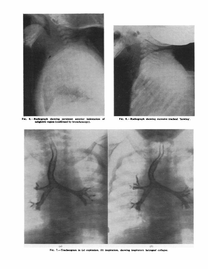
Fw;. 6.-Radiograph showing persistent anterior indentation ofsubgkttic region (confirmed by bronchoscopy).
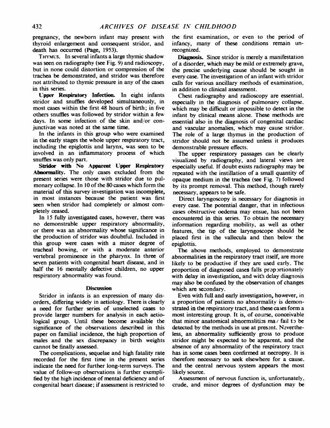
FJG. 8.-Radiograph showing excessive tracheal 'bowmg.
.I..I
*
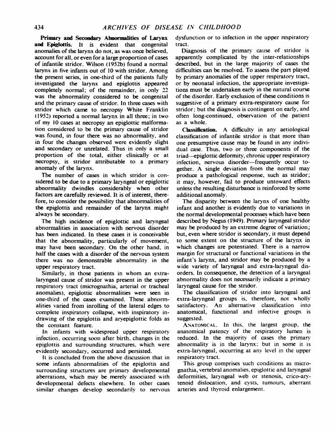
FiG. 7.-Tracheogram in (a) expiration. (b) inspiration, showing inspiratorv laryngea! collapse.

THE INFANT WITH STRIDOR
undiminished. Narrowing of the area of cross-sectionof available space in the airway must occur if theepiglottis is disproportionately large, and a primaryenlargement was in a few cases considered toaccount for the sucking in. In patients with extra-laryngeal narrowing of the airway a similar effectmay be produced; an example was a case withtracheal collapse, where the epiglottis, though smalland firm, was drawn into the larynx on inspiration.
In the second and larger group of cases, however,no anomaly beyond an excessive flabbiness of thetissues was demonstrated. This is apparently due toan exaggeration of the normal characteristics of theinfant larynx, as compared with that of the adult.
larynx. In four patients with stridor due to aprimary intra-laryngeal anomaly the followingconditions were found. In one there was oedema ofthe laryngeal mucosa, which persisted for a fewweeks. This infant produced a tremendous 'crow' oninspiration, and was cyanosed and distressed from afew hours after birth; the oedema and stridorsubsided concurrently. In one newborn infant anabnormal, swollen left arytenoid was found. Inanother (a mongol) the larynx was stenosed, anassociation which has previously been described(Bonham Carter and Lovel, 1951). In the remain-ing case, with aphonia as well as stridor, a persistentindentation in the immediate subglottic region wasseen on radiography (Fig. 6) and confirmed bybronchoscopy, though its nature was not deter-mined.
In two other infants unilateral vocal cord paralysiswas observed. In one it was associated with birthtrauma. In the other it was apparently due toinvolvement of the recurrent laryngeal nerve bygross cardiac enlargement (see p. 426).
Trachea. Intrinsic defects were first considered.
INTRINsIc DrFEcrs. In three cases inspiratorytracheal collapse was the only anomaly demon-strated (Fig. 7). In one superficially similar case thecollapse was considered to be secondary and notprimary (see p. 429).
In a fourth child there was a marked degree oftracheal 'bowing' (Fig. 8) which persisted during thefirst three years of life; stridor also ceased at the sameage. Bending of a tube through which air is passinghas an obstructive effect similar to that producedby narrowing, and it was concluded that the bowingin this case was productive of stridor. In severalother infants with stridor a lesser degree of bowingwas seen, and a minor degree may be observed intracheal radiographs of normal infants. The casedescribed, however, was the only one in which thebowing could not be annulled by full extension ofthe infant's head, and was considered so extremea variation from the normal as to be pathological.
PRESSURE BY ANoMALous ARTERIES. In one patientwith a vascular ring the diagnosis was made from theradiological evidence of distortion and constrictionof the trachea and oesophagus. In two others, withan aberrant left subclavian and an anomalousinnominate artery respectively, the radiologicaldiagnosis was confirmned by angiography (Apley,1949).THYROID. A case of thyroid enlargement with
stridor due to compression of the trachea has beendescribed earlier. It is, perhaps, surprising that thethyroid is not a commoner cause of tracheal obstruc-tion, since in the newborn infant thyroid tissue mayextend between the trachea and oesophagus andthe main part of a goitre may even lie behind thetrachea (Hamne, 1946). Moreover, if the treatmentof goitre with iodine or with thiouracil in adultwomen is continued during the last weeks of
FiG. 9.-Chest radiographs showing large thvmus in a child at (a) 4 months of age wshen stridor was present. and (b) 16 mor.ths whenstridor had ceased. No compression or distortion of the trachea could be demonstrated.
431
ARCHIVES OF DISEASE IN CHILDHOOD
pregnancy, the newborn infant may present withthyroid enlargement and consequent stridor, anddeath has occurred (Page, 1953).
THYMUS. In several infants a large thymic shadowwas seen on radiography (see Fig. 9) and radioscopy,but in none could distortion or compression of thetrachea be demonstrated, and stridor was thereforenot attributed to thymic pressure in any of the casesin this series.
Upper Respiratory Infection. In eight infantsstridor and snuffles developed simultaneously, inmost cases within the first 48 hours of birth; in fiveothers snuffles was followed by stridor within a fewdays. In some infection of the skin and/or con-junctivae was noted at the same time.
In the infants in this group who were examinedin the early stages the whole upper respiratory tract,including the epiglottis and larynx, was seen to beinvolved in an inflammatory process of whichsnuffles was only part.
Stridor with No Apparent Upper RespiratoryAbnormality. The only cases excluded from thepresent series were those with stridor due to pul-monary collapse. In 10 of the 80 cases which form thematerial of this survey investigation was incomplete,in most instances because the patient was firstseen when stridor had completely or almost com-pletely ceased.
In 15 fully investigated cases, however, there wasno demonstrable upper respiratory abnormality,or there was an abnormality whose significance inthe production of stridor was doubtful. Included inthis group were cases with a minor degree oftracheal bowing, or with a moderate anteriorvertebral prominence in the pharynx. In three ofseven patients with congenital heart disease, and inhalf the 16 mentally defective children, no upperrespiratory abnormality was found.
DiscussionStridor in infants is an expression of many dis-
orders, differing widely in aetiology. There is clearlya need for further series of unselected cases toprovide larger numbers for analysis in each aetio-logical group. Until these become available thesignificance of the observations described in thispaper on familial incidence, the high proportion ofmales and the sex discrepancy in birth weightscannot be finally assessed.The complications, sequelae and high fatality rate
recorded for the first time in the present seriesindicate the need for further long-term surveys. Thevalue of follow-up observations is further exempli-fied by the high incidence of mental deficiency and ofcongenital heart disease; if assessment is restricted to
the first examination, or even to the period ofinfancy, many of these conditions remain un-recognized.
Diagnosis. Since stridor is merely a manifestationof a disorder, which may be mild or extremely grave,the precise underlying cause should be sought inevery case. The investigation of an infant with stridorcalls for various ancillary methods of examination,in addition to clinical assessment.
Chest radiography and radioscopy are essential.especially in the diagnosis of pulmonary collapse.which may be difficult or impossible to detect in theinfant by clinical means alone. These methods areessential also in the diagnosis of congenital cardiacand vascular anomalies, which may cause stridor.The role of a large thymus in the production ofstridor should not be assumed unless it producesdemonstrable pressure effects.The upper respiratory passages can be clearly
visualized by radiography, and lateral views areespecially useful. If doubt exists radiography may berepeated with the instillation of a small quantity ofopaque medium in the trachea (see Fig. 7) followedby its prompt removal. This method, though rarelynecessary, appears to be safe.
Direct laryngoscopy is necessary for diagnosis inevery case. The potential danger, that in infectiouscases obstructive oedema may ensue, has not beenencountered in this series. To obtain the necessaryinformation regarding mobility, as well as otherfeatures, the tip of the laryngoscope should beplaced first in the vallecula and then below theepiglottis.The above methods, employed to demonstrate
abnormalities in the respiratory tract itself, are morelikely to be productive if they are used early. Theproportion of diagnosed cases falls prop )rtionatelywith delay in inve,tigation, and with delay diagnosismay also be confused by the observation of changeswhich are sezondary.Even with full and early investigation, however, in
a proportion of patients no abnormality is demon-strated in the respiratory tract, and these cases form amost interesting group. It is, of course, conceivablethat minor anatomical abnormaliti-s ma i fail to bedetected by the methods in use at pres-nt. N-verthe-less, an abnormality sufficiently gross to producestridor might be expected to be apparent, and theabsence of any abnormality of the respiratory tracthas in some cases been confirmed at necropsy. It istherefore necessary to seek elsewhere for a cause.and the central nervous system appears the mostlikely source.
Assessment of nervous function is, unfortunately,crude, and minor degrees of dysfunction may be
432

THE INFANT WITH STRIDOR
undetectable. Electro-encephalography for thispurpose was found to be unhelpful, and the mostsatisfactory method available is long-term clinicalobservation with particular attention to mental andneuromuscular development. Even so, in a smallproportion of cases no evidence of abnormalityother than transient stridor is found; in these it istempting to speculate that stridor is produced bytemporary nervous dysfunction.
Nervous Dysfunctio and Stridor. In half the casesof disorder of the central nervous system in which thelarynx was examined an epiglottic or laryngealabnormality was found; there was, therefore, in thisgroup a high incidence of these abnormalities, ascompared with normal infants. The abnormality wasusually one of movement, i.e., the epiglottis andarytenoids were drawn into the larynx on inspiration,just as occurs with the so-called primary lax epig-lottis; in a few cases the epiglottis was also markedlyinrolled, and in one it was folded transversely onitself.
In one infant, with clinical signs of markedcerebral irritation from birth, respiratory difficultyand stridor began at the same time. They graduallyreceded after the signs of intracranial disturbancedisappeared, finally to be lost in the fifth month.Laryngoscopy and other examinations were repeated,but no abnormality could be demonstrated in therespiratory systemi. This case probably correspondsto those where intracranial damage, producingstridor inter alia, has been confirmed at necropsy(Thomas, 1921).
In two fatal cases with meningo-myelocoele andparalysis of the legs and anal sphincter, clinical andvost-mortem evidence could be collated. In the first(Table 3, Case 1) the respiratory tract appearednormal in all respects at necropsy; nevertheless,during life, with each inspiration the epiglottis wasdrawn into the larynx so forcibly as to obstruct theairway almost completely. Moreover, when theepiglottis was grasped with forceps the stridor wasabolished. In the second (Table 3, Case 4) noabnormality of the respiratory tract was found dur-ing life or after death. In these two cases the absenceof an anatomical abnormality of the respiratorytract makes it reasonable to attribute the stridorto the gross neurological anomalies, even thoughdetailed study of the central nervous system was notfeasible.
In Case 9 (Table 3) the brain at necropsy appearedmacroscopically normal, though the child had beenmentally defective and spastic, and had had achalasiaof the cardia. The only noteworthy feature in theupper respiratory tract was moderate infolding of thelateral epiglottic margins; the stridor, like the
achalasia, was therefore considered to have beenproduced by neurological dysfunction. Anotherinfant with a clinically normal respiratory tract wasblind and spastic and mentally defective.
In the above cases the cause of stridor couldreasonably be attributed to nervous dysfunction.In a larger number of children with mental defect,but unaccompanied by gross physical anomaliesor spasticity, stridor also occurred. In these theevidence justifies only a statement that the two condi-tions are frequently associated, though there mayconceivably be a causal relationship similar to thatalready considered.Upper Respiratory Infection as a Cause of Persis-
tent Stridor. Observations on the association betweenstridor and upper respiratory infection, and thesequence of events in these cases, have previouslybeen described. It is suggested that infection canproduce dysfunction and secondary deformitiesin the larynx which may, I think erroneously, beconsidered as primary.The association observed between upper respira-
tory infection and stridor would be expected fromvarious factors which apply particularly to the infant.They have been clearly summarized by Holinger andJohnston (1950). The first is the smal size of theinfant larynx. The glottic orifice of the newborninfant has a cross-sectional area of 14 sq. mm.Oedema to a depth of 1 mm., which would give riseto hoarseness in the adult, will reduce this area to5 sq. mm. and give rise to respiratory difficulty andstridor in the infant. The second factor is thearrangement of the mucosa and sub-mucosa. Themucosa, while firmly attached to the posteriorsurface of the epiglottis, is loosely attached on theanterior surface and along the aryepiglottic folds(Lederer, 1946); inflammatory extravasation andoedema will accordingly be localized mainly to thefront and sides of the epiglottis, and will cause itslateral edges to curl in and the tip to be bent back-wards. From the changes seen in infants who wereexamined frequently from the earliest stages itappears that if these induced deformities are main-tained for a sufficient length of time they may persisteven after the inflammatory reaction has subsided.
In a survey of 30 infants with stridor Bowman andJackson (1939) attributed the stridor in two tohypertrophic laryngitis. Stridor beginning withacute laryngitis may therefore persist if the laryngitisbecomes chronic; but the observations recorded heresuggest that even when laryngitis has resolved stridormay persist. It does so evidently because of persistentdeformity of the epiglottis and neighbouring tissueswhich develops while a transient infection ispresent.
433
ARCHIVES OF DISEASE IV CHILDHOOD
Primar and Secondary Abnormalities of Larynxand Epiglottis. It is evident that congenitalanomalies of the larynx do not, as was once believed,account for all, or even for a large proportion of casesof infantile stridor. Wilson (1952b) found a normallarynx in five infants out of 10 with stridor. Amongthe present series, in one-third of the patients fullyinvestigated the larynx and epiglottis appearedcompletely normal; of the remainder, in only 22
was the abnormality considered to be congenitaland the primary cause of stridor. In three cases withstridor which came to necropsy White Franklin(1952) reported a normal larynx in all three; in twoof my 10 cases at necropsy an epiglottic malforma-tion considered to be the primary cause of stridorwas found, in four there was no abnormality, andin four the changes observed were evidently slightand secondary or unrelated. Thus in only a smallproportion of the total, either clinically or atnecropsy, is stridor attributable to a primaryanomaly of the larynx.The number of cases in which stridor is con-
sidered to be due to a primary laryngeal or epiglotticabnormality dwindles considerably when otherfactors are carefully reviewed. It is of interest, there-fore, to consider the possibility that abnormalities ofthe epiglottis and remainder of the larynx mightalways be secondary.The high incidence of epiglottic and laryngeal
abnormalities in association with nervous disorderhas been indicated. In these cases it is conceivablethat the abnormality, particularly of movement,may have been secondary. On the other hand, in
half the cases with a disorder of the nervous systemthere was no demonstrable abnormality in theupper respiratory tract.
Similarly, in those patients in whom an extra-laryngeal cause of stridor was present in the upper
respiratory tract (micrognathia, arterial or trachealanomalies), epiglottic abnormalities were seen inone-third of the cases examined. These abnorm-alities varied from inrolling of the lateral edges tocomplete inspiratory collapse, with inspiratory in-
drawing of the epiglottis and aryepiglottic folds as
the constant feature.In infants with widespread upper respiratory
infection, occurring soon after birth, changes in theepiglottis and surrounding structures, which were
evidently secondary, occurred and persisted.It is concluded from the above discussion that in
some infants abnormalities of the epiglottis andsurrounding structures are primary developmentalaberrations, which may be merely associated withdevelopmental defects elsewhere. In other cases
similar changes develop secondarily to nervous
dysfunction or to infection in the upper respiratorytract.
Diagnosis of the primary cause of stridor isapparently complicated by the inter-relationshipsdescribed, but in the large majority of cases thedifficulties can be resolved. To assess the part playedby primary anomalies of the upper respiratory tract,or by neonatal infection, the appropriate investiga-tions must be undertaken early in the natural courseof the disorder. Early exclusion of these conditions issuggestive of a primary extra-respiratory cause forstridor; but the diagnosis is contingent on early, andoften long-continued, observation of the patientas a whole.
Classification. A difficulty in any aetiologicalclassification of infantile stridor is that more thanone presumptive cause may be found in any indivi-dual case. Thus, two or three components of thetriad-epiglottic deformity, chronic upper respiratoryinfection, nervous disorder-frequently occur to-gether. A single deviation from the normal mayproduce a pathological response, such as stridor;it may, however, fail to produce untoward effectsunless the resulting disturbance is reinforced by someadditional anomaly.The disparity between the larynx of one healthy
infant and another is evidently due to variations inthe normal developmental processes which have beendescribed by Negus (1949). Primary laryngeal stridormay be produced by an extreme degree of variation;but, even where stridor is secondary, it must dependto some extent on the structure of the larynx inwhich changes are potentiated. There is a narrowmargin for structural or functional variations in theinfant's larynx, and stridor may be produced by awide variety of laryngeal and extra-laryngeal dis-orders. In consequence, the detection of a laryngealabnormality does not necessarily indicate a primarylaryngeal cause for the stridor.The classification of stridor into laryngeal and
extra-laryngeal groups is, therefore, not whollvsatisfactory. An alternative classification intoanatomical, functional and infective groups issuggested.ANATOMICAL. In this, the largest group, the
anatomical patency of the respiratory lumen isreduced. In the majority of cases the primaryabnormality is in the larynx: but in some it isextra-laryngeal, occurring at anv level in the upperrespiratory tract.
This group comprises such conditions as micro-gnathia, vertebral anomalies, epiglottic and laryngealdeformities, laryngeal web or stenosis, crico-ary-tenoid dislocation, and cysts, tumours, aberrantarteries and thyroid enlargement.
434

THE INFANT WITH STRIDOR 435FUNCTONAL. In this group the functional patency
of the lumen is reduced. The primary abnormalitymay occur in the respiratory tract or in the nervoussystem.Examples of the former are tracheal collapse and
inspiratory indrawing of the anatomically normalepiglottis. Nervous disorders may be peripheral(affecting the recurrent laryngeal nerve) or, morecommonly, central. Included in this group are therare neonatal tetany and laryngismus stridulus, anddevelopmental and traumatic disorders of the brain.
INFECTIVE. In this group the respiratory lumen isnarrowed by inflammatory processes, the effects ofwhich may persist even when the inflammation hassubsided.
This group comprises many of those cases in whichstridor and widespread upper respiratory infectiondevelop concurrently in the early neonatal period.
Observations are recorded on 80 cases with stridordeveloping in early infancy. To determine thenatural history of the condition progress wasassessed clinically and by ancillary methods forperiods up to several years. Attention was directednot only to the sequence of any changes in the larynx,but also to general physical and mental development.
Stridor occurred in 11 of 14 siblings in fivefamilies. In the whole series there was a male pre-ponderance, in the ratio of five boys to three girls.The average birth weight of males was significantlyhigher than that of females with stridor, and of un-selected infants of either sex.
In seven cases a congenital cardiac malformationwas present, though in only one was it the cause ofstridor. Three patients in the series were mongols;16 others were mentally retarded.Feeding difficulties occurred in half the total
number. Pulmonary complications were frequent.Chest deformities developed in many cases but werealmost invariably transient.
Eleven patients died, and in 10 necropsy wasperformed. Apart from two cases of meningo-
myelocoele and one of thyroid enlargement, deathwas invariably due to pulmonary infection.
In the largest group of cases stridor was producedby an anatomical anomaly which impaired thepatency of the upper respiratory tract; in themajority of these the anomaly was epiglottic. In asecond group stridor was considered to be due todysfunction of the nervous system. In a thirdgroup stridor originated with an upper respiratoryinfection.The mechanism of production of stridor in these
groups is discussed, with particular reference toprimary and secondary changes in the larynx. It isemphasized that in the investigation of infantilestridor it is essential to make early and repeatedobservations, not only of the larynx and respiratorytract, but of the patient as a whole.
It is a pleasure to acknowledge my indebtedness tomany colleagues, without whom this survey could nothave been completed. They include Mr. A. D. Batemanand Mr. H. D. Fairman for instruction and advice,and Mr. V. E. Negus and Mr. J. Angell James for helpfulsuggestions on laryngological aspects: Professor A. V.Neale and Dr. B. D. Corner for allowing me access tosome of their cases; Drs. H. J. Johnson, J. M. Sheachand A. C. P. D. Thomson for their cooperation in theradiology of the respiratory tract: Professor T. F. Hewerand Drs. J. Heathcote, C. D. Cross and M. C. H.Dodgson for advice on pathological specimens: andDr. G. Herdan for help with statistical problems.
REFEENXCES
Apley, J. (1949). Proc. roy. Soc. Med.. 42, 918.Bonham Carter, R. E. and LoveL, K. (1951). Ibid.. 44. 293.Bowman, J. E. and Jackson, C. L. (1939). J. Pediat., 15. 476.Caffey. J. (1950). Pediatric X-ray Diagnosis, 2nd ed., Chicago.Ckerf, L. H. (1950). J. Amer. med. Ass., 143.1231.Finlay, H. V. L. (1949). Archives of Diseases in Childhood. 24. 219.Franklin, A. White (1952). Proc. roy. Soc. Med.. 45. 358.Hamne. B. (1946). Act. med. scand._ Suppl. 170. p. 399.Holinger, P. H. and Johnston, K. C. (1950). J .4mer. med. Ass.. 143,
1229.Lederer. F. L. (1946). Diseases of the Ear, Nose and Throat, 5th ed.,
Philadelphia.Naish, J. and Wallis, H. R. E. (1948). Brit. med. J., 1. 541.Negus, V. E. (1949). The Comparative Anatomy and Physiology of the
Larynx, London.Page, A. P. M. (1953). Personal communication.Schwartz. L. (1944). Arch. otolatryng., Chicago. 39,403.Tlhomas, -. (1921). Mschr. Kinderheilk., 22, 223.Wilson, T. G. (1952a). Proc. roy. soc. Med., 45. 355.- (1952b). J. Laryng., 66, 437.