the influence of natural head position on the cervical...
TRANSCRIPT

Research ArticleThe Influence of Natural Head Position on the CervicalSagittal Alignment
Kuan Wang,1,2 Zhen Deng,1,3 Zhengyan Li,1,4 Huihao Wang,1,4 and Hongsheng Zhan1,4
1Shi’s Center of Orthopedics and Traumatology, Shuguang Hospital Affiliated to Shanghai University of TCM,Shanghai 201203, China2Tongji Hospital, Tongji University School of Medicine, Shanghai 200092, China3Baoshan Branch, Shuguang Hospital Affiliated to Shanghai University of TCM, Shanghai 201900, China4Institute of Traumatology & Orthopedics, Shanghai Academy of TCM, Shanghai 201203, China
Correspondence should be addressed to Huihao Wang; [email protected] andHongsheng Zhan; [email protected]
Received 24 March 2017; Revised 6 June 2017; Accepted 3 July 2017; Published 13 August 2017
Academic Editor: Wei Yao
Copyright © 2017 Kuan Wang et al. This is an open access article distributed under the Creative Commons Attribution License,which permits unrestricted use, distribution, and reproduction in any medium, provided the original work is properly cited.
Introduction. This study investigated the relationship between the parameters related to the natural head position and cervicalsegmental angles and alignment of patients with neck pain. Material and Methods. The lateral radiographs of the cervical spinewere collected from 103 patients and were used to retrospectively analyze the correlation between the natural headposition, cervical local sagittal angles, and alignment. Sagittal measurements were as follows: cervical curvatureclassification, slope of McGregor’s line (McGS), local sagittal angles (C0–C2 angle, C2–C5 angle, C5–C7 angle, and C2–C7angle), T1 slope, center of gravity of the head to sagittal vertical axis (CG–C7 SVA), and local sagittal alignment (C0–C2SVA and C2–C7 SVA). Results. McGS was significantly correlated to C0–C2 angle (r = 0 57), C0–C2 SVA (r = −0 53), C2–C7SVA (r = −0 28), and CG–C7 SVA (r = −0 47). CG–C7 SVA was also significantly correlated to curvature type (r = 0 27), C5–C7angle (r = −0 37), and C2–C7 angle (r = −0 39). Conclusions. A backward shift with an extended head position may accompanya relatively normal curvature of the cervical spine. The effect of posture control in relieving abnormal mechanical state of thecervical spine needs to be further confirmed by biomechanical analysis.
1. Introduction
Cervical curvature is one of the clinical assessments whichcould reflect the mechanical state of the cervical spine. Witha normal lordotic curvature, the least amount of energy isspent to maintain the horizontal gaze in the upright position[1]. Under this condition, tensile and compression loads onspinal structures are smallest compared to other cervicalalignments [2, 3].
The cervical spine connects the skull and thoracic verte-brae and supports the mass of the head. It is a multijointstructure that allows complex movement of the neck. Inrecent years, many studies have used X-ray imaging toexplore the relationship between parameters related to cervi-cal sagittal alignment, involving the mass point of the head,C2 vertebral center, T1 slope, and other measurements [4].
Lee et al. found that T1 slope and C2–C7 Cobb angle werestrongly correlated [5], whereas the cranial offset andC2–C7 Cobb angle were in moderate correlation. Nunez-Pereira et al. found that the occiput–C2 angle and C2–C7Cobb angle also showed a moderate correlation [6]. Tanget al. found a close relationship between the C2–C7 sagittalvertical axis (C2–C7 SVA) and health-related quality of lifein patients who underwent surgery of the cervical spine [7].
The above studies have shown that the segmental angle,alignment of the cervical spine, and the position of the firstthoracic spine were interdependent and may contribute tothe progression of cervical degeneration and quality of life.These studies focused a lot on the effect of cervicothoracicjunction towards sagittal alignment of the cervical spine,but less attention was paid to the head position. As alreadyknown, the head position could be controlled freely by our
HindawiJournal of Healthcare EngineeringVolume 2017, Article ID 2941048, 7 pageshttps://doi.org/10.1155/2017/2941048

mind in a certain range of motion, and it needs a littlemoment to move the head in a neutral zone [8]. For a propertransmission of the head weight, there should be somerelationship between the position of the head in the naturalstanding position, the local sagittal angles, and the alignmentof the cervical spine. The natural head position in the sagittalplane may include the rotation and translation degrees offreedom, which could be represented by a head tilt andforward/backward head shift [9] or by McGregor slope(McGS) and C0–C7 sagittal vertical axis (CG–C7 SVA) [10,11]. We hypothesized that these parameters of the head posi-tion have some impacts on the cervical segmental angles andalignment.
Therefore, the objective of this study was to investigatethe relationship between the parameters related to the natu-ral head position and the cervical alignment, through retro-spectively analyzing the cervical lateral radiographs of 103patients with neck pain.
2. Materials and Methods
2.1. Study Design. This study was a retrospective data anal-ysis of consecutive patients who underwent sagittal planecervical spine X-ray from January 2015 to January 2016.Inclusion criteria were adult neck pain patients (>18 yearsof age), with symptoms like neck stiffness, pain, and localtenderness, with or without limited cervical activity. TheirX-ray examination can be normal or with mild degenera-tion. Exclusion criteria included patients with ankylosingspondylitis, tumor, cervical fracture or dislocation, diffuseidiopathic bone hypertrophy, and other specific diseasesor mutations.
2.2. Radiographic Analysis. The lateral radiographs of thecervical spine were taken when patients were in the standingposition. The measurements included cervical curvature clas-sification (Figure 1) and parameters related to the headposition and cervical alignment (Figure 2). The entire mea-surements included the following parameters:
(1) Cervical curvature classification [12]: the cervicalcurvature was divided into three types including lor-dosis, straight/sigmoid, and kyphosis as types 1–3. Toclassify the curvature, a line was drawn to connect themidpoint of the C2 inferior end plate and C7 superiorend plate. Then, the center of each vertebral bodyfromC3 toC6was located by connecting the diagonalsof each vertebral body in the sagittal plane, and the dis-tance from each center to line AB (see Figure 1) wasmeasured. If all centers of vertebral bodies were infrontof the line and themaximumdistancewasgreaterthan 2mm, the curvature was classified into thelordosis group; if the centers were in front of or behindthe line but the maximum value of distance was nomore than 2mm, the curvature was classified into thestraight group. For the sigmoid group, the centerswerein front of or behind the line, but the maximum valueof distance was greater than 2mm. For the kyphosisgroup, the centers were all behind the line connectingthe C2–C7 endplates, and the maximum value ofdistance was greater than 2mm.
(2) Slope of McGregor’s line (McGS) [13]: McGregorslopewasdefinedas the slopeof the line that connectedthe posterior margin of the hard palate and foramenmagnum against the horizontal plane. This value wasreported to be strongly correlated with the chin-browvertical angle (CBVA) (r = 0 862) which was onemeasurement of horizontal gaze. So this value wasused in the current study as an angular assessment ofthe natural head position. A positive value means thatthe head was in the extension position with ascendinggaze, and a negative value means that the head was inthe flexion position with descending gaze.
(3) Local sagittal angles: local sagittal angles includedC0–C2 angle, C2–C5 angle, C5–C7 angle, and C2–C7 Cobb angle. C0–C2 angle was defined as the anglebetween the inferior endplates of C2 and McGregor’sline [6]. C5–C7 angle denoted the angle between theC5 superior endplate and C7 inferior endplate [14].
C2
C7
A
B
A
B
A
B
A
B
≥2 mm <2 mm
≥2 mm
≥2 mm
Figure 1: The cervical curvature types (from left to right: lordosis, straight, sigmoid, and kyphosis).
2 Journal of Healthcare Engineering

C2–C7 Cobb angle denoted the angle between the C2inferior endplate and C7 inferior endplate [5]. In thecurrent study, to see the effect of the head position onthe middle cervical spine, C2–C5 angle was defined asthe value of C2–C7 Cobb angle subtracted by C5–C7angle.
(4) T1 slope [10]: T1 slope was defined as the anglebetween the line that was parallel to the superiorend plate of T1 and the horizontal plane.
(5) The center of gravity of the head to sagittal verticalaxis (CG–C7 SVA): CG–C7 SVA was defined asthe distance between a plumb line dropped fromthe anterior margin of the external auditory canaland the posterior superior corner of C7. This valuereflected relative translation of the head against theC7 vertebra [10].
(6) Local sagittal alignment: C2–C7 sagittal vertical axis(C2–C7 SVA) was defined as the distance between aplumb line dropped from the centroid of C2 andthe posterior superior corner of C7 [7]. This valuereflected the relative translation of the C2 vertebraagainst the C7 vertebra. To see the effect of the headposition on the sagittal alignment of the upper cervi-cal spine, we defined the C0–C2 sagittal vertical axis(C0–C2 SVA) as the value of CG–C7 SVA subtractedby C2–C7 SVA.
The above parameters concerning the head position andcervical alignment have been widely used in previous studieswith good reliability [10]. These measurements were proc-essed independently by two experienced radiologists, andthe measured values were averaged to produce the results.
The study protocol was approved by the medical ethics com-mittee of our hospital.
To investigate the effect of the natural head position onthe cervical alignment, the subjects were divided into fourgroups according to their CG–C7 SVA and McGS values.The median CG–C7 SVA was used for the division of theforward shift head position or backward shift head position.Lafage el al. reported the mean value of McGS and equationsgenerated with linear regression between CBVA and McGS[13]. According to their results, 0.87° was the mean value ofMcGS, and this value was used for the division of theextended or flexed cranial position. Therefore, in terms oftranslation and rotation of the head position in the sagittalplane, four groups included forward shift with the extendedhead position (FE), backward shift with the extended headposition (BE), forward shift with the flexed head position(FF), and backward shift with the flexed head position (BF).
2.3. Statistical Analysis. Data were analyzed by SPSS 20.0software. Values were presented as mean± SD. Spearman’scorrelation analyses were performed to examine associationsamong the selected variables. The curvature type numbers 1–3 were treated as ordinal categorical variables in the currentstudy. If the curvature type number is negatively correlatedwith a local segmental angle, an increase in the curvature typenumber is correlated with a decrease in the angle. This resultsshould be interpreted as that the curvature type numberwould be more close to 1 (lordosis type) rather than 3(kyphosis type), with a decrease in the local segmental angle.The comparison of each radiographic parameter among fourhead positions was determined by using one-way ANOVAwith Bonferroni’s post hoc test. A P value equal to or lessthan 0.05 was set up as threshold for statistical significance.
McGS
T1 slope CG–C7 SVA
C2–C7 SVAC5–C7 angle
C2–C7 angle
C0–C2 angle
C2–C5 angle C0–C2 SVA
Figure 2: The measurement of parameters related to natural head position and cervical alignment (McGS, slope of McGregor’s line; SVA,sagittal vertical axis).
3Journal of Healthcare Engineering

3. Results
3.1. Demographic and Cervical Curvature Classification.There were 103 subjects included in this study. Among thesesubjects, 33 subjects were male and 70 were female. Themean age was 37.4± 12.3 years. There were 35 subjects allo-cated to the lordosis group, 56 subjects to the straight or sig-moid group (only one subject was classified as sigmoidcurvature; thus, the two groups were combined), and 12 sub-jects to the kyphosis group. The mean age of each group was39.4± 13.2, 37.3± 11.4, and 32.7± 13.6 years, respectively.There was no significant difference in age and genderbetween each group (P > 0 05).
3.2. Measurement of Parameters and Correlation Analysis.The results of radiographic measurements related to the headposition and cervical alignment are shown in Table 1. Theresults of Spearman’s correlation analyses are shown inTable 2. McGS was found to be significantly correlated toC0–C2 angle (r = 0 57), C0–C2 SVA (r = −0 53), C2–C7SVA (r = −0 28), and CG–C7 SVA (r = −0 47) but not signif-icantly correlated to curvature classification, C2–C5 angle,C5–C7 angle, and C2–C7 angle. CG–C7 SVA was also signif-icantly correlated to curvature type (r = 0 27), C5–C7 angle(r = −0 37), and C2–C7 angle (r = −0 39).
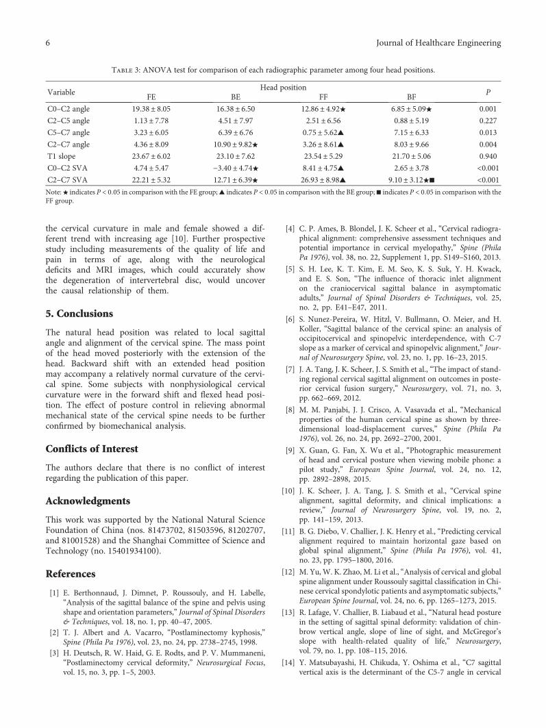
3.3. The Influence of Different Head Positions. Figure 3 showsthe distribution of cervical curvatures in different headpositions. Among the four groups (FE, BE, BF, and FFgroups), the proportion of subjects with lordosis curvaturein the BE group was most compared to those in othergroups, and the proportion of subjects with straight orsigmoid curvature was most in the FE group. In the FFgroup, the proportion of subjects with kyphosis curvaturewas most compared to those in other groups. There wereonly four subjects in the BF group, with three subjectsclassified with straight/sigmoid curvatures. Table 3 showsthe comparison of each radiographic parameter amongfour head positions determined by one-way ANOVAwith Bonferroni’s post hoc test. C0–C2 angle, C5–C7angle, C2–C7 angle, C0–C2 SVA, and C2–C7 SVA aresignificantly different among the four head positions. C2–
C5 angle and T1 slope are not significantly differentamong the four head positions.
4. Discussion
In this study, the lateral radiographs of the cervical spinewere used to analyze the relationship between the naturalhead position and cervical alignment. The measured param-eters related to cervical alignment (e.g., C2–C7 angle and T1slope) were consistent with the reported data [5, 6, 12]. Thisstudy provides some insight into the effect of the head posi-tion on the cervical spine alignment.
Since T1 was the fixed end of the cervical spine and thecervical column is influenced by the weight of the head, it isexpected that the T1 slope might play a determining role inthe curvature of the cervical spine. In the current study, T1slope was in positive correlation with C2–C7 angle. Theresults agreed with the data reported by Lee et al. [5].
The translation or rotation of the head is controlled bythe nervous system to keep an economic posture [14]. Dueto the large vision field, people could maintain a horizontalgaze with varying head positions within a certain range. Inthe current study, the CG–C7 SVA was found to be in nega-tive correlation with curvature type. This means that it ismore likely to be kyphotic of the cervical spine with the ante-rior translation of the head. Furthermore, the CG–C7 SVAwas found to be negatively correlated with C5–C7 angleand C2–C7 angle but not significantly correlated withC2–C5 angle. This result suggested that a forward shiftof the head in the natural head position might indicate aloss of lordosis in the lower cervical spine.
This study also found that the extended or flexedposition of the head represented by McGS was notcorrelated with local sagittal angle except C0–C2 angle.This might be because most flexion and extension rangeof motion is found at C0–C2 compared to mid andlower cervical regions [8]. Thus, it has a great compen-satory space for the flexion/extension of the head andminimizes the impact on the local segmental anglebelow C2. Additionally, the McGS was found to be innegative correlation with C0–C2 SVA, C2–C7 SVA,and CG–C7 SVA, which means that the mass point ofthe head moves posteriorly with the extension of thehead. The results suggested that the rotation in the nat-ural head position might be indirectly involved in thebalance control of the cervical spine by adjusting thetranslation of the head. A study by Tang et al. foundthat patients who underwent posterior cervical fusionsurgery reported a decrease in the quality of life withan increased value of C2–C7 SVA [7]. According toour results, the anterior translation of the head mightaccompany cranial flexion as well as a flattening of thecervical lordosis. To balance the extensor moment pro-duced by the flexed head, the extensor muscles need toproduce additional extension torque and the fatigue ofthese muscles might lead to neck or upper back symp-toms. Therefore, it might be more appropriate to keepthe head in a neutral position rather than a flexed posi-ton to prevent neck and back pain.
Table 1: Radiographic measurements related to natural headposition and cervical alignment.
Variable N Min Max Mean SD
McGS (°) 103 −12.00 20.90 6.14 6.10
C0–C2 angle (°) 103 0.80 34.80 16.61 7.39
C2–C5 angle (°) 103 −19.70 21.50 2.88 7.71
C5–C7 angle (°) 103 −11.80 17.30 4.52 6.61
C2–C7 angle (°) 103 −16.80 30.30 7.40 9.55
T1 slope (°) 103 8.70 43.40 23.31 6.64
C0–C2 SVA (mm) 103 −16.50 22.30 1.36 6.77
C2–C7 SVA (mm) 103 −4.60 49.80 17.91 8.54
CG–C7 SVA (mm) 103 −12.00 72.10 19.27 13.12
McGS: slope of McGregor’s line; SVA: sagittal vertical axis.
4 Journal of Healthcare Engineering

With the development of modern electronic devices, aforward head posture is common among people [15] andthe forward head posture has been associated with muscu-loskeletal pain in previous studies [16, 17]. Although thetotal number of subjects with kyphosis curvature wassmall in the current study, the proportion of subjects withthe kyphosis type curvature was most in the FF group.This result suggested that the forward shift with the flexedhead position may be associated with cervical kyphosisthus leading to the unbalance of a mechanical state. Incontrast, the proportion of the lordosis type was most in
the BE group, meaning that a backward shift with anextended head position was more likely to be accompaniedwith a normal cervical curvature. The cervical spine couldbe divided into three columns (one vertebral body and twofacet joints on the same level) [10]. With the backwardshift and the extended head position, the weight of thehead would be more loaded on the posterior column ofthe cervical spine. Since we found that C2–C5 angle wasnot significantly different among the four head positions,this position (BE) might relieve the load on the interverte-bral disc of C5–C7 segments and be helpful in compensat-ing the additional load caused by nonphysiological cervicalcurvature like kyphosis and straight/sigmoid type. In thecurrent study, some subjects with kyphotic curvature werefound to have a forward shift and flexed position of thehead. Whether posture control (e.g., keeping a backwardshift and extended head position) is helpful to thesesubjects in relieving abnormal mechanical state of thecervical spine needs to be further confirmed by biome-chanical analysis.
Some limitations existed in this study. Since thisstudy was a retrospectively radiograph analysis, it couldnot accurately reflect the causal relationship betweeneach parameter and no healthy subjects as the controlgroup. Age is also an important factor affecting the cur-vature. In the current study, there is a trend that theage of subjects with nonphysiological cervical curvature(straight, sigmoid, and kyphosis) become younger. Also,
Table 2: The correlation analysis between parameters.
VariableCurvature
typeMcGS
C0–C2angle
C2–C5angle
C5–C7angle
C2–C7angle
T1slope
C0–C2SVA
C2–C7SVA
CG–C7SVA
Curvaturetype
r1
−0.11 0.38 −0.61 −0.33 −0.73 −0.40 0.42 0.06 0.27
P 0.29 <0.01 <0.01 <0.01 <0.01 <0.01 <0.01 0.55 0.01
McGSr
10.57 0.05 0.08 0.12 −0.05 −0.53 −0.28 −0.47
P <0.01 0.63 0.42 0.24 0.59 <0.01 <0.01 <0.01
C0–C2 angler
1−0.51 −0.17 −0.53 −0.07 0.12 0.18 0.18
P <0.01 0.08 <0.01 0.46 0.24 0.07 0.07
C2–C5 angler
1−0.12 0.69 0.29 −0.46 0.14 −0.16
P 0.24 <0.01 <0.01 <0.01 0.15 0.11
C5–C7 angler
10.58 0.48 −0.22 −0.38 −0.37
P <0.01 <0.01 0.03 <0.01 <0.01
C2–C7 angler
10.55 −0.53 −0.15 −0.39
P <0.01 <0.01 0.13 <0.01
T1 sloper
1−0.05 0.28 0.13
P 0.61 <0.01 0.21
C0–C2 SVAr
10.39 0.79
P <0.01 <0.01
C2–C7 SVAr
10.85
P <0.01
CG–C7 SVAr
1P
Note: bold font indicates statistical significance (P < 0 05). McGS: slope of McGregor’s line; SVA: sagittal vertical axis.
37
24.32%
64.87%
10.81%48
47.92%
41.67%
10.41%14
14.29%
64.28%
21.43%4
25.00%
75.00%
FE BE FF BF
Figure 3: The proportion of each cervical curvature type and totalnumber of subjects in different head position (FE, forward shiftwith extended head position; BE, backward shift with extendedhead position; FF, forward shift with flexed head position; BF,backward shift with flexed head position; black square, lordosis;gray square, straight or sigmoid; white square, kyphosis).
5Journal of Healthcare Engineering
the cervical curvature in male and female showed a dif-ferent trend with increasing age [10]. Further prospectivestudy including measurements of the quality of life andpain in terms of age, along with the neurologicaldeficits and MRI images, which could accurately showthe degeneration of intervertebral disc, would uncoverthe causal relationship of them.
5. Conclusions
The natural head position was related to local sagittalangle and alignment of the cervical spine. The mass pointof the head moved posteriorly with the extension of thehead. Backward shift with an extended head positionmay accompany a relatively normal curvature of the cervi-cal spine. Some subjects with nonphysiological cervicalcurvature were in the forward shift and flexed head posi-tion. The effect of posture control in relieving abnormalmechanical state of the cervical spine needs to be furtherconfirmed by biomechanical analysis.
Conflicts of Interest
The authors declare that there is no conflict of interestregarding the publication of this paper.
Acknowledgments
This work was supported by the National Natural ScienceFoundation of China (nos. 81473702, 81503596, 81202707,and 81001528) and the Shanghai Committee of Science andTechnology (no. 15401934100).
References
[1] E. Berthonnaud, J. Dimnet, P. Roussouly, and H. Labelle,“Analysis of the sagittal balance of the spine and pelvis usingshape and orientation parameters,” Journal of Spinal Disorders& Techniques, vol. 18, no. 1, pp. 40–47, 2005.
[2] T. J. Albert and A. Vacarro, “Postlaminectomy kyphosis,”Spine (Phila Pa 1976), vol. 23, no. 24, pp. 2738–2745, 1998.
[3] H. Deutsch, R. W. Haid, G. E. Rodts, and P. V. Mummaneni,“Postlaminectomy cervical deformity,” Neurosurgical Focus,vol. 15, no. 3, pp. 1–5, 2003.
[4] C. P. Ames, B. Blondel, J. K. Scheer et al., “Cervical radiogra-phical alignment: comprehensive assessment techniques andpotential importance in cervical myelopathy,” Spine (PhilaPa 1976), vol. 38, no. 22, Supplement 1, pp. S149–S160, 2013.
[5] S. H. Lee, K. T. Kim, E. M. Seo, K. S. Suk, Y. H. Kwack,and E. S. Son, “The influence of thoracic inlet alignmenton the craniocervical sagittal balance in asymptomaticadults,” Journal of Spinal Disorders & Techniques, vol. 25,no. 2, pp. E41–E47, 2011.
[6] S. Nunez-Pereira, W. Hitzl, V. Bullmann, O. Meier, and H.Koller, “Sagittal balance of the cervical spine: an analysis ofoccipitocervical and spinopelvic interdependence, with C-7slope as a marker of cervical and spinopelvic alignment,” Jour-nal of Neurosurgery Spine, vol. 23, no. 1, pp. 16–23, 2015.
[7] J. A. Tang, J. K. Scheer, J. S. Smith et al., “The impact of stand-ing regional cervical sagittal alignment on outcomes in poste-rior cervical fusion surgery,” Neurosurgery, vol. 71, no. 3,pp. 662–669, 2012.
[8] M. M. Panjabi, J. J. Crisco, A. Vasavada et al., “Mechanicalproperties of the human cervical spine as shown by three-dimensional load-displacement curves,” Spine (Phila Pa1976), vol. 26, no. 24, pp. 2692–2700, 2001.
[9] X. Guan, G. Fan, X. Wu et al., “Photographic measurementof head and cervical posture when viewing mobile phone: apilot study,” European Spine Journal, vol. 24, no. 12,pp. 2892–2898, 2015.
[10] J. K. Scheer, J. A. Tang, J. S. Smith et al., “Cervical spinealignment, sagittal deformity, and clinical implications: areview,” Journal of Neurosurgery Spine, vol. 19, no. 2,pp. 141–159, 2013.
[11] B. G. Diebo, V. Challier, J. K. Henry et al., “Predicting cervicalalignment required to maintain horizontal gaze based onglobal spinal alignment,” Spine (Phila Pa 1976), vol. 41,no. 23, pp. 1795–1800, 2016.
[12] M. Yu,W. K. Zhao, M. Li et al., “Analysis of cervical and globalspine alignment under Roussouly sagittal classification in Chi-nese cervical spondylotic patients and asymptomatic subjects,”European Spine Journal, vol. 24, no. 6, pp. 1265–1273, 2015.
[13] R. Lafage, V. Challier, B. Liabaud et al., “Natural head posturein the setting of sagittal spinal deformity: validation of chin-brow vertical angle, slope of line of sight, and McGregor’sslope with health-related quality of life,” Neurosurgery,vol. 79, no. 1, pp. 108–115, 2016.
[14] Y. Matsubayashi, H. Chikuda, Y. Oshima et al., “C7 sagittalvertical axis is the determinant of the C5-7 angle in cervical
Table 3: ANOVA test for comparison of each radiographic parameter among four head positions.
VariableHead position
PFE BE FF BF
C0–C2 angle 19.38± 8.05 16.38± 6.50 12.86± 4.92★ 6.85± 5.09★ 0.001
C2–C5 angle 1.13± 7.78 4.51± 7.97 2.51± 6.56 0.88± 5.19 0.227
C5–C7 angle 3.23± 6.05 6.39± 6.76 0.75± 5.62▲ 7.15± 6.33 0.013
C2–C7 angle 4.36± 8.09 10.90± 9.82★ 3.26± 8.61▲ 8.03± 9.66 0.004
T1 slope 23.67± 6.02 23.10± 7.62 23.54± 5.29 21.70± 5.06 0.940
C0–C2 SVA 4.74± 5.47 −3.40± 4.74★ 8.41± 4.75▲ 2.65± 3.78 <0.001C2–C7 SVA 22.21± 5.32 12.71± 6.39★ 26.93± 8.98▲ 9.10± 3.12★■ <0.001Note:★ indicates P < 0 05 in comparison with the FE group;▲ indicates P < 0 05 in comparison with the BE group;■ indicates P < 0 05 in comparison with theFF group.
6 Journal of Healthcare Engineering

sagittal alignment,” The Spine Journal, vol. 17, no. 5,pp. 622–626, 2017.
[15] Y. Brink, Q. Louw, K. Grimmer, and E. Jordaan, “The spinalposture of computing adolescents in a real-life setting,” BMCMusculoskeletal Disorders, vol. 15, p. 212, 2014.
[16] G. P. Szeto, L. Straker, and S. Raine, “A field comparison ofneck and shoulder postures in symptomatic and asymptom-atic office workers,” Applied Ergonomics, vol. 33, no. 1,pp. 75–84, 2002.
[17] K. T. Lau, K. Y. Cheung, K. B. Chan, M. H. Chan, K. Y. Lo,and T. T. Chiu, “Relationships between sagittal postures ofthoracic and cervical spine, presence of neck pain, neckpain severity and disability,” Manual Therapy, vol. 15,no. 5, pp. 457–462, 2010.
7Journal of Healthcare Engineering

RoboticsJournal of
Hindawi Publishing Corporationhttp://www.hindawi.com Volume 2014
Hindawi Publishing Corporationhttp://www.hindawi.com Volume 2014
Active and Passive Electronic Components
Control Scienceand Engineering
Journal of
Hindawi Publishing Corporationhttp://www.hindawi.com Volume 2014
International Journal of
RotatingMachinery
Hindawi Publishing Corporationhttp://www.hindawi.com Volume 2014
Hindawi Publishing Corporation http://www.hindawi.com
Journal of
Volume 201
Submit your manuscripts athttps://www.hindawi.com
VLSI Design
Hindawi Publishing Corporationhttp://www.hindawi.com Volume 201
Hindawi Publishing Corporationhttp://www.hindawi.com Volume 2014
Shock and Vibration
Hindawi Publishing Corporationhttp://www.hindawi.com Volume 2014
Civil EngineeringAdvances in
Acoustics and VibrationAdvances in
Hindawi Publishing Corporationhttp://www.hindawi.com Volume 2014
Hindawi Publishing Corporationhttp://www.hindawi.com Volume 2014
Electrical and Computer Engineering
Journal of
Advances inOptoElectronics
Hindawi Publishing Corporation http://www.hindawi.com
Volume 2014
The Scientific World JournalHindawi Publishing Corporation http://www.hindawi.com Volume 2014
SensorsJournal of
Hindawi Publishing Corporationhttp://www.hindawi.com Volume 2014
Modelling & Simulation in EngineeringHindawi Publishing Corporation http://www.hindawi.com Volume 2014
Hindawi Publishing Corporationhttp://www.hindawi.com Volume 2014
Chemical EngineeringInternational Journal of Antennas and
Propagation
International Journal of
Hindawi Publishing Corporationhttp://www.hindawi.com Volume 2014
Hindawi Publishing Corporationhttp://www.hindawi.com Volume 2014
Navigation and Observation
International Journal of
Hindawi Publishing Corporationhttp://www.hindawi.com Volume 2014
DistributedSensor Networks
International Journal of