the innate immune response to infections€¦ · the innate immune response to infections...
TRANSCRIPT
1
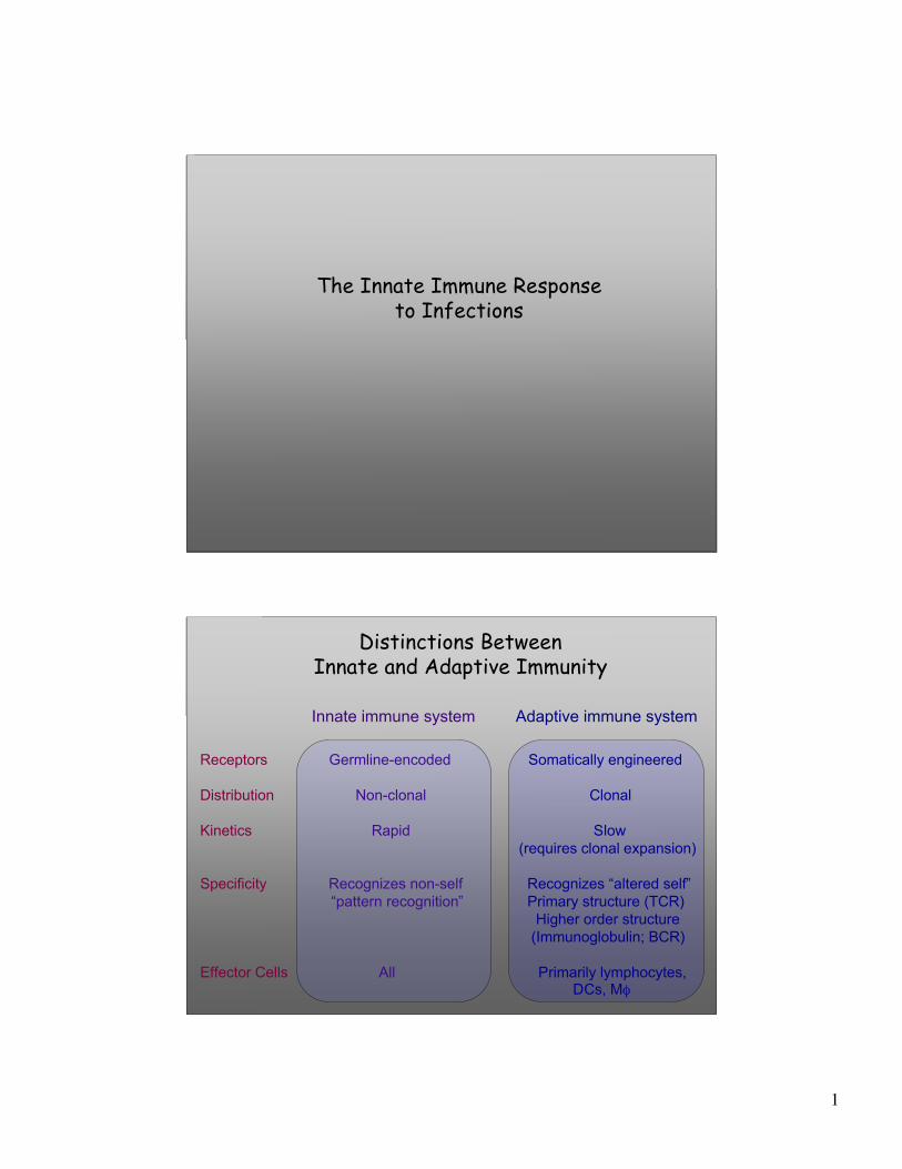
The Innate Immune Responseto Infections
Distinctions BetweenInnate and Adaptive Immunity
Innate immune system Adaptive immune system
Receptors Germline-encoded Somatically engineered
Distribution Non-clonal Clonal
Kinetics Rapid Slow(requires clonal expansion)
Specificity Recognizes non-self Recognizes “altered self” “pattern recognition” Primary structure (TCR)
Higher order structure (Immunoglobulin; BCR)
Effector Cells All Primarily lymphocytes, DCs, Mφ
2
What Really Happens During the Lag Period Before the Acquired Immune Response?
Innate immunity
Acquired immunity
Receptors Important in Innate Immunity
TLRsLectinsGPCRs
Production ofcytokines & chemokines
GPCR = G protein-coupled receptorsTLRs = Toll-like receptorsLectin: A molecule that binds carbohydrates
3
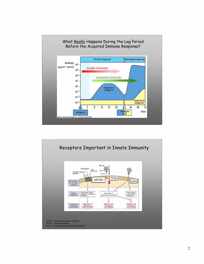
Phagocytosis of IgG-coated Targets by Macrophages
3 min 10 min
Most, but not all Leukocytes Can Perform Phagocytosis

4
Opsonic vs Non-opsonic Phagocytosis
• Non-opsonic phagocytosis is typically mediated by cellsurface receptors on leukocytes that recognize repeatingcarbohydrate subunits (comprising “molecular patterns”) onmicrobes.
• Opsonic phagocytosis is typically mediated by depositionof proteins (e.g., antibodies) on microbes that target them forrecognition by specific phagocytic receptors on leukocytes.
(<Latin opsonare, to buy provisions<Greek opsonein, condiment
"Opsonin is what you butter the disease germs with to makeyour white blood corpuscles eat them.”
-G.B. Shaw, The Doctor’s Dilemma
The Biology of Complement
5
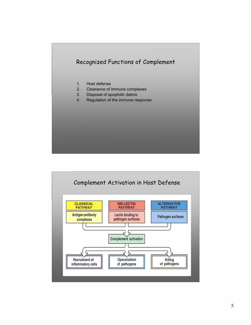
Recognized Functions of Complement
1. Host defense2. Clearance of immune complexes3. Disposal of apoptotic debris4. Regulation of the immune response
Complement Activation in Host Defense
6
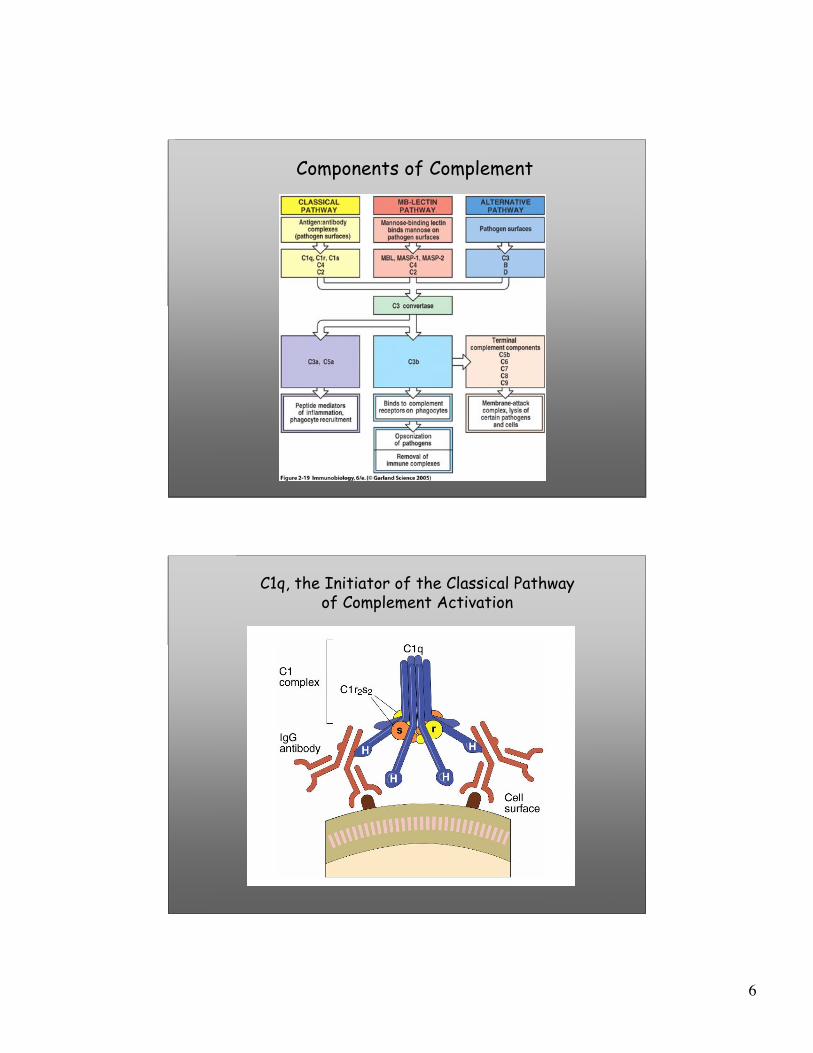
Components of Complement
C1q, the Initiator of the Classical Pathwayof Complement Activation
7
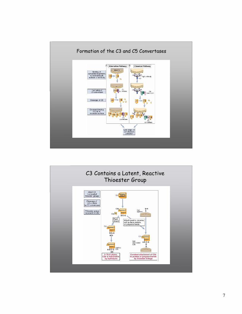
Formation of the C3 and C5 Convertases
C3 Contains a Latent, ReactiveThioester Group
8
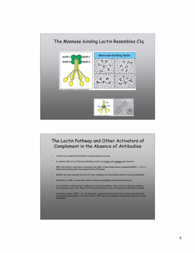
The Mannose-binding Lectin Resembles C1q
The Lectin Pathway and Other Activators ofComplement in the Absence of Antibodies
• A lectin is a molecule that binds to carbohydrate structures
• A collectin (like C1q or Mannose Binding Lectin) is a lectin with collagen-like features
• MBL first binds to mannose on bacterial cell walls. It then binds serine proteases MASP-1, -2 or -3(Mannose binding lectin Associated Serine Protease)
• MASPs can then activate C4 and C2, thus creating a C3 convertase without involving antibodies
• Deficiency in MBL is associated with increased susceptibility to bacterial infections
• It is simplistic to think of each “pathway” as acting in isolation. Thus, once the classical pathwayhas produced some C3b, these C3b molecules produce more C3b using the alternative pathway
• C-reactive protein (CRP) – An “acute phase” protein produced by the liver, binds to bacterial cellwall lipopolysaccharides. C1q then binds to CRP and thus activates complement without involvingantibodies.
9
The Complement System is Critical for Innate Immunity and is Triggered
by Multiple Ligands
C3 Convertase
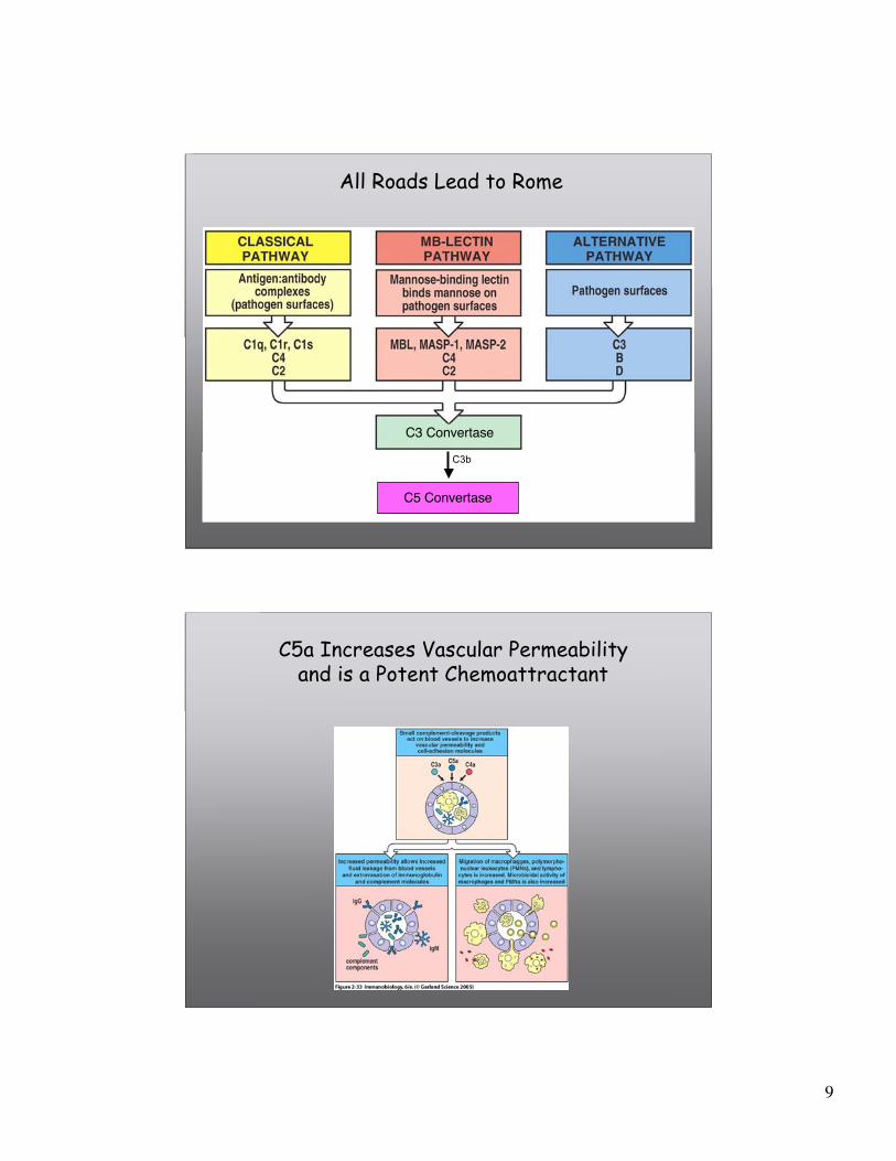
All Roads Lead to Rome
C3bC5 Convertase
C3b
C5a Increases Vascular Permeabilityand is a Potent Chemoattractant
10
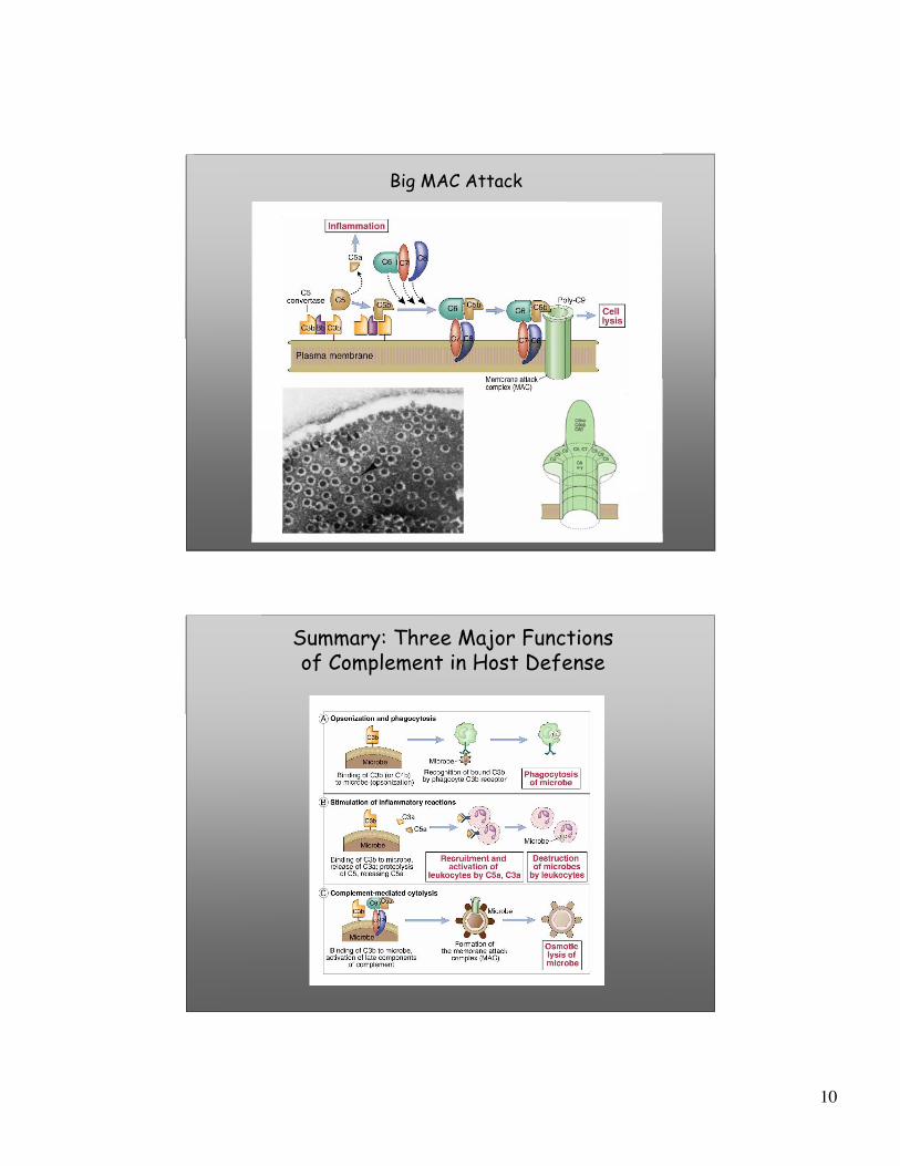
Big MAC Attack
Summary: Three Major Functionsof Complement in Host Defense
11
Summary: Complement
1. Complement is an ancient system of host defense that has well-defined functions in host defense: it opsonizes microbes (C3b,C3bi), stimulates inflammation (C3a, C4a, C5a), and mediates lysisof pathogens by the membrane attack complex (C5-9).
2. Additional functions of complement include clearance of immunecomplexes and apoptotic debris. These functions have majorimplications for the emergence of autoimmunity.
3. Among the known inherited complement deficiencies includeLeukocyte Adhesion Deficiency (LAD) and complement componentdeficiencies; these are associated with frequent infections and, inthe latter case, autoimmunity.
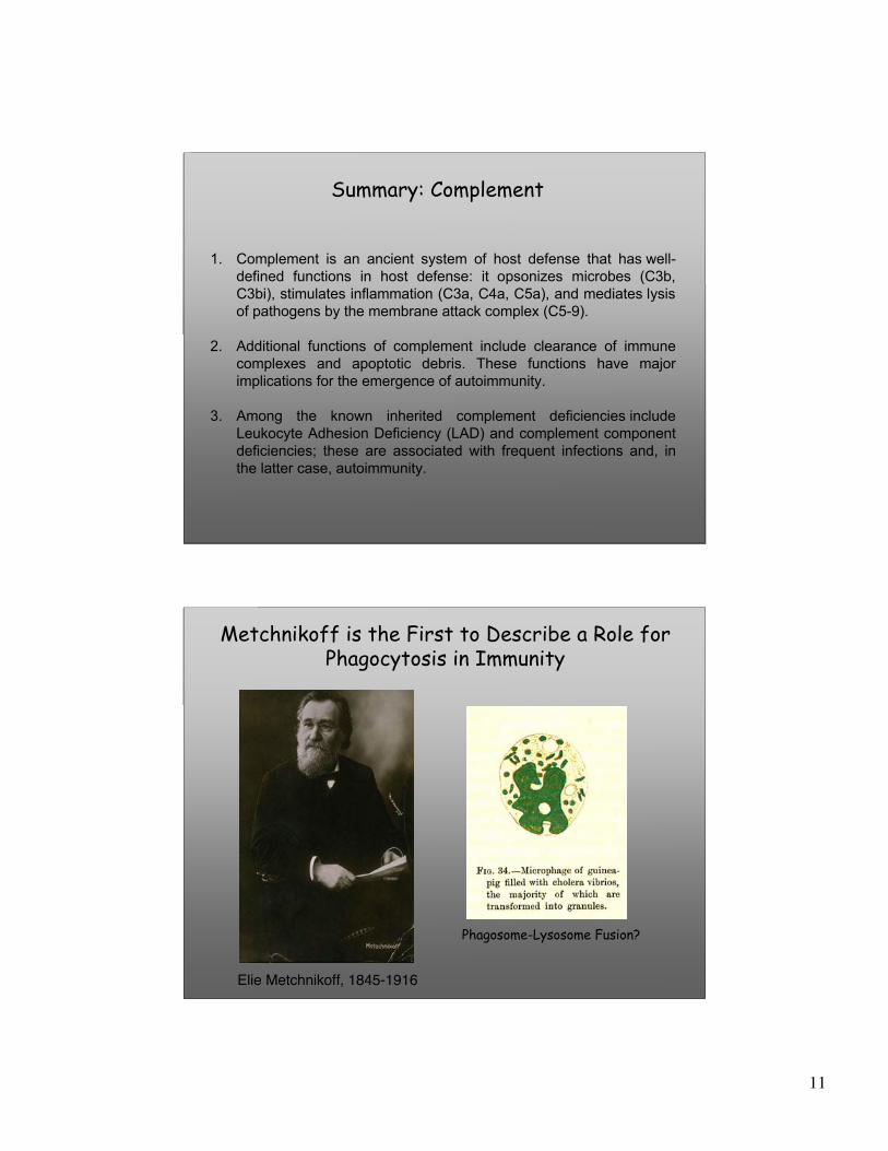
Elie Metchnikoff, 1845-1916
Phagosome-Lysosome Fusion?
Metchnikoff is the First to Describe a Role forPhagocytosis in Immunity

12
Post-phagocytic Events:Phagosome-Lysosome Fusion
Lysosomes
Pathogen Macrophage
Phagolysosomes
0-3 min 1-5 min 30 min-hrs
Phagocytosis of Bacteriais Followed by Phagosome-Lysosome Fusion
From: Allen et al., J. Exp. Med. 191:115, 2000
13
What happens following pathogen ingestion?
Post-phagocytic Events
Pathogen Macrophage
14
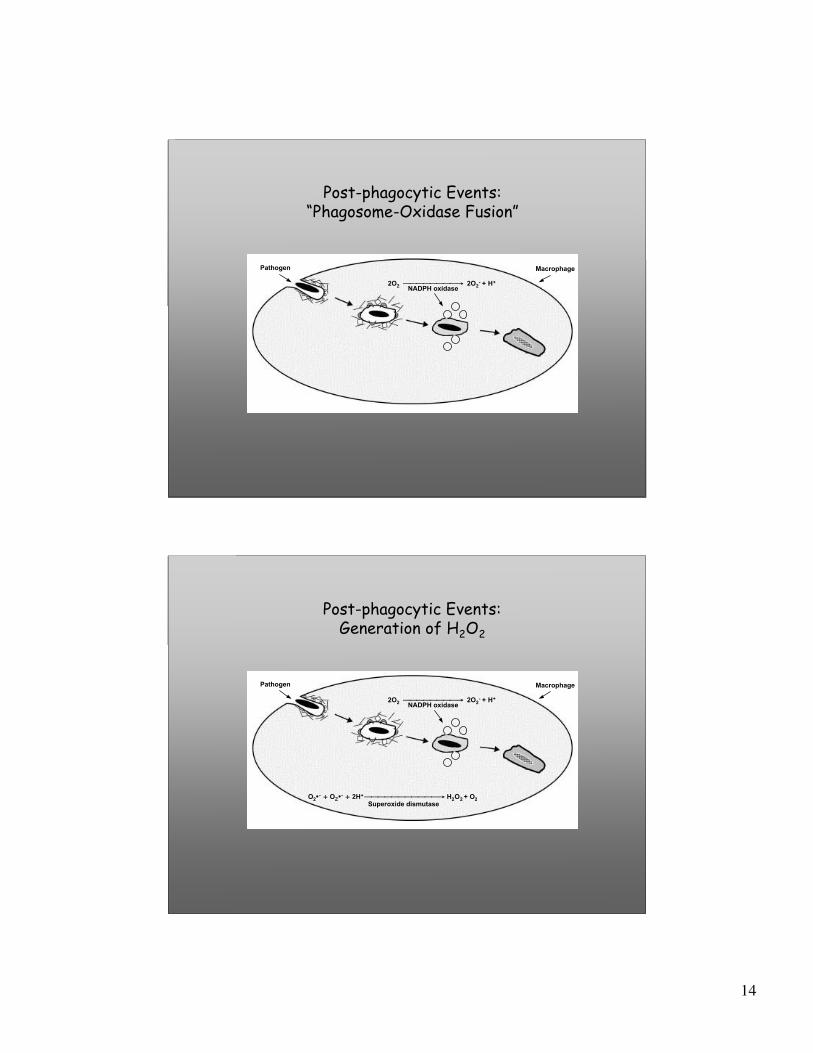
Post-phagocytic Events:“Phagosome-Oxidase Fusion”
NADPH oxidase
Pathogen Macrophage
2O2 → 2O2- + H+
Post-phagocytic Events:Generation of H2O2
NADPH oxidase
Pathogen Macrophage
2O2 → 2O2- + H+
O2•- + O2•- + 2H+ → H2O2 + O2Superoxide dismutase

15
Post-phagocytic Events:Myeloperoxidase Activity
NADPH oxidase
Pathogen Macrophage
2O2 → 2O2- + H+
O2•- + O2•- + 2H+ → H2O2 + O2Superoxide dismutase
H2O2 + Cl- → HOCl + OH•Myeloperoxidase
Reactive oxygen species: O2-•, HOCl, H2O2, O3
Post-phagocytic Events:Peroxynitrite Production
NADPH oxidase
Pathogen Macrophage
2O2 → 2O2- + H+
O2•- + O2•- + 2H+ → H2O2 + O2Superoxide dismutase
H2O2 + Cl- → HOCl + OH•Myeloperoxidase
2O2- + NO• → ONOO-
Peroxynitrite
Reactive oxygen species: O2-•, HOCl, H2O2, O3
Reactive nitrogen species: ONOO-
16
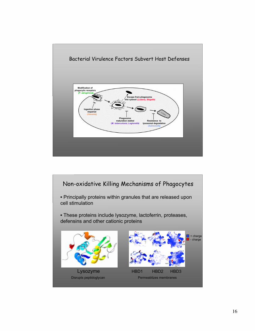
Bacterial Virulence Factors Subvert Host Defenses
Phagosomematuration stalled
(M. tuberculosis; Legionella)
Ingestion phaseimpaired(Yersinia)
Resistance tolysosomal degradation
(Salmonella)
Modification ofphagocytic receptors
(P. aeruginosa)
Escape from phagosome into cytosol (Listeria, Shigella)
• Principally proteins within granules that are released uponcell stimulation
• These proteins include lysozyme, lactoferrin, proteases, defensins and other cationic proteins
Non-oxidative Killing Mechanisms of Phagocytes
Lysozyme HBD1 HBD2 HBD3
+ charge- charge
Disrupts peptidoglycan Permeablizes membranes

17
Phagocytosis: Not Just for Bugs
Phagocytosis is the Principal Mechanismof Disposal of Apoptotic Corpses
MacrophageApoptotic Thymocyte
From: Jennings et al., Am. J. Resp. Cell Mol. Biol. 32:108, 2005
• Phagocytosis is the means of disposal ofapoptotic corpses, and occurs continuouslyduring the lifetime of an individual.
• In this setting, phagocytosis is notaccompanied by inflammation, but ratherleads to an “anti-inflammatory” signal (theproduction of TGF-β).
• As apoptotic corpses contain manypotential self antigens, the lack of anappropriate anti-inflammatory signal hasthe potential to trigger autoimmunity.
18
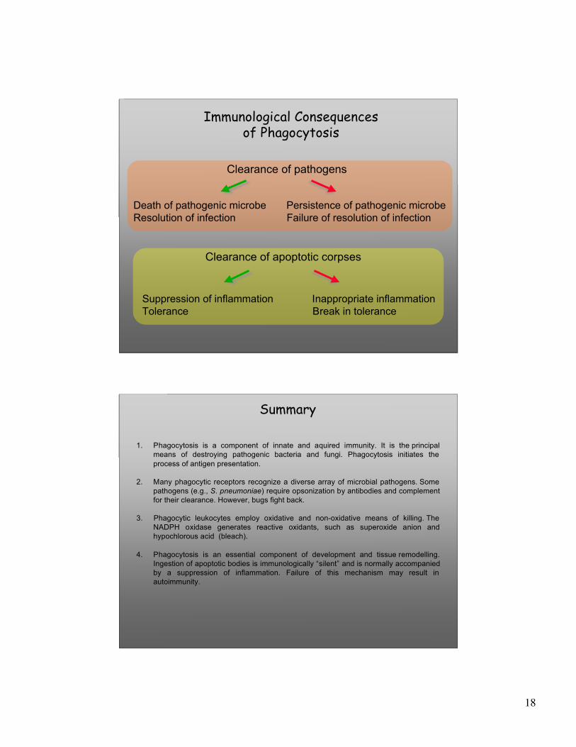
Immunological Consequencesof Phagocytosis
Death of pathogenic microbe Persistence of pathogenic microbeResolution of infection Failure of resolution of infection
Clearance of pathogens
Clearance of apoptotic corpses
Suppression of inflammation Inappropriate inflammationTolerance Break in tolerance
Summary
1. Phagocytosis is a component of innate and aquired immunity. It is the principalmeans of destroying pathogenic bacteria and fungi. Phagocytosis initiates theprocess of antigen presentation.
2. Many phagocytic receptors recognize a diverse array of microbial pathogens. Somepathogens (e.g., S. pneumoniae) require opsonization by antibodies and complementfor their clearance. However, bugs fight back.
3. Phagocytic leukocytes employ oxidative and non-oxidative means of killing. TheNADPH oxidase generates reactive oxidants, such as superoxide anion andhypochlorous acid (bleach).
4. Phagocytosis is an essential component of development and tissue remodelling.Ingestion of apoptotic bodies is immunologically “silent” and is normally accompaniedby a suppression of inflammation. Failure of this mechanism may result inautoimmunity.
19
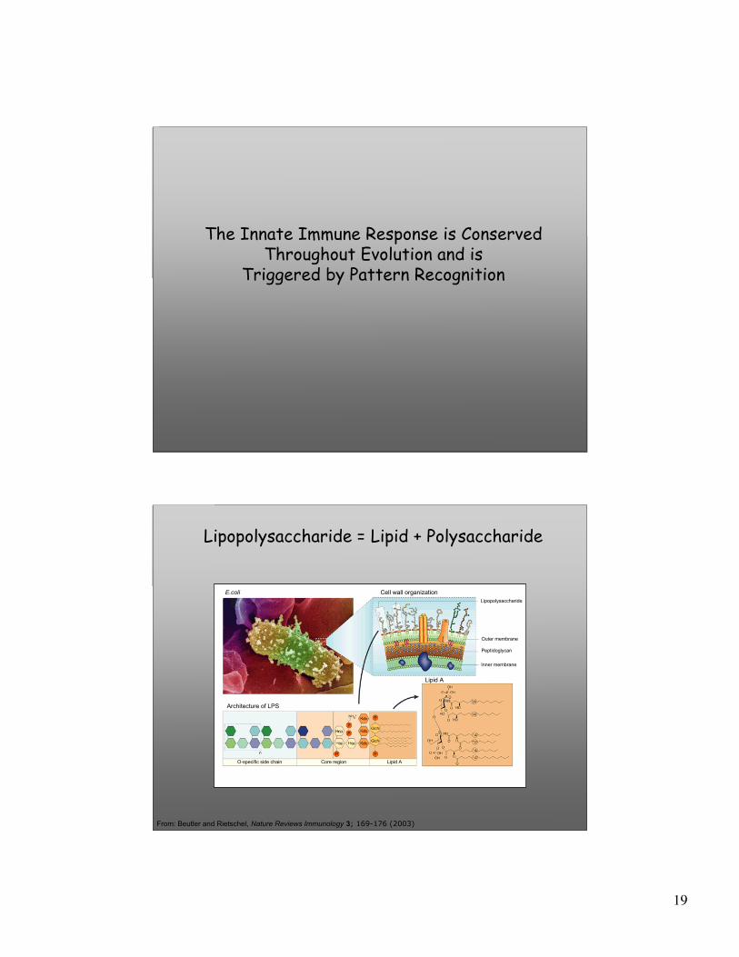
The Innate Immune Response is Conserved Throughout Evolution and is
Triggered by Pattern Recognition
Lipopolysaccharide = Lipid + Polysaccharide
From: Beutler and Rietschel, Nature Reviews Immunology 3; 169-176 (2003)
E.coli Cell wall organization
Architecture of LPS
Lipopolysaccharide
Outer membrane
Peptidoglycan
Inner membrane
O-specific side chain Core region Lipid A
Lipid A

20
Diversity of“Pathogen-associated Molecular Patterns” (PAMPs)
From: Akira et al., Cell 124:783, 2006
Innate Immune Receptors for PAMPs
• Toll-like receptors (TLRs)• Complement• Collectins (e.g., Surfactant Protein-A)• Scavenger receptors• Pentraxins (e.g., CRP)• Lectins (e.g., Dectin-1)• CD14• NOD-like receptors (NLRs)• RIG-1-like receptors
21
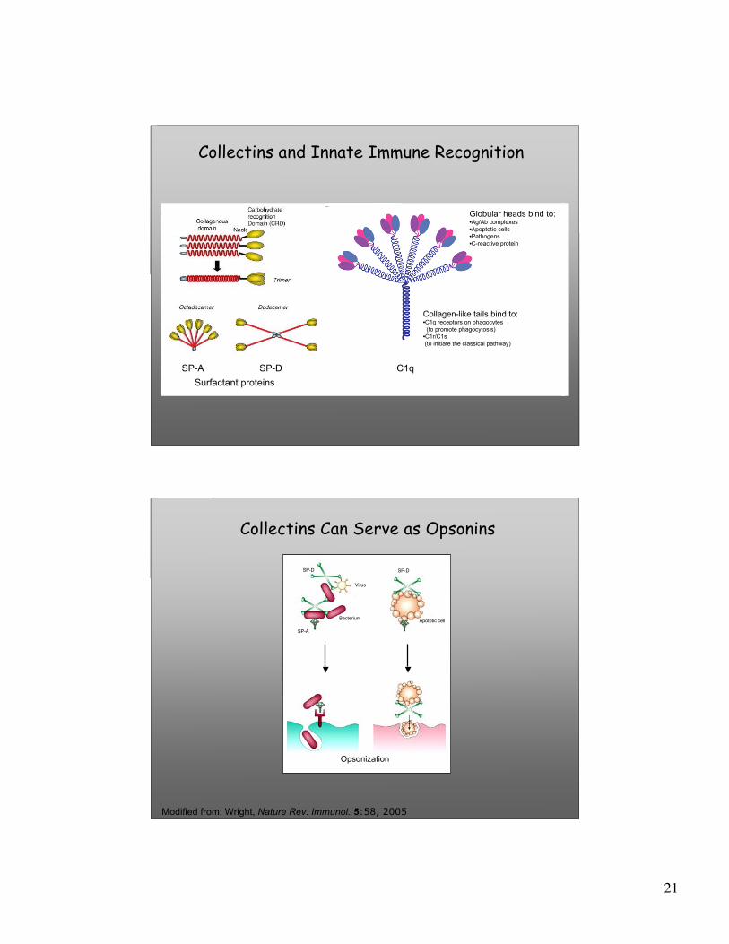
Collectins and Innate Immune Recognition
SP-A SP-D C1qSurfactant proteins
Globular heads bind to:•Ag/Ab complexes•Apoptotic cells•Pathogens•C-reactive protein
Collagen-like tails bind to:•C1q receptors on phagocytes (to promote phagocytosis)•C1r/C1s (to initiate the classical pathway)
Collectins Can Serve as Opsonins
Modified from: Wright, Nature Rev. Immunol. 5:58, 2005
Bacterium
Virus
Apototic cell
SP-DSP-D
SP-A
Opsonization
22
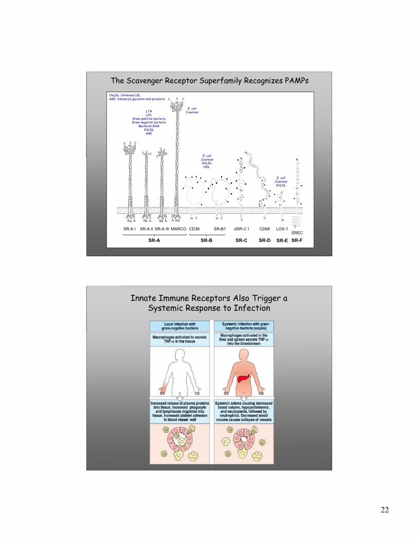
C C C
C
C
SoSo
C
C
SoSo
CL
SRECLOX-1CD68dSR-C ISR-B1CD36MARCOSR-A II
NNNNNNN
N
N
N
N
N
C C
SR-A I
NNN
C C C
C C C
CC
C
C**
SR-CSR-B
C
C C
NN N
SR-A
SR-A III
SoSo
C
CL
E
-Complement control protein (CCP)
-Somatomedin B
-C-type Lectin
-Epidermal growth factor (EGF)
-Partial Epidermal growth factor (EGF)
-Potential N-linked glycosylation
-Potential O-linked glycosylation
KEY TO DOMAINS
E
E
E
E
E
E
E
SR-D SR-E SR-F
LTALPS
Gram-positive bacteriaGram-negative bacteria
Bacterial DNAOxLDLAGE
E. coliS.aureus
E. coliS.aureusOxLDLHDL
E. coliS.aureusOxLDL
OxLDL: Oxidized LDLAGE: Advanced glycation end-products
The Scavenger Receptor Superfamily Recognizes PAMPs
Innate Immune Receptors Also Trigger aSystemic Response to Infection

23
History of Endotoxin Research
Modified from: Beutler and Rietschel, Nature Reviews Immunology 3; 169-176 (2003)
Endotoxin = LPS
C3H/HeJmutationreported
TNF mediatesLPS-induced shock
Discoveryof CD14
LPS = TLR4
TLR2 is the receptorfor PG
Pasteur, Koch: the germ theory
of disease
Pfeiffer describes“endotoxin”
Primary structure of
LPS reported
The post-microbial era began with the discoveries of Koch and Pasteur (1865). Four phases of discovery are depicted: the recognition thatinfection is” poisonous” (red); search for poisons culminating in the identification of endotoxin (green); the chemical and biologicalcharacteriztaion of endotoxin (orange); the identification of the endotoxin receptor and its role in promoting the immune response (purple)
24
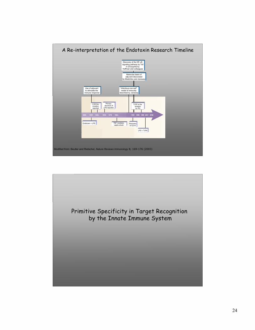
A Re-interpretation of the Endotoxin Research Timeline
Modified from: Beutler and Rietschel, Nature Reviews Immunology 3; 169-176 (2003)
Discovery of the NF-κB signaling pathway by Toll
in Drosophila by Hoffman and colleagues
Molecular basis ofadjuvant discovered
by Medzhitov and Janeway
“Infectious-non-self” model of immunity
described by Janeway
Use of adjuvant to stimulate the
immune response
Endotoxin = LPS TNF mediates septic shock
TLR2 is the receptorfor PG
LPS = TLR4
Primary structure of
LPS reported
C3H/HeJmutationreported
Discoveryof CD14
Primitive Specificity in Target Recognition by the Innate Immune System

25
Ligand Specificity of TLRs
Adaptor proteins, kinases
From: Luster, Curr. Opin. Immunol. 14:129, 2002
Specificity of TLR Transcriptional Programs
26
From: Moynagh,Trends Immunol. 26:469, 2005
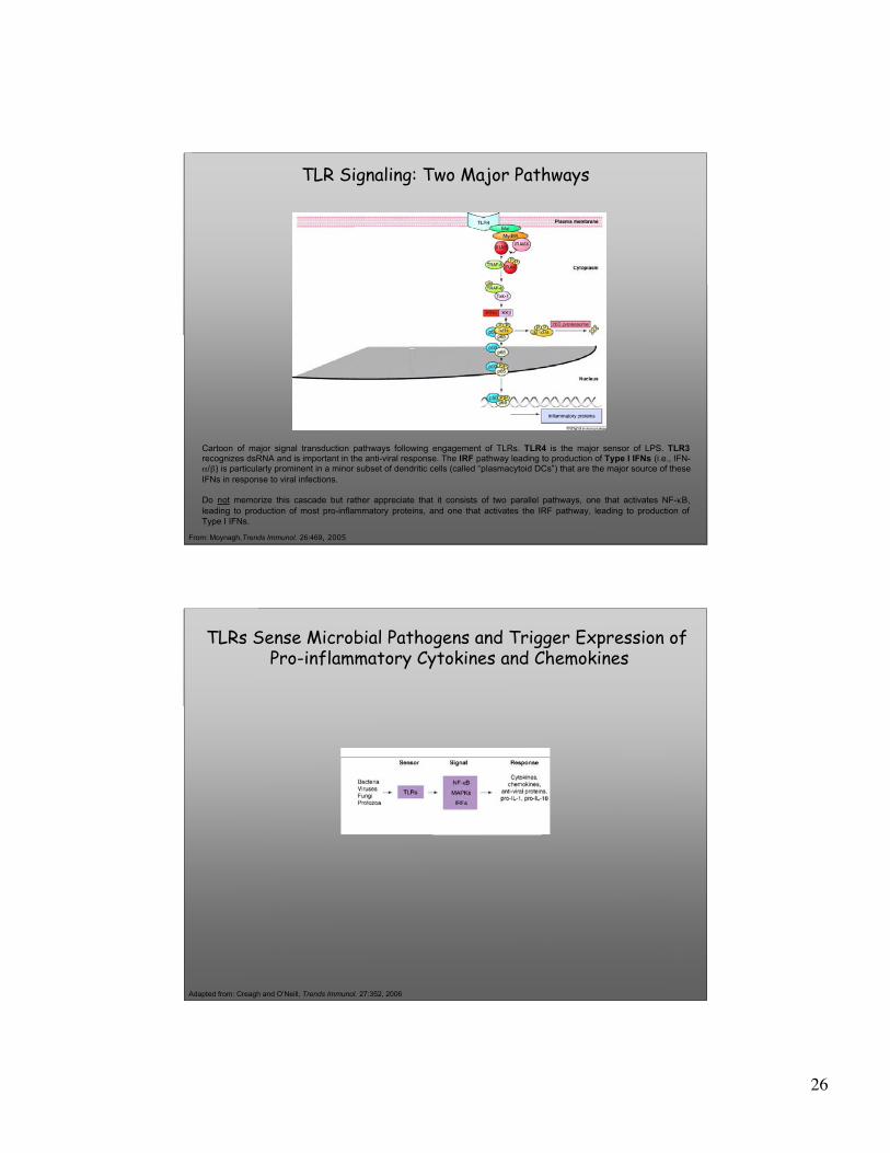
TLR Signaling: Two Major Pathways
Cartoon of major signal transduction pathways following engagement of TLRs. TLR4 is the major sensor of LPS. TLR3recognizes dsRNA and is important in the anti-viral response. The IRF pathway leading to production of Type I IFNs (i.e., IFN-α/β) is particularly prominent in a minor subset of dendritic cells (called “plasmacytoid DCs”) that are the major source of theseIFNs in response to viral infections.
Do not memorize this cascade but rather appreciate that it consists of two parallel pathways, one that activates NF-κB,leading to production of most pro-inflammatory proteins, and one that activates the IRF pathway, leading to production ofType I IFNs.
TLRs Sense Microbial Pathogens and Trigger Expression of Pro-inflammatory Cytokines and Chemokines
Adapted from: Creagh and O’Neill, Trends Immunol. 27:352, 2006