the insulin-like growth factor binding protein bp-25 is expressed by human breast cancer cells
TRANSCRIPT

Vo1.158, No. 1,1989
Janua~ 16,1989
BIOCHEMICAL AND BIOPHYSICAL RESEARCH COMMUNICATIONS
Pages 38-44
THE INSULIN-LIKE GROWTH FACTOR BINDING PROTEIN BP-25
IS EXPRESSED BY HUMAN BREAST CANCER CELLS
D. Yee*, R.E. Favoni*, R. Lupu*, K.J. Cullen*, G.S. Lebovic, K.K. Huff*, P.D.K. Lee#, Y.L. Lee+, D.R. Powell+, R.B. Dickson*, N. Rosen*, and M.E. Lippman*
Medicine Branch, National Cancer Institute Bethesda, Maryland 20892
#Division of Endocrinology, The Children's Hospital Denver, Colorado 80218
+Department of Pediatrics, Baylor College of Medicine Houston, Texas 77054
Received September 27, 1988
Specific binding proteins are thought to modulate the effects of IGF-I. Previous work has demonstrated that media conditioned by human breast cancer cells contains IGF-I binding activity. Radiolabelled IGF-I incubated with serum-free conditioned media from the breast cancer cell line MDA-MB 231 eluted with an apparent M.W. of 35-40 kDa when analyzed by gel filtration chromatography at pH 7.4. The M.W. of this binding activity corresponded to that of BP-25, a binding protein cloned from the hepatocellular carcinoma cell line HepG2. Two breast cancer cell lines, MDA-MB 231 and Hs578T, were found to express BP-25 RNA. Specific BP-25 radioimmunoassay detected BP-25 production in the conditioned media of these two cell lines. Immunoprecipitation confirmed that metabolically labelled MDA-MB 231 released 30 kDa BP-25 into its medium. This study demonstrates that some breast cancer ceils express the IGF-I binding protein, BP-25. © 1 9 8 9 Academlc P r e s s , I n c .
IGF-I is a mediator of the effects of growth hormone and a mitogen for both
normal and malignant cell lines. These mitogenic effects are thought to be a result of the
interaction between IGF-I and the type I IGF receptor, however IGF-I is also associated
with specific serum and extracellular binding proteins that may also modulate the
biological actions of IGF-I(1).
We have previously shown that IGF-I is a potent mitogen for human breast cancer
cell lines and these cells produce an IGF-I related protein that is associated with a binding
* Current address: Lombardi Cancer Research Center, Georgetown University Medical Center, Washington, D.C. 20007
Abbreviations used: Insulin-like growth factor I-IGF-I, Molecular weight-M.W., Radioimmunoassay-RIA, Sodium dodecyl sulfate-SDS, Polyacrylamide gel electrophoresis-PAGE, Improved minimal essential media-IMEM, Phosphate buffered saline-PBS
0006-291X/89 $1.50 Copyright © 1989 by Academic Press, Inc. All rights of reproduction in any form reserved. 38

VOI. 158, No. 1, 1989 BIOCHEMICAL AND BIOPHYSICAL RESEARCH COMMUNICATIONS
activity (2). Using gel filtration chromatography, we have found that the binding activity
produced by the breast cancer cell line MDA-MB 231 has an apparent M.W. of 28-33
kDa. Since a cDNA for a low molecular weight IGF-I binding protein (BP-25) has been
recently isolated from both placenta and the human hepatocellular carcinoma cell line
HepG2 (3, 4, 5), we have examined breast cancer cell line RNA for BP-25 expression.
Additionally, we have examined serum-free media conditioned by breast cancer cells for
BP-25 protein production by RIA and immunoprecipitation with a specific BP-25
antiserum.
Materials and Methods
Cell lines The cell line MCF-7 was obtained from the Michigan Cancer Foundation, Detroit
MI. 172 is a normal mammary epithelial cell line derived from reduction mammoplasty tissue and was a gift from Martha Stampfer, Lawrence Berkeley Laboratories, Berkeley, CA. All other cell lines were obtained from American Type Culture Collection, RockviUe, MD. Cancer cell lines were maintained in IMEM (Biofluids, Rockville, MD) with 2mM glutamine, 0.05 mg/ml gentamicin and 10% fetal calf serum (Gibco, Detroit, MI). 172 was maintained as previously described (6).
RNA was obtained using the guanadinium thiocyanate method (7).
Binding studies Serum-free conditioned media was obtained as previously described (8) . The
conditioned media was concentrated 100 fold by ultrafiltration using an Amicon filter with a 5 kDa M.W. cutoff.
The conditioned medium was filtered on a 1.5cm x 85cm Sephadex G-100 column, equilibrated with lx PBS pH 7.4, at a flow rate of 15 ml/hr. 2ml fractions were collected and 1 ml aliquots were measured on an LKB 1275 gamma counter.
IGF-I binding activity was assayed by the charcoal separation method as previously described (9).
Northern analysis and RNAse protection assay A 970 bp BamHI fragment of the BP-25 cDNA (3) was subcloned into a pGem
vector (Promega, Madison, WI). This fragment was random primed labelled and used as a probe for Northern analysis. For RNAse protection assays antisense RNA probes were transcribed and labelled with 32p UTP according to the instructions of the manufacturer. Northern blots utilizing 10~tg of total RNA were performed as previously described (10). Final washing was done in .1X SSC, 0.1% SDS at 65°C for 15 minutes.
RNAse protection assays were performed as previously described(ll). Briefly, 30~tg of total RNA was hybridized with probe in an 80% formamide (vol/vol) buffer. Samples were hybridized for 12-16 hours at 50°C followed by digestion with RNAse A. The samples were precipitated with 1 ~tg tRNA and 2 volumes of absolute ethanol. The pellets were lyophilized then resuspended in an 80% formamide loading buffer. The samples were run on a 6% polyacrylamide gel with 8M urea. pBR322 was digested and endlabeUed with 32p dCTP and used as size standards. The dried gels were exposed to X- ray film in the presence of a Quanta III (Dupont, Wilmington, DE) intensifying screen.
Radioimmunoassav of conditioned media Cells (2 x -107) were placed in T-75 flasks (Costar, Cambridge, MA) and grown in
IMEM with 10% fetal calf serum. When the cells approached confluence, 10 ml of serum-free conditioned media was collected per flask and lyophiUized as described (8).
BP-25 levels in the conditioned media samples were analyzed by RIA using standards and tracer prepared from purified HepG2 cell-derived BP-25 (9) and a rabbit polyclonal antiserum raised against the same protein (12). The specificity of this RIA for BP-25 has been validated in separate experiments (P.D.K. Lee, manuscript in preparation). Briefly, lyophilized samples were reconstituted to 0.5x original volume in RIA buffer (PBS, oH 7.4, 0.1% BSA). 100~tl of standard or sample was incubated with 100~tl of
39

Vot. 158, No. 1, 1989 BIOCHEMICAL AND BIOPHYSICAL RESEARCH COMMUNICATIONS
1:1000 dilutions of antiserum for 1 hour at room temperature, followed by a 16 hour incubation with 5-10K cpm/tube 12SI-BP-25 at 4°C. Bound counts were separated using agarose-immobilized goat anti-rabbit immunoglobulin (Biorad, Richmond, CA). Samples were assayed in duplicate and after serial dilution. Data were analyzed using the IBM-PC Data Reduction Program (M.L. Jaffee and Assoc., Silver Spring, MD).
Metabolic Labellin~ and Immunoprecipitation of BP-25 protein In vitro labeling of proteins was performed using 35S cysteine and 3sS methionine
as previously described (13). 35S labelled proteins released in the conditioned medium from MDA-MB 231, HepG2, and MCF-7 were immunoprecipitated with 10~tg of BP-25 antiserum as previously described(14). Precipitates were separated by electrophoresis on 10% SDS/PAGE gels, and subsequently autoradiographed. Pre-labelled M.W. markers (Biorad) were run in parallel lanes.
Results and Discussion: Radioiodinated IGF-I (lng - specific activity 2000 Ci/mmol) was
incubated with lml of 100x serum-free concentrated media conditioned by the breast
cancer cell line MDA-MB 231. Gel filtration chromatography on the Sephadex G-100
column at pH 7.4 shows that labelled IGF-I incubated with conditioned media eluted with
an apparent M.W. of 35-40 kDa, while labelled IGF-I alone eluted at the expected M.W
of 7.5 kDa(Fig 1). Addition of excess unlabelled IGF-I (10 Ixg) could partially displace
10 67 45
I I
8
6
4
2
L 0
X
EL_ £D
40
n 1 O0 E
",2 o
80
v 6 0
~ 4o
~ 2o m
- - 0
4O
/°,O,o.oo.oooo o 'o
\ 0
o o
80 1 O0
o
(3/ I I 60 120
IGF-I Frochon N u m b e r
I
80 120 160 Froc t ion N u m b e r
Figure 1-Gel filtration of binding protein-radiolabelled IGF-I complex. 1 ml of lOOx concentrated conditioned media from the cell line MDA-MB 231 was incubated with labelled IGF-I at 4°C overnight and chromatograpbed at pH 7.4 with Sephadex G-100. Closed circles represent labelled IGF-I and conditioned media. Open circles represent labelled IGF-I, conditioned media plus 10~g unlabelled IGF-I. The positions of protein standards 67 kDa bovine serum albumin, 43 kDa ovalbumin and labelled IGF-I alone are marked. Inset: 100x concentrated conditioned media from cell line MDA-MB 231 was run on the same colunm, with eluted fractions tested for IGF binding protein activity by charcoal binding assay. The positions of the protein standards are noted.
40
Vol. 158, No. 1, 1989 BIOCHEMICAL AND BIOPHYSICAL RESEARCH COMMUNICATIONS
A.
? ' -
B.
622-
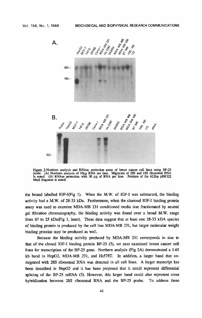
Fimn'¢ 2-Northern analysis and RNAse protection assay of breast cancer c¢11 lines using BP-25 .probe. (A) Northern analysis of 10gg RNA per lane. Migration of 28S and 18S ribosomal RNA is noted. (B) RNAse protection with 30 gg of RNA per lane. Position of the 622bp pBR322 MspI fragment is noted.
the bound labelled IGF-I(Fig 1). When the M.W. of IGF-I was subtracted, the binding
activity had a M.W. of 28-33 kDa. Furthermore, when the charcoal IGF-I binding protein
assay was used to examine MDA-MB 231 conditioned media size fractionated by neutral
gel filtration chromatography, the binding activity was found over a broad M.W. range
from 67 to 25 kDa(Fig 1, inset). These data suggest that at least one 28-33 kDA species
of binding protein is produced by the cell line MDA-MB 231, but larger molecular weight
binding proteins may be produced as well.
Because the binding activity produced by MDA-MB 231 corresponds in size to
that of the cloned IGF-I binding protein BP-25 (3), we next examined breast cancer cell
lines for transcription of the BP-25 gene. Northern analysis (Fig 2A) demonstrated a 1.65
kb band in HepG2, MDA-MB 231, and Hs578T. In addition, a larger band that co-
migrated with 28S ribosomal RNA was detected in all cell lines. A larger transcript has
been described in HepG2 and it has been proposed that it could represent differential
splicing of the BP-25 mRNA (3). However, this larger band could also represent cross
hybridization between 28S ribosomal RNA and the BP-25 probe. To address these
41
Vol. 158, No. 1, 1989 BIOCHEMICAL AND BIOPHYSICAL RESEARCH COMMUNICATIONS
M D A - M B 2 3 1 H E P - G 2
E , ,
1 2 3 4 5 6
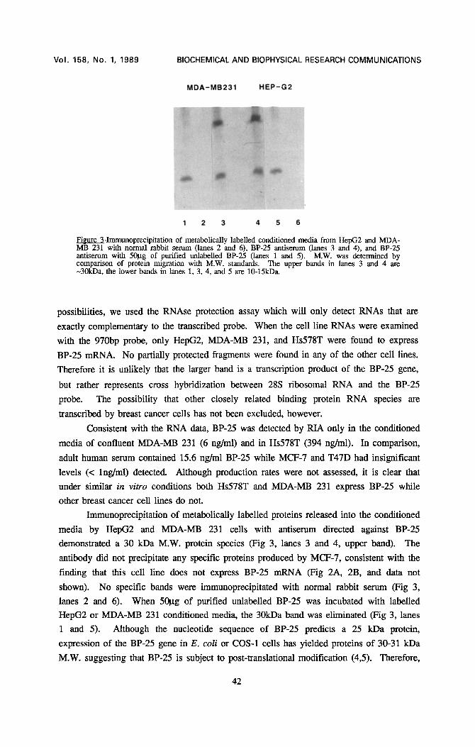
Figure 3-Immunoprecipitation of metabolically labelled conditioned media from HepG2 and MDA- MB 231 with normal rabbit serum (lanes 2 and 6), BP-25 antiserum (lanes 3 and 4), and BP-25 antiserum with 501.tg of purified unlabelled BP-25 (lanes 1 and 5). M.W. was determined by comparison of protein migration with M.W. standards. The upper bands in lanes 3 and 4 are -30kDa, the lower bands in lanes 1, 3, 4, and 5 are 10-t5kD&
possibilities, we used the RNAse protection assay which will only detect RNAs that are
exactly complementary to the transcribed probe. When the cell line RNAs were examined
with the 970bp probe, only HepG2, MDA-MB 231, and Hs578T were found to express
BP-25 mRNA. No partially protected fragments were found in any of the other cell lines.
Therefore it is unlikely that the larger band is a transcription product of the BP-25 gene,
but rather represents cross hybridization between 28S ribosomal RNA and the BP-25
probe. The possibility that other closely related binding protein RNA species are
transcribed by breast cancer cells has not been excluded, however.
Consistent with the RNA data, BP-25 was detected by RIA only in the conditioned
media of confluent MDA-MB 231 (6 ng/ml) and in Hs578T (394 ng/ml). In comparison,
adult human serum contained 15.6 ng/ml BP-25 while MCF-7 and T47D had insignificant
levels (< lng/ml) detected. Although production rates were not assessed, it is clear that
under similar in vitro conditions both Hs578T and MDA-MB 231 express BP-25 while
other breast cancer cell lines do not.
Immunoprecipitation of metabolically labelled proteins released into the conditioned
media by HepG2 and MDA-MB 231 cells with antiserum directed against BP-25
demonstrated a 30 kDa M.W. protein species (Fig 3, lanes 3 and 4, upper band). The
antibody did not precipitate any specific proteins produced by MCF-7, consistent with the
finding that this cell line does not express BP-25 mRNA (Fig 2A, 2B, and data not
shown). No specific bands were immunoprecipitated with normal rabbit serum (Fig 3,
lanes 2 and 6). When 501~g of purified unlabelled BP-25 was incubated with labelled
HepG2 or MDA-MB 231 conditioned media, the 30kDa band was eliminated (Fig 3, lanes
1 and 5). Although the nucleotide sequence of BP-25 predicts a 25 kDa protein,
expression of the BP-25 gene in E. coli or COS-1 cells has yielded proteins of 30-31 kDa
M.W. suggesting that BP-25 is subject to post-translational modification (4,5). Therefore,
42

VoI. 158, No. 1, 1989 BIOCHEMICAL AND BIOPHYSICAL RESEARCH COMMUNICATIONS
the size of the protein detected in breast cancer cells agrees with that of the expressed
gene product. This suggests that authentic BP-25 is produced by these cells.
A 10-15 kDa protein was also identified by immunoprecitation of HepG2 and
MDA-MB 231 conditioned media (Fig. 3, lanes 1,3,4, and 5, lower band) and unlabelled
BP-25 did not compote with this smaller protein (Fig 3, lanes 1 and 5). BP-25 and other
smaller M.W. IGF-I binding proteins have homologous NH2 terminal amino acid
sequences (14) and these smaller binding proteins could potentially cross react with the
polyclonal BP-25 antiserum. Alternatively, the smaller M.W. protein could represent
another unidentified protein that binds to BP-25.
This study demonstrates that some human breast cancer cell lines express BP-25.
By cross linking conditioned media with labelled IGF-I, DeLeon et al have identified 45,
36, and 29 kDa IGF-I binding protein species in MDA-MB 231 (15). MCF-7 did not
contain the 29 kDa binding protein. Since MDA-MB 231 expresses BP-25 mRNA and
protein while MCF-7 does not, it appears that the 29 kDa protein identified by cross
linking and by immunoprecipitation is BP-25. Immunoprecipitation of MDA-MB 231
conditioned media did not identify any of the higher M.W. species binding proteins. This
suggests that the larger binding proteins represents either a different species of IGF-I
binding protein or a multimeric form of BP-25. However, since MCF-7 produces the 45
and 36 kDa binding proteins (14) yet expresses no BP-25, it is unlikely that these higher
molecular weight species are a result of post-translational modification of BP-25. The
function and relative abundance of the higher M.W. binding proteins produced by breast
cancer ceils has not yet been fully defined. It is likely that an interaction between these
different binding proteins, IGF-I, and the type I IGF receptor modulate the mitogenic
effect of IGF-I.
The binding protein purified from amniotic fluid is identical to BP-25 and has been
shown to enhance the mitogenic effects of IGF-I in human fibroblasts (16). Since IGF-I
is a potent mitogen for breast cancer cells, expression of BP-25 could be important in
regulating the growth of breast cancer.
Acknowledgements-The authors would like to thank A. Wellstein and G. Zugmaier for advice and helpful discussions.
References 1. Baxter, R.C. (1986) Adv. Clin. Chem. 25, 49-115 2. Huff, K.K., Kaufman, D., Gabbay, K.H., Spencer, E.M., Lippman, M.E., and
Dickson, R.B. (1986) Canc. Res. 46, 4613-4619 3. Lee, Y.L., Hintz, R.L., James, P.M., Lee, P.D.K., Shively, J.E., and Powell, D.R.
(1988) Mol. Endo. 2, 404-411 4. Brewer, M.T., Stetler, G.L., Squires, C.H., Thompson, R.C., Busby, W.H., and
Clemmons, D.R. (1988) Biochem. Biophys. Res. Comm. 152, 1289-1297 5. Brinkman, A., Groffen, C., Kortleve, D.J., Geurts van Kessel, A., and Drop, S.L.S.
(1988) Embo J. 7, 2417-2423 6. Hammond, S.L., Ham, R.G., and Stampfer, M.R. (1984) Proc. Natl. Acad. Sci.
USA 81, 5435-5439
43

Vol. 158, No. 1, 1989 BIOCHEMICAL AND BIOPHYSICAL RESEARCH COMMUNICATIONS
7. Chirgwin, LM., F'rzybia, A.I., MacDonald, R.J., and Rutter, W.J. (1979) Biochemistry 18, 5294-5299
8. Huff, K.K., Knabbe, C., Lindsey, R., Kaufman, D., Bronzert, D., Lippman, M.E., and Dickson, R.B. (1988) Mol. Endo. 2, 200-208
9. Powell, D.R., Lee, P.D.K., Shively, J.E., Eckenhausen, M., and Hintz, R.L. (1987) J. Chrom. 420, 163-170
10. Davis, L.G., Dibner, M.D., Battey, J.F. (1986) Basic Methods in Molecular Biology, p. 143 Elsevier Science Publishing, New York
11. Veillette, A., Foss, F.M., Sausville, E.A., Bolen, J.B., and Rosen, N. (1987) Oncogene Res. 1, 357-374
12. Lee, P.D.K., Powell, D.R., Li, C.H., Bohn, H., Liu, F., and Hintz, R.L. (1988) Biochem. Biophys. Res. Comm. 152, 1131-1137
13. Bronzert, D.A., Pantazis, P., Antoniades, H.N., Kasid, A., Davisdson, N., Dickson, R.B., and Lippman, M.E. (1987) Proc. Natl. Acad. Sci. USA 84, 5762-5767
14. Bell, S.C. and Keyte, J.W. (1988) Endo. 123, 1202-1204 15. DeLeon, D.D., Bakker, B., Wilson, D.M., Hintz, R.L., and Rosenfeld, R.G. (1988)
Biochem. Biophys. Res. Comm. 152, 398-406 16. Elgin, R.G., Busby, Jr., W.H., and Clemmons, D.R. (1987) Proc. Natl. Acad. Sci.
USA 84, 3254-3258
44