the inter-relationship between clinical …481858/s4228429_phd... · regressions to identify...
TRANSCRIPT
P a g e | 1
THE INTER-RELATIONSHIP BETWEEN CLINICAL OUTCOMES AS MEASURED BY
SAGITTAL PLANE ALIGNMENT OF IMPLANT COMPONENTS, PERI-OPERATIVE
KINEMATICS, CLINICAL RATINGS SYSTEMS, STRENGTH, BALANCE AND
FUNCTIONAL PERFORMANCE ASSESSMENT IN PATIENTS POST TOTAL KNEE
ARTHROPLASTY
JOYCE ANTONY
Bachelor of Medicine/Bachelor of Surgery (The University of Queensland - 2013)
A thesis submitted for the degree of Doctor of Philosophy at
The University of Queensland in 2017
School of Medicine
P a g e | 2
ABSTRACT
Background
Total Knee Arthroplasty (TKA) is a surgical procedure to replace the weight-bearing surfaces
of the knee joint in order to relieve pain and disability from osteoarthritis and other arthritic
conditions. The surgery involves resecting the diseased or damaged joint surfaces of the knee
and resurfacing with metal and polyethylene prosthetic components shaped to allow
continued motion of the knee and relief from pain.
The gold standard for measurement of success of TKA used by national joint registries is the
‘time to revision’ with a 92.8% fourteen year implant survival rate for primary TKA in
Australia. With significant improvements in implant design and survival, most patients are
primarily concerned about objective functional outcomes such as knee kinematics and
strength to evaluate the success of their total knee arthroplasty (TKA). It is estimated that up
to 23% of patients are dissatisfied with their replaced knee due to residual pain or limited
range of motion and function. Therefore, various subjective and objective surrogate measures
of outcome have been devised to obtain a better reflection of success post TKA. However,
there is no consensus in regards to the most optimal, consistent and reliable outcome measure.
Aim and Objective
The aim of this research project was to investigate the inter-relationship between the clinical
outcomes as measured by the sagittal plane alignment of implant components, peri-operative
kinematics, validated clinical rating systems, strength, balance and functional performance
tests in patients at least one year post TKA at a regional academic hospital. The primary
objective was to investigate the correlation between the radiographic alignment of the femoral
and tibial implant components in the sagittal plane and the post-operative kinematic data in
patients who have undergone navigational total knee arthroplasty. This research project had
two secondary objectives. The first was to investigate the influence of strength, post-operative
range of motion (ROM) and Timed Up and Go (TUG) on balance. The second was to
investigate how the strength, post-operative ROM, TUG and balance contribute to two
validated clinical rating systems - Oxford Knee Score (OKS) and Knee Society Score (KSS).
P a g e | 3
Setting and Design
This was a retrospective, observational, cohort study of 94 patients (105 knees) who had
undergone TKA from February 2009 to December 2012 by two consultant orthopaedic
surgeons at the Rockhampton Hospital (RBH).
Methodology and Material
The list of participants was extracted from the computer-assisted total knee arthroplasty (CAS
TKA) database. The pre- and intra-operative kinematic data - maximum flexion angle (MFA),
maximum extension angle (MEA) and ROM was extracted. The sagittal plane component
alignment was determined by measuring the femoral implant flexion/extension angle (sFCA),
the posterior condylar offset (PCO), and the tibial implant slope (TS) on existing post-
operative lateral radiographs.
Clinical outcome measures were collected during the regular post-operative patient follow-up
at the Orthopaedic Outpatients Clinic. These comprised of two validated rating scores –
Oxford Knee Score and Knee Society Score; measurement of isometric strength (flexion and
extension) and balance using a Nintendo Wii platform; and assessment of function using the
Timed ‘Up & Go’ (TUG) test. All the data collected was then combined, de-identified and
analysed - descriptive analysis of all measured variables followed by multiple linear
regressions to identify predictors of post-operative kinematics, balance and clinical rating
systems. All analyses were conducted using STATA SE for Windows and a p-value less than
0.05 was considered statistically significant.
Results
Although the MFA was influenced by gender (p=0.04); age, gender and pre-operative
kinematics did not otherwise influence post-operative knee kinematics. The prediction model
for MFA was statistically significant (p=0.03) and accounted for 8.4% of the variance. FCA
(R2=0.3, p=0.01) and PCO (R2=0.2, p=0.05) were statistically significant predictors of MFA.
However, the prediction models for ROM and MEA did not achieve statistical significance.
FCA (R2=0.2, p=0.02) was also a statistically significant predictor with ROM.
Extension strength, Flexion strength, ROM and TUG were used in regression analyses to
predict balance. TUG was the only significant moderate predictor of balance on a single leg
with eyes open (R2=0.2, p=0.05).
P a g e | 4
Multiple linear regression analysis with OKS as the dependent variable achieved statistical
significance (p<0.001) and accounted for 35.2% of the variance. ROM (R2=0.1, p=0.02) and
TUG (R2=-0.9, p<0.001) were both found to be statistically significant predictors of OKS.
Conclusion
The most important findings of this study are that the FCA demonstrates weak positive
correlation with MFA and ROM and that PCO demonstrates weak positive correlation with
MFA. However, TS does not contribute significantly to knee kinematics after TKA. This is
clinically relevant as orthopaedic surgeons can increase the PCO in cruciate retaining TKA
and the FCA within therapeutic limits to improve knee kinematics. Flexion strength,
extension strength and ROM are unlikely to be significant predictors of balance. However,
TUG is a moderate predictor of balance confirming the close relationship between walking
ability and balance. Moreover, this study demonstrates that TUG and ROM are moderate
predictors of OKS and that strength and balance are unlikely to contribute to patient
satisfaction after TKA.

P a g e | 5
DECLARATION BY AUTHOR
This thesis is composed of my original work, and contains no material previously published
or written by another person except where due reference has been made in the text. I have
clearly stated the contribution by others to jointly-authored works that I have included in my
thesis.
I have clearly stated the contribution of others to my thesis as a whole, including statistical
assistance, survey design, data analysis, significant technical procedures, professional
editorial advice, and any other original research work used or reported in my thesis. The
content of my thesis is the result of work I have carried out since the commencement of my
research higher degree candidature and does not include a substantial part of work that has
been submitted to qualify for the award of any other degree or diploma in any university or
other tertiary institution. I have clearly stated which parts of my thesis, if any, have been
submitted to qualify for another award.
I acknowledge that an electronic copy of my thesis must be lodged with the University
Library and, subject to the policy and procedures of The University of Queensland, the thesis
be made available for research and study in accordance with the Copyright Act 1968 unless a
period of embargo has been approved by the Dean of the Graduate School.
I acknowledge that copyright of all material contained in my thesis resides with the copyright
holder(s) of that material. Where appropriate I have obtained copyright permission from the
copyright holder to reproduce material in this thesis.
P a g e | 6
Publications during candidature
Peer-Reviewed Papers
1. Antony J, Tetsworth K, Hohmann E. Influence of Sagittal Plane Component
Alignment on Kinematics after Total Knee Arthroplasty. Knee Surg Sports Traumatol
Arthrosc, 2016. doi: 10.1007/s00167-016-4098-x
Conference Poster Presentations
1. Antony J, Tetsworth K, Bryant A, Clark R, Hohmann E. Objective Predictors of
Subjective Patient Satisfaction Post Total Knee Arthroplasty. Australian Orthopaedic
Association Annual Scientific Meeting, Brisbane, Australia. 11-15, October, 2015.
2. Antony J, Tetsworth K, Bryant A, Clark R, Hohmann E. Total Knee Arthroplasty –
The Influence of Post Operative Functional Outcome Measures on Balance. Australian
Orthopaedic Association Annual Scientific Meeting, Melbourne, Australia. 12-16,
October, 2014.
Publications included in this thesis
No publications included.
Contributions by others to the thesis
Professor E. Hohmann contributed significantly to the conception of the project with minor
contributions from Associate Professor A. Bryant. Dr. R. Clark significantly facilitated data
collection by providing access to software to measure balance and strength using Nintendo
Wii platforms. Professor E. Hohmann contributed significantly to statistical analysis of data.
Professor E. Hohmann and Associate Professor K. Tetsworth contributed significantly to
drafting and editing of written work.
Statement of parts of the thesis submitted to qualify for the award of another degree
None

P a g e | 7
ACKNOWLEDGEMENTS
I would like to thank Professor E. Hohmann for the opportunity to undertake clinical research
in the field of Surgery at the School of Medicine, University of Queensland. I am grateful for
his excellent supervision, sound advice, remarkable patience and encouragement throughout
the study.
I wish to express my thanks to all colleagues for their useful discussions and technical advice.
I thank Dr. R. Clark for providing access to his software to facilitate measurement of balance
and strength using the Nintendo Wii platform. I thank Associate Professor K. Tetsworth and
Associate Professor A. Bryant for their valuable discussions and suggestions.
I thank the University of Queensland for financial assistance in the form of the University of
Queensland Research Scholarship (UQRS) throughout the duration of my PhD candidature.
This support was critical in the successful completion of the study.
I would like to dedicate this thesis to my dear father, mother, brother and most of all to my
wife whose love and patience during these difficult years is much appreciated.

P a g e | 8
Keywords
Total Knee Arthroplasty, Outcome, Alignment, Flexion, Extension, Range of Motion,
Strength, Balance, Patient Reported Outcome Measure
Australian and New Zealand Standard Research Classifications (ANZSRC)
ANZSRC code: 110323, Surgery, 100%
Fields of Research (FoR) Classification
FoR code: 1103, Clinical Sciences, 100%

P a g e | 9
TABLE OF CONTENTS
Content Page Number
Title Page 1
Abstract 2
Declaration by author 5
Publications and Contributions 6
Acknowledgements 7
Keywords, ANZSRC & FoR Classifications 8
Table of Contents 9
List of Tables and Figures 13
List of Abbreviations Used in the Thesis 14
Chapter 1 – Introduction 16
1.1 Key Concepts 17
1.2 Rationale and Research Approach
1.2.1 Rationale
1.2.2 Research Approach
18
18
19
1.3 Background and Motivation 19
Chapter 2 – Review of Literature 20
2.1 Anatomy of the Knee Joint
2.1.1 Skeletal
2.1.2 Menisci
2.1.3 Ligaments
21
21
21
22
P a g e | 10
2.1.4 Innervation
2.1.5 Musculature
2.1.6 Vasculature
2.1.7 Bursae
2.1.8 Joint Capsule
23
24
24
24
25
2.2 Pathology
2.2.1 Osteoarthritis
2.2.2 Rheumatoid Arthritis
2.2.3 Other Pathology
25
25
26
27
2.3 Management
2.3.1 Non-Surgical Management
2.3.2 Surgical Management
27
27
27
2.4 Total Knee Arthroplasty
2.4.1 Introduction to Total Knee Arthroplasty
2.4.2 Indications and Contraindications
2.4.3 Time to Failure/Revision
2.4.4 Other Outcome Measures
2.4.5 Role of Alignment in TKA
2.4.6 Overall Alignment
2.4.7 Alignment in Transverse Plane
2.4.8 Alignment in Coronal Plane
2.4.9 Alignment in Sagittal Plane
28
28
30
30
31
35
36
36
36
37

P a g e | 11
Chapter 3 - Hypothesis development 40
Chapter 4 - Methodology 44
4.1 Study Design 45
4.2 Patient Selection and Setting 45
4.3 Inclusion and Exclusion Criteria 45
4.4 Surgery, Post-surgical Care and Follow-up 46
4.5 Sample Size 46
4.6 Variables 47
4.7 Data Analysis 55
4.8 Ethics Application 57
4.9 Citation Style 57
Chapter 5 - Results 58
5.1 Descriptive Analysis 59
5.2 Hypothesis 1 63
5.3 Hypothesis 2 63
5.4 Hypothesis 3 64
Chapter 6 - Discussion 66
6.1 Hypothesis 1 67
6.2 Hypothesis 2 69
6.3 Hypothesis 3 72
6.4 Limitations 74
6.5 Internal and External Validity 75

P a g e | 12
6.5.1 Internal Validity
6.5.2 External Validity
6.6 Conclusion
75
75
76
List of References 77
P a g e | 13
LIST OF TABLES AND FIGURES
Table/Figure Title Page Number
Fig 1 Anterior view of the knee joint 21
Fig 2 Intra-articular anatomy of the knee 22
Fig 3 Innervation of the knee joint 23
Fig 4 Vasculature of the knee joint 24
Fig 5 X-ray of normal and arthritic knee 25
Fig 6 Pathogenesis of rheumatoid arthritis 26
Fig 7 Total knee arthroplasty 29
Fig 8 Alignment of femoral and tibial components in the
coronal, sagittal and transverse planes
35
Fig 9 Femoral component angle in the sagittal plane 37
Fig 10 Posterior condylar offset 38
Fig 11 Tibial slope in the sagittal plane 38
Fig 12 Post-operative measurement of MEA, MFA and ROM
using goniometer
48
Fig 13 A true lateral knee X-ray demonstrating FCA, PCO
and TS
49
Fig 14 Assessment of strength using Nintendo Wii Platform 52
Fig 15 Assessment of balance using Nintendo Wii platform 53
Fig 16 Summary of data collection process 55
Table 1 Descriptive analysis of measured variables 59
Table 2 Influence of confounders – gender, site and bilateral 62
Fig 17 Alignment predictors of kinematics 63
Fig 18 Relative importance of predictors for SLEO with 95%
bootstrap confidence intervals
63
Fig 19 Pearson’s product moment correlation between Knee
Society Score and Oxford Knee Score
64
Fig 20 Relative importance of predictors for Oxford Knee
Score with 95% bootstrap confidence intervals
65
Fig 21 Objective predictors of Oxford Knee Score 65

P a g e | 14
LIST OF ABBREVIATIONS USED IN THE THESIS
Abbreviation Full Form
6MW Six Minute Walk Test
ACL Anterior Cruciate Ligament
BBS Berg Balance Scale
BMI Body Mass Index
CAS Computer-Assisted
cFCA Femoral Component Angle in the Coronal Plane
CoP Centre of Pressure
CT Computed Tomography
cTCA Tibial Component Angle in the Coronal Plane
DMARD Disease Modifying Anti-Rheumatic Drugs
IKDC International Knee Documentation Committee
KOOS Knee Injury and Osteoarthritis Outcome Score
KOOS-PS Knee Injury and Osteoarthritis Outcome Score Physical Function
Short Form
KOS-ADL Knee Outcome Survey Activities of Daily Living Scale
KSS Knee Society Score
MCID Minimum Clinically Important Difference
MEA Maximum Extension Angle
MFA Maximum Flexion Angle
MRI Magnetic Resonance Imaging
NHMRC National Health and Medical Research Council
OA Osteoarthritis
OKS Oxford Knee Score
PCL Posterior Cruciate Ligament
PCO Posterior Condylar Offset
PROM Patient Reported Outcome Measure
PSPG Patient Specific Positioning Guides
RA Rheumatoid Arthritis
RBH Rockhampton Base Hospital
ROM Range of Motion
SCT Stair Climbing Test

P a g e | 15
sFCA Femoral Component Angle in the Sagittal Plane
SH-36 Short Form - 36
SLEO Single Leg Eyes Open
TKA Total Knee Arthroplasty
TLEO Two Legs Eyes Open
TLEC Two Legs Eyes Closed
TS Tibial Slope
TUG Time Up and Go
WOMAC Western Ontario and McMaster Universities Arthritis Index

P a g e | 16

P a g e | 17
1.0 INTRODUCTION
1.1 Key Concepts
There has been a significant increase in the number of Total Knee Arthroplasty (TKA)
procedures being performed every year since its introduction in 1973 [1-3]. Failure of TKA
has traditionally been measured by time to revision [1, 2]. However, one in five patients are
dissatisfied with the outcome of the operation without necessarily requiring a revision [4].
Patients most commonly attribute their dissatisfaction to persistent pain, lack of improvement
in function and perception of alignment [1, 4]. Complex interactions between the patient, the
surgeon and the implant dependant factors contribute to the outcome of TKA [4, 5].
Historically, alignment has received great attention as an important objective determinant of
outcome and implant survival from laboratory investigations demonstrating correlation with
increased stress and wear of the polyethylene component [5, 6]. Numerous methods and
techniques such as Computer-Assisted (CAS) TKA, patient specific positioning guides
(PSPG), mobile bearing TKA and high flexion TKA have been devised to improve
component and overall alignment [7-9]. A recent randomised control study by Todesca et al,
demonstrated superior accuracy of implant positioning and improved functional outcome in
patients who underwent CAS TKA compared to conventional TKA [10].
As a result of the persisting patient dissatisfaction in spite of the improvement in alignment,
there has been a drive to obtain patient reported outcome measures [1]. National joint
registries such as the Swedish, New Zealand and England, Wales and Northern Ireland have
utilised patient reported outcome measures since 1997 for the purpose of quality improvement
[11-13]. Patient reported questionnaires such as the OKS, Western Ontario and McMaster
Universities Arthritis Index (WOMAC), KSS, Knee Injury and Osteoarthritis Outcome Score
(KOOS) and Lysholm Knee Scoring Scale have been utilised as cost-efficient, reproducible
and reliable outcome measurement tools [14]. However, these clinical rating systems have
issues of their own, particularly the lack of a minimum clinically important difference,
purpose-specific utility, validation across languages and interpretational challenges [1, 14].
Therefore, it is important to complement these subjective measures of outcome with objective
outcome measures such as strength, balance, proprioception and functional performance
assessments [1]. Laubenthal et al, demonstrated that a minimum ROM of 90 degrees is
required for activities of daily living with higher level activities like running and cycling
P a g e | 18
dependant on increased ROM [15]. More recently, Ha et al, demonstrated that increased ROM
post TKA is an important factor for functional outcome and patient satisfaction, particularly
in the Asian population [16]. Quadriceps and hamstring strength provide further objective
measures of clinical outcome in patients post TKA as they been attributed to the return of
normal gait pattern [17]. Age and osteoarthritis are associated with the destruction of
proprioceptive fibres and deterioration in balance with documented improvement post TKA
resulting in decreased falls risk [18, 19]. There has been increased reliance on functional
performance assessments such as TUG, six-minute walk (6MW) test and stair climbing test
(SCT) to determine the functional ability in orthopaedic wards and to predict falls-risk [20].
Although these outcome measures in isolation have serious limitations, supplementing
subjective and objective outcome measures provide a better reflection of patient satisfaction
and function post TKA [1, 21]. However, the correlation between and the extent of influence
of the outcome measures on each other is largely unknown [1]. Identifying the pivotal
outcome measures will enable the orthopaedic community to focus attention and allocate
resources to ensure an increase in success rates of TKA.
1.2 Rationale and Research Approach
1.2.1 Rationale:
Historically, both subjective and objective outcome measures have been used by orthopaedic
surgeons to quantify the success of TKA [1]. There has been considerable disagreement about
the most optimal, reliable, reproducible and cost-effective outcome measure [1, 14]. Whilst
alignment has been perceived as one of the most important determinants of implant survival,
alignment in the sagittal plane and its relationship to outcome as measured by intra-operative
navigational kinematics has not been understood [5, 6]. Hence, this research will facilitate
understanding of the relationship between sagittal plane alignment of implant components
(sFCA, PCO, tibial slope) and post-operative kinematics (MFA, MEA and ROM).
Balance is a key outcome measure as it is predictive of the quality of life and functional
ability of patients post TKA [22, 23]. Although it is known that TKA results in an
improvement in balance, the relationship between balance and both subjective and objective
clinical outcome measures is not well understood [18, 23]. Hence, this research will
investigate the relationships between balance and outcome as measured by quadriceps and
hamstring strength, post-operative ROM and TUG.
P a g e | 19
Subjective outcome measures such as OKS and KSS have been used as a reproducible, cost-
effective method of measuring outcome in patients [14, 24]. These clinical rating systems
have undergone validation and have been used extensively for assessing patients post TKA
[14]. However, the inter-relationship between subjective and objective outcome measures has
been largely unanswered [1, 21]. Hence, this research will investigate the relationship
between subjective outcomes as measured by KSS and OKS and objective outcomes as
measured by quadriceps and hamstring strength, post-operative ROM and TUG.
1.2.2 Research approach:
This research project was designed as a retrospective, observational, cohort study. The cohort
was defined as patients who underwent CAS TKA at RBH from February, 2009 to December,
2012.
1.3 Background and Motivation
The research candidate is a medical graduate from the University of Queensland working as a
junior doctor at the Royal Brisbane and Women’s Hospital with a keen interest in the field of
surgery. Therefore it was quite natural to select a research topic relevant to surgery for the
purpose of this research higher degree. Being a medical alumnus of the University of
Queensland and being familiar with its global research reputation, the decision was made to
pursue the research higher degree at this institution. Great surgeons like Dr. Harvey Cushing
and Sir John Charnley, were not only good clinicians, but they also devoted their time to
extensive research and academics. I wish to follow in the footsteps of these individuals who I
consider my role models and hope that the concurrent MBBS/PhD program of the Clinician
Scientist Pathway at the University of Queensland will enable me to improve my clinical
acumen while helping me become a good researcher and academic.

P a g e | 20

P a g e | 21
2.0 LITERATURE REVIEW
2.1 Anatomy of the Knee Joint
2.1.1 Bone:
Fig 1: Anterior view of the knee joint with and without the patella [25]
The knee joint is a complex hinge joint permitting flexion, extension and slight rotation and
gliding [25, 26]. It is formed by the articulation of four bones i.e. the femur, the tibia, the
fibula and the patella as demonstrated in Fig 1 [26]. Orthopaedic surgeons describe three
compartments of the knee joint: medial, lateral and patellofemoral [26, 27]. The distal portion
of the femur widens to form the articular surfaces i.e. the lateral and medial condyles [26].
Distally, the femur articulates with the lateral and medial condyles of the proximal tibia
separated by the inter-condylar eminence [26]. Anteriorly, the inter-condylar fossa or
trochlear groove of the femoral condyles articulates with the posterior patella [27]. The patella
is the largest sesamoid bone in the body with a variable anatomy, but is usually flat,
proximally curved and distally tapered as demonstrated in Fig 1 [28]. The shaft of the tibia
and fibula are attached together by the interosseous membrane to form a syndesmosis [26].
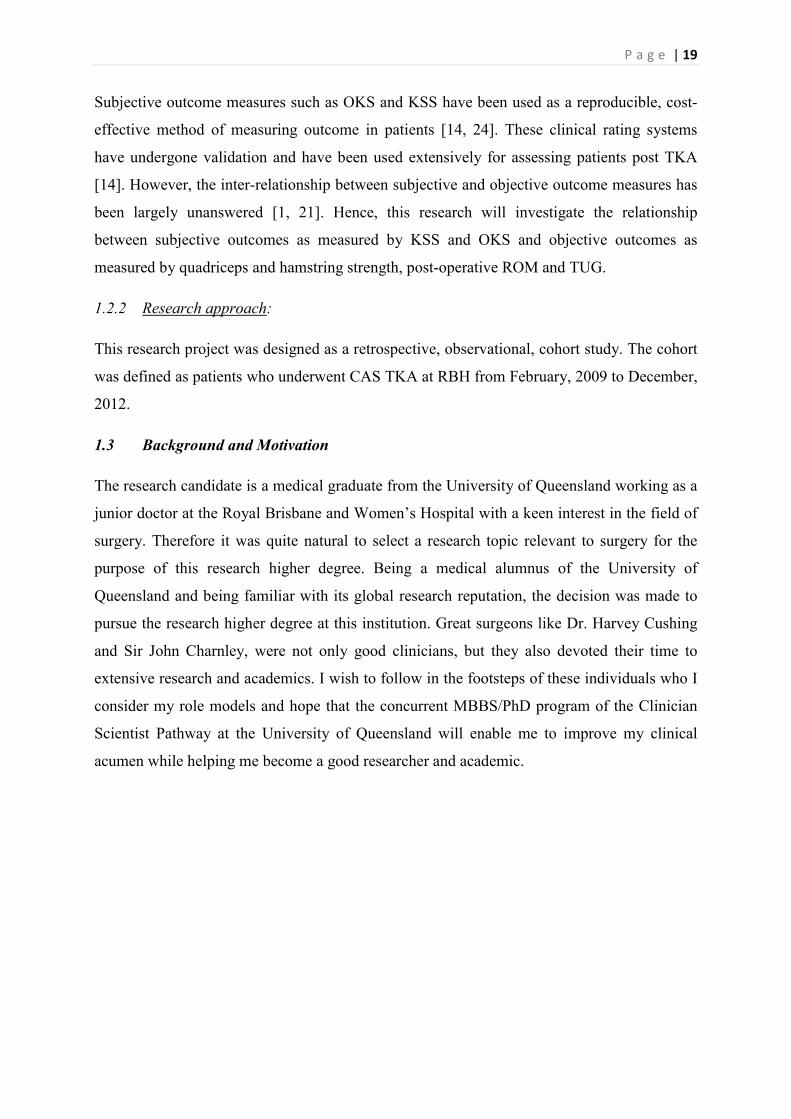
2.1.2 Menisci:
The lateral and medial menisci are paired, crescentic fibro-cartilaginous discs that extend
from the inter-condylar eminence to the periphery of the tibial plateau as demonstrated in Fig
2 [29]. While providing a cushion for weight bearing, it also deepens the articulations to
provide increased stability like the glenoid and acetabular labrum of the shoulder and hip joint
P a g e | 22
respectively [26, 29]. Hence, these menisci are thinner and concave in the middle and thicker
and convex in the periphery [26, 30]. The periphery in comparison is also relatively well-
vascularised from the capillaries branching off the fibrous capsule and synovial membrane
allowing good healing [29]. The medial meniscus has a characteristic C-shape and adheres
medially to the medial collateral ligament, anteriorly to the anterior cruciate ligament and
posteriorly to the posterior cruciate ligament rendering it less mobile and susceptible to
combined injury with the medial collateral ligament [29, 30]. The wider lateral meniscus has
the tendency to get trapped between the femur and tibia causing “clunking” in some patients.
It can be discoid shaped in 5% of the population [30].
Fig 2: Intra-articular anatomy of the knee [25]
2.1.3 Ligaments:
The stability of the knee joint is maintained by a group of intra-articular and extra-articular
ligaments [26]. The intra-articular ligaments are the anterior cruciate ligament, posterior
cruciate ligament and the posterior meniscofemoral ligament [26, 30]. The anterior cruciate
ligament originates from the anterior inter-condylar eminence of the tibial plateau and moves
superior, posterior and lateral to attach to the postero-medial portion of the lateral femoral
condyle [31]. The posterior cruciate ligament originates from the posterior inter-condylar
eminence of the tibial plateau and moves superior, anterior and medial along the medial
aspect of the anterior cruciate to attach to the antero-lateral portion of the medial femoral
condyle [32]. There are significant inconsistencies in the presence and size of the
meniscofemoral ligament which originates from the posterior horn of the lateral meniscus and
inserts onto the medial femoral condyle adjacent to the posterior cruciate [29, 30]. The extra-
P a g e | 23
articular ligaments are the patellar ligament, medial collateral ligament, lateral collateral
ligament, oblique popliteal ligament and arcuate popliteal ligament [26]. The tendon of the
quadriceps femoris completely surrounds the patella after which it becomes the patella
ligament to attach to the tibial tuberosity [28]. The fibular collateral ligament, separated from
the lateral meniscus by the popliteus tendon, originates from the lateral epicondyle of the
femur and attaches to the fibular head [26, 33]. It protects the knee joint against varus stress
[33]. The tibial collateral ligament originates from the medial epicondyle of the femur and
attach to the medial condyle of the tibia [26, 34]. It protects the knee joint against valgus
stress [33, 34]. An expansion of the semimembranosus tendon, the oblique popliteal ligament,
originates from the medial tibial condyle and attaches to the lateral femoral condyle [26, 29].
The arcuate popliteal ligament originates at the fibular head, moves over the tendon of the
popliteus and blends with the posterior fibrous capsule of the knee [25, 26]. Both the oblique
and arcuate popliteal ligaments reinforce the joint capsule posteriorly [26].
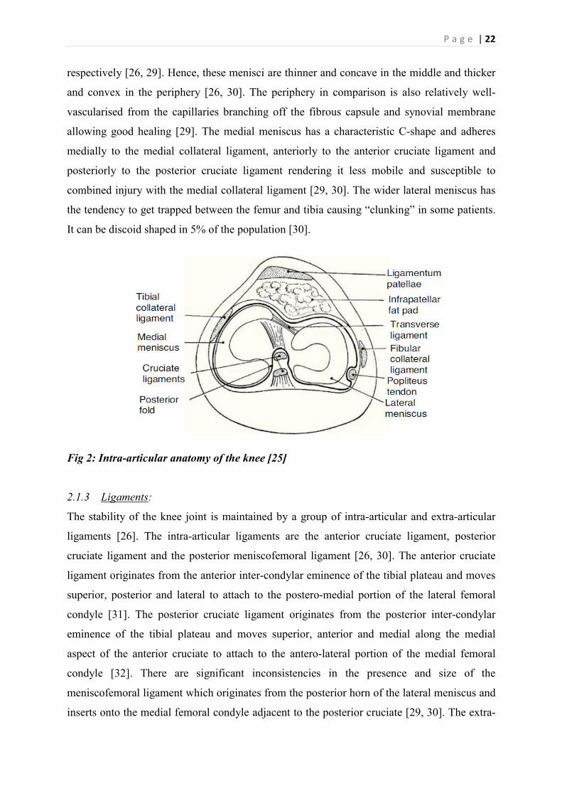
2.1.4 Innervation:
Innervation of the knee joint is by the obturator and femoral nerves of the lumbar plexus and
tibial and common peroneal nerves of the sacral plexus as demonstrated in Fig 3 [29, 35].
Sensation over the medial aspect of the knee is by the infrapatellar branch of the saphenous
nerve [28, 36]. Sensation over the areas of knee is by the peripatellar plexus comprising
branches of the femoral and lateral femoral cutaneous nerves [28, 35].
Fig 3: Innervation of the knee joint [36]
P a g e | 24
2.1.5 Musculature:
The knee joint is capable of flexion and extension with minimal internal and external rotation
[26]. The primary extensors of the knee joint are the vastus medialis, vastus intermedius,
vastus lateralis and rectus femoris which together form the quadriceps femoris [25, 26]. The
primary flexors of the knee joint are semitendinosus, semimembranosus and biceps femoris
[25]. Other weak flexors include gracilis, sartorius, gastrocneumius and popliteus [26].
Internal rotation is achieved by semimembranosus, semitendinosus, popliteus, gracilis and
Sartorius [25, 26]. Lateral rotation of the knee is predominantly achieved by the biceps
femoris [26].
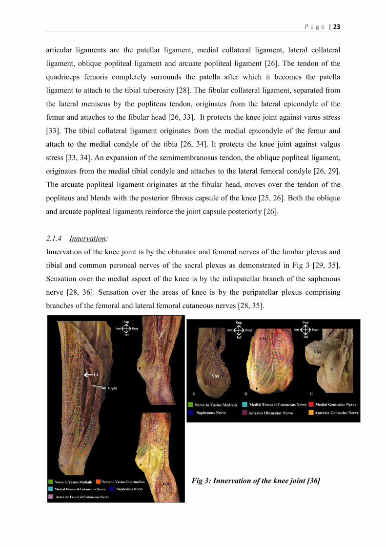
2.1.6 Vasculature:
The knee joint is supplied by a rich
arterial anastamosis formed by the
popliteal artery (superior, middle and
inferior genicular branches), femoral
artery (descending genicular branch),
lateral femoral circumflex artery
(descending branch), circumflex
fibular artery and tibial recurrent
arteries (anterior and posterior) as
demonstrated in Fig 4 [25, 37]. The
venous drainage is by deep veins of
the same name as the corresponding
arteries and they follow the arterial
system [25].
Fig 4: Vasculature of the knee joint [37]
2.1.7 Bursae:
There are numerous bursae that surround the knee [26]. Their arrangement around the knee is
highly variable and complex [25]. The pes anserine bursa is located deep to the pes anserinus,
formed by the tendons of gracilis, sartorius, and semitendinosus, 4-5cm inferior to the antero-
medial joint line of the knee [25, 26]. The semimembranosus bursa can be found in the
popliteal fossa [25]. Other bursae of clinical significance include the supra-patellar bursa,
infra-patellar bursa and pre-patellar bursa [25, 26].
P a g e | 25
2.1.8 Joint capsule:
The joint capsule of the knee joint has two components [26]. The external fibrous layer is
generally thin with localized thickenings due to the extra-articular ligaments of the knee [26,
29]. The internal synovial membrane lines the articular surfaces and produces synovial fluid
which lubricates the knee joint and provides nutrients [26].
2.2 Pathology
Damage to any of the three compartments of the knee joint can be a result of osteoarthritis
(idiopathic, post-traumatic), inflammatory arthritis (rheumatoid arthritis, sero-negative
spondyloarthropathies, etc), osteonecrosis, tumours or congenital deformities [2].
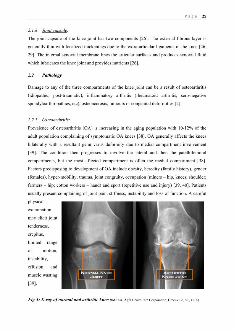
2.2.1 Osteoarthritis:
Prevalence of osteoarthritis (OA) is increasing in the aging population with 10-12% of the
adult population complaining of symptomatic OA knees [38]. OA generally affects the knees
bilaterally with a resultant genu varus deformity due to medial compartment involvement
[39]. The condition then progresses to involve the lateral and then the patellofemoral
compartments, but the most affected compartment is often the medial compartment [38].
Factors predisposing to development of OA include obesity, heredity (family history), gender
(females), hyper-mobility, trauma, joint congruity, occupation (miners – hip, knees, shoulder;
farmers – hip; cotton workers – hand) and sport (repetitive use and injury) [39, 40]. Patients
usually present complaining of joint pain, stiffness, instability and loss of function. A careful
physical
examination
may elicit joint
tenderness,
crepitus,
limited range
of motion,
instability,
effusion and
muscle wasting
[39].
Fig 5: X-ray of normal and arthritic knee (IMPAX, Agfa HealthCare Corporation, Greenville, SC, USA)
P a g e | 26
Primary modality of investigation is plain radiography [40, 41]. Three views are commonly
used: anteroposterior – obtained with the patient standing (stressed) to assess the medial and
lateral compartments; lateral – standing to assess the patellofemoral joint narrowing and
patellar position; tangential patellar view (sunrise, skyline or merchant view) – to assess
patellofemoral compartment [28, 39]. Classic radiographic findings include joint space
narrowing, subchondral sclerosis, subchondral cysts and osteophytes as demonstrated in Fig 5
[38, 42]. These radiographic findings can be graded to determine severity by using systems
such as the Kellgren-Lawrence grading system and the Ahlback classification [42]. Advanced
imaging modalities like Computed Tomography and Magnetic Resonance Imaging are not
required in assessing OA joints for
TKA, but may be useful in
identifying other pathologies such
as meniscal tears and osteonecrosis
[39, 41].
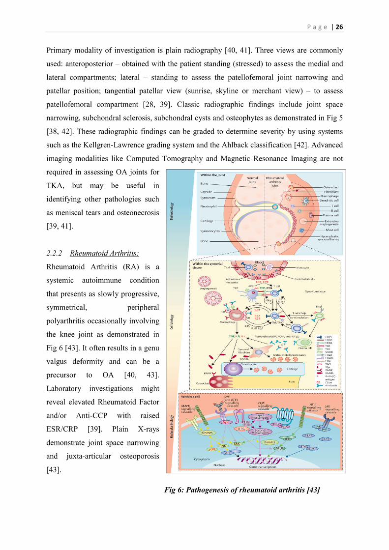
2.2.2 Rheumatoid Arthritis:
Rheumatoid Arthritis (RA) is a
systemic autoimmune condition
that presents as slowly progressive,
symmetrical, peripheral
polyarthritis occasionally involving
the knee joint as demonstrated in
Fig 6 [43]. It often results in a genu
valgus deformity and can be a
precursor to OA [40, 43].
Laboratory investigations might
reveal elevated Rheumatoid Factor
and/or Anti-CCP with raised
ESR/CRP [39]. Plain X-rays
demonstrate joint space narrowing
and juxta-articular osteoporosis
[43].
Fig 6: Pathogenesis of rheumatoid arthritis [43]

P a g e | 27
2.2.3 Other pathology:
Sero-negative spondyloarthropathies produce histologically similar synovitis to RA but
without the production of rheumatoid factor and an increased association with HLA-B27 [44].
Osteonecrosis can be due to a multitude of factors such as sickle cell disease, Caisson’s
disease, medications, endocrine disorders (Cushing’s and diabetes mellitus), trauma, HIV,
irradiation and alcohol abuse [45]. It can present as joint pain with MRI being the best
modality of investigation [39, 45].
2.3 Management
Treatment options for arthritic knees involve non-surgical and surgical management [40].
2.3.1 Non-Surgical Management:
Non-surgical management includes non-pharmacological management such as weight loss for
overweight individuals; use of walking aids to take load off the knees; strengthening,
stretching and conditioning exercises; use of heat/ice packs; wedge insoles or bracing [40,
46]. Pharmacological management involves use of paracetamol, NSAIDs, intra-articular
corticosteroid injections and in the case of inflammatory arthritis DMARDs (disease
modifying anti-rheumatic drugs) [40, 43]. Recent studies have reaffirmed the utility of
autologous platelet rich plasma and hyaluronic acid in the treatment of mild to moderate
osteroarthritis [47].
2.3.2 Surgical Management:
Surgical interventions include arthroscopy, osteotomy, arthroplasty and arthrodesis [40, 48,
49]. Arthroscopic synovectomy produces good outcomes in patients with diseased rheumatoid
synovium, but the use of arthroscopic lavage and debridement in OA remains controversial
and under investigation [48, 50]. Arthroscopic lavage and debridement in OA is strictly
indicated for patients who experience mechanical symptoms such as locking-unlocking due to
underlying meniscal pathology [40]. Osteotomy remains a viable option in younger patients
with non-inflammatory, unicompartmental disease [40, 48]. A medial compartment
localisation producing a genu varus deformity is amenable to a proximal tibial osteotomy and
a lateral compartment localisation producing a genu valgus deformity is amenable to
osteotomy of the supracondylar region of the distal femur [50]. Osteotomy in the selective
case produces favourable outcomes although controversy surrounds the outcome of secondary
P a g e | 28
conversion to a total knee arthroplasty [48, 50]. W-Dahl et al reported a 30% cumulative
revision rate at 10 years for high tibial osteotomy with increased risk of revision in the female
gender and with increasing age [51].
Unicompartmental knee arthroplasty began in the 1950s and is experiencing a current
resurgence [48]. While detractors claim incomplete pain relief, early implant failure and
increased difficulty of subsequent revisions, proponents attribute these to flaws in early
designs [48, 50]. In Australia, the cumulative percent revision at 11 years for primary
unicompartmental knee arthroplasty is 16.4% compared to 6.4% for primary total knee
arthroplasty [2]. National joint registry data show that the risk of re-revision after a
conversion from unicompartmental to TKA is also higher than if a TKA was performed as the
primary procedure [1]. Unicompartmental knee arthroplasty has the advantage of preservation
of ligamentous integrity restoring natural kinematics of the knee, less extensive surgery, lower
risk of infection and preservation of bone stock of the remaining two compartments for
subsequent revision surgeries [1, 52]. Indications for unicompartmental knee arthroplasty are
strict and include – arthritic wear of single compartment; age and activity level compatible for
arthroplasty; BMI less than 30kg/m2; intact ligament system; and moderate axis deformity
that is correctable to less than 7-10 degrees varus or valgus after tibial augmentation spacer
[1, 50, 52]. Rheumatoid arthritis is considered a contraindication [52]. Arthrodesis or surgical
fusion of the knee joint is often reserved as a salvage procedure for patients with multiple
prosthetic infections/revisions or failed extensor mechanism [49]. This procedure has the
advantage of pain relief, but compromises the functional ability of the knee joint and is hence
considered a last resort [39, 48, 49].
2.4 Total Knee Arthroplasty
2.4.1 Introduction to Total Knee Arthroplasty:
Total knee arthroplasty (TKA) was first introduced by Insall and colleagues as the “total
condylar prosthesis” at the Hospital for Special Surgery in 1973 [3]. Total Knee Arthroplasty
(TKA) is a surgical procedure to replace the weight-bearing surfaces of the knee joint in order
to relieve pain and disability from osteoarthritis and other arthritic conditions [1]. The surgery
involves resecting the diseased or damaged joint surfaces of the knee and resurfacing with
metal and polyethylene prosthetic components shaped to allow continued motion of the knee
and relief from pain as demonstrated in Fig 7 [1, 53].

P a g e | 29
Fig 7: Total knee arthroplasty. A – anteroposterior radiographs of bilateral knees and hips;
B – femoral and tibial components; C- incision to open the knee joint; D- attachment of jig
to femur; E – femoral cuts and resection of bone; F – attachment of tibial jig; G – tibial cut
and resection of bone; H – implantation of femoral component; I – implantation of tibial
component [53]
TKA is conventionally performed with the aid of intra-medullary or extra-medullary jig-based
alignments [54]. CAS TKA was introduced in 1997 and can utilise either image-based
systems – preoperative CT or intraoperative fluoroscopy for collection of morphological
information; or imageless systems – to overcome concerns about radiation exposure by using
a virtual model supplemented by registration data [55]. CAS-TKA incorporates three basic
components: the computer platform, the tracking system, and the rigid body marker [56].
Todesca et al, examined 225 patients prospectively and randomly assigned them to
conventional and CAS TKA to demonstrate superior accuracy of implant positioning and
better functional outcomes of CAS TKA [10, 57].
TKA can be posterior cruciate substituting where the PCL is excised; or posterior cruciate
retaining when the PCL is preserved [58]. Fixation for TKA include cemented, cement-less or
hybrid options [59]. The Cochrane review investigating the efficacy of fixation options for
P a g e | 30
TKA by Nakama et al, demonstrate smaller displacement of the tibial component with
cemented fixation in the short term with increased subsequent risk of loosening and no
demonstrable evidence of any difference in functional performance [59]. Cemented primary
procedures have a lower rate of second revision compared to other methods of fixation when
the first revision is undertaken with five years of the primary procedure. The method of
fixation does not have an effect on the outcome of the first revision after five years [2].
TKA is considered to be both cost effective and highly successful with a one year mortality
rate of 1.0% and a ten year mortality rate of 24% [2, 13]. By 2015, 534,717 knee
replacements were performed in Australia of which 443,948 were primary TKA, 46701 were
partial TKA and 44068 were revision knee replacements [2]. In Australia, the total number of
TKA performed has increased by 88.3% since 2003 and 4.7% over the last year [2]. This is
comparable to the Scandinavian countries such as Sweden where the incidence of TKA has
more than doubled since 1998 [11]. This increase in incidence of TKA can be attributed to the
increasing prevalence of osteoarthritis, accounting for 94-98% of TKAs performed, in an ever
growing geriatric population [1, 2].
2.4.2 Indications and Contraindications:
The main indication for TKA is pain relief of arthritic knees confirmed by radiography that
have failed prolonged non-surgical intervention and do not meet the indication for other
surgical procedures [1, 20]. Restoration of function and correction of deformity are secondary
outcomes in TKA and are not primary indications to perform the procedure [1, 39]. Absolute
contraindications to perform TKA include evidence of an active infection, a non-functioning
extensor mechanism and vascular compromise to lower extremity [1]. Neurological
conditions such as polio, stroke and those that affect the lower extremities can be considered a
relative contraindication, but neither age nor weight (BMI) is a contraindication to TKA [1,
60].
2.4.3 “Time to Failure/Revision” – Traditional Outcome Measure:
The gold standard for measurement of success of TKA used by national joint registries is the
‘time to revision’ with a 92.8% fourteen year implant survival rate for primary TKA in
Australia [2]. The revision rate as measured by mean revisions per 100 observed component
years varies between countries – Sweden: 0.71; Australia: 0.83; Norway: 1.27; Finland: 1.77;
New Zealand: 1.93; Denmark: 2.51 [61]. The most common reason for failure, which
P a g e | 31
accounts for 28.7% of all revisions, is aseptic loosening/lysis usually due to implant wear [1,
2]. Infections account for 22.4% of all revisions [2]. Contributors to failure also include
postoperative pain (especially patellofemoral pain – 20.9%), instability (6.3%) and stiffness,
which together contribute to a further 30-40% of all revisions [1, 2]. Numerous factors
increase the risk for failure and revision [1, 11]. Age, an important prognostic factor is
inversely related to the risk of revision with 2.5 times higher revision rates in patients who are
younger than 65 years of age [2, 11]. Males also have a significantly increased risk of revision
due to a higher incidence of prosthetic infections [2]. The diagnosis is known to influence
outcome with primary TKA for rheumatoid arthritis demonstrating lower revision rates and
that for osteonecrosis and other inflammatory arthritis demonstrating higher revision rates
than that for OA [2, 11]. The class and type of implants also affect the risk of revision and
national joint registries keep an active record of failure rates to identify implants that perform
poorly [11]. Other factors include patient dependant factors such as obesity and surgeon
dependant factors such as surgical technique, experience, skill and postoperative care protocol
[1, 2, 62]. However, determining the success of TKA purely by assessing the ‘time to
revision’ is not reflective of patient satisfaction and functioning on a daily basis and could
potentially underestimate problems experienced post TKA.
2.4.4 Other Outcome Measures:
It is estimated that up to 23% of patients are dissatisfied with their replaced knee due to
residual pain or limited range of motion and function [1, 4]. Therefore various subjective and
objective surrogate measures of outcome have been devised to obtain a better reflection of
success post TKA [1]. However, there is no consensus in regards to the most optimal,
consistent and reliable outcome measure.
2.4.4.1 Range of Motion:
One of the primary outcomes that most patients are concerned about is the ROM acquired by
the knee post surgery [63, 64]. Laubenthal et al, demonstrated that a minimum ROM of 90
degrees is required for activities of daily living with higher level activities like running and
cycling dependant on increased ROM [15]. More recently, Ha et al, demonstrated that
increased ROM post TKA is an important factor for functional outcome and patient
satisfaction, particularly in the Asian population [16]. The factors suspected of contributing to
the post TKA kinematics have been extensively investigated [63-65]. The most reliable
predictor of post-operative ROM is pre-operative ROM [64-66]. This could be due to peri-
P a g e | 32
articular soft tissue contractions resulting in a soft tissue imbalance and requiring soft tissue
releases in carefully selected cases [6, 65]. Posterior cruciate substituting prosthesis is
associated with statistically significant improvement in range of motion in comparison with
cruciate retaining prosthesis [58, 67, 68]. Ishii et al, investigated the change in ROM during
pre-operative, intra-operative and post-operative periods across posterior cruciate ligament
sacrificing and retaining prostheses [66]. Significant correlations were observed between ‘pre-
operative and intra-operative ROM’ and ‘pre-operative and post-operative ROM’ across both
prostheses [66]. Factors suspected of influencing the postoperative ROM and still under
investigation include gender, implant design, patellar resurfacing, patellar height and
postoperative physiotherapy [63-65, 67].
Knee extension ROM is significantly correlated with extensor mechanism strength and
physical function as assessed by Short Form 36 (SF-36) [69]. ROM, flexion contractures and
extension lag also contribute to the calculation of KSS [70]. Hence, the ROM acquired post-
operatively does influence both subjective and objective clinical outcome measures.
Although, there is a significant correlation between ‘intra-operative and post-operative ROM’
in patients who underwent posterior cruciate sacrificing TKA, the influence of intra-operative
kinematic data on outcome measures is not well understood and warrants further investigation
[66].
2.4.4.2 Strength:
Another outcome measure is the flexion and extension strength after TKA as patients are
concerned about their ability to climb stairs and engage in physical activities [17, 71].
Quadriceps strength is intricately associated with performance and the loss of extensor
mechanism could result in a collapse when the knee is flexed [5, 71]. Quadriceps weakness
due to atrophy and neuromuscular activation deficits is intrinsic to the pathogenesis and
natural progression of osteoarthritis [71]. There is an immediate decline in the strength of the
extensor mechanism immediately post TKA attributed to the surgical approach and knee
swelling followed by significant improvement which plateaus after six to twelve months [71-
73]. Factors that contribute to quadriceps strength post TKA include early surgery, implant
design, surgical approach, neuromuscular electrical stimulation, extension ROM and post-
operative rehabilitation [20, 60, 69]. Currently, hamstring strength is attributed decreased
functional significance compared to the extensor mechanism as hamstring weakness only
becomes apparent in high intensity activities like running and uphill walking [17, 71].
P a g e | 33
However, as the incidence of TKA in the younger population increases, hamstring strength
will acquire enhanced functional significance [17].
2.4.4.3 Clinical Outcome Rating Systems:
Numerous outcomes scores such as International Knee Documentation Committee (IKDC),
Subjective Knee Evaluation Form, Knee Injury and Osteoarthritis Outcome Score (KOOS),
Knee Injury and Osteoarthritis Outcome Score Physical Function Short Form (KOOS-PS),
Knee Outcome Survey Activities of Daily Living Scale (KOS-ADL), Lysholm Knee Scoring
Scale and Activity Rating Scale (ARS) have been used to investigate the outcome of TKA
[14]. The limitation faced by outcome scores is the inability to objectively assess the function
of the knee without being biased by the overall function of the patient [24, 74]. A major
limitation is the lack of a minimum clinically important difference (MCID) i.e. the least
difference in scores that demonstrate a clinically significant difference [14]. These rating
systems are limited also by their lack of validation across languages and their purpose specific
utility – a rating system validated for tibial plateau fractures cannot then be used to assess
outcome of cruciate reconstruction despite being an indicator of knee function unless
subsequently also validated in cruciate reconstruction patients [1, 14]. The different outcome
scores variably weigh the different factors (pain, function, ROM) contributing to outcome and
opinions vary about the most appropriate clinical rating system [14, 74]. The Western Ontario
and McMaster University Osteoarthritis Index (WOMAC), SF-36, Oxford Knee Score (OKS)
and Knee Society Score (KSS) have undergone validation and demonstrate sufficient inter-
rater reliability for assessment of outcome [70, 74]. The OKS is a 12-item questionnaire for
patients undergoing TKA that assesses outcome in the last 4 weeks with a score out of 48
[14]. It demonstrates good sensitivity to change in outcome and adequate internal consistency
across multiple languages [14, 74]. It has been widely accepted due to its simplicity and ease
of use with the lack of a minimum clinically important difference being a major weakness
[14]. The original KSS was developed in 1989 and included a knee score to assess the knee
and a functional score to assess the ability to undertake activities of daily living [24]. This
was later abandoned to make way for the new KSS that is broadly applicable across sex, age,
activity level and implant type [24, 70]. The KSS is a validated and responsive questionnaire
to assess both subjective and objective performance before and after TKA [70]. Rating
systems, most of which are self-reporting questionnaires, fail to reflect physical ability and
overestimate performance [21].

P a g e | 34
2.4.4.4 Proprioception and Balance:
Balance and proprioception are important predictive factors for the quality of a patient’s life
and the functional level achieved post TKA [22]. There is a steady decline in proprioception
with age even in patients with normal knees [23]. Moreover, the pathogenesis of osteoarthritis
also involves loss of the proprioceptive ability of the knee from destruction of intra-articular
cartilage, ligaments and tendons which result in deteriorating balance and increased falls-risk
[19, 23]. Despite this destruction of intra-articular proprioceptive structures, TKA seems to
restore the soft tissue balance and hence the proprioceptive ability of the extra-articular
structures resulting in a decline in falls-risk, atleast in the short term [23]. Improvement in
balance and proprioception is observed in the first 6 months post surgery after which it
plateaus [75, 76]. Bascuas et al, analysed the relationship between balance and clinical
outcome measures such as BMI, pain, range of motion, strength (flexion and extension), gait
velocity and KSS in 44 patients [18]. A statistically significant negative correlation was
demonstrated between age and improvement in balance [18]. Schwartz et al, also
demonstrated a statistically significant positive correlation between balance and the clinical
outcome measures - SF-36, OKS, TUG and four square step test [22]. Balance is also better in
patients with bilateral TKA as opposed to unilateral TKA [77]. Other factors that
predict/influence balance and proprioception include preoperative proprioceptive training,
joint space, mal-alignment, soft tissue balance and early postoperative rehabilitation [19, 75,
78-80].
2.4.4.5 Timed Up and Go:
Although clinical rating systems are efficient and cost-effective, there is growing evidence for
the inclusion of performance tests such as Berg Balance Score (BBS), TUG, 6MW test and
SCT in the analysis of TKA outcomes [20, 21]. The TUG test is considered to be a reliable
and valid measure of change in functional mobility of inpatients in orthopaedic wards, but is
not a good predictor of length of stay [81]. Beauchet et al, demonstrated that TUG is closely
associated with a past history of falls [82]. Although, its potential to predict future falls is
limited, there is an improvement in correlation with standardization of testing conditions and
control of significant potential confounders such as age, gender and co-morbidities [21, 82].
Poor performance in these functional tests preoperatively is strongly associated with poor
performance postoperatively [20, 82]. Other factors such as age and declining mental health
also predict poor performance in functional tests [20].
P a g e | 35
2.4.5 Role of Alignment in TKA:
The alignment of implant components is a surgeon dependant factor that improves with
experience and skill and demands significant attention and resources [5, 83]. The surgeon
attempts to restore the overall alignment of the limb to pre-disease state by carefully aligning
the implant components in the coronal, sagittal and transverse planes with the joint line at an
appropriate level and good soft tissue balance in flexion and extension without limiting the
range of motion or putting undue pressure on the polyethylene plate [6]. Numerous strategies
such as navigational TKA (CAS TKA), patient specific positioning guides (PSPG), mobile
bearing prosthesis and high flexion TKA have been devised in order to minimize component
and overall mal-alignment [7-9]. The alignment in TKA can be determined clinically or by
employing radiography, fluoroscopy, the Perth CT protocol, MRI and navigational
investigations with each modality subject to its inherent limitations [84, 85].
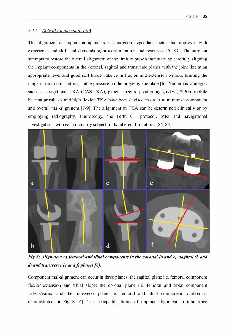
Fig 8: Alignment of femoral and tibial components in the coronal (a and c), sagittal (b and
d) and transverse (e and f) planes [6].
Component mal-alignment can occur in three planes: the sagittal plane i.e. femoral component
flexion/extension and tibial slope; the coronal plane i.e. femoral and tibial component
valgus/varus; and the transverse plane i.e. femoral and tibial component rotation as
demonstrated in Fig 8 [6]. The acceptable limits of implant alignment in total knee

P a g e | 36
arthroplasty are considered to be very narrow [86]. Mal-alignment was attributed as the
primary cause in only 2.2% of revisions after primary TKA [2]. This is because mal-
alignment manifests as other modes of failure such as implant loosening/lysis, patellofemoral
pain, instability, fracture, patellar erosion and implant wear thus contributing a more
significant role in outcome/survival than directly attributed [2, 5]. However, the question of
which alignment parameters are critical and their relative importance is still largely
unanswered.
2.4.6 Overall Alignment:
The mechanical axis of the limb is the imaginary straight line drawn from the centre of the
femoral head to the centre of the ankle as assessed on a long leg anteroposterior radiograph
[6]. An off-centre loading would cause collapse on one side or lift-off on the other resulting in
component loosening and thus failure [5]. Post operative limb alignment of 0±3 degrees
relative to the mechanical axis is considered to maximize implant durability and improve
clinical outcome [87, 88]. However, Parratte et al concluded that a strict adherence to a
neutral postoperative mechanical axis did not improve implant survival [89]. Ritter et al,
studied 6070 knees to define a neutral overall alignment as a tibiofemoral angle of 2.5-7.4
degrees valgus and demonstrated improved implant survival when this neutral alignment was
achieved [62]. Hence, it is difficult to conclude on the ideal overall alignment for the
population in general as it likely to be highly patient-specific.
2.4.7 Alignment in Transverse Plane:
Although there is a high variability in the reported rotational alignment of the femoral and
tibial components in TKA, axial mal-rotation with a tibiofemoral mismatch of more than 5
degrees is associated with increased risk of failure and poor clinical and functional outcomes
[90-92]. Mal-rotation of the femoral or tibial components is traditionally considered a cardinal
sin affecting patellar tracking and contributing to patellar subluxation, dislocation and
patellofemoral pain [5]. However in modern orthopaedics, the advent of high quality
polyethylene inserts and implant design have significantly decreased the observable
functional/clinical deficits from rotational mal-alignment within limits [93].
2.4.8 Alignment in Coronal Plane:
Varus/valgus mal-alignment is the commonest cause of early loosening and is the most
investigated alignment parameter after the overall alignment of the limb [62]. The coronal
P a g e | 37
femoral component angle (cFCA) is the angle between the longitudinal axis of the femoral
component and the mechanical axis of the femur in the coronal plane and every attempt is
made not to exceed 8 degrees of valgus [62]. A good coronal femoral alignment is
significantly associated with improved clinical and functional outcomes [90, 92]. The coronal
tibial component angle (cTCA) is the angle between the longitudinal axis of the tibial
component and the mechanical axis of the tibia in the coronal plane and every attempt is made
to achieve 90 degrees [62]. A perfectly neutral (perpendicular) tibial component in the coronal
plane is important in order to prevent early failure and produce good clinical outcome [90,
92].
2.4.9 Alignment in Sagittal Plane:
The component alignment in the sagittal plane is the least studied of all alignment parameters
in determining outcome after TKA [6]. The radiographic parameters in the sagittal plane as
ideally determined on a long leg, weighted, lateral x-ray include femoral component
flexion/extension, tibial slope and posterior condylar offset (PCO) [94].
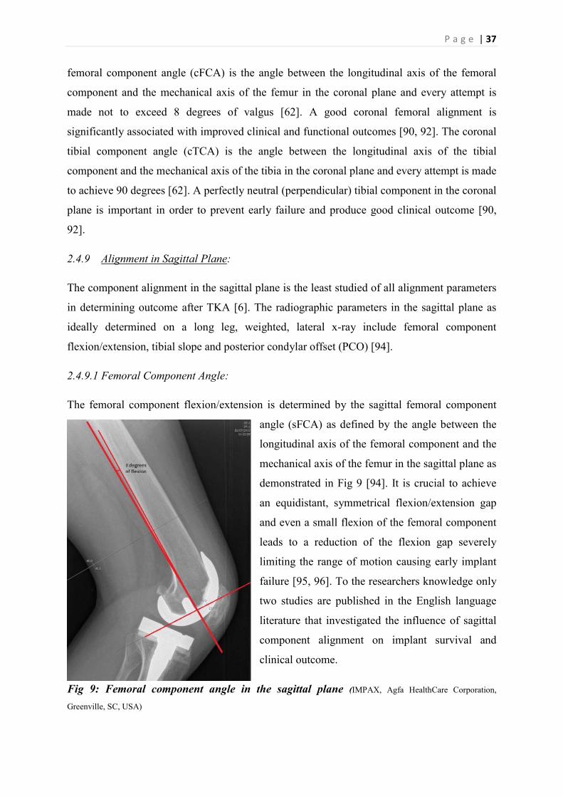
2.4.9.1 Femoral Component Angle:
The femoral component flexion/extension is determined by the sagittal femoral component
angle (sFCA) as defined by the angle between the
longitudinal axis of the femoral component and the
mechanical axis of the femur in the sagittal plane as
demonstrated in Fig 9 [94]. It is crucial to achieve
an equidistant, symmetrical flexion/extension gap
and even a small flexion of the femoral component
leads to a reduction of the flexion gap severely
limiting the range of motion causing early implant
failure [95, 96]. To the researchers knowledge only
two studies are published in the English language
literature that investigated the influence of sagittal
component alignment on implant survival and
clinical outcome.
Fig 9: Femoral component angle in the sagittal plane (IMPAX, Agfa HealthCare Corporation,
Greenville, SC, USA)

P a g e | 38
The earlier research by Faris et al investigated 623 knee replacements with the sagittal
femoral component ranging from 20 degrees in extension to 20 degrees in flexion, but found
no correlation with the ROM achieved [97]. The later research by Murphy et al investigated
the effect of femoral component flexion on ROM (knee flexion and extension), outcome
scores (WOMAC, SF-36), quadriceps strength, functional tests (timed stand test, SCT) and
satisfaction [94]. A strong correlation between femoral component flexion, improved ROM
and worsening scores for the mental component of SF-
36 was observed [94].
2.4.9.2 Posterior Condylar Offset:
The PCO is defined as the maximal sagittal plane
distance between the posterior femoral condyle and the
posterior femoral cortex as demonstrated in Fig 10 [94,
98]. Malviya et al demonstrated that PCO is an
important predictor of knee flexion [99]. In contrast,
Bauer et al did not find any correlation between PCO
and post-operative knee flexion [100]. Therefore, there
is a clear need for further comprehensive investigation to
determine the influence of PCO on clinical outcome
measures particularly ROM.
Fig 10: Posterior condylar offset (IMPAX, Agfa HealthCare Corporation, Greenville, SC, USA)
2.4.9.3 Tibial slope:
Fig 11: Tibial slope in the sagittal plane (IMPAX, Agfa
HealthCare Corporation, Greenville, SC, USA)
Tibial slope is the angle between the longitudinal axis
of the tibial component and the mechanical axis of the
tibia in the sagittal plane as demonstrated in Fig 11
[101]. An increase in tibial slope could limit extension
and increase flexion and a decrease in tibial slope could
result in laxity on extension and stiffness on flexion [6].
Increased tibial slope can improve knee flexion by
facilitating posterior femoral rollback as a result of
P a g e | 39
increased tension in posterior cruciate ligament [60]. Bellemans et al demonstrated an average
gain of 1.7 degrees of flexion with every degree of increased posterior tibial slope [98].
Malviya et al also showed a positive correlation between tibial slope and ROM [99].
However, this is under scrutiny as recently, authors have demonstrated that an increased tibial
slope failed to produce any significant improvement in ROM [100-102]. Therefore, the
concept of an ideal tibial slope remains as elusive as that of an ideal sFCA and an ideal PCO.

P a g e | 40

P a g e | 41
The aim of this research project was to investigate the inter-relationship between the clinical
outcomes as measured by the sagittal plane alignment of implant components, peri-operative
kinematics, validated clinical rating systems, strength, balance and functional performance
tests in patients at least one year post TKA at a regional academic hospital. This research
project had a primary objective and two secondary objectives.
Although the relationship between alignment and clinical outcome has been extensively
investigated, the role of sagittal component alignment, in particular the sFCA, PCO and tibial
slope, has been poorly understood [94]. There is conflicting evidence about the influence of
sFCA, PCO and tibial slope on ROM or flexion [94, 95, 97-99, 101]. Faris et al investigated
623 knee replacements with the sagittal femoral component ranging from 20 degrees in
extension to 20 degrees in flexion, but found no correlation with the ROM achieved [97].
However, Murphy, et al investigated the effect of femoral component flexion on ROM (knee
flexion and extension) and demonstrated a strong correlation [94]. Similarly, Malviya et al
demonstrated that PCO is an important predictor of knee flexion and; Bauer et al did not find
any correlation between PCO and post-operative knee flexion [99, 100]. Bellemans et al
demonstrated an average gain of 1.7 degrees of flexion with every degree of increased
posterior tibial slope [98]. Malviya et al also showed a positive correlation between tibial
slope and ROM [99]. However, this is under scrutiny as recently, other researchers have
demonstrated that an increased tibial slope failed to produce any significant improvement in
ROM [100-102]. Hence, the relationship between sagittal plane component alignment as
measured by sFCA, PCO and tibial slope and clinical outcome as measured by intra-operative
kinematic data (MFA, MEA and ROM) currently lacks clarity. The primary objective of this
research project was to determine the correlation between radiological sagittal plane
alignment of the implant components, as defined by sFCA, PCO and tibial slope and the post-
operative kinematics of the knee, as defined by MFA, MEA and ROM, in patients post CAS
TKA. Therefore, it was hypothesized (H1) that there will be a correlation between
component alignment in the sagittal plane and intra-operative navigational kinematic
data in patients post CAS TKA such that increased sFCA, PCO and tibial slope would
increase the MFA and hence the ROM.
Balance is a strong predictor of the quality of life and functional ability of patients post TKA
[22]. Bascuas et al analysed the relationship between balance and clinical outcome measures;
they demonstrated a statistically significant negative relationship between age and balance,
despite their small sample size [18]. Corrigan et al demonstrated a significant correlation
P a g e | 42
between hamstring to quadriceps ratio and balance in ACL deficient knees, suggesting a
relatively greater correlation of flexion strength in comparison to extension strength with
functional outcome in patients [103, 104]. However, Lee et al could not demonstrate a
statistically significant correlation between hamstring strength and balance in patients with
ACL deficient (Rho=0.239, p=0.506) and ACL intact (Rho=0.367, p=0.297) knees [105].
Schwartz et al demonstrated a statistically significant positive correlation between balance
and TUG [22]. Hence, any possible relationship between balance and post-operative flexion
strength, extension strength, range of motion or TUG currently lacks clarity. The first
secondary objective of this research project was to determine the relationship between
balance, as defined by Two Leg Eyes Open test, Two Leg Eyes Closed test and Single Leg
Eyes Open test, and the clinical outcome measures, as defined by flexion strength, extension
strength, ROM and TUG, in patients post TKA. Therefore, it was hypothesized (H2) that
there will be a correlation between balance and the clinical outcome measures such that
improved strength would result in improved balance.
Few clinical ratings systems such as OKS and KSS have been validated and considered
broadly applicable across sex, age, activity level and implant type [14, 70]. While Laubenthal
et al demonstrated that a minimum ROM of 90 degrees is required for activities of daily
living, with higher-level activities like running and cycling dependent on increased ROM;
Thomsen et al demonstrated no relationship between ROM and the ability to perform
activities of daily living [15, 106]. Schwartz et al demonstrated a statistically significant
positive correlation between balance and the Short Form -36 (SF-36) and Oxford Knee Score
(OKS) [22]. Hence, any possible relationship between OKS and post-operative flexion
strength, extension strength, ROM, balance, or TUG currently lacks clarity. The second
secondary objective of this research project is to determine the correlation between subjective
outcome measures, as defined by OKS and KSS, and objective outcome measures, as defined
by flexion strength, extension strength, TUG, ROM and balance, in patients post TKA.
Therefore, it was hypothesized (H3) that there will be a correlation between subjective
and objective outcome measures such that improved strength, ROM and balance would
result in improved patient satisfaction.
In summary, this research project is a significant addition to the current literature on outcome
measures after TKA. The study design selected was a retrospective observational cohort study
which enabled examination of the correlation between the outcome measures. The cohort
refers to patients who have undergone CAS TKA at RBH from February 2009 to December
P a g e | 43
2012. A cohort study provided level III-2 evidence according to the NHMRC evidence
hierarchy and adequately answered the research questions posed. A randomised controlled
trial or quasi-randomised controlled trial will require intentional suboptimal alignment of
components in subjects which would be unethical.

P a g e | 44
P a g e | 45
4.1 Study Design
This was a retrospective cohort study of 94 participants (105 knees) that underwent computer
assisted (CAS) primary TKA between February 2009 and December 2012 at a single
institution.
4.2 Patient Selection and Setting
Subjects were recruited from the Orthopaedics Outpatient Department at Rockhampton Base
Hospital. Patients who returned for their regular follow-up appointment and met the inclusion
criteria were invited to participate in the study. This clinic caters for most of the public
patients in the wider Central Queensland catchment area requiring total joint arthroplasty.
Referrals to this clinic are largely through General Practitioners working in Central
Queensland. The average time from referral to be seen was six months and the average time
on the waiting list for a joint replacement was six months. During this waiting period, subjects
were encouraged to remain active and aim for a health body mass index. No referral to
physiotherapy was initiated due to the nature of the project; however, if a subject elected to be
treated by a private physiotherapist in the pre-operative interval, the treating surgeon did not
discourage it. Regular analgesics for pain control was encouraged and supervised by the
General Practitioner. All participants had been operated on by one of two experienced
orthopaedic surgeons with a subspecialty interest in knee arthroplasty (EH and RK). Both
surgeons work as full time consultant orthopaedic surgeons in a public hospital with over 25
years of operative experience.
4.3 Inclusion and Exclusion Criteria
The following inclusion criteria were used:
Patients who have undergone CAS TKA at Rockhampton Base Hospital from
February, 2009 to December, 2012.
Minimum 1 year post TKA.
Community ambulators at the time of data collection - in order to reduce the risk of
falls and injury to participants.
The following exclusion criteria were used:
Individual below the age of 40 years.

P a g e | 46
Non osteoarthritic aetiology for joint degeneration
People with an intellectual or mental impairment.
Women who are pregnant and the human foetus.
People highly dependent on medical care.
Past medical history of connective tissue disorders such as Ehlers Danlos, Marfan’s,
etc.
4.4 Surgery, Post-surgical Care and Follow-up
The medial para-patellar approach was utilized for all cases as demonstrated in Fig 7. In
addition, two 4.5 mm pins were placed into the distal femur and proximal tibia to attach
trackers for the navigation system (Stryker OrthoMap Precision Knee Navigation System).
All patients received a posterior cruciate retaining implant (Stryker Triathlon Total Knee
Replacement System).
Both surgeons utilized standard pre- and post-operative protocols to minimize confounders in
the study. Post-operatively patients were mobilized on day one by the physiotherapist, active
ROM was encouraged and continuous passive motion machines were used. Patients were
discharged when the following discharge criteria were met: active pain-free ROM 0-90
degrees; satisfactory wound healing; safe independent mobilisation; and no post-operative
complications.
All patients were instructed to attend routine follow-up appointments at six weeks, three
months, six months, one year, and annually thereafter.
4.5 Sample Size
A sample size calculation was performed. The calculation was designed to provide the
number of cases required to discover a statistically significant (p = 0.05) correlation for a
maximum of five co-variates with a medium effect size (f2 = 0.15). The sample size
calculation based on these parameters indicated that 91 patients were needed to provide a
statistical power of 80%. Therefore, taking into account a 10% participant attrition/drop-out
rate, it is the intention of the researcher to recruit at least 100 participants.

P a g e | 47
4.6 Variables
The following outcome measures were used: kinematic variables (pre-, intra- and post-
operative measurements for MFA, MEA and ROM); sagittal plane alignment of implant
components (sFCA, PCO, tibial slope); clinical rating systems (OKS and KSS); isometric
strength (knee flexion and extension); balance (two legs eyes open – TLEO; two legs eyes
closed – TLEC; single leg eyes open – SLEO); and TUG. The navigational computer database
was used to develop the list of potential participant. The navigational kinematic data of
potential participants was then extracted.
4.6.1 Kinematic Data
Pre-operative kinematics: navigational data extracted
o MFA – the maximum knee flexion acquired during passive flexion of the knee
by the surgeon post administration of anaesthesia and prior to commencement
of surgery as registered by the navigational computer.
o MEA - the maximum knee extension acquired during passive extension of the
knee by the surgeon post administration of anaesthesia and prior to
commencement of surgery as registered by the navigational computer.
o ROM – the range of motion during passive flexion-extension of the knee by
the surgeon post administration of anaesthesia and prior to commencement of
surgery as registered by the navigational computer.
Intra-operative kinematics: navigational data extracted
o MFA – the maximum knee flexion acquired during passive flexion of the knee
by the surgeon post implant in situ as registered by the navigational computer.
o MEA - the maximum knee extension acquired during passive extension of the
knee by the surgeon post implant in situ as registered by the navigational
computer.
o ROM – the range of motion during passive flexion-extension of the knee by
the surgeon post implant in situ as registered by the navigational computer.
P a g e | 48
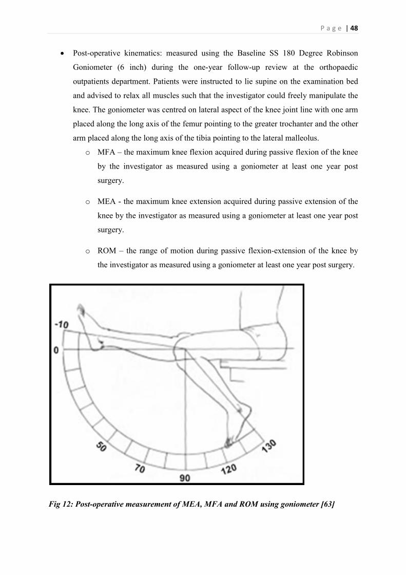
Post-operative kinematics: measured using the Baseline SS 180 Degree Robinson
Goniometer (6 inch) during the one-year follow-up review at the orthopaedic
outpatients department. Patients were instructed to lie supine on the examination bed
and advised to relax all muscles such that the investigator could freely manipulate the
knee. The goniometer was centred on lateral aspect of the knee joint line with one arm
placed along the long axis of the femur pointing to the greater trochanter and the other
arm placed along the long axis of the tibia pointing to the lateral malleolus.
o MFA – the maximum knee flexion acquired during passive flexion of the knee
by the investigator as measured using a goniometer at least one year post
surgery.
o MEA - the maximum knee extension acquired during passive extension of the
knee by the investigator as measured using a goniometer at least one year post
surgery.
o ROM – the range of motion during passive flexion-extension of the knee by
the investigator as measured using a goniometer at least one year post surgery.
Fig 12: Post-operative measurement of MEA, MFA and ROM using goniometer [63]
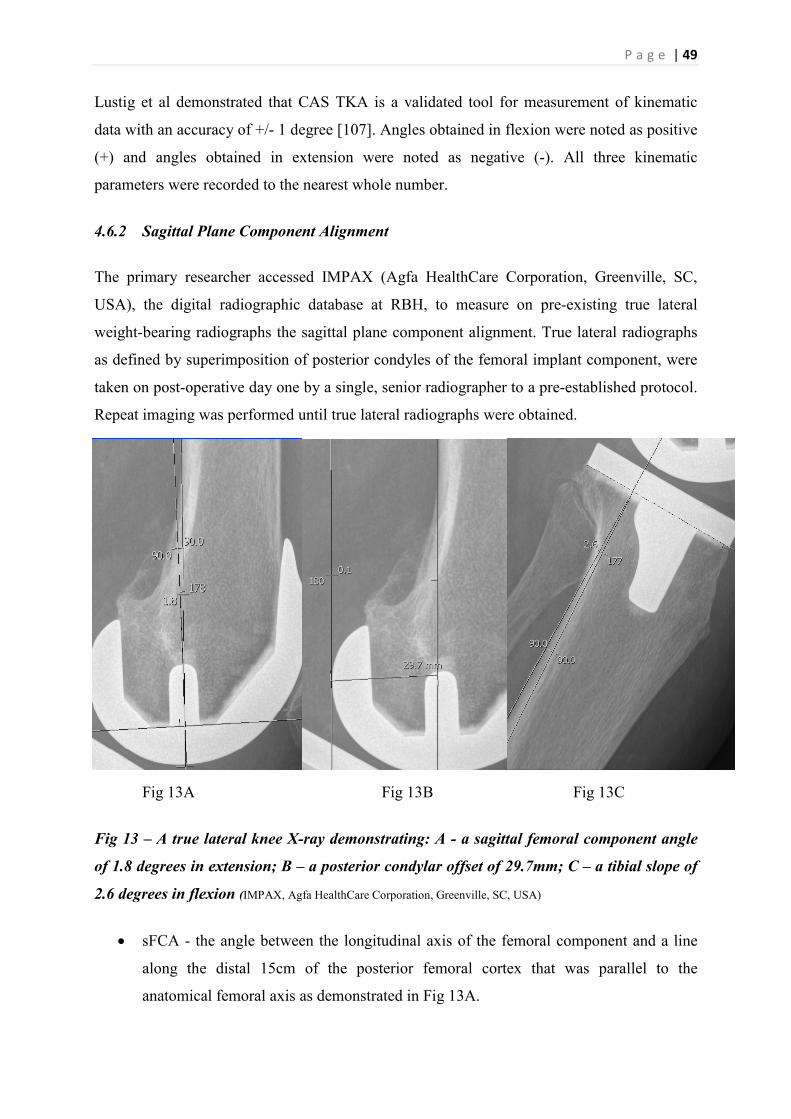
P a g e | 49
Lustig et al demonstrated that CAS TKA is a validated tool for measurement of kinematic
data with an accuracy of +/- 1 degree [107]. Angles obtained in flexion were noted as positive
(+) and angles obtained in extension were noted as negative (-). All three kinematic
parameters were recorded to the nearest whole number.
4.6.2 Sagittal Plane Component Alignment
The primary researcher accessed IMPAX (Agfa HealthCare Corporation, Greenville, SC,
USA), the digital radiographic database at RBH, to measure on pre-existing true lateral
weight-bearing radiographs the sagittal plane component alignment. True lateral radiographs
as defined by superimposition of posterior condyles of the femoral implant component, were
taken on post-operative day one by a single, senior radiographer to a pre-established protocol.
Repeat imaging was performed until true lateral radiographs were obtained.
Fig 13A Fig 13B Fig 13C
Fig 13 – A true lateral knee X-ray demonstrating: A - a sagittal femoral component angle
of 1.8 degrees in extension; B – a posterior condylar offset of 29.7mm; C – a tibial slope of
2.6 degrees in flexion (IMPAX, Agfa HealthCare Corporation, Greenville, SC, USA)
sFCA - the angle between the longitudinal axis of the femoral component and a line
along the distal 15cm of the posterior femoral cortex that was parallel to the
anatomical femoral axis as demonstrated in Fig 13A.

P a g e | 50
PCO - maximal sagittal plane distance between the posterior femoral condyle and the
posterior femoral cortex as demonstrated in Fig 13B.
Tibial slope - the angle between the longitudinal axis of the tibial component and a
line along the proximal 15cm of the posterior tibial cortex that was parallel to the
anatomical axis of the tibia as demonstrated in Fig 13C.
All three alignment parameters were recorded with an accuracy of one decimal place.
When patients attended their regular post-operative follow-up clinic at the Orthopaedics
Outpatients Department, the following clinical outcome measures were recorded:
4.6.3 Clinical Rating Systems
The Clinical Rating Systems used for this study were:
OKS - Participants were asked to complete the questionnaire to best reflect the
outcome of the operated knee in the last 4 weeks. The Oxford Knee Score consists of
twelve items, each with a score of one to four (from worst to best): pain; difficulty
washing and drying self; difficulty getting in and out the car/public transport; walking
duration; pain on standing; limp; ability to kneel; night pain; interference with work;
giving way; ability to do shopping and ability to descend stairs. A total score out of 48
was then obtained.
KSS – The researcher then completed the questionnaire in consultation with the
participant. The Knee Society Clinical Rating System categorises subjects to identify
those subjects whose function may be undermined by factors other than the knee in
question:
A: No substantial disease in the contralateral knee
B: Substantial arthrosis
C: Multiple joint involvement or generalised disability
It then assessed subjects by two scores: the Knee score and the Function score. The
Knee score is comprised of pain (50 points), range of motion (25 points with a
maximum of 125 degrees) and stability, measuring antero-posterior and mediolateral
stability separately (25 points). Deductions are made for flexion contracture,
extension lag, and alignment. The Function score comprised of walking distance (50
points), and stair climbing (50 points) with deductions for using a walking aid. The

P a g e | 51
clinical examination component of the KSS was performed by the principal
investigator (JA) who was experienced in orthopaedic examination:
o Range of motion (ROM): ROM was measured using a goniometer on the
lateral aspect of knee from full passive extension to full passive flexion. The
centre of the goniometer was placed at the tibiofemoral articulation, with one
arm following the line of the femur and the other following the line of the tibia.
o Antero-posterior displacement: The investigator massaged the subject’s
hamstrings to help in relaxation, and then sat on the foot with the subject’s
permission with the knee flexed at 90. The investigator then drew the tibia
anteriorly on the femur and estimated this displacement to the nearest whole
number in millimeters (mm). Similar posterior displacement of the tibia on the
femur was estimated using the same methodology.
o Mediolateral displacement: The subjects’ lower limb was held under the
investigator’s axilla and supported with one hand while the other hand applied
valgus and varus stress to the knee at 30 degrees of knee flexion. The angle
formed in the coronal plane between the femur and tibia was estimated in
degrees to the nearest whole number and added to correlate to a score.
o Flexion contracture: The subject was asked to straighten both knees flat on the
bed, and passive extension or the lack thereof was measured on the operated
leg with the goniometer.
o Extension lag: If full extension was not achieved actively, the investigator
gently passively extended the affected knee. The difference between active
and passive extension was measured in degrees and correlated to a score.
o Alignment: The angle between a line between the anterior superior iliac spine
and the centre of the patella and a line between the centre of the patella and the
centre of the ankle will be measured in degrees and correlated to a score.
4.6.4 Strength
The use of Nintendo Wii platform as a novel, cheap, accurate and reproducible measure of
force was previously validated by Bartlett et al [108]. Notably, by using the Nintendo Wii
platform, measurement error due to variations and limitations in tester’s strength was
eliminated. Participants were asked to sit on a comfortable chair with Velcro strapping placed
across the waist and chest to stabilize the torso and to push against the Nintendo Wii platform
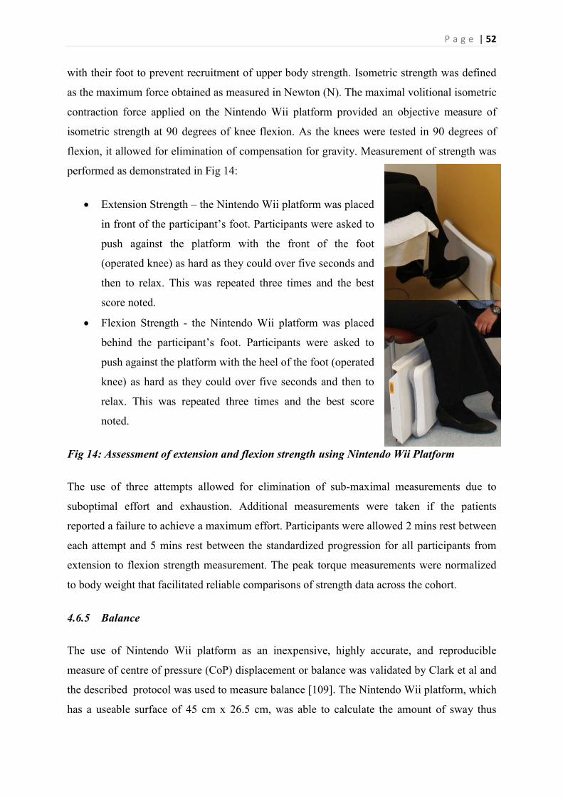
P a g e | 52
with their foot to prevent recruitment of upper body strength. Isometric strength was defined
as the maximum force obtained as measured in Newton (N). The maximal volitional isometric
contraction force applied on the Nintendo Wii platform provided an objective measure of
isometric strength at 90 degrees of knee flexion. As the knees were tested in 90 degrees of
flexion, it allowed for elimination of compensation for gravity. Measurement of strength was
performed as demonstrated in Fig 14:
Extension Strength – the Nintendo Wii platform was placed
in front of the participant’s foot. Participants were asked to
push against the platform with the front of the foot
(operated knee) as hard as they could over five seconds and
then to relax. This was repeated three times and the best
score noted.
Flexion Strength - the Nintendo Wii platform was placed
behind the participant’s foot. Participants were asked to
push against the platform with the heel of the foot (operated
knee) as hard as they could over five seconds and then to
relax. This was repeated three times and the best score
noted.
Fig 14: Assessment of extension and flexion strength using Nintendo Wii Platform
The use of three attempts allowed for elimination of sub-maximal measurements due to
suboptimal effort and exhaustion. Additional measurements were taken if the patients
reported a failure to achieve a maximum effort. Participants were allowed 2 mins rest between
each attempt and 5 mins rest between the standardized progression for all participants from
extension to flexion strength measurement. The peak torque measurements were normalized
to body weight that facilitated reliable comparisons of strength data across the cohort.
4.6.5 Balance
The use of Nintendo Wii platform as an inexpensive, highly accurate, and reproducible
measure of centre of pressure (CoP) displacement or balance was validated by Clark et al and
the described protocol was used to measure balance [109]. The Nintendo Wii platform, which
has a useable surface of 45 cm x 26.5 cm, was able to calculate the amount of sway thus

P a g e | 53
delivering an objective measure of balance. Participants were instructed to stand on the
platform with their hands placed on their hips or by their side, looking straight ahead fixed at
an imaginary point and to minimize the amount of sway by remaining as still as possible for
the duration of the trial as demonstrated in Fig 15. Three balance tasks were used based on
their varying difficulty and common use in literature:
o Two Legs Eyes Open – the right foot
on the right pedal, the left foot on the
left pedal and eyes open, the amount
of sway was assessed for one minute.
o Two Legs Eyes Closed – the right
foot on the right pedal, the left foot
on the left pedal and eyes closed, the
amount of sway was assessed for one
minute.
o Single Leg Eyes Open – the foot of
the operated knee on the centre of the
platform, the foot of the non-
operated knee slightly raised off the
platform, the amount of sway was
assessed for ten seconds. Fig 14A Fig 14B
Fig 15: Assessment of balance (centre of pressure displacement – path velocity) using
Nintendo Wii Platform. A – Two Legs Eyes Open/Closed with the right foot on the right
pedal, the left foot on the left pedal. B – Single Leg Eyes Open with the foot of the operated
knee on the centre of the platform and the foot of the non-operated knee slightly raised off
the platform.
The Nintendo Wii platform was interfaced with a laptop computer (operating on Microsoft
Windows 7) using the same C++ custom-written software (Labview 8.5 National Instruments,
Austin, TX, U.S.A) validated by Clark et al and a bluetooth connection. The platform was
calibrated by placing a variety of known loads at different positions that has been shown to
greatly improve the measurement reliability and reduce measurement errors using the
protocol described by Clark et al [109]. Participants were given three attempts at each balance

P a g e | 54
task to allow for elimination of sub-maximal measurements due to suboptimal effort and
exhaustion. The data obtained was automatically recorded in a secure folder on the laptop and
the best performance as measured by the total centre of pressure path velocity (cm/sec) was
extracted for data analysis. Additional measurements were taken if the patients reported a
failure to achieve a maximum performance. Participants were allowed 2 mins rest between
each attempt and 5 mins rest between each balance task. All trials were completed with
participants wearing their regular footwear and the order of progression was standardized for
all patients.
4.6.6 TUG
For the “timed up and go test” (TUG), the protocol utilised by Bade et al was used for this
project, and was defined as the time taken to perform the test in seconds (sec) [20]. TUG has
excellent test-retest reliability. Participants were asked to sit on a comfortable chair with their
feet planted on the ground behind the starting line.
o Standard TUG - upon prompting, participants are required to stand up, walk 3
meters, turn around, walk back and sit back down.
o Counting TUG - upon prompting, participants are required to perform the
standard TUG while counting backwards from a randomly allotted number
between 20 and 100, out loud so it can be heard by the investigator.
o Water TUG - upon prompting, participants are required to perform the
standard TUG while carrying a cup with water full to 1 cm below the brim.
Participants were permitted and encouraged to use the arms of the chair for support during
rising and sitting if needed to reduce risk of falls and injury. Participants were given an
attempt to warm-up and establish the baseline. Participants were then encouraged to perform
better than their baseline. The peak performance after three attempts at each task was noted.
Additional attempts were permitted if the patients reported a failure to achieve a maximum
performance. Participants were allowed 2 mins rest between each attempt and 5 mins rest
between each TUG task. All trials were completed with participants wearing their regular
footwear and walking aids with a standardized order of progression for all patients. Time was
recorded to the nearest 0.1 sec.

1. Participants identified and navigational data recorded
2. Sagittal plane alignment of components recorded - sFCA, tibial slope, PCO
3. Participants seen at regular followat Orthopaedics Outpatients Department
4. Strength and balance platforms
5. Clincal rating system Functional test
These tests provide a comprehensive
total knee arthroplasty. After this phase of data collection, participant information was de
identified. The data collection process is summarized in Figure
Figure 16: Summary of data collecti
4.7 Data Analysis
A pilot study of 20 patients with three observers and three repeated measurements was
performed to determine the inter
parameters. Means, standard
parameters, kinematic data, outcome scores, balance, strength and TUG
H1: States that there will be a correlation between component alignment in the sagittal plane
and intra-operative navigational kinematic data in patients post CAS TKA
radiographical variables (sFCA, PCO and tibial slope) and three intra
variables (MFA, MEA and ROM).
models:
MFA (dependent variable)
MEA (dependent variable)
1. Participants identified and navigational data
2. Sagittal plane alignment of components sFCA, tibial slope, PCO
3. Participants seen at regular follow-up clinic at Orthopaedics Outpatients Department
4. Strength and balance - Nintendo Wii platforms
5. Clincal rating system - KSS and OKS; Functional test - TUG
These tests provide a comprehensive evaluation of the participant’s clinical improvement after
After this phase of data collection, participant information was de
identified. The data collection process is summarized in Figure 16.
: Summary of data collection process.
A pilot study of 20 patients with three observers and three repeated measurements was
performed to determine the inter- and intra-observer reliability for measurement of
standard deviations and 95% confidence intervals
outcome scores, balance, strength and TUG was
States that there will be a correlation between component alignment in the sagittal plane
nal kinematic data in patients post CAS TKA
radiographical variables (sFCA, PCO and tibial slope) and three intra-operative kinematic
variables (MFA, MEA and ROM). Data was analysed using three multiple
dent variable) – sFCA, PCP and Tibial slope (independent variables)
dependent variable) – sFCA, PCP and Tibial slope (independent variables)
P a g e | 55
clinical improvement after
After this phase of data collection, participant information was de-
A pilot study of 20 patients with three observers and three repeated measurements was
observer reliability for measurement of alignment
s of the alignment
was calculated.
States that there will be a correlation between component alignment in the sagittal plane
nal kinematic data in patients post CAS TKA. There are three
operative kinematic
multiple linear regression
dependent variables)
dependent variables)

P a g e | 56
ROM (dependent variable) – sFCA, PCP and Tibial slope (independent variables)
H2: States that there will be a correlation between balance and the clinical outcome measures
in patients post TKA. There are three balance variables (Two Legs Eyes Open, Two Legs
Eyes Closed and Single Leg Eyes Open) and five clinical outcome measures (flexion strength,
extension strength, intra-operative ROM, TUG and KSS). Data was analysed using three
multiple linear regression models followed by calculation of relative importance metrics for
statistically significant models:
Two Legs Eyes Open (dependent variable) - flexion strength, extension strength, post-
operative ROM and TUG (independent variables)
Two Legs Eyes Closed (dependent variable) - flexion strength, extension strength,
post-operative ROM and TUG (independent variables)
Single Leg Eyes Open (dependent variable) - flexion strength, extension strength,
post-operative ROM and TUG (independent variables)
H3: States that there will be a correlation between subjective and objective outcome measures
in patients post TKA. There are two subjective outcome measures (OKS and KSS) and five
objective outcome measures (flexion strength, extension strength, post-operative ROM, TUG
and balance). If the Pearson product-moment correlation coefficient between OKS and KSS is
greater than or equal to 0.7, then data was analysed using one multiple linear regression with
OKS as the independent variable followed by calculation of relative importance metrics.
Otherwise, data was analysed using two linear regressions:
OKS (dependent variable) – flexion strength, extension strength, post-operative ROM,
TUG and balance (independent variables)
KSS (dependent variable) – flexion strength, extension strength, post-operative ROM,
TUG and balance (independent variables)
A p-value less than 0.05, was considered statistically significant. All analyses was conducted
using STATA SE (Version 12.0 ; StataCorp, College Station, Texas, USA) for Windows.

P a g e | 57
4.8 Ethics Application
Ethical clearance was sought from the Central Queensland Human Research Ethics
Committee as this research is to be conducted within a Queensland Health facility i.e.
Rockhampton Base Hospital. An expedited ethical clearance was also sought from the
University of Queensland Human Research Ethics Committee as the principal researcher is
undertaking this research project as part of his proposed Doctor of Philosophy at the
University of Queensland. Ethical clearance was obtained from both these ethics committees
(Central Queensland - HREC/12/QCQ/21; School of Medicine – 2012-SOMILRE-0056).
4.9 Citation Style
The citation style used is consistent with the publication target: Journal of Arthroplasty.

P a g e | 58
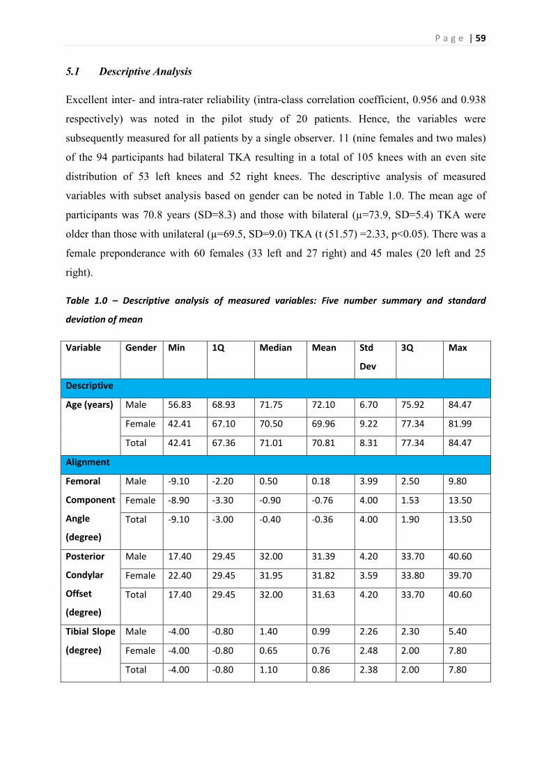
P a g e | 59
5.1 Descriptive Analysis
Excellent inter- and intra-rater reliability (intra-class correlation coefficient, 0.956 and 0.938
respectively) was noted in the pilot study of 20 patients. Hence, the variables were
subsequently measured for all patients by a single observer. 11 (nine females and two males)
of the 94 participants had bilateral TKA resulting in a total of 105 knees with an even site
distribution of 53 left knees and 52 right knees. The descriptive analysis of measured
variables with subset analysis based on gender can be noted in Table 1.0. The mean age of
participants was 70.8 years (SD=8.3) and those with bilateral (µ=73.9, SD=5.4) TKA were
older than those with unilateral (µ=69.5, SD=9.0) TKA (t (51.57) =2.33, p<0.05). There was a
female preponderance with 60 females (33 left and 27 right) and 45 males (20 left and 25
right).
Table 1.0 – Descriptive analysis of measured variables: Five number summary and standard
deviation of mean
Variable Gender Min 1Q Median Mean Std
Dev
3Q Max
Descriptive
Age (years) Male 56.83 68.93 71.75 72.10 6.70 75.92 84.47
Female 42.41 67.10 70.50 69.96 9.22 77.34 81.99
Total 42.41 67.36 71.01 70.81 8.31 77.34 84.47
Alignment
Femoral
Component
Angle
(degree)
Male -9.10 -2.20 0.50 0.18 3.99 2.50 9.80
Female -8.90 -3.30 -0.90 -0.76 4.00 1.53 13.50
Total -9.10 -3.00 -0.40 -0.36 4.00 1.90 13.50
Posterior
Condylar
Offset
(degree)
Male 17.40 29.45 32.00 31.39 4.20 33.70 40.60
Female 22.40 29.45 31.95 31.82 3.59 33.80 39.70
Total 17.40 29.45 32.00 31.63 4.20 33.70 40.60
Tibial Slope
(degree)
Male -4.00 -0.80 1.40 0.99 2.26 2.30 5.40
Female -4.00 -0.80 0.65 0.76 2.48 2.00 7.80
Total -4.00 -0.80 1.10 0.86 2.38 2.00 7.80

P a g e | 60
Strength
Extension
Strength
(Newton)
Male 8.30 29.90 40.50 40.71 17.70 53.90 92.50
Female 8.40 18.50 22.10 24.38 9.02 30.52 50.20
Total 8.30 20.30 27.90 31.38 15.64 40.50 92.50
Flexion
Strength
(Newton)
Male 4.10 8.40 10.70 11.52 4.97 12.90 29.70
Female 2.10 5.38 7.95 8.53 4.31 10.50 26.00
Total 2.10 7.00 9.00 9.81 4.82 11.60 29.70
Function
Timed Up
and Go
(seconds)
Male 6.00 7.35 7.79 9.02 3.51 8.47 21.47
Female 6.69 8.41 9.63 10.53 3.43 11.07 23.80
Total 6.00 7.65 8.88 9.89 3.53 10.68 23.80
Kinematics
Pre-MEA
(degree)
Male -6.00 1.00 4.50 5.20 6.23 9.00 19.50
Female -7.50 0.25 4.50 6.44 8.00 12.00 29.50
Total -7.50 0.50 4.50 5.90 7.28 10.63 29.50
Pre-MFA
(degree)
Male 88.50 110.0 117.50 116.20 10.52 123.50 133.00
Female 87.00 98.50 113.50 109.90 11.45 117.00 137.00
Total 87.00 104.40 114.80 112.60 11.43 120.50 137.00
Pre-ROM
(degree)
Male 76.00 104.00 113.00 111.00 13.00 118.50 137.00
Female 66.50 93.50 104.00 103.50 15.05 115.20 136.50
Total 66.50 96.38 108.00 106.72 14.61 117.00 137.00
Intra-MEA
(degree)
Male -4.00 -1.38 0.00 0.67 3.44 1.38 11.50
Female -38.00 -1.75 0.00 -1.14 6.22 1.00 8.00
Total -38.00 -1.50 0.00 -0.39 5.30 1.00 11.50
Intra-MFA
(degree)
Male 73.00 106.10 115.50 113.40 13.72 122.50 136.50
Female 80.00 103.80 114.00 112.60 12.74 123.00 138.50
Total 73.00 104.50 115.00 112.90 13.10 123.00 138.50
Intra-ROM
(degree)
Male 76.50 107.20 115.00 112.80 13.77 122.40 136.00
Female 89.00 102.50 114.00 113.70 13.33 123.50 147.50
Total 76.50 103.50 114.50 113.30 13.45 123.50 147.50

P a g e | 61
Post-MEA
(degree)
Male -5.00 0.00 0.00 1.89 3.58 5.00 15.00
Female -5.00 0.00 0.00 2.00 3.71 5.00 10.00
Total -5.00 0.00 0.00 1.95 3.64 5.00 15.00
Post-MFA
(degree)
Male 70.00 100.00 110.00 108.40 12.73 120.00 130.00
Female 50.00 98.75 105.00 103.20 11.23 110.00 120.00
Total 50.00 100.00 105.00 105.40 12.11 110.00 130.00
Post-ROM
(degree)
Male 60.00 100.00 110.00 106.50 14.82 115.00 130.00
Female 40.00 95.00 105.00 101.20 12.74 110.00 120.00
Total 40.00 95.00 105.00 103.50 13.85 110.00 130.00
Balance
Path
Velocity –
TLEO
(cm/sec)
Male 0.68 0.96 1.31 1.46 0.72 1.65 3.89
Female 0.61 0.92 1.04 1.30 0.75 1.47 5.66
Total 0.61 0.93 1.14 1.37 0.74 1.59 5.66
Path
Velocity –
TLEC
(cm/sec)
Male 0.95 1.39 1.97 2.22 1.02 3.00 4.96
Female 0.85 1.35 1.66 1.95 1.04 2.13 6.26
Total 0.85 1.35 1.73 2.07 1.03 2.58 6.26
Path
Velocity –
SLEO
(cm/sec)
Male 2.05 4.61 5.46 5.81 2.23 6.70 11.59
Female 0.86 4.06 4.62 4.70 1.61 5.43 10.36
Total 0.86 4.14 4.93 5.23 2.00 5.90 11.59
Clinical Rating Systems
Oxford
Knee Score
Male 20.00 34.00 43.00 39.33 8.29 46.00 48.00
Female 18.00 33.75 40.00 37.75 7.64 44.00 48.00
Total 18.00 34.00 41.00 38.43 7.92 44.00 48.00
Knee
Society
Score
Male 33.00 66.00 79.00 74.57 13.50 85.00 90.00
Female 26.00 67.00 78.00 73.87 14.79 84.25 89.00
Total 26.00 67.00 78.00 74.17 14.19 85.00 90.00
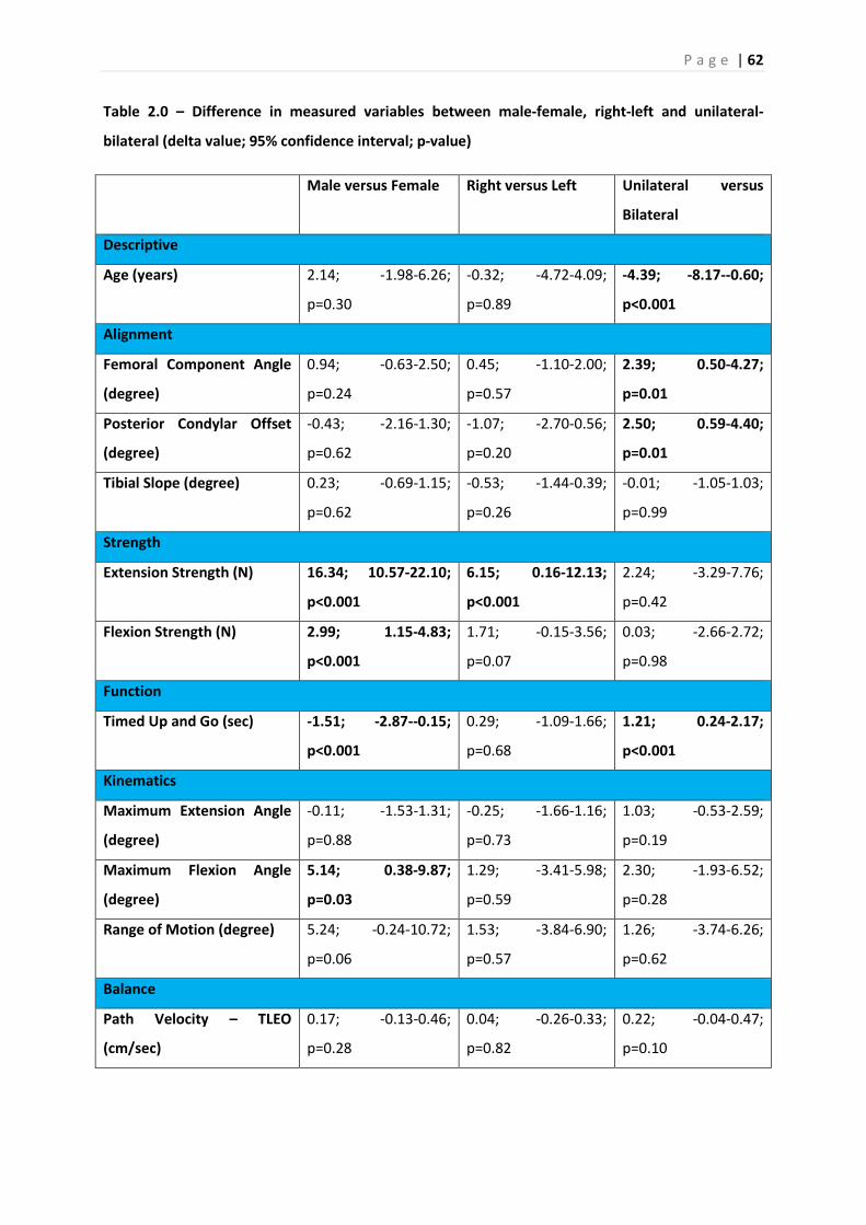
P a g e | 62
Table 2.0 – Difference in measured variables between male-female, right-left and unilateral-
bilateral (delta value; 95% confidence interval; p-value)
Male versus Female Right versus Left Unilateral versus
Bilateral
Descriptive
Age (years) 2.14; -1.98-6.26;
p=0.30
-0.32; -4.72-4.09;
p=0.89
-4.39; -8.17--0.60;
p<0.001
Alignment
Femoral Component Angle
(degree)
0.94; -0.63-2.50;
p=0.24
0.45; -1.10-2.00;
p=0.57
2.39; 0.50-4.27;
p=0.01
Posterior Condylar Offset
(degree)
-0.43; -2.16-1.30;
p=0.62
-1.07; -2.70-0.56;
p=0.20
2.50; 0.59-4.40;
p=0.01
Tibial Slope (degree) 0.23; -0.69-1.15;
p=0.62
-0.53; -1.44-0.39;
p=0.26
-0.01; -1.05-1.03;
p=0.99
Strength
Extension Strength (N) 16.34; 10.57-22.10;
p<0.001
6.15; 0.16-12.13;
p<0.001
2.24; -3.29-7.76;
p=0.42
Flexion Strength (N) 2.99; 1.15-4.83;
p<0.001
1.71; -0.15-3.56;
p=0.07
0.03; -2.66-2.72;
p=0.98
Function
Timed Up and Go (sec) -1.51; -2.87--0.15;
p<0.001
0.29; -1.09-1.66;
p=0.68
1.21; 0.24-2.17;
p<0.001
Kinematics
Maximum Extension Angle
(degree)
-0.11; -1.53-1.31;
p=0.88
-0.25; -1.66-1.16;
p=0.73
1.03; -0.53-2.59;
p=0.19
Maximum Flexion Angle
(degree)
5.14; 0.38-9.87;
p=0.03
1.29; -3.41-5.98;
p=0.59
2.30; -1.93-6.52;
p=0.28
Range of Motion (degree) 5.24; -0.24-10.72;
p=0.06
1.53; -3.84-6.90;
p=0.57
1.26; -3.74-6.26;
p=0.62
Balance
Path Velocity – TLEO
(cm/sec)
0.17; -0.13-0.46;
p=0.28
0.04; -0.26-0.33;
p=0.82
0.22; -0.04-0.47;
p=0.10

P a g e | 63
Path Velocity – TLEC
(cm/sec)
0.27; -0.14-0.68;
p=0.19
0.03; -0.38-0.44;
p=0.87
-0.01; -0.52-0.51;
p=0.98
Path Velocity – SLEO
(cm/sec)
1.11; 0.29-1.93;
p<0.001
0.86; 0.03-1.68;
p<0.001
0.37; -0.37-1.11;
p=0.32
Clinical Rating Systems
Oxford Knee Score 1.59; -1.55-4.72;
p=0.32
-0.24; -3.32-
2.84;p=0.88
-4.52; -7.02--2.02;
p<0.05
Knee Society Score 0.70; -4.81-6.21;
p=0.80
-2.26; -7.77-3.26;
p=0.42
-6.81; -11.76--1.85;
p<0.05
5.2 Hypothesis 1
Alignment parameters, sFCA, PCO and tibial slope, were used in a regression analysis to
predict kinematics, MFA, MEA and ROM. The prediction model for MFA was statistically
significant (p=0.03) and accounted for 8.4% of the variance. Both FCA (R2=0.3, p=0.01) and
PCO (R2=0.2, p=0.05) are statistically significant weak predictors of MFA as noted in Fig 17.
The prediction model for ROM did not achieve statistical significance, but accounted for
6.5% of the variance. FCA (R2=0.2, p=0.02) was a statistically significant weak predictor of
ROM as noted in Fig 17. The prediction model for MEA was not statistically significant
(p=0.38) and accounted for only 3.0% of the variance.
Fig 17: Femoral Component Angle (FCA) and Posterior Condylar Offset (PCO) were
statistically significant predictors of Maximum Flexion Angle (MFA); Femoral Component
Angle was also a statistically significant predictor of Range of Motion (ROM).
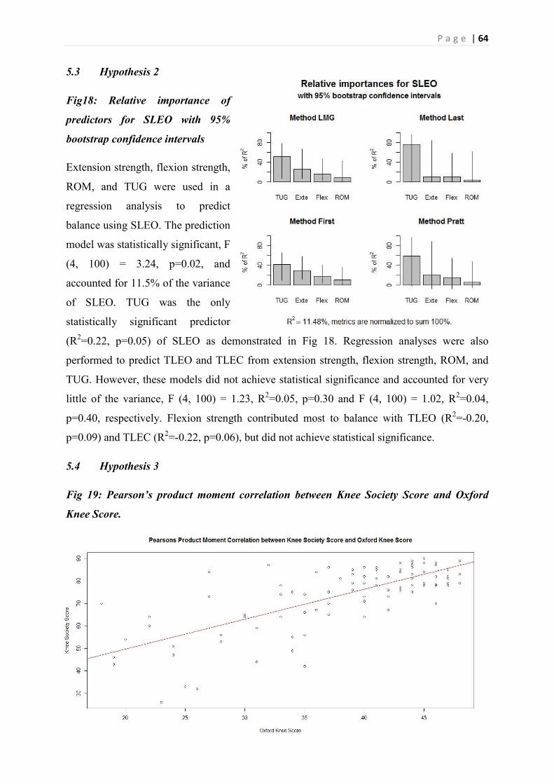
P a g e | 64
5.3 Hypothesis 2
Fig18: Relative importance of
predictors for SLEO with 95%
bootstrap confidence intervals
Extension strength, flexion strength,
ROM, and TUG were used in a
regression analysis to predict
balance using SLEO. The prediction
model was statistically significant, F
(4, 100) = 3.24, p=0.02, and
accounted for 11.5% of the variance
of SLEO. TUG was the only
statistically significant predictor
(R2=0.22, p=0.05) of SLEO as demonstrated in Fig 18. Regression analyses were also
performed to predict TLEO and TLEC from extension strength, flexion strength, ROM, and
TUG. However, these models did not achieve statistical significance and accounted for very
little of the variance, F (4, 100) = 1.23, R2=0.05, p=0.30 and F (4, 100) = 1.02, R2=0.04,
p=0.40, respectively. Flexion strength contributed most to balance with TLEO (R2=-0.20,
p=0.09) and TLEC (R2=-0.22, p=0.06), but did not achieve statistical significance.
5.4 Hypothesis 3
Fig 19: Pearson’s product moment correlation between Knee Society Score and Oxford
Knee Score.

P a g e | 65
40 60 80 100 120
20
30
40
ROM
OK
S
10 15 20
20
30
40
TUG
OK
S
There was a statistically significant positive correlation between Oxford Knee Score and Knee
Society Score (r = 0.74, p=0.001) as demonstrated in Fig 19. Therefore, extension strength,
flexion strength, balance, ROM, and TUG were used in a regression analysis to predict OKS.
The prediction model was statistically significant, F (7, 80) = 6.19, p=0.001, and accounted
for 35.15% of the variance of OKS. ROM (R2=0.34, p=0.02) and TUG (R2=-0.40, p=0.001)
were found to be statistically significant predictors of OKS as demonstrated in Fig 20 and Fig
21.
Fig20: Relative importance of predictors for Oxford Knee Score with 95% bootstrap
confidence intervals.
However, flexion strength (R2=0.16, p=0.75), extension strength (R2=0.29, p=0.26), and
balance as measured with TLEO (R2=0.01, p=0.79), TLEC (R2=0.16, p=0.19) and SLEO
(R2=0.13, p=0.85) did not achieve statistical significance.
Fig 21: Range of motion and Timed Up and Go were statistically significant predictors of
Oxford Knee Score.

P a g e | 66

P a g e | 67
6.1 Hypothesis 1
The most important findings of this study are that the femoral component angle (FCA) was a
statistically significant predictor of maximum flexion angle (MFA) and range of motion
(ROM) and that the posterior condylar offset (PCO) was a statistically significant predictor of
MFA. However, tibial slope (TS) does not contribute significantly to knee kinematics after
TKA. With significant improvements in implant design and survival, patients are increasingly
concerned about objective functional outcomes such as knee kinematics to evaluate the
success of their total knee arthroplasty (TKA) [1]. The influence of sagittal plane component
alignment, particularly the femoral component angle (FCA), tibial slope (TS) and posterior
condylar offset (PCO) on knee kinematics is contentious.
Most surgeons aim to place the femoral component in neutral alignment to the femoral axis in
the sagittal plane [110]. The FCA was a statistically significant predictor of MFA and ROM
in patients with osteoarthritis who underwent primary TKA using a posterior cruciate
retaining implant. Although statistically significant, FCA was a weak predictor for MFA (one
degree increase in FCA, increased the MFA by 0.3 degrees) and ROM (one degree increase in
FCA, increased the ROM by 0.2 degrees). This is similar to the finding in a recent study that
flexing the femoral implant in cruciate retaining TKA provides an immediate increase in knee
flexion [94]. The increase in flexion could be secondary to the increase in the PCO derived
from flexing the femoral component. However, these findings are in contrast with an earlier
study that demonstrated no correlation between implant position and knee range of motion
[97]. This could be due to the retrospective nature of their study and the use of non-
standardized radiographs that could have induced significant measurement errors. Increased
failure rates have been demonstrated in knees with femoral component flexed greater than
three degrees [111]. Therefore, although flexion of the femoral component confers increased
knee flexion, the risk of early aseptic failure diminishes any significant clinical benefits.
Therefore, this study demonstrates that within the therapeutic range of femoral component
flexion, we could potentially achieve 0.9 degrees of increased flexion and 0.6 degrees of
ROM, which would not be considered clinically significant. Moreover, a theoretical
disadvantage to flexing the femoral component is loss of knee extension. However, this and
previous studies do not demonstrate any statistically significant association between FCA and
MEA [94].
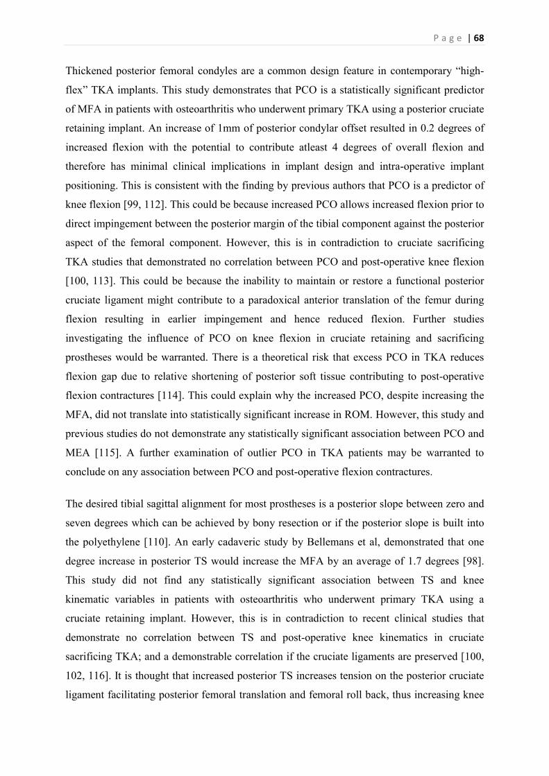
P a g e | 68
Thickened posterior femoral condyles are a common design feature in contemporary “high-
flex” TKA implants. This study demonstrates that PCO is a statistically significant predictor
of MFA in patients with osteoarthritis who underwent primary TKA using a posterior cruciate
retaining implant. An increase of 1mm of posterior condylar offset resulted in 0.2 degrees of
increased flexion with the potential to contribute atleast 4 degrees of overall flexion and
therefore has minimal clinical implications in implant design and intra-operative implant
positioning. This is consistent with the finding by previous authors that PCO is a predictor of
knee flexion [99, 112]. This could be because increased PCO allows increased flexion prior to
direct impingement between the posterior margin of the tibial component against the posterior
aspect of the femoral component. However, this is in contradiction to cruciate sacrificing
TKA studies that demonstrated no correlation between PCO and post-operative knee flexion
[100, 113]. This could be because the inability to maintain or restore a functional posterior
cruciate ligament might contribute to a paradoxical anterior translation of the femur during
flexion resulting in earlier impingement and hence reduced flexion. Further studies
investigating the influence of PCO on knee flexion in cruciate retaining and sacrificing
prostheses would be warranted. There is a theoretical risk that excess PCO in TKA reduces
flexion gap due to relative shortening of posterior soft tissue contributing to post-operative
flexion contractures [114]. This could explain why the increased PCO, despite increasing the
MFA, did not translate into statistically significant increase in ROM. However, this study and
previous studies do not demonstrate any statistically significant association between PCO and
MEA [115]. A further examination of outlier PCO in TKA patients may be warranted to
conclude on any association between PCO and post-operative flexion contractures.
The desired tibial sagittal alignment for most prostheses is a posterior slope between zero and
seven degrees which can be achieved by bony resection or if the posterior slope is built into
the polyethylene [110]. An early cadaveric study by Bellemans et al, demonstrated that one
degree increase in posterior TS would increase the MFA by an average of 1.7 degrees [98].
This study did not find any statistically significant association between TS and knee
kinematic variables in patients with osteoarthritis who underwent primary TKA using a
cruciate retaining implant. However, this is in contradiction to recent clinical studies that
demonstrate no correlation between TS and post-operative knee kinematics in cruciate
sacrificing TKA; and a demonstrable correlation if the cruciate ligaments are preserved [100,
102, 116]. It is thought that increased posterior TS increases tension on the posterior cruciate
ligament facilitating posterior femoral translation and femoral roll back, thus increasing knee
P a g e | 69
flexion [116]. However, this correlation between increased TS and MFA (2.3 degrees of
increased flexion per degree increase in tibial slope) in cruciate retaining prosthesis was only
demonstrable from one to six degrees of posterior tibial slope after which it plateaus [116].
The lack of correlation in our study cohort could be attributed to the fact that greater than
50% of our participants had tibial slope < 1 degree or > 6 degree, which is outside the window
of correlation demonstrated by Chambers et al [116]. Given that the influence of TS on knee
flexion can only be demonstrated in a controlled cadaveric setting, with previous authors
failing to demonstrate any correlation in cruciate sacrificing studies and a narrow window of
correlation in cruciate retaining studies, the clinical significance of the influence of TS on
knee kinematics may be negligible.
The prediction models for knee kinematics (MEA, MFA and ROM) achieved low variance,
suggesting that other untested variables may contribute more significantly to post-operative
kinematics. The final knee kinematics is the culmination of a complex set of multi-factorial
interactions, not limited to pre-operative kinematics, component alignment in the coronal
plane, component size, bony resection and soft tissue release, with each specific factor
contributing very little to kinematics individually [63, 65]. However, this study demonstrates
that FCA is a significant predictor of MFA and ROM; and PCO is a significant predictor of
MFA.
6.2 Hypothesis 2
The most important finding of this study is that TUG is a moderate predictor of balance in
single leg stance with eyes open in patients with osteoarthritis who underwent primary TKA
using a posterior cruciate retaining implant. Balance is considered an important outcome
measure, as it is highly predictive of the quality of life and function of patients after TKA
[22]. The findings of this study are consistent with that previously demonstrated by Schwartz
et al who performed static balance measures in patients after TKA; they demonstrated a
significant (R2=0.51, p<0.001) association between TUG and their static balance measures
[22].
Patients who undergo TKA demonstrate improved single leg balance when compared to those
with high tibial osteotomy [23]. This is because restoration of joint space facilitates re-
recruitment of dynamic knee stabilizers to facilitate restoration of proprioception and balance
which in turn improves functional mobility that is measured by TUG [23]. This association
between TUG and balance helps explain why TUG is indicative of a prior history of falls,
P a g e | 70
and why TUG is also considered a reliable and valid measure of change in functional mobility
of inpatients on orthopaedic wards [81, 82].
However, this association with TUG was not reflected in balance when tested with two legs,
either with eyes open or with eyes closed. Butler et al demonstrated that patients with total
ankle arthroplasty had significantly poorer balance compared to those following hip or knee
arthroplasty, suggesting the greater importance of the ankle in maintaining balance [117].
Hence, a possible explanation is that in two-legged stance the contralateral leg, particularly
the ankle, provides sufficient afferent input to avoid increased sway, confounding any
association between balance and TUG.
The influence of ROM and isometric flexion and extension strength on balance in patients
post TKA has never previously been investigated. Although flexion strength in this study was
found to be the most significant contributor to balance with two legs, both with eyes open and
closed, it did not achieve statistical significance. Corrigan et al demonstrated a significant
correlation between hamstring to quadriceps ratio and balance in ACL deficient knees,
suggesting a relatively greater correlation of flexion strength in comparison to extension
strength with functional outcome in patients [103, 104]. However, Lee et al could not
demonstrate a statistically significant correlation between hamstring strength and balance in
patients with ACL deficient (Rho=0.239, p=0.506) and ACL intact (Rho=0.367, p=0.297)
knees [105]. One possible explanation could be that the contribution of dynamic hamstring
muscles is only clinically relevant in high intensity activities like running or uphill walking
[17]. Hence, hamstring strength is unlikely to be a significant contributor to static balance, but
is relatively more important than quadriceps strength in maintaining balance post TKA.
A change in joint position causes a change in muscle length or velocity, and this initiates a
stretch reflex at the level of the spinal cord [118]. Intrafusal muscle fibres convey impulses
via gamma motor neurons to monosynaptically transmit signals via alpha motor neurons that
then cause contraction of the extrafusal muscle fibres [118]. This forms much of the basis of
quadriceps strengthening exercises by physiotherapists post TKA. Quadriceps weakness is
intrinsic to the pathogenesis and natural progression of osteoarthritis with an immediate
decline in the strength of the extensor mechanism immediately post TKA, followed by
significant improvement subsequently which plateaus after six to twelve months [71]. This
study demonstrates that extension strength is not a predictor of balance in patients post TKA.
This could be because in elderly patients and those with osteoarthritis who have lost the
P a g e | 71
proprioceptive ability of the knee, proprioception is predominantly mediated supraspinally,
communicated by the posterior column-medial lemniscus pathway to the cerebrum or dorsal
and ventral spinocerebellar tract to the cerebellum [118]. Stevens-Lapsley et al demonstrated
that despite a faster improvement in muscle strength following minimally invasive TKA, this
did not translate into improved functional performance [119]. Moreover, decreased EMG
amplitudes in the vastus lateralis and biceps femoris in patients post TKA compared to control
patients with OA, suggests that the improvement in proprioception and balance could be
secondary to relative over-recruitment of other dynamic knee stabilizers [23]. Hence, it is
possible that while a minimum quadriceps and hamstring strength is paramount for ranging
the knee, particularly when being loaded without “giving way”, any further strength gain
might not add to functional performance.
This study also demonstrates that ROM is not a predictor of balance in patients post TKA.
Watanabe et al demonstrated that intra-operative joint gaps at 120-135 degrees and at 10
degrees of flexion was positively correlated with post-operative flexion and extension
respectively, to emphasise the importance of joint gaps at flexion and extension to preserve or
increase ROM [120]. Although it is suspected that greater joint gaps and soft tissue imbalance
could adversely affect joint stability, it has been demonstrated that the clinical range of joint
gaps do not affect balance [120]. Hence, it could be that ROM and balance share a non-linear
relationship where the majority of clinically significant ROM has minimal or no impact on
balance; a further study of outlier ROMs would be required to determine the tipping point of
balance. Moreover, Pua et al demonstrated that knee extensor strength is significantly
correlated with extension ROM and mediates the influence of knee ROM on physical function
[69]. As extension strength is not a predictor of balance, ROM would not be significantly
associated with balance.
The small value of the coefficient of determination (R2) achieved by these regression models
and the inability to achieve statistical significance suggests that ROM, flexion strength, and
extension strength are not significant predictors of balance. This is unlikely to be a Type II
error, and is possibly explained by other unexamined factors that could contribute more
substantially to balance. However, the more likely explanation is that balance is the
culmination of a complex set of multi-factorial interactions, with each specific factor
contributing very little to balance individually.
P a g e | 72
6.3 Hypothesis 3
Total knee arthroplasty (TKA) is cost effective and highly successful in the management of
arthritic knees with a 94.4% ten year implant survival rate [121]. Recently, the Swedish,
United Kingdom and New Zealand national joint registries have adopted PROMs as cost-
efficient, reproducible and reliable tools to report patient satisfaction and hence the success of
TKA [1, 12]. However, despite the improved implant survival, decreased revision rates and
exceptional objective outcomes after total knee arthroplasty (TKA), there is evidence that up
to 23% of patients are dissatisfied with their replaced knee [1]. There is very little
understanding of the relationship between subjective and objective functional outcome
measures used in TKA patients. The most important findings of this study are that TUG and
ROM are both significant predictors of patient satisfaction after TKA.
The results of this study demonstrate that ROM is a statistically significant moderate predictor
of OKS, suggesting that patient satisfaction improved with increased ROM as previous
authors have demonstrated. Pua et al demonstrated a correlation between flexion ROM and
SF-36 physical function, and Christen et al demonstrated positive correlations for ROM with
both the Knee Injury and Osteoarthritis Outcome Score (0.33) and the Knee Society Score
(0.40) [122, 123]. Increased patient satisfaction in patients who achieved increased ROM
could be a reflection of the increased functional ability particularly in the Asian population
who frequently use high flexion in activities of daily living [16]. Laubenthal et al
demonstrated that a minimum ROM of 90 degrees is required for activities of daily living,
with higher-level activities like running and cycling dependent on increased ROM [15].
However, Thomsen et al demonstrated no relationship between ROM and the ability to
perform activities of daily living [106]. This could be because all of Thomsen’s participants
achieved >95 degrees of flexion [106]. Hence, achieving ROM of at least 90 degrees is
crucial for post-operative patient satisfaction, but any further gains are most likely clinically
irrelevant in the elderly, Caucasian cohort.
The results of this study also demonstrate that TUG is a statistically significant moderate
predictor of OKS, suggesting that the faster a patient could complete the TUG test, the more
satisfied the patient was post-operatively. Rossi et al demonstrated a similar correlation for
TUG with the aggregate WOMAC score (0.59) and physical function dimension (0.63) [124].
This could explain why TUG is considered a reliable and valid measure of change in
functional mobility of inpatients on orthopaedic wards and hence would reflect patient

P a g e | 73
satisfaction [81]. However, Beard et al demonstrated that a difference of nine points for
cohort studies and four points for case-control studies is the minimum clinically significant
difference for OKS, suggesting that the correlations for ROM and TUG with OKS, though
statistically significant, may not be clinically significant [125]. Hence, it is possible that
functional outcome measures do not substitute for, or are not predictive of, PROMs but are
instead complementary to patient satisfaction in measuring outcomes after TKA.
This study also found that isometric flexion and extension strength are not statistically
significant predictors of patient satisfaction. This could be because of a non-linear association
between extension strength and physical function, with a strength threshold below which the
muscle strength and physical function are closely related, but above which the association is
diminished [122]. Moreover, the contribution of dynamic hamstring muscles is only clinically
relevant in high intensity activities like running or uphill walking, and again may not be
appreciated by the typical geriatric recipients of TKA [17]. This study demonstrates that
balance is not a statistically significant predictor of patient satisfaction. This contradicts the
findings by Schwartz et al, that static and dynamic balance measures were statistically
significant predictors of SF-36 and OKS [22]. However, the study by Schwartz et al was
limited by a high dropout rate of 24.3% and compliance between 62% and 68% for PROMs,
introducing significant attrition and selection bias due to the elimination of patients who may
have had poor post-operative outcomes. In those patients who have already become compliant
with the use of mobility aides during the lengthy lead-up to surgery, further balance gains
may not translate into patient satisfaction [126]. Moreover, it is possible that increased
reliance on the contra-lateral limb, to negate the pre-operative loss of balance related to the
arthritic knee, may then result in a failure of the patient to appreciate any subsequent balance
gains resulting from the TKA.
The prediction model for Oxford Knee Score achieved low variance (35%), suggesting that
other untested variables may contribute more significantly to subjective patient satisfaction
post TKA. Post-operative patient satisfaction is the culmination of a complex set of multi-
factorial interactions, not limited to post-operative pain, stiffness, perception of alignment,
ROM and functional ability, with each specific factor contributing very little individually to
overall patient satisfaction [127].

P a g e | 74
6.4 Limitations
Hypothesis 1 has certain limitations. It is thought that measurement of PCO on radiographs do
not correlate with those of the medial or lateral condyles on computed tomography (CT)
[128]. However, CT exposes patients to higher radiation, cannot be utilised in patients who
are claustrophobic or cannot lie still for the duration of the test and is susceptible to
significant component artefacts predisposing to measurement errors. Hence, this study utilized
true lateral radiographs taken by a single senior radiographer to a pre-determined protocol on
the first day post operation for measurement of sagittal plane component alignment.
Numerous confounders are thought to influence post-operative kinematics. Pre-operative
diagnosis and ROM are considered the most significant predictors of post-operative ROM
[65]. Moreover, previous authors have investigated numerous factors not limited to age, sex,
body mass index, patellar thickness and height, component size and design, posterior cruciate
ligament status and radiological alignment for association with knee kinematics, with
conflicting evidence in literature [65, 129]. This study was limited to non-obese patients with
osteoarthritis undergoing primary cruciate retaining TKA with a standardized implant design
in order to minimize confounders. Moreover, this study accounted for age, gender and pre-
operative kinematics to mitigate the influence of confounders. While male participants
demonstrated 2.147 degrees of increased flexion in comparison to female participants, other
variables failed to demonstrate any statistically significant influence on post-operative knee
kinematics. Furthermore, the influence of gender can be explained by the need for larger
prosthesis in males resulting in increased PCO and hence knee flexion. Therefore, this was a
carefully designed prospective study to minimise the influence of confounders while
maintaining the external validity. It is possible that the combination of FCA, PCO and TS
may be more important in determining post-operative knee kinematics, rather than in
isolation, with deviation of one value being remedied by alteration of the others. Future
studies should strive to investigate each of these variables by controlling the other variables in
neutral position.
Moreover, Hypothesises 2 and 3 also have certain limitations. Systemic inflammatory arthritis
such as rheumatoid arthritis could potentially result in different outcomes and influence
outcome measures. The effect of chronic inflammatory disease on outcome is not well
established, with some studies reporting better outcomes in patients with rheumatoid arthritis
and others in patients with osteoarthritis [64, 130]. The pre-operative levels activity levels and
duration of post-operative physiotherapy were not controlled due to the retrospective design
P a g e | 75
of the study; both these variables could have potentially influenced our results. However,
there are no pre- or post-operative physiotherapy protocols that can be universally applied to
all TKA patients due to the highly individualised deficits and pathology, rendering futile most
attempts to standardize these variables [131]. Similarly, the Body Mass Index of patients
could also be considered a potential confounder of outcome. However, Napier, et al (2014)
showed that morbidly obese patients did not have a statistically significantly greater
complication rate compared to non-obese patients [132]. Patient recruitment from a single
institution, as opposed to a multi-center study, and the convenience sampling employed for
participant selection may be considered to weaken the external validity of the study.
However, close examination reveals that participant characteristics such as age, side and
gender distribution, incidence of bilateral TKA, and others are closely reflective of the general
population, thus preserving the external validity of the study.
6.5 Internal and External Validity
6.5.1 Internal Validity:
The research project limited investigation to participants with a single implant design.
Different implants designs are a confounding factor that can affect the navigational kinematic
data and the clinical outcome. Standardizing the implant design strengthens the internal
validity of the research, but weakens the external validity.
Selection of participants was dependent upon the follow-up of patients. Patients who
presented to the follow-up clinic were invited to take part in the research. Although no
patients declined the invitation to participate, the research did not include patients who failed
to turn up to follow-up clinic. It is difficult to postulate if loss to follow-up was due to the lack
of interest of high functioning patients or the inability of poorly mobilising patients. However,
this introduces a selection bias due to loss to follow-up.
6.5.1 External Validity:
CAS TKA is performed at RBH by two different orthopaedic surgeons and there was no set
operative protocol. Although the surgical procedure is standardized, the varying skill levels of
the different surgeons could influence the outcomes. However, the use of more than one
surgeon strengthens the external validity of the research project.

P a g e | 76
Hawthorne effect – it is likely that participants might outperform their general performance
levels during investigation. This is likely to result in falsely elevated results that cannot be
generalised to the entire population.
6.6 Conclusion
The most important finding of this study is that the femoral component angle in the sagittal
plane is predictor of knee flexion and range of motion. Given the increased risk of implant
failure with greater than three degrees of flexion, the clinical relevance of femoral component
flexion might be limited. Another important finding is that the posterior condylar offset is also
a predictor of knee flexion. However, the benefits of increasing the PCO might be limited to
only 4 degrees of increased flexion in cruciate retaining TKA with a theoretical risk of flexion
contractures. It is important to note that this study failed to demonstrate a statistically
significant correlation between tibial slope and knee kinematics. Although in vitro studies
have shown tibial slope to influence knee kinematics, the lack of this observation in cruciate
sacrificing studies combined with a narrow window of correlation (zero to seven degrees of
posterior tibial slope) in cruciate retaining studies, suggest that its clinical relevance might be
limited.
This study suggests that TUG is a moderately significant predictor of balance on a single leg
following TKR. Knee flexion strength, knee extension strength, and ROM do not contribute
significantly to balance following TKR, for either single or two-leg stance. The results of this
study also suggest that TUG and ROM are both statistically significant predictors of
subjective patient reported outcomes following TKA. Despite the functional benefits of
flexion strength, extension strength, and balance in patients after TKA, these functional
outcome measures may not contribute significantly to post-operative patient satisfaction.
Functional outcome measures are complementary to PROMs in measuring post-operative
outcome. Further investigations are warranted to determine what additional factors may
contribute significantly to patient satisfaction after TKA.
P a g e | 77
LIST OF REFERENCES
1. Carr, A.J., et al., Knee replacement. Lancet, 2012. 379(9823): p. 1331-40. 2. Australian Orthopaedic Association National Joint Replacement Registry. Annual Report.
Adelaide:AOA. 2015. 3. Insall, J., A.J. Tria, and W.N. Scott, The total condylar knee prosthesis: the first 5 years. Clin
Orthop Relat Res, 1979(145): p. 68-77. 4. Choi, Y.J. and H.J. Ra, Patient Satisfaction after Total Knee Arthroplasty. Knee Surg Relat Res,
2016. 28(1): p. 1-15. 5. Moreland, J.R., Mechanisms of failure in total knee arthroplasty. Clin Orthop Relat Res,
1988(226): p. 49-64. 6. Sikorski, J.M., Alignment in total knee replacement. J Bone Joint Surg Br, 2008. 90(9): p. 1121-
7. 7. Miyasaka, T., et al., Accuracy of Computed Tomography-Based Navigation-Assisted Total
Knee Arthroplasty: Outlier Analysis. J Arthroplasty, 2016. 8. Fransen, B.L., et al., No differences between fixed- and mobile-bearing total knee
arthroplasty. Knee Surg Sports Traumatol Arthrosc, 2016. 9. Kim, M.S., et al., Two- to Four-Year Follow-up Results of Total Knee Arthroplasty Using a New
High-Flexion Prosthesis. Knee Surg Relat Res, 2016. 28(1): p. 39-45. 10. Todesca, A., et al., Conventional versus computer-navigated TKA: a prospective randomized
study. Knee Surg Sports Traumatol Arthrosc, 2016. 11. Swedish Knee Arthroplasty Register. Annual Report. 2015. 12. National Joint Registry for England, Wales and Northern Ireland. 12th Annual Report. 2015. 13. The New Zealand Joint Registry. Sixteen Year Report - January 1999 to December 2014. 2015. 14. Ramkumar, P.N., J.D. Harris, and P.C. Noble, Patient-reported outcome measures after total
knee arthroplasty: a systematic review. Bone Joint Res, 2015. 4(7): p. 120-7. 15. Laubenthal, K.N., G.L. Smidt, and D.B. Kettelkamp, A quantitative analysis of knee motion
during activities of daily living. Phys Ther, 1972. 52(1): p. 34-43. 16. Ha, C.W., et al., Increased Range of Motion Is Important for Functional Outcome and
Satisfaction After Total Knee Arthroplasty in Asian Patients. J Arthroplasty, 2016. 31(6): p. 1199-203.
17. Silva, M., et al., Knee strength after total knee arthroplasty. J Arthroplasty, 2003. 18(5): p. 605-11.
18. Bascuas, I., et al., Balance 1 year after TKA: correlation with clinical variables. Orthopedics, 2013. 36(1): p. e6-12.
19. Attfield, S.F., et al., Soft-tissue balance and recovery of proprioception after total knee replacement. J Bone Joint Surg Br, 1996. 78(4): p. 540-5.
20. Bade, M.J., et al., Predicting poor physical performance after total knee arthroplasty. J Orthop Res, 2012. 30(11): p. 1805-10.
21. Swinkels, A. and T.J. Allain, Physical performance tests, self-reported outcomes, and accidental falls before and after total knee arthroplasty: An exploratory study. Physiother Theory Pract, 2013.
22. Schwartz, I., et al., Balance is an important predictive factor for quality of life and function after primary total knee replacement. J Bone Joint Surg Br, 2012. 94(6): p. 782-6.
23. Wodowski, A.J., et al., Proprioception and Knee Arthroplasty: A Literature Review. Orthop Clin North Am, 2016. 47(2): p. 301-9.
24. Insall, J.N., et al., Rationale of the Knee Society clinical rating system. Clin Orthop Relat Res, 1989(248): p. 13-4.
25. Saavedra, M.A., et al., Clinical anatomy of the knee. Reumatol Clin, 2012. 8 Suppl 2: p. 39-45. 26. Flandry, F. and G. Hommel, Normal anatomy and biomechanics of the knee. Sports Med
Arthrosc, 2011. 19(2): p. 82-92.
P a g e | 78
27. Tecklenburg, K., et al., Bony and cartilaginous anatomy of the patellofemoral joint. Knee Surg Sports Traumatol Arthrosc, 2006. 14(3): p. 235-40.
28. Fox, A.J., F. Wanivenhaus, and S.A. Rodeo, The basic science of the patella: structure, composition, and function. J Knee Surg, 2012. 25(2): p. 127-41.
29. Fox, A.J., A. Bedi, and S.A. Rodeo, The basic science of human knee menisci: structure, composition, and function. Sports Health, 2012. 4(4): p. 340-51.
30. De Smet, A.A., How I diagnose meniscal tears on knee MRI. AJR Am J Roentgenol, 2012. 199(3): p. 481-99.
31. Siegel, L., C. Vandenakker-Albanese, and D. Siegel, Anterior cruciate ligament injuries: anatomy, physiology, biomechanics, and management. Clin J Sport Med, 2012. 22(4): p. 349-55.
32. Voos, J.E., et al., Posterior cruciate ligament: anatomy, biomechanics, and outcomes. Am J Sports Med, 2012. 40(1): p. 222-31.
33. Quarles, J.D. and R.G. Hosey, Medial and lateral collateral injuries: prognosis and treatment. Prim Care, 2004. 31(4): p. 957-75, ix.
34. Schein, A., et al., Structure and function, injury, pathology, and treatment of the medial collateral ligament of the knee. Emerg Radiol, 2012. 19(6): p. 489-98.
35. Buckenmaier, C.C., 3rd, Anaesthesia for outpatient knee surgery. Best Pract Res Clin Anaesthesiol, 2002. 16(2): p. 255-70.
36. Burckett-St Laurant, D., et al., The Nerves of the Adductor Canal and the Innervation of the Knee: An Anatomic Study. Reg Anesth Pain Med, 2016. 41(3): p. 321-7.
37. Thompson, J.C.N.B.S.N.s.C.O.A.S.L., US: Saunders, 2009. 38. Hunter, D.J., Osteoarthritis. Best Pract Res Clin Rheumatol, 2011. 25(6): p. 801-14. 39. Sinusas, K., Osteoarthritis: diagnosis and treatment. Am Fam Physician, 2012. 85(1): p. 49-56. 40. Bennell, K.L., D.J. Hunter, and R.S. Hinman, Management of osteoarthritis of the knee. BMJ,
2012. 345: p. e4934. 41. Hayashi, D., F.W. Roemer, and A. Guermazi, Imaging for osteoarthritis. Ann Phys Rehabil
Med, 2016. 59(3): p. 161-9. 42. Petersson, I.F., et al., Radiographic osteoarthritis of the knee classified by the Ahlback and
Kellgren & Lawrence systems for the tibiofemoral joint in people aged 35-54 years with chronic knee pain. Ann Rheum Dis, 1997. 56(8): p. 493-6.
43. Smolen, J.S., D. Aletaha, and I.B. McInnes, Rheumatoid arthritis. Lancet, 2016. 44. Zochling, J. and E.U. Smith, Seronegative spondyloarthritis. Best Pract Res Clin Rheumatol,
2010. 24(6): p. 747-56. 45. Karim, A.R., et al., Osteonecrosis of the knee: review. Ann Transl Med, 2015. 3(1): p. 6. 46. Filardo, G., et al., Non-surgical treatments for the management of early osteoarthritis. Knee
Surg Sports Traumatol Arthrosc, 2016. 24(6): p. 1775-85. 47. Lana, J.F., et al., Randomized controlled trial comparing hyaluronic acid, platelet-rich plasma
and the combination of both in the treatment of mild and moderate osteoarthritis of the knee. J Stem Cells Regen Med, 2016. 12(2): p. 69-78.
48. Choong, P.F. and M.M. Dowsey, Update in surgery for osteoarthritis of the knee. Int J Rheum Dis, 2011. 14(2): p. 167-74.
49. Jones, R.E., R.D. Russell, and M.H. Huo, Alternatives to revision total knee arthroplasty. J Bone Joint Surg Br, 2012. 94(11 Suppl A): p. 137-40.
50. Richmond, J.C., Surgery for osteoarthritis of the knee. Med Clin North Am, 2009. 93(1): p. 213-22, xii.
51. A, W.D., O. Robertsson, and L.S. Lohmander, High tibial osteotomy in Sweden, 1998-2007: a population-based study of the use and rate of revision to knee arthroplasty. Acta Orthop, 2012. 83(3): p. 244-8.
52. Deschamps, G. and C. Chol, Fixed-bearing unicompartmental knee arthroplasty. Patients' selection and operative technique. Orthop Traumatol Surg Res, 2011. 97(6): p. 648-61.
P a g e | 79
53. Gan, Y., et al., Accuracy and efficacy of osteotomy in total knee arthroplasty with patient-specific navigational template. Int J Clin Exp Med, 2015. 8(8): p. 12192-201.
54. Cheng, T., et al., Little clinical advantage of computer-assisted navigation over conventional instrumentation in primary total knee arthroplasty at early follow-up. Knee, 2012. 19(4): p. 237-45.
55. Bauwens, K., et al., Navigated total knee replacement. A meta-analysis. J Bone Joint Surg Am, 2007. 89(2): p. 261-9.
56. Bae, D.K. and S.J. Song, Computer assisted navigation in knee arthroplasty. Clin Orthop Surg, 2011. 3(4): p. 259-67.
57. Brin, Y.S., et al., Imageless computer assisted versus conventional total knee replacement. A Bayesian meta-analysis of 23 comparative studies. Int Orthop, 2011. 35(3): p. 331-9.
58. Luo, S.X., et al., Posterior cruciate substituting versus posterior cruciate retaining total knee arthroplasty prostheses: a meta-analysis. Knee, 2012. 19(4): p. 246-52.
59. Nakama, G.Y., et al., Cemented, cementless or hybrid fixation options in total knee arthroplasty for osteoarthritis and other non-traumatic diseases. Cochrane Database Syst Rev, 2012. 10: p. CD006193.
60. Greene, K.A. and J.R. Schurman, 2nd, Quadriceps muscle function in primary total knee arthroplasty. J Arthroplasty, 2008. 23(7 Suppl): p. 15-9.
61. Labek, G., et al., Revision rates after total joint replacement: cumulative results from worldwide joint register datasets. J Bone Joint Surg Br, 2011. 93(3): p. 293-7.
62. Ritter, M.A., et al., The effect of alignment and BMI on failure of total knee replacement. J Bone Joint Surg Am, 2011. 93(17): p. 1588-96.
63. Dennis, D.A., et al., Factors affecting flexion after total knee arthroplasty. Clin Orthop Relat Res, 2007. 464: p. 53-60.
64. Harvey, I.A., et al., Factors affecting the range of movement of total knee arthroplasty. J Bone Joint Surg Br, 1993. 75(6): p. 950-5.
65. Gatha, N.M., et al., Factors affecting postoperative range of motion after total knee arthroplasty. J Knee Surg, 2004. 17(4): p. 196-202.
66. Ishii, Y., et al., Range of motion during the perioperative period in total knee arthroplasty. Arch Orthop Trauma Surg, 2008. 128(8): p. 795-9.
67. Dennis, D.A., et al., Range of motion after total knee arthroplasty: the effect of implant design and weight-bearing conditions. J Arthroplasty, 1998. 13(7): p. 748-52.
68. Jiang, C., et al., Posterior Cruciate Ligament Retention versus Posterior Stabilization for Total Knee Arthroplasty: A Meta-Analysis. PLoS One, 2016. 11(1): p. e0147865.
69. Pua, Y.H., et al., Knee extension range of motion and self-report physical function in total knee arthroplasty: mediating effects of knee extensor strength. BMC Musculoskelet Disord, 2013. 14: p. 33.
70. Scuderi, G.R., et al., The new Knee Society Knee Scoring System. Clin Orthop Relat Res, 2012. 470(1): p. 3-19.
71. Meier, W., et al., Total knee arthroplasty: muscle impairments, functional limitations, and recommended rehabilitation approaches. J Orthop Sports Phys Ther, 2008. 38(5): p. 246-56.
72. Holm, B., et al., Loss of knee-extension strength is related to knee swelling after total knee arthroplasty. Arch Phys Med Rehabil, 2010. 91(11): p. 1770-6.
73. Yoshida, Y., et al., Examining outcomes from total knee arthroplasty and the relationship between quadriceps strength and knee function over time. Clin Biomech (Bristol, Avon), 2008. 23(3): p. 320-8.
74. Davies, A.P., Rating systems for total knee replacement. Knee, 2002. 9(4): p. 261-6. 75. Cho, S.D. and C.H. Hwang, Improved single-limb balance after total knee arthroplasty. Knee
Surg Sports Traumatol Arthrosc, 2012. 76. Warren, P.J., et al., Proprioception after knee arthroplasty. The influence of prosthetic design.
Clin Orthop Relat Res, 1993(297): p. 182-7.

P a g e | 80
77. Bakirhan, S., et al., A comparison of static and dynamic balance in patients with unilateral and bilateral total knee arthroplasty. Eklem Hastalik Cerrahisi, 2009. 20(2): p. 93-101.
78. Labraca, N.S., et al., Benefits of starting rehabilitation within 24 hours of primary total knee arthroplasty: randomized clinical trial. Clin Rehabil, 2011. 25(6): p. 557-66.
79. Gstoettner, M., et al., Preoperative proprioceptive training in patients with total knee arthroplasty. Knee, 2011. 18(4): p. 265-70.
80. Wada, M., et al., Joint proprioception before and after total knee arthroplasty. Clin Orthop Relat Res, 2002(403): p. 161-7.
81. Yeung, T.S., et al., The timed up and go test for use on an inpatient orthopaedic rehabilitation ward. J Orthop Sports Phys Ther, 2008. 38(7): p. 410-7.
82. Beauchet, O., et al., Timed Up and Go test and risk of falls in older adults: a systematic review. J Nutr Health Aging, 2011. 15(10): p. 933-8.
83. Bargren, J.H., J.D. Blaha, and M.A. Freeman, Alignment in total knee arthroplasty. Correlated biomechanical and clinical observations. Clin Orthop Relat Res, 1983(173): p. 178-83.
84. Hauschild, O., et al., Correlation of radiographic and navigated measurements of TKA limb alignment: a matter of time? Knee Surg Sports Traumatol Arthrosc, 2010. 18(10): p. 1317-22.
85. Harvie, P., K. Sloan, and R.J. Beaver, Three-dimensional component alignment and functional outcome in computer-navigated total knee arthroplasty: a prospective, randomized study comparing two navigation systems. J Arthroplasty, 2011. 26(8): p. 1285-90.
86. Fang, D.M., M.A. Ritter, and K.E. Davis, Coronal alignment in total knee arthroplasty: just how important is it? J Arthroplasty, 2009. 24(6 Suppl): p. 39-43.
87. Choong, P.F., M.M. Dowsey, and J.D. Stoney, Does accurate anatomical alignment result in better function and quality of life? Comparing conventional and computer-assisted total knee arthroplasty. J Arthroplasty, 2009. 24(4): p. 560-9.
88. Jeffery, R.S., R.W. Morris, and R.A. Denham, Coronal alignment after total knee replacement. J Bone Joint Surg Br, 1991. 73(5): p. 709-14.
89. Parratte, S., et al., Effect of postoperative mechanical axis alignment on the fifteen-year survival of modern, cemented total knee replacements. J Bone Joint Surg Am, 2010. 92(12): p. 2143-9.
90. Rousseau, M.A., J.Y. Lazennec, and Y. Catonne, Early mechanical failure in total knee arthroplasty. Int Orthop, 2008. 32(1): p. 53-6.
91. Czurda, T., et al., The association between component malalignment and post-operative pain following navigation-assisted total knee arthroplasty: results of a cohort/nested case-control study. Knee Surg Sports Traumatol Arthrosc, 2010. 18(7): p. 863-9.
92. Longstaff, L.M., et al., Good alignment after total knee arthroplasty leads to faster rehabilitation and better function. J Arthroplasty, 2009. 24(4): p. 570-8.
93. Kim, D., et al., Comparison of the tibiofemoral rotational alignment after mobile and fixed bearing total knee arthroplasty. Knee Surg Sports Traumatol Arthrosc, 2012. 20(2): p. 337-45.
94. Murphy, M., et al., Does flexion of the femoral implant in total knee arthroplasty increase knee flexion: A randomised controlled trial. Knee, 2012.
95. Matziolis, G., et al., Increased flexion position of the femoral component reduces the flexion gap in total knee arthroplasty. Knee Surg Sports Traumatol Arthrosc, 2012. 20(6): p. 1092-6.
96. Higuchi, H., et al., Relationship between joint gap difference and range of motion in total knee arthroplasty: a prospective randomised study between different platforms. Int Orthop, 2009. 33(4): p. 997-1000.
97. Faris, P.M., M.A. Ritter, and E.M. Keating, Sagittal plane positioning of the femoral component in total knee arthroplasty. J Arthroplasty, 1988. 3(4): p. 355-8.
98. Bellemans, J., et al., The influence of tibial slope on maximal flexion after total knee arthroplasty. Knee Surg Sports Traumatol Arthrosc, 2005. 13(3): p. 193-6.
99. Malviya, A., et al., Predicting range of movement after knee replacement: the importance of posterior condylar offset and tibial slope. Knee Surg Sports Traumatol Arthrosc, 2009. 17(5): p. 491-8.
P a g e | 81
100. Bauer, T., et al., Influence of posterior condylar offset on knee flexion after cruciate-sacrificing mobile-bearing total knee replacement: a prospective analysis of 410 consecutive cases. Knee, 2010. 17(6): p. 375-80.
101. Seo, S.S., et al., Clinical results associated with changes of posterior tibial slope in total knee arthroplasty. Knee Surg Relat Res, 2013. 25(1): p. 25-9.
102. Kim, K.H., S.I. Bin, and J.M. Kim, The Correlation between Posterior Tibial Slope and Maximal Angle of Flexion after Total Knee Arthroplasty. Knee Surg Relat Res, 2012. 24(3): p. 158-63.
103. Alfano, L.N., et al., Correlation of knee strength to functional outcomes in Becker muscular dystrophy. Muscle Nerve, 2013. 47(4): p. 550-4.
104. Corrigan, J.P., W.F. Cashman, and M.P. Brady, Proprioception in the cruciate deficient knee. J Bone Joint Surg Br, 1992. 74(2): p. 247-50.
105. Lee, H.M., C.K. Cheng, and J.J. Liau, Correlation between proprioception, muscle strength, knee laxity, and dynamic standing balance in patients with chronic anterior cruciate ligament deficiency. Knee, 2009. 16(5): p. 387-91.
106. Thomsen, M.G., et al., Do patients care about higher flexion in total knee arthroplasty? A randomized, controlled, double-blinded trial. BMC Musculoskelet Disord, 2013. 14: p. 127.
107. Lustig, S., et al., The accuracy of acquisition of an imageless computer-assisted system and its implication for knee arthroplasty. Knee, 2011. 18(1): p. 15-20.
108. Bartlett, H.L., L.H. Ting, and J.T. Bingham, Accuracy of force and center of pressure measures of the Wii Balance Board. Gait Posture, 2014. 39(1): p. 224-8.
109. Clark, R.A., et al., Validity and reliability of the Nintendo Wii Balance Board for assessment of standing balance. Gait Posture, 2010. 31(3): p. 307-10.
110. Gromov, K., et al., What is the optimal alignment of the tibial and femoral components in knee arthroplasty? Acta Orthop, 2014. 85(5): p. 480-7.
111. Kim, Y.H., et al., The relationship between the survival of total knee arthroplasty and postoperative coronal, sagittal and rotational alignment of knee prosthesis. Int Orthop, 2014. 38(2): p. 379-85.
112. Bellemans, J., et al., Fluoroscopic analysis of the kinematics of deep flexion in total knee arthroplasty. Influence of posterior condylar offset. J Bone Joint Surg Br, 2002. 84(1): p. 50-3.
113. Hanratty, B.M., et al., The influence of posterior condylar offset on knee flexion after total knee replacement using a cruciate-sacrificing mobile-bearing implant. J Bone Joint Surg Br, 2007. 89(7): p. 915-8.
114. Onodera, T., et al., Posterior femoral condylar offset after total knee replacement in the risk of knee flexion contracture. J Arthroplasty, 2013. 28(7): p. 1112-6.
115. Han, H., et al., Changes in femoral posterior condylar offset and knee flexion after PCL-substituting total knee arthroplasty: comparison of anterior and posterior referencing systems. Knee Surg Sports Traumatol Arthrosc, 2016. 24(8): p. 2483-8.
116. Chambers, A.W., et al., Effect of Posterior Tibial Slope on Flexion and Anterior-Posterior Tibial Translation in Posterior Cruciate-Retaining Total Knee Arthroplasty. J Arthroplasty, 2016. 31(1): p. 103-6.
117. Butler, R.J., et al., Unipedal Balance Is Affected by Lower Extremity Joint Arthroplasty Procedure 1 Year Following Surgery. J Arthroplasty, 2014.
118. Bennell, K.L., et al., Update on the role of muscle in the genesis and management of knee osteoarthritis. Rheum Dis Clin North Am, 2013. 39(1): p. 145-76.
119. Stevens-Lapsley, J.E., et al., Minimally invasive total knee arthroplasty improves early knee strength but not functional performance: a randomized controlled trial. J Arthroplasty, 2012. 27(10): p. 1812-1819 e2.
120. Watanabe, T., et al., Intraoperative joint gaps affect postoperative range of motion in TKAs with posterior-stabilized prostheses. Clin Orthop Relat Res, 2013. 471(4): p. 1326-33.
121. Australian Orthopaedic Association National Joint Replacement Registry. Annual Report. Adelaide:AOA. 2014.
P a g e | 82
122. Pua, Y.H., et al., Associations of self-report physical function with knee strength and knee range-of-motion in total knee arthroplasty possible nonlinear and threshold effects. J Arthroplasty, 2013. 28(9): p. 1521-7.
123. Christen, M., E. Aghayev, and B. Christen, Short-term functional versus patient-reported outcome of the bicruciate stabilized total knee arthroplasty: prospective consecutive case series. BMC Musculoskelet Disord, 2014. 15: p. 435.
124. Rossi, M.D., et al., Mobility and perceived function after total knee arthroplasty. J Arthroplasty, 2006. 21(1): p. 6-12.
125. Beard, D.J., et al., Meaningful changes for the Oxford hip and knee scores after joint replacement surgery. J Clin Epidemiol, 2015. 68(1): p. 73-9.
126. Pata, R.W., K. Lord, and J. Lamb, The effect of Pilates based exercise on mobility, postural stability, and balance in order to decrease fall risk in older adults. J Bodyw Mov Ther, 2014. 18(3): p. 361-7.
127. Van Onsem, S., et al., A New Prediction Model for Patient Satisfaction After Total Knee Arthroplasty. J Arthroplasty, 2016.
128. Ishii, Y., et al., Changes in the medial and lateral posterior condylar offset in total knee arthroplasty. J Arthroplasty, 2011. 26(2): p. 255-9.
129. Kotani, A., A. Yonekura, and R.B. Bourne, Factors influencing range of motion after contemporary total knee arthroplasty. J Arthroplasty, 2005. 20(7): p. 850-6.
130. Singh, J.A. and D.G. Lewallen, Better functional and similar pain outcomes in osteoarthritis compared to rheumatoid arthritis after primary total knee arthroplasty: a cohort study. Arthritis Care Res (Hoboken), 2013. 65(12): p. 1936-41.
131. Pozzi, F., L. Snyder-Mackler, and J. Zeni, Physical exercise after knee arthroplasty: a systematic review of controlled trials. Eur J Phys Rehabil Med, 2013. 49(6): p. 877-92.
132. Napier, R.J., et al., Intra-operative and short term outcome of total knee arthroplasty in morbidly obese patients. Knee, 2014. 21(3): p. 784-8.