the journal of biological vol. 264, no. 20, of …the journal of biological chemistry 0 1989 by the...
TRANSCRIPT

THE JOURNAL OF BIOLOGICAL CHEMISTRY 0 1989 by The American Society for Biochemistry and Molecular Biology, Inc. Vol. 264, No. 20, Issue of July 15, pp. 11822-11826.1989
Printed in U. S. A .
Role of a Luciferin-binding Protein in the Circadian Bioluminescent Reaction of Gonyaulax polyedra”
(Received for publication, January 17, 1989)
David Morse$, A. M. Pappenheimer, Jr., and J. Woodland Hastings From the Department of Cellular and Developmental Biology, The Biological Laboratories, Harvard University, Cambridge, Massachusetts 02138
A luciferin-binding protein (LBP), which binds and protects from autoxidation the substrate of the circa- dian bioluminescent reaction of Gonyaulax polyedra, has been purified to near homogeneity. The purified protein is a dimer with two identical 72-kDa subunits, and an isoelectric point of 6.7. LBP is a major compo- nent of the cells, comprising about 1% of the total protein during the night phase, but drops to only about 0.1% during the day. The luciferin is protected from autoxidation by binding to LBP, and one luciferin is bound per dimer at alkaline pH (ICa = 5 X 10’~”) . The protein undergoes a conformational change with re- lease of luciferin at pH values below 7, concurrent with an activation of Gonyaulax luciferase. LBP thus has a dual role in the circadian bioluminescent system.
Among unicellular marine algae that contribute signifi- cantly to the phosphorescence of the ocean, the dinoflagellate Gonyaulax polyedra has been widely studied. In Gonyaulax extracts, three components have been shown to be involved in luminescence (Hastings and Dunlap, 1986): the enzyme luciferase, the substrate luciferin, and a luciferin-binding pro- tein (LBP).’ Bioluminescence in vivo is under circadian con- trol which ensures that luminescence occurs primarily during the night phase (Johnson and Hastings, 1986).
During night phase, light is emitted as brief (-100 ms) flashes from tiny organelles termed scintillons, which can be observed in the living cells by their bioluminescence or by the fluorescence of the substrate luciferin (Johnson et al., 1985). The scintillons have been shown to contain luciferase (Nicolas et al., 1987) and LBP (Morse et al., 1989a). Scintillons are extremely fragile so that most of the bioluminescent system components appear in the soluble fraction after extraction. They may be isolated in low yield as intact vesicles by extrac- tion of cells at pH 8 and purified by isopycnic density gradient centrifugation. These vesicles will emit flashes of light, in vitro, when the pH is rapidly lowered to 6 (Fogel et al., 1972).
In Gonyaulax, the velocity of the luciferase-catalyzed reac- tion at pH 6 is at least 100 times greater than at pH 8 (Krieger and Hastings, 1968). The binding of luciferin by LBP also shows a strong pH dependence and is far weaker at pH 6 than at pH 8 (Fogel and Hastings, 1971; Krieger, 1972). These
* This research was supported in part by National Institutes of Health Grant GM19536 and National Science Foundation Grant DMB 8616522. The costs of publication of this article were defrayed in part by the payment of page charges. This article must therefore be hereby marked “advertisement” in accordance with 18 U.S.C. Section 1734 solely to indicate this fact.
$ Recipient of a fellowship from the Medical Research Council of Canada.
The abbreviations used are: LBP, luciferin-binding protein; SDS, sodium dodecyl sulfate.
complementary pH profiles have led to the postulate that luciferase and LBP act synergistically in the in vivo control of light emission (Hastings and Dunlap, 1986).
In this paper we describe the isolation of highly purified luciferin-binding protein and some of its properties. In partic- ular, we have been interested in the effect of pH on the binding of luciferin. Our results suggest that LBP undergoes a reversible conformational change at pH values below neu- trality to a form that no longer binds luciferin. The role of LBP in the control over the bioluminescent reaction will be discussed in relation to flashing and the circadian changes in the bioluminescent system.
MATERIALS AND METHODS
Growth, Harvest, and Extraction of Cells-Cultures of the biolu- minescent dinoflagellates G. polyedra (for the isolation of luciferase and luciferin-binding protein) and Pyrocystis lunulu (from which luciferin was extracted) were grown in f/2 medium under alternating periods of 12 h light (cool white fluorescent lamps; 150 microein- steins/m2/s) and 12 h dark (Dunlap and Hastings, 1981a) to a cell density of about lo7 cells/liter. Gonyaulax cells were harvested in the middle of a dark period by filtration on Whatman 541 filter paper after a 1-h exposure to bright fluorescent light; this inhibits the mechanically stimulated flashing which occurs during filtration SO that less luciferin is lost during harvesting (Bode et al., 1963). Higher luciferin levels facilitate the purification of LBP. P. lunulu cells could be harvested at any time, since the luciferin level (-100 times that of G. polyedra) does not vary with time of day. Harvests were routinely done during the day phase in order to ensure photoinhibition of flashing.
The Luciferase Assay for Luciferin-binding Protein-The luciferase assay involves rapid mixing of luciferase and luciferin in 1 ml of assay buffer (0.2 M phosphate, pH 6.3, containing 0.25 mM EDTA and 0.1 mg/ml bovine serum albumin) a t room temperature in a scintillation vial. Light emission in quanta/s was measured with a photomultiplier tube calibrated with the standard of Hastings and Weber (1963) and corrected to the luminol standard by multiplying by 2.8. A unit of luciferase is defined as that amount which will produce an initial light intensity of 1OI6 quanta/s at a luciferin concentration of 0.2 pM.
During the in uitro luminescent reaction, light intensity rises rapidly to an initial peak ( l o ) and then decays (Fig. 1). Although the decay is not strictly exponential, the total number of quanta emitted was approximated by I, X t%/ln 2 where t% is the time needed for the light intensity to decay to one-half Io. Moles of luciferin were calcu- lated by dividing the total quanta by the quantum yield (0.22) and Avogadro’s number (Dunlap and Hastings, 1981a).
The luciferase assay was used to measure the amount of luciferin released from LBP by a drop in pH from 8.5 to 6.3. The amount of luciferin released was taken to equal the amount of LBP when, prior to the measurement, the LBP had been saturated by a 10-min incubation with an excess of luciferin at pH 8.5 and the free ligand removed by gel filtration (Sulzman et al., 1978).
Purification of Luciferase-Freshly harvested cells from 15 liters of culture were resuspended in 40 ml of extraction buffer (100 mM Tris, pH 8.5, containing 10 mM EDTA and 10 mM dithiothreitol) and broken by a single pass through a French press (6,000 p.s.i.). Cell debris was removed by centrifugation at 15,000 X g for 15 min. Extraction buffer saturated with ammonium sulfate and adjusted to
11822

A Luciferin-binding Protein in Gonyaulax 11823
pH 8.5 was added, and the protein precipitating between 30 and 50% saturation was collected. The pellet was resuspended in 4 ml of extraction buffer and desalted by passage through a 2.5 X 25-cm P- 10 (Pharmacia LKB Biotechnology Inc.) column equilibrated with column buffer (extraction buffer diluted 1:lOO containing 5 mM 8- mercaptoethanol and 3 mM NaCl). The brown protein-containing fractions were pooled and diluted with column buffer until the con- ductance was less than 0.7 mmho. The pooled fractions were then loaded onto a 2.5 X 15-cm DEAE-Bio-Gel A column equilibrated with column buffer and washed with the same buffer. The washings were discarded and a salt gradient from 3 to 333 mM NaCl in column buffer was used to elute luciferase (peak at 1.2 mmho conductance) and separate it from LBP (peak at 5 mmho conductance). LBP-free luciferase-containing fractions were pooled and used as a stock solu- tion for luciferin assays. Aliquots of luciferase were stored at -70 "C in column buffer containing 20% glycerol and 150 mM NaCl. The concentration of luciferase in the stocks was estimated to be about 7
Purification of Binding Protein-LBP fractions eluted from the DEAE-Bio-Gel A column were pooled and concentrated by precipi- tation with 75% saturated ammonium sulfate. The precipitate, resus- pended in 4 ml of extraction buffer, was chromatographed on a Sephacryl S-300 column (Pharmacia) which had been equilibrated with column buffer containing 100 D M NaCl. Fractions containing LBP activity were pooled and concentrated by adsorption on a small (2.5 X 2 cm) hydroxylapatite column equilibrated in S-300 buffer; the protein was eluted with a gradient from 1 to 500 mM sodium phos- phate, pH 8.5, in S-300 buffer. The elution of LBP was followed under UV light by the blue fluorescence of its bound luciferin.
Preparation of Luciferin-The luciferin used in these experiments was isolated from day phase P. lunufa, a photosynthetic dinoflagellate in which the luciferin content is at least 100 times that of Gonyauh (Seliger et al., 1969). Cells from 15 liters of culture were added to 75 ml of 2 mM K2HP04, pH 8.5, containing 5 mM (3-mercaptoethanol at 100 "C. After 20 s the extract was rapidly chilled in an ice bath, sparged with argon, and the pH adjusted to 8 with NaOH. Because free luciferin is extremely sensitive to inactivation by oxygen, all buffers were sparged with pure argon for 30 min before use. The extract was centrifuged at 27,000 X g for 20 min, and the supernate diluted with dHpO to a conductance of less than 0.7 mmho before loading onto a coarse DEAE-cellulose column (Sigma). A gradient from 0 to 500 mM KC1 in 2 mM KzHPOI containing (3-mercaptoeth- anol was used to elute luciferin, measured by its absorption at 390 nm. This luciferin was used as a stock solution and stored in 1-ml aliquots at -70 "c under argon, a t a typical concentration of 4 p M as determined both by absorption at 390 nm (EM = 2.8 X lo4 cm" M - ~ ) and by the quanta emitted in the luciferase reaction (Dunlap and Hastings 1981a, 1981b).
Antibody Preparation-Several milligrams of purified LBP were treated with 1.3 mM glutaraldehyde at 37 "C for 30 min, and the excess glutaraldehyde was removed by overnight dialysis against phosphate-buffered saline, pH 7.2. The sample in 1 ml was emulsified by sonication with 1 volume of complete Freund's adjuvant and injected subcutaneously into rabbits. A booster injection of 1 mg of LBP was given after 1 month, and the animals were bled 2 weeks later. Affinity-purified antibody was eluted with 0.2 M glycine, pH 2.8, from anti-LBP serum-treated nitrocellulose strips to which pu- rified LBP had been transferred after SDS-PAGE (Olmsted, 1981). Before use, it was dialyzed against 10 mM Tris-buffered saline, pH 7.2.
Protein Measurements-Protein was measured by reaction either with the Folin reagent of Lowry et al. (1951), the Biuret method, or by the Coomassie Blue dye binding assay (Bio-Rad).
Electrophoresis and Western Blotting-SDS-polyacrylamide gel electrophoresis was performed on 8.5% gels according to the method of Laemmli (1970). The gels were either stained with Coomassie Blue or were transferred to nitrocellulose for Western blots (Towbin et al., 1979) using "'1-protein A and autoradiography for detection of bound antibody.
The isoelectric point of LBP was determined by isoelectric focusing of the purified protein using the PhastSystem (Pharmacia) and PI markers supplied by the manufacturer.
Equilibrium Dialysis-Equilibrium dialysis was carried out using lucite microchambers (Eisen, 1964) each containing two small glass beads and separated by a polycarbonate membrane (Spectrum Co.). The total volume was 300-400 pl. Because of the great sensitivity of free luciferin to inactivation by oxygen, all solutions were repeatedly degassed under vacuum and saturated with argon (<I ppm oxygen).
d m l .
Dilutions of luciferin, made under argon, were added to one chamber and LBP (1-2 p ~ ) to the other. The chambers were filled and sealed under argon. After 16-20 h equilibration by rotation on a vertical turntable at 4 "C, triplicate 20-40-pl samples were removed from each chamber and assayed for luciferin. It is essential to use polycarbonate membranes since at pH values below neutrality ordinary dialysis membranes are impermeable to luciferin.
RESULTS
Luciferase Kinetics-As shown in Fig. 1, light emitted by the luminescent reaction rapidly reaches a maximum and then decays. The decay in light emission approximates first order kinetics until the reaction is at least 50% complete. As shown in Fig. 2, at a fixed initial luciferin concentration, both Io and k (i.e. In Z/t,) are proportional to the luciferase con- centration over at least a 10-fold range. On the other hand, at constant luciferase, Io is proportional to added substrate
=i 1 0 0 1 0 20 30 40 50 6 0 7 0 80 90 100
time (sec) FIG. 1. Luminescent decay curve. Light intensity produced
from the oxidation of luciferin released from 20 pg of purified LBP in assay buffer catalyzed by 0.25 pg of luciferase. The amount of luciferin can be approximated by (IO X tH)/(ln 2 X 0.22 N ) (where lo = initial light intensity; tBh = 5 s; N is Avogadro's number) as 0.055 nM.
60 0.2
c 0.0 0.1 0 2 0.3 0 4
[Luciferase] (pglrnl)
FIG. 2. Kinetic parameters as a function of luciferase con- centration. Both the rate constant k (or In 2/4, in s-') (0) and the initial light intensity (Z,,, in quanta/s) (0) increase with increasing luciferase concentration. The amount of substrate (calculated from Z0/(0.22 N X k ) to be 0.27 pM) is thus constant for all assays.
11824 A Luciferin-binding Protein in Gonyaulax
while k remains constant over a wide range of luciferin con- centration (Fig. 3). The first order exponential approximation holds for luciferin concentrations up to 1 or 2 ~ L M but deviates as the enzyme begins to become saturated.
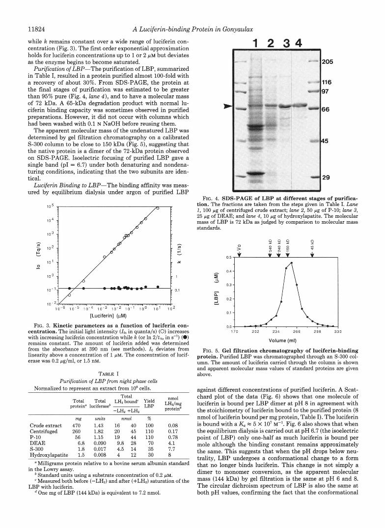
Purification of LBP-The purification of LBP, summarized in Table I, resulted in a protein purified almost 100-fold with a recovery of about 30%. From SDS-PAGE, the protein at the final stages of purification was estimated to be greater than 95% pure (Fig. 4, lane 4 ) , and to have a molecular mass of 72 kDa. A 65-kDa degradation product with normal lu- ciferin binding capacity was sometimes observed in purified preparations. However, it did not occur with columns which had been washed with 0.1 N NaOH before reusing them.
The apparent molecular mass of the undenatured LBP was determined by gel filtration chromatography on a calibrated S-300 column to be close to 150 kDa (Fig. 5), suggesting that the native protein is a dimer of the 72-kDa protein observed on SDS-PAGE. Isoelectric focusing of purified LBP gave a single band (PI = 6.7) under both denaturing and nondena- turing conditions, indicating that the two subunits are iden- tical.
Luciferin Binding to LBP-The binding affinity was meas- ured by equilibrium dialysis under argon of purified LBP
' 0 5 /" 8" I
[Luciferin] (pM)
FIG. 3. Kinetic parameters as a function of luciferin con- centration. The initial light intensity (Zo, in quanta/s) (0) increases with increasing luciferin concentration while k (or In 2/t,, in s-l) (0) remains constant. The amount of luciferin added was determined from the absorbance at 390 nm (see methods). Zo deviates from linearity above a concentration of 1 p ~ . The concentration of lucif- erase was 0.2 pg/ml, or 1.5 nM.
TABLE I Purification of LBP from night phase cells
Normalized to represent an extract from 10' cells. Total
protein" luciferase* LBP LH2/mg Total Total LH2 bound' Yield nmol
-LH, +LHo proteind ~~~ ~ _ _ _ _ _ _ ~ ~ ~~
mg units nmol % Crude extract 470 1.43 16 40 100 0.08 Centrifuged 260 1.82 20 45 110 0.17 P-10 56 1.15 19 44 110 0.78 DEAE 6.8 0.090 9.8 28 70 4.1 S-300 1.8 0.017 4.5 14 35 7.7 Hydroxylapatite 1.5 0.008 4 12 30 8 ' Milligrams protein relative to a bovine serum albumin standard
in the Lowry assay. Standard units using a substrate concentration of 0.2 p ~ . Measured both before (-LH2) and after (+LH2) saturation of the
One mg of LBP (144 kDa) is equivalent to 7.2 nmol. LBP with luciferin.
1 2 3 4
FIG. 4. SDS-PAGE of L B P at different stages of purifica- tion. The fractions are taken from the steps given in Table I. Lane 1, 100 pg of centrifuged crude extract; lane 2, 50 pg of P-10; lane 3, 25 pg of DEAE and lane 4, 10 pg of hydroxylapatite. The molecular mass of LBP is 72 kDa as judged by comparison to molecular mass standards.
170 202 234 266 298 3
Volume (ml)
FIG. 5. Gel filtration chromatography of luciferin-binding protein. Purified LBP was chromatographed through an S-300 col- umn. The amount of luciferin carried through the column is shown and apparent molecular mass values of standard proteins are given above.
against different concentrations of purified luciferin. A Scat- chard plot of the data (Fig. 6) shows that one molecule of luciferin is bound per LBP dimer at pH 8 in agreement with the stoichiometry of luciferin bound to the purified protein (8 nmol of luciferin bound per mg protein, Table I). The luciferin is bound with a K, = 5 X lo7 M-'. Fig. 6 also shows that when the equilibrium dialysis is carried out at pH 6.7 (the isoelectric point of LBP) only one-half as much luciferin is bound per mole although the binding constant remains approximately the same. This suggests that when the pH drops below neu- trality, LBP undergoes a conformational change to a form that no longer binds luciferin. This change is not simply a dimer to monomer conversion, as the apparent molecular mass (144 kDa) by gel filtration is the same at pH 6 and 8. The circular dichroism spectrum of LBP is also the same at both pH values, confirming the fact that the conformational

A Luciferin-binding Protein in Gonyaulax 11825
0.0 0.2 0.4 0.6 0.8 1 .o
Bound FIG. 6. Stoichiometry of luciferin binding to LBP. The
amount of luciferin bound to LBP, determined by equilibrium di- alysis, is plotted according to the Scatchard equation r/c = ( n - r) K., where r is the amount of luciferin bound per mol of protein and c the concentration of free luciferin. The 3c intercept yields n, the total amount of substrate bound per mole of protein (calculated for the 144-kDa dimer). The association constant (K.) is the same at the two pH values, pH 6.7 (0) and pH 8 (W), although at pH 6.7, the total luciferin bound is half. Lines drawn are based on a linear regression.
6.2 6.7 7.2 7 . 7
PH FIG. 7. Dependence of luciferase and LBP activities on pH.
The amount of luciferin bound to LBP as a function of the pH (solid circles) was determined by equilibrium dialysis as described under “Materials and Methods.” Data are plotted as the fraction of LBP which contains bound luciferin, and the theoretical curve drawn through the data is that of a 4-electron transfer. A similar theoretical curve is also drawn for the activity of luciferase as a function of pH with data based on points (open circles) taken from Krieger (1972).
change does not involve dissociation into monomers. Similar CD measurements using purified luciferin were also identical at the two pH values.
The effect of pH on binding of luciferin is shown in Fig. 7. The data are best fitted to the theoretical curve for a 4- electron transfer:
pH = 0.25 log ~
LBP-LH2 LBP
It is of interest that a similar curve can be drawn through the
TABLE I1 Daily variations in LBP content
Normalized to an extract from 10’ cells.
bound“ LH2
LBPb bound/mg bound/mg nmol LH, nmol LH2
-LH2 +LH, total protein‘ LBP
nmol mg
Day 2 3.2 0.6 0.01 5.3 Night 1.2 38 5 0.13 7.6
“The amount of luciferin bound to LBP is shown both before (-LH,) and after (+LH2) an incubation of the sample with an excess of luciferin (see “Materials and Methods”).
* The amount of LBP (in milligrams) was determined from densi- tometric scans of Western blots using affinity-purified anti-LBP; purified LBP was run as a standard.
e The total protein was determined using the Biorad assay.
data of Krieger (1972) on the effect of pH on luciferase activity (Fig. 7).
Circadian Changes in Amounts of Extractable LBP-The amount of luciferin which can be bound by G. polyedra cell extracts has been previously reported to depend upon the time of day the extractions are made, with a maximum during the night (Sulzman et al., 1978). As shown in Table 11, both by luciferin binding assay and by Western blots probed with affinity purified anti-LBP, cellular LBP levels are 8-10 times higher during the night than during the day. This rhythm persists under constant dim light. LBP is a major component of night phase Gonyaulax cells, comprising about 1% of total cellular protein (2% of the soluble protein, calculated from the amount and yield of the purified protein in Table I). This is far greater than the amount of luciferase, which was esti- mated by Dunlap and Hastings (1981a) to constitute only about 0.1% of the soluble protein. The molar ratio of LBP (144 kDa) to luciferase (135 kDa) is thus about 20 to 1 in the extracts.
DISCUSSION
The LBP that has been isolated from Gonyaulux is a dimer consisting of two identical 72,000-Da subunits that are tightly held together by noncovalent bonds. The subunits may be separated by detergents such as SDS. At pH values above neutrality, each dimeric molecule binds one molecule of lu- ciferin (Ka = 5 x lo7 M-’), but below pH 7, LBP appears to undergo a reversible conformational change to a form that is no longer capable of binding luciferin, even though the two subunits remain bound to one another.
The present studies, when taken together with the earlier observations of Krieger and Hastings (1968), may be ex- plained by assuming a series of independent (or separate) reactions. The first is the equilibrium between LBP and luciferin at alkaline pH.
LBP + L H Z - LBP-LH, (1)
As we have shown, this reaction has an association constant of about 5 X lo7 M-’, and the equilibrium is relatively inde- pendent of small changes in pH. The second reaction involves a major conformational change of the binding protein to a form that no longer binds luciferin when the pH drops below neutrality. This conformational change is 50% complete at pH 6.7, the isoelectric point of LBP, and if LBP contains bound luciferin the pH drop will cause its release.
LBP-LH, + 4H+ + HaLBP (inactive form) + LH2 (2)
Finally, the released luciferin is oxidized by luciferase in the light producing reaction:

11826 A Luciferin-binding Protein in Gonyauhx
luciferase LHP + Oz-L=O + Hz0 + hv. (3)
Within the cell during night phase, the amount of LBP may reach a maximum of 1% of the total cellular protein. Based on the light produced during exhaustive mechanical stimulation of Gonyaulax (10' quanta/cell; Seliger et al., 1969), the amount of luciferin has been estimated to be of the same order of magnitude. This suggests that virtually all of the cellular luciferin is bound to LBP. Luciferin is thus protected from inactivation by autoxidation: and can be liberated in small amounts to react with luciferase and O2 to produce a flash when stimulated by a small pH change. The localization of luciferin-specific fluorescence in the scintillons (Johnson et al., 1985) is in good agreement with immunocytochemical studies suggesting that LBP is located only in the ~cintillons.~ From Table I it may be calculated that the crude extract from 10' night phase Gonyaulax cells contains about 5 mg of LBP. Assuming a scintillon diameter of 0.5 pm and about 400 s~intillons/cell,~ it may be calculated that the total scintillon volume in 10' cells is about 3 pl. Considering the approxima- tions involved in arriving at these figures, it is remarkable that we can conclude within an order of magnitude that the scintillon volume is sufficient to accommodate the entire bioluminescent system.
Because Gonyaulax scintillons contain a 20-fold or greater excess both of binding protein and luciferin over their lucifer- ase content, only a slight decrease in pH will result in release of sufficient luciferin to cause a flash of light. Such small changes in pH could be caused by an action potential triggered by mechanical stimulation, as has been shown to be the case in Noctiluca niliaris (Eckert and Sibaoka, 1968; Nawata and Sibaoka, 1979). As in Noctiluca, we would expect that such a stimulated response could be repeated many times within a given scintillon. Indeed isolated scintillons of Gonyaulax, brought to pH 6 in vitro, are also stimulated to flash. They will flash again if brought back to neutrality, provided with fresh luciferin and subjected to a second pH drop.
The maximum rate of synthesis of LBP (and presumably luciferase and luciferin as well) occurs during the early hours of night phase, soon after evolution of O2 by photosynthesis has stopped. After reaching a maximum, the amount of LBP remains constant for about 6 h and then rapidly decreases, until during day phase only about 10% remains (Morse et al., 1989b). When photosynthesis stops at the onset of night phase, the p02 will rapidly fall to a low value, and it seems probable that the cytoplasmic pH will slowly decrease due to accumulation of the products of glycolysis. Toward the end of night phase, Gonyaulax cells emit light as a steady glow which
Nonspecific binding to bovine serum albumin at concentrations of M or greater also protects luciferin against autoxidation (Bode and Hastings, 1963).
L. Fritz, D. Morse, M. T. Nicolas, and J. W. Hastings, unpublished data.
terminates soon after daylight (Johnson and Hastings, 1986; Hastings and Dunlap, 1986). This suggests that a drop in cytoplasmic pH might be related to the onset of the glow. However, this possibility is not easily reconciled with early experiments, in which cells were shown to emit successive glow peaks, about 24 h apart, after being placed in constant darkness (Sweeney and Hastings, 1958; Hastings, 1960).4 The onset of the glow peak coincides also with the disappearance of the scintillons and their contents from the cells,4 which might well be its most immediate cause.
and to Tsuysung Park of Brandeis University who carried out the Acknowledgments-We are grateful to Professor Gerald Fasman
circular dichroism measurements on luciferin and LBP and to ThBrke Wilson for valuable criticism.
REFERENCES Bode, V. C., and Hastings, J. W. (1963) Arch. Biochem. Biophys. 103,
Bode, V. C., DeSa, R., and Hastings, J. W. (1963) Science 141,913-
Dunlap, J. C., and Hastings, J . W. (1981a) J. Biol. Chem. 256,10509-
Dunlap, J. C., and Hastings, J. W. (1981b) Biochemistry 20,983-989 Eckert, R., and Sibaoka, T. (1968) J. Gen. Physiol. 52,258-282 Eisen, H. N. (1964) Methods in Medical Research, Vol. 10, p. 106,
Fogel, M., and Hastings, J. W. (1971) Arch. Biochem. Biophys. 142,
Fogel, M., Schmitter, R., and Hastings, J. W. (1972) J. Cell Sci. 11,
Hastings, J. W. (1960) Cold Spring Harbor Symp. Quant. Biol. 25,
Hastings, J. W., and Dunlap, J. C. (1986) Methods Enzymol. 133,
Hastings, J. W., and Weber, G. (1963) J. Opt. SOC. Am. 53, 1410-
Johnson, C. H., and Hastings, J. W. (1986) Am. Sci. 74, 29-36 Johnson, C. H., Inoue, S., Flint, A., and Hastings, J. W. (1985) J.
Krieger, N. R. (1972) The Biochemistry of Bioluminescence in Gon-
Krieger, N. R., and Hastings, J. W. (1968) Science 161,586-589 Laemmli, U. K. (1970) Nature 227,680-685 Lowry, 0. H., Rosebrough, N. J., Farr, A. L., and Randall, R. J. (1951)
Morse, D., Milos, P., Roux, E., and Hastings, J. W. (1989a) Proc.
Morse, D., Fritz, L., Pappenheimer, A. M., Jr., and Hastings, J. W.
Nawata, T., and Sibaoka, T. (1979) J. Comp. Physiol. 134, 137-149 Nicolas, M.-T., Nicolas, G., Johnson, C. H., Bassot, J.-M., and
Olmsted, J. B. (1981) J. Biol. Chem. 256,11955-11957 Hastings, J. W. (1987) J. Cell Biol. 105, 723-735
Seliger, H. H., Biggley, W. H., and Swift, E. (1969) Photochm.
Sulzman, F. N., Krieger, N. R., Gooch, V. D., and Hastings, J. W.
Sweeney, B. M., and Hastings, J. W. (1958) J. Protozool. 5, 217-224 Towbin, H., Staehelin, T., and Gordon, J. (1979) Proc. Natl. Acud.
488-499
915
10518
Yearbook Publishers, Chicago
310-321
305-317
313-143
307-327
1415
Cell Biol. 100,1435-1446
yaulax polyedra. PhD dissertation, Harvard University
J. Biol. Chem. 193,265-275
Natl. Acad. Sci. U. S. A. 86, 172-176
(198913) J. Chemilum. & Biolum. 3, 79-83
Photobiol. 10, 227-232
(1978) J. Comp. Physiol. 128, 251-257
Sci. U. S. A. 76, 4350
For unknown reasons, we are not able to reproduce these results with our present strains of Gonyaulax.