the journal of rheumatology volume 35, no. 12 efficacy of ... · leukoencephalopathy syndrome....
TRANSCRIPT
The Journal of Rheumatology Volume 35, no. 12
Efficacy of Infliximab in Cogan's Syndrome
RAWAN GHADBAN, MARIE COURET and THIERRY ZENONE
http://www.jrheum.org/content/35/12/2456J Rheumatol 2008;35;2456-2458
http://www.jrheum.org/alerts 1. Sign up for TOCs and other alerts
http://jrheum.com/faq 2. Information on Subscriptions
http://jrheum.com/reprints_permissions 3. Information on permissions/orders of reprints
in rheumatology and related fields. Silverman featuring research articles on clinical subjects from scientists working
is a monthly international serial edited by Earl D.The Journal of Rheumatology
of RheumatologyThe Journal on August 18, 2019 - Published by www.jrheum.orgDownloaded from
of RheumatologyThe Journal on August 18, 2019 - Published by www.jrheum.orgDownloaded from
2449LettersPersonal non-commercial use only. The Journal of Rheumatology Copyright © 2008. All rights reserved.
Inverse Association Between Obesity and AntinuclearAntibodies in WomenTo the Editor:More than 70% of all autoimmune illnesses occur in women, a figure thathas been attributed to stimulation of the Th2 response by estrogens.However, a little-explored relationship is that which may exist betweenobesity and autoimmune disorders in women. Since the discovery of lep-
tin1 it has been known that the cytokine-producing capacity of adipose tis-sue is high. Serum concentration of leptin is 3- to 4-fold higher in womenthan in men. However, little is known about the reasons for this difference.To date no studies have investigated the relationship between leptin andautoimmune disease in the general population. Our aim was to determinewhether there was any association, in the general population, between obe-sity and the presence of antinuclear antibodies (ANA).
We studied the first 702 individuals enrolled in the “CDC de Canarias”cohort study, whose participants were drawn randomly from the adult gen-eral population. Some of the findings for this cohort study have beenreported earlier2. Obesity was identified as body mass index (BMI) ≥ 30.Abdominal obesity was considered to exist when waist circumference was≥ 88 cm in women or ≥ 102 cm in men3, and also when waist circumfer-ence was ≥ 80 cm in women and ≥ 94 in4. Obesity was also identified asa waist/height ratio of ≥ 0.55.
ANA titer was measured with an indirect immunofluorescence tech-nique that used HEp-2 cells as the substrate (Nova Lite™, InovaDiagnostics, San Diego, CA, USA). Samples were considered ANA-posi-tive when fluorescence was seen at a serum dilution of ≥ 1/40. All ANA-positive samples were tested by serial double dilution to the highest dilutionthat yielded fluorescence. Leptin concentration was measured with anenzyme linked immunosorbent assay (ng/mL, Biosource®) with a within-assay coefficient of variation of 3.6%, a between-assay coefficient of varia-tion of 6.8%) and a detection limit of 0.1 ng/ml. Proportions were comparedwith Pearson’s chi-squared test, and continuous variables with Student’s ttest. Logistic regression model were fitted for each measurement of obesity(independent variable), with ANA being the dependent variable.
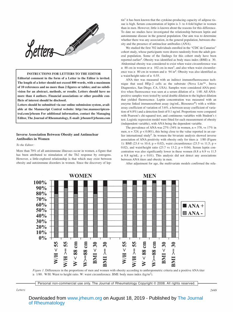
The prevalence of ANA was 27% (34% in women, n = 378, vs 17% inmen, n = 324; p < 0.001), this being close to the value reported in an ear-lier international study5. In women the bivariate analysis showed inverseassociation of ANA positivity with obesity only for titers ≥ 1/80 (Figure1): BMI (23.4 vs 10.4; p = 0.02), waist circumference (23.5 vs 11.5; p =0.02), and waist/height ratio (23.7 vs 13.2; p = 0.04). Serum leptin con-centration was also significantly lower in these women (8.8 ± 6.9 vs 11.5± 8.8 ng/mL; p = 0.01). This analysis did not detect any associationsbetween ANA titers and obesity in men.
After adjustment for age, the multivariate models confirmed the rela-
INSTRUCTIONS FOR LETTERS TO THE EDITOREditorial comment in the form of a Letter to the Editor is invited.The length of a letter should not exceed 800 words, with a maximumof 10 references and no more than 2 figures or tables; and no subdi-vision for an abstract, methods, or results. Letters should have nomore than 4 authors. Financial associations or other possible con-flicts of interest should be disclosed.Letters should be submitted via our online submission system, avail-able at the Manuscript Central website: http://mc.manuscriptcen-tral.com/jrheum For additional information, contact the ManagingEditor, The Journal of Rheumatology, E-mail: [email protected]
Figure 1. Differences in the proportions of men and women with obesity according to anthropometric criteria and a positive ANA titer≥ 1/80. W/H: Waist to height ratio. W: waist circumference. BMI: body mass index (kg/m2).
of RheumatologyThe Journal on August 18, 2019 - Published by www.jrheum.orgDownloaded from

2450 The Journal of Rheumatology 2008; 35:12
Personal non-commercial use only. The Journal of Rheumatology Copyright © 2008. All rights reserved.
tionship between positive ANA titer and obesity in women, and the absenceof this relationship in men (Table 1). In multivariate analyses to contrastANA-negative persons against ANA-positive participants in whom the titerwas ≥ 1/80, the inverse association between ANA titer and obesity inwomen was stronger (odds ratio further from the null value) than in mod-els that included ANA titers ≥ 1/40 (Table 1).
This inverse relationship we found between ANA and obesity inwomen from the general population has not been reported previously.Improvements in nutritional status have been paralleled by an increase inthe susceptibility to autoimmune diseases6,7. Given that leptin acceleratesthe onset or progression of some autoimmune diseases, and that it alsostimulates the secretion of autoantibodies in vitro8, it might be predictedthat obesity and elevated leptin concentrations would be associated with anincrease in ANA. However, we found no such association in men, and incontrast to our expectations, in women we observed that leptin concentra-tion and obesity correlated inversely with ANA. This finding was bolsteredby the fact that the association became stronger at higher ANA titers.
One possible explanation for the low prevalence of ANA in womenwith overweight or obesity is related to the lack of response to leptin inobese individuals. Obesity in humans presents hyperleptinemia togetherwith both central and peripheral resistance to the action of this hormone.Leptin resistance has been well documented9: as BMI increases, the dimin-ishing response to leptin can impair ANA production by B lymphocytes. Tounderstand why the inverse relationship between obesity and ANA titerappears only in women, we may speculate whether sex hormones play arole in leptin resistance10. The main limitation of our study is its cross-sec-tional design; this prevents us from establishing a causal relationship forthe variables we analyzed. The main strength of our study, in contrast, liesin the large sample of individuals drawn randomly from the general popu-lation. We conclude that in women in the general population there is aninverse association between positive ANA titer and obesity defined on thebasis of anthropometric criteria or serum leptin concentration.
DELIA ALMEIDA GONZÁLEZ, Research Unit, Nuestra Señora de laCandelaria University Hospital, Santa Cruz de Tenerife; ANTONIOCABRERA DE LEÓN, MD, Research Unit, Nuestra Señora de laCandelaria University Hospital; Hospital San Juan de Dios, Tenerife; andSchool of Medicine, University of La Laguna, Canary Islands, Spain;MARÍA C. RODRÍGUEZ PÉREZ, MD; SANTIAGO DOMÍNGUEZCOELLO, MD; ANA GONZÁLEZ HERNÁNDEZ, Research Unit,Nuestra Señora de la Candelaria University Hospital; RAFAEL CASTROFUENTES, School of Medicine, University of La Laguna;ARMANDO AGUIRRE JAIME; and BUENAVENTURA BRITO DIÁZ,MD, Research Unit, Nuestra Señora de la Candelaria University Hospital.Address reprint requests to Dr. Antonio Cabrera de León, Hospital SanJuan de Dios, Carretera Santa Cruz – La Laguna, 53, 38009 Santa Cruzde Tenerife, Canary Islands, Spain. E-mail: [email protected]
Supported by the Fundación Canaria de Investigación y Salud and theFondo de Investigación Sanitaria (grant PI 070934). We thankK. Shashok for translating the original manuscript into English.
REFERENCES1. Zhang Y, Proenca R, Maffei M, Barone M, Leopold L, Friedman
JM. Positional cloning of the mouse obese gene and its humanhomologue. Nature 1994;372:425-32.
2. Cabrera de León A, Santiago Domínguez Coello, Rguez Pérez MC,et al. A simple clinical score for type 2 diabetes mellitus screening.Diab Res Clin Pract 2008;80:128-33.
3. Third Report of the National Cholesterol Education Program(NCEP) Expert Panel on Detection, Evaluation, and Treatment ofHigh Blood Cholesterol in Adults (Adult Treatment Panel III) FinalReport. Circulation 2002;106:3188-90.
4. The IDF consensus worldwide definition of the metabolicsyndrome. Available at: http://www.idf.org/home/index.cfm?node=
Table 1. Relationship between antinuclear antibody (ANA) status and anthropometric and biochemical indica-tors of obesity in men and women. Each row shows results for the logistic regression model with ANA status asthe age-adjusted dependent variable. Rows in boldface type show the results with same model as the precedingrow for an ANA titer ≥ 1/80.
Dependent Variable: ANAWomen (n = 378)* Men (n = 324)*
Indicator OR 95% CI p OR 95% CI p
Waist circumference, cm 0.978 0.960, 0.997 0.023 0.988 0.962, 1.015 0.386Waist circumference, cm 0.971 0.948, 0.995 0.020 0.992 0.955, 1.030 0.668Abdominal obesity ATPIII† 0.575 0.359, 0.919 0.021 1.118 0.577, 2.167 0.740Abdominal obesity ATPIII† 0.445 0.247, 0.804 0.007 1.074 0.420, 2.748 0.882Waist/height ratio, cm 0.969 0.944, 0.995 0.020 0.992 0.947, 1.039 0.736Waist/height ratio, cm 0.962 0.930, 0.996 0.028 1.016 0.951, 1.085 0.638Abdominal obesity/height†† 0.460 0.280, 0.770 0.003 0.992 0.947, 1.039 0.736Abdominal obesity/height†† 0.393 0.206, 0.752 0.005 1.157 0.435, 3.079 0.770Abdominal obesity IDF** 0.550 0.322, 0.940 0.029 0.872 0.451, 1.686 0.685Abdominal obesity IDF** 0.462 0.235, 0.909 0.025 0.831 0.346, 1.996 0.678Body mass index, kg/m2 0.941 0.898, 0.986 0.010 0.988 0.923, 1.057 0.717Body mass index, kg/m2 0.921 0.866, 0.979 0.009 1.002 0.912, 1.101 0.965Obesity body mass index*** 0.553 0.344, 0.888 0.014 1.231 0.646, 2.347 0.527Obesity body mass index*** 0.502 0.274, 0.921 0.026 1.38 0.555, 3.430 0.489Leptin 0.760 0.593, 0.975 0.031 0.952 0.761, 1.192 0.668Leptin 0.629 0.465, 0.852 0.003 1.057 0.744, 1.501 0.758
* For models that included all participants with a positive ANA titer: n = 378 in women (248 negative, 138 pos-itive) and n = 324 in men (268 negative, 56 positive). For models that included only ANA-positive titers ≥ 1/80:n = 318 women (248 negative, 70 positive) and n = 292 men (368 negative, 24 positive). † Abdominal obesityaccording to NCEP ATPIII criteria: > 88 cm in women, > 102 cm in men. †† Waist/height ratio: < 0.55 = 1;≥ 0.55 = 2. ** Abdominal obesity according to International Diabetes Federation criteria: > 80 cm in women,> 94 cm in men. *** Body mass index: < 30 = 1; ≥ 30 = 2.
of RheumatologyThe Journal on August 18, 2019 - Published by www.jrheum.orgDownloaded from
2451LettersPersonal non-commercial use only. The Journal of Rheumatology Copyright © 2008. All rights reserved.
1429. Accessed Sept 24 2008.5. Tan EM, Feltkamp TE, Smolen JS, Butcher B, Dawkins R, Fritzler
MJ. Range of antinuclear antibodies in “healthy” individuals.Arthritis Rheum 1997;40:1601-11.
6. Harbige LS. Nutrition and autoimmunity with emphasis oninfection and autoimmune disease. Nutr Health 1996;10:285-12.
7. Black P. Why is the prevalence of allergy and autoimmunityincreasing? Trends Immunol 2001;22:354-55.
8. Ren H, Zhao H, Wang T, Yang Y, Han Z, Liu B et al. Leptinenhances in vitro secretion of IgG antiplatelet antibodies bysplenocytes and peripheral blood mononuclear cells from patientswith chronic idiopathic thrombocytopenic purpura. Clin Immunol2006;120:205-11.
9. Brito Diáz B, Rodriguez Pérez MC, Cabrera de León A. Thevicious circle of leptin and obesity. Curr Nutr Food Sci2006;2:361-73.
10. Maeso Fortuny MC, Brito Diáz B, Cabrera de León A. Leptin,estrogens and cancer. Mini Rev Med Chem 2006;6:897-07.
J Rheumatol 2008;35:12; doi:10.3899/jrheum.080322
Reversible Basal Ganglia and Amygdala Lesions in CentralNervous System LupusTo the Editor:Central nervous system (CNS) involvement in systemic lupus erythemato-sus (SLE) is common, with 25% to 60% of patients having neurologicalsymptoms, and a higher percentage displaying CNS pathology in imaging
studies or on post mortem examination1,2. The most common clinical man-ifestations are cognitive decline, psychosis, seizures, and strokes.Abnormalities on magnetic resonance imaging (MRI) scans are usuallynonspecific, such as cortical atrophy or scattered focal high-intensitywhite-matter signals3. Occasionally, larger infarcts or hemorrhages may beseen. We describe a young girl with CNS lupus who presented with thevery uncommon MRI findings of hyperintense lesions in the basal ganglia,amygdala, and cerebellum, which resolved completely after immunosup-pressive treatment, and discuss the implications for the pathogenesis ofCNS involvement in SLE.
A 17-year-old girl was admitted in status epilepticus. She had a historyof SLE diagnosed 4 months previously when she developed a butterflymalar rash, joint pain (knees, wrists, fingers), and fatigue. She was main-tained on 60 mg prednisone daily. On examination, blood pressure was115/75 mm Hg and there were no focal neurological findings. An erythe-matous malar rash was noted. Blood tests [positive antinuclear antibodies(ANA) and anti-dsDNA antibodies] confirmed a diagnosis of SLE. CranialMRI scan done the next day was normal. Cerebrospinal fluid examinationrevealed elevated protein (270 mg/dl; normal 20–40), normal glucose, andmild lymphocytic pleocytosis (18 leukocytes, predominantly lympho-cytes). A diagnosis of neuropsychiatric lupus (NPSLE) was made; seizureswere rapidly controlled with intravenous lorazepam (4 mg) and fospheny-toin (1 g loading dose, maintenance 100 mg 8-hourly). Immunosuppressivetherapy was continued with prednisone 80 mg per day. An electroen-cephalogram (EEG) 1 week after initial seizures showed diffuse slowing,but no epileptogenic activity. There were no further seizures from the sec-ond day of admission. Three weeks later she had new complaints of fatigueand generalized body weakness. There were no neurological symptoms andneurological examination was normal. A repeat EEG 4 days before these
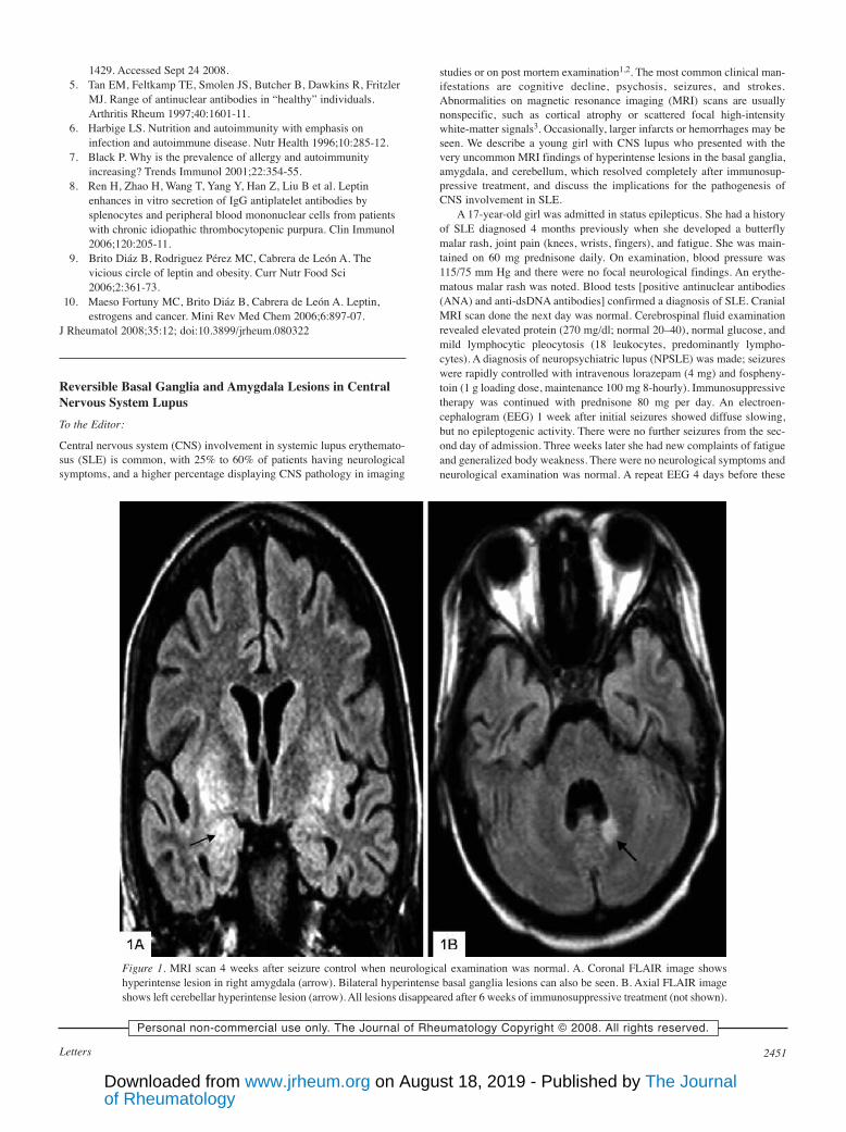
Figure 1. MRI scan 4 weeks after seizure control when neurological examination was normal. A. Coronal FLAIR image showshyperintense lesion in right amygdala (arrow). Bilateral hyperintense basal ganglia lesions can also be seen. B. Axial FLAIR imageshows left cerebellar hyperintense lesion (arrow). All lesions disappeared after 6 weeks of immunosuppressive treatment (not shown).
of RheumatologyThe Journal on August 18, 2019 - Published by www.jrheum.orgDownloaded from

2452 The Journal of Rheumatology 2008; 35:12
Personal non-commercial use only. The Journal of Rheumatology Copyright © 2008. All rights reserved.
new symptoms showed some improvement with mild diffuse slowing, butno epileptogenic activity. There was generalized muscle pain but no weak-ness. There was no joint swelling. Investigations revealed that she hadincreased levels of creatine kinase (CK), from 3656 U/l to 4370 U/l (nor-mal 24–195), myoglobinuria, and proteinuria (300 mg/dl). An electromyo-graphic study revealed early recruitment of low-amplitude, polyphasicmotor unit potentials, with frequent denervation potentials consistent withan acute myopathic process such as myositis. She was treated with 1 gintravenous methylprednisolone for 5 days and continued on 80 mg oralprednisone; CK levels gradually decreased over the following 3 weeks.
Although she had no new neurological symptoms or signs, a followupcranial MRI scan was done 4 weeks after the initial admission for seizures.It unexpectedly revealed hyperintense lesions in bilateral basal ganglia,right amygdala, and left cerebellum (Figure 1 and 2A). The basal gangliainvolvement was striking, with involvement of the caudate nucleus, globuspallidus, putamen, and external capsule bilaterally (Figure 2A). There wasno evidence of restricted diffusion to suggest infarction. Erythrocyte sedi-mentation rate was 120 mm/h (normal 0–20); CK level was still markedlyelevated at 1800. ANA, anti-dsDNA antibodies, antiribonucleoprotein, andanti-SM serum antibodies were positive. Lupus anticoagulant, antiphos-pholipid, and anti-SSB antibodies were negative. Serum complement C3,C4, and CH50 levels were normal. She had proteinuria (300 mg/dl); renaland hepatic function tests were normal. Mycophenolate mofetil 1 g twicedaily was added to prednisone 80 mg daily, and she had an uneventful clin-ical recovery from myositis over the next month. A repeat cranial MRI scanafter 6 weeks showed complete resolution of all hyperintense lesions inbasal ganglia, amygdala, and cerebellum (Figure 2B).
Our patient was unique because of the bilateral involvement of basalganglia and right amygdala and cerebellum. Involvement of the basal gan-
glia4-6 and amygdala7 has very rarely been reported in SLE. Basal gangliainvolvement was described in one patient with seizures4 and in patientswith chorea or parkinsonian symptoms5,6. In contrast, in our patient basalganglia and amygdala lesions were noted 4 weeks after seizures, whilethere was an exacerbation of lupus with myositis and proteinuria, but withno neurological symptoms or signs. Conversely, the cranial MRI scan at thetime she presented in status epilepticus was completely normal. NormalMRI scans following seizures or psychosis in SLE has been noted3, andmay reflect that small high-intensity lesions due to edema may be rapidlyreversible and hence are not detected. More commonly, transient corticalhyperintense lesions can be seen for the first few days on brain MRI scansafter multiple seizures8. However, in our patient the initial MRI scan donethe day after seizures was normal, and abnormalities were noted on thebrain MRI scan 4 weeks later, when she had no neurological symptoms orsigns and had had no seizures for 4 weeks. As well, the location of lesionsin the basal ganglia and cerebellum (rather than in cortical gray matter)suggested that the brain MRI abnormalities were not related to the priorepisode of seizures.
Reversible brain lesions can be seen in NPSLE9-11 secondary to casesof reversible posterior leukoencephalopathy syndrome9, multiple seizures,or reversible edema secondary to ischemic lesions. The absence of restrict-ed diffusion on brain MRI scan and absence of any lesion on followupbrain MRI scans makes ischemic lesions less likely in our patient.Reversible posterior leukoencephalopathy syndrome has been described inmany conditions including acute hypertension, eclampsia, chemotherapy,and SLE, and is characterized by headaches, seizures, blindness, and pari-etooccipital vasogenic edema on MRI scans9. This diagnosis should alwaysbe considered in lupus patients with reversible brain lesions. However, in
Figure 2. A. Axial MRI FLAIR image 4 weeks after seizure control when neurological examination was normal shows bilateral,extensive, hyperintense lesions in basal ganglia [globus pallidus, putamen (arrows) and in caudate nucleus (C)]. B. Axial MRIFLAIR image after 6 weeks of immunosuppressive treatment is normal, with resolution of all lesions.
of RheumatologyThe Journal on August 18, 2019 - Published by www.jrheum.orgDownloaded from

2453LettersPersonal non-commercial use only. The Journal of Rheumatology Copyright © 2008. All rights reserved.
our patient the absence of hypertension and the distribution of lesions inbasal ganglia and cerebellum (without more typical posterior parietooccip-ital involvement on MRI scan) makes it less likely. Further, immunosup-pressive therapy can trigger and should be avoided in reversible posteriorleukoencephalopathy syndrome. However, our patient improved whileunder immunosuppression with high-dose prednisone and mycophenolatemofetil, making this diagnosis less likely.
The pathogenesis of neuropsychiatric lupus is not well understood12.Vascular compromise in lupus from an embolus or thrombus has been asso-ciated with antibodies that bind cardiolipin or phospholipids and may occurin any organ including the CNS13. However, CNS involvement can alsooccur, as is likely in our patient, with no evidence of ischemia2. It has beenhypothesized that for CNS disease to occur the pathological agent, whichmay be an antibody or cytokine or cells capable of producing these mole-cules, has to cross the blood-brain barrier12. Specifically, CNS lupus hasbeen linked to autoantibodies to ribosomal P proteins14 and more recentlyto antibodies to double-stranded DNA, which have been shown to crossre-act with the neuronal NMDA glutamate receptor and produce neuronalinjury15. It is likely that many other still undiscovered autoantibodies existthat can crossreact with different brain proteins. The location of these pro-teins may determine the specific region of the brain that gets involved. Forexample, mice injected with anti-NMDA receptor antibodies have beenreported to develop specific damage to the amygdala16, as seen in ourpatient.
Under normal circumstances antibodies cannot cross the blood-brainbarrier. However, anti-NMDA receptor antibodies have been found inlupus brain15, implying that breaches of blood-brain barrier do occur inpatients with SLE. Many factors, such as status epilepticus17, infection,stress, hypertension, or nicotine exposure, are known to disrupt the blood-brain barrier18. We speculate that in our patient breaches of the blood-brainbarrier at the time she was undergoing an acute systemic exacerbation oflupus (with presumed higher levels of autoantibodies) allowed entry ofpathogenic autoantibodies to the brain. The disruption of the blood-brainbarrier may have been secondary to the prior status epilepticus (eventhough it occurred 4 weeks before brain lesions were first detected).Further, the absence of clinical neurological abnormalities and the com-plete resolution of all cerebral lesions on MRI scans suggest that autoanti-bodies can sometimes cause nonlethal reversible damage to neurons. It isalso likely that early immunosuppression may have reduced the severity ofneuronal damage.
We conclude that cranial MRI scans should be obtained during acuteexacerbations of lupus or following events that may disrupt the blood-brainbarrier. This may reveal unexpected CNS lesions, which could assist moreeffective planning of dosage and duration of immunosuppressive therapy.More research is needed to determine the significance and prognosis offindings obtained by MRI FLAIR studies, and careful clinical correlationfor new neurological abnormalities is important and should be taken intoconsideration before any decisions about therapy are made.
SATYAKAM BHAGAVATI, MD, Department of Neurology; JAI CHOI,MD, Neuroradiology Division, Department of Radiology, SUNY DownstateMedical Center, 450 Clarkson Avenue, Brooklyn, New York 11203, USA.Address reprint requests to Dr. Bhagavati; E-mail: [email protected]
REFERENCES1. Brey RL, Holliday SL, Saklad AR, et al. Neuropsychiatric
syndromes in lupus. Prevalence using standardized definitions.Neurology 2002;58:1214-20.
2. Jennekens FG, Kater L. The central nervous system in systemiclupus erythematosus. Part 2. Pathogenetic mechanisms of clinicalsyndromes. A literature investigation. Rheumatology Oxford2002;41:619-30.
3. Sibbitt WL, Sibbitt RR, Brooks WM. Neuroimaging inneuropsychiatric systemic lupus erythematosus. Arthritis Rheum
1999;42:2026-38.4. Shibata M, Kibe T, Fujimoto S, et al. Diffuse central nervous
system lupus involving white matter, basal ganglia, thalami andbrain stem. Brain Dev 1999;21:337-40.
5. Kashihara K, Nakashima S, Kohira I, et al. Hyperintense basalganglia on T1 weighted MR images in a patient with centralnervous system lupus and chorea. Am J Neuroradiol1998;19:284-6.
6. Kwong KL, Chu R, Wong SN. Parkinsonism as unusualneurological complication in childhood systemic lupuserythematosus. Lupus 2000;9:474-7.
7. Emmer BJ, Grond J, Steup-Beekman GM, et al. Selectiveinvolvement of the amygdala in systemic lupus erythematosus.PloS Medicine 2006;3:2285-90.
8. Kim JA, Chung JI, Yoon PH, et al. Transient MR signal changes inpatients with generalized tonicoclonic seizure or status epilepticus:perictal diffusion-weighted imaging. Am J Neuroradiol2001;22:1149-60.
9. Mak A, Chan BPL, Yeh IB, et al. Neuropsychiatric lupus andreversible posterior leucoencephalopathy syndrome: a challengingdilemma. Rheumatology Oxford 2008;47:256-62.
10. Appenzeller S, Faria A, Marini R, et al. Focal transient lesions ofthe corpus callosum in systemic lupus erythematosus. ClinRheumatol 2006;25:568-71.
11. Sibbitt WL, Brooks WM, Haseler LJ, et al. Spin-spin relaxation ofbrain tissues in systemic lupus erythematosus. A method forincreasing the sensitivity of magnetic resonance imaging forneuropsychiatric lupus. Arthritis Rheum 1995;38:810-8.
12. Guerrero JS, Aranow C, Mackay M, et al. Neuropsychiatric lupuserythematosus reconsidered. Nature Clin Pract Rheumatol2008;4:112-3.
13. Roldan JF, Brey RL. Neurologic manifestations of theantiphospholipid syndrome. Curr Rheumatol Rep 2007;9:109-15.
14. Bonfa E, Golombek SJ, Kaufman LD, et al. Association betweenlupus psychosis and anti-ribosomal P protein antibodies. N EnglJ Med 1987;317:265-71.
15. Kowal C, DeGiorgio LA, Lee JY, et al. Human lupusautoantibodies against NMDA receptors mediate cognitiveimpairment. Proc Nat Acad Sci USA 2006;103:19854-9.
16. Huerta PT, Kowal C, DeGiorgio LA, et al. Immunity and behavior:antibodies alter emotion. Proc Natl Acad Sci USA2006;103:678-83.
17. Correale J, Rabinowicz AL, Heck CN, Smith TD, Loskota WJ,DeGiorgio CM. Status epilepticus increases CSF levels ofneuron-specific enolase and alters the blood-brain barrier.Neurology 1998;50:1388-91.
18. Abbot NJ, Mendonca LL, Dolman DE. The blood brain barrier insystemic lupus erythematosus. Lupus 2003;12:908-15.
J Rheumatol 2008;35:12; doi:10.3899/jrheum.080310
Blockade of Interleukin 1 Receptor in Still’s Disease AffectsActivation of Peripheral T-LymphocytesTo the Editor:Adult-onset Still’s disease (AOSD) is a rare systemic inflammatory diseasecharacterized by arthralgias, transient cutaneous eruption, and high fluctu-ating fever1. No pathognomonic biological marker has yet been identified,making diagnosis difficult.
Recent pathophysiological findings showed that AOSD is characterizedby a particular cytokine profile, suggesting implication of Th1 immuneresponse. The overwhelming implication of interleukin 1 (IL-1) in the sys-temic inflammatory reaction seen during the course of AOSD has justifiedthe administration of agents blocking IL-1 receptor (anakinra), which gave
of RheumatologyThe Journal on August 18, 2019 - Published by www.jrheum.orgDownloaded from

2454 The Journal of Rheumatology 2008; 35:12
Personal non-commercial use only. The Journal of Rheumatology Copyright © 2008. All rights reserved.
some good results3,4. We describe a case of AOSD with an activatedmacrophage syndrome that responded to anakinra. The progression of acti-vated T cells was associated with AOSD activity.
A 41-year-old woman presented in April 2001 with a history of inter-mittent fever, arthralgias, and cutaneous lesions progressing since 1986.AOSD was diagnosed in 1999 according to Yamaguchi’s criteria5. Since1999 she had received hydroxychloroquine and corticosteroids, resulting inremission for 2 years until January 2001. She presented with spiking feversup to 39°C, polyarthritis, and an evanescent rash. Biological findings werewhite blood cell (WBC) count 30,000/mm3 (90% neutrophils), elevatedliver enzyme (5 times the normal level), and high ferritin level (2000ng/ml, normal 10–240 ng/ml; 9% glycosylated). C-reactive protein anderythrocyte sedimentation rate were elevated to 60 mg/dl and 85 mm/h,respectively. Immunologically, she exhibited strong and permanent T-lym-phocyte activation, with 25%–60% of CD3+ T cells expressing the HLADR+ cluster (N < 15%). The latter are usually 80% CD8+ cytotoxic T cellsexpressing perforin and granzyme B, and being CCR7– CD45RA+.
The antimalarial drug was stopped in April 2001 and methotrexate (15mg/wk) was started, with no response. She received different therapeuticagents — intravenous immunoglobulins, azathioprine, thalidomide, inflix-imab, etanercept, adalimumab — with no improvement.
In October 2002, while she was taking corticosteroids (20 mg/day), aza-thioprine, and thalidomide, she developed severe pancytopenia (WBC count1100/mm3, hemoglobin 9.3 g/dl, platelets 35,000/mm3) in the context ofmacrophage activation syndrome, documented by a bone marrow examina-tion (Figure 1). Cyclosporine was started and resulted in improvement of pan-cytopenia within 10 days, but not lymphocyte activation (45% CD3+DR+). InJanuary 2003, infliximab was reintroduced because of clinical and hemato-logical relapse. This treatment was considered partially efficient.
Figure 1. Bone marrow aspiration; cytopathology shows macrophages(arrow) that have active hemophagocytosis with intracellular blood cells,confirming the diagnosis of macrophage activation syndrome (Wright-Giemsa stain, original magnification ×400).
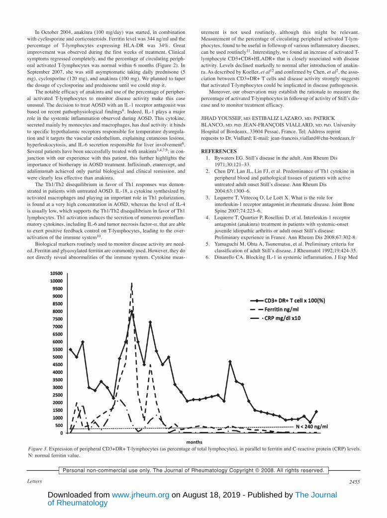
Figure 2. Expression of peripheral CD3+DR+ T lymphocytes (as percentages of total lymphocytes) as a function of time and the different therapeutic agents(indicated below). CD3+DR+ T-lymphocyte levels seemed to parallel disease activity, with generally high rates until October 2004, when the patient wasclinically symptomatic; October 2002 and January 2003 correspond to AOSD flares. Normal levels were attained only after introduction of anakinra, October2004, which achieved complete clinical and biological remission. N: normal value.
of RheumatologyThe Journal on August 18, 2019 - Published by www.jrheum.orgDownloaded from
2455Letters
In October 2004, anakinra (100 mg/day) was started, in combinationwith cyclosporine and corticosteroids. Ferritin level was 344 ng/ml and thepercentage of T-lymphocytes expressing HLA-DR was 34%. Greatimprovement was observed during the first weeks of treatment. Clinicalsymptoms regressed completely, and the percentage of circulating periph-eral activated T-lymphocytes was normal within 6 months (Figure 2). InSeptember 2007, she was still asymptomatic taking daily prednisone (5mg), cyclosporine (120 mg), and anakinra (100 mg). We planned to taperthe dosage of cyclosporine and prednisone until we could stop it.
The notable efficacy of anakinra and use of the percentage of peripher-al activated T-lymphocytes to monitor disease activity make this caseunusual. The decision to treat AOSD with an IL-1 receptor antagonist wasbased on recent pathophysiological findings6. Indeed, IL-1 plays a majorrole in the systemic inflammation observed during AOSD. This cytokine,secreted mainly by monocytes and macrophages, has dual activity: it bindsto specific hypothalamic receptors responsible for temperature dysregula-tion and it targets the vascular endothelium, explaining cutaneous lesions,hyperleukocytosis, and IL-6 secretion responsible for liver involvement6.Several patients have been successfully treated with anakinra3,4,7,9; in con-junction with our experience with this patient, this further highlights theimportance of biotherapy in AOSD treatment. Infliximab, etanercept, andadalimumab achieved only partial biological and clinical remission, andwere clearly less effective than anakinra.
The Th1/Th2 disequilibrium in favor of Th1 responses was demon-strated in patients with untreated AOSD. IL-18, a cytokine synthesized byactivated macrophages and playing an important role in Th1 polarization,is found at a very high concentration in AOSD, whereas the level of IL-4is usually low, which supports the Th1/Th2 disequilibrium in favor of Th1lymphocytes. Th1 activation induces the secretion of numerous proinflam-matory cytokines, including IL-6 and tumor necrosis factor-α, that are ableto exert positive feedback control on T-lymphocytes, leading to the over-activation of the immune system10.
Biological markers routinely used to monitor disease activity are need-ed. Ferritin and glycosylated ferritin are commonly used. However, they donot directly reveal abnormalities of the immune system. Cytokine meas-
urement is not used routinely, although this might be relevant.Measurement of the percentage of circulating peripheral activated T-lym-phocytes, found to be useful in followup of various inflammatory diseases,can be used routinely11. Interestingly, we found an increase of activated T-lymphocyte CD3+CD8+HLADR+ that is closely associated with diseaseactivity. Levels declined markedly to normal after introduction of anakin-ra. As described by Koeller, et al12 and confirmed by Chen, et al2, the asso-ciation between CD3+DR+ T cells and disease activity strongly suggeststhat activated T-lymphocytes could be implicated in disease pathogenesis.
Moreover, our observation may establish the rationale to measure thepercentage of activated T-lymphocytes in followup of activity of Still’s dis-ease and to monitor treatment efficacy.
JIHAD YOUSSEF, MD; ESTIBALIZ LAZARO, MD; PATRICKBLANCO, MD, PhD; JEAN-FRANÇOIS VIALLARD, MD, PhD, UniversityHospital of Bordeaux, 33604 Pessac, France. Tel: Address reprintrequests to Dr. Viallard; E-mail: [email protected]
REFERENCES1. Bywaters EG. Still’s disease in the adult. Ann Rheum Dis
1971;30:121–33.2. Chen DY, Lan JL, Lin FJ, et al. Predominance of Th1 cytokine in
peripheral blood and pathological tissues of patients with activeuntreated adult onset Still’s disease. Ann Rheum Dis2004;63:1300–6.
3. Lequerre T, Vittecoq O, Le Loët X. What is the role forinterleukin-1 receptor antagonist in rheumatic disease. Joint BoneSpine 2007;74:223–6.
4. Lequerre T, Quartier P, Rosellini D, et al. Interleukin-1 receptorantagonist (anakinra) treatment in patients with systemic-onsetjuvenile idiopathic arthritis or adult onset Still’s disease:Preliminary experience in France. Ann Rheum Dis 2008;67:302-8.
5. Yamaguchi M, Ohta A, Tsunematsu, et al. Preliminary criteria forclassification of adult Still’s disease. J Rheumatol 1992;19:424-35.
6. Dinarello CA. Blocking IL-1 in systemic inflammation. J Exp Med
Figure 3. Expression of peripheral CD3+DR+ T-lymphocytes (as percentage of total lymphocytes), in parallel to ferritin and C-reactive protein (CRP) levels.N: normal ferritin value.
Personal non-commercial use only. The Journal of Rheumatology Copyright © 2008. All rights reserved.
of RheumatologyThe Journal on August 18, 2019 - Published by www.jrheum.orgDownloaded from

2456 The Journal of Rheumatology 2008; 35:12
Personal non-commercial use only. The Journal of Rheumatology Copyright © 2008. All rights reserved.
2005;201:1355–9.7. Vasques Godinho FM, Parreira Santos MJ, Canas da Silva J.
Refractory adult onset Still’s disease successfully treated withanakinra. Ann Rheum Dis 2005;64:647–8.
8. Haraoui B, Bourrelle D, Kaminska E. Anakinra in the treatment ofadult onset Still’s disease [abstract]. Ann Rheum Dis 2004;63Suppl 1:263.
9. Fitzgerald AA, Leclercq SA, Yan A, et al. Rapid responses toanakinra in patients with refractory adult-onset Still’s disease.Arthritis Rheum 2005;52:1794–803.
10. Choi JH, Suh CH, Lee YM, et al. Serum cytokine profiles inpatients with adult onset Still’s disease. J Rheumatol2003;30:2422–7.
11. Blanco P, Pitard V, Viallard JF, et al. Increase in activated CD8+ Tlymphocytes expressing perforin and granzyme B correlates withdisease activity in patients with systemic lupus erythematosus.Arthritis Rheum 2005;52:201-11.
12. Koeller M, Kiener H, Simonitsch I, et al. Destructivelymphadenopathy and T-lymphocyte activation in adult-onset Still’sdisease. Br J Rheumatol 1995;34:984–8.
J Rheumatol 2008;35:12; doi:10.3899/jrheum.080150
Efficacy of Infliximab in Cogan’s SyndromeTo the Editor:Cogan’s syndrome is a rare chronic inflammatory disease that most com-monly affects adults in the third decade, with no gender predominance.Interstitial keratitis is the characteristic ocular feature, although this is notmandatory for the diagnosis; moreover, ocular manifestations such as scle-ritis, episcleritis, and uveitis may occur. The vestibuloauditory manifesta-tions consist of a Ménière’s-like disease including vertigo, nausea, vomit-ing, tinnitus, and hearing loss. The diagnosis is made upon the associationof eye disease and vestibuloauditory dysfunction, with or without systemicfeatures (fever, asthenia, weight loss, arthritis, arthralgias, aortitis, andabdominal pain)1-4.
A 48-year-old woman was admitted to our hospital in May 2001 fol-lowing a sudden bilateral hearing loss with vertigo and tinnitus. One yearpreviously, she had had a history of polyarthritis of the wrists and metacar-pophalangeal joints, without morning stiffness, which was treated as aseronegative arthritis with nonsteroidal antiinflammatory drugs and low-dose oral prednisone. She also had an episode of painful red eye for whichno medical attention had been given. She had tapered the prednisone from10 mg/day to 7.5 mg/day 3 weeks before the appearance of the vestibu-loauditory manifestations.
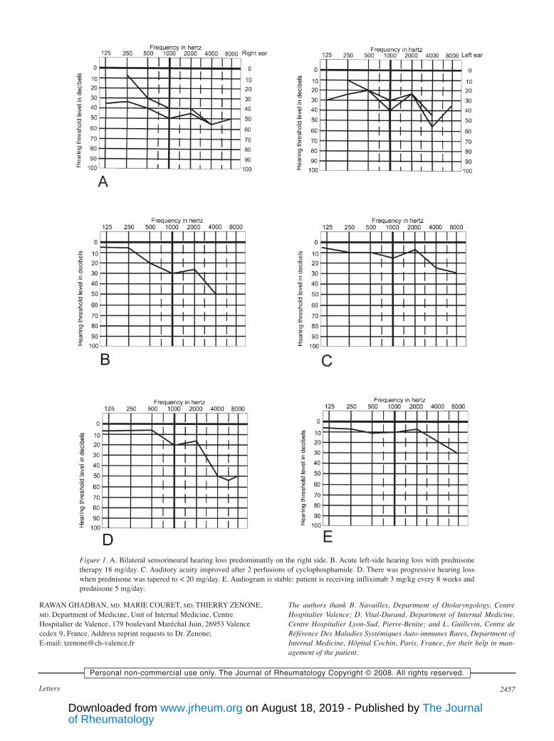
Upon admission, the clinical examination was not significant. Shecomplained of polyarthralgia without arthritis. Otolaryngeal examinationconfirmed a bilateral sensorineural hearing loss predominantly on the rightside (Figure 1A). The ophthalmologic examination showed signs of prioranterior uveitis. Chest radiograph, cerebral and auditory conduct magneticresonance imaging (MRI), electroencephalogram, electrocardiogram,transthoracic cardiac echography, and thoracic MRI were all normal.Laboratory tests showed normal complete blood count, erythrocyte sedi-mentation rate, C-reactive protein, renal function, and complements.Treponemic pallidum serologies, rheumatoid factors, antinuclear antibod-ies, anti-DNA antibodies, antiphospholipid antibodies, and antineutrophilcytoplasm antibodies were all negative.
The diagnosis of Cogan’s syndrome was established upon the associa-tion of vestibuloauditory, ocular, and systemic manifestations. The patientdid not fulfil the classification criteria for any other systemic disease suchas rheumatoid arthritis, spondyloarthropathy, or inflammatory bowel dis-ease. Treatment with prednisone 60 mg/day was started, which resulted inimprovement in the audiogram and the polyarthralgias. Prednisone was
slowly tapered, but a relapse with acute bilateral worsening of hearing lossoccurred at a dose of 14 mg/day. She was admitted again and started pulsetherapy of methylprednisolone 240 mg/day for 5 days. This resulted in apartial improvement. Then methotrexate (MTX) 17.5 mg weekly wasadded. She continued to have progressive hearing loss of the right ear untilshe lost hearing completely in October 2002, while being treated withsteroids (60 mg/day initially and slowly tapered) and MTX. A new relapsewith acute hearing loss on the left side occurred with prednisone therapy of18 mg/day in March 2003 (Figure 1B).
Because of continued hearing loss despite steroids and MTX, pulsetherapy of intravenous cyclophosphamide 750 mg was started in June2004, for 6 months, along with methylprednisolone 250 mg/day for thefirst 5 days. The auditory acuity improved after 2 perfusions of cyclophos-phamide (Figure 1C). Azathioprine 150 mg/day was started after the sixthperfusion of cyclophosphamide. Although the patient was treated with aza-thioprine and prednisone, relapses continued to occur, with a progressivehearing loss every time the prednisone was tapered to a dose less than 20mg/day (Figure 1D).
Treatment with infliximab 3 mg/kg (200 mg) every 3 weeks was initi-ated in April 2005. There was a rapid improvement of the audiogram of theleft ear after 2 perfusions (Figure 1E). Treatment intervals were thenextended to 8 weeks. Prednisone was successfully tapered to a daily doseof 5 mg, with a complete remission for 3 years after starting infliximab.The patient is still receiving infliximab (3 mg/kg every 8 weeks) and pred-nisone (5 mg/day) in June 2008. The audiogram is stable, as shown inFigure 1E.
Treatment of Cogan’s syndrome is difficult, and the only information wefind in the literature is based upon clinical case reports; no organized seriesof treatments has been published. Systemic corticosteroids are the mainstayof treatment for inner ear disease and/or systemic vasculitis, other immuno-suppressive drugs being used in case of treatment failure or as corticosteroid-sparing therapy3. Failure to aggressively treat immune-mediated hearing lossmay lead to profound and permanent hearing loss. Repeated disease flareslead to loss of hearing despite control of hearing damage initially1. Earlytreatment after onset of hearing loss was shown to give better results5.However, severe hearing loss may occur despite treatment with high-dosesteroids and immunosuppressive agents1,4. So it is not clear that providingaggressive immunosuppressive treatment alters the longterm outcome.
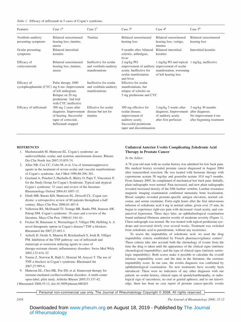
The effect of tumor necrosis factor-α blockers in Cogan’s syndromewas recently investigated1, and we identified 5 cases6-8 of Cogan’s syn-drome treated with infliximab (Table 1). In our case there was a resistanceand failure of steroids and other immunosuppressive agents (azathioprine,MTX, and cyclophosphamide). Although the patient was treated with highdoses of methylprednisolone and immunosuppressive agents, a completehearing loss of the right ear and progressive hearing loss on the left sideoccurred. Infliximab, started 29 months after the right ear hearing loss and4 years after the diagnosis, showed good efficacy, with improvement ofauditory acuity of the left ear after the second perfusion. There was noimprovement in the right ear, which was already deaf 29 months before thetreatment was started. Oral prednisone was successfully tapered to a dailydose of 5 mg, with no relapses and a good tolerance after 3 years of treat-ment with infliximab.
The effect of etanercept was investigated in an open-label prospectivestudy9 including 23 patients with bilateral immune-mediated cochleo-vestibular disorder or symptoms of Ménière’s disease; 3 of these patientshad Cogan’s syndrome. Etanercept was not effective in preserving orimproving hearing loss, but 2 of 3 patients who had Cogan’s syndromeshowed improvement in word identification and recognition9. However,etanercept was not helpful in preventing hearing loss in Cogan’s syndrome.
Infliximab might be an alternative therapy for Cogan’s syndrome, espe-cially in cases of failure of corticosteroids and immunosuppressive thera-py. However, treatment might be more effective when started at an earlystage of the disease, and mainly for inner-ear disease, when the lesions arestill reversible. The decision to consider infliximab as first-line therapyafter the onset of hearing loss is worth investigating.
of RheumatologyThe Journal on August 18, 2019 - Published by www.jrheum.orgDownloaded from
2457LettersPersonal non-commercial use only. The Journal of Rheumatology Copyright © 2008. All rights reserved.
RAWAN GHADBAN, MD; MARIE COURET, MD; THIERRY ZENONE,MD, Department of Medicine, Unit of Internal Medicine, CentreHospitalier de Valence, 179 boulevard Maréchal Juin, 26953 Valencecedex 9, France. Address reprint requests to Dr. Zenone;E-mail: [email protected]
The authors thank B. Navailles, Department of Otolaryngology, CentreHospitalier Valence; D. Vital-Durand, Department of Internal Medicine,Centre Hospitalier Lyon-Sud, Pierre-Benite; and L. Guillevin, Centre deRéférence Des Maladies Systémiques Auto-immunes Rares, Department ofInternal Medicine, Hôpital Cochin, Paris, France, for their help in man-agement of the patient.
Figure 1. A. Bilateral sensorineural hearing loss predominantly on the right side. B. Acute left-side hearing loss with prednisonetherapy 18 mg/day. C. Auditory acuity improved after 2 perfusions of cyclophosphamide. D. There was progressive hearing losswhen prednisone was tapered to < 20 mg/day. E. Audiogram is stable: patient is receiving infliximab 3 mg/kg every 8 weeks andprednisone 5 mg/day.
of RheumatologyThe Journal on August 18, 2019 - Published by www.jrheum.orgDownloaded from
2458 The Journal of Rheumatology 2008; 35:12
Personal non-commercial use only. The Journal of Rheumatology Copyright © 2008. All rights reserved.
REFERENCES1. Mazlumzadeh M, Matteson EL. Cogan’s syndrome: an
audiovestibular, ocular, and systemic autoimmune disease. RheumDis Clin North Am 2007;33:855-74.
2. Allen NB, Cox CC, Cobo M, et al. Use of immunosuppressiveagents in the treatment of severe ocular and vascular manifestationsof Cogan’s syndrome. Am J Med 1990;88:296–301.
3. Grasland A, Pouchot J, Hachulla E, Blétry O, Papo T, Vinceneux P,for the Study Group for Cogan’s Syndrome. Typical and atypicalCogan’s syndrome: 32 cases and review of the literature.Rheumatology Oxford 2004;43:1007-15.
4. Gluth MB, Baratz KH, Matteson EL, Driscoll CL. Cogan syn-drome: a retrospective review of 60 patients throughout a halfcentury. Mayo Clin Proc 2006;81:483-8.
5. Vollertsen RS, McDonald TJ, Younge BR, Banks PM, Stanson AW,Ilstrup DM. Cogan’s syndrome: 18 cases and a review of theliterature. Mayo Clin Proc 1986;61:344–61.
6. Fricker M, Baumann A, Wermelinger F, Villiger PM, Helbling A. Anovel therapeutic option in Cogan’s disease? TNF-a blockers.Rheumatol Int 2007;27:493-5.
7. Aeberli D, Oertle S, Mauron H, Reichenbach S, Jordi B, VilligerPM. Inhibition of the TNF pathway: use of infliximab andetanercept as remission inducing agents in cases oftherapy-resistant chronic inflammatory disorders. Swiss Med Wkly2002;132:414–22
8. Touma Z, Nawwar R, Hadi U, Hourani M, Arayssi T. The use ofTNF-a blockers in Cogan’s syndrome. Rheumatol Int2007;27:995-6.
9. Matteson EL, Choi HK, Poe DS, et al. Etanercept therapy forimmune-mediated cochleovestibular disorders: A multi-centeropen-label, pilot study. Arthritis Rheum 2005;53:337-42.
J Rheumatol 2008;35:12; doi:10.3899/jrheum.080203
Unilateral Anterior Uveitis Complicating Zoledronic AcidTherapy in Prostate CancerTo the Editor:A 78-year-old man with no ocular history was admitted for low back pain.His medical history revealed prostate cancer diagnosed in August 2004after transurethral resection. He was treated with hormone therapy withcyproterone acetate 50 mg/day and goserelin acetate 10.8 mg/3 months.From January 2005, he complained of mechanical low back pain. Initially,plain radiographs were normal. Pain increased, and new plain radiographsrevealed increased density of the fifth lumbar vertebra. Lumbar resonancemagnetic imaging examination confirmed metastatic bone localization.Blood samples revealed prostate-specific antigen elevation, normal cal-cemia, and serum creatinine. Forty-eight hours after the first intravenousinfusion of zoledronic acid 4 mg in normal saline, given over 15 min, hebegan to experience right-eye pain with decreased visual acuity, and con-junctival hyperemia. Three days later, an ophthalmological examinationfound unilateral fibrinous anterior uveitis of moderate severity (Figure 1).Chest radiograph was normal. He was treated with topical prednisone eye-drops and recovered slowly over several weeks. Treatment was switchedfrom zoledronic acid to pamidronate, without any recurrence.
To assess the imputability of zoledronic acid, we used the drugimputability criteria established by French pharmacovigilance centers1.These criteria take into account both the chronology of events from thetime the drug is taken until the appearance of the clinical signs (intrinsicchronological imputability), and the type of clinical signs (intrinsic semio-logic imputability). Both scores make it possible to calculate the overallintrinsic imputability score; and the data in the literature, the extrinsicimputability score. In our case, the uveitis diagnosis was confirmed byophthalmological examination. No new treatments have recently beenintroduced. There were no indicators of any other diagnosis with ourpatient: no ocular history, clinical signs of spondyloarthropathy, or radio-logical sign of sarcoidosis, no oral or genital aphtosis; and to our knowl-edge, there has been no case report of prostate cancer-specific uveitis
Table 1. Efficacy of infliximab in 5 cases of Cogan’s syndrome.
Features Case 16 Case 27 Case 36 Case 48 Case 58
Vestibulo-auditory Bilateral sensorineural Tinnitus Bilateral sensorineural Bilateral sensorineural Bilateral sensorineuralpresenting symptoms hearing loss, tinnitus, hearing loss hearing loss, vertigo, hearing loss
ataxia tinnitusOcular presenting Bilateral interstitial 9 months after, bilateral Bilateral interstitial Interstitial keratitissymptoms keratitis scleritis, arthralgias, keratitis
feverEfficacy of Bilateral sensorineural Ineffective for ocular 2 mg/kg PO: 1 mg/kg PO and topical: 1 mg/kg, ineffectivecorticosteroids hearing loss, tinnitus, and vestibulo-auditory improvement of auditory improvement of ocular
ataxia manifestations acuity. Ineffective for manifestation, worseningocular manifestation of left hearing lossand fever
Efficacy of Pulse therapy 1000 Ineffective for ocular Effective for ocularcyclophosphamide (CYC) mg 6 mo. Improvement and vestibulo-auditory manifestations, but
of left audiogram. manifestations relapse of scleritis onRelapse on 20 mg 5 mg prednisone and CYCprednisone. 2nd trialwith CYC ineffective
Efficacy of infliximab 300 mg 2 years after Effective for ocular 300 mg effective for 3 mg/kg 3 weeks after 3 mg/kg 30 monthsdiagnosis. Improvement disease but not for ocular disease, diagnosis. Improvement after diagnosis.of hearing. Successful tinnitus improvement of of auditory acuity No improvement 4 motaper of corticoids. auditory acuity. after first perfusion after beginning treatmentInfliximab stopped Successful prednisone
taper and discontinuation
of RheumatologyThe Journal on August 18, 2019 - Published by www.jrheum.orgDownloaded from

2459LettersPersonal non-commercial use only. The Journal of Rheumatology Copyright © 2008. All rights reserved.
(semiologic criterion: S3). The clinical signs of uveitis occurred 48 h afterthe first intravenous infusion. Uveitis was controlled with topical pred-nisone eyedrops with no relapse (chronological criterion: C3). The overallintrinsic imputability score (I4) strongly suggests the involvement of zole-dronic acid. Bisphosphonates are widely used in patients with hypercal-cemia of malignancy and cancer metastatic to bone, osteoporosis, andPaget’s disease. Most frequent general side effects include clinical troublessuch as influenza-like symptoms, nausea, bone pain, and biological pertur-bation such as transient hypocalcemia. Some previous inflammatory ocu-lar adverse events have been reported with other bisphosphonates2, usual-ly with the nitrogen-containing bisphosphonates (alendronate, pami-dronate, zoledronate, risedronate)3-5, except in one case (clodronate)6. Themechanism of the inflammation is unclear, but may be explained by thefact that the nitrogen-containing bisphosphonates cause elevated levels ofproinflammatory cytokines, including interleukin 6 and tumor necrosis fac-tor-α7. No ocular or patient predisposing factors are known. Only 3 recentcases reported uveitis as a complication of zoledronic acid8-10. Zoledronicacid is characterized by its high antiresorptive potency and short infusiontime11,12. In the HORIZON (Health Outcomes and Reduced Incidence withZoledronic Acid Once Yearly) trial conducted in postmenopausal womenwith osteoporosis12, patients treated with zoledronic acid had an absoluteincrease of approximately 0.69% in inflammatory ocular adverse events(mainly conjunctivitis) during the first 15 days after infusion in compari-son with controls, but no case of uveitis. Finally, the extrinsic imputabilityscore (B2) also suggests the involvement of zoledronic acid.
All physicians should be aware of uveitis as a possible complication ofzoledronic acid therapy. They should instruct patients to immediatelyreport eye trouble such as pain or decreased visual acuity in order to treatthe onset of symptoms with specific therapy, after ophthalmological exam-ination. This may be true especially after the first infusion, which is thetime of occurrence in 3 of the 4 published cases. Repeating the infusions ofzoledronic acid in these patients, even with prophylactic topical steroidsand atropine, may not be safe. Indeed, one case report of uveitis associat-ed with clodronate relapsed when rechallenged with the same drug6. Inanother case, in which the original bisphosphonate was replaced by a dif-ferent drug of the same class, eye inflammation was reduced and eventual-ly resolved with continued use, suggesting the development of immuno-logical tolerance13. This was the rationale to switch to pamidronate, and wedid not observe any relapse of uveitis.
FRÉDÉRIC BANAL, MD; KARINE BRIOT, MD; GHAZI AYOUB, MD;MAXIME DOUGADOS, MD; CHRISTIAN ROUX, MD, PhD, RenéDescartes University, Medicine Faculty; APHP Cochin Hospital,Department of Rheumatology B, Paris, France. Address reprint requeststo Prof. C. Roux, Rhumatologie B, Hôpital Cochin, 27 rue du FaubourgSaint-Jacques, 75014 Paris, France. E-mail: [email protected]
REFERENCES1. Bégaud B, Evreux JC, Jouglard J, Lagier G. Imputation of the
unexpected or toxic effects of drugs. Actualization of the methodused in France. Therapie 1985;40:111-8.
2. Fraunfelder FW, Fraunfelder FT. Bisphosphonates and ocularinflammation. N Engl J Med 2003;348:1187-8.
3. Siris ES. Bisphosphonates and iritis. Lancet 1993;341:436-7.4. Malik AR, Campbell SH, Toma NM. Bilateral uveitis after
alendronate. Br J Ophthalmol 2002;86:1443.5. Ghose K, Waterworth R, Trolove P, Highton J. Uveitis associated
with pamidronate. Aust NZ J Med 1994;24:320.6. Fietta P, Manganelli P, Lodigiani L. Clodronate induced uveitis.
Ann Rheum Dis 2003;62:378.7. Sauty A, Pecherstorfer M, Zimmer-Roth I, et al. Interleukin-6 and
tumor necrosis factor alpha levels after bisphosphonates treatmentin vitro and in patients with malignancy. Bone 1996;18:133-9.
8. El Saghir NS, Otrock ZK, Bleik JH. Unilateral anterior uveitiscomplicating zoledronic acid therapy in breast cancer. BMCCancer 2005;6:156.
9. Durnian JM, Olujohungbe A, Kyle G. Bilateral acute uveitis andconjunctivitis after zoledronic acid therapy. Eye 2005;19:221-2.
10. Moore MM, Beith JM. Acute unilateral anterior uveitis and scleritisfollowing a single infusion of zoledronate for metastatic breast can-cer. Med J Aust 2008;188:370-1.
11. Reid IR, Miller P, Lyles K, et al. Comparison of a single infusionof zoledronic acid with risedronate for Paget’s disease. N EnglJ Med 2005;353:898-908.
12. Black DM, Delmas PD, Eastell R, et al. Once-yearly zoledronicacid for treatment of postmenopausal osteoporosis. N Engl J Med2007;356:1809-22.
13. Benderson D, Karakunnel J, Kathuria S, Badros A. Scleritiscomplicating zoledronic acid infusion. Clin Lymphoma Myeloma2006;7:145-7. J Rheumatol 2009;36:1; doi 10.3899
J Rheumatol 2008;35:12; doi:10.3899/jrheum.080273
CorrectionEder L, Zisman D, Rimar D, Barzilai M, Rahat M, Laor A,Bitterman H, Rozenbaum M, Rozner I, Feld J. Subclinicalatherosclerosis in patients with psoriatic arthritis. Reply; let-ter. J Rheumatol 2008:35:2070-1. The correct spelling of theauthor’s name is Rosner. We regret the error.
Figure 1. Right-eye acute anterior uveitis.
of RheumatologyThe Journal on August 18, 2019 - Published by www.jrheum.orgDownloaded from