the journal of rheumatology volume 70, no. who gets
TRANSCRIPT

Pers
onal
, non
-com
mer
cial
use
onl
y. T
he J
ourn
al o
f Rhe
umat
olog
y. C
opyr
ight
© 2
004.
All
right
s re
serv
ed.
The Journal of Rheumatology 2004, Volume 31, Supplement 7010
From the Tufts University School of Medicine, Tufts University, Marion,Massachusetts, USA.
E.L. Radin, MD, Adjunct Professor of Orthopaedic Surgery.
Address reprint requests to Dr. E.L. Radin, PO Box 561, Marion, MA02738, USA.
Who gets osteoarthritis (OA)? The answer is reasonablystraightforward. As concluded by Drs. Felson and Hochbergelsewhere in these proceedings1,2, OA affects seniors,Caucasians more than Orientals, women, large people,people with greater bone density, and athletes who are atrisk; and it affects them because of what they do. There isnot much argument about this. However, in discussing thistopic we need to define what we are speaking about: asymp-tomatic radiographic OA is of no interest to me — it doesn’trequire treatment. I’m interested only in symptomatic OA.
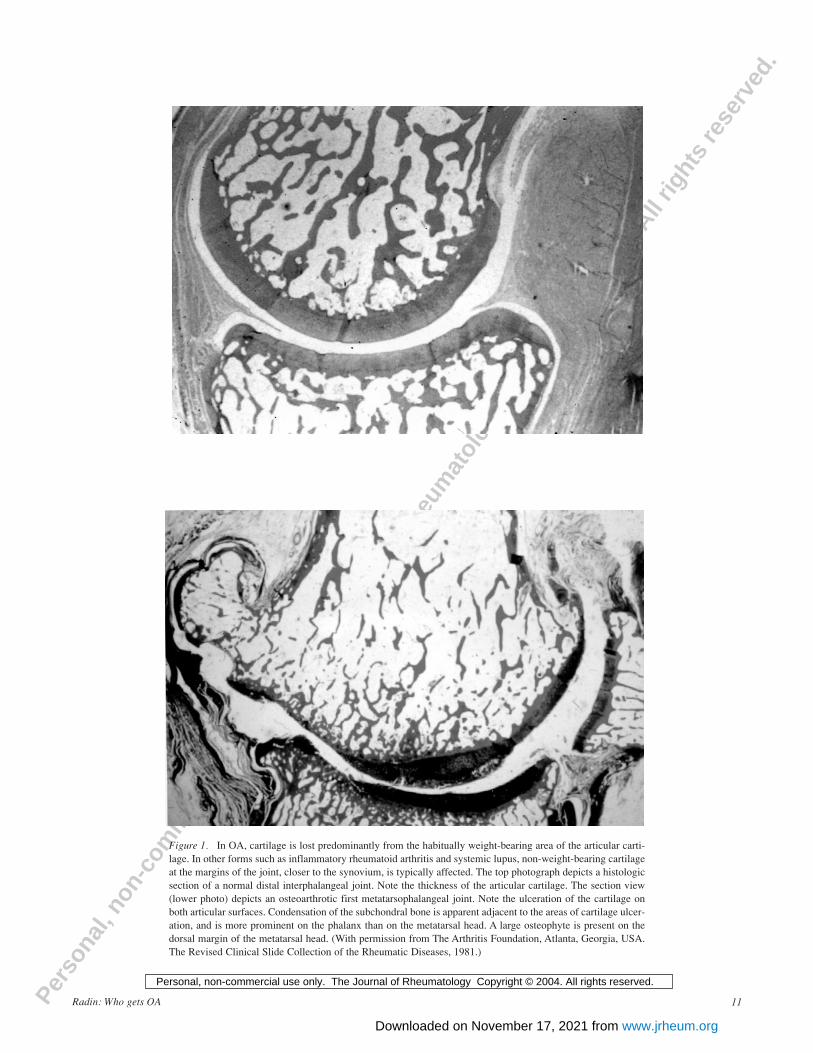
One of the problems in studying OA is that we includelemons and limes among the oranges. My definition of OA issimple: I differentiate OA from other forms of joint diseasebecause of the mechanical factors involved in its developmentand progression. OA initially involves the loss of habituallyweight-bearing articular cartilage (Figure 1). This contrastsOA with rheumatoid arthritis, lupus, and other connectivetissue diseases that affect joints, in which the primary cause isinflammatory; and it is the non-weight-bearing cartilage,close to the synovium, that is initially affected.
In inflammatory arthritis the central articular cartilagebecomes involved as inflammatory change progresses. Inmechanically-induced OA, the non-weight-bearing articularcartilage is involved only as the joint undergoes remodeling.Also, sclerosis and thickening of the subchondral plate arehallmark features of OA, whereas juxtaarticular osteo-porosis characterizes the inflammatory forms of arthritis.Statistically, the presence of osteoporosis appears to sparejoints from OA.
As Dr. Burr discusses in these proceedings3, stiffening ofthe subchondral bone is not a critical primary pathogeneticcause of OA, as we had thought earlier. Rather, reactivationof the primary center of ossification, thickening of thesubchondral plate, and loss of habitually load-bearing artic-ular cartilage appear to be cardinal features of the patho-physiology of OA.
Osteophytes should not be thought of as pathognomonicof OA. They occur also with other conditions. The osteo-phyte, indeed, is good tissue: it contains good hyaline carti-lage, good bone, a good subchondral plate, and goodcalcified cartilage. If osteophytes are present, it is hard tobelieve that the cells in most of the patients with OA are sicksince they can still create normal tissues.
In short, the characteristic of OA is joint failure, drivenby mechanical factors. As Felson and others have pointedout1,2, some OA joints may not be quite right from amechanical standpoint at the outset. The inflammatoryresponse in OA is not primary, but secondary. Attempts tosuppress inflammation in OA with nonsteroidal antiinflam-matory drugs, if they are at all effective, may hasten theprogression of the disease4.
The relationship between Heberden’s nodes and OA isvariable. Smythe5 established that there are several causes ofHeberden’s nodes, only one of which is related to general-ized OA. OA affects mainly the axial skeleton and largeappendicular joints. Inflammatory arthritis is very different— it can affect any joint. Interestingly, in inflammatoryarthritis it is secondary mechanical changes that lead tosurgery.
OA is a proliferative condition, and is characterized by anabundance of new tissue. Under these circumstances itseems inappropriate to call OA a degenerative process. Newarticular cartilage and new bone are being formed. Theproblem, of course, is that this new cartilage and new bonedo not form in the right places and do not do the joint anygood.
Because of its multiple etiologies, OA probably shouldnot be considered a disease. It represents the failure of anorgan (the synovial joint) and is analogous to heart failure orkidney failure. OA can begin in any of the articular or peri-articular tissues. Sometimes it begins in the bone, some-times in the cartilage, sometimes in the ligaments.Sometimes the defect lies in an abnormality in neuromus-cular control. This is why efforts to identify markers of OAhave proved so frustrating. The organ failure represented byOA does not begin in the same tissue in each case, andchanges in one tissue within the joint are related to changesin other tissues within that organ. If changes begin in thecartilage, the bone will be altered; if changes in the bone areprimary, the articular cartilage will be affected. If ligamentsare damaged, everything will be affected.
When the cells are involved in joint failure, as in the raregenetically based generalized joint destruction described byAla-Kokko, et al6, in patients with a point mutation in thecDNA that codes for Type II collagen, the cartilage failureexpresses itself as a mechanical problem. There are prob-ably many genetic aberrations behind the various etiologiesof OA. For example, the shape of our joints, the basicdensity of our bones, the way we walk and move, are allbasically inherited. It is important that we begin thinking ofsubgroups of patients with OA. If we do not do so, epidemi-
Who Gets Osteoarthritis and Why?ERIC L. RADIN
Personal, non-commercial use only. The Journal of Rheumatology Copyright © 2004. All rights reserved.
www.jrheum.orgDownloaded on November 17, 2021 from
Pers
onal
, non
-com
mer
cial
use
onl
y. T
he J
ourn
al o
f Rhe
umat
olog
y. C
opyr
ight
© 2
004.
All
right
s re
serv
ed.
Radin: Who gets OA 11
Figure 1. In OA, cartilage is lost predominantly from the habitually weight-bearing area of the articular carti-lage. In other forms such as inflammatory rheumatoid arthritis and systemic lupus, non-weight-bearing cartilageat the margins of the joint, closer to the synovium, is typically affected. The top photograph depicts a histologicsection of a normal distal interphalangeal joint. Note the thickness of the articular cartilage. The section view(lower photo) depicts an osteoarthrotic first metatarsophalangeal joint. Note the ulceration of the cartilage onboth articular surfaces. Condensation of the subchondral bone is apparent adjacent to the areas of cartilage ulcer-ation, and is more prominent on the phalanx than on the metatarsal head. A large osteophyte is present on thedorsal margin of the metatarsal head. (With permission from The Arthritis Foundation, Atlanta, Georgia, USA.The Revised Clinical Slide Collection of the Rheumatic Diseases, 1981.)
Personal, non-commercial use only. The Journal of Rheumatology Copyright © 2004. All rights reserved.
www.jrheum.orgDownloaded on November 17, 2021 from
Pers
onal
, non
-com
mer
cial
use
onl
y. T
he J
ourn
al o
f Rhe
umat
olog
y. C
opyr
ight
© 2
004.
All
right
s re
serv
ed.
ologic studies will only add to our confusion. The causes ofjoint failure are the same as those leading to failure of otherorgans: congenital, developmental, post-traumatic, postin-fectious.
Although increased load, such as that associated withobesity, is associated with knee OA, Maquet has suggestedthat the increase in thigh girth and the resultant wider-basedstance alter the physiological axis of the knee joint, pushingit into varus7. Although a preponderance of the evidenceseems to show that obesity is not associated with hip OA,Hochberg and his colleagues (see above) have challengedthis1,2.
One must agree that the affected joint tissues are exces-sively loaded in OA. As mentioned above, the evidence thatinadequacy of joint tissues is a cause of OA seems to bebased upon a small number of patients. More commonly,OA is a sequel to serious joint trauma, such as from contactsports, a fall, or a motor vehicle accident. Or the damage canbe minor, but repetitive. The manner in which physiologi-cally reasonable loads are applied is critical to the continuedhealth of a joint. Loads that are applied too quickly damagejoints and the supporting musculoskeletal structures,causing microinjury. Because these tissues are viscoelastic,they contain a water component. When these tissues areloaded, the movement of this interstitial water spares thetissue matrices from deleterious loads. If we think aboutwhat would cause damage to an extracellular matrix thatcontains water and proteoglycans, collagen fibers, and othermolecular constituents, and we exclude circumstances thatreally pound on the tissue (e.g., driving into a tree or playingtackle on the football team) but consider activities of dailyliving, it is apparent that a damaging load must be deliveredvery rapidly — more rapidly than the extracellular water canbe displaced. If the conditions permit water to move withinthe tissue, the matrix will be spared from damage. This is thebeauty of viscoelasticity and the secret of viscoelastic shockabsorption. Thus, in order to be damaging, loads on jointsmust be delivered very rapidly. If they are, they will producemicrodamage, which is cumulative over time and provokesa healing response. Thus, the offending loads need not besupraphysiologic in magnitude — they merely need to bedelivered too quickly.
We know that aging is associated with increasing incoor-dination8. In studies over the last 2 decades we found one in3 adults to have micro-incoordination, a phenomenon werefer to as microklutziness9. Because we believe that OA isbest characterized pathophysiologically by damage to thejoint and attempted repair, we have hypothesized that micro-incoordination can be both a cause and a mitigating factor inthe etiology of OA.
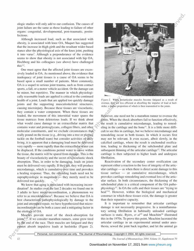
Muscles provide most of the shock-absorption forjoints9,10. If we consider marathon runners, some grow tiredby the end of the race. Their muscles are fatigued and theycannot absorb impulsive loads at heelstrike (Figure 2).
However, one need not be a marathon runner to overuse thejoints. When the shock absorbers fail to function effectively,the result is cumulative microdamage, leading to remod-eling in the cartilage and the bone11. It is a little more diffi-cult to see this in cartilage, but we believe microdamage andremodeling occur in both tissues. In which it occurs firstmay not be relevant. It even occurs, albeit slowly, in thecalcified cartilage, where the result is enchondral ossifica-tion, leading to thickening of the subchondral plate andsubsequent thinning of the articular cartilage12. The articularcartilage is then subjected to higher loads and undergoesfibrillation.
Reactivation of the secondary center ossification canrepresent either a reaction to the loss of integrity of the artic-ular cartilage — as when there is direct acute damage to thistissue surface — or cumulative microdamage, whichprovokes cartilage remodeling and eventual loss of the artic-ular surface. In both circumstances, the thickening of thesubchondral plate is a critical component of the OA patho-physiology13. In OA the cells and their tissues are “trying toheal”14. However, within the biological and mechanicalconstruct in which these tissues exist, the damage is greaterthan their reparative capacity.
It is important to remember that articular cartilagedamage is not necessarily progressive. In a noninflamma-tory setting fibrillation in habitually unloaded cartilagesurfaces is static. Byers, et al15 and Meachim16 illustratedthis in the 1970s. To prove this point, Meachim lacerated thearticular cartilage in an experimental animal under anes-thesia, sewed the joint back together, and let the animal go
The Journal of Rheumatology 2004, Volume 31, Supplement 7012
Figure 2. When periarticular muscles become fatigued as a result ofoveruse, they are less efficient at absorbing the impulse of load at heel-strike, a higher proportion of which is then transmitted to the joints.
Personal, non-commercial use only. The Journal of Rheumatology Copyright © 2004. All rights reserved.
www.jrheum.orgDownloaded on November 17, 2021 from

Pers
onal
, non
-com
mer
cial
use
onl
y. T
he J
ourn
al o
f Rhe
umat
olog
y. C
opyr
ight
© 2
004.
All
right
s re
serv
ed.
about its business. When he reexamined the joint some timelater, there was no evidence of progression of the cartilagedamage17. Vertical fibrillations and/or softening of articularcartilage (chondromalacia) can be nonprogressive changes(Figure 3). Arthroscopists know that this is the case inhumans as well as in experimental animals.
Given that the chondrocytes are multiplying and activelysynthesizing new matrix molecules at an increased rate inOA and that this disease represents joint failure at the organlevel, it is important to recognize that in order to get a jointto heal we need only to surgically change the mechanicalenvironment. Why isn’t this done more often? Because it is
Figure 3. As shown by Meachim17, laceration of the articular cartilage with a scalpel does not lead to progres-sive cartilage damage as long as the laceration does not penetrate into the subchondral bone (upper panel). Asimilar phenomenon is seen in humans with chondromalacia patellae, which is also nonprogressive and rarelyleads to progressive damage (lower panel).
Radin: Who gets OA 13
Personal, non-commercial use only. The Journal of Rheumatology Copyright © 2004. All rights reserved.
www.jrheum.orgDownloaded on November 17, 2021 from
Pers
onal
, non
-com
mer
cial
use
onl
y. T
he J
ourn
al o
f Rhe
umat
olog
y. C
opyr
ight
© 2
004.
All
right
s re
serv
ed.
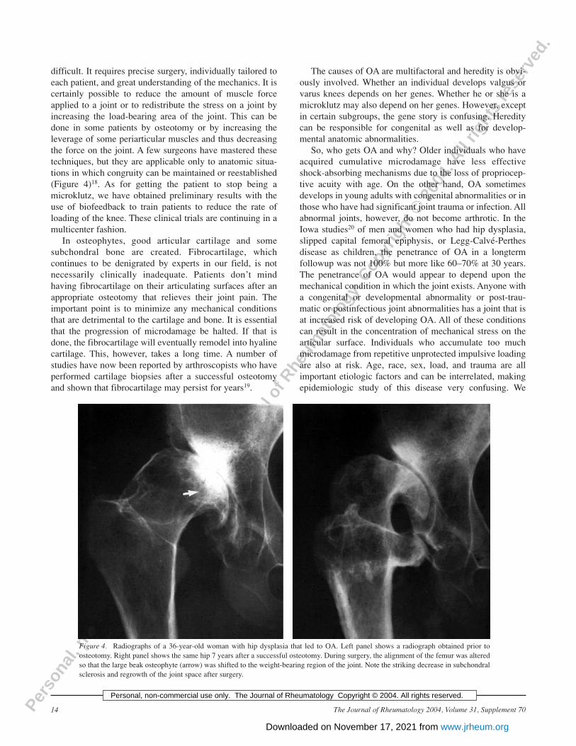
difficult. It requires precise surgery, individually tailored toeach patient, and great understanding of the mechanics. It iscertainly possible to reduce the amount of muscle forceapplied to a joint or to redistribute the stress on a joint byincreasing the load-bearing area of the joint. This can bedone in some patients by osteotomy or by increasing theleverage of some periarticular muscles and thus decreasingthe force on the joint. A few surgeons have mastered thesetechniques, but they are applicable only to anatomic situa-tions in which congruity can be maintained or reestablished(Figure 4)18. As for getting the patient to stop being amicroklutz, we have obtained preliminary results with theuse of biofeedback to train patients to reduce the rate ofloading of the knee. These clinical trials are continuing in amulticenter fashion.
In osteophytes, good articular cartilage and somesubchondral bone are created. Fibrocartilage, whichcontinues to be denigrated by experts in our field, is notnecessarily clinically inadequate. Patients don’t mindhaving fibrocartilage on their articulating surfaces after anappropriate osteotomy that relieves their joint pain. Theimportant point is to minimize any mechanical conditionsthat are detrimental to the cartilage and bone. It is essentialthat the progression of microdamage be halted. If that isdone, the fibrocartilage will eventually remodel into hyalinecartilage. This, however, takes a long time. A number ofstudies have now been reported by arthroscopists who haveperformed cartilage biopsies after a successful osteotomyand shown that fibrocartilage may persist for years19.
The causes of OA are multifactoral and heredity is obvi-ously involved. Whether an individual develops valgus orvarus knees depends on her genes. Whether he or she is amicroklutz may also depend on her genes. However, exceptin certain subgroups, the gene story is confusing. Hereditycan be responsible for congenital as well as for develop-mental anatomic abnormalities.
So, who gets OA and why? Older individuals who haveacquired cumulative microdamage have less effectiveshock-absorbing mechanisms due to the loss of propriocep-tive acuity with age. On the other hand, OA sometimesdevelops in young adults with congenital abnormalities or inthose who have had significant joint trauma or infection. Allabnormal joints, however, do not become arthrotic. In theIowa studies20 of men and women who had hip dysplasia,slipped capital femoral epiphysis, or Legg-Calvé-Perthesdisease as children, the penetrance of OA in a longtermfollowup was not 100% but more like 60–70% at 30 years.The penetrance of OA would appear to depend upon themechanical condition in which the joint exists. Anyone witha congenital or developmental abnormality or post-trau-matic or postinfectious joint abnormalities has a joint that isat increased risk of developing OA. All of these conditionscan result in the concentration of mechanical stress on thearticular surface. Individuals who accumulate too muchmicrodamage from repetitive unprotected impulsive loadingare also at risk. Age, race, sex, load, and trauma are allimportant etiologic factors and can be interrelated, makingepidemiologic study of this disease very confusing. We
The Journal of Rheumatology 2004, Volume 31, Supplement 7014
Figure 4. Radiographs of a 36-year-old woman with hip dysplasia that led to OA. Left panel shows a radiograph obtained prior toosteotomy. Right panel shows the same hip 7 years after a successful osteotomy. During surgery, the alignment of the femur was alteredso that the large beak osteophyte (arrow) was shifted to the weight-bearing region of the joint. Note the striking decrease in subchondralsclerosis and regrowth of the joint space after surgery.
Personal, non-commercial use only. The Journal of Rheumatology Copyright © 2004. All rights reserved.
www.jrheum.orgDownloaded on November 17, 2021 from
Pers
onal
, non
-com
mer
cial
use
onl
y. T
he J
ourn
al o
f Rhe
umat
olog
y. C
opyr
ight
© 2
004.
All
right
s re
serv
ed.
suggest that our understanding of OA can be improved inthe future by looking closely at individual subgroups.
In summary, if we are going to treat OA nonoperatively,joint loading, anatomy, and alignment count. Total jointarthroplasty is a procedure that represents the abject failureof medical management but, fortunately, is available and hasmade many patients more comfortable. Joints can heal if wecan determine how to return habitually loaded contact areasto conditions of normal stresses and rates of loading. Jointmotion is important because it induces the metaplasia ofcells to produce the kinds of tissue we want in the joint21. Bycontrast, without movement, cartilage atrophies22.
Figure 4 shows a successful hip osteotomy 7 years aftersurgery. During the procedure the medial teardrop osteo-phyte was shifted into the weight-bearing area, increasing itssurface area. Even with a larger weight-bearing area, the hipabductor muscles had to be lengthened to assure an adequatedecrease in intraarticular stress23. As a result, this patient’spain has subsided, the subchondral sclerosis that was presentinitially has receded, and the joint space has reappeared. Themajor point: after a well done osteotomy the patient’s jointpain — which isn’t apparent radiographically but arisesfrom synovial inflammation or from the subchondral bone— can disappear.
When we operate on late stage OA, there may be noevidence of inflammation, but only a very thick capsule andsynovium. Trabecular microfracture, although it does notcause stiffening of the bone, may, if sufficiently widespread,be a significant factor because it obliterates the intertrabec-ular channels, thereby increasing the intraosseous pressure.As with any tissue damage, this is a trigger for remodelingand reactivation of the secondary center of ossification. Thepathophysiologic importance of trabecular microfracture inreactivation of the secondary center of ossification andsubchondral thickening in OA cannot be overemphasized.
Osteotomy can achieve a very satisfactory result incertain joints, but it is important to know in which joint thiswill be the case. As shown in Figure 4, it is very importantto realign the joint, to bring the teardrop exactly into whatwe can predict will be the load-bearing area. Osteotomy isexacting surgery, performed in the operating room withprotractors. For good results, the surgeon must come within1° to 2° of his objective. The operation must be planned andindividualized for the pathoanatomy of each patient.Because of the variations in pathoanatomy, not every patientis a suitable candidate for osteotomy. This is why total jointreplacement is so popular — it is a much easier procedurethan osteotomy and, unlike the latter, is applicable to almostall patients with OA.
REFERENCES1. Felson DT. Obesity and vocational and avocational overload of the
joint as risk factors for osteoarthritis. J Rheumatol 2004;31 Suppl70:2-5.
2. Hochberg MC. Do risk factors for incident hip osteoarthritis (OA)differ from those for progression of hip OA? J Rheumatol 2004;31Suppl 70:6-9.
3. Burr DB. The importance of subchondral bone in the progression ofosteoarthritis. J Rheumatol 2004;31 Suppl 70:77-80.
4. Palmoski M, Brandt K. In vivo effect of aspirin on canineosteoarthritic cartilage. Arthritis Rheum 1983;26:994-1001.
5. Smythe HA. The mechanical pathogenesis of generalizedosteoarthritis. J Rheumatol 1983;10 Suppl 9:11-12.
6. Ala-Kokko L, Baldwin CT, Moskowitz RW, Prockop DJ. Singlebase mutation in the type II procollagen gene (COL2A1) as a causeof primary osteoarthritis associated with a mild chondrodysplasia.Proc Natl Acad Sci USA 1990;87:6565-8.
7. Maquet PGP. Biomechanics of the knee and the surgical treatmentof osteoarthritis with application to the pathogenesis. New York:Springer-Verlag; 1976.
8. Levin HS, Benton AL. Age effects in proprioceptive feedbackperformance. Gerontol Clin Basel 1973;15:161-9.
9. Radin EL, Yang KH, O’Connor JJ, Riegger C, Kish VL.Relationship between lower limb dynamics and knee joint pain. J Orthop Res 1991;9:398-405.
10. Radin EL, Whittle MW, Yang KH, et al. The heelstrike transient, itsrelationship with the angular velocity of the shank, and the effectsof quadriceps paralysis. In: Lantz SA, King AI, editors. Advancesin bioengineering. New York: American Society of MechanicalEngineering; 1986:121-3.
11. Radin EL, Schaffler MB, Gibson G, Tashman S. Osteoarthrosis asthe result of repetitive trauma. Osteoarthritic disorders. Am AcadOrthop Surg 1995;13:197-203.
12. Burr DB, Schaffler MB. The involvement of subchondral mineralized tissues in osteoarthrosis. Quantitative microscopicevidence. Micro Res Tech 1997;37:343-57.
13. Radin EL, Burr DB, Fyhrie D, Brown TD, Boyd RD.Characteristics of joint loading as it applies to osteoarthrosis. In:Mow VC, Woo SL-Y, Ratcliffe T, editors. Symposium onBiomechanics of Diarthrodial Joints, Vol. I. New York: Springer-Verlag; 1990:437-51.
14. Radin EL, Burr DB. Hypothesis: Joints can heal. Semin ArthritisRheum 1984;13:293-302.
15. Byers PD, Contempomi CA, Farkas TA. Post-mortem study of thehip joint. III. Correlation between observations. Ann Rheum Dis1976;35:122-6.
16. Meachim G. Articular cartilage lesions in the Liverpool population.Ann Rheum Dis 1975;34 Suppl:122-4.
17. Meachim G. The effect of scarification on articular cartilage in therabbit. J Bone Joint Surg Br 1963;45:150-61.
18. Radin EL. Osteoarthrosis. In: Wright V, Radin EL, editors.Mechanics of human joints. Physiology, pathophysiology, and treatment. New York: Marcel Dekker; 1993:341-54.
19. Fujisawa Y, Masuhara K, Shiomi S. The effect of high tibialosteotomy on osteoarthritis of the knee. An arthroscopic study of 54knee joints. Orthop Clin North Am 1979;3:585-608.
20. Weinstein SL. Long-term follow-up of pediatric orthopaedic conditions. Natural history and outcomes of treatment. J Bone JointSurg Am 2000;82:980-90.
21. Convery FR, Akeson WH, Keown GH. The repair of large osteochondral defects. An experimental study in horses. ClinOrthop 1972;82:253-62.
22. Palmoski M, Perricone E, Brandt KD. Development and reversal ofa proteoglycan aggregation defect in normal canine knee cartilageafter immobilization. Arthritis Rheum 1979;22:508-17.
23. Pauwels F. Biomechanics of the normal and diseased hip.Theoretical foundation. Techniques and results of treatment. Anatlas. New York: Springer-Verlag; 1976.
Radin: Who gets OA 15
Personal, non-commercial use only. The Journal of Rheumatology Copyright © 2004. All rights reserved.
www.jrheum.orgDownloaded on November 17, 2021 from