the lasker/irrf initiative for innovation in vision …€“ i – restoring vision to the blind...
TRANSCRIPT

Restoring Vision to the Blind
The Lasker/IRRF Initiative
for Innovation in Vision Science

About the Albert and Mary Lasker Foundation: Founded in 1942, the Albert and Mary Lasker Foundation envisions a healthier world through sustained support for basic and clinical medical research. The Foundation works to accomplish its mission through education and advocacy and, most notably, through a prestigious annual awards program, now in its 70th year. Lasker Award winners are selected by their peers, who, like themselves, include the world’s most accomplished and well-respected medical research scientists, and thus the award represents a special honor. The Foundation’s education and advocacy missions focus on engaging the public and policymakers on the importance of robust medical research programs and the funding to make them possible. The Lasker Foundation is also dedicated to supporting and inspiring the next generation of research scientists. For more information about the Lasker Foundation and its programs, visit http://www.laskerfoundation.org.
About the International Retinal Research Foundation: The International Retinal Research Foundation (IRRF) upholds a commitment to accelerate and sustain targeted research efforts into the diseases of the human eye, especially those affecting the retina and macula, to discover the causes, preventions, and cures of retinal and macular degenerative diseases and diabetic retinopathy. The IRRF will accomplish its mission by providing financial support of vision research directly, as well as through training fellowships, public awareness programs, and the promotion of the exchange of research findings. For more information about the IRRF, please visit www.irrfonline.org.
�
�
�
�
�
�
�
�
�
�
�
�� ��� �������� ���� ���������������������������� �� ���� ������� �
�������������� ������ � ��������������� �������� � !����������������
� ������� �� ��������� "��� ����������%�������������������� ���� ��� � ������
����� �� ��������� � ������������ �� ���������� �� �� �� �����������
����������� �� !� � ���������� ������!���� �������������������������
� ������ � ���"�������� ������������������ ����������!��� � �������
���"��� ����������"���"�
�
��� �� ���� ��������� ������ � ��������������#����$����������
������� �������� � �� �������������� � ��� � ���� �������������� �
��� � ������� ������ !� �� ���� ����� ��� �������� �� �����������!�
��������� ���� ���� �!��� � ������!������� ������ ��������������
� � � ���� ���� � ������� ����� ������� "��� ���������������������
������������ �������������������������������������� � ������� ��� !���� ���
������������������� ���������!��������� � ����������!������ �
��������������� � ����� ����� � ������������"�������� ����������������
�� �����!��� � ����������"��������� "���"�
�
�
�
�
�
�
�
�
�
�
�
�
�� ��� �������� ���� ���������������������������� �� ���� ������� �
�������������� ������ � ��������������� �������� � !����������������
� ������� �� ��������� "��� ����������%�������������������� ���� ��� � ������
����� �� ��������� � ������������ �� ���������� �� �� �� �����������
����������� �� !� � ���������� ������!���� �������������������������
� ������ � ���"�������� ������������������ ����������!��� � �������
���"��� ����������"���"�
�
��� �� ���� ��������� ������ � ��������������#����$����������
Cover: Optic vesicle-like structures derived from human induced pluripotent stem cells can self assemble into rudimentary retinal laminae, and following 50 days of differentiation, form an outer neuroblastic layer of proliferating progenitor cells (identified by immunolabeling with the retinal progenitor marker VSX2, in red, and the mitotic cell marker Ki67, in green) and an inner layer of putative retinal ganglion cells (immunolabeled with HuCD, a marker for post-mitotic neurons, in purple). Image produced by the Gamm Laboratory, University of Wisconsin School of Medicine and Public Health. Reprinted from: Wright LS, Phillips MJ, Pinilla I, Hei D, Gamm D. Induced pluripotent stem cells as custom therapeutics for retinal repair: Progress and rationale. Exp Eye Res. 123:161-72, 2014.

– i –
Restoring Vision to the Blind The Lasker/IRRF Initiative for Innovation in Vision Science
December 2014Table of Contents
PageProject Background and Acknowledgements ..................................................................................i
Introduction ..................................................................................................................................1 Chapter 1The New Age of Implanted Visual Prostheses .................................................................................5
Chapter 2Optogenetics ................................................................................................................................21
Chapter 3Gene Therapy for Vision Loss: The Road Ahead ..........................................................................35 Chapter 4 Stem Cells and Transplantation ....................................................................................................49
Chapter 5 Endogenous Regeneration ............................................................................................................61 Chapter 6 Neuroprotection ...........................................................................................................................71 Chapter 7 Advancements in Vision Aids for the Visually Impaired ...............................................................81
Chapter 8 Evaluating Visual Function, Endpoints ........................................................................................91
Concluding Remarks .................................................................................................................101 Appendix 1 Joint Advisory Board and Collaborating Executives ....................................................................102
Appendix 2 Steering Committee ...................................................................................................................103
Appendix 3 Participants ................................................................................................................................105
Appendix 4 Abbreviations .............................................................................................................................114
Index .........................................................................................................................................115

– ii –
Photographs: Larry A. Donoso and Bryan William Jones

– iii –
Restoring Vision to the BlindThe Lasker/IRRF Initiative for Innovation in Vision Science
Project Background and Acknowledgements
The Albert and Mary Lasker Foundation (Lasker) and the International Retinal Research Foundation (IRRF) entered into a 10-year collaboration that began July 15, 2008 with the goal of identifying knowledge gaps in vision research and developing innovative strategies to advance retinal research and accelerate discovery of sight-saving treatments and prevention of retinal degenerative diseases. Restoring Vision to the Blind is the third report by the Lasker/IRRF Initiative for Innovation in Vision Science. The Initiative’s first report, Astrocytes and Glaucomatous Neurodegeneration, was published in November 2010. The Initiative then examined diabetic retinopathy, one of the leading causes of visual impairment and blindness in the world, and issued its report, Diabetic Retinopathy: Where We Are and A Path to Progress, in November 2012.
John E. Dowling, Gordon and Llura Gund Professor of Neurosciences at Harvard University, chairs the Initiative, with the guidance of a Lasker/IRRF Joint Advisory Board and collaborating executives (Appendix 1), and each study is undertaken using a Steering Committee (SC) of bench and clinical scientists with expertise in interdisciplinary fields and the combined skills, knowledge, and experience necessary to identify key issues and hurdles confronting vision scientists. The SC identifies leaders in diverse fields to participate in workshops during which key impediments to research progress are identified. These workshops are followed by a plenary session at which small groups focus on specific targeted areas and develop a framework of innovative multidisciplinary approaches to accelerate discovery and its translation to clinical application. The results of these sessions are published by the Initiative for wide distribution within the research community and to potential funders and other organizations interested in advancing research in retinal degenerative diseases.
In the fall of 2012, the Initiative undertook a bold new investigation of potential breakthroughs to restore vision lost as a result of retinal degenerative disease. The Initiative chose to explore the major question of which approaches are most promising and most likely to benefit the greatest number of blind individuals. Given the breadth and complexities of this topic, the Initiative invited a small group of scientists, who later formed the SC (Appendix 2), to meet at the Institut de la Vision in Paris. There, they looked at all of the major research efforts underway and clarified the objectives of this effort: how best to exploit the newest insights and cutting edge technologies to re-establish light sensitivity, and restore visual perception damaged or destroyed by retinal degeneration. The SC also identified the key scientists who participated in this Initiative (Appendix 3). Two workshops were subsequently held in Woods Hole, Massachusetts in the summer of 2013, followed by a plenary session in March 2014.
We were honored to be joined at the opening of the plenary meeting by Sanford D. Greenberg and his wife Susan. Following a colorful introduction by Peter McDonnell, Director of the Johns Hopkins’ Wilmer Eye Institute, Mr. Greenberg gave a compelling keynote address to the Initiative participants, sharing his inspirational journey from the despondent days following his loss of sight while an undergraduate at Columbia to his uncompromising achievements in academia, government, business, and philanthropy. With that same determination, he is now focused on galvanizing global research

– iv –
efforts toward the goal of ending blindness forever. Mr. Greenberg’s presentation can be downloaded at https://www.dropbox.com/s/qyiie2wenje1w9q/SandyG.mp4.
The Initiative thanks the Boards of Directors of the Albert and Mary Lasker Foundation and the International Retinal Research Foundation for their support; the Initiative’s Joint Advisory Board and SC, for their counsel; the Discussion Leaders who guided the development of the key issues discussed in this report and the scribes who recorded the discussions and drafted chapter texts; and all participants, for their energy, expertise, and lively discourse. Special thanks go to Karen M. Wright, Project Administrator for her diligent and essential administrative direction; to Meredith Graves, Project Manager, for her logistical support; and to Sandra Blackwood, Executive Director of the IRRF, and Claire Pomeroy, President of Lasker, for their constancy and contributions to this endeavor.
The Initiative is most appreciative to José-Alain Sahel, and his staff at the Institut de la Vision, Paris, for so memorably hosting the October 2012 planning session. The Initiative gratefully acknowledges both the Howard Hughes Medical Institute for its very generous in-kind contribution by making available the facilities at its Janelia Farm Research Campus in Ashburn, Virginia, for the Initiative’s plenary session, and the staff of the National Academy of Sciences’ J. Eric Jonsson Center in Woods Hole, Massachusetts, for their gracious hospitality during the two summer workshops.
The Initiative gratefully acknowledges the Association for Research in Vision and Ophthalmology (ARVO) for publishing this report as a special edition of its online journal Translational Vision Science and Technology (TVST), making it available to all members of ARVO and, by its listing in PubMed, to any other interested parties. The TVST special edition may be viewed at http://www.tvstjournal.org/toc/tvst/3/7.
For further information about the Initiative, please contact: Karen M. Wright, Project Administrator at [email protected]. For additional copies of this report, please make your request to Meredith Graves, Project Manager at [email protected].

– 1 –
Introduction
John E. Dowling The notion that restoring vision to the blind is possible has long been thought to be fanciful. However, beginning as far back as the 1960s vision scientists began to investigate the possibility of restoring vision to the blind by activating neurons in the visual pathways beyond the eye, namely in the visual cortex. These early experiments showed that it is possible to elicit visual sensations in humans by electrically stimulating neurons in the visual cortex. Most blindness is caused by defects in the eye. It can be caused, first of all, by damage to the optical pathways that are required for the focusing of a sharp image on the light-sensitive retinal photoreceptors that line the back of the eye. Today, it is generally possible to cure these optical impediments. Cataract surgery to remove an opaque lens and replace it with an artificial lens is carried out routinely in many parts of the world, and corneal transplants with natural or artificial corneas are generally successful. It should be noted, however, that in those parts of the world where such procedures are not available, blindness remains common because of such defects. It is estimated that there may be as many as 20 million blind people in the world because of cataracts.
The major cause of untreatable blindness throughout much of the world today is retinal degenerative disease, most often because of a loss of photoreceptor cells but also, especially in glaucoma, a loss of third order neurons of the retina, the retinal ganglion cells whose axons form the optic nerve and carry the visual signal from the eye to the higher visual centers such as the cortex. Because most retinal degenerations cause blindness by destroying the photoreceptors, much emphasis in the quest to cure blindness is to restore photoreceptive function in the blind eyes, or to substitute for the loss of photoreceptor function.
Most success so far has come from two approaches. First, retinal prostheses have been developed that electrically stimulate the second or third order retinal neurons, namely the retinal bipolar or ganglion cells. Indeed, two types of prostheses have been successfully implanted in blind human patients and have restored light sensitivity and low-acuity vision to the patients. The second approach has been successful for patients with specific gene defects that severely compromise photoreceptor function, and the treatment consists of injecting a viral construct containing the normal gene into the eye, thus replacing the defective gene. Again, substantial improvement in vision, especially light sensitivity, has been demonstrated in these patients. A newer approach, not yet tested in humans but which soon will be, is in essence a combination of the above two approaches, namely imparting light sensitivity to retinal neurons via genetic means called optogenetics. Genes that code for light-sensitive molecules linked to an ion channel or pump are introduced into various retinal cells, most often bipolar or ganglion cells. In animals treated this way, the treated cells are stimulated by light, causing the opening of ion channels or activating ion pumps, both of which permit ions to flow across the cell outer membranes, thus electrically activating them. This technique was recently applied in blind animals with some remaining cone cells, but which had lost light sensitivity because the outer segments of the cells (which contain the light-sensitive molecules—the visual pigments) had degenerated. Once light sensitivity was restored to these cones, downstream retinal pathways could be activated and the animals showed visual behavioral responses.

– 2 –
Another approach to replace damaged or destroyed photoreceptor cells is to transplant healthy photoreceptor cells into the eyes of blind animals. This strategy has had limited success so far—the number of transplanted cells that survive and integrate into the retinal circuitry is quite limited—but some restoration of electrical activity recorded from the eyes occurs and the animals do show some behavioral responses to light. Stem cells, which in theory can differentiate into any cell type, have also been introduced into blind eyes, including some human eyes, again with very limited and largely undocumented success. Investigators are now inducing stem cells maintained in culture to differentiate into photoreceptor cells and then are injecting such cells into eyes whose photoreceptor cells have degenerated; this approach appears promising and may be more successful.
In addition to direct deleterious effects of a disease process or gene defect in the photoreceptor cells themselves, such defects can also occur in the associated retinal pigment epithelial cells, and this can cause photoreceptor death. The photoreceptor cells and overlying retinal pigment epithelium are intimately connected, and they depend upon each other to function. The isomerization of vitamin A, to generate the 11-cis retinoid molecule needed to regenerate the visual pigment molecules after light exposure, occurs mainly in the retinal pigment epithelium, and the phagocytosis and digestion of spent outer segment material as well as recycling lipids occurs in the retinal pigment epithelial cells. Compromise of any of these retinal pigment epithelial cell functions results in photoreceptor cell degeneration in both animals and humans. Thus, gene therapy to correct retinal pigment epithelial cell defects or transplantation of retinal pigment epithelial cells into diseased retinas has been accomplished with promising results. Indeed, the first gene therapy treatment in humans, described above, was for a gene defect in the retinal pigment epithelial cells. Retinal pigment epithelial cells grow readily in culture and are readily transplanted. Unlike photoreceptor cells, they do not need to integrate into the retinal circuitry but interact only with the photoreceptor cells, which they do readily.
It has long been known that nonmammalian species such as amphibians and fish can regenerate retinal cells endogenously but mammals, including humans, cannot. Why can these cold-blooded vertebrates do this but we can’t? This is an intriguing question that is now receiving substantial attention. If we could regenerate our retinal cells, presumably we could cure not only blindness caused by photoreceptor degeneration, but blindness caused by degeneration of any retinal cell including the ganglion cells. In fish, for example, new neurons are formed throughout life, and the axons of the newly formed ganglion cells extend into the rest of the brain and make appropriate connections. In mammals, not only do ganglion cells not regenerate, but their axons do not regrow in large numbers after the optic nerve is damaged or cut.
From what cells does the regeneration in the nonmammalian species occur? This may differ among species, but certainly retinal pigment epithelial cells and Müller glial cells appear to be involved. In fish, the formation of new retinal cells throughout life comes from a region in the retinal periphery called the marginal zone, whose cells may derive from the retinal pigment epithelium, whereas when the fish retina is damaged, new retinal neurons derive from Müller cells that dedifferentiate and appear to behave like stem cells. That is, after dedifferentiation these cells first proliferate and then generate progenitors for repairing the retina.
The objective of the present initiative was to evaluate the various approaches presently underway to cure blindness caused by retinal degenerative disease, to identify the most promising and feasible

– 3 –
approaches and to indicate the major problems and issues that must be overcome to make an approach useful and effective in restoring vision to the blind. In addition to discussing the approaches outlined above, we also considered other topics that may impact the various approaches being undertaken. For example, the retinal prostheses that have been developed so far provide only low-level vision. Many devices have been developed over the years to help those who are visually impaired and have low-level vision. Can some of these devices be of use to those who have low vision restored as a result of an implanted visual prosthesis? Another example would be for a device to allow a retina made light-sensitive via optogenetics to adapt over a range of intensities and to have greater sensitivity to light, something optogenetically-induced vision is unlikely able to accomplish by itself.
Another area we considered is that of neuroprotection, neuroactive substances that protect neurons and often slow down degeneration in a diseased retina. Can such molecules be used in conjunction with other restorative approaches to enhance their effectiveness? So, for example, we know that after photoreceptors degenerate in a retina, the retina undergoes substantial remodeling, and this could limit success when restoring photoreceptor function, especially if the visual loss is long standing.
A final topic discussed was that of end points—what is the best way to measure the return of visual function in previously blind patients? The gold standard to evaluate vision ordinarily is visual acuity—how many lines on an eye chart can a person read. But there is much more to vision than just acuity, although acuity is certainly critical if we are to restore reading, driving, face recognition, and so forth to blind individuals.
In the chapters that follow, the topics introduced above are described in detail with indications as to what the major questions are that need to be addressed and how to go about answering these questions where possible.
The chapters of this report were written based on the discussions held during the targeted sessions held during the plenary meeting. All members of a session had the opportunity to comment upon and contribute to each chapter, and everyone who participated in the workshops and plenary session had the opportunity to comment upon the final report. We believe this is a consensus document, and we thank all who were involved and contributed so generously with their time. We hope this report is useful and hastens the day when we can restore vision to the blind.

– 4 –

– 5 –
Chapter 1The New Age of Implanted Visual Prostheses
Discussion Leaders: Eberhart Zrenner and Bradley Greger
Scribe: Daniel Rathbun
Session Participants: David Birch, John Dowling, Erika Ellis, Fred Fitzke, Donald Hood, Alan Laties, Daniel Palanker, John Pezaris, Joseph Rizzo, Gary Rubin, Ronald Schuchard, Dirk Trauner, James Weiland, and Frank Werblin
Introduction
The timing of the Lasker/IRRF (International Retinal Research Foundation) Initiative on Restoring Vision to the Blind in March of 2014 was particularly opportune given that the first commercial sales of implanted visual prostheses (the Argus II) occurred in 2011 (Rizzo et al., 2014), and a second commercial device (the Alpha-IMS [Institut für Maschinelle Sprachverarbeitung]) entered the market in 2013 (Zrenner, 2013). We are therefore at a perfect point to look back on the successes of visual prostheses as well as to look forward to what the future may hold. The core question for the Implanted Visual Prostheses session, which was fine-tuned by the session members and provided a framework for our discussions was: How to provide useful visual information to patients blind from lesions in the afferent visual pathway by means of safe and efficient electronic implants?
Accomplishments to Date
Several strategies have been employed to electrically activate the neurons that remain after loss of vision. These can loosely be arranged according to which neuron along the visual pathway is being targeted (Fig. 1.1). For extensive reviews of these different strategies, the reader is directed to recent review articles: Chuang, Margo, & Greenberg, 2014; Guenther, Lovell, & Suaning, 2012; Luo and da Cruz, 2014; Matthaei et al., 2011; Maynard, 2001; Weiland, Cho, and Humayun, 2011; Zrenner, 2013. For a further history of how the principal research groups and concepts emerged, the reader is directed to Dowling, 2005.
Given the clinical successes and the diversity of strategies employed by retinal implants, a brief overview of their merits relative to each other is appropriate. The most common retinal implants can be classified as epiretinal, subretinal, or suprachoroidal according to the placement of their electrode arrays (Fig. 1.2). Typically, epiretinal and suprachoroidal implants have employed extraocular light sensors, whereas subretinal implants couple light sensors with the stimulating electrodes at the position of lost photoreceptors to ensure that the sensors exploit natural eye movements. In subretinal and suprachoroidal implants, bipolar cells are targeted for stimulation in hopes that by activating the retinal network as early as possible residual neural processing of bipolar and amacrine cells can be exploited. Epiretinal implants employ a simpler surgical procedure than subretinal implants to target the ganglion cells for more direct control of the output signals of the retina. Even simpler and less

– 6 –
invasive is suprachoroidal placement in which a scleral tunnel through the back of the eye is used to insert the array. To date, only preliminary clinical experiments with suprachoroidal implants have been conducted in which a small number of widely spaced electrodes were stimulated directly through a laboratory computer without the accompanying camera system.
Recent Advances in Retinal Stimulation: Clinical Applications
Of the many devices investigated, retina-based devices have shown the greatest success. These devices are limited, however, to eye diseases that destroy the photoreceptors but leave, at the very least, the ganglion cells intact for direct stimulation (Kellner, 2000). Of those diseases, retinitis pigmentosa (RP) has been the most attractive candidate because some affected individuals reach near-complete blindness at a relatively early age, and the inner (neural) retina is relatively spared. In comparison, age-related macular degeneration (AMD) has a late onset and does not cause total blindness for many years, typically leaving the retinal periphery functional.
At present, there are seven major ongoing retinal prosthesis projects that have either implanted test subjects or have concrete plans to do so in the near future. The Argus II developed by Second Sight (Second Sight Medical Products, Lausanne, Switzerland) has been implanted during clinical trials in over 30 patients in both Europe and the United States (Humayun et al., 2012) where it has also been approved for commercial sale (in 2011 and 2013, respectively [Rizzo et al., 2014]). Currently, approximately 45 commercial devices have been implanted. In comparison, the Alpha-IMS system of Retina Implant AG has been implanted during clinical trials in over 40 patients (Zrenner, 2013) and in 2013 received CE (Conformité Européenne) approval for commercial sale in the European Union with the first sales expected in 2014 (W. Wrobel, personal communication,). In addition to these two commercial devices, the IRIS device developed by Intelligent Medical Implants (IMI) was implanted in 20 patients in 2003 and 2004 (Hornig et al., 2007). This IMI (Innovative Medicines Initiative) device has been acquired by Pixium, which is conducting renewed clinical trials. Furthermore, devices developed by the Bionic Vision Australia consortium (Saunders et al., 2014) the Boston Retina Implant Project (Rizzo, 2011), Nidek Co., Ltd. (Nidek Co., Aichi, Japan) (Fujikado et al., 2011), and the Stanford-based photovoltaic retinal prosthesis (PRIMA) (Mandel et al., 2013; Mathieson et al., 2012) have announced plans to enter clinical trials in the next few years. Functional results from some of these retinal implants will be discussed below.
Recent Advances in Optic Nerve and Thalamic Stimulation
Optic nerve stimulation seeks to create action potentials in the axons of the ganglion cells. Whereas epiretinal stimulation targets these ganglion cells at or very near their cell bodies in order to preserve retinotopy (Fried, Lasker, Desai, Eddington, & Rizzo, 2009; Jensen, Rizzo, Ziv, Grumet, & Wyatt, 2003; Sekirnjak et al., 2008), optic nerve stimulation targets these axons at the bottleneck of the visual system where about one million axons from the entire retina are tightly packed into the 3.5-mm wide optic nerve. Regrettably, the greatest drawback to optic nerve stimulation, especially for surface stimulation, is that resultant light perceptions (phosphenes) are irregular in both shape and visual location, requiring sophisticated image processing algorithms to create a meaningful visual scene. To date, two patients have been implanted with optic nerve cuff devices in Belgium, and ongoing work is

– 7 –
targeted at optimizing optic nerve electrode arrays and the related surgical techniques and stimulation paradigms (Brelen, Vince, Gerard, Veraart, & Delbeke, 2010; Lu et al., 2013; Sakaguchi et al., 2009).
A relatively new approach seeks to stimulate the targets of retinal ganglion cell axons in the lateral geniculate nucleus (LGN) of the thalamus (Pezaris & Reid, 2007).One potential advantage of this approach compared with retinal stimulation is that while the whole visual space is topographically represented in the compact LGN, the crucial foveal representation comprises a larger proportion of the neural tissue than the fovea in the retina. A potential advantage of LGN stimulation compared to the cortical approach is that the neural representation for vision in the LGN is less complex, although much work still remains in understanding the encoding of the space-and time-varying neuronal signals that pass through the LGN.
Recent Advances in Cortical Stimulation
The earliest multielectrode prosthesis for vision was on the surface of a patient’s visual cortex in July of 1967, and was an array of flat electrodes, embedded in silicone, and activated wirelessly through transdermal radio transmission (Brindley & Lewin, 1968). This implant demonstrated that electrical
perceptions. Building on this work another group continued to develop cortical stimulation devices for decades afterward(Dobelle, 2000; Dobelle & Mladejovsky, 1974). As semiconductor microfabrication developed, penetrating intracorticalmicrostimulation (ICMS) was investigated by other groups to overcome some of the limitations of cortical surfacestimulation. ICMS should theoretically allow for higher spatial resolution when penetrating implants finally enter clinicaltrials (Bak et al., 1990; Bradley et al., 2005; Davis et al., 2012).
It should be noted that many more devices are under development than could reasonably be presented here, and thedetails of each research project change on a monthly basis. For this reason, interested readers are encouraged to visit thecontinuously updated list at http://www.eye-tuebingen.de/zrenner/retimplantlist/ for the latest information.
How Good Is Restored Vision?
Figure 1.1. Overview of the visual system as related to visual prostheses. In most retinal dystrophies, the first order photoreceptor neurons (rods
and cones) are lost. Thus, second order neurons (bipolar cells) are the earliest viable target, typically for subretinal and suprachoroidal devices.
Epiretinal devices typically target retinal ganglion cells (RGCs), the third order neurons that form the output of the retina. Likewise, optic nerve
devices target these neurons either within the eye at the nerve head or outside of the eye. The fourth order neurons, relay cells of the thalamic lateral
geniculate nucleus (LGN) are targeted with penetrating electrode arrays. Finally, cortical implants target the fifth and higher order neurons found in
the primary visual cortices. (Modified from Krey, H.F., & Brauer, H. (1998). Chibret Augenatlas: Eine Repetition fur Artze mit Zeigetafeln furPatienten. Munich: Chibret Med Serv.)
Article 35
The Lasker/IRRF Initiative for Innovation in Vision Science
Figure 1.1. Overview of the visual system as related to visual prostheses. In most retinal dystrophies, the first order pho-toreceptor neurons (rods and cones) are lost. Thus, second order neurons (bipolar cells) are the earliest viable target, typi-cally for subretinal and suprachoroidal devices. Epiretinal devices typically target retinal ganglion cells (RGCs), the third order neurons that form the output of the retina. Likewise, optic nerve devices target these neurons either within the eye at the nerve head or outside of the eye. The fourth order neurons, relay cells of the thalamic lateral geniculate nucleus (LGN) are targeted with penetrating electrode arrays. Finally, cortical implants target the fifth and higher order neurons found in the primary visual cortices. (Modified from Krey, H.F., & Brauer, H. (1998). Chibret Augenatlas: Eine Repetition für ¨ Artze mit Zeigetafeln ffürr Patienten. Munich: Chibret Med Serv.)
– 8 –
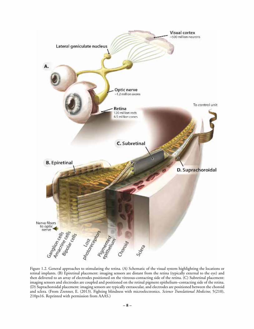
Figure 1.2. General approaches to stimulating the retina. (A) Schematic of the visual system highlighting the locations or retinal implants. (B)Epiretinal placement: imaging sensors are distant from the retina (typically external to the eye) and then delivered to an array of electrodes positionedon the vitreous-contacting side of the retina. (C) Subretinal placement: imaging sensors and electrodes are coupled and positioned on the retinalpigment epithelium–contacting side of the retina. (D) Suprachoroidal placement: imaging sensors are typically extraocular, and electrodes arepositioned between the choroid and sclera. (From Zrenner, E. (2013). Fighting blindness with microelectronics. Science Translational Medicine,5(210), 210ps16. Reprinted with permission from AAAS.)
http://tvstjournal.org/doi/full/10.1167/tvst.3.7.3 TVST j 2014 j Vol. 3 j No. 7 j Article 36
The Lasker/IRRF Initiative for Innovation in Vision Science
Figure 1.2. General approaches to stimulating the retina. (A) Schematic of the visual system highlighting the locations or retinal implants. (B) Epiretinal placement: imaging sensors are distant from the retina (typically external to the eye) and then delivered to an array of electrodes positioned on the vitreous-contacting side of the retina. (C) Subretinal placement: imaging sensors and electrodes are coupled and positioned on the retinal pigment epithelium–contacting side of the retina. (D) Suprachoroidal placement: imaging sensors are typically extraocular, and electrodes are positioned between the choroid and sclera. (From Zrenner, E. (2013). Fighting blindness with microelectronics. Science Translational Medicine, 5(210), 210ps16. Reprinted with permission from AAAS.)

– 9 –
stimulation of the visual system could produce light percepts (phosphenes) with reliable location, shape, and size; however, it was inconveniently bulky, required high currents, occasionally caused pain upon stimulation, and was unable to produce sufficient coverage of the visual space for useful perceptions. Building on this work another group continued to develop cortical stimulation devices for decades afterward (Dobelle, 2000; Dobelle & Mladejovsky, 1974). As semiconductor microfabrication developed, penetrating intracortical microstimulation (ICMS) was investigated by other groups to overcome some of the limitations of cortical surface stimulation. ICMS should theoretically allow for higher spatial resolution when penetrating implants finally enter clinical trials (Bak et al., 1990; Bradley et al., 2005; Davis et al., 2012). It should be noted that many more devices are under development than could reasonably be presented here, and the details of each research project change on a monthly basis. For this reason, interested readers are encouraged to visit the continuously updated list at http://www.eye-tuebingen.de/zrenner/retimplantlist/for the latest information.
How Good Is Restored Vision?
Visual function is notoriously difficult to quantify, especially in low-vision subjects (Geruschat, Bittner, & Dagnelie, 2012). Even the seemingly simple metric of visual acuity can be measured with a broad range of tests that only loosely correlate with each other. In addition to visual acuity, an important feature of visual function is the location and size of the visual field. In some cases, it makes more sense to assess restoration of the visually-guided behaviors restored to a patient by an implant in terms of improved quality of life (see also Chapter 8). Furthermore, given the psychological impact of blindness, psychological benefit should also be considered.
Of the nearly 100 patients implanted with either an Argus II or an Alpha-IMS, the three best visual acuities achieved to date are 20/1262 with an Argus II device using a grating orientation test (Humayun et al., 2012), 20/1000 with a prototype that preceded the Alpha-IMS device measured with Landolt C test (Zrenner et al., 2011), and most recently, 20/546 with an Alpha-IMS implant using the same Landolt C test (Stingl et al., 2013). With the Argus II device, patients receive stimulation from 60 electrodes over an area of approximately 10˚ x 20˚ of visual angle, whereas with the Alpha-IMS device, they receive input from 1500 electrodes over an area of 11˚ x 11˚. Beyond visual acuity, implanted patients have demonstrated object localization, discrimination, and identification; motion detection and discrimination; letter identification; and limited reading where none of these tasks were possible either before implantation or afterward with the implants turned off. Furthermore, patients can perform simple orientation and mobility tasks using the devices. All of these tasks have been measured in the laboratory to enable comparison among test subjects but have also been reported in real-world use by a limited subset of patients.
What Is the Value of an Implant?
Recent clinical trials have established that visual prostheses can provide visual information that is useful in daily life. Looking forward, it can be anticipated that, with continuing development, these devices have a high probability of acceptance by the RP patient population. The risks associated with implanted visual prostheses in terms of serious adverse events (SAE) have been shown to be low (Humayun et al., 2012; Stingl et al., 2013). However, limited real-world experience currently precludes a complete assessment of the cost/benefit tradeoff that can be expected with such devices. In

– 10 –
evaluating this tradeoff, the seemingly contradictory outcome goals of patient independence and social connectedness need to be carefully evaluated. Critically, such evaluation needs to consider the life situation of each individual patient. For example, while one person may value reading highly, a sports enthusiast may prefer faster signals with lower spatial resolution (although both normal reading and sporting activities lie beyond current visual prosthesis capabilities). Given this context-dependence of the utility of restored vision, it is important to identify the most important aspects of visual function. For most visually guided tasks such as reading and navigation, nonprosthetic solutions are well developed (see Chapter 7). Therefore, we propose that the two primary goals of prosthetic vision should be to improve the subject’s independence and social connectedness. Developing meaningful measures for these two should, therefore, be a top priority for psychophysical testing of implanted prostheses in the future. Validated questionnaires for such assessments are discussed further in Chapter 8. Furthermore, in quantifying regained independence capabilities). Given this context-dependence of the utility of restored vision, it is important to identify the most important aspects of visual function. For most visually guided tasks such as reading and navigation, nonprosthetic solutions are well developed (see Chapter 7). Therefore, we propose that the two primary goals of prosthetic vision should be to improve the subject’s independence and social connectedness. Developing meaningful measures for these two should, therefore, be a top priority for psychophysical testing of implanted prostheses in the future. Validated questionnaires for such assessments are discussed further in Chapter 8. Furthermore, in quantifying regained independence and connectedness and integrating them with more traditional measures of visual function, the goal should be to facilitate cost-benefit analyses such as the quality-adjusted life year (QALY) for use by individuals, clinicians, insurers, researchers, governments, and research funding agencies. For general information on how the usefulness of medical interventions is evaluated, see Fanshel & Bush, 1970; Pliskin, Shepard, & Weinstein, 1980; for specific evaluations of implantable visual prostheses, see Vaidya et al., 2014; Wrobel 2010, as an example. The final monetary and societal value of an implant device will be a deciding factor in whether it can prove viable either as a commercial venture or as a humanitarian/societal endeavor.
Managing Expectations
The development of the Argus II and Alpha-IMS implants has been the culmination of decades of research and the investment of enormous capital resources from many governmental, charitable, and commercial entities. However, it is vital that we as a community are careful not to oversell the capabilities of our devices. As a general rule of thumb, these two devices have yielded extraordinary visual restoration in approximately one-quarter of the patients tested. Useful gains in daily function were seen in another one-quarter. The final one-half of patients realized only rudimentary functional gains, like simple light perception or localization of bright versus dark areas, which is although of limited use, appreciated by blind people. Only relatively few had no benefit at all, despite undergoing hours of surgery and weeks of recovery. Of the one-quarter of patients who have extraordinary gains, only a handful have come close to the theoretical limits of visual acuity possible based on the device limitations. Although we hope to see typical results continue to improve as the devices move into mainstream medicine and as manufacturing and surgical techniques improve further, such improvements have yet to be demonstrated.
A Goggle System for Image Preprocessing One realm in which further improvements may be achieved with existing implanted devices is in more sophisticated preprocessing of the images prior to their conversion into electrical stimulation patterns.

– 11 –
Since an external camera is implicit in its design, Second Sight and its collaborators have already made strides in image preprocessing, including face localization software to activate the electrodes corresponding to a face location, implementing zoom to resize the visual scene onto the electrode array, and the direct presentation of Braille letters (Dorn et al., 2013; Guerra et al., 2013; Lauritzen et al., 2012). With the Alpha-IMS, the camera is an integrated part of the subretinally implanted device and cannot easily be modified. However, an attractive option for this device is to incorporate Google Glass, Oculus Rift, or one of the other head-mounted displays currently being developed. This modification also simplifies the process of testing and updating image processing algorithms. Notably, goggles have always been an integral component of the Stanford photovoltaic system, which can use conventional liquid crystal display (LCD) or dot matrix display (DMD) displays, or a novel holographic projection for enhanced brightness (Goetz, Mandel, Manivanh, Palanker, & Cizmar, 2013). Integration of eye tracking into the system enables location-specific image processing, such as correction of the radial spread in the fovea (Asher, Segal, Baccus, Yaroslavsky, & Palanker, 2007). Given the broad applicability of such a goggle system for low-vision aids, optogenetics, and photopharmacology in addition to implantable prostheses, there is a strong case to be made for development of a standardized platform custom-built to meet the needs of the blind community. For additional discussion of this issue, see Chapter 7.
Consensus Statements Regarding Implantable Visual Prostheses: • They can provide useful vision in daily life. • They have a high probability of acceptance by blind RP patients.
• The associated risks are low. • Evaluation of the cost-benefit tradeoff requires further investigation. • Patient expectations should be carefully managed. • Primary outcome measures should focus on improvement of both independence and social connectedness. • Better image preprocessing will improve prosthetic vision.
Short-Term Research Goals
Improve Phosphene Reliability – Epiretinal Approach Irregularity and inconsistency of phosphene perception across different electrodes during direct epiretinal stimulation of ganglion cells continues to limit the effectiveness of this strategy and requires significant training (published and public statements have ranged from several months to up to 3 years) (Cosendai, 2014; Cosendai et al., 2014; Humayun et al., 2012). One possible reason is the large variation in distance between the electrodes and target cells. Another is that it is difficult to stimulate only local ganglion cells without also activating axonal fibers of other cells that pass under the electrodes, producing arcuate

– 12 –
percepts instead of a single dot. Multiple studies in animal models have shown that axonal stimulation can be avoided only by using longer stimuli, which activate inner retinal neurons rather than ganglion cells.
Increase Stimulation Frequency – Subretinal Approach
Although subretinal stimulation has yielded the best-restored visual acuity via visual prosthetics to this date, creating perceptions at a high stimulus frequency remains problematic. This is likely due to adaptation of the neural network to high frequency pulse trains. Experiments are currently underway in multiple labs to better understand the complex responses generated by subretinal stimulation and harness them to produce better visual perception.
Improve Spatial Resolution – Most Devices It has been asserted that to be useful in daily life, retinal prostheses should include at least 500 pixels spread over an area of approximately 10° x 15° in the central visual field (Fornos, Sommerhalder, Rappaz, Safran, & Pelizzone, 2005; Perez Fornos, Sommerhalder, Pittard, Safran, & Pelizzone, 2008; Sommerhalder et al., 2003; Sommerhalder et al., 2004). Therefore, it is important that devices that do not currently meet these minimal requirements are either modified to increase the number of pixels and/or stimulation area or to compensate in some other way. Nevertheless, based on physical limitations, it will be difficult to go beyond a pitch of 50 lm for either epi- or subretinal stimulation without employing sophisticated methods like current focusing or current steering (Eiber, Lovell, & Suaning, 2013).
Improve Contrast
Electrical stimulation results in visual perceptions that differ from those occurring with natural retinal signaling originating in photoreceptors. Further research of the neural signaling might elucidate protocols to enhance the perceptual range and contrast of the image. Furthermore, contrast enhancement can improve the spatial resolution up to the limit set by electrode spacing.
Image Preprocessing
Prosthetic devices discussed in this chapter transform images from the visual world into electrical signals. Ideally, image processing should compensate for the missing signal processing in the lost part of the neural network, the altered state of existing retinal processing, where relevant, and the input-output relationship between electrical stimulation and neural response. For epiretinal devices, retinal ganglion cell (RGC) spike trains can be driven at rates in excess of 500 Hz, where each pulse drives a separate spike (Cai, Ren, Desai, Rizzo, & Fried, 2011). Subretinal devices stimulate nonspiking inner retinal neurons and rely on conversion of these signals into RGC spiking via the retinal network. A more detailed understanding of how stimulation is converted into spike patterns in various types of ganglion cells should help with optimization of the signal preprocessing. In the case of direct stimulation of the ganglion cells, a complete input/output model of retinal visual processing (encoder) is required to define the spike pattern that should be generated for a particular visual stimulus (Nirenberg & Pandarinath, 2012). With an extraocular camera, such ‘‘encoding’’ of the images should also include information about eye movements. The higher up in the visual system a prosthesis is situated, the more visual processing must be incorporated into such an encoder. Furthermore, beyond compensating
– 13 –
for the substitution of retinal processing with an implant system, image preprocessing also holds the potential to enhance artificial percepts.
Improve and Standardize Assessment of Performance
Until recently, patient studies have been focused on demonstrating the safety and rudimentary effectiveness of prosthetic implants. For example, the Food and Drug Administration Investigational Device Exemption (IDE) guidance for retinal prostheses recommends testing letter acuity, grating acuity, spatial mapping, form vision, orientation/mobility, activities of daily living, and patient reported outcomes (Cohen EL, 2013). These recommended tests represent a good starting point but cannot provide a full picture of the utility of visual prostheses. To better characterize what is actually gained by the patients, we recommend the widespread engagement of psychophysicists with the appropriate expertise, as well as orientation and mobility specialists incorporating the following improvements to current assessment methods: (1) incorporating cognitive load testing and measuring response latency to contextualize current performance measures, (2) documenting device usage with embedded electronics and questionnaires to assess how much and for what purposes patients actually use the device at home, and (3) assessing the economic benefit of the device with standardized measures such as various formulations of the QALY as discussed above (also see Evaluating Visual Function, Endpoints, Chapter 8).
Long-Term Research Goals How Can We Increase Both Visual Resolution and Visual Field Size of the Implants? To date, the best-restored acuity in patients is 20/546, corresponding to a gap in the Landolt C of approximately 1.8 sensor units (126 µm) (Stingl et al., 2013). Recent results with subretinal stimulation in rats demonstrated that prosthetic acuity may reach the theoretical limit of the sampling density of the arrays, 65 µm (Palanker et al., 2014). Accordingly, it is reasonable to assume that even better visual acuity might be achieved by decreasing the pitch between electrodes further. In addition to visual acuity though, most definitions of legal blindness include a minimum allowable visual field (20° in the United States) specifically because of the importance of the field size in visual function. Therefore, we must also strive to increase the area of restored vision while at the same time improve acuity. This topic is the focus of a recent review (Eiber, Lovell, & Suaning, 2013). Notably, increasing field size may be achieved by implanting several autonomous implants (Mathieson et al., 2012), possibility also proposed for the Retina Implant Alpha-IMS.
Investigate Potential for Implantation During the Critical Period in Young Children Although cochlear implants were first marketed as an aid for lip reading in adults, it was eventually realized that young children with congenital deafness benefit the most from implantation. Since the critical period for development of the auditory system, especially for language skills, ends well before adulthood, children are being implanted in the early years of life. It is reasonable to expect that a similar situation may exist for congenital forms of blindness like Usher’s syndrome and Leber’s congenital amaurosis (LCA). Indeed, the success of eye patching in amblyopia attests to the utility of early intervention in the visual system.

– 14 –
What Is the Role of Neural Plasticity in Processing Prosthetic Vision?
Nearly all patient studies to date have observed that training and motivation help maximize the benefits of an implant. Therefore, the role of training and experience should be enthusiastically investigated, including the potential role of retinal and cortical plasticity driven by prosthetic stimulation.
How and When Can We Expand Applicability of Prosthetic Vision Beyond RP Patients?
To date, the vast majority of patients with retinal implants have one of the dozens of forms of RP. Heterogeneity of RP may underlie the broad variability of functional results. However, AMD is a fast growing patient population, and therefore, a key question is under which circumstances they may benefit from visual prostheses. To date, few attempts have been made to adapt existing retinal implants for treatment of AMD out of fear of damaging residual peripheral vision.
Do Implants Slow Degeneration?
A surprising byproduct of prosthetic research was the discovery that electrical stimulation of the retina, even below levels necessary to elicit phosphenes may have neurotrophic effect and slow the progression of retinal degeneration (Morimoto et al., 2007; Pardue, Ciavatta, & Hetling, 2014; Schatz et al., 2011). It may therefore prove beneficial to implant retinal prostheses earlier to not only replace the vision that eventually will be lost, but to also delay retinal degeneration outside the implanted area. It is plausible that creating continuity between degenerating natural vision and prosthetic vision may improve the effectiveness of prosthetic vision alone by minimizing reorganization of the retinal circuit and degenerative plasticity in the visual cortex. In support of this hypothesis, a transcorneal electrical stimulation (TES) device (OkuStim; Okuvision GmbH, Reutlingen, Germany), which has received the CE mark for commercial sale in Europe has been shown to yield visual improvement in RP patients preceding complete vision loss (Schatz et al., 2011). That said, unpublished data from at least one of these same authors found that negative retinal plasticity and retinal remodeling was accelerated with the introduction of electrical stimulation. Additional parametric studies are required to look at current load, frequency, and other parameters of stimulation.
When Will There Be a Commercial Cortical Implant?
Given that the earliest visual implants were cortical devices, it is perhaps surprising that the first two commercial visual implants are both situated in the retina. Despite the lack of success in realizing a clinical device through cortical stimulation, many groups remain committed to developing cortical devices and work to ensure that a cortical prosthesis for restoration of vision will one day achieve clinical use.
Final Remarks
Retinal implants have recently been approved for clinical use with acceptable risk/benefit tradeoffs. Still, major improvements are both necessary and possible. Given the ongoing interest in brain–machine interfaces for both clinical and research applications, we expect that improvement of retinal implants will also continue in the coming years. These improvements are certain to translate to enhanced benefits for blind patients. Further development of cortical implants may allow restoration of sight to

– 15 –
patients who cannot benefit from retinal approaches due to complete loss of their retinal neurons or even the whole eye.
References
Asher, A., Segal, W., Baccus, S., Yaroslavsky, L., & Palanker, D. (2007). Image processing for a high-resolution optoelectronic retinal prosthesis. IEEE Transactions on Bio-Medical Engineering, 54, 993–1004.
Bak, M., Girvin, J.P., Hambrecht, F.T., Kufta, C.V., Loeb, G.E., & Schmidt, E.M. (1990). Visual sensations produced by intracortical microstimulation of the human occipital cortex. Medical & Biological Engineering & Computing, 28, 257–259.
Bradley, D.C., Troyk, P.R., Berg, J.A., Bak, M., Cogan, S., Erickson, R., . . . Kufta, C. (2005). Visuotopic mapping through a multichannel stimulating implant in primate V1. Journal of Neurophysiology, 93, 1659–1670.
Brelen, M.E., Vince, V., Gerard, B., Veraart, C., & Delbeke, J. (2010). Measurement of evoked potentials after electrical stimulation of the human optic nerve. Investigative Ophthalmology & Visual Science, 51, 5351–5355.
Brindley, G.S., & Lewin, W.S. (1968). The sensations produced by electrical stimulation of the visual cortex. Journal of Physiology, 196, 479–493.
Cai, C., Ren, Q., Desai, N.J., Rizzo, J.F. III, & Fried, S.I. (2011). Response variability to high rates of electric stimulation in retinal ganglion cells. Journal of Neurophysiology, 106, 153–62.
Cohen EL, B. (2013). Investigational device exemption (IDE) guidance for retinal prostheses guidance for industry and food and drug administration staff. ed. USDoHaH Services, FaD Administration, CfDaR Health, OoSaE Laboratories, OoD Evaluation. Retrieved from http://www.fda.gov/ medicaldevices/deviceregulationandguidance/guidancedocuments/ucm341954.htm.
Cosendai, G. (2014). Implantat Argus II: Wo Stehen wir. Concept Ophthalmologie, 12–14.
Cosendai, G., da Cruz, L., Sahel, J.A., Stanga, P.E., Hafezi, F., & Greenberg, R.J. (2014). Group AIS. Clinical trials update from the Argust II Retinal Prosthesis system. In International Congress of German Ophthalmic Surgeons. 2014. Nürnberg, Germany.
Chuang, A.T., Margo, C.E., & Greenberg, P.B. (2014). Retinal implants: A systematic review. British Journal of Ophthalmology, 98, 852–856.

– 16 –
Davis, T.S., Parker, R.A., House, P.A., Bagley, E., Wendelken, S., Normann, R.A., & Greger, B. (2012). Spatial and temporal characteristics of V1 microstimulation during chronic implantation of a microelectrode array in a behaving macaque. Journal of Neural Engineering, 9, 065003.
Dobelle, W.H. (2000). Artificial vision for the blind by connecting a television camera to the visual cortex. ASAIO Journal, 46, 3–9.
Dobelle, W.H., & Mladejovsky, M.G. (1974). Phosphenes produced by electrical stimulation of human occipital cortex, and their application to the development of a prosthesis for the blind. Journal of Physiology, 243, 553–576.
Dorn, J.D., Ahuja, A.K., Caspi, A., da Cruz, L., Dagnelie, G., Sahel, J.A., . . . Greenberg, R.J. (2013). The detection of motion by blind subjects with the epiretinal 60-electrode. (Argus II ) Retinal Prosthesis. JAMA Ophthalmology, 131, 183–189. Dowling, J. (2005). Artificial human vision. Expert Review of Medical Devices, 2, 73–85. Eiber, C.D.,
Lovell, N.H., & Suaning, G.J. (2013). Attaining higher resolution visual prosthetics: a review of the factors and limitations. Journal of Neural Engineering, 10, 011002.
Fanshel, S., & Bush, J.W.A. (1970). Health-Status Index and its Application to Health-Services Outcomes, 18, 1021–1066. Retrieved from http://dx.doi.org/10.1287/opre.18.6.1021.
Fornos, A.P., Sommerhalder, J., Rappaz, B., Safran, A.B., & Pelizzone, M. (2005). Simulation of artificial vision, III: do the spatial or temporal characteristics of stimulus pixelization really matter? Investigative Ophthalmology & Visual Science, 46, 3906–3912.
Fried, S.I., Lasker, A.C., Desai, N.J., Eddington, D.K., & Rizzo, J.F. III. (2009). Axonal sodium-channel bands shape the response to electric stimulation in retinal ganglion cells. Journal of Neurophysiology, 101, 1972–1987.
Fujikado, T., Kamei, M., Sakaguchi, H., Kanda, H., Morimoto, T., Ikuno, Y., . . . Nishida, K. (2011). Testing of semichronically implanted retinal prosthesis by suprachoroidal-transretinal stimulation in patients with retinitis pigmentosa. Investigative Ophthalmology & Visual Science, 52, 4726–4733.
Geruschat, D.R., Bittner, A.K., & Dagnelie, G. (2012). Orientation and mobility assessment in retinal prosthetic clinical trials. Optometry and Visual Science, 89, 1308–1315.
Goetz, G.A., Mandel, Y., Manivanh, R., Palanker, D.V., & Cizmar, T. (2013). Holographic display system for restoration of sight to the blind. Journal of Neural Engineering, 10, 056021. Gorostiza, P., Arosio, D., & Bregestovski, P. (2013). Molecular probes and switches for functional analysis of receptors, ion channels and synaptic networks. Frontiers in Molecular Neuroscience, 6, 48.
Guenther, T., Lovell, N.H., & Suaning, G.J. (2012). Bionic vision: System architectures: A review. Expert Review of Medical Devices, 9, 33–48.

– 17 –
Guerra, S., Stanga, P., Merlini, F., Sahel, J., Mohand-Said, S., daCruz, L., . . . Greenberg, R. (2013). Detection of human faces by blind patients implanted with the Argust II Retinal Prosthesis System. Presented at the Artificial Vision meeting, August 8–9, 2013, Aachen, Germany.
Hornig, R., Zehnder, T., Velikay-Parel, M., Laube, T., Feucht, M., & Richard, G. (2007). The IMI retinal implant system. In: Humayun, M.S., Weiland, J.D. Chader, G., Greenbaum, E. (Eds.), Artificial sight: Basic research, biomedical engineering, and clinical advances (pp. 111–128). New York: Springer-Verlag.
Humayun, M.S., Dorn, J.D., da Cruz, L., Dagnelie, G., Sahel, J.A., Stanga, P.E., . . . Cideciyan, A.V. (2012). Interim results from the international trial of Second Sight’s visual prosthesis. Ophthalmololgy, 119, 779–788.
Jensen, R.J., Rizzo, J.F. III, Ziv, O.R., Grumet, A., & Wyatt, J. (2003). Thresholds for activation of rabbit retinal ganglion cells with an ultrafine, extracellular microelectrode. Investigative Ophthalmology & Visual Science, 44, 3533– 3543.
Kellner, U. (2000). Hereditary Retinal Dystrophies. Stuttgart Georg Thieme Verlag.
Krey, H.F., & Brauer, H. (1998). Chibret Augenatlas: Eine Repetition für Ärtze mit Zeigetafeln für Patienten. Munich, Germany: Chibret Med Serv.
Lauritzen, T.Z., Harris, J., Mohand-Said, S., Sahel, J.A., Dorn, J.D., McClure, K., & Greenberg, R.J. (2012). Reading visual braille with a retinal prosthesis. Frontiers in Neuroscience, 6, 168.
Luo, Y.H., & da Cruz, L. (2014). A review and update on the current status of retinal prostheses (bionic eye). British Medical Bulletin, 109, 31–44.
Lu, Y., Yan, Y., Chai, X., Ren, Q., Chen, Y., & Li, L. (2013). Electrical stimulation with a penetratingoptic nerve electrode array elicits visuotopic cortical responses in cats. Journal of Neural Engineering, 10, 036022. Mandel, Y., Goetz, G., Lavinsky, D., Huie, P., Mathieson, K., Wang, L., . . . Kamins, T. (2013). Cortical responses elicited by photovoltaic subretinal prostheses exhibit similarities to visually evoked potentials. Nature Communications, 4, 1980.
Mathieson, K., Loudin, J., Goetz, G., Huie, P., Wang, L., Kamins, T.I., . . . Galambos, L. (2012). Photovoltaic retinal prosthesis with high pixel density. Nature Photonics, 6, 391–397.
Matthaei, M., Zeitz, O., Keserü, M., Wagenfeld, L., Hornig, R., Post, N., & Richard, G. (2011). Progress in the development of vision prostheses. Ophthalmologica, 225, 187–192.
Maynard, E.M. (2001). Visual prostheses. Annual Review of Biomedical Engineering, 3, 145–168.

– 18 –
Morimoto, T., Fujikado, T., Choi, J.S., Kanda, H., Miyoshi, T., Fukuda, Y., & Tano, Y. (2007). Transcorneal electrical stimulation promotes the survival of photoreceptors and preserves retinal function in royal college of surgeons rats. Investigative Ophthalmology & Visual Science, 48, 4725–4732.
Nirenberg, S., & Pandarinath, C. (2012). Retinal prosthetic strategy with the capacity to restore normal vision. Proceedings of the National Academy of Sciences of the United States of America, 109, 15012–15017.
Palanker, D., Lorach, H., Goetz, G., Mandel, Y., Smith, R., Boinagrov, D., & Lei, X. (2014). Photovoltaic restoration of sight in rats with retinal degeneration: Assessment of spatial resolution and visual functions. Investigative Ophthalmology & Visual Science, 55, E-Abstract 5964.
Pardue, M.T., Ciavatta, V.T., & Hetling, J.R. (2014). Neuroprotective effects of low level electrical stimulation therapy on retinal degeneration. Advances in Experimental Medicine and Biology, 801, 845–851.
Perez Fornos, A., Sommerhalder, J., Pittard, A., Safran, A.B., & Pelizzone, M. (2008). Simulation of artificial vision: IV. Visual information required to achieve simple pointing and manipulation tasks. Vision Research, 48, 1705–1718.
Pezaris, J.S., & Reid, R.C. Demonstration of artificial visual percepts generated through thalamic microstimulation. (2007). Proceedings of the National Academy of Sciences of the United States of America, 104, 7670–7675.
Pliskin, J.S., Shepard, D.S., & Weinstein, M.C. (1980). Utility functions for life years and health status. Operations Research, 28, 206–224.
Rizzo, J.F. III. (2011). Update on retinal prosthetic research: the Boston Retinal Implant Project. Journal of Neuroophthalmology, 31, 160–168.
Rizzo, S., Belting, C., Cinelli, L., Allegrini, L., Genovesi-Ebert, F., Barca, F., & di Bartolo, E. (2014). The Argus II Retinal Prosthesis: 12-Month Outcomes from a Single-Study Center. American Journal of Ophthalmology, 157, 1282– 1290.
Sakaguchi, H., Kamei, M., Fujikado, T., Yonezawa, E., Ozawa, M., Cecilia-Gonzalez, C., . . . Ustariz-Gonzalez, O. (2009). Artificial vision by direct optic nerve electrode (AV-DONE) implantation in a blind patient with retinitis pigmentosa. J Artificial Organs: The Official Journal of the Japanese Society for Artificial Organs, 12, 206–209.
Saunders, A.L., Williams, C.E., Heriot, W., Briggs, R., Yeoh, J., Nayagam, D.A., . . . McCombe, M. (2014). Development of a surgical procedure for implantation of a prototype suprachoroidal retinal prosthesis. Clinical & Experimental Ophthalmology, 42, 665–672, doi: 10.1111/ceo.12287.

– 19 –
Schatz, A., Rock, T., Naycheva, L., Willmann, G., Wilhelm, B., Peters, T., . . . Bartz-Schmidt, K.U. (2011). Transcorneal electrical stimulation for patients with retinitis pigmentosa: a prospective, randomized, sham-controlled exploratory study. Investigative Ophthalmology & Visual Science, 52, 4485–4496.
Sekirnjak, C., Hottowy, P., Sher, A., Dabrowski, W., Litke, A.M., & Chichilnisky, E.J. (2008) High-resolution electrical stimulation of primate retina for epiretinal implant design. Journal of Neuroscience, 28, 4446–4456.
Sommerhalder, J., Oueghlani, E., Bagnoud, M., Leonards, U., Safran, A.B., & Pelizzone, M. (2003). Simulation of artificial vision: I. Eccentric reading of isolated words, and perceptual learning. Vision Research, 43, 269–283.
Sommerhalder, J., Rappaz, B., de Haller, R., Fornos, A.P., Safran, A.B., & Pelizzone, M. (2004). Simulation of artificial vision: II. Eccentric reading of full-page text and the learning of this task. Vision Research, 44, 1693–1706.
Stingl, K., Bartz-Schmidt, K.U., Besch, D., Braun, A., Bruckmann, A., Gekeler, F., . . . Greppmaier, U. (2013). Artificial vision with wirelessly powered subretinal electronic implant alpha-IMS. Proceedings Biological Sciences/The Royal Society, 280, 20130077.
Vaidya, A., Borgonovi, E., Taylor, R.S., Sahel, J.A., Rizzo, S., Stanga, P.E., . . . Kukreja, A. (2014). The cost-effectiveness of the Argus II retinal prosthesis in retinitis pigmentosa patients. BMC Ophthalmology, 14, 49.
Weiland, J.D., Cho, A.K., & Humayun, M.S. (2011). Retinal prostheses: current clinical results and future needs. Ophthalmology, 118, 2227–2237.
Wrobel, W.G., & AG. Retina Implant. (2010). The value of retinal implants. Biomedizinische Technik, 55, 1.
Zrenner, E. (2013). Fighting blindness with microelectronics. Science Translational Medicine, 5, 210ps16.
Zrenner, E., Bartz-Schmidt, K.U., Benav, H., Besch, D., Bruckmann, A., Gabel, V.P., . . . Gekeler, F. (2011). Subretinal electronic chips allow blind patients to read letters and combine them to words. Proceedings Biological Sciences/The Royal Society, 278, 1489–97.

– 20 –

– 21 –
Chapter 2 Optogenetics
Discussion Leaders: Botond Roska and David Pepperberg
Scribe: Laura Bryant
Session Participants: Doreen Agboh, David Birch, Larry Donoso, Sheila Nirenberg, Zhuo-Hua Pan, Serge Picaud, Stephen Van Hooser, Luk Vandenberghe, Frank Werblin, and Feng Zhang
Introduction
Many blinding diseases result from the dysfunction and/or death of rod and cone photoreceptors that generate neural signals in response to light, and thus enable photosensitivity of the retina. Following the loss of photoreceptor function, the nonphotoreceptor cells of the retina often remain largely intact and potentially capable of function. However, the absence of functional photoreceptors leaves these cells without light-generated input signals. Multiple groups are working to develop new therapies for photoreceptor degenerative diseases by making the remaining retinal cells directly sensitive to light. In one of these approaches, termed ‘‘optogenetics,’’ investigators introduce the gene for a light-sensitive protein into the plasma membrane (i.e., surface membrane) of light-insensitive cells (Boyden, Zhang, Bamberg, Nagel, & Deisseroth, 2005; Nagel et al., 2003). The expressed protein acts either as a light-sensitive ion channel or a light-sensitive pump, and thereby produces a membrane current (i.e., an electrical signal) in the cell. The most commonly used optogenetic proteins are members of the channelrhodopsin family (Nagel et al., 2002; Nagel et al., 2003), which are ion channels that cause cell depolarization when activated; and members of the halorhodopsin family (Bamberg, Tittor, & Oesterhelt, 1993; Han & Boyden, 2007; Schobert & Lanyi, 1982; Zhang et al., 2007), which are ion pumps that cause hyperpolarization. In a second approach, referred to as ‘‘photopharmacology’’ or ‘‘optopharmacology,’’ a light-sensitive small molecule, termed a photoswitch, is interfaced with native ion channels in the plasma membrane. Here, the absorption of light by the small molecule alters the structure of the ion channel to which it is bound, causing the channel to generate an electrical signal. Photoswitches developed to date include diethylamine-azobenzene-quaternary ammonium (DENAQ), which interfaces with hyperpolarization-activated cation channels (Ih) (Tochitsky et al., 2014), and (E)-N-(2-Aminoethyl)-4-((4-((4-(4-hydroxy-3,5-diisopropylphenyl)butanamido)methyl)phenyl)diazenyl)benzamide (MPC088), which interfaces with a class of receptors (GABAA) that are activated by gamma-aminobutyric acid (GABA) (Yue et al., 2012). The chemical structures of both DENAQ and MPC088 incorporate a derivative of azobenzene, a chemical that undergoes cis/trans photoisomerization and mediates the light sensitivity of these photoswitches. Photoswitch technology has also been used in combination with genetic engineering to generate cysteine-substituted variants of native ion channels. Upon the delivery of a photoswitch containing a thiol-reactive group (maleimide), these channels covalently bind the photoswitch to the substituted cysteine residue, enabling photoregulation of the channel’s activity (Banghart, Borges, Isacoff, Trauner, & Kramer, 2004; Caporale et al., 2011; Yue et al., 2012). Both the optogenetic and photopharmacological approaches for vision restoration are aimed, in essence, at bypassing the dysfunctional photoreceptors and engineering a light responsiveness
– 22 –
to remaining healthy retinal cells. Common to both approaches is the delivery of the needed reagents into the eye by intravitreal or subretinal injection. Intravitreal injection is a well-established procedure already in wide use by ophthalmologists for delivering therapeutic drugs to the retina. Subretinal injection is a more delicate procedure, requiring highly trained surgeons.
What Do We Need From a Light-Sensing Molecule?
The success of optogenetic and photopharmacologic therapies will depend on the ability of the introduced lightsensing protein or photoswitch to stimulate adequately their cell targets. This leads to the seemingly straightforward question of what qualities are needed from the light-sensing molecule to achieve meaningful stimulation. Clearly important are a sufficiently high concentration and photosensitivity of the introduced molecule and sufficiently fast kinetics of the induced electrical response to light. However, there are caveats that govern the choice of ideal values for these properties. For example, a higher level of expression of an optogenetic protein or a higher concentration of an introduced photoswitch will produce a larger response of the cell to light, but increasing the level of either type of photosensor increases the likelihood of causing an immune response or other type of cellular toxicity. A further potential pitfall must be considered when the light-sensitive molecule is targeted to ganglion cells, the axonal processes of which extend over relatively large distances from the cell body. That is, a high-light sensor concentration in the axons of a given ganglion cell, by generating a response of the cell to light falling anywhere over the large distance covered by its axon, could reduce the spatial resolution of the cell’s light response. A possible optogenetic solution to circumvent this problem is to promote the localization of the expressed photo-protein (say, channelrhodopsin) to a desired region of the ganglion cell (in the present case, the cell body rather than the axonal region). This can be achieved by identifying the amino acid segment (the ‘‘motif ’’) of a native protein that mediates targeting of the protein to sites within the cell body, and then incorporating, as an appendage to the channelrhodopsin gene to be delivered, the nucleotide sequence that encodes this protein motif (Greenberg, Pham, & Werblin, 2011; Wu et al., 2013). A possible solution to circumvent this problem is to use subcellular motifs to reduce the axonal expression in retinal ganglion cells (Greenberg et al., 2011; Wu et al., 2013).
The photosensitivity of the optogenetic protein or photopharmacological molecule is another important consideration. Channels consisting of an optogenetically introduced protein or native channels that are pharmacologically made light responsive are not highly efficient. That is, activation of the channel, resulting from the absorbance of a photon, produces only a small membrane current. For an optogenetic channel, the amount of current passed by the channel (and thus the size of the electrical response mediated by a single channel) can be increased in two ways: by making the activated channel more permeable to the desired ions, or by lengthening the period that the channel remains open. Currently, many optogenetic proteins have faster kinetics than necessary, and lengthening the channel-open period could increase the photosensitivity (i.e., yield a greater time-integrated passage of ions). Longer channel-open periods can be expected to decrease the temporal resolution of the light-generated response.
Developing optogenetic and/or photopharmacological agents that could respond to infrared (IR) or near-IR light represents a particularly attractive goal, for two reasons. First, even in cases of advanced-stage photoreceptor degeneration, some visual function mediated by native rods and/or cones may

– 23 –
remain. Because the retina ordinarily lacks sensitivity to IR light (i.e., because the visual pigments of the rods and cones do not absorb light at IR wavelengths), IR-induced signals in the retina could minimize or avoid cross-talk with residual native visual signaling that persists despite the disease. Second, the fact that IR illumination largely avoids the excitation of native photoreceptors would likely make IR-sensitive optogenetic/photopharmacological agents valuable for testing in primate models, where (by contrast with, e.g., mouse) there currently exist no genetic strains that exhibit dysfunction or loss of the native rods and cones.
Chemical Versus Genetic Approaches
Optogenetic and photopharmacological strategies have distinct advantages and limitations. The use of genetically encoded optogenetic proteins allows for a one-time treatment of the retina with a long-lasting effect (Busskamp et al., 2010; Doroudchi et al., 2011; Ivanova, Hwang, Pan, & Troilo, 2010; Lagali et al., 2008). The genetic approach also allows investigators to use genetic promoters as well as viral tropism to limit the expression of the optogenetic protein to the desired cell type. However, optogenetic treatment is irreversible. Thus, if the treatment causes an immune response or another adverse effect, it cannot be removed and protein expression will continue. Furthermore, in its current form, it is not possible to increase or decrease the dose after administration to the patient, and if another, better treatment is developed later, the patient would likely be ineligible to receive it. By contrast, the effect of photopharmacological molecules (photoswitches) is temporary. The photoswitches will degrade over a course of days, allowing discontinuation of treatment if adverse events occur (Polosukhina et al., 2012). It would also be possible to adjust the dose over time for each patient to find the optimal dosage. However, the temporary nature of the pharmacological approach is a disadvantage because it requires multiple intravitreal injections, which is not a trivial procedure. It is possible that photoswitches could be packaged in a time-release polymer, thereby lengthening the period between treatments, or perhaps administered as eye drops, entirely removing the need for intra-ocular injections.
Potential Roles of Light-Projecting Goggles
As presently conceived, signaling processes in the diseased retina initiated by an optogenetic protein or chemical photoswitch will, in multiple ways, differ from those operating in the healthy retina. One key difference relates to the operation, in normally functioning rods and cones, of a cascade of biochemical reactions that amplifies the signal initiated by the photoactivation of visual pigment protein in these cells. Through this phototransduction amplification mechanism, the absorption of light by just a few of the densely packed, large number of visual pigment molecules in the photoreceptor leads to the transient closure of many channels in the photoreceptor plasma membrane and thus, the generation of a highly amplified membrane current response. The inability of optogenetic/photopharmacological therapies to allow a photosensor concentration that approaches the normal abundance of visual pigment (due to toxicity risk; see above), and the overall lower amplification (relative to that achieved by the normal biochemical cascade) achieved by these therapeutic photosensors, emphasize the need for boosting the intensity of light incident on the treated retina. That is, at least for the immediate future, it seems likely that satisfactory vision restoration by optogenetic/photopharmacological approaches will require, in addition to treatment of the retina with the photosensor agents, an intensity enhancement of the

– 24 –
visual scene being viewed by the patient. This intensity boost of the visual scene can, in principle, be achieved by a goggle-style electronic device that in real time captures an image of the scene, amplifies the luminous features, and, at wavelengths to which the photosensors respond, projects the enhanced image onto the retina (Goetz et al., 2013).
Although introducing an optogenetic protein or photoswitch into the diseased retina can be anticipated to enable photogeneration of membrane currents by the targeted retinal cells, the nonnative nature of this light-response mechanism, as well as possible abnormalities of signal processing associated with remodeling occurring in the diseased retina, may lead to substantial alterations in the way that the photosensitized cells and other remaining retinal cells process these signals. As a result, the spatial and temporal properties of ganglion cell responses to a given visual scene could differ from the responses produced by the same scene in the normal retina. Thus, an additional likely requirement of the goggle electronic device just referred to will be the capability to transform light of the visual scene into a spatiotemporally altered array of projected intensities that compensates for the altered signal processing in the retina. The goal here is to enable a normally encoded output of the retina to the brain despite abnormal signal processing within the retina. It is important to keep in mind that since the spiking in RGCs is produced in response to changes in the light intensity on photoreceptors, eye movements are an essential component in this signal processing chain. Without eye movements the retina would adapt to a static scene and elicit no response. Therefore, conversion of the video stream into a retinal ‘‘code’’ should include eye tracking.
Yet, a further possible requirement of the goggles may be the need for this device to modify its projected image in response to changes in the eye’s position. In the healthy eye, the fixation point is routinely changed as a visual scene is scanned. Thus, in the absence of an eye-tracking capability by the goggles, if the patient were to change the point of fixation, the goggle-projected, spatially restricted image originally directed at a retinal location that has been made lightsensitive would suddenly be directed to a retinal location where no light-sensitive cells are present. An eye-tracking capability of the goggles could perhaps enable dynamic modification of the relevant ‘‘region of interest’’ of the image, based on the eye’s position, which is projected on the light-sensitive area of the retina. Image processing based on eye tracking can provide location-specific corrections, such as a radial stretch in the fovea (Asher, Segal, Baccus, Yaroslavsky, & Palanker, 2007). Alternatively, the device could perhaps project onto the retina a wide-field image that is larger than the light-sensitive area. In this latter case, however, energy expended by the device to project photons to light-insensitive regions would be wasted.
Which Cells to Target
Another factor to be considered is which cells to target with optogenetic or photopharmacological therapy. There are four main options: (1) a nontargeted approach, in which for simplicity there is no engineering of the delivered gene or vector (optogenetic), or of the delivered chemical photoswitch (photopharmacological), to promote the therapy’s action on a particular cell type; and the specific targeting of (2) ganglion cells, (3) bipolar cells, or (4) remnant cone photoreceptor cell bodies. Each has distinct advantages and limitations in different states of retinal degeneration (Sahel & Roska, 2013).

– 25 –
Nontargeted optogenetic vision restoration was the first to be used for optogenetic vision restoration in mice (Bi et al., 2006), and nontargeted reagents are also currently used in photopharmacology. The belief is that the natural, sophisticated processing of the retina, with its 20-circuit mosaics each extracting a different feature from the visual, can be sidelined, and that the plasticity of the brain can allow patients to relearn interpreting the visual scene using the new and simplified language that the resensitized retina provides. This is a radical view, but the plasticity of the brain cannot be underestimated, and likely our brain will extract as much information as possible from any sensory signal provided to it. However, the extent to which this will produce useful vision remains to be tested.
Ganglion cells relay visual signals to the brain. There are approximately 20 types of ganglion cells in primates (Dacey, 2004) and in other mammals, and each of these implements a different retinal code that gives rise to 20 neuronal representations of the visual scene (Roska & Meister, 2014). Similar to the nontargeted approach, targeting ganglion cells bypasses all of the retinal processing that takes place in a healthy retina. This limits the ability of the therapy to approximate normal vision. One possible way to overcome this limitation is by recreating the neural code for 1 of the 20 ganglion cell types, using the goggles to transform the light signal into an approximation of normal processing of that type and stimulating the ganglion cells accordingly (Nirenberg & Pandarinath, 2012). Another possibility is to target expression of the optogenetic agent to just one type of ganglion cell that is thought to be the most important. Since the gene expression patterns of retinal cell types are rather different, it is likely that in the future it will be possible to target at least some specific types. Finally, if one could target more than one type, it could become possible to use optogenetic tools with different spectral sensitivities to play back their own neural code. Ganglion cell targeting is especially attractive in late-stage degeneration in those patients where remodeling of the retina may make normal retinal processing impossible (Marc, Jones, Watt, & Strettoi, 2003).
Targeting ganglion cells using intravitreal injection is currently problematic, since such injections in primates lead primarily to labeling in the foveal region where ganglion cells are not laid down on a mosaic. However, this is currently a technical limitation and is likely to be solved by new virus capsids or by subretinal injection. So far, only one primate species has been tested; other species may be different.
Targeting bipolar cells would allow more of the retinal processing to be preserved, although some processing would still be lost. Unlike photoreceptors, bipolar cells are preserved even in late-stage degeneration. Bipolar cells provide a compromise between preserving as much of the signal as possible and being applicable to patients in late stage retinal degeneration. However, there are several important limitations. For example, optogenetically transducing a large number of bipolar cells likely requires a subretinal injection. Also, it is possible that late-stage retinal degeneration may cause enough retinal remodeling to distort retinal processing (Marc & Jones, 2003).
Many people with genetic retinal degeneration have a rod–cone dystrophy. That is, the rods degenerate first, followed by a slower cone degeneration. This results in remnant cone cell bodies, where the outer segments have degenerated but the cell is still alive and may survive for many years (Cotter & Noell, 1984). These remnant cones no longer respond to light. Transducing the remnant cones with halorhodopsin can restore light responses to the cones. Targeting cones, by utilizing most of the retinal processing, has the potential to recreate vision with the greatest similarity to normal vision (Busskamp

– 26 –
et al., 2010). The remnant cones appear to delay retinal remodeling, preserving the circuitry of the cells below them in the retina (Jones & Marc, 2005). However, this strategy relies on the presence of remnant cones, which are observed only in the fovea at late stages of degeneration (Milam, Li, & Fariss, 1998). Many patients with advanced-stage retinal degeneration will not be eligible for this treatment. Also, it remains unknown whether optogenetic treatment would prevent or slow cone death. If it does not, some of those who are eligible for the treatment may eventually lose the benefit through cone death.
Given the distinct advantages and limitations of targeting three different retinal cell types, it is likely that each approach will be beneficial for different subgroups of patients. Patients who still have a large number of remnant cones would likely benefit most from targeting the photoreceptors. Those who have a more advanced degeneration will likely benefit from targeting bipolar cells, while the most severely affected would require using ganglion cells as the target cell type.
Fovea Versus Periphery
One question that was raised was whether it would be better to target the fovea or the periphery of the retina. Targeting the fovea seems intuitive because that is the area that provides the highest acuity. However, the fovea also has a different cellular arrangement than the rest of the retina. In most of the retina there is a highly organized mosaic arrangement of cells at every level, and the visual information is sent in a relatively straight line from the photoreceptor to the bipolar cell to the ganglion cell. Because of this mosaic structure, elements of the visual scene ‘‘map’’ in straightforward fashion to (i.e., correspond in simple manner with) locations on the retina. However, in the fovea there are no ganglion cells or bipolar cells directly under the photoreceptors. Rather, these cells are pushed aside to allow light to reach the photoreceptors most efficiently (Kolb, 1995). The cells are arranged in a ring around the fovea, highly organized, but not arranged in a mosaic as they are elsewhere in the retina. If we target the ganglion cells or bipolar cells in the fovea with optogenetic proteins and then project an image onto the retina, the neuronal representation of the image would be distorted. One way to at least partially remedy this problem is to stimulate the foveal ganglion cells with images that have been purposely ‘‘predistorted’’ by a goggle-type device to compensate for the ring-arrangement of the lightsensitive targeted cells. This predistortion capability of the goggles would have to be accompanied by an eye-tracking ability of high spatial precision, to follow changes in fixation (see above). A further challenge related to the positioning of foveal ganglion cells is that their ring arrangement occupies three (not just two) dimensions. Therefore, even predistortion of a two-dimensional image projected onto this three-dimensional array would not in itself enable fully adequate photostimulation of all of the cells.
Targeting the periphery would preserve the map. However, it does not provide the same acuity as the fovea, and it is not the central part of vision, which is the area that is the most useful to patients. It is possible that patients who are treated in the periphery will develop a ‘‘pseudo-fovea’’ similar to patients with age-related macular degeneration, who use a peripheral area of the retina to fixate on images (Schuchard, 2005).
Targeting remnant cone cell bodies in the fovea would bypass the problem of the inner retina’s re-arrangement since the cones are still arranged in a spatial map that correlates with the spatial properties of the image.
– 27 –
Possibility of Immune Response
Another consideration to take into account is the possibility of an immune response to optogenetic proteins or photoswitches, especially when introduced at high levels. Although the eye is normally immune privileged by comparison with other parts of the body, certain eye diseases can compromise the blood–retina barrier (Vinores et al., 1995). The route of administration of an optogenetic protein (intravitreal or subretinal), the specific protein to be expressed, and the specific serotype of the viral vector that is used to target the cells could be important determinants of whether an immune reaction is evoked. More work in primates is needed to characterize the immune response.
Animal Models and Evaluation of Efficacy
There are currently no animal models that allow straightforward evaluation of the clinical feasibility of optogenetic or photoswitch therapies. Each model has limitations in its ability to simulate the human retina well enough for us to draw firm conclusions about the efficacy.
Mice
Mice are widely used as a first model. They breed relatively quickly, have strains with genetic mutations resulting in retinal degeneration of various speeds, and their small size keeps housing costs low compared with larger animal models. However, their eyes are very small compared with those of humans, which results in a proportionally larger area of the retina being transduced by a subretinal and intravitreal treatment. This could cause overestimation of the efficacy of the treatment. Furthermore, mice do not have a fovea, which is a key feature in the human retina. Also, the permeability of the inner limiting membrane is different in mice than in humans or primates. The inner limiting membrane is thinner in mice and lacks the variation in thickness in different parts of the retina that is seen in larger animal models (Dalkara et al., 2009).
Dogs
Dogs have eyes that are more comparable in size to human eyes. Also, there are naturally occurring dog models of blindness. However, dogs, like mice, do not have a true fovea, although they have a fovea-like bouquet of cone receptors that is affected by inherited macular degenerations (Beltran et al., 2014). Also, the inner limiting membrane in dogs differs from the inner limiting membrane in humans, although it is more similar than mouse to the human inner limiting membrane (Balaratnasingam et al., 2009). Another limitation of the dog model is the amount of time needed for the retina to degenerate. In order to evaluate optogenetic or photopharmacologic treatment, the dog would need to be at a very advanced stage of degeneration, which takes a year or more depending on the specific dog model used (Kijas et al., 2002; Ropstad et al., 2008; Suber et al., 1993).
Pigs
There is a transgenic mini-pig model of retinitis pigmentosa that has an autosomal dominant mutation in rhodopsin (Ross et al., 2012; Scott, Fernandez de Castro, Kaplan, & McCall, 2014). This model has many advantages. Pigs have large eyes that are structurally similar to human eyes, and they degenerate
– 28 –
relatively quickly (Scott et al., 2014). However, the pig also does not have a true fovea (Beauchemin, 1974). Also, aging the pigs is expensive and time-consuming, and the persistence of robust cones makes degeneration times lengthy, further increasing the costs of using this model (Fernandez de Castro et al., 2014).
Rabbits
Rabbits are another large-eye animal in which a transgenic model of RP has been made. Rabbits are smaller and far cheaper than pigs but also do not have a fovea. However, disease progression and retinal rewiring in the rabbit RP model closely parallel autosomal dominant RP in humans (Jones et al., 2011). Another potential disadvantage of rabbits is that much of the retina is avascular, which can result in greater retinal degeneration after subretinal injection as compared with more vascularized retinas.
Monkeys
Macaque monkeys are a very good model for the human eye. Monkey eyes possess a fovea, and the properties of the inner limiting membrane appear to be similar to that of the human retina (Yin et al., 2011). Additionally, monkeys can be trained to perform complex visually based tasks, which allows a better understanding of the level of conscious vision achievable using optogenetics or photoswitch therapy. Unfortunately, there are no well-established models of blindness in which cell deterioration and resulting vision loss closely mimic the disease processes evident in humans. Local retinal degeneration can be induced by subretinal placement of a thin implant, which causes rapid demise of photoreceptors above it due to chronic separation from the retinal pigment epithelium (RPE) (Mandel et al., 2013). Alternatively, photoreceptors can be selectively coagulated over large areas using pattern scanning laser irradiation. However, new, innovative ways of evaluating the efficacy of treatment in the presence of functional photoreceptors, or ways of inducing retinal degeneration in primates, are needed. Because all animal models have limitations, the only way to truly evaluate the quality of vision obtainable from optogenetics or photopharmacology is through human trials. It will be essential to make sure that the patients enrolled have reasonable expectations of the therapy. A successful therapy would provide useful visual function to patients.
Future Directions
1. Develop an IR-sensing optogenetic protein or photoswitch. IR stimulation would allow for better efficacy studies and could allow earlier treatment because it would not overlap with normal visual function in the retina.
2. Develop eye drops capable of delivering photopharmacological drugs to the retina. Eye drops would remove the barriers to re-administration of a photoswitch. Without the need for frequent injections, photoswitches would provide an opportunity to validate optogenetics without the risks associated with permanent treatment.
3. Optimize the kinetics of photo-sensor activation and de-activation of optogenetic photo-proteins and photopharmacological reagents. The sensitivity of optogenetic proteins could be increased by

– 29 –
increasing the amount of time the channels are open and increasing the permeability of the channels to the desired ions. Maximizing the sensitivity of the proteins or photoswitches will lower the required amount of light for effective stimulation of the retina.
4. Develop viral-mediated targeting of optogenetic sensors to specific inner retinal neurons.
5. Develop better animal models for blindness. The current models of blindness are inadequate to allow us to evaluate efficacy, especially in the fovea. Ideally, we would need a model of blindness in monkeys. Within this overall objective there is a need for further development and testing of chemicals/drugs that, when administered systemically or intravitreally, cause widespread photoreceptor degeneration in animals. For example, pharmaceutical companies have compounds that have failed in their intended development for clinical application because they cause retinal degeneration in one or more species. The identification and validation of these compounds could enlarge the toolbox of compounds available for studies of retinal degeneration in a variety of experimental animals, both large and small.
6. Human trials. There are several optogenetic therapies that are very close to human trials. All of the preclinical data so far show that optogenetic therapy is safe and is capable of inducing light-driven activity in the degenerated retina.
7. The success of optogenetic or photopharmacological strategies for vision rescue will likely depend on better understanding of the mechanisms of retinal remodeling, and the incorporation of this knowledge into the design for therapeutic treatment. Once remodeling and negative plasticity ensue, interventions by these treatments may be coopted into plasticity as the remodeling ‘‘program’’ continues. It will thus be important to define windows of opportunity when optogenetic and photopharmacological interventions will be most successful.
Summary
The paragraphs above summarize research progress toward therapies that seek to restore vision in late-stage photoreceptor degenerative disease by engineering light sensitivity of the inner retina by genetic or pharmacological approaches. The proof of concept studies have shown that it is possible to take advantage of the innate retinal circuitry to restore light sensitivity (Bi et al., 2006; Busskamp et al., 2010; Doroudchi et al., 2011; Lagali et al., 2008; Tochitsky et al., 2014). Although the first optogenetic proteins were naturally occurring channels isolated from algae and archaea, many new variants have been developed to optimize their utility in probing the function of neural circuits. Now that optogenetics has been repurposed as a treatment for blindness, new proteins should be engineered with the specific requirements for vision in mind. With several groups moving forward to clinical trials, it will be exciting to see the level of vision that will be obtained.

– 30 –
References
Asher, A., Segal, W., Baccus, S., Yaroslavsky, L., & Palanker, D. (2007). Image processing for a high-resolution optoelectronic retinal prosthesis. Institute of Electrical and Electronics Engineers Professional Technical Group on Bio- Medical Engineering, 54, 993–1004.
Balaratnasingam, C., Morgan, W.H., Johnstone, V., Pandav, S.S., Cringle, S.J., & Yu, D.Y. (2009). Histomorphometric measurements in human and dog optic nerve and an estimation of optic nerve pressure gradients in human. Experimental Eye Research, 89, 618–628.
Bamberg, E., Tittor, J., & Oesterhelt, D. (1993). Light-driven proton or chloride pumping by halorhodopsin. Proceedings of the National Academy of Sciences of the United States of America, 90, 639–643.
Banghart, M., Borges, K., Isacoff, E., Trauner, D., & Kramer, R.H. (2004). Light-activated ion channels for remote control of neuronal firing. Nature Neuroscience, 7, 1381–1386.
Beauchemin, M.L. (1974). The fine structure of the pig’s retina. Albrecht von Graefes Archiv für klinische und experimentelle Ophthalmologie, 190, 27–45.
Beltran, W.A., Cideciyan, A.V., Guziewicz, K.E., Iwabe, S., Swider, M., Scott, E.M., . . . Savina, S.V. (2014). Canine retina has a primate fovea-like bouquet of cone photoreceptors which is affected by inherited macular degenerations. PLoS One, 9, e90390.
Bi, A., Cui, J., Ma, Y.P., Olshevskaya, E., Pu, M., Dizhoor, A.M., & Pan, Z.H. (2006). Ectopic expression of a microbial-type rhodopsin restores visual responses in mice with photoreceptor degeneration. Neuron, 50, 23–33.
Boyden, E.S., Zhang, F., Bamberg, E., Nagel, G., & Deisseroth, K. (2005). Millisecond-timescale, genetically targeted optical control of neural activity. Nature Neuroscience, 8, 1263–1268.
Busskamp, V., Duebel, J., Balya, D., Fradot, M., Viney, T.J, Siegert, S., . . . Groner, A.C. 2010. Genetic reactivation of cone photoreceptors restores visual responses in retinitis pigmentosa. Science, 329, 413–417.
Caporale, N., Kolstad, K.D., Lee, T., Tochitsky, I., Dalkara, D., Trauner, D., . . . Kramer, R. (2011). LiGluR restores visual responses in rodent models of inherited blindness. Molecular Therapy: The Journal of the American Society of Gene Therapy, 19, 1212–1219.
Cotter, J.R., & Noell, W.K. (1984). Ultrastructure of remnant photoreceptors in advanced hereditary retinal degeneration. Investigative Ophthalmology & Visual Science, 25, 1366–1375.
Dacey, D. (2004). Origins of perception: Retinal ganglion cell diversity and the creation of parallel visual pathways. In: M.S. Gazzaniga, (Ed.), The cognitive neurosciences III (pp. 281). Cambridge, MA: MIT Press.

– 31 –
Dalkara, D., Kolstad, K.D., Caporale, N., Visel, M., Klimczak, R.R., Schaffer, D.V., & Flannery, J.G. (2009). Inner limiting membrane barriers to AAV-mediated retinal transduction from the vitreous. Molecular Therapy: The Journal of the American Society of Gene Therapy, 17, 2096–2102.
Doroudchi, M.M., Greenberg, K.P., Liu, J., Silka, K.A., Boyden, E.S., Lockridge, J.A., & Arman, A.C. (2011). Virally delivered channelrhodopsin-2 safely and effectively restores visual function in multiple mouse models of blindness. Molecular Therapy: The Journal of the American Society of Gene Therapy, 19, 1220–1229.
Fernandez de Castro, J.P., Scott, P.A., Fransen, J.W., Demas, J., DeMarco, P.J., Kaplan, H.J., & McCall, M.A. (2014). Cone photoreceptors develop normally in the absence of functional rod photoreceptors in a transgenic swine model of retinitis pigmentosa. Investigative Ophthalmology & Visual Science, 55, 2460–2468.
Greenberg, K.P., Pham, A., & Werblin, F.S. (2011). Differential targeting of optical neuromodulators to ganglion cell soma and dendrites allows dynamic control of center-surround antagonism. Neuron, 69, 713–720.
Goetz, G.A., Mandel, Y., Manivanh, R., Palanker, D.V., & Cizmar, T. (2013). Holographic display system for restoration of sight to the blind. Journal of Neural Engineering, 10, 056021.
Han, X., & Boyden, E.S. (2007). Multiple-color optical activation, silencing, and desynchronization of neural activity, with single-spike temporal resolution. PLoS One, 2, e299.
Ivanova, E., Hwang, G.-S., Pan, Z.-H., Troilo, D. (2010). Evaluation of AAV-mediated expression of chop2-GFP in the marmoset retina. Investigative Ophthalmology & Visual Science, 51, 5288–5296.
Jones, B.W., & Marc, R.E. (2005). Retinal remodeling during retinal degeneration. Experimental Eye Research, 81, 123– 137.
Jones, B.W., Kondo, M., Terasaki, H., Watt, C.B., Rapp, K., Anderson, J., . . . Lin Y. (2011). Retinal remodeling in the tg P347L rabbit, a large-eye model of retinal degeneration. The Journal of Comparative Neurology, 519, 2713–2733.
Kijas, J.W., Cideciyan, A.V., Aleman, T.S., Pianta, M.J., Pearce-Kelling, S.E., Miller, B.J., . . . Jacobson, S.G. (2002). Naturally occurring rhodopsin mutation in the dog causes retinal dysfunction and degeneration mimicking human dominant retinitis pigmentosa. Proceedings of the National Academy of Sciences of the United States of America, 99, 6328– 6333.
Kolb, H. (1995). Simple anatomy of the retina. In: H. Kolb, E. Fernandez, R. Nelson (Eds.), Webvision: The organization of the retina and visual system. Salt Lake City, Utah. Retrieved from http://www.webvision.med.utah.edu.

– 32 –
Kondo, M., Sakai, T., Komeima, K., Kurimoto, Y., Ueno, S., Nishizawa, Y., .. . . Usukura J. (2009). Generation of a transgenic rabbit model of retinal degeneration. Investigative Ophthalmology & Visual Science, 50, 1371–1377.
Lagali, P.S., Balya, D., Awatramani, G.B., Munch, T.A., Kim, D.S., Busskamp, V., . . . Cepko, C.L. (2008). Lightactivated channels targeted to ON bipolar cells restore visual function in retinal degeneration. Nature Neuroscience. 11, 667–675.
Mandel, Y., Goetz, G., Lavinsky, D., Huie, P., Mathieson, K., Wang, L. ...Kamins, T. (2013). Cortical responses elicited by photovoltaic subretinal prostheses exhibit similarities to visually evoked potentials. Nature Communications, 4, 1980.
Marc, R.E., & Jones, B.W. (2003). Retinal remodeling in inherited photoreceptor degenerations, Molecular Neurobiology, 28, 139–147.
Marc, R.E., Jones, B.W., Watt, C.B., & Strettoi, E. (2003). Neural remodeling in retinal degeneration. Progress in Retinal and Eye Research, 22, 607–655.
Milam, A.H., Li, Z.Y., & Fariss, R.N. (1998). Histopathology of the human retina in retinitis pigmentosa. Progress in Retinal and Eye Research, 17, 175–205.
Nagel, G., Ollig, D., Fuhrmann, M., Kateriya, S., Musti, A.M., Bamberg, E., & Hegemann, P. (2002). Channelrhodopsin-1: A light-gated proton channel in green algae. Science, 296, 2395–2398.
Nagel, G., Szellas, T., Huhn, W., Kateriya, S., Adeishvili, N., Berthold, P.,. . .Ollig, D, (2003). Channelrhodopsin-2, a directly light-gated cation-selective membrane channel. Proceedings of the National Academy of Sciences of the United States of America, 100, 13940–13945.
Nirenberg, S., & Pandarinath, C. (2012). Retinal prosthetic strategy with the capacity to restore normal vision. Proceedings of the National Academy of Sciences of the United States of America, 109, 15012–15017.
Polosukhina, A., Litt, J., Tochitsky, I., Nemargut, J., Sychev, Y., De Kouchkovsky, I.,. . . Huang, T. (2012). Photochemical restoration of visual responses in blind mice. Neuron, 75, 271–282.
Ropstad, E.O., Narfstrom, K., Lingaas, F., Wiik, C., Bruun, A., Bjerkas, E. (2008). Functional and structural changes in the retina of wire-haired dachshunds with early-onset cone-rod dystrophy. Investigative Ophthalmology & Visual Science, 49, 1106–1115.
Roska, B., & Meister, M. (2014). The retina dissects the visual scene into distinct features. In: J.H. Werner & L.M. Chalupa (Eds.), The new visual neurosciences (pp. 163–183). Cambridge, MA: MIT Press.

– 33 –
Ross, J.W., Fernandez de Castro, J.P., Zhao, J., Samuel, M., Walters, E., Rios, C., . . . Bray-Ward, P. (2012). Generation of an inbred miniature pig model of retinitis pigmentosa. Investigative Ophthalmology & Visual Science, 53, 501–507.
Sahel, J.A., & Roska, B. (2013). Gene therapy for blindness. Annual Review of Neuroscience, 36, 467–488. Schobert, B., & Lanyi, J.K. (1982). Halorhodopsin is a light-driven chloride pump. The Journal of Biological Chemistry, 257, 10306–10313.
Schuchard, R.A. (2005). Preferred retinal loci and macular scotoma characteristics in patients with age-related macular degeneration. Canadian Journal of Ophthalmology, 40, 303–312.
Scott, P.A., Fernandez de Castro, J.P., Kaplan, H.J., & McCall, M.A. (2014). A Pro23His mutation alters prenatal rod photoreceptor morphology in a transgenic swine model of retinitis pigmentosa. Investigative Ophthalmology & Visual Science, 55, 2452–2459.
Suber, M.L., Pittler, S.J., Qin, N., Wright, G.C., Holcombe, V., Lee, R.H., Craft, C.M., . . . Lolley, R.N. (1993). Irish setter dogs affected with rod/cone dysplasia contain a nonsense mutation in the rod cGMP phosphodiesterase betasubunit gene. Proceedings of the National Academy of Sciences of the United States of America, 90, 3968–3972.
Tochitsky, I., Polosukhina, A., Degtyar, V.E., Gallerani, N., Smith, C.M., Friedman, A., . . . Van Gelder, R.N. (2014). Restoring visual function to blind mice with a photoswitch that exploits electrophysiological remodeling of retinal ganglion cells. Neuron, 81, 800–813.
Vinores, S.A., Kuchle, M., Derevjanik, N.L., Henderer, J.D., Mahlow, J., Green, W.R., & Campochiaro, P.A. (1995). Blood-retinal barrier breakdown in retinitis pigmentosa: Light and electron microscopic immunolocalization. Histology and Histopathology, 10, 913–923.
Wu, C., Ivanova, E., Zhang, Y., & Pan, Z.-H. (2013). AAV-mediated subcellular targeting of optogenetic tools in retinal ganglion cells. PloS One, 8, e66332.
Yin, L., Greenberg, K., Hunter, J.J., Dalkara, D., Kolstad, K.D., Masella, B.D., . . . Wolfe, R. (2011). Intravitreal injection of AAV2 transduces macaque inner retina. Investigative Ophthalmology & Visual Science, 52, 2775–2783.
Yue, L., Pawlowski, M., Dellal, S.S., Xie, A., Feng, F., Otis, T.S., . . . Bruzik, K.S. (2012). Robust photoregulation of GABA(A) receptors by allosteric modulation with a propofol analogue. Nature Communications, 3, 1095.
Zhang, F., Wang, L.P., Brauner, M., Liewald, J.F., Kay, K., Watzke, N., . . . Wood, P.G. (2007). Multimodal fast optical interrogation of neural circuitry. Nature, 446, 633–639.

– 34 –

– 35 –
Chapter 3 Gene Therapy for Vision Loss: The Road Ahead
Discussion Leaders: José-Alain Sahel and Luk Vandenberghe
Scribe: Laura Bryant
Session Participants: Doreen Agboh, Robert Anderson, Larry Benowitz, Connie Cepko, Dennis Clegg, Thierry Léveillard, Bryan Jones, Brian Mansfield, Zhuo-Hua Pan, Pamela Raymond, Botond Roska, Paul Sieving, and Feng Zhang
Introduction
Gene therapy currently is one of the most promising treatments for genetic blindness. With the success of the phase I/ II trials for Leber’s congenital amaurosis (LCA) RPE65 and choroideremia (CHM), gene transfer to the retina has been shown to be technically safe, and with the potential to lead to efficacy in the treatment of retinal forms of blindness through gene augmentation in recessive or X-linked disease (MacLaren et al., 2014; Simonelli et al., 2010). A multitude of single gene disorders including retinitis pigmentosa (RP), caused by defects in over 60 different genes, remain. The challenges to bring this technology to patients in the wide spectrum of blinding disorders were discussed. There are several technical and logistical issues that are seen as hurdles in the development path that are either specific to particular approaches/indications or shared among many.
Method of Administration
There are two main routes of administration for retinal gene therapy; that is, through subretinal or intravitreal injection. Subretinal injection delivers the therapeutic vector adjacent to the retinal pigment epithelium (RPE) and photoreceptor cells by generating a retinal elevation (also referred to as bleb). By appropriately selecting a gene transfer vector, high level targeting of both cell types can be achieved. In animal models and humans, a saline formulated vector solution is absorbed, and the retina reattaches within a couple of days with limited to no consequences (Martin, Klein, & Quigley, 2002). Due to the efficiency of gene transfer, the relevance of RPE and photoreceptor gene defects in many retinal blinding disorders, and the proven safety of this injection method, it is used most commonly for outer retinal targeting. The area that is transduced is limited to the bleb; that is, the area directly around the injection site, leaving the majority of the retina untreated. Subretinal injections are more challenging technically than intravitreal injection and require surgery. Subretinal injections are not routine clinical procedures and require a highly specialized skill.
Intravitreal injections are used routinely to administer pharmacological treatment to the eye and retina. Intravitreal injections theoretically permit the vector to be exposed to a wide area of the retina from the vitreoretinal surface. However, observations in small and large animal models have shown gene transfer efficiency to be low, and primarily targeting a fraction of retinal ganglion cells suggesting a barrier for vector diffusion and/or transduction. Data in rodents indicate the inner limiting membrane (ILM) to be one barrier that traps and prevents penetration of the vector into the retina (Dalkara et al., 2009).

– 36 –
While molecular engineering of the vector has been shown to improve this, additional methods to minimize potential hurdles pharmacologically or surgically were discussed. Additionally, once past the ILM, diffusion through the ganglion cell layer, inner nuclear layer, and associated synaptic layers is needed to reach the photoreceptors and RPE. Novel mutations in adeno-associated virus (AAV) capsids are capable of achieving limited photoreceptor transduction following an intravitreal injection, but whether the efficacy is high enough for a therapeutic effect has not been determined (Kay et al., 2013). An intravitreal injection targets primarily retinal ganglion cells (Igarashi et al., 2013), a relevant target for optogenetic therapy for vision restoration (Bi et al., 2006) and gene augmentation in hereditary optic neuropathies (Koilkonda et al., 2014). Intravitreal injections also can be used to target Müller glial cells, which can be used to express neurotrophic agents. Besides the lower efficiency for transduction of photoreceptors and RPE, intravitreal injections are also more likely to induce a self-limiting immune response than a subretinal injection (Maclachlan et al., 2011).
Intravenous injection also has been considered for retinal gene transfer due to its less invasive nature and theoretical ability to target the entire retina through the vasculature (Bemelmans et al., 2013). However, a systemic administration of the virus would remove the advantage of treating the relatively immune privileged eye and limiting biodistribution. It also would necessitate a much higher amount of virus and, therefore, be even more likely to induce an immune response. The amount of virus needed also could pose a financial barrier, since the virus is expensive to produce. Intravenous (IV) injection could be an attractive option in mice, since they are much smaller and can be treated very young, but currently it is impractical for humans. Additionally, administering the vector systemically could lead to more off target effects when the transgene is expressed in tissues outside of the eye.
Preclinical Models to Evaluate Safety and Efficacy of Retinal Gene Therapy
Many different models are available for testing retinal gene therapy, but none of them is ideal. The mouse is the most common model used for preliminary research. Mouse strains are available with many of the mutations that affect people (Baehr & Frederick, 2009; Chang et al., 2002). If a model is not already available, a new genetically modified animal model can be developed in approximately five months. However, mice do not always recapitulate human disease phenotype even when the exact mutation is replicated in mice. For example, in humans, a mutation in ABCA4 that results in partial inactivation of the gene leads to photoreceptor cell death (Kjellstrom, 2014). Complete loss of function results in a more severe phenotype. It would be expected that an ABCA4 knockout mouse would result in severe retinal degeneration, but instead the mice have little or no photoreceptor cell death (Conley et al., 2012; Mata et al., 2001) and retain normal structure and function even at advanced age. Loss of ABCA4 does result in accumulation of a waste product (A2E) in photoreceptors. In contrast, in humans, loss of ABCA4 results in very early photoreceptor degeneration. The fact that the same mutation can cause a severe phenotype in humans, but a mild phenotype in mice illustrates the problems with using mice as a disease model. An important limitation of the mouse as model for human retinal disease is related to species-specific differences in the proportion and distribution of photoreceptor types. As noted in Chapter 2, mice do not have a fovea and relatively few cone photoreceptor cells centrally compared to humans; both are critical features in high acuity human vision. The small size of the mouse eye is another problem when evaluating gene therapy efficacy. A subretinal injection results in a proportionally larger area of the mouse retina coming into contact with the vector. An intravitreal injection has less volume to become diluted in mice than it does in humans.

– 37 –
Also, the inner limiting membrane is thinner in mice and likely to be more permissive to the virus. There now are transgenic models of blindness in pigs (Ross et al., 2012; Scott et al., 2014). These provide a better model of the human retina than mice. The pig eye is closer in size to the human eye, and pigs have a pseudomacula (Beauchemin, 1974). However, because of the absence of a genuine macula, the pig is not an ideal model of human retinal degeneration. Again as noted in Chapter 2, pigs are expensive to house. Additionally, degeneration time can be lengthy (Fernandez de Castro et al., 2014). Rabbits have an eye approximately two-thirds the size of a human eye, and there is a transgenic model of RP in rabbits (Kondo et al., 2009). Rabbits have long been used in ophthalmological research, but they do not even have a pseudomacula, again making the correlation with human disease less than ideal. Also, because much of the rabbit retina is avascular, subretinal injections can cause retinal damage.
Canine models of blindness also are available (Kijas et al., 2002; Miyadera, Acland, & Aguirre, 2012; Ropstad et al., 2008; Suber et al., 1993). We currently do not have any transgenic canine models of blindness, but there are several colonies of naturally occurring models of blindness in dogs. Dogs are a good model for human disease, since they have relatively large eyes, approximately two-thirds the size of human eyes, and are structurally similar to human eyes. However, they do have several significant differences from human eyes. Most importantly, they do not have a fovea. Dogs do have a cone-enriched region that can be affected by forms of macular degeneration (Beltran, et al., 2014). While not ideal, dogs provide significant advantages over mice as a model for development of gene therapy techniques; indeed, the proof of principle studies leading to RPE65 gene therapy were done in dogs, and gene therapy for achromatopsia has been accomplished recently in dogs (Acland et al., 2001; Beltran et al., 2012; Komaromy et al., 2010; Komaromy et al., 2013). Some models also are available in cats, such as the Abyssinian cat with a spontaneous mutation in the CEP290 LCA gene, but with their individualism, and strong adaptive behaviors, they are more difficult to perform visual function tests on, and have not been used widely.
A compelling alternative is to verify gene transfer and vector targeting in retinas from human cadaver eyes. Methods have been established to culture human retina for up to two weeks, although it does result in loss of photoreceptor outer segments, probably due to the removal of the RPE (Johnson & Martin, 2008). Cultured retina would not model the delivery methods, since the retina is essentially bathed in the virus, but it does provide a valuable tool to validate the expression profile of the promoter and the viral tropism to the desired cell type (Fradot et al., 2011). Furthermore, gene expression and protein processing can be evaluated in the human tissue, giving a better prediction of potential problems that might arise in clinical trials, such as protein accumulation in the endoplasmic reticulum (ER) or cellular toxicity. While diseased donor eyes are difficult to access, studies on eyes from donors without apparent ophthalmic disease are thought to be highly informative. Limitations of this model include the inability to model surgical route of delivery, host immunity, and the relatively short time that retinal tissue can be cultured in relation to the onset of expression of our gene transfer vectors.
Primates are by far the best model available for the healthy human retina. Their eyes are very similar to human eyes in size and structure. Primates have a fovea as well as an inner limiting membrane with properties similar to humans (Yin et al., 2011). However, because there are no primate genetic models of blindness, they have limited use in testing the efficacy of gene therapy treatments for most retinal degenerative disease. Assessments of safety can be done in primates, but these studies make

– 38 –
the important and possibly erroneous assumption that healthy and diseased retinal cells will respond in similar ways to the treatment. Moreover, the healthy eye is relatively immune privileged, but degeneration can affect the blood–retina barrier and allow a more robust immune response (Vinores et al., 1995).
Transgenic models for some diseases (such as Huntington’s) have been produced in monkeys, but these do not include any form of retinal degeneration (Niu et al., 2014). Developing a transgenic primate model of blindness would be very expensive to create and to maintain. Most mutations resulting in blindness are rare; therefore, a transgenic monkey model would be applicable to a limited patient population with a specific subtype of blindness. There currently are 212 genes and 252 separate loci involved in retinal degenerative disease (Daiger, Sullivan, & Bowne, 2014). Even when looking at a single form of retinal degeneration, there are multiple genetic causes. Retinitis pigmentosa affects approximately 1 in 3500 people, and so far no gene has been identified that causes more than 10% of these cases. This means that at best, a transgenic monkey would be able to model 1/10 of the patients with an already rare disease (Wang et al., 2005).
What Efficacy Is Needed to Preserve or Restore Vision?
An important consideration in designing a therapy for restoring vision is to identify the threshold for success. Early treatment is correlated with better clinical outcomes. Ideally, treatment would begin before degeneration, but this generally is not possible. By the time the patient is diagnosed, significant photoreceptor loss often has occurred. When patients have lost a majority of rod photoreceptors, secondary cone degeneration begins, which, in humans, ultimately leads to gradually decreasing central vision. It is obvious that early treatment is better, but it remains largely undefined what constitutes the optimal window of treatment, and this likely varies by disease or disease gene mutation. Identifying the therapeutic window is critical in designing an efficacious treatment. Therefore, strategies that broaden the timing to achieve treatment effect are highly desirable.
According to psychophysical measurements, patients can lose 90% of their cone photoreceptors in the fovea and still have useful visual acuity (Geller & Sieving, 1993). Given the large amount of photoreceptor cell death that can occur before significant visual impairment, early genetic diagnosis is one key method to achieve this. It is possible that correcting the genetic mutation and restoring function may not be enough to halt the degenerative processes in the retina, especially if retinal rewiring has already begun. The time course of disease progression will reach a critical state in which the retina has suffered enough damage that further degeneration is independent of the initial insult. For example, a mutation in rhodopsin, which is expressed in rods, will first cause degeneration of rods. Secondary cone degeneration will follow the primary rod degeneration. Correcting the rhodopsin mutation after most of the rods have already died would not halt the degenerative process because the cone degeneration is not the direct result of the rhodopsin mutation, but rather results from the loss of the rod photoreceptors.
A more complex question is identifying the window of opportunity for correcting RPE mutations. Gene therapy studies already have demonstrated that correcting RPE65 will restore visual function. However, eventually in the degeneration process fundamental changes to the connectivity of the retina and activation of the glial cells may preclude correction by gene therapy. There is no doubt

– 39 –
that gene therapy is valuable and at the very least extends the amount of time that the patient has functional vision. However the question does remain: will retinal degeneration stop after treatment or proceed with the modification of retaining photoreceptor function longer than would otherwise occur? Photoreceptor degeneration was shown to progress in the canine model and in humans, despite the sustained improvement in vision, after RPE65 gene augmentation therapy when the therapy was administered after significant degeneration had already occurred (Cideciyan et al., 2013). These findings emphasize the need for halting the retinal degeneration process, in addition to improving retinal function. Combination therapies for retinal dystrophies should be evaluated that include correcting the underlying mutation as well as supplying trophic support to improve cell survival. The chimera created by Fulton Wong (Huang et al., 1993) is important to this. Although the chimera had patchy distribution of cells from an otherwise normal albino mouse and pigmented rhodopsin mouse, the retinal distribution of disease was as in the pigmented mouse but slower. It was concluded that although the dystrophy was due to the presence of cells with a mutation, cells with the mutation were no more or less likely to degenerate than cells without a mutation. Thus, transfecting a small proportion of cells or incomplete reversal of the genetic defect would predictably fail to reverse the degeneration completely. In some families with AD RP, visual loss occurs in the presence of many viable rods and cones, presumably due to cell dysfunction rather than cell death (Massoff, Johnson, and Finkelstein, 1981; Lyness et al., 1985; Kemp, Faulkner, and Jacobson, 1988).
Retinal Remodeling
Any gene therapy intervention or treatment will have to take into account progressive negative plasticity in the retina as photoreceptors are lost, and subsequent alterations in Müller glia and neuronal circuitry ensue (Jones & Marc, 2005). Once photoreceptors begin to die, a programmed series of alterations to metabolism and connectivity are initiated. Any interventions with gene therapies will have to take these alterations into account. Thus, exploring the previously mentioned ‘‘windows of opportunity’’ will be important. There may be points at which retinal rescue in the face of programmed retinal remodeling is impossible. Additionally, administration of the gene therapy via subretinal injections is complicated by retinal remodeling. During retinal degeneration, Müller glia are activated and their distal processes form a scar that isolates the retina from the subretinal space (Jones & Marc, 2005). Any treatment that involves subretinal injection will have to occur prior to Müller seal formation, since surgical detachment of the glial seal will be traumatic and likely do more harm than good. Therefore, in late stage retinal degenerations, intravitreal injections might be preferred.
Why Develop Gene Therapy for Ultra-Rare Diseases?
There is some debate about whether the value of studying rare and ultra-rare diseases balances the large cost of developing a gene therapy that will likely never be profitable. According to the United States Food and Drug Administration (FDA), a rare disease is one that affects fewer than 200,000 Americans. The European Union defines a rare disease as a disease that affects fewer than 5 in 10,000 people (Hennekam, 2011). An ultra-rare disease generally affects fewer than 1 in 50,000 people (Hughes, Tunnage, & Yeo, 2005). Among diseases causing blindness, RP affects 1 in 4000 people and LCA affects approximately 1 in 50,000. There are over 60 distinct genetic causes for RP and at least 15 different genes that cause LCA. Creating a gene therapy for any one of these genes would qualify as treating an ultra-rare disease. Developing a gene therapy for even the most prevalent form of RP would

– 40 –
have a very limited patient population available for clinical trials. It will take a lot of effort to find the patients who are eligible for the treatment and would likely lead to logistical problems with follow-up when many patients do not live near a treatment center. Finally, given the time and cost of running a clinical trial for a slow progressing orphan retinal disease, there is a lack of economic incentive for pharmaceutical and biotech companies to develop a treatment with a limited number of patients who would purchase the final product, especially if the trial itself depletes a significant number of the treatable population before it reaches market and distribution. The FDA Office of Orphan Products Development (OOPD) provides incentives for sponsors to develop products for rare diseases and has been used to bring more than 45 products to marketing approval since 1983 (FDA, 2014). The EU also offers a range of incentives to encourage the development of medicines for rare diseases, including tax credits on clinical research, reduced charges, and ten years of market exclusivity (EMA, 2014).
However, gene therapy provides what no other therapy can: a permanent solution to a genetic problem that requires only one administration. The eye is an ideal place to develop gene therapy. The relative immune privilege in the retina removes some of the negative effects possible from treatment with a viral capsid. The relatively small area of the retina is also attractive since it can be treated with a single low volume (<1 mL) injection into each eye as opposed to a muscular or liver disease that requires a higher volume and multiple injections. The blood–retina barrier ensures that the majority of the vector remains in the eye, although a small amount likely escapes. The spatial isolation of the eye prevents the protein from being expressed in other tissues even if a ubiquitous promoter is used.
Treating rare and ultra-rare diseases in the retina may provide the building blocks for other gene therapy trials. Validating the safety of the capsid used in the retina allows other trials that use the same capsid to utilize safety data gained in the first trial. Validating a capsid/promoter combination to target certain cell types (photoreceptor or RPE) could lead to a ‘‘cut and paste’’ approach, in which a different gene could be inserted into the vector with the reasonable expectation that it would be expressed in the desired cell type. This type of approach might reduce the extensiveness of some of the IND-enabling studies and even phase 1 clinical studies, leading to time and cost savings that could make developing a gene therapy for other related diseases easier and cheaper than the first therapy, effectively lowering the barriers.
New Approaches and Future Studies
1. The gene addition or augmentation studies in LCA due to the RPE65 defect has spurred the development and exploration of new technologies and therapeutic approaches that aim at addressing limitations of the first generation of retinal gene therapies. Indeed, a significant effort has been devoted to the discovery, characterization, and optimization of vector technologies that alter tropism or increase gene transfer efficiency by altering, for example, the AAV capsid proteins or modifying the genomic structure of the viral vector genomes (Dalkara et al., 2009; Kay et al., 2013; Natkunarajah et al., 2008; Vandenberghe & Auricchio, 2012). To address the limited therapeutic window of gene augmentation strategies, two approaches have generated substantial interest and are discussed in separate chapters: optogenetic therapy for vision restoration and neuroprotective strategies to delay retinal degeneration. These also have been reviewed extensively by Sahel and Roska (2013).

– 41 –
2. A more recent prospect is that of corrective genome editing therapy; that is, editing the patient’s own genome to eliminate disease-causing mutations, as an attractive theoretical modality of gene therapy. In current therapies, the gene either remains episomal and, therefore, is not integrated into the patient’s genome, or the gene is inserted randomly into the genome. By targeting the mutated gene in situ, at the wild type locus, we theoretically could correct the mutation while leaving the gene under the control of the endogenous promoters and enhancers; genes that exceed the packing capacity of viral vectors also could be targeted.
a. Several methods currently are being developed for genome editing, including zinc finger nucleases (ZFNs), transcription activator-like effector nucleases (TALENs), and the newly developed clustered, regularly interspaced, short palindromic repeat RNA-guided nucleases like the CRISPR/Cas9 system (Gaj, Gersbach, & Barbas, 2013). Each relies on the delivery of a gene-editing system into the affected cells using viral mediated gene therapy. All three systems function by inducing double-stranded DNA breaks at specifically targeted location in the genome. The DNA break can be repaired in a way that prevents expression of the target gene by inducing a missense or nonsense mutation (Ran et al., 2013; Sung et al., 2014). This strategy would completely correct the mutated gene, but is more challenging, since it involves the additional step of incorporating the template sequence. The site then can be repaired by the error-prone nonhomologous end-joining, with the intended result of a missense or nonsense mutation that will prevent expression of the targeted gene (Ran et al., 2013; Sung et al., 2014). Alternatively, homologous repair can be used to incorporate a template sequence to correct a genetic mutation (Ran et al., 2013; Rouet, Smih, & Jasin, 1994).
The ZFNs, TALENs, and the CRISPR/Cas9 systems differ in the strategy they use to target the desired DNA sequence and the method used to cleave the DNA. ZFNs were the first form of directed genome editing used as a gene therapy (Urnov et al., 2005). ZFNs target specific genetic sequences using combinations of zinc fingers, which are approximately 30 amino acids in length and target three base pairs each. By using an array of 3 to 6 zinc fingers, the ZFN can target a sequence of 9 to 18 base pairs long and usually are used in pairs (Mani et al., 2005; Miller et al., 2007). The DNA break is induced by the FokI nuclease (Ramalingam et al., 2011). ZFNs are large, difficult to design and can be difficult to target to the desired site (Kim & Kim, 2014). TALENs also use the FokI nuclease, but they use a different method to target the desired DNA sequence (Miller et al., 2011). TALENs are made up of 33 to 35 amino acid modules that target a single nucleotide (Deng et al., 2012). These modules can be combined to target specifically the desired location in the chromosome (Zhang et al., 2011). TALENs are much larger than ZFNs, making them even more difficult to deliver to the target cells (Gaj et al., 2013). Both ZFNs and TALENs depend on a coding that relates their amino acid binding sequence to a specific nucleotide sequence. The simpler code of the TALEN makes them cheaper to develop and provide a more flexible platform that can be modified to target more sites than ZFNs are capable of targeting. CRISPR is the newest form of genome editing (Yin et al., 2014). As opposed to ZFNs and TALENs, the CRISPR/Cas9 system targets the desired DNA sequence using a guide RNA that is approximately 20 nucleotides long, making it by far the smallest and easiest to administer platform. It uses the Cas9 nuclease, which, unlike FokI, does not require dimerization to function (Jinek et al., 2012). Furthermore, the guide RNA is relatively easy to design and inexpensive to produce (Sander & Joung, 2014). The potential

– 42 –
for off-target binding of the guide RNA still is in question and strategies are being developed to increase the specificity (Fu et al., 2014; Kuscu et al., 2014).
b. There are some key hurdles to overcome before CRISPR can be used therapeutically to fix mutated genes. Perhaps most importantly, the corrected gene segment and Cas9 would have to be transduced into a large number of cells to be effective. Also, Cas9 could cause some off target cleavage, especially at high concentrations and when present in the cell for an extended duration. One possible solution would be to design a self-targeting Cas9that will cleave itself at a lower rate than the target gene or have inducible expression of the CRISPR/Cas9 components and the nuclease in particular. This would allow the enzyme to be active for a very limited time and limit the off target effects. Another potential problem for retinal gene modification is that CRISPR requires endogenous DNA repair mechanisms to perform genome editing (Auer et al., 2014). DNA repair generally takes place during DNA replication. Since post-mitotic neurons do not undergo DNA replication, traditional repair mechanisms do not occur. Initially, it was believed that post-mitotic neurons did not undergo DNA repair; however, it now is known that active loci still undergo repair (Lee & McKinnon, 2007). Since the genes that would be targets for genome editing are actively transcribed in the target cells, CRISPR should be capable of genome editing at the target loci. CRISPR is especially promising for targeting gain-of- function mutations in which silencing of the mutated allele would be sufficient to preserve the cell. Using Cas9, the sequence of the gene could be disrupted in a way that would prevent translation of that allele. The challenging aspect of the therapy would be to make the targeting sufficiently specific to target only the mutated allele.
3. Alternatives to traditional gene therapy, where the mutated gene is corrected, are more generic gene therapy approaches that would be broadly applicable regardless of the actual gene that is mutated. One such approach is optogenetics, which is covered in Chapter 2. Optogenetics is a gene therapy approach in which a light-sensitive protein is inserted into either remnant photoreceptors or inner retinal cells to restore some level of light perception after photoreceptor degeneration. This approach would result in a lower level of visual function than traditional gene therapy approaches that seek to prevent photoreceptor degeneration, but the patients it could benefit would not be limited to those suffering from specific and very rare diseases. Furthermore, these therapies would have a larger window of opportunity, since optogenetics could be effective even after complete photoreceptor cell death.
4. Another idea is to knock down the neural retinal leucine zipper (Nrl) protein somatically in patients who have a mutation that primarily affects rods. When Nrl is absent, rods develop a cone-like morphology. This approach was tested in rd1 mice that normally lose all rod photoreceptors by one month of age. When Nrl was knocked out, photoreceptor cell death was prevented (Montana et al., 2013). The rods did not function properly, but they did not degenerate. Preventing cell death, even without preserving rod function would be extremely beneficial to patients with rod-cone dystrophies, since it would prevent or delay the secondary cone degeneration, thereby preserving normal daylight vision. However, mutations in Nrl are associated with retinal degeneration, although on a slower time course (Yoshida et al., 2004). Therefore, knocking out Nrl as a treatment for blindness would most likely result in a slower degeneration, but not a halt to degeneration.
5. One problem with the way research currently is done is that knowledge is not readily shared and findings are insufficiently cross-validated by multiple groups, particularly on the available gene transfer tools. A

– 43 –
platform to share information and experience other than through journal articles would be welcomed and may limit redundant effort, especially in reporting negative results (e.g., AAVX does not transduce horizontal cells following subretinal injection at a certain dose in a certain model). However, there is no easy way to share technical issues and solutions in a way that is accessible and convenient to search, and maintenance of such a platform would come at cost and significant effort. One model or option for achieving this is Addgene (www.addgene.org), a nonprofit organization that makes plasmid reagents accessible and provides information on those.
Conclusion
Gene therapy is highly promising as a solution for genetic diseases without therapeutic alternatives. Using this paradigm in vision loss is even more attractive due to the accessibility and relative safety of the eye. A variety of new approaches build on the gene therapy platform, including gene replacement for monogenetic diseases, genome editing to knockout dominant mutations and generic approaches, like optogenetics. Genome editing in particular is opening up new opportunities in disease modeling and treating gain-of-function mutations. The main hurdle facing gene therapy is the high cost associated with developing treatments and bringing them to clinical trials in the context of orphan diseases, and the barriers presented by the lack of knowledge sharing. Building a better platform for data sharing would go a long way toward enabling the development of new gene therapies.
References
Acland, G.M., Aguirre, G.D., Ray, J., Zhang, Q., Aleman, T.S., Cidesiyan, A.V., . . . Bennett, J. (2001). Gene therapy restores vision in a canine model of childhood blindness. Nature Genetics, 28, 92–95.
Auer, T.O., Duroure, K., De Cian, A., Concordet, J.P., & Del Bene, F. (2014). Highly efficient CRISPR/Cas9-mediated knock-in in zebrafish by homology-independent DNA repair. Genome Research, 24, 142–153.
Baehr, W., & Frederick, J.M. (2009). Naturally occurring animal models with outer retina phenotypes. Vision Research, 49, 2636–2652.
Beauchemin, M.L. (1974). The fine structure of the pig’s retina. Albrecht Von Graefes Archives für Klinische Experimentelle Ophthalmolie, 190, 27–45.
Beltran, W.A. Cideciyan, A.V., Lewin, A.S., Iwabe, S., Khanna, A., Sumaroka, A., . . . Aguirre, G.D. (2012). Gene therapy rescues photoreceptor blindness in dogs and paces the way for treating human X-linked retinitis pigmentosa. Proceedings of the National Academy of Science of the United States of America, 109, 2132–2137.
Beltran, W.A., Cideciyan, A.V., Guziewicz, K.E., Iwabe, S., Swider, M., Scott, E.M., . . . Aguirre, G.D. (2014). Canine retina has a primate fovea-like bouquet of cone photoreceptors which is affected by inherited macular degenerations. PLoS One, 9, e90390.
Bemelmans, A.P., Duque, S., Riviere, C., Astord S., Desrosiers M., Marais, T., . . . Barkats, M. (2013). A single intravenous AAV9 injection mediates bilateral gene transfer to the adult mouse retina. PLoS One, 8, e61618.

– 44 –
Bi, A., Cui, J., Ma, Y.P., Olshevskaya, E., Pu, M., Dizhoor, A.M., & Pan, Z.H. (2006). Ectopic expression of a microbial-type rhodopsin restores visual responses in mice with photoreceptor degeneration. Neuron, 50, 23–33.
Chang, B., Hawes, N.L., Hurd, R.E., Davisson, M.T., Nusinowitz, S., & Heckenlively, J.R. (2002). Retinal degeneration mutants in the mouse. Vision Research, 42, 517–525.
Cideciyan, A.V., Jacobson, S.G., Beltran, W.A., Sumaroka, A., Swider, M., Iwabe, S., . . . Aguirre, C.D. (2013). Human retinal gene therapy for Leber congenital amaurosis shows advancing retinal degeneration despite enduring visual improvement. Proceedings of the National Academy of Science of the United States of America, 110, E517–E525.
Conley, S.M., Cai, X., Makkia, R., Wu, Y., Sparrow, J.R., & Naash, M.I. (2012). Increased cone sensitivity to ABCA4 deficiency provides insight into macular vision loss in stargardt’s dystrophy. Biochimica et Biophysica Acta, 1822, 1169– 1179.
Daiger, S.P., Sullivan, L.S., & Bowne, S.J. (2014). RetNet: Retinal Information Network [Internet], Houston, Texas: The University of Texas Health Science Center; c2014 [cited 2014 7/27]. Available from: http://www.sph.uth.tmc.edu/ RetNet/. Dalkara, D., Kolstad, K.D.,
Caporale, N., Visel, M., Klimczak, R.R., Schaffer, D.V., & Flannery, J.G. (2009). Inner limiting membrane barriers to AAV-mediated retinal transduction from the vitreous. Molecular Therapy, 17, 2096– 2102.
Deng, D., Yan, C., Pan, X., Mahfouz, M., Wang, J., Zhu, J.K., . . . Yan, N. (2012). Basis for sequence-specific recognition of DNA by TAL effectors. Science, 335, 720–723. Developing Products for Rare Diseases & Conditions Silver Spring, MD: U.S. Food and Drug Administration; c2014 [cited 2014 08/08]. Available from: http://www.fda.gov/forindustry/DevelopingProductsforrareDiseasesConditions/ default.htm.
Fernandez de Castro, J.P., Scott, P.A., Fransen, J.W., Demas, J., DeMarco, P.J., Kaplan, H.J., & McCall, M.A. (2014). Cone photoreceptors develop normally in the absence of functional rod photoreceptors in a transgenic swine model of retinitis pigmentosa. Investigative Ophthalmology and Visual Science, 55, 2460–2468.
Fradot, M., Busskamp, V., Forster, V., Cronin, T., Léveillard, T., Bennett, J., . . . Picaud, S. (2011). Gene therapy in ophthalmology: Validation on cultured retinal cells and explants from postmortem human eyes. Human Genetic Therapy, 22, 587–593.
Fu, Y., Sander, J.D., Reyon, D., Cascio, V.M., & Joung, J.K. (2014). Improving CRISPR-cas nuclease specificity using truncated guide RNAs. Nature Biotechnology, 32, 279–284.
Gaj, T., Gersbach, C.A., Barbas, C.F. III. (2013). ZFN, TALEN, and CRISPR/cas-based methods for genome engineering. Trends in Biotechnology, 31, 397–405.
Geller, A.M., & Sieving, P.A. (1993). How many cones are required to ‘‘See’’: Lessons from Stargardt’s Macular Dystrophy and from modeling with degenerate photoreceptor arrays. In J.G. Hollyfield et al. (Eds.), Retinal degeneration, (pp. 25–33). New York: Plenum Press.

– 45 –
Hennekam, R.C. (2011). Care for patients with ultra-rare disorders. European Journal of Medical Genetics, 54, 220–224.
Huang, P.C., Gaitan, A.E., Hao, Y., Petters, R.M., & Wong, F. (1993). Cellular interactions implicated in the mechanism of photoreceptor degeneration in transgenic mice expressing a mutant rhodopsin gene. Proceedings of the National Academy of Science of the United States of America, 90, 8484–8488.
Hughes, D.A., Tunnage, B., & Yeo, S.T. (2005). Drugs for exceptionally rare diseases: Do they deserve special status for funding? The Quarterly Journal of Medicine, 98, 829–836.
Igarashi, T., Miyake, K., Asakawa, N., Miyake, N., Shimada, T., & Takahashi, H. (2013). Direct comparison of administration routes for AAV8-mediated ocular gene therapy. Current Eye Research, 38, 569–577.
Jinek, M., Chylinski, K., Fonfara, I., Hauer, M., Doudna, J.A., & Charpentier, E. (2012). A programmable dual-RNAguided DNA endonuclease in adaptive bacterial immunity. Science, 337, 816–821.
Johnson, T.V., & Martin, K.R. (2008). Development and characterization of an adult retinal explant organotypic tissue culture system as an in vitro intraocular stem cell transplantation model. Investigative Ophthalmology and Visual Science, 49, 3503–3512.
Jones, B.W., & Marc, R.E. (2005). Retinal remodeling during retinal degeneration. Experimental Eye Research, 81, 123– 137.
Kay, C.N., Ryals, R.C., Aslanidi, G.V., Min, S.H., Ruan, Q., Sun, J., . . . Boye, S.E. (2013). Targeting photoreceptors via intravitreal delivery using novel, capsid-mutated AAV vectors. PLoS One, 8, e62097.
Kemp, C.M., Faulkner, D.J., & Jacobson, S.G. (1988). Rhodopsin levels in autosomal dominant retinitis pigmentosa. Investigative Ophthalmology and Visual Science, 29, 1235–1241.
Kijas, J.W., Cideciyan, A.V., Aleman, T.S., Pianta, M.J., Pearce-Kelling, S.E., Miller, B.J., . . . Acland, G.M. (2002). Naturally occurring rhodopsin mutation in the dog causes retinal dysfunction and degeneration mimicking human dominant retinitis pigmentosa. Proceedings of the National Academy of Science of the United States of America, 99, 6328– 6333.
Kim, H., & Kim, J.S. (2014). A guide to genome engineering with programmable nucleases. Nature Reviews Genetics, 15, 321–334.
Kjellstrom, U. (2014). Association between genotype and phenotype in families with mutations in the ABCA4 gene. Molecular Vision, 20, 89–104.
Koilkonda, R.D., Yu, H., Chou, T.H., Feuer, W.J., Ruggeri, M., Porciatti, V., . . . Guy, J. (2014). Safety and effects of the vector for the leber hereditary optic neuropathy gene therapy clinical trial. JAMA Ophthalmol. 132, 409–420.

– 46 –
Komaromy, A.M., Alexander, J.J., Rowland, J.S., Garcia, M.M., Chiodo, V.A., Kaya, A., . . . Aguirre, G.D. (2010). Gene therapy rescues cone function in congenital achromatopsia. Human Molecular Genetics, 19, 2581–2593.
Komaromy, A.M., Rowlan, J.S., Corr, A.T., Reinstein, S.L., Boye, S.L., Cooper, A.E., . . . Aguirre, G.D. (2013). Transient photoreceptor deconstruction by ciliary neurotrophic factor enhances rAAV-mediated cone functional rescue in CNGB3-achromatopsia adults. Molecular Therapy, 21, 1131–1141.
Kondo, M., Sakai, T., Komeima, K., Kurimoto, Y., Ueno, S., Nishizawa, Y., . . . Terasaki, H. (2009). Generation of a transgenic rabbit model of retinal degeneration. Investigative Ophthalmology and Visual Science, 50, 1371–1377.
Kuscu, C., Arslan, S., Singh, R., Thorpe, J., & Adli, M. (2014). Genome-wide analysis reveals characteristics of off-target sites bound by the Cas9 endonuclease. Nature Biotechnology, 32, 677–683.
Lee, Y., & McKinnon, P.J. (2007). Responding to DNA double strand breaks in the nervous system. Neuroscience, 145, 1365–1374.
Lyness, A.L., Ernst, W., Quinlan, M.P., Clover, G.M., Arden, G.B., Carter, R.M., . . . Parker, J.A. (1985). A clinical, psychophysical, and electroretinographic survey of patients with autosomal dominant retinitis pigmentosa. British Journal of Ophthalmology, 69, 326–339.
Maclachlan, T.K., Lukason, M., Collins, M., Munger, R., Isenberger, E., Rogers, C., . . . Wadsworth, S. (2011). Preclinical safety evaluation of AAV2-sFLT01- a gene therapy for age-related macular degeneration. Molecular Therapy, 19, 326–334.
MacLaren, R.E., Groppe, M., Barnard, A.R., Cottriall, C.L., Tolmachova, T., Seymour, L., . . . Seabra, M.C. (2014). Retinal gene therapy in patients with choroideremia: Initial findings from a phase 1/2 clinical trial. Lancet, 383, 1129– 1137.
Mani, M., Smith, J., Kandavelou, K., Berg, J.M., & Chandrasegaran, S. (2005). Binding of two zinc finger nuclease monomers to two specific sites is required for effective double-strand DNA cleavage. Biochemical and Biophysical Research Communications, 334, 1191–1197.
Martin, K.R., Klein, R.L., & Quigley, H.A. (2002). Gene delivery to the eye using adeno-associated viral vectors. Methods, 28, 267–275.
Massoff, R.W., Johnson, M.A., & Finkelstein, D. (1981). Peripheral absolute threshold spectral sensitivity in retinitis pigmentosa. Documenta Ophthalmologica, 51, 289–346.
Mata, N.L., Tzekov, R.T., Liu, X., Weng, J., Birch, D.G., & Travis, G.H. (2001). Delayed dark-adaptation and lipofuscin accumulation in abcr./- mice: Implications for involvement of ABCR in age-related macular degeneration. Investigative Ophthalmology and Visual Science, 42, 1685–1690.
– 47 –
Miller, J.C., Holmes, M.C., Wang, J., Guschin, D.Y., Lee, Y.L., Rupniewski, I., . . . Rebar, E.J. (2007). An improved zinc-finger nuclease architecture for highly specific genome editing. Nature Biotechnology, 25, 778–785.
Miller, J.C., Tan, S., Qiao, G., Barlow, K.A., Wang, J., Xia, D.F., . . . Rebar, E.J. (2011). A TALE nuclease architecture for efficient genome editing. Nature Biotechnology, 29, 143–148.
Miyadera, K., Acland, G.M., & Aguirre, G.D. (2012). Genetic and phenotypic variation of inherited retinal diseases in dogs: the power of within and across breed studies. Mammalian Genome, 23, 40–61.
Montana, C.L., Kolesnikov, A.V., Shen, S.Q., Myers, C.A., Kefalov, V.J., & Corbo, J.C. (2013). Reprogramming of adult rod photoreceptors prevents retinal degeneration. Proceedings of the National Academy of Science of the United States of America, 110, 1732–1737.
Natkunarajah, M., Trittibach, P., McIntosh, J., Duran, Y., Barker, S.E., Smith, A.J., . . . Ali, R.R. (2008). Assessment of ocular transduction using single-stranded and self-complementary recombinant adeno-associated virus serotype 2/8. Gene Therapy, 15, 463–467.
Niu, Y., Shen, B., Cui, Y., Chen, Y., Wang, J., Wang, L., . . . Sha, J. 2014. Generation of gene-modified cynomolgus monkey via Cas9/RNA-mediated gene targeting in one-cell embryos. Cell, 156, 836–843. Orphan incentives [Internet], London, UK: EMA; c2014 [cited 2014 08/08]. Available from: http://www.ema.europa.eu/ ema/index.jsp?curl.pages/regulation/general/general_content_000393.jsp.
Ramalingam, S., Kandavelou, K., Rajenderan, R., & Chandrasegaran, S. (2011). Creating designed zinc-finger nucleases with minimal cytotoxicity. Journal of Molecular Biology, 405, 630–641.
Ran, F.A., Hsu, P.D., Wright, J., Agarwala, V., Scott, D.A., & Zhang, F. (2013). Genome engineering using the CRISPR-Cas9 system. Nature Protocols, 8, 2281–2308.
Ropstad, E.O., Narfstrom, K., Lingaas, F., Wiik, C., Bruun, A., & Bjerkas, E. (2008). Functional and structural changes in the retina of wire-haired dachshunds with early-onset cone-rod dystrophy. Investigative Ophthalmology and Visual Science, 49, 1106–1115.
Ross, J.W., Fernandez de Castro, J.P., Zhao, J., Samuel, M., Walters, E., Rios, C., . . . Kaplan, H.J. (2012). Generation of an inbred miniature pig model of retinitis pigmentosa. Investigative Ophthalmology and Visual Science, 53, 501–507.
Rouet, P., Smih, F., & Jasin, M. (1994). Expression of a site-specific endonuclease stimulates homologous recombination in mammalian cells. Proceedings of the National Academy of Science of the United States of America, 91, 6064–6068.
Sahel, J.A., & Roska, B. (2013). Gene therapy for blindness. Annual Review of Neuroscience, 36, 467–488.
Sander, J.D., & Joung, J.K. (2014). CRISPR-cas systems for editing, regulating and targeting genomes. Nature Biotechnology, 32, 347–355.

– 48 –
Scott, P.A., Fernandez de Castro, J.P., Kaplan, H.J., & McCall, M.A. (2014). A Pro23His mutation alters prenatal rod photoreceptor morphology in a transgenic swine model of retinitis pigmentosa. Investigative Ophthalmology and Visual Science, 55, 2452–2459.
Simonelli, F., Maguire, A.M., Testa, F., Pierce, E.A., Mingozzi, F., Bennicelli, J.L., . . . Auricchio, A. (2010). Gene therapy for Leber’s congenital amaurosis is safe and effective through 1.5 years after vector administration. Molecular Therapy, 18, 643–650.
Suber, M.L., Pittler, S.J., Qin, N., Wright, G.C., Holcombe, V., Lee, R.H., . . . Hurwitz, R.L. (1993). Irish setter dogs affected with rod/cone dysplasia contain a nonsense mutation in the rod cGMP phosphodiesterase beta-subunit gene. Proceedings of the National Academy of Science of the United States of America, 90, 3968–3972.
Sung, Y.H., Jin, Y., Kim, S., & Lee, H.W. (2014). Generation of knockout mice using engineered nucleases. Methods, 69, 85–93.
Urnov, F.D., Miller, J.C., Lee, Y.L., Beausejour, C.M., Rock, J.M., Augustus, S., . . . Holmes, M.C. (2005). Highly efficient endogenous human gene correction using designed zinc-finger nucleases. Nature, 435, 646–651.
Vandenberghe, L.H., & Auricchio, A. (2012). Novel adeno-associated viral vectors for retinal gene therapy. Gene Therapy, 19, 162–168.
Vinores, S.A., Kuchle, M., Derevjanik, N.L., Henderer, J.D., Mahlow, J., Green, W.R., & Campochiaro, P.A. (1995). Blood-retinal barrier breakdown in retinitis pigmentosa: Light and electron microscopic immunolocalization. Histology and Histopathology, 10, 913–923.
Wang, D.Y., Chan, W.M., Tam, P.O., Baum, L., Lam, D.S., Chong, K.K., . . . Pang, C.P. (2005). Gene mutations in retinitis pigmentosa and their clinical implications. Clinica Chimica Acta, 351, 5–16.
Yin, H., Xue, W., Chen, S., Bogorad, R.L., Benedetti, E., Grompe, M., . . . Anderson D.G. (2014). Genome editing with Cas9 in adult mice corrects a disease mutation and phenotype. Nature Biotechnology, 32, 551–553.
Yin, L., Greenberg, K., Hunter, J.J., Dalkara, D., Kolstad, K.D., Masella, B.D., . . . Merigan, W.H. (2011). Intravitreal injection of AAV2 transduces macaque inner retina. Investigative Ophthalmology and Visual Science, 52, 2775–2783.
Yoshida, S., Mears, A.J., Friedman, J.S., Carter, T., He, S., Oh, E., . . . Swaroop, A. (2004). Expression profiling of the developing and mature nrl-/- mouse retina: Identification of retinal disease candidates and transcriptional regulatory targets of nrl. Human Molecular Genetics, 13, 1487–1503.
Zhang, F., Cong, L., Lodato, S., Kosuri, S., Church, G.M., & Arlotta, P. (2011). Efficient construction of sequencespecific TAL effectors for modulating mammalian transcription. Nature Biotechnology, 29, 149–153.
– 49 –
Chapter 4 Stem Cells and Transplantation
Discussion Leaders: Jane Sowden and David Beebe
Scribe: Curtis Powell
Session Participants: Alan Bird, Connie Cepko, Dennis Clegg, David Gamm, Daniel Goldman, Peter Hitchcock, David Hyde, Amy Laster, Pamela Raymond, Jeffrey Stern, and Donald Zack
Introduction
Stem cells are primitive cells characterized by (1) a capacity for self-renewal, and (2) at least some level of potency, or ability to differentiate into other cell types. Human embryonic stem cells (ESCs), for example, have the capacity to differentiate into all of the cell types of the human body. Thus, as evident during development, a single cell can give rise to every tissue of an organism. One can easily imagine the immense potential that stem cells have for regenerative medicine and the treatment of disease, developmental defects, aging, and accidental tissue damage. Because of this potential, stem cell technologies aimed at the restoration of vision loss due to retinal disease or injury have emerged as a field of great clinical interest over the last decade.
Many diseases that result in vision loss are neural degenerations, which can lead to the death of retinal neurons and/or retinal pigment epithelial cells (RPE). A regenerative event is needed for the replacement of these lost cell types. Whereas endogenous stem cells have been identified in many adult tissues, including the adult human eye, endogenous human eye stem cells do not regenerate or replace retinal neurons and/or RPE, although there are current efforts to stimulate such an event (see Chapter 5). Because of this, the potential of other stem cell sources that can be transplanted into the eye (or that can generate cell populations to be transplanted) is being evaluated. The source of these cells varies, but most often includes either stem cells isolated at various stages of development or induced pluripotent stem cells (iPSCs).
The purpose of this targeted session was to call together researchers at the forefront of stem cell transplantation technologies aimed at the restoration of vision in order to evaluate the current progress of stem cell–based therapeutic strategies to improve vision. The session opened with discussion of the wide range of potential applications for stem cell research. These included using iPSCs to model developmental and degenerative disorders of the eye, for drug testing and gene correction, and engineering transplanted cells to secrete neuronal survival factors (‘‘ex vivo’’ gene therapy). The participants agreed that the session would then focus on prospects for restoration of vision that has been lost to retinal degenerative disease, particularly by the replacement of RPE and photoreceptor cells, as rapid progress is being made in these areas. In this session a brief review of the recent successes in RPE and photoreceptor transplantation was discussed, as well as their limitations and deficiencies. Many detailed reviews of these topics are available for those interested in a more thorough exploration of these fields (Croze and Clegg, 2014; Fernandez-Robredo et al., 2013; Pearson, 2014; Ramsden et al., 2013; Reynolds and Lamba, 2014; Rowland, Buchholz, & Clegg, 2012; Westenskow, Kurihara,

– 50 –
& Friedlander, 2014; Wright, Phillips, Pinilla, Hei, & Gamm, 2014). Finally, recommendations were provided to direct and stimulate future research and ultimately, to make these therapies available to patients suffering from vision loss.
Transplantation of RPE
The RPE is a monolayer of pigmented hexagonal cells underlying the photoreceptor cells of the retina. This layer of polarized cells performs many functions that are critical to the homeostasis and proper functioning of the retina (Strauss, 2005). Age-related macular degeneration (AMD) and Stargardt macular dystrophy (SMD) are characterized by the degeneration or dysfunction of the RPE, which can induce photoreceptor atrophy and/or death. Thus, cell replacement via stem cell transplantation therapies could be a promising treatment for these diseases. Human RPE transplantations have already been performed using autologous, fetal, and cadaver RPE in a small number of cases and have reported some limited visual recovery (da Cruz, Chen Ahmado, Greenwood, & Coffey, 2007).
Conditions necessary for the production of highly enriched, pigmented RPE suitable for transplantation have been developed using human ESCs (hESCs) (Klimanskaya et al., 2004) and human iPSCs (hiPSCs) (Buchholz et al., 2009; Hirami et al., 2009), and recently identified RPE stem cells resident in the adult RPE layer (Salero et al., 2012). Biochemical analyses of hESC-RPE and hiPSC-RPE suggest that they are highly homologous to prenatal RPE in cellular structure and in gene and protein expression profiles; furthermore, in vitro analyses suggest that hESC-RPE and hiPSC-RPE can recapitulate the primary functions of RPE (Kokkinaki, Sahibzada, & Golestaneh, 2011; Meyer et al., 2011; Osakada, 2009; Singh et al., 2013; Ukrohne et al., 2012). Most in vivo analyses of the efficacy of hESC-RPE and hiPSC-RPE have been performed using the RCS rat, which harbors a mutation in Mertk, a gene crucial for photoreceptor outer segment phagocytosis. This mutation results in RPE dysfunction and
Figure 4.1. RPE monolayers derived from human embryonic stem cells can be grown on a synthetic parylene substrate to provide a biocompatible replace-ment for Bruch’s membrane (scale bar . 200 µm) (Image used with permission of Britney Pennington and Dennis Clegg.).
Transplantation of RPE
The RPE is a monolayer of pigmented hexagonal cells underlying the photoreceptor cells of the retina. This layer ofpolarized cells performs many functions that are critical to the homeostasis and proper functioning of the retina (Strauss,2005). Age-related macular degeneration (AMD) and Stargardt macular dystrophy (SMD) are characterized by thedegeneration or dysfunction of the RPE, which can induce photoreceptor atrophy and/or death. Thus, cell replacementvia stem cell transplantation therapies could be a promising treatment for these diseases. Human RPE transplantationshave already been performed using autologous, fetal, and cadaver RPE in a small number of cases and have reported somelimited visual recovery (da Cruz, Chen Ahmado, Greenwood, & Coffey, 2007).
Conditions necessary for the production of highly enriched, pigmented RPE suitable for transplantation have beendeveloped using human ESCs (hESCs) (Klimanskaya et al., 2004) and human iPSCs (hiPSCs) (Buchholz et al., 2009;Hirami et al., 2009), and recently identified RPE stem cells resident in the adult RPE layer (Salero et al., 2012).Biochemical analyses of hESC-RPE and hiPSC-RPE suggest that they are highly homologous to prenatal RPE in cellularstructure and in gene and protein expression profiles; furthermore, in vitro analyses suggest that hESC-RPE and hiPSC-RPE can recapitulate the primary functions of RPE (Kokkinaki, Sahibzada, & Golestaneh, 2011; Meyer et al., 2011;Osakada, 2009; Singh et al., 2013; Ukrohne et al., 2012). Most in vivo analyses of the efficacy of hESC-RPE and hiPSC-RPE have been performed using the RCS rat, which harbors a mutation in Mertk, a gene crucial for photoreceptor outersegment phagocytosis. This mutation results in RPE dysfunction and the progressive loss of photoreceptors, which is alsoa feature of AMD. Subretinal injection of hESC-RPE or hiPSC-RPE into RCS rats can promote photoreceptor survivaland function and can result in improved visual function compared with sham treatment (Carr et al., 2009; Gamm et al.,2007; Idelson et al., 2009; Lu et al., 2009; Lund et al., 2006; Ukrohne et al., 2012; Vugler et al., 2008). The mechanismby which the transplanted RPE cells promote photoreceptor survival is incompletely understood. As transplantation ofhuman fetal neural progenitors achieves a similar effect (da Cruz et al., 2007), noncell autonomous effects, such assecreted factors, or stimulation of endogenous macrophages, may play a role. Recent demonstration of the developmentalplasticity of RPE stem cells and their potential to generate mesenchymal progeny, including osteocytes (Salero et al.,2012), highlights the importance of assessing phenotypic stability of stem cell–derived RPE cells used for therapeutictransplantation.
In subretinal transplantation studies in the RCS rat model, discussed above, RPE cells were delivered in the form of abolus. These researchers reported numerous challenges associated with this form of delivery: (1) the aged and damagedbasement membrane is not an adequate substrate for transplanted RPE, (2) generating a polarized monolayer oftransplanted RPE cells is difficult, (3) survival of transplanted cells is low, and (4) disruption of the blood–brain barrierincreases the chances of detrimental immune responses (Carr et al., 2009; Idelson et al., 2009; Lu et al., 2009; Sugino et
Figure 4.1. RPE monolayers derived from human embryonic stem cells can be grown on a synthetic parylene substrate to provide a biocompatiblereplacement for Bruch’s membrane (scale bar ¼ 200 lm) (Image used with permission of Britney Pennington and Dennis Clegg.).
Article 634
The Lasker/IRRF Initiative for Innovation in Vision Science

– 51 –
the progressive loss of photoreceptors, which is also a feature of AMD. Subretinal injection of hESC-RPE or hiPSC-RPE into RCS rats can promote photoreceptor survival and function and can result in improved visual function compared with sham treatment (Carr et al., 2009; Gamm et al., 2007; Idelson et al., 2009; Lu et al., 2009; Lund et al., 2006; Ukrohne et al., 2012; Vugler et al., 2008). The mechanism by which the transplanted RPE cells promote photoreceptor survival is incompletely understood. As transplantation of human fetal neural progenitors achieves a similar effect (da Cruz et al., 2007), noncell autonomous effects, such as secreted factors, or stimulation of endogenous macrophages, may play a role. Recent demonstration of the developmental plasticity of RPE stem cells and their potential to generate mesenchymal progeny, including osteocytes (Salero et al., 2012), highlights the importance of assessing phenotypic stability of stem cell–derived RPE cells used for therapeutic transplantation.
In subretinal transplantation studies in the RCS rat model, discussed above, RPE cells were delivered in the form of a bolus. These researchers reported numerous challenges associated with this form of delivery: (1) the aged and damaged basement membrane is not an adequate substrate for transplanted RPE,
(2) generating a polarized monolayer of transplanted RPE cells is difficult,
(3) survival of transplanted cells is low, and
(4) disruption of the blood–brain barrier increases the chances of detrimental immune responses (Carr et al., 2009; Idelson et al., 2009; Lu et al., 2009; Sugino et al., 2012). To circumvent some of these challenges, alternative delivery methods are being developed which employ semipermeable or porous scaffolds (Fig. 4.1) (Lu et al., 2012; Mathieson et al., 2012; Sheridan, Williams, & Grierson, 2004; Stanzel et al., 2014; Williams et al., 2005).
Table. List of Approved and Ongoing Clinical Trials Using Stem Cell Technologies for the Treatment of AMD Therapy Developer Status of Product
Suspension hESC-RPE cells
Suspension hESC-RPE cells
Suspension neural progenitors
Suspension cord blood mesenchymal stem cellsiPSC-RPE monolayerAutologous adult RPE
iPSC-RPE monolayer on a biodegradable scaffoldhESC-RPE monolayer on a polyester scaffold
hESC-RPE monolayer on a parylene scaffold
Advanced Cell Technologies, Inc. (Marlborough, MA, USA)Cell Cure, Inc. (Alameda, CA, USA)Stem Cells, Inc. (Newark, CA, USA)Janssen Pharma, Inc. (Titusville, NJ, USA)Riken CDB (Kobe, Japan) NY Neural Stem Cell Institute (Rensselaer, NY, USA)NEI (Bethesda, MD, USA)
London Project to Cure Blindness / Pfizer (London, UK)The California Project to Cure Blindness
Phase 1 clinical trials for dry AMD and Stargardt
Preclinical Studies; dry AMD
Phase 1 clinical trial; dry AMD
Phase 1 clinical trial; dry AMD
Approved for clinical research for wet AMDPreclinical studies; dry AMD
Preclinical studies; dry AMD
Approved for phase 1/2 clinical trial for wet AMD and RPE tears
Preclinical studies, IND enabling studies underway; dry AMD
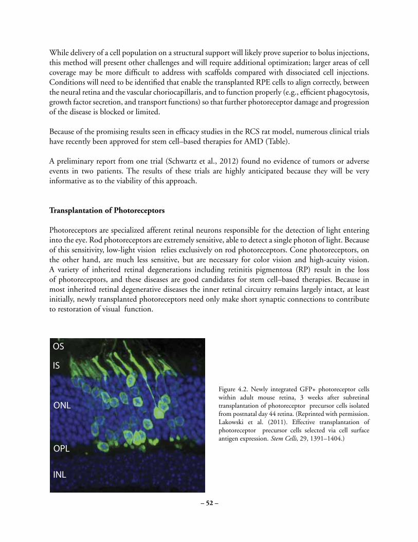
– 52 –
While delivery of a cell population on a structural support will likely prove superior to bolus injections, this method will present other challenges and will require additional optimization; larger areas of cell coverage may be more difficult to address with scaffolds compared with dissociated cell injections. Conditions will need to be identified that enable the transplanted RPE cells to align correctly, between the neural retina and the vascular choriocapillaris, and to function properly (e.g., efficient phagocytosis, growth factor secretion, and transport functions) so that further photoreceptor damage and progression of the disease is blocked or limited.
Because of the promising results seen in efficacy studies in the RCS rat model, numerous clinical trials have recently been approved for stem cell–based therapies for AMD (Table).
A preliminary report from one trial (Schwartz et al., 2012) found no evidence of tumors or adverse events in two patients. The results of these trials are highly anticipated because they will be very informative as to the viability of this approach.
Transplantation of Photoreceptors
Photoreceptors are specialized afferent retinal neurons responsible for the detection of light entering into the eye. Rod photoreceptors are extremely sensitive, able to detect a single photon of light. Because of this sensitivity, low-light vision relies exclusively on rod photoreceptors. Cone photoreceptors, on the other hand, are much less sensitive, but are necessary for color vision and high-acuity vision. A variety of inherited retinal degenerations including retinitis pigmentosa (RP) result in the loss of photoreceptors, and these diseases are good candidates for stem cell–based therapies. Because in most inherited retinal degenerative diseases the inner retinal circuitry remains largely intact, at least initially, newly transplanted photoreceptors need only make short synaptic connections to contribute to restoration of visual function.
Figure 4.2. Newly integrated GFP+ photoreceptor cells within adult mouse retina, 3 weeks after subretinal transplantation of photoreceptor precursor cells isolated from postnatal day 44 retina. (Reprinted with permission. Lakowski et al. (2011). Effective transplantation of photoreceptor precursor cells selected via cell surface antigen expression. Stem Cells, 29, 1391–1404.)
Newly integrated GFPþ photoreceptor cells within adult mouse retina, 3 weeks after subretinal transplantation of photoreceptorprecursor cells isolated from postnatal day 44 retina. (Reprinted with permission. Lakowski et al. (2011). Effective transplantation of photoreceptorprecursor cells selected via cell surface antigen expression. Stem Cells, 29, 1391–1404.)
Article 636
The Lasker/IRRF Initiative for Innovation in Vision Science

– 53 –
The identification of an ideal donor cell for efficient photoreceptor transplantation has proven challenging. Transplantation of intact sheets of fetal human retina in two RP patients reported subjective visual improvements (Radtke, Aramant, Seiler, & Petry, 1999). However, transplanted partial or whole retinal sheets derived from rat or rabbit embryonic, or neonatal pig retina, showed very limited integration into the recipient retinal circuitry of animal models (Ghosh, Juliusson, Arner, & Ehinger, 1999; Ghosh, Wong, Johansson, Bruun, & Petters, 2004; Seiler et al., 2008; Turner, Seiler, Aramant, & Blair, 1988). Other research in rodent models has employed transplantation of dissociated cells: brain-derived neural stem cells, progenitors isolated from immature retinas, and ESC- and iPSC-derived retinal donor cells, each with varying degrees of success. Details of these studies are available in recent reviews (Pearson, 2014; Reynolds & Lamba, 2014; Wright, Phillips, Pinilla, Hei, & Gamm, 2014). Currently, subretinal transplantations using suspensions of post-mitotic, photoreceptor precursor cells (cells that are already specified to differentiate into rod photoreceptors) have been the most successful (Lakowski et al., 2010; MacLaren et al., 2006; Pearson et al., 2012; Warre-Cornish, Barber, Sowden, Ali, & Pearson, 2014). Some of these transplanted rod precursor cells are able to migrate into the adult retina, differentiate, and acquire morphological features comparable with mature photoreceptor cells (Fig. 4.2) (Bartsch et al., 2008; Eberle et al., 2012; MacLaren et al., 2006; Warre-Cornish et al., 2014).
Transplantation of rod precursors (isolated from postnatal mouse retina) into a genetic model of rod dysfunction has been shown to improve rod-mediated vision following integration of more than 25,000 new rod photoreceptors (Pearson et al., 2012). These recent studies in animal models are resolving the type and developmental stage of differentiated hESC/iPSC-derived cells that will be optimal for clinical photoreceptor transplantation therapy.
efficient than transplantation of rod precursor cells isolated directly from the neonatal retina (Gonzalez-Cordero et al.,2013; West et al., 2012). Notably, ESC-derived rod precursors isolated at a developmental stage similar to postnatal days4 to 8 were shown to integrate more efficiently compared with cells at other stages (Gonzalez-Cordero et al., 2013). Giventhe extensive proliferative capacity and differentiation potential of pluripotent stem cells, for effective and safetransplantation, optimal differentiated progeny need to be purified and proliferative cells excluded (Gonzalez-Cordero etal., 2013; Lakowski et al., 2011; West et al., 2012). Further development of these therapies will require refinement ofmethods to generate sufficiently high numbers of transplantable rod and cone cells and to increase their long-termsurvival, integration, and function post transplantation. In addition, the success of these transplantations is limited by theintegrity of the outer limiting membrane (OLM) and the extent of gliosis in the recipient tissue, both of which remain tobe explored in greater detail (Barber et al., 2013; Pearson et al., 2010). Even with these challenges, stem cell–based cellreplacement therapies for treatment of retinal diseases resulting in the loss of photoreceptors look very promising.
Human iPSC derived multilayered retina-like structures. RECOVERINþphotoreceptor precursor cells (red) observed in the thickened
ONL. (Reprinted with permission. Phillips, M.J., Wallace, K.A., Dickerson, S.J., Miller, M.J., Verhoeven, A.D., Martin, J.M., . . . Gamm, D.M.
(2012). Blood-derived human iPS cells generate optic vesible-like structures with the capacity to form retinal laminae and develop synapses.
Investigative Ophthalmology & Visual Science, 53(4), 2007–2019.)
Article 637
The Lasker/IRRF Initiative for Innovation in Vision Science
Figure 4.3. Human iPSC derived multilayered retina-like structures. RECOVERIN+photoreceptor precursor cells (red) observed in the thickened ONL. (Reprinted with permission. Phillips, M.J., Wallace, K.A., Dickerson, S.J., Miller, M.J., Verhoeven, A.D., Martin, J.M., . . . Gamm, D.M. (2012). Blood-derived human iPS cells generate optic vesible-like structures with the capacity to form retinal laminae and develop synapses. Investigative Ophthalmology & Visual Science, 53(4), 2007–2019.)
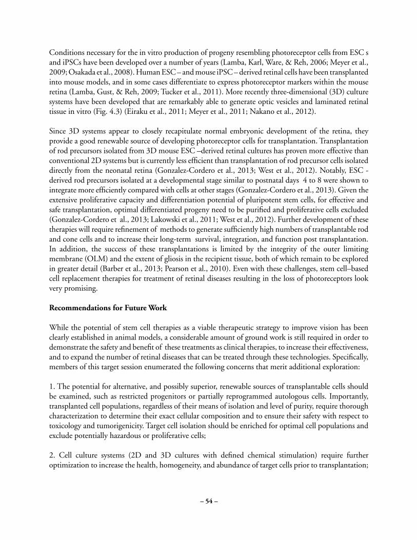
– 54 –
Conditions necessary for the in vitro production of progeny resembling photoreceptor cells from ESC s and iPSCs have been developed over a number of years (Lamba, Karl, Ware, & Reh, 2006; Meyer et al., 2009; Osakada et al., 2008). Human ESC – and mouse iPSC – derived retinal cells have been transplanted into mouse models, and in some cases differentiate to express photoreceptor markers within the mouse retina (Lamba, Gust, & Reh, 2009; Tucker et al., 2011). More recently three-dimensional (3D) culture systems have been developed that are remarkably able to generate optic vesicles and laminated retinal tissue in vitro (Fig. 4.3) (Eiraku et al., 2011; Meyer et al., 2011; Nakano et al., 2012).
Since 3D systems appear to closely recapitulate normal embryonic development of the retina, they provide a good renewable source of developing photoreceptor cells for transplantation. Transplantation of rod precursors isolated from 3D mouse ESC –derived retinal cultures has proven more effective than conventional 2D systems but is currently less efficient than transplantation of rod precursor cells isolated directly from the neonatal retina (Gonzalez-Cordero et al., 2013; West et al., 2012). Notably, ESC - derived rod precursors isolated at a developmental stage similar to postnatal days 4 to 8 were shown to integrate more efficiently compared with cells at other stages (Gonzalez-Cordero et al., 2013). Given the extensive proliferative capacity and differentiation potential of pluripotent stem cells, for effective and safe transplantation, optimal differentiated progeny need to be purified and proliferative cells excluded (Gonzalez-Cordero et al., 2013; Lakowski et al., 2011; West et al., 2012). Further development of these therapies will require refinement of methods to generate sufficiently high numbers of transplantable rod and cone cells and to increase their long-term survival, integration, and function post transplantation. In addition, the success of these transplantations is limited by the integrity of the outer limiting membrane (OLM) and the extent of gliosis in the recipient tissue, both of which remain to be explored in greater detail (Barber et al., 2013; Pearson et al., 2010). Even with these challenges, stem cell–based cell replacement therapies for treatment of retinal diseases resulting in the loss of photoreceptors look very promising.
Recommendations for Future Work
While the potential of stem cell therapies as a viable therapeutic strategy to improve vision has been clearly established in animal models, a considerable amount of ground work is still required in order to demonstrate the safety and benefit of these treatments as clinical therapies, to increase their effectiveness, and to expand the number of retinal diseases that can be treated through these technologies. Specifically, members of this target session enumerated the following concerns that merit additional exploration:
1. The potential for alternative, and possibly superior, renewable sources of transplantable cells should be examined, such as restricted progenitors or partially reprogrammed autologous cells. Importantly, transplanted cell populations, regardless of their means of isolation and level of purity, require thorough characterization to determine their exact cellular composition and to ensure their safety with respect to toxicology and tumorigenicity. Target cell isolation should be enriched for optimal cell populations and exclude potentially hazardous or proliferative cells;
2. Cell culture systems (2D and 3D cultures with defined chemical stimulation) require further optimization to increase the health, homogeneity, and abundance of target cells prior to transplantation;

– 55 –
3. While current target cell delivery strategies include transplantation of cell suspensions, boluses, or combinations of cells and support matrices (gels and scaffolds), these approaches require additional exploration and optimization (likely specific for each target disease);
4. Including additives at the time of transplantation to stimulate neuroprotection and/or the health and survival of transplanted cells (e.g., rod-derived cone viability factor or growth factors cotransplanted with photoreceptors), while at the same time promoting their stability (maintaining them in a desired, nonproliferative cell state) should be considered. Furthermore, efforts to increase the number of integrated cells, to stimulate their migration (when needed), and to enhance their proper localization post transplantation should be explored. Transplantation might be combined with additional treatments to bring about repair of damaged tissue, such as Bruch’s membrane. Long-term studies of these metrics would prove useful;
5. Approaches to deliver RPE and photoreceptors (or other combinations of cells) simultaneously using support matrices should be developed, as this type of combinatorial treatment may augment the survival of transplanted cells and may be beneficial for retinal diseases that result in the loss of multiple cells types, such as Best’s disease, AMD, and so on;
6. Additional strategies to measure the efficacy of the therapy and functionality of the cells following transplantation should be developed;
7. Because stem cell transplantation into the eye stimulates both a gliotic and an immune response, the events accompanying these responses, as well as any benefit that results from modulating these responses, should be documented. While it is common practice to immunosuppress following transplantation, systemic immunosuppression is costly and detrimental to the patient, and it is possible that aspects of the immune response are beneficial to the success of these therapies. It was also suggested that developing engineered cells that provide for localized immunosuppression in the eye could be beneficial; and
8. There is a great need for additional animal models, particularly to advance cone replacement therapies (since rodent retinas have relatively few cones and lack a fovea) and to develop treatments for more complex diseases such as AMD. Additional models to better gauge the potential and safety of the transplants and to enhance the development of these therapies prior to clinical trial would be desirable.
9. Although it is our hope and belief that stem cell transplantation technologies will prove beneficial for the treatment of multiple retinal diseases, a critical step in the development of these treatments is the design of carefully thought-out clinical trials. This targeted session stressed the importance of the following with regard to the design of current and future trials:
a. Clear rationale in the design of clinical trials is essential. While solely observing the gross physiological outcome of stem cell treatments can be informative, there is a growing need for conclusive clinical studies involving stepwise and standardized outcome measures; b. The identification of target patient groups is essential for the successful interpretation of clinic trials. A better mechanism to classify patients with different manifestations of and stages of target

– 56 –
retinal diseases is needed (in particular for AMD). Small, focused studies based on these classifications may be more informative; c. Development of new imaging modalities to visualize and to track transplanted cells in patients and to measure the efficacy of these treatments for patients is needed; and
d. Importantly, patients who undergo clinical treatments should be given a clear explanation of expected outcomes (based on experimental evidence).
10. Finally, this targeted session spent a considerable amount of time discussing challenges that arise through the commercialization of these stem cell treatments in selective regions of the world, in particular when these treatments have not been sufficiently founded on rigorous experimental and clinical results. While it is our goal to provide these treatments to patients as quickly as possible, we realize that to do so in an undisciplined fashion could prove detrimental to the recipients of these treatments or be of no benefit to them, and could hinder the development of these technologies through the generation of negative public perceptions. The safety and success of these procedures requires a global effort. We call on scientists and clinicians from around the world to ensure that these treatments are done in a stepwise and careful fashion, while ensuring safety and with public reporting of trial results. It is essential to work together on this global goal of restoring vision to the blind.
References
Barber, A.C., Hippert, C., Duran, Y., West, E.L., Bainbridge, J.W., Warre-Cornish, K., . . . Pearson, R.A. (2013). Repair of the degenerate retina by photoreceptor transplantation. Proceedings of the National Academy of Sciences of the United States of America, 110, 354–359.
Bartsch, U., Oriyakhel, W., Kenna, P.F., Linke, S., Richard, G., Petrowitz, B., . . . Ader, M. (2008). Retinal cells integrate into the outer nuclear layer and differentiate into mature photoreceptors after subretinal transplantation into adult mice. Experimental Eye Research, 86, 691–700.
Buchholz, D.E., Hikita, S.T., Rowland, T.J., Friedrich, A.M., Hinman, C.R., Johnson, L.V., & Clegg, D.O. (2009). Derivation of functional retinal pigmented epithelium from induced pluripotent stem cells. Stem Cells, 27, 2427–2434.
Carr, A.J., Vugler, A.A., Hikita, S.T., Lawrence, J.M., Gias, C., Chen, L.L., . . . Coffey, P.J. (2009). Protective effects of human iPS-derived retinal pigment epithelium cell transplantation in the retinal dystrophic rat. PLoS One, 4, e8152.
Croze, R.H., & Clegg, D.O. (2014). Differentiation of pluripotent stem cells into retinal pigmented epithelium. Developments in Ophthalmology, 53, 81–96.
da Cruz, L., Chen, F.K., Ahmado, A., Greenwood, J., & Coffey, P. (2007). RPE transplantation and its role in retinal disease. Progress in Retinal Eye Research, 26, 598–635.

– 57 –
Eberle, D., Kurth, T., Santos-Ferreira, T., Wilson, J., Corbeil, D., & Ader, M. (2012). Outer segment formation of transplanted photoreceptor precursor cells. PLoS One, 7, e46305.
Eiraku, M., Takata, N., Ishibashi, H., Kawada, M., Sakakura, E., Okuda, S., . . . Sasai, Y. (2011). Self-organizing opticcup morphogenesis in three-dimensional culture. Nature, 472, 51–56.
Fernandez-Robredo, P., Sancho, A., Johnen, S., Recalde, S., Gama, N., Thurmann, G., . . . García-Layana, A. (2013). Current treatment limitations in age-related macular degeneration and future approaches based on cell therapy and tissue engineering. Journal of Ophthalmology, 2014, 510285.
Gamm, D.M., Wang, S., Lu, B., Girman, S., Holmes, T., Bischoff, N., . . . Lund, R.D. (2007). Protection of visual functions by human neural progenitors in a rat model of retinal disease. PLoS One, 2, e338.
Ghosh, F., Juliusson, B., Arner, K., & Ehinger, B. (1999). Partial and full-thickness neuroretinal transplants. Experimental Eye Research, 68, 67–74.
Ghosh, F., Wong, F., Johansson, K., Bruun, A., & Petters, R.M. (2004). Transplantation of full-thickness retina in the rhodopsin transgenic pig. Retina, 24, 98–109.
Gonzalez-Cordero, A., West, E.L., Pearson, R.A., Duran, Y., Carvalho, L.S., Chu, C.J., . . . Ali, R.R. (2013). Photoreceptor precursors derived from three-dimensional embryonic stem cell cultures integrate and mature within adult degenerate retina. Nature Biotechnology, 31, 741–747.
Hirami, Y., Osakada, F., Takahashi, K., Okita, K., Yamanaka, S., Ikeda, H., . . . Takahashi, M., (2009). Generation of retinal cells from mouse and human induced pluripotent stem cells. Neuroscience Letters, 458, 126–131.
Idelson, M., Alper, R., Obolensky, A., Ben-Shushan, E., Hemo, I., Tachimovich-Cohen, N., . . . Reubinoff, B. (2009). Directed differentiation of human embryonic stem cells into functional retinal pigment epithelium cells. Cell Stem Cell, 5, 396–408.
Klimanskaya, I., Hipp, J., Rezai, K.A., West, M., Atala, A., & Lanza, R. (2004). Derivation and comparative assessment of retinal pigment epithelium from human embryonic stem cells using transcriptomics. Cloning Stem Cells, 6, 217– 245.
Kokkinaki, M., Sahibzada, N., & Golestaneh, N. (2011). Human induced pluripotent stem-derived retinal pigment epithelium (RPE) cells exhibit ion transport, membrane potential, polarized vascular endothelial growth factor secretion, and gene expression pattern similar to native RPE. Stem Cells, 29, 825–835.
Lakowski, J., Baron, M., Bainbridge, J., Barber, A.C., Pearson, R.A., Ali, R.R., . . . Sowden, J.C. (2010). Cone and rod photoreceptor transplantation in models of the childhood retinopathy Leber congenital amaurosis using flow-sorted Crx-positive donor cells. Human Molecular Genetics, 19, 4545–4559.

– 58 –
Lakowski, J., Han, Y.T., Pearson, R.A., Gonzalez-Cordero, A., West, E.L., Gualdoni, S., . . . Sowden, J.C. (2011). Effective transplantation of photoreceptor precursor cells selected via cell surface antigen expression. Stem Cells, 29, 1391–1404.
Lamba, D.A., Gust, J., & Reh, T.A. (2009). Transplantation of human embryonic stem cell-derived photoreceptors restores some visual function in Crx-deficient mice. Cell Stem Cell, 4, 73–79.
Lamba, D.A., Karl, M.O., Ware, C.B., & Reh, T.A. (2006). Efficient generation of retinal progenitor cells from human embryonic stem cells. Proceedings of the National Academy of Sciences of the United States of America, 103, 12769– 12774.
Lu, B., Malcuit, C., Wang, S., Girman, S., Francis, P., Lemieux, L., . . . Lund, R. (2009). Long-term safety and function of RPE from human embryonic stem cells in preclinical models of macular degeneration. Stem Cells, 27, 2126–2135.
Lu, B., Zhu, D., Hinton, D., Humayun, M.S., & Tai, Y.C. (2012). Mesh-supported submicron parylene-C membranes for culturing retinal pigment epithelial cells. Biomedical Microdevices, 14, 659–667.
Lund, R.D., Wang, S., Klimanskaya, I., Holmes, T., Ramos-Kelsey, R., Lu, B., . . . Lanza, R. (2006). Human embryonic stem cell-derived cells rescue visual function in dystrophic RCS rats. Cloning Stem Cells, 8, 189–199.
MacLaren, R.E., Pearson, R.A., MacNeil, A., Douglas, R.H., Salt, T.E., Akimoto, M., . . . Ali, R.R. (2006). Retinal repair by transplantation of photoreceptor precursors. Nature, 444, 203–207.
Mathieson, K., Loudin, J., Goetz, G., Huie, P., Wang, L., Kamins, T.I., . . . Palanker, D. (2012). Photovoltaic retinal prosthesis with high pixel density. Nature Photonics, 6, 391–397.
Meyer, J.S., Howden, S.E., Wallace, K.A., Verhoeven, A.D., Wright, L.S., Capowski, E.E., . . . Gamm, D.M. (2011). Optic vesicle-like structures derived from human pluripotent stem cells facilitate a customized approach to retinal disease treatment. Stem Cells, 29, 1206–1218.
Meyer, J.S., Shearer, R.L., Capowski, E.E., Wright, L.S., Wallace, K.A., McMillan, E.L., . . . Gamm, D.M. (2009). Modeling early retinal development with human embryonic and induced pluripotent stem cells. Proceedings of the National Academy of Sciences of the United States of America, 106, 16698–16703.
Nakano, T., Ando, S., Takata, N., Kawada, M., Muguruma, K., Sekiguchi, K., . . . Sasai, Y. (2012). Self-formation of optic cups and storable stratified neural retina from human ESCs. Cell Stem Cell, 10, 771–785.
Osakada, F., Ikeda, H., Mandai, M., Wataya, T., Watanabe, K., Yoshimura, N., . . . Takahashi, M. (2008). Toward the generation of rod and cone photoreceptors from mouse, monkey and human embryonic stem cells. Nature Biotechnology, 26, 215–224.
Osakada, F., Jin, Z.B., Hirami, Y., Ikeda, H., Danjyo, T., Watanabe, K., . . . Takahashi, M. (2009). In vitro differentiation of retinal cells from human pluripotent stem cells by small-molecule induction. Journal of Cell Science, 122, 3169–3179.

– 59 –
Pearson, R.A. (2014). Advances in repairing the degenerate retina by rod photoreceptor transplantation. Biotechnology Advancements, 32, 485–491.
Pearson, R.A., Barber, A.C., Rizzi, M., Hippert, C., Xue, T., West, E.L., . . . Ali, R.R. (2012). Restoration of vision after transplantation of photoreceptors. Nature, 485, 99–103.
Pearson, R.A., Barber, A.C., West, E.L., MacLaren, R.E., Duran, Y., Bainbridge, J.W., . . . Ali, R.R. (2010). Targeted disruption of outer limiting membrane junctional proteins (Crb1 and ZO-1) increases integration of transplanted photoreceptor precursors into the adult wild-type and degenerating retina. Cell Transplant, 19, 487–503.
Phillips, M.J., Wallace, K.A., Dickerson, S.J., Miller, M.J., Verhoeven, A.D., Martin, J.M., . . . Gamm, D.M. (2012). Blood-derived human iPS cells generate optic vesicle-like structures with the capacity to form retinal laminae and develop synapses. Investigative Ophthalmology & Visual Science, 53, 2007–2019.
Radtke, N.D., Aramant, R.B., Seiler, M., & Petry, H.M. (1999). Preliminary report: indications of improved visual function after retinal sheet transplantation in retinitis pigmentosa patients. American Journal of Ophthalmology, 128, 384–387.
Ramsden, C.M., Powner, M.B., Carr, A.J., Smart, M.J., da Cruz, L., & Coffey P.J. (2013). Stem cells in retinal regeneration: Past, present and future. Development, 140, 2576–2585.
Reynolds, J., & Lamba, D.A. Human embryonic stem cell applications for retinal degenerations. (2014). Experimental Eye Research, 123, 151–160.
Rowland, T.J., Buchholz, D.E., & Clegg, D.O. (2012). Pluripotent human stem cells for the treatment of retinal disease. Journal of Cell Physiology, 227, 457–66.
Salero, E., Blenkinsop, T.A., Corneo, B., Harris, A., Rabin, D., Stern, J.H., . . . Temple, S. (2012). Adult human RPE can be activated into a multipotent stem cell that produces mesenchymal derivatives. Cell Stem Cell, 10, 88–95.
Schwartz, S.D., Hubschman, J.P., Heilwell, G., Franco-Cardenas, V., Pan, C.K., Ostrick, M., . . . Lanza, R. (2012). Embryonic stem cell trials for macular degeneration: a preliminary report. Lancet, 379, 713–720.
Seiler, M.J., Thomas, B.B., Chen, Z., Wu, R., Sadda, S.R., & Aramant, R.B. (2008). Retinal transplants restore visual responses: trans-synaptic tracing from visually responsive sites labels transplant neurons. European Journal of Neuroscience, 28, 208–220.
Sheridan, C., Williams, R., & Grierson, I. (2004). Basement membranes and artificial substrates in cell transplantation. Graefe’s Archive for Clinical and Experimental Ophthalmology, 242, 68–75.
Singh, R., Phillips, M.J., Kuai, D., Meyer, J., Martin, J.M., Smith, M.A., . . . Gamm, D.M. (2013). Functional analysis of serially expanded human iPS cell-derived RPE cultures. Investigative Ophthalmology & Visual Science, 54, 6767– 6778.

– 60 –
Stanzel, B.V., Liu, Z., Somboonthanakij, S., Wongsawad, W., Brinken, R., Eter, N., . . . Blenkinsop, T.A. (2014). Human RPE stem cells grown into polarized RPE monolayers on a polyester matrix are maintained after grafting into rabbit subretinal space. Stem Cell Reports, 2, 64–77.
Strauss, O. (2005). The retinal pigment epithelium in visual function. Physiology Review, 85, 845–881.
Sugino, I.K., Sun, Q., Wang, J., Nunes, C.F., Cheewatrakoolpong, N., Rapista, A., . . . Zarbin, M.A. (2012). Comparison of FRPE and human embryonic stem cell-derived RPE behavior on aged human Bruch’s membrane. Investigative Ophthalmology & Visual Science, 52, 4979–4997.
Tucker, B.A., Park, I.H., Qi, S.D., Klassen, H.J., Jiang, C., Yao, J., . . . Young, M.J. (2011). Transplantation of adult mouse iPS cell-derived photoreceptor precursors restores retinal structure and function in degenerative mice. PLoS One, 6, e18992.
Turner, J.E., Seiler, M., Aramant, R., & Blair, J.R. (1988). Embryonic retinal grafts transplanted into the lesioned adult rat retina. Progress in Brain Research, 78, 131–139.
Ukrohne, T.U., Westenskow, P.D., Kurihara, T., Friedlander, D.F., Lehmann, M., Dorsey, A.L., . . . Friedlander, M. (2012). Generation of retinal pigment epithelial cells from small molecules and OCT4 reprogrammed human induced pluripotent stem cells. Stem Cells Translational Medicine, 1, 96–109.
Vugler, A., Carr, A.J., Lawrence, J., Chen, L.L., Burrell, K., Wright, A., . . . Coffey, P. (2008). Elucidating the phenomenon of HESC-derived RPE: anatomy of cell genesis, expansion and retinal transplantation. Experimental Neurology, 214, 347–361.
Warre-Cornish, K., Barber, A.C., Sowden, J.C., Ali, R.R., & Pearson, R.A. (2014). Migration, integration and maturation of photoreceptor precursors following transplantation in the mouse retina. Stem Cells Development, 23, 941–954.
West, E.L., Gonzalez-Cordero, A., Hippert, C., Osakada, F., Martinez-Barbera, J.P., Sowden, J.C., . . . Ali, R.R. (2012). Defining the integration capacity of embryonic stem cell-derived photoreceptor precursors. Stem Cells, 30, 1424–1435.
Westenskow, P.D., Kurihara, T., & Friedlander, M. (2014). Utilizing stem cell-derived RPE cells as a therapeutic intervention for age-related macular degeneration. Advances in Experimental Medicine and Biology, 801, 323–329.
Williams, R.L., Krishna, Y., Dixon, S., Haridas, A., Grierson, I., & Sheridan, C. (2005). Polyurethanes as potential substrates for sub-retinal retinal pigment epithelial cell transplantation. Journal of Materials Science. Materials in Medicine, 16, 1087–1092.
Wright, L.S., Phillips, M.J., Pinilla, I., Hei, D., & Gamm, D.M. (2014). Induced pluripotent stem cells as custom therapeutics for retinal repair: Progress and rationale. Experimental Eye Research, 123, 161–172.

– 61 –
Chapter 5Endogenous Regeneration
Discussion Leaders: Daniel Goldman and David Hyde
Scribe: Curtis Powell
Session Participants: John Ash, David Beebe, John Dowling, David Gamm, Peter Hitchcock, Amy Laster, Matthew LaVail, Pamela Raymond, Tonia Rex, Josh Sanes, Jane Sowden, Jeffrey Stern, Sally Temple, and Donald Zack
Introduction
The human eye is made up of many tissues, each of which directly or indirectly influences our visual perception of the world. Tissues that directly impact vision include the cornea, lens, retina, and optic nerve. The cornea and lens serve to focus light as it enters into the eye; this light is detected and analyzed by the retina and the visual signal communicated to the rest of the brain via retinal ganglion cell (RGC) axons that form the optic nerve. Because they directly influence our vision, damage to any one of these tissues, due to disease or injury, can dramatically impact one’s quality of life. Many approaches are being developed to aid those who suffer from vision loss due to damage of these tissues; some of which have been quite successful, like corneal transplants, corneal epithelial regeneration, and artificial intraocular lenses. However, approaches to treat damage of the retina and optic nerve have proven more challenging.
Unlike mammals, some vertebrate species are able to repair damage to the retina and optic nerve via endogenous regenerative events. Two fundamentally different categories of neuronal regeneration are important in the eye: neurogenesis (cell proliferation leading to replacement of neurons that have been lost) and axonal regeneration (regrowth of retinal ganglion cell axons following damage to the optic nerve). Depending on which cells are damaged, and the vertebrate species being studied, regeneration of retinal neurons through neurogenesis is accomplished by mitotic activation of retinal pigment epithelial (RPE) cells, retinal progenitor cells in the ciliary marginal zone (CMZ), or Müller glia. For optic nerve regeneration, RGCs whose axons are severed exhibit an axonal growth-permissive state. In both types of regeneration, immune cells and injury-related changes likely play a critical but poorly understood role. Importantly, these regenerative events ultimately can lead to the restoration of visually mediated behaviors.
While mammals do not actively demonstrate these modes of regeneration in response to retinal disease or injury, current research supports the possibility that at least some of these regenerative cell types maintain an intrinsic regenerative potential in mammals and that this potential could be harnessed for retinal repair if the proper stimuli were provided. One can easily imagine how advantageous it would be to treat human retinal disease using endogenous reparative strategies.
The purpose of this targeted session was to review the current state of research aimed at stimulating endogenous regeneration of retinal neurons and axons in the optic nerve and to provide guidance for

– 62 –
future research. To this end, we provide a brief background describing the various modes of retinal and optic nerve regeneration employed by species that exhibit a robust regenerative response (amphibians and teleost fish) and those that demonstrate a limited regenerative response (birds). A discussion of progress made in initiating endogenous regenerative events in nonregenerative species (mammals) is then presented. Finally, recommendations are provided to stimulate and direc future research in this field.
Amphibians
Amphibians are the only adult animals known to possess the ability to regenerate the entire retina after its removal. In urodele amphibians, such as newts, the new retina arises predominantly from the RPE (Okada, 1980); however, a small domain of the new retina is derived from the circumferential germinal zone (CGZ): a ring of retinal progenitor cells at the periphery of the neural retina (Fischer, Bosse, & El-Hodiri, 2013). Amazingly, regeneration of the neural retina occurs without the addition of exogenous factors and without preserving the vascular membrane of the eye. In anuran amphibians, the mode of regeneration varies by species, but the integrity of the vascular membrane is universally crucial (Reh & Nagy, 1987). Similar to urodele amphibians, regenerated retinal neurons in Xenopus laevis are derived from the RPE (Yoshii et al., 2007). In contrast, retinal regeneration in Xenopus tropicalis is carried out exclusively by the CGZ (Miyake & Araki, 2014).
Because the studies mentioned above involve the removal of the entire neural retina, these models by necessity also require growth of a new optic nerve. Optic nerve regeneration in amphibians can also be stimulated without such a drastic injury (e.g., following optic nerve crush or cut). This damage leads to the activation of RGCs, which establish a developmental-like growth permissive state, ultimately restoring vision (Stelzner, Bohn, & Strauss, 1986).
Teleost Fish
Although teleost fish are unable to replace an entire retina, they do mount a robust regenerative response following damage to the retina or optic nerve. Similar to the amphibian retina, the teleost retina possesses a CGZ to facilitate persistent neurogenesis and retinal growth throughout the life of the fish (Johns, 1977). The Müller glia in the inner nuclear layer of the fish retina also retain a radial glial, stem-cell-like neurogenic ability. Under normal circumstances, they undergo sporadic, self-renewing divisions in the inner nuclear layer to produce retinal progenitors that migrate to the outer nuclear layer (Bernardos et al., 2007), where they proliferate as rod precursors and are committed to differentiate as rod photoreceptors (Johns & Fernald, 1981). When the retina is injured and neurons are lost, Müller glial proliferation is enhanced and the resulting retinal progenitors (Fig. 5.1) differentiate into various types of retinal neurons (Bernardos et al., 2007; Fausett & Goldman, 2006; Fimbel et al., 2007; Thummel et al., 2008). Interestingly, intraocular injection of agents that impact a variety of signaling pathways can also stimulate Müller glia proliferation, even in the absence of overt loss of retinal neurons, and a small number of the resulting cells can express neuronal markers (Ramachandran, Zhao, & Goldman, 2011; Wan, Ramachandran, & Goldman, 2012). Thus, Müller glia are primarily responsible for the ability of teleost fish to regenerate any of the lost neuronal cell types following retinal damage (Bernardos et al., 2007; Fausett & Goldman, 2006; Fimbel et al.,

– 63 –
2007; Thummel et al., 2008). This Müller glia-derived regenerative response represents a fundamental difference between amphibians and teleost fish in their modes of retinal regeneration.
Teleost fish also exhibit a robust ability to regenerate their optic nerve that leads to restoration of lost sight (Bernhardt, 1999; McDowell et al., 2004). This regenerative response is regulated by gene expression programs and signaling cascades in RGCs after optic nerve injury (Fig. 5.2; Elsaeidi et al., 2014; Kato et al., 2013; Veldman et al., 2007). Unlike mammals, in fish most RGCs do not die following optic nerve lesion.
Birds
In contrast to amphibians and teleost fish, adult birds do not regenerate a damaged retina; however, the embryonic and post-hatch chick retina does possess a limited regenerative potential. If a region of
Amphibians
Amphibians are the only adult animals known to possess the ability to regenerate the entire retina after its removal. Inurodele amphibians, such as newts, the new retina arises predominantly from the RPE (Okada, 1980); however, a smalldomain of the new retina is derived from the circumferential germinal zone (CGZ): a ring of retinal progenitor cells at theperiphery of the neural retina (Fischer, Bosse, & El-Hodiri, 2013). Amazingly, regeneration of the neural retina occurswithout the addition of exogenous factors and without preserving the vascular membrane of the eye. In anuran amphibians,the mode of regeneration varies by species, but the integrity of the vascular membrane is universally crucial (Reh & Nagy,1987). Similar to urodele amphibians, regenerated retinal neurons in Xenopus laevis are derived from the RPE (Yoshii et al.,2007). In contrast, retinal regeneration in Xenopus tropicalis is carried out exclusively by the CGZ (Miyake & Araki, 2014).
Because the studies mentioned above involve the removal of the entire neural retina, these models by necessity alsorequire growth of a new optic nerve. Optic nerve regeneration in amphibians can also be stimulated without such a drasticinjury (e.g., following optic nerve crush or cut). This damage leads to the activation of RGCs, which establish adevelopmental-like growth permissive state, ultimately restoring vision (Stelzner, Bohn, & Strauss, 1986).
Teleost Fish
Although teleost fish are unable to replace an entire retina, they do mount a robust regenerative response followingdamage to the retina or optic nerve. Similar to the amphibian retina, the teleost retina possesses a CGZ to facilitatepersistent neurogenesis and retinal growth throughout the life of the fish (Johns, 1977). The M€uller glia in the innernuclear layer of the fish retina also retain a radial glial, stem-cell-like neurogenic ability. Under normal circumstances, theyundergo sporadic, self-renewing divisions in the inner nuclear layer to produce retinal progenitors that migrate to theouter nuclear layer (Bernardos et al., 2007), where they proliferate as rod precursors and are committed to differentiate asrod photoreceptors (Johns & Fernald, 1981). When the retina is injured and neurons are lost, M€uller glial proliferation isenhanced and the resulting retinal progenitors (Fig. 5.1) differentiate into various types of retinal neurons (Bernardos etal., 2007; Fausett & Goldman, 2006; Fimbel et al., 2007; Thummel et al., 2008). Interestingly, intraocular injection ofagents that impact a variety of signaling pathways can also stimulate M€uller glia proliferation, even in the absence of overt
Injury-dependent M€uller glia proliferation in zebrafish retina. Zebrafish retinas were mechanically injured and 4 days later retinalsections prepared and immunofluorescence used to identify glutamine synthetase-positive M€uller glia (red) and BrdU-positive proliferating M€ullerglia-derived progenitors (green). (Image courtesy of Jin Wan, MBNI, University of Michigan).
http://tvstjournal.org/doi/full/10.1167/tvst.3.7.7 TVST j 2014 j Vol. 3 j No. 7 j Article 743
The Lasker/IRRF Initiative for Innovation in Vision Science
loss of retinal neurons, and a small number of the resulting cells can express neuronal markers (Ramachandran, Zhao, &Goldman, 2011; Wan, Ramachandran, & Goldman, 2012). Thus, M€uller glia are primarily responsible for the ability ofteleost fish to regenerate any of the lost neuronal cell types following retinal damage (Bernardos et al., 2007; Fausett &Goldman, 2006; Fimbel et al., 2007; Thummel et al., 2008). This M€uller glia-derived regenerative response represents afundamental difference between amphibians and teleost fish in their modes of retinal regeneration.
Teleost fish also exhibit a robust ability to regenerate their optic nerve that leads to restoration of lost sight (Bernhardt,1999; McDowell et al., 2004). This regenerative response is regulated by gene expression programs and signaling cascadesin RGCs after optic nerve injury (Fig. 5.2; Elsaeidi et al., 2014; Kato et al., 2013; Veldman et al., 2007). Unlikemammals, in fish most RGCs do not die following optic nerve lesion.
Birds
Figure 5.2. Enhanced optic nerve regeneration following Sfpq morpholino knockdown. Regenerating zebrafish optic axons were lesioned andback-labeled with fluorescein dextran. Whole mount retinas were examined under a fluorescent microscope to visualize RGCs regenerating theiroptic axon as indicated by fluorescein in RGC bodies (green). Morpholino-modified antisense oligonucleotides (MOs) were used to knockdown theexpression of specific proteins in RGCs. Control MO shows the number of RGCs whose axons have regenerated past the lesion site without proteinknockdown. Note that Sfpq (splicing factor proline glutamine rich) knockdown resulted in increased numbers of back-labeled RGCs indicatingimproved axonal regeneration. (Photo courtesy of Rose Elsaeidi, MBNI, University of Michigan).
Article 744
The Lasker/IRRF Initiative for Innovation in Vision Science
Figure 5.1. Injury-dependent Müller glia proliferation in zebrafish retina. Zebrafish retinas were mechanically injured and 4 days later retinal sections prepared and immunofluorescence used to identify glutamine synthetase-positive Müller glia (red) and BrdU-positive proliferating Müller glia-derived progenitors (green). (Image courtesy of Jin Wan, MBNI, University of Michigan).
Figure 5.2. Enhanced optic nerve regeneration following Sfpq morpholino knockdown. Regenerating zebrafish optic axons were lesioned and back-labeled with fluorescein dextran. Whole mount retinas were examined under a fluorescent micro-scope to visualize RGCs regenerating their optic axon as indicated by fluorescein in RGC bodies (green). Morpholino-modified antisense oligonucleotides (MOs) were used to knockdown the expression of specific proteins in RGCs. Control MO shows the number of RGCs whose axons have regenerated past the lesion site without protein knockdown. Note that Sfpq (splicing factor proline glutamine rich) knockdown resulted in increased numbers of back-labeled RGCs indicating improved axonal regeneration. (Photo courtesy of Rose Elsaeidi, MBNI, University of Michigan).

– 64 –
the neural retina is removed from the chick embryo within the first four days of incubation, an RPE-dependent regenerative event ensues, generating a new retina (Coulombre & Coulombre, 1965). Like teleost fish, Müller glia also can serve as a source of retinal progenitors in the chick retina following excitotoxic injury, but unlike teleosts, they do so only through the first few days post-hatch (Fischer & Reh, 2001). Intraocular injection of exogenous growth factors can enhance this response and even initiate Müller glia proliferation in the absence of injury (Fischer et al., 2002; Fischer & Reh, 2002). In contrast to Müller glia-dependent regeneration in teleost fish, the majority of Müller glia-derived cells in the chick retina either do not survive or fail to differentiate (Fischer & Bongini, 2010; Fischer & Reh, 2001).
While the majority of work using the chick model system has focused on regeneration of retinal neurons, there is some evidence of a limited potential for RGC-dependent optic nerve regeneration; however, both regenerative abilities decline rapidly post-hatch (Halfter & Deiss, 1984).
Mammals
While RPE, CMZ, and Müller glia can generate neuronal progenitors following retina damage in some vertebrate species, these cell types do not do so in mammals. In contrast to amphibians, teleost fish, and birds, the mammalian retina does not add retinal neurons after birth. However, pigmented cells in the mammalian CMZ can proliferate and express neuronal markers under certain circumstances (Cicero et al., 2009). Interestingly, lid fusion stimulated proliferation of progenitor-like cells in the retinal periphery in juvenile macaques (Tkatchenko et al., 2006), perhaps suggesting a source of cells for regeneration in primates. Furthermore, the human RPE harbors multipotent cells that, under certain conditions, can be activated and may serve as a potential source of progenitors for repair (Salero et al., 2012). Activation of RPE proliferation and regeneration contributes to RPE layer wound repair that supports the neural retina (Lopez et al., 1995). After activation, RPE stem cell progeny can be directed to differentiate into RPE or cells that express neuronal markers in cell culture. The fact that the (presumptive) RPE domain can transdifferentiate into neural retina in mammalian embryos lacking MITF, Pax6, or Pax2 (Baumer et al., 2003; Bharti et al., 2012) indicates that specific stages of RPE could exhibit neurogenic potential, with appropriate manipulation. While strategies to direct RPE differentiation into neurons in vivo may result in a potential therapy for retinal diseases, these approaches will need to be carefully modulated to avoid fibrosis and epiretinal membrane formation.
Although mammalian Müller glia do not normally generate neurons in vivo, they do express low levels of markers associated with a retinal progenitor state (Jadhav, Roesch, & Cepko, 2009), and they can generate neurons and glia when placed in cell culture (Giannelli et al., 2011; Singhal et al., 2012). A number of studies suggest that mammalian Müller glia can be coaxed to proliferate and generate a small number of cells with neuronal markers in vivo by combining retinal damage with growth factor stimulation (Karl et al., 2008; Osakada et al., 2007; Wan et al., 2008). These studies also suggest that this Müller glia response may decline with age.
Similarly, in mammals, RGCs lose their intrinsic capacity for axonal growth soon after birth (Moore et al., 2009). In adult mammals, there is no appreciable regenerative response following damage to the optic nerve in the absence of exogenously introduced stimuli. Some success has been achieved with enhancing the intrinsic potential of RGCs or modulating inhibitory extrinsic factors, but even with

– 65 –
these changes, a large percentage of the RGCs do not survive and only a small percentage of RGC axons regrow past the optic chiasm (de Lima, Habboub, & Benowitz, 2012; Fischer et al., 2012; Sun et al., 2011).
Recommendations for Future Work
While these results delineate a stark difference between the regenerative response of mammals following damage to the retina or optic nerve relative to amphibians or teleost fish, studies do suggest some level of endogenous regeneration can be achieved under certain circumstances. Because research suggests that mammalian Müller glia, RPE cells, and RGCs possess an intrinsic regenerative potential, the majority of our discussion in this targeted session focused on a better understanding of these cell types so that their regenerative potential could be realized. What follows are questions, the answers to which would greatly benefit the field:
1. What are the mechanisms that drive regeneration in species where these processes are robust?
2. What are the mechanisms that limit avian and mammalian Müller glia, RPE cells, and RGCs from mounting a more robust regenerative response either in vivo or when placed in cell culture?
3. Are there molecular differences between the regeneration-permissive retinas and the regeneration-limited retinas? Are these differences intrinsic to the Müller glia, RPE cells, or RGCs?
4. What early developmental programs enhance the regenerative capacity of Müller glia, RPE cells, and RGCs?
5. What role does the retinal/optic nerve environment have in the regenerative capacity of Müller glia, RPE cells, and RGCs? Do inhibitory environments need to be neutralized or are stimulatory signals sufficient for a regenerative response?
6. What molecular mechanisms underlie and distinguish the gliotic/fibrotic response versus the regenerative responses resulting from Müller glia and RPE cell activation. Can the gliotic/fibrotic responses to retinal injury be shifted to regenerative?
7. How are the proliferative/regenerative responses of Müller glia, RPE, and RGCs terminated to prevent tumor formation and exuberant axonal growth in the regeneration-permissive retinas?
8. What pathfinding and synaptogenic mechanisms are used by RGC axons in the adult brain so they reform functional connections with appropriate brain targets?
9. What role does the immune system and inflammation play in stimulating and repressing retinal and optic nerve regeneration?
10. Can more realistic disease models be developed in model organisms to help guide regenerative strategies for vision restoration in a disease context?

– 66 –
11. Can Müller glia, RPE cells, and RGC cell cultures and retinal explants be used to identify small molecules that stimulate regeneration?
Answering these questions will require investigation of a variety of biological systems, including animal models that have already proven their value in driving discovery. In addition, the advent of human stem cell technologies and the development of protocols to efficiently grow retina and RPE cells in 2D and 3D configurations, now provide powerful tools to investigate and test hypotheses on human cells, with the ultimate goal of translating discoveries into therapies
References
Baumer, N., Marquardt, T., Stoykova, A., Spieler, D., Treichel, D., Ashery-Padan, R., & Gruss, P. (2003). Retinal pigmented epithelium determination requires the redundant activities of Pax2 and Pax6. Development, 130, 2903–2915.
Bernardos, R.L., Barthel, L.K., Meyers, J.R., & Raymond, P.A. (2007). Late-stage neuronal progenitors in the retina are radial Muller glia that function as retinal stem cells. Journal of Neuroscience, 27, 7028–7040.
Bernhardt, R.R. (1999). Cellular and molecular bases of axonal regeneration in the fish central nervous system. Experimental Neurology, 157, 223–240.
Bharti, K., Gasper, M., Ou, J., Brucato, M., Clore-Gronenborn, K., Pickel, J., & Arnheiter, H. (2012). A regulatory loop involving PAX6, MITF, and WNT signaling controls retinal pigment epithelium development. PLoS Genetics, 8, e1002757.
Cicero, S.A., Johnson, D., Reyntjens, S., Frase, S., Connell, S., Chow, L.M., . . . Dyer, M.A. (2009). Cells previously identified as retinal stem cells are pigmented ciliary epithelial cells. Proceedings of the National Academy of Sciences of the United States of America, 106, 6685–6690.
Coulombre, J.L., & Coulombre, A.J. (1965). Regeneration of neural retina from the pigmented epithelium in the chick embryo. Developmental Biology, 12, 79–92.
de Lima, S., Habboub, G., & Benowitz, L.I. (2012). Combinatorial therapy stimulates long-distance regeneration, target reinnervation, and partial recovery of vision after optic nerve injury in mice. International Review of Neurobiology, 106, 153–172.
Elsaeidi, F., Bemben, M.A., Zhao, X.F., & Goldman, D. (2014). Jak/Stat signaling stimulates zebrafish optic nerve regeneration and overcomes the inhibitory actions of Socs3 and Sfpq. Journal of Neuroscience, 34, 2632–2644.
Fausett, B.V., & Goldman, D. (2006). A role for alpha1 tubulin-expressing Müller glia in regeneration of the injured zebrafish retina. Journal of Neuroscience, 26, 6303–6313.
Fimbel, S.M., Montgomery, J.E., Burket, C.T., & Hyde, D.R. (2007). Regeneration of inner retinal neurons after intravitreal injection of ouabain in zebrafish. Journal of Neuroscience, 27, 1712–1724.

– 67 –
Fischer, A.J., & Bongini, R. (2010). Turning Muller glia into neural progenitors in the retina. Molecular Neurobiology, 42, 199–209.
Fischer, A.J., Bosse, J.L., & El-Hodiri, H.M. (2013). The ciliary marginal zone (CMZ) in development and regeneration of the vertebrate eye. Experimental Eye Research, 116, 199–204.
Fischer, A.J., McGuire, C.R., Dierks, B.D., & Reh, T.A. (2002). Insulin and fibroblast growth factor 2 activate a neurogenic program in Muller glia of the chicken retina. Journal of Neuroscience, 22, 9387–9398.
Fischer, A.J., & Reh, T.A. (2001). Muller glia are a potential source of neural regeneration in the postnatal chicken retina. Nature Neuroscience, 4, 247–252.
Fischer, A.J., & Reh, T.A. (2002). Exogenous growth factors stimulate the regeneration of ganglion cells in the chicken retina. Developmental Biology, 251, 367–379.
Fischer, D. (2012). Stimulating axonal regeneration of mature retinal ganglion cells and overcoming inhibitory signaling. Cell Tissue Research, 349, 79–85.
Giannelli, S.G., Demontis, G.C., Pertile, G., Rama, P., & Broccoli, V. (2011). Adult human Muller glia cells are a highly efficient source of rod photoreceptors. Stem Cells, 29, 344–356.
Halfter, W., & Deiss, S. (1984). Axon growth in embryonic chick and quail retinal whole mounts in vitro. Developmental Biology, 102, 344–355.
Jadhav, A.P., Roesch, K., & Cepko, C.L. (2009). Development and neurogenic potential of Muller glial cells in the vertebrate retina. Progress in Retinal and Eye Research, 28, 249–262.
Johns, P.R. (1977). Growth of the adult goldfish eye. III. Source of the new retinal cells. The Journal of Comparative Neurology, 176, 343–357.
Johns, P.R., & Fernald, R.D. (1981). Genesis of rods in teleost fish retina. Nature, 293, 141–142.
Karl, M.O., Hayes, S., Nelson, B.R., Tan, K., Buckingham, B., & Reh, T.A. (2008). Stimulation of neural regeneration in the mouse retina. Proceedings of the National Academy of Sciences of the United States of America, 105, 19508–19513.
Kato, S., Matsukawa, T., Koriyama, Y., Sugitani, K., & Ogai, K. (2013). A molecular mechanism of optic nerve regeneration in fish: The retinoid signaling pathway. Progress in Retinal and Eye Research, 37, 13–30.
Lopez, P.F., Yan, Q., Kohen, L., Rao, N.A., Spee, C., Black, J., & Oganesian, A. (1995). Retinal pigment epithelial wound healing in vivo. Archives of Ophthalmology, 113, 1437–1446.

– 68 –
McDowell, A.L., Dixon, L.J., Houchins, J.D., & Bilotta, J. (2004). Visual processing of the zebrafish optic tectum before and after optic nerve damage. Visual Neuroscience, 21, 97–106.
Miyake, A., & Araki, M. (2014). Retinal stem/progenitor cells in the ciliary marginal zone complete retinal regeneration: A study of retinal regeneration in a novel animal model. Developmental Neurobiology, 74, 739–756.
Moore, D.L., Blackmore, M.G., Hu, Y., Kaestner, K.H., Bixby, J.L., Lemmon, V.P., & Goldberg, J.L. (2009). KLF family members regulate intrinsic axon regeneration ability. Science, 326, 298–301.
Okada, T.S. (1980). Cellular metaplasia or transdifferentiation as a model for retinal cell differentiation. Current Topics in Developmental Biology, 16, 349–380.
Osakada, F., Ooto, S., Akagi, T., Mandai, M., Akaike, A., & Takahashi, M. (2007). Wnt signaling promotes regeneration in the retina of adult mammals. Journal of Neuroscience, 27, 4210–4219.
Ramachandran, R., Zhao, X.F., & Goldman, D. (2011). Ascl1a/Dkk/beta-catenin signaling pathway is necessary and glycogen synthase kinase-3beta inhibition is sufficient for zebrafish retina regeneration. Proceedings of the National Academy of Sciences of the United States of America, 108, 15858–15863.
Reh, T.A., & Nagy, T. (1987). A possible role for the vascular membrane in retinal regeneration in Rana catesbienna tadpoles. Developmental Biology, 122, 471–482.
Salero, E., Blenkinsop, T.A., Corneo, B., Harris, A., Rabin, D., Stern, J.H., & Temple, S. (2012). Adult human RPE can be activated into a multipotent stem cell that produces mesenchymal derivatives. Cell Stem Cell, 10, 88–95.
Singhal, S., Bhatia, B., Jayaram, H., Becker, S., Jones, M.F., Cottrill, P.B., . . . Limb, G.A. (2012). Human Muller glia with stem cell characteristics differentiate into retinal ganglion cell (RGC) precursors in vitro and partially restore RGC function in vivo following transplantation. Stem Cells Translational Medicine, 1, 188–199.
Stelzner, D.J., Bohn, R.C., & Strauss, J.A. (1986). Regeneration of the frog optic nerve. Comparisons with development. Neurochemical Pathology, 5, 255–288.
Sun, F., Park, K.K., Belin, S., Wang, D., Lu, T., Chen, G., . . . He, Z. (2011). Sustained axon regeneration induced by co-deletion of PTEN and SOCS3. Nature, 480, 372–375.
Thummel, R., Kassen, S.C., Montgomery, J.E., Enright, J.M., & Hyde, D.R. (2008). Inhibition of Muller glial cell division blocks regeneration of the light-damaged zebrafish retina. Developmental Neurobiology, 68, 392–408.
Tkatchenko, A.V., Walsh, P.A., Tkatchenko, T.V., Gustincich, S., & Raviola, E. (2006). Form deprivation modulates retinal neurogenesis in primate experimental myopia. Proceedings of the National Academy of Sciences of the United States of America, 103, 4681–4686.

– 69 –
Veldman, M.B., Bemben, M.A., Thompson, R.C., & Goldman, D. (2007). Gene expression analysis of zebrafish retinal ganglion cells during optic nerve regeneration identifies KLF6a and KLF7a as important regulators of axon regeneration. Developmental Biology, 312, 596–612.
Wan, J., Ramachandran, R., & Goldman, D. (2012). HB-EGF is necessary and sufficient for Muller glia dedifferentiation and retina regeneration. Developmental Cell, 22, 334–347.
Wan, J., Zheng, H., Chen, Z.L., Xiao, H.L., Shen, Z.J., & Zhou, G.M. (2008). Preferential regeneration of photoreceptor from Muller glia after retinal degeneration in adult rat. Journal of Ophthalmic and Vision Research, 48, 223–234.
Yoshii, C., Ueda, Y., Okamoto, M., & Araki, M. (2007). Neural retinal regeneration in the anuran amphibian Xenopus laevis post-metamorphosis: transdifferentiation of retinal pigmented epithelium regenerates the neural retina. Developmental Biology, 303, 45–56.

– 70 –

– 71 –
Chapter 6Neuroprotection
Discussion Leaders: Matthew LaVail and Robert Anderson
Scribe: Blake Hopiavuori
Session Participants: John Ash, Larry Benowitz, Christine Curcio, Jacque Duncan, Thierry Léveillard, Bryan Jones, Brian Mansfield, Tonia Rex, José-Alain Sahel, Paul Sieving, Sally Temple, and Donald Zack
Introduction
Retinal cell death is the leading cause of vision loss in the majority of blinding conditions. Research focused on understanding the mechanism by which retinal cells die provides the cornerstone on which we will build and develop therapies meant to target those mechanisms. Neuroprotective therapies are one approach that is aimed at preventing, slowing, and ultimately, reversing the neurodegenerative processes that drive retinal diseases.
Targeting Cell Death Mechanisms
There are many different diseases that cause neurodegeneration in the eye, some of which are inherited, such as X-linked retinitis pigmentosa (XLRP), and others that are widespread throughout the population with their underlying causes poorly understood, such as age-related macular degeneration (AMD). Regardless, they, as well as many other retinal degenerations, have one thing in common, cell death (Huckfeldt & Vavvas, 2013). It has been shown that in early cases of diabetic retinopathy, there is a significant increase in the activation of proapoptotic signaling, suggesting that these target molecules are some of the first responders in the early stage of retinal degeneration, thus making them excellent targets for therapy (Valverde et al., 2013). Programed cell death, or apoptosis, has been and will continue to be a promising target in the search for neuroprotective therapies. Several neuroprotective agents from monoclonal antibodies to small inhibitory RNAs to small peptide inhibitors have been developed to target both intrinsic and extrinsic apoptotic pathways. Such targets include caspases, Fas ligand/receptor complex, BAX/Bcl2, and various components of the PI3K pathway including ERK- and Akt-dependent neuroprotection via erythropoietin (EPO) (Chinskey, Besirli, & Zacks, 2014; Ha, Shanmugam et al., 2014; Huang, Li, Qui, Gonzalez, & Challa, 2013; Mo et al., 2013; Shen et al., 2010; Zacks, Boehlke, Richards, & Zheng, 2007). A highly informative review by Chinskey et al. (2014) outlines the current strategies in retinal neuroprotection, spanning several cellular mechanisms including apoptosis, necrosis, autophagy, and inflammation, as well as targeting calpains.
Retinal Ganglion Cells or Microglia?
Given the many cell–cell interactions in the central nervous system, including the retina, the targeting of cells other than degenerating retinal neurons or photoreceptors must be considered. For example, there is an increasingly large amount of recent research in the field of glaucoma that supports the

– 72 –
idea that microglia are actually a more promising target for neuroprotection than are retinal ganglion cells (Rojas et al., 2014). There is a considerable body of data suggesting that microglia, in fact, maintain important neuroprotective functions that are specific to the different stages of microglial cells
Table 6.1. Examples of Neuroprotective Agents That Have Been Effective in Photoreceptor and Retinal Ganglion Cell Degeneration
FGF-2, FGF-5, FGF-18 a2-adrenergic agonists BDNF (and related) Melatonin receptor antagonists NT-3 Melatonin CNTF (and related) Levobetaxalol IL-1β Calcium channel blockers GDNF PBN (free-radical scavenger) PEDF Caspase-3 inhibitors LEDGF Dimethylthiourea HSP 25/HSP-70 Ginko biloba extract RdCVF Estradiol & its derivatives Erythropoietin Antioxidants
(M1, M2, etc.) (Ardeljan & Chan, 2013). It will be important to determine what microglial cells are secreting at the different stages and attempt to harness their protective reactivity. It is unknown how microglia assemble/disassemble their scaffolding to maintain such a rapid and dynamic response following their activation. Microglia make a promising target, because every time the retina is damaged, regardless of the type of damage, there is robust activation of these glial cells (Harada & Harada, 2004). Thus, the response of microglia may hold the key to identifying a compound, or pool of compounds, that can be utilized to provide neuroprotective treatment for retinal degenerations, regardless of the type of mutation, stage of progression, or underlying pathology. In some disease states, however, subpopulations of microglia have been shown to promote neurodegeneration, making them excellent potential targets for therapy (Arroba, Alvarez-Lindo, van Rooijen, & de la Rosa, 2011; Arroba, Alvarez-Lindo, van Rooijen, & de la Rosa, 2014). Thus, the activation of glial cells can be harmful at times.
A Focus on Molecules
Several different molecules have been tested and used as neuroprotective agents. The rationale for the use of these has been varied; some have been shown to be neuroprotective in other regions of the nervous system, some are known as growth factors that influence cell survival at different times during development, some are involved in (or inhibitors of ) cell death pathways, and some were identified empirically or by serendipity in the past 20 years. Listed in Table 6.1 are some neuroprotective agents that have shown efficacy in treating photoreceptor and retinal ganglion cell degenerations.
Highlighting CNTF
One of the most remarkable breakthroughs in recent years has been the discovery and clinical development of ciliary neurotrophic factor (CNTF). It is one of the best-studied neurotrophic factors

– 73 –
for neuroprotection in the retina, and it was first shown by LaVail et al. (1992) to promote rod photoreceptor survival in light-induced degeneration. Several groups have shown this cytokine to be effective in a variety of different animal species and models of inherited retinal degeneration. Specifically, CNTF can rescue photoreceptors in 13 different models of retinal degeneration in four different species of animals (Sieving et al., 2006). As outlined in an excellent review by Wen, Tao, Li, & Sieving (2012), CNTF has shown tremendous therapeutic effects on photoreceptors and retinal ganglion cells (RGCs) in the mammalian retina. Despite the positive effect of CNTF in many retinal degenerations, the mechanism of rescue remains controversial, with the cellular localization of CNTF receptors in the retina and the possible involvement of Müller cells as intermediaries debated (reviewed by Wen et al., 2012). CNTF was predominantly thought to act only on rod photoreceptors until a phase 1 clinical trial for CNTF-encapsulated cell technology (ECT) implants (Sieving et al., 2006) showed that one patient with late-stage retinitis pigmentosa (RP) showed an improvement in his vision based on best-corrected visual acuity (Sieving et al., 2006). The argument was made that since the rods are essentially gone in these patients, the improvement was most likely mediated by enhanced cone function. There are currently patients within this population who have been living with their ECT-CNTF implants for 7 years, but the phase 2 clinical trial ended after 24 months (Birch et al., 2013),
Table 6.2. Some Mechanisms and Therapeutic Targets for the Identification of New Neuroprotective Agents
Antioxidants Inhibitors of RIP kinases Stimulation of glycolysis, pentose pathway, & oxidation Inhibitors of the inflammasome Mitochondrial activity and biogenesis UPR pathways PI3K signaling Chaperones JAK/STAT signaling pathways Electron carriers AMP-activated protein kinase (AMPK) Kinase inhibitors Sirtuin (Sirt) family stress regulators Intrinsic and extrinsic cell death mechanisms
and patients have not been evaluated consistently on a long-term basis after the end of the clinical trial. It will be invaluable to organize a long-term study to determine whether there has been any ongoing improvement/protection in these patients’ vision and, more importantly, if none is observed, we need to determine if the ECT implants are still producing and releasing CNTF to the retina.
CNTF has been shown directly to induce cone outer segment regeneration in the S334ter-3 rhodopsin transgenic rat model of retinal degeneration (Li et al., 2010). Consistent with this important finding, CNTF has been shown to reduce the rate of cone cell loss in a subset of participants in a phase 2 clinical trial of long-term CNTF release in human patients with RP (Talcott et al., 2011); this evaluation was possible by using an adaptive optics scanning laser ophthalmoscope (AOSLO) to image individual cone photoreceptors (and other cells) over the course of 24 to 36 months. This technology is currently being used to assess patterns of photoreceptor loss in several types of retinal degeneration (Mkrtchyan et al., 2012; Ratnam, Carroll, Porco, Duncan, & Roorda, 2013; Zayit-Soudry, Duncan, Syed, Menghini, & Roorda, 2013), and a randomized clinical trial of CNTF is underway using AOSLO cone spacing measures as the primary outcome measure (www.clinicaltrials.gov, clinical trial identifier NCT 01530659). The ability to visualize individual photoreceptor cells with AOSLO is

– 74 –
a revolutionary imaging technique that has the potential to provide objective, sensitive measures of photoreceptor survival in eyes treated with neuroprotective and other therapeutic approaches.
With its ECT delivery platform, Neurotech USA has initiated clinical trials for CNTF in glaucoma (www. clinicaltrials.gov, clinical trial identifier NCT01408472), which could reveal another benefit for this neuroprotectant that goes beyond photoreceptor degenerations.
Identification and Implementation of Novel Neuroprotectants
The most efficient way to identify novel agents may be with high throughput screens of small molecule libraries with read-outs in human primary cell lines and searches for novel agents that can be obtained from biological sources like tissue and plant extracts. In addition, closer evaluation of specific signaling pathways/cellular mechanisms may help us identify new pathways, molecules, and mechanisms of degeneration that can subsequently be targeted. Some excellent and promising examples of known mechanisms that have pointed to therapeutic targets are listed in Table 6.2.
Compound metabolite screening is currently being done by several groups, including work involving a microscale screening protocol with which 1280 different compounds can be screened using primary retinal neurons (Fuller et al., 2014). Another lab has screened over 800 extracts from 30 different plants containing putative molecules that protect cone photoreceptors (Léveillard, unpublished). This is very promising work and could identify several new molecules to be purified and characterized for use in neuroprotective therapies.
In this regard, the models with which we test the efficacy of a neuroprotective agent must be improved and, more importantly, standardized. The mechanism(s) of action of each new agent needs to be tested in models in which the cell death pathways activated at each stage of disease are known. In the future, we may be able to use human induced pluripotent stem (iPS) cells/self-organizing optic cups as a powerful tool for analyzing a variety of different agents in combination with gene therapy, or even in the setting of a specific set of mutations that have already been induced prior to development of these optic cups. The caveat is that to date they are unable to form functional outer segments, but there are several groups working on this. Once that bridge has been crossed, these will serve as excellent in vitro models for testing neuroprotective agents and laying extensive ground work in support of those agents, which can then be tested in more advanced model systems (Eiraku et al., 2011).
In addition to providing a valuable testing ground, human stem cell approaches can foster the discovery of novel neuroprotective agents and mechanisms. While several fundamental pathways cross from animal model to human, it is likely that species differences in neurotrophic support also exist. Pluripotent stem cells offer the great advantage of producing abundant human retinal neurons and retinal pigment epithelial (RPE) cells, which can be cultured as a mixture or purified to identify pertinent cell–cell trophic mechanisms, and enable molecular analysis. Highly pure populations of human RPE can be generated from fetal eyes (Hu & Bok, 2014) and a recently described adult RPE stem cell (Salero et al., 2012), which can also help distinguish human-specific, RPE-derived factors that support rod and cone photoreceptors.

– 75 –
While human stem cell models are developing and can provide great value in vitro, in order to form a bridge to the clinic, a small nonhuman primate would be an ideal midpoint model prior to human clinical trials. There is much focus on preserving cone function and macular vision in humans, yet we have no standardized animal models that allow us to test the ability of neuroprotective agents to preserve/restore this type of visual function.
One of the most frustrating limitations in the generation and identification of novel neuroprotective agents is the constant struggle over intellectual property. A real world example of this is the unfortunate situation surrounding Rod-Derived Cone Viability Factor (Léveillard et al., 2004), which was shown to demonstrate strong preclinical evidence for rescuing cone function (Léveillard & Sahel, 2010; Yang et al., 2009), yet has lacked any apparent further development for 8 years by the company that holds the intellectual property. The high cost of this particular product licensing fee, should the therapy prove effective, has dissuaded other companies and foundations from investing in its clinical development. Cost recovery and a reasonable return on investment are key to the engagement of pharma and investors.
Extending the use of drugs already approved for different indications to the retina is another route worth pursuing, when they impact pathways of interest. There are several challenges in this. While still under intellectual property (IP) protection, the companies marketing the drugs need to support the effort. The size of the orphan market relative to the larger market they already have; a lack of expertise in orphan and/or retinal diseases; a fear of revealing toxicities that might impact the existing indication; a lack of sufficiently convincing retinal efficacy data in animals (especially since there are no nonhuman primate models of retinal disease); and simply the length of time and costs of running orphan drug trials in slowly progressing diseases are significant barriers to companies. Foundations and governments can help address these concerns by providing funding but also in providing natural history studies and well-designed registries that help accelerate trial enrollment and definition of endpoints. These remain important because currently the Food and Drug Administration usually does not accept unilateral eye treatment in which the contralateral eye can serve as an internal control for the eye that receives the treatment. Since the variation between patients is often much greater than the variation in disease severity and progression between eyes of a given patient, any efforts that can help address this challenge are welcome.
There is a substantial need for the identification of new neuroprotective agents; however, these intellectual property walls must be torn down or at least made substantially easier to navigate if we are to see significant translation from the identification/screening process to clinical trials with real-life applications.
Recommendations
1. New models need to be developed and standardized:
a. Ultimately, we need to develop the means to test neuroprotective agents in human cell products in two-dimensional (2D) and 3D configurations and subsequently human patients in well- organized clinical studies that will allow us to drive our research toward application in human patients.

– 76 –
b. As an alternative to larger nonhuman primates, which are costly and difficult to manage, we suggest using small nonhuman primates, which can be used as a starting/intermediate model for therapies and treatment; new gene editing approaches (e.g., CRISPR technology, could be highly beneficial in building disease-relevant animal models).
c. Addressing the challenges surrounding improving the self-organizing optic cups from iPS cells as a starting model will be important. This is potentially an extremely useful tool, and if photoreceptor outer segments can be generated, these iPS cells can be engineered to carry mutations associated with a specific disease that will allow investigators to target therapies and isolate changes in neuroprotective molecules in response to a single disease state.
2. A focus on mechanisms:
a. It will be critically important to focus time and resources to generate a better understanding of cell death mechanisms and pathways that respond to various insults, as well as identifying and targeting the upstream events that lead to the activation of those mechanisms.
3. Improved organization, standardization, and accessibility of data via establishment of an international data base and encouraging a more open/collaborative effort between research groups:
a. Develop an international repository of data for: delivery methods, bioavailability, biodistribution, negative results, animal models, vectors used and their efficiency, technical data for methods that have been shown to work, as well as array/transcriptome data.
b. An example of this type of concept is the Knowledge Base for Sensory Systems, which is based at the Institut de la Vision in France.
c. Consider formation of a national core facility that would specialize in performing tests with novel compounds to create standardization in protocols (i.e., means of injection/injury) and avoid failure to reproduce results from one group to another.
4. Identification of new neuroprotective agents based on known mechanisms/therapeutic targets:
a. Based on known mechanisms/therapeutic targets: these include traditional neuroprotective molecules/growth factors; antioxidation enzymes; agents that stimulate glycolysis, pentose pathway and oxidation; mitochondria activity, PI3K, JAK/STAT, AMPK, SIRT; inhibitors of RIPK; as well as inhibitors of the inflammasome.
b. Based on studies of human stem cell–derived products aimed at seeking novel neurotrophic agents.

– 77 –
References
Ardeljan, D., & Chan, C.C. (2013). Aging is not a disease: distinguishing age-related macular degeneration from aging. Progress in Retinal and Eye Research, 37, 68–89.
Arroba, A.I., Alvarez-Lindo, N., van Rooijen, N., & de la Rosa, E.J. (2011). Microglia-mediated IGF-I neuroprotection in the rd10 mouse model of retinitis pigmentosa. Investigative Ophthalmology & Visual Science, 52, 9124–9130.
Arroba, A.I., Alvarez-Lindo, N., van Rooijen, N., & de la Rosa, E.J. (2014). Microglia-Müller glia crosstalk in the rd10 mouse model of retinitis pigmentosa. Advances in Experimental Medicine and Biology, 801, 373–379.
Besirli, C.G., Chinskey, N.D., Zheng, Q.D., & Zacks, D.N. (2010). Inhibition of retinal detachment-induced apoptosis in photoreceptors by a small peptide inhibitor of the fas receptor. Investigative Ophthalmology & Visual Science, 51, 2177–2184.
Birch, D.G., Weleber, R.G., Duncan, J.L., Jaffe, G.L., Tao, W., & Ciliary Neurotrophic Factor Retinitis Pigmentosa Study Groups. (2013). Randomized trial of ciliary neurotrophic factor delivered by encapsulated cell intraocular implants for retinitis pigmentosa. American Journal of Ophthalmology, 156, 283–292, e1.
Chinskey, N.D., Besirli, C.G., & Zacks, D.N. (2014). Retinal cell death and current strategies in retinal Neuroprotection. Current Opinion in Ophthalmology, 25, 228–233.
Eiraku, M., Takata, N., Ishibashi, H., Kawada, M., Sakakura, E., Okuda, S., . . . Sasai, Y. (2011). Self-organizing opticcup morphogenesis in three-dimensional culture. Nature, 472, 51–56.
Fuller, J.A., Shaw, G.C., Bonnet-Wersinger, D., Hansen, B.S., Berlinicke, C.A., Inglese, J., & Zack, D.J. (2014). A high content screening approach to identify molecules neuroprotective for photoreceptor cells. Advances in Experimental Medicine and Biology, 801, 773–781.
Ha, Y., Shanmugam, A.K., Markand, S., Zorrilla, E., Ganapathy, V., & Smith, S.B. (2014). Sigma receptor 1 modulates ER stress and Bcl2 in murine retina. Cell & Tissue Research, 356, 15–27.
Huckfeldt, R.M., & Vavvas, D.G. (2013). Neuroprotection for retinal detachment. International Ophthalmology Clinics, 53, 105–117.
Harada, T., & Harada, C. (2004). Function of glial cell network as a modulator of neural cell death during retinal degeneration. Nihon Ganka Gakkai Zasshi, 108, 674–681.
Hu, J., & Bok, D. (2014). The use of cultured human retinal pigment epithelium in studies of the classical retinoid visual cycle and retinoid-based disease processes. Experimental Eye Research, 126, 46–50.

– 78 –
Huang, W., Li, G., Qiu, J., Gonzalez, P., & Challa, P. (2013), P. Protective effects of resveratrol in experimental retinal detachment. PLoS One, 8, e75735.
LaVail, M.M., Unoki, K., Yasumura, D., Matthes, M.T., Yancopoulos, G.D., & Steinberg, R.H. (1992). Multiple growth factors, cytokines, and neurotrophins rescue photoreceptors from the damaging effects of constant light. Proceedings of the National Academy of Sciences of the United States of America, 89, 11249–11253.
Léveillard, T., Mohand-Said, S., Lorentz, O., Hicks, D., Fintz, A.C., Clerin, E., . . . Sahel, J.A. (2004). Identification and characterization of rod-derived cone viability factor. Nature Genetics, 36, 755–759.
Léveillard, T., & Sahel, J. (2010). Rod-derived cone viability factor for treating blinding diseases: from clinic to redox signaling. Science Translational Medicine, 2, 26.
Li, Y., Tao, W., Luo, L., Huang, D., Kauper, K., Stabila, P., . . . Wen, R. (2010). CNTF induces regeneration of cone outer segments in a rat model of retinal degeneration. PLoS One, 5, e9495.
Mkrtchyan, M., Lujan, B.J., Merino, D., Thirkill, C.E., Roorda, A., & Duncan, J.L. (2012). Outer retinal structure in patients with acute zonal occult outer retinopathy. American Journal of Ophthalmology, 153, 757–768, e1.
Mo, M.S., Li, H.B., Wang, B.Y., Wang, S.L., Zhu, Z.L., & Yu, X.R. (2013). PI3K/AKT and NF-kB activation following intravitreal administration of 17 beta-estradiol: neuroprotection of the rat retina from light-induced apoptosis. Neuroscience, 228, 1–12.
Ratnam, K., Carroll, J., Porco, T.C., Duncan, J.L., & Roorda, A. (2013). Relationship between foveal cone structure and clinical measures of visual function in patients with inherited retinal degenerations. Investigative Ophthalmology & Visual Science, 54, 5836–5847.
Rojas, B., Gallego, B.I., Ramirez, A.I., Salazar, J.J., de Hoz, R., Valiente-Soriano, F.J., . . . Ramirez, J.M. (2014). Microglia in mouse retina contralateral to experimental glaucoma exhibit multiple signs of activation in all retinal layers. Journal of Neuroinflammation, 11, 133.
Salero, E., Blenkinsop, T.A., Corneo, B., Harris, A., Rabin, D., Stern, J.H., & Temple, S. (2012). Adult human RPE can be activated into a multipotent stem cell that produces mesenchymal derivatives. Cell Stem Cell, 10, 88–95.
Sieving, P.A., Caruso, R.C., Tao, W., Coleman, H.R., Thompson, D.J., Fullmer, K.R., & Bush, R.A. (2006). Ciliary neurotrophic factor (CNTF) for human retinal degeneration: phase I trial of CNTF delivered by encapsulated cell intraocular implants. Proceedings of the National Academy of Sciences of the United States of America, 103, 3896–3901.
Shen, J., Wu, Y., Xu, J.Y., Zhang, J., Sinclair, S.H., Yanoff, M., . . . Xu, G.T. (2010). ERK- and Akt-dependent neuroprotection by erythropoietin (EPO) against glyoxal-AGEs via modulation of Bcl-xL, Bax, and BAD. Investigative Ophthalmology & Visual Science, 51, 35–46.

– 79 –
Talcott, K.E., Ratnam, K., Sundquist, S.M., Lucero, A.S., Lujan, B.J., Tao, W., . . . Duncan, J.L. (2011). Longitudinal study of cone photoreceptors during retinal degeneration and in response to ciliary neurotrophic factor treatment. Investigative Ophthalmology & Visual Science, 52, 2219–2226.
Valverde, A.M., Miranda, S., García-Rodriguez, M., González-Rodriguez, A., Hernández, C., & Simó, R. (2013). Proapoptotic and survival signaling in the neuroretina at early stages of diabetic retinopathy. Molecular Vision, 19, 47– 53.
Wen, R., Tao, W., Li, Y., & Sieving, P.A. (2012). CNTF and retina. Progress in Retinal and Eye Research, 31, 136–151.
Yang, Y., Mohand-Said, S., Danan, A., Simonutti, M., Fontaine, V., Clerin, E., . . . Sahel, J.A. (2009). Functional cone rescue by RdCVF protein in a dominant model of retinitis pigmentosa. Molecular Therapy: The Journal of the American Society of Gene Therapy, 17, 787–795.
Zacks, D.N., Boehlke, C., Richards, A.L., & Zheng, Q.D. (2007). Role of the Fas-signaling pathway in photoreceptor neuroprotection. Archives of Ophthalmology, 125, 1389–1395.
Zayit-Soudry, S., Duncan, J.L., Syed, R., Menghini, M., & Roorda, A.J. (2013). Cone structure imaged with adaptive optics scanning laser ophthalmoscopy in eyes with nonneovascular age-related macular degeneration. Investigative Ophthalmology & Visual Science, 54, 7498–7509.

– 80 –

– 81 –
Chapter 7Advancements in Vision Aids for the Visually Impaired
Discussion Leaders: Frank Werblin and Daniel Palanker
Scribe: Daniel Rathbun
Session Participants: Serge Picaud, Eberhart Zrenner, John Pezaris, James Weiland, Bradley Greger, Hugo Marx, Stephen Van Hooser, Joe Rizzo, and Dirk Trauner
Introduction
Visual impairment is a significant limitation of visual capability resulting from either disease or trauma, which cannot be restored by conventional means, such as refractive correction or medication. Ocular disorders, which can lead to visual impairments include retinal degeneration, albinism, cataracts, glaucoma, corneal disorders, diabetic retinopathy, congenital disorders, infection, and macular problems. Visual impairment caused by brain and nerve disorders is usually termed cortical visual impairment. Census data of 2010 in the United States project that 13 million Americans aged 40 and older will have a visual impairment or be blind by the year 2050 (visual impairment and blindness increase in the over-40 population this past decade, 2012).
According to the World Health Organization (Arditi & Rosenthal, 1998), when the best-corrected vision in the better eye is in the following ranges, it is considered:
1. 20/30 to 20/60 – near-normal vision or mild vision loss; 2. 20/70 to 20/160 – moderate visual impairment, or moderate low vision; 3. 20/200 to 20/400 – severe visual impairment, or severe low vision; 4. 20/500 to 20/1000 – profound visual impairment, or profound low vision; 5. Below 20/1000 – near-total visual impairment, or near total blindness; and 6. No light perception – total visual impairment, or total blindness.
There are also levels of visual impairment based on loss of the visual field. In the United States, any person with bestcorrected visual acuity (BCVA) below 20/200 or visual field smaller than 20˚ in the better-seeing eye is considered legally blind (Medicare Vision Rehabilitation Services Act of 2003 HR 1902 IH, 2003).
The possibilities for the development of visual aids for the low vision and blind communities have expanded dramatically with the development of smartphone technologies and image recognition algorithms, and hold even bigger promise with the rapid advancement of video goggles. The proliferation of light tablet computers with high-resolution displays, high-speed processors and low power consumption, cameras of high resolution and small size, as well as connectivity of these devices to omnipresent network, offer broad new horizons to the development of sophisticated devices for augmentation of and compensation for visual impairment.

– 82 –
This review is divided into two main areas: (1) devices currently available and proposed to aid the low vision and blind community, and (2) algorithms helpful for visual aids, visual augmentation, and restoration of sight to the blind.
Devices
Optical and Electronic Magnifiers
A multiplicity of the optical and electronic devices for the visually impaired are already available on the market, and more are being developed to help with mobility of the patients beyond the traditional guide dog and white cane, with reading and with other daily functions. The most common visual aids are optical and electronic magnifiers, shown in Figures 7.1 and 7.2. Modern displays with high resolution, wide-dynamic range, and good contrast, as well as software for contrast enhancement and reversal (white text on black background) allow much more comfortable reading at the desk than just a magnifying glass. However, these simple devices fail to offer help with navigation and object recognition at home or in the supermarket. An implantable telescope (Fig. 7.3) is now available at many major low-vision clinics in the United States. However, so far such devices have not gained popularity due to severe reduction in the visual field and lack of reversibility due to implantation.
Figure 7.1. Optical magnifier - the most popular prescription for the low vision patient. It is very useful for the static task of reading but fails for more mobile tasks such as navigation, object recognition, and ‘‘cooking dinner at the stove.’’
Devices
Optical and Electronic Magnifiers
A multiplicity of the optical and electronic devices for the visually impaired are already available on the market, andmore are being developed to help with mobility of the patients beyond the traditional guide dog and white cane, withreading and with other daily functions. The most common visual aids are optical and electronic magnifiers, shown inFigures 7.1 and 7.2. Modern displays with high resolution, wide-dynamic range, and good contrast, as well as software forcontrast enhancement and reversal (white text on black background) allow much more comfortable reading at the deskthan just a magnifying glass. However, these simple devices fail to offer help with navigation and object recognition athome or in the supermarket.
An implantable telescope (Fig. 7.3) is now available at many major low-vision clinics in the United States. However, sofar such devices have not gained popularity due to severe reduction in the visual field and lack of reversibility due toimplantation.
Mobile Digital Devices
A different class of mobile visual aids is based on video goggles, where an image captured by a head-mounted camera isdisplayed on a near-the-eye display, such as eSight or the Jordy goggles shown in Figure 7.4. Modern cameras offerelectronic zoom, autofocus, and adaptation to ambient lighting in a small package at moderate cost. However, so far theseproducts have failed on the market due to narrow visual field and cumbersome adjustment of parameters, such as contrastor brightness, with a set of knobs; the cost is also perceived as not offering sufficient value for the money (Culham,Chabra, & Rubin, 2004; Culham, Chabra, & Rubin, 2009).
Resolution, contrast, and the visual field of the video goggles keep improving, with Oculus Rift already providingstereoscopic vision with 1920! 1080 resolution, and >110" of the visual angle. However, the social awkwardness of the
Figure 7.1. Optical magnifier - the most popular prescription for the low vision patient. It is very useful for the static task of reading but fails for
more mobile tasks such as navigation, object recognition, and ‘‘cooking dinner at the stove.’’
Figure 7.2. A digital camera acquires the image, which is then digitally magnified and presented on electronic display. With the widespreadavailability of electronic books and other media, comfortable reading on a large display becomes even more natural.
Article 955
The Lasker/IRRF Initiative for Innovation in Vision Science
Figure 7.2. A digital camera acquires the image, which is then digitally magnified and presented on electronic display. With the widespread availability of electronic books and other media, comfortable reading on a large display becomes even more natural.
Devices
Optical and Electronic Magnifiers
A multiplicity of the optical and electronic devices for the visually impaired are already available on the market, andmore are being developed to help with mobility of the patients beyond the traditional guide dog and white cane, withreading and with other daily functions. The most common visual aids are optical and electronic magnifiers, shown inFigures 7.1 and 7.2. Modern displays with high resolution, wide-dynamic range, and good contrast, as well as software forcontrast enhancement and reversal (white text on black background) allow much more comfortable reading at the deskthan just a magnifying glass. However, these simple devices fail to offer help with navigation and object recognition athome or in the supermarket.
An implantable telescope (Fig. 7.3) is now available at many major low-vision clinics in the United States. However, sofar such devices have not gained popularity due to severe reduction in the visual field and lack of reversibility due toimplantation.
Mobile Digital Devices
A different class of mobile visual aids is based on video goggles, where an image captured by a head-mounted camera isdisplayed on a near-the-eye display, such as eSight or the Jordy goggles shown in Figure 7.4. Modern cameras offerelectronic zoom, autofocus, and adaptation to ambient lighting in a small package at moderate cost. However, so far theseproducts have failed on the market due to narrow visual field and cumbersome adjustment of parameters, such as contrastor brightness, with a set of knobs; the cost is also perceived as not offering sufficient value for the money (Culham,Chabra, & Rubin, 2004; Culham, Chabra, & Rubin, 2009).
Resolution, contrast, and the visual field of the video goggles keep improving, with Oculus Rift already providingstereoscopic vision with 1920! 1080 resolution, and >110" of the visual angle. However, the social awkwardness of the
Figure 7.1. Optical magnifier - the most popular prescription for the low vision patient. It is very useful for the static task of reading but fails for
more mobile tasks such as navigation, object recognition, and ‘‘cooking dinner at the stove.’’
Figure 7.2. A digital camera acquires the image, which is then digitally magnified and presented on electronic display. With the widespreadavailability of electronic books and other media, comfortable reading on a large display becomes even more natural.
http://tvstjournal.org/doi/full/10.1167/tvst.3.7.9 TVST j 2014 j Vol. 3 j No. 7 j Article 955
The Lasker/IRRF Initiative for Innovation in Vision Science

– 83 –
Mobile Digital Devices A different class of mobile visual aids is based on video goggles, where an image captured by a head-mounted camera is displayed on a near-the-eye display, such as eSight or the Jordy goggles shown in Figure 7.4. Modern cameras offer electronic zoom, auto focus, and adaptation to ambient lighting in a small package at moderate cost. However, so far these products have failed on the market due to narrow visual field and cumbersome adjustment of parameters, such as contrast or brightness, with a set of knobs; the cost is also perceived as not offering sufficient value for the money (Culham, Chabra, & Rubin, 2004; Culham, Chabra, & Rubin, 2009).
Resolution, contrast, and the visual field of the video goggles keep improving, with Oculus Rift already providing stereoscopic vision with 1920x1080 resolution, and >110° of the visual angle. However, the social awkwardness of the bulky head wear devices is another barrier to acceptance on the market. Like hearing aids, patients do not want to advertise their disability by wearing a signpost on their heads. The goggles also cut off socially essential eye contact. Even the low vision patient would like to be able to look other people ‘‘in the eyes.’’ A solution to some of these limitations is being developed by Lumus Inc. (see Lumus website). A thin (1.6 mm) semitransparent display allows seeing the world through the glass, and also being seen from outside. Small size, light weight, and ergonomic design of these video goggles (Fig. 7.5) make them appear similar to regular optical glasses, minimizing the social awkwardness of the electronic eye wear. High resolution images (1280x720 pixels) in the visual field of up to 40°, with contrast of 250:1, offer comfortable viewing of the displayed information. This information could include magnified and enhanced version of the text or the objects, path guidance, face recognition, and other aspects of augmented reality.
bulky headwear devices is another barrier to acceptance on the market. Like hearing aids, patients do not want to advertisetheir disability by wearing a signpost on their heads. The goggles also cut off socially essential eye contact. Even the low-vision patient would like to be able to look other people ‘‘in the eyes.’’ A solution to some of these limitations is beingdeveloped by Lumus Inc. (see Lumus website). A thin (1.6 mm) semitransparent display allows seeing the world throughthe glass, and also being seen from outside. Small size, light weight, and ergonomic design of these video goggles (Fig. 7.5)make them appear similar to regular optical glasses, minimizing the social awkwardness of the electronic eyewear. High-resolution images (1280! 720 pixels) in the visual field of up to 40", with contrast of 250:1, offer comfortable viewing ofthe displayed information. This information could include magnified and enhanced version of the text or the objects, pathguidance, face recognition, and other aspects of augmented reality.
Figure 7.3. A small telescope is inserted in place of the conventional intra ocular lens. It provides magnification for the patient’s central vision, butit reduces the visual field.
Figure 7.4. Top: Video goggles marketed by eSight. Bottom: A mobile visual aid called the ‘‘Jordy’’ marketed by Enhanced Vision. Both models
suffer from a small field of view and cumbersome knobs to adjust parameters.
http://tvstjournal.org/doi/full/10.1167/tvst.3.7.9 TVST j 2014 j Vol. 3 j No. 7 Article 956
The Lasker/IRRF Initiative for Innovation in Vision Science
bulky headwear devices is another barrier to acceptance on the market. Like hearing aids, patients do not want to advertisetheir disability by wearing a signpost on their heads. The goggles also cut off socially essential eye contact. Even the low-vision patient would like to be able to look other people ‘‘in the eyes.’’ A solution to some of these limitations is beingdeveloped by Lumus Inc. (see Lumus website). A thin (1.6 mm) semitransparent display allows seeing the world throughthe glass, and also being seen from outside. Small size, light weight, and ergonomic design of these video goggles (Fig. 7.5)make them appear similar to regular optical glasses, minimizing the social awkwardness of the electronic eyewear. High-resolution images (1280! 720 pixels) in the visual field of up to 40", with contrast of 250:1, offer comfortable viewing ofthe displayed information. This information could include magnified and enhanced version of the text or the objects, pathguidance, face recognition, and other aspects of augmented reality.
A small telescope is inserted in place of the conventional intra ocular lens. It provides magnification for the patient’s central vision, butit reduces the visual field.
Top: Video goggles marketed by eSight. Bottom: A mobile visual aid called the ‘‘Jordy’’ marketed by Enhanced Vision. Both models
suffer from a small field of view and cumbersome knobs to adjust parameters.
http://tvstjournal.org/doi/full/10.1167/tvst.3.7.9 TVST j 2014 j Vol. 3 j No. 7 j Article 956
The Lasker/IRRF Initiative for Innovation in Vision Science
bulky headwear devices is another barrier to acceptance on the market. Like hearing aids, patients do not want to advertisetheir disability by wearing a signpost on their heads. The goggles also cut off socially essential eye contact. Even the low-vision patient would like to be able to look other people ‘‘in the eyes.’’ A solution to some of these limitations is beingdeveloped by Lumus Inc. (see Lumus website). A thin (1.6 mm) semitransparent display allows seeing the world throughthe glass, and also being seen from outside. Small size, light weight, and ergonomic design of these video goggles (Fig. 7.5)make them appear similar to regular optical glasses, minimizing the social awkwardness of the electronic eyewear. High-resolution images (1280! 720 pixels) in the visual field of up to 40", with contrast of 250:1, offer comfortable viewing ofthe displayed information. This information could include magnified and enhanced version of the text or the objects, pathguidance, face recognition, and other aspects of augmented reality.
Figure 7.3. A small telescope is inserted in place of the conventional intra ocular lens. It provides magnification for the patient’s central vision, butit reduces the visual field.
Figure 7.4. Top: Video goggles marketed by eSight. Bottom: A mobile visual aid called the ‘‘Jordy’’ marketed by Enhanced Vision. Both models
suffer from a small field of view and cumbersome knobs to adjust parameters.
http://tvstjournal.org/doi/full/10.1167/tvst.3.7.9 TVST j 2014 j Vol. 3 Article 956
The Lasker/IRRF Initiative for Innovation in Vision Science
Figure 7.3. A small telescope is inserted in place of the conventional intraocular lens. It provides magnification for the patient’s central vision, but it reduces the visual field.
bulky headwear devices is another barrier to acceptance on the market. Like hearing aids, patients do not want to advertisetheir disability by wearing a signpost on their heads. The goggles also cut off socially essential eye contact. Even the low-vision patient would like to be able to look other people ‘‘in the eyes.’’ A solution to some of these limitations is beingdeveloped by Lumus Inc. (see Lumus website). A thin (1.6 mm) semitransparent display allows seeing the world throughthe glass, and also being seen from outside. Small size, light weight, and ergonomic design of these video goggles (Fig. 7.5)make them appear similar to regular optical glasses, minimizing the social awkwardness of the electronic eyewear. High-resolution images (1280! 720 pixels) in the visual field of up to 40", with contrast of 250:1, offer comfortable viewing ofthe displayed information. This information could include magnified and enhanced version of the text or the objects, pathguidance, face recognition, and other aspects of augmented reality.
A small telescope is inserted in place of the conventional intra ocular lens. It provides magnification for the patient’s central vision, butit reduces the visual field.
Top: Video goggles marketed by eSight. Bottom: A mobile visual aid called the ‘‘Jordy’’ marketed by Enhanced Vision. Both models
suffer from a small field of view and cumbersome knobs to adjust parameters.
http://tvstjournal.org/doi/full/10.1167/tvst.3.7.9 TVST j 2014 j Vol. 3 j No. 7 j Article 956
The Lasker/IRRF Initiative for Innovation in Vision Science
Figure 7.4. Left: Video goggles marketed by eSight. Right: A mobile visual aid called the ‘‘Jordy’’ marketed by Enhanced Vision. Both models suffer from a small field of view and cumbersome knobs to adjust parameters.

– 84 –
Which patients would benefit the most from electronic goggles? Patients with tunnel vision (advanced stages of retinitis pigmentosa and glaucoma) could benefit from a zoomed-out view, widening their visual angle. Age-related macular degeneration patients with reduced central vision could benefit from magnification and enhanced contrast of the image, especially if presented to the preferred retinal locus (PRL).
Video Goggles for Restoration of Sight to the Blind
Similar video goggles could be used for optogenetic (Busskamp et al., 2010; Lagai et al., 2008), photopharmacological (Polosukhina et al., 2012; Tochitsky et al., 2014) or photovoltaic (Mandel et al., 2013; Matheison et al., 2012) restoration of sight to the blind. These approaches introduce additional challenges: the video goggles should provide very bright pulsed illumination at specific wavelengths (blue or yellow) to activate channel- or halorhodopsin in optogenetic approaches, azobenzene-based photoswitches in photopharmacological approaches, or near-infrared for photovoltaic implants. Direct activation of the retinal ganglion cells with pulse trains mimicking the natural firing patterns in optogenetic or photopharmacological approaches will also need direct control of the pixels in the digital light processing (DLP) or liquid-crystal display (LCD) array, a feature that will require custom electronic controllers. In addition, calculation and delivery of the ‘‘natural retinal code-like’’ trains of pulses for direct activation of retinal ganglion cells (RGCs) will require eye tracking to monitor movements of the visual scene on the retina. Eye tracking also allows more advanced image processing, including radial stretch and local magnification on the fovea (Asher, Segal, Baccus, Yaroslavsky, & Palanker, 2007), as described in the section Algorithmic Developments below.
Alternative Sensory Substitution
An alternative approach to help the visually impaired patients is sensory substitution. One technique, called ‘‘Brainport’’ (Arnoldussen & Fletcher, 2012), includes an array of vibrating pixels that represent patterns of the visual scene on the tongue, as illustrated in Figure 7.6.
Which patients would benefit the most from electronic goggles? Patients with tunnel vision (advanced stages ofretinitis pigmentosa and glaucoma) could benefit from a zoomed-out view, widening their visual angle. Age-relatedmacular degeneration patients with reduced central vision could benefit from magnification and enhanced contrast of theimage, especially if presented to the preferred retinal locus (PRL).
Video Goggles for Restoration of Sight to the Blind
Similar video goggles could be used for optogenetic (Busskamp et al., 2010; Lagai et al., 2008), photopharmacological(Polosukhina et al., 2012;Tochitsky et al., 2014) or photovoltaic (Mandel et al., 2013;Matheison et al., 2012) restoration of sightto the blind. These approaches introduce additional challenges: the video goggles should provide very bright pulsed illuminationat specific wavelengths (blue or yellow) to activate channel- or halorhodopsin in optogenetic approaches, azobenzene-basedphotoswitches in photopharmacological approaches, or near-infrared for photovoltaic implants. Direct activation of the retinalganglion cells with pulse trains mimicking the natural firing patterns in optogenetic or photopharmacological approaches willalso need direct control of the pixels in the digital light processing (DLP) or liquid-crystal display (LCD) array, a feature that willrequire custom electronic controllers. In addition, calculation and delivery of the ‘‘natural retinal code-like’’ trains of pulses fordirect activation of retinal ganglion cells (RGCs) will require eye tracking tomonitormovements of the visual scene on the retina.Eye tracking also allows more advanced image processing, including radial stretch and local magnification on the fovea (Asher,Segal, Baccus, Yaroslavsky, & Palanker, 2007), as described in the section Algorithmic Developments below.
Alternative Sensory Substitution
An alternative approach to help the visually impaired patients is sensory substitution. One technique, called‘‘Brainport’’ (Arnoldussen & Fletcher, 2012), includes an array of vibrating pixels that represent patterns of the visualscene on the tongue, as illustrated in Figure 7.6.
Figure 7.5. Lumus video goggles with a semi-transparent display which allows overlaying digital images over the visual scene - a representation
called ‘‘augmented reality.’’ (Photo courtesy of Lumus Inc.)
Figure 7.6. The ‘‘Brainport’’ generates a tactile display as an array of vibrating ‘‘pixels’’ placed upon the tongue. Patients have shown remarkable
prowess using this device to perform sporting activities, for example. (Photo courtesy of Wicab.)
http://tvstjournal.org/doi/full/10.1167/tvst.3.7.9 TVST j 2014 j Vol. 3 j No. 7 j Article 957
The Lasker/IRRF Initiative for Innovation in Vision Science
Figure 7.5. Lumus video goggles with a semi-transparent display which allows overlaying digital images over the visual scene - a representation called ‘‘augmented reality.’’ (Photo courtesy of Lumus Inc.)
Which patients would benefit the most from electronic goggles? Patients with tunnel vision (advanced stages ofretinitis pigmentosa and glaucoma) could benefit from a zoomed-out view, widening their visual angle. Age-relatedmacular degeneration patients with reduced central vision could benefit from magnification and enhanced contrast of theimage, especially if presented to the preferred retinal locus (PRL).
Video Goggles for Restoration of Sight to the Blind
Similar video goggles could be used for optogenetic (Busskamp et al., 2010; Lagai et al., 2008), photopharmacological(Polosukhina et al., 2012;Tochitsky et al., 2014) or photovoltaic (Mandel et al., 2013;Matheison et al., 2012) restoration of sightto the blind. These approaches introduce additional challenges: the video goggles should provide very bright pulsed illuminationat specific wavelengths (blue or yellow) to activate channel- or halorhodopsin in optogenetic approaches, azobenzene-basedphotoswitches in photopharmacological approaches, or near-infrared for photovoltaic implants. Direct activation of the retinalganglion cells with pulse trains mimicking the natural firing patterns in optogenetic or photopharmacological approaches willalso need direct control of the pixels in the digital light processing (DLP) or liquid-crystal display (LCD) array, a feature that willrequire custom electronic controllers. In addition, calculation and delivery of the ‘‘natural retinal code-like’’ trains of pulses fordirect activation of retinal ganglion cells (RGCs) will require eye tracking tomonitormovements of the visual scene on the retina.Eye tracking also allows more advanced image processing, including radial stretch and local magnification on the fovea (Asher,Segal, Baccus, Yaroslavsky, & Palanker, 2007), as described in the section Algorithmic Developments below.
Alternative Sensory Substitution
An alternative approach to help the visually impaired patients is sensory substitution. One technique, calledBrainport’’ (Arnoldussen & Fletcher, 2012), includes an array of vibrating pixels that represent patterns of the visualscene on the tongue, as illustrated in Figure 7.6.
Figure 7.5. Lumus video goggles with a semi-transparent display which allows overlaying digital images over the visual scene - a representation
called augmented reality.’’ (Photo courtesy of Lumus Inc.)
Figure 7.6. The ‘‘Brainport’’ generates a tactile display as an array of vibrating ‘‘pixels’’ placed upon the tongue. Patients have shown remarkable
prowess using this device to perform sporting activities, for example. (Photo courtesy of Wicab.)
http://tvstjournal.org/doi/full/10.1167/tvst.3.7.9 TVST j 2014 j Vol. 3 j No. 7 j Article 957
The Lasker/IRRF Initiative for Innovation in Vision Science
Figure 7.6. The ‘‘Brainport’’ generates a tactile display as an array of vibrating ‘‘pixels’’ placed upon the tongue. Patients have shown remarkable prowess using this device to perform sporting activities, for example. (Photo courtesy of Wicab.)
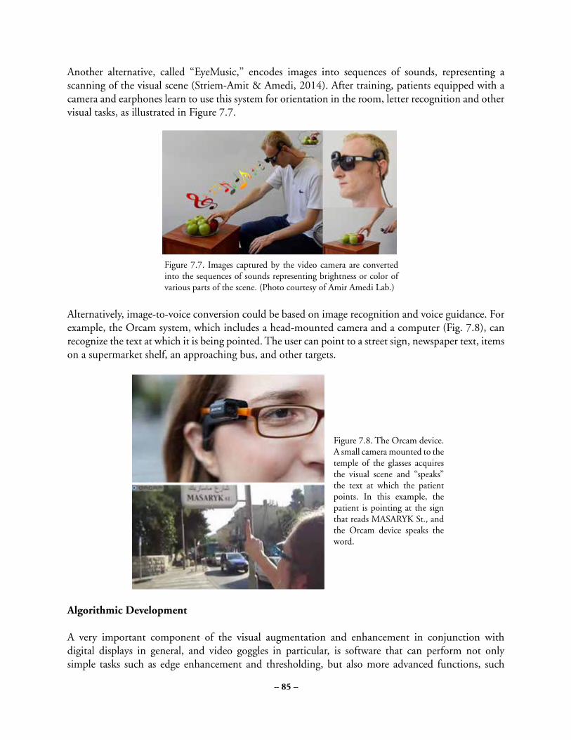
– 85 –
Another alternative, called ‘‘EyeMusic,’’ encodes images into sequences of sounds, representing a scanning of the visual scene (Striem-Amit & Amedi, 2014). After training, patients equipped with a camera and earphones learn to use this system for orientation in the room, letter recognition and other visual tasks, as illustrated in Figure 7.7.
Alternatively, image-to-voice conversion could be based on image recognition and voice guidance. For example, the Orcam system, which includes a head-mounted camera and a computer (Fig. 7.8), can recognize the text at which it is being pointed. The user can point to a street sign, newspaper text, items on a supermarket shelf, an approaching bus, and other targets.
Algorithmic Development
A very important component of the visual augmentation and enhancement in conjunction with digital displays in general, and video goggles in particular, is software that can perform not only simple tasks such as edge enhancement and thresholding, but also more advanced functions, such
Another alternative, called ‘‘EyeMusic,’’ encodes images into sequences of sounds, representing a scanning of the visualscene (Striem-Amit & Amedi, 2014). After training, patients equipped with a camera and earphones learn to use thissystem for orientation in the room, letter recognition and other visual tasks, as illustrated in Figure 7.7.
Alternatively, image-to-voice conversion could be based on image recognition and voice guidance. For example, theOrcam system, which includes a head-mounted camera and a computer (Fig. 7.8), can recognize the text at which it isbeing pointed. The user can point to a street sign, newspaper text, items on a supermarket shelf, an approaching bus, andother targets.
Algorithmic Development
A very important component of the visual augmentation and enhancement in conjunction with digital displays ingeneral, and video goggles in particular, is software that can perform not only simple tasks such as edge enhancement andthresholding, but also more advanced functions, such as image recognition and simplification. This should allow easily
Figure 7.7. Images captured by the video camera are converted into the sequences of sounds representing brightness or color of various parts of the
scene. (Photo courtesy of Amir Amedi Lab.)
Figure 7.8. The Orcam device. A small camera mounted to the temple of the glasses acquires the visual scene and ‘‘speaks’’ the text at which thepatient points. In this example, the patient is pointing at the sign that reads MASARYK St., and the Orcam device speaks the word.
Article 958
The Lasker/IRRF Initiative for Innovation in Vision Science
Another alternative, called ‘‘EyeMusic,’’ encodes images into sequences of sounds, representing a scanning of the visualscene (Striem-Amit & Amedi, 2014). After training, patients equipped with a camera and earphones learn to use thissystem for orientation in the room, letter recognition and other visual tasks, as illustrated in Figure 7.7.
Alternatively, image-to-voice conversion could be based on image recognition and voice guidance. For example, theOrcam system, which includes a head-mounted camera and a computer (Fig. 7.8), can recognize the text at which it isbeing pointed. The user can point to a street sign, newspaper text, items on a supermarket shelf, an approaching bus, andother targets.
Algorithmic Development
A very important component of the visual augmentation and enhancement in conjunction with digital displays ingeneral, and video goggles in particular, is software that can perform not only simple tasks such as edge enhancement andthresholding, but also more advanced functions, such as image recognition and simplification. This should allow easily
Figure 7.7. Images captured by the video camera are converted into the sequences of sounds representing brightness or color of various parts of the
scene. (Photo courtesy of Amir Amedi Lab.)
Figure 7.8. The Orcam device. A small camera mounted to the temple of the glasses acquires the visual scene and ‘‘speaks’’ the text at which thepatient points. In this example, the patient is pointing at the sign that reads MASARYK St., and the Orcam device speaks the word.
http://tvstjournal.org/doi/full/10.1167/tvst.3.7.9 TVST j 2014 j Vol. 3 j No. 7 j Article 958
The Lasker/IRRF Initiative for Innovation in Vision Science
Figure 7.7. Images captured by the video camera are converted into the sequences of sounds representing brightness or color of various parts of the scene. (Photo courtesy of Amir Amedi Lab.)
Figure 7.8. The Orcam device. A small camera mounted to the temple of the glasses acquires the visual scene and ‘‘speaks’’ the text at which the patient points. In this example, the patient is pointing at the sign that reads MASARYK St., and the Orcam device speaks the word.

– 86 –
as image recognition and simplification. This should allow easily recognizable symbolic or cartoon representation of the objects, ‘‘Platonic’’ ideas rather than real objects. One common example is the character recognition and representation of the clean and sharp fonts instead of the fuzzy text in the actual scene. Other objects could include simplified contours of the doors, tables, chairs, guide lines on sidewalks, faces, and so on. A recent advance is the depth encoding into the image, the Project Tango by Google (Fig. 7.9).
This technology maps the distances to the objects in the visual field and encodes them in a false color on top of the actual contours of the objects in the scene. Such a technique could provide additional guidance to the depth and spatial relationship of the objects that might be hard to achieve for the visually impaired. Additional help with navigation is offered by GPS, accelerometers and gyroscopes, internal navigation systems, and detailed maps, which allow comfortable orientation both indoors and outdoors. Modern user interfaces are also becoming much more intuitive and multifunctional than knobs and switches of previous devices. Devices can be controlled by gestures, voice, touch screen, and other nonintrusive actions. Similar algorithms for image simplification and enhanced scarcity (such as representation of the contours) should help with optical approaches to restoration of sight. In addition, sequential rather than simultaneous activation of pixels in the prosthetic approach may help to reduce cross-talk between simultaneously activated pixels, and thereby increase the contrast in the image. Selective activation of different retinal cell types with these techniques (achieved with selective expression of transgenes in optogenetics, selective binding of photoswitches in photopharmacology, or the specific location of electrodes and use of stimulation waveforms optimized for specific cell layers in electronic prostheses) may further help improve proper interpretation of the stimulation patterns by the brain. For example, stimulation of the inner nuclear layer performed at sufficiently high frame rate may allow for flicker fusion (Lorach et. al., 2014). Alternatively, direct activation of specific types of RGCs may include encoding of the projected visual scene into bursts of pulses corresponding to patterns of natural activity that RGCs would produce in response to images projected onto the healthy retina. Because image location on the retina is affected by eye movements, the latter type of activation will require precise eye tracking. Eye tracking also enables remapping of the retinal images to properly account for radial spread of the cells near the fovea (Asher et al., 2007) for restoration of the central vision, as illustrated in Figure 7.10. Similarly, it can be used for dynamic magnification of the parts of the image corresponding to the PRL, creating an effect of the magnifying glass following the eye’s direction of gaze, as illustrated in Figure 7.11.
Figure 7.9. Google ‘‘Project Tango’’ is a smartphone-sized device that maps the immediate environment in 3D, and displays the color coded depth. This could be very useful for the low vision patient as a way of orienting for both indoor and outdoor environments.
recognizable symbolic or cartoon representation of the objects, ‘‘Platonic’’ ideas rather than real objects. One commonexample is the character recognition and representation of the clean and sharp fonts instead of the fuzzy text in the actualscene. Other objects could include simplified contours of the doors, tables, chairs, guide lines on sidewalks, faces, and soon.
A recent advance is the depth encoding into the image, the Project Tango by Google (Fig. 7.9). This technology mapsthe distances to the objects in the visual field and encodes them in a false color on top of the actual contours of the objectsin the scene. Such a technique could provide additional guidance to the depth and spatial relationship of the objects thatmight be hard to achieve for the visually impaired.
Additional help with navigation is offered by GPS, accelerometers and gyroscopes, internal navigation systems, anddetailed maps, which allow comfortable orientation both indoors and outdoors. Modern user interfaces are also becomingmuch more intuitive and multifunctional than knobs and switches of previous devices. Devices can be controlled bygestures, voice, touch screen, and other nonintrusive actions.
Similar algorithms for image simplification and enhanced scarcity (such as representation of the contours) should helpwith optical approaches to restoration of sight. In addition, sequential rather than simultaneous activation of pixels in theprosthetic approach may help to reduce cross-talk between simultaneously activated pixels, and thereby increase thecontrast in the image. Selective activation of different retinal cell types with these techniques (achieved with selectiveexpression of transgenes in optogenetics, selective binding of photoswitches in photopharmacology, or the specificlocation of electrodes and use of stimulation waveforms optimized for specific cell layers in electronic prostheses) mayfurther help improve proper interpretation of the stimulation patterns by the brain. For example, stimulation of the innernuclear layer performed at sufficiently high frame rate may allow for flicker fusion (Lorach et. al., 2014). Alternatively,direct activation of specific types of RGCs may include encoding of the projected visual scene into bursts of pulsescorresponding to patterns of natural activity that RGCs would produce in response to images projected onto the healthyretina. Because image location on the retina is affected by eye movements, the latter type of activation will require preciseeye tracking.
Eye tracking also enables remapping of the retinal images to properly account for radial spread of the cells near thefovea (Asher et al., 2007) for restoration of the central vision, as illustrated in Figure 7.10. Similarly, it can be used fordynamic magnification of the parts of the image corresponding to the PRL, creating an effect of the magnifying glassfollowing the eye’s direction of gaze, as illustrated in Figure 7.11.
Recommendations for Future Research
1. Further advancement of the semitransparent video goggles with low weight and ergonomic design should minimizethe social awkwardness of the electronic eyewear. Higher resolution (beyond extended video graphics array [XVGA]),wider visual field (>40!), high brightness and contrast should allow comfortable viewing of the displayed informationoverlaid on the natural scene;
2. Advanced image processing, and especially image recognition, are other very promising directions of future research.These include text recognition with conversion into audio or into magnified and sharpened image, face recognition,and simplification of the object representation by the retention of essential features and removal of less importantdetails, etc.;
Figure 7.9. Google ‘‘Project Tango’’ is a smartphone-sized device that maps the immediate environment in 3D, and displays the color coded
depth. This could be very useful for the low vision patient as a way of orienting for both indoor and outdoor environments.
Article 959
The Lasker/IRRF Initiative for Innovation in Vision Science

– 87 –
Figure 7.10. A. Foveal pit and parafoveal area of the human retina, with diagrammatic illustration of the radial spread of the connections betweenthe photoreceptors in the fovea to bipolar and ganglion cells in the capillary-free zone. B. Visual scene on the photoreceptor plane, with a red crossindicating the fixation point for a particular direction of gaze. C. Same information remapped according to positions of the bipolar cells displacedfrom the fovea. Black disk corresponds to the central 2 degrees of the visual field with absent inner retinal neurons.
Figure 7.11. Image captured by the camera, processed for magnification, edge enhancement and contrast enhancement and projected from thevideo goggles onto the parafoveal region of the retina. Additional smart processing for dynamic magnification in the fovea is enabled by eye tracking.
http://tvstjournal.org/doi/full/10.1167/tvst.3.7.9 TVST j 2014 j Vol. 3 j No. 7 j Article 960
The Lasker/IRRF Initiative for Innovation in Vision Science
Figure 7.10. A. Foveal pit and parafoveal area of the human retina, with diagrammatic illustration of the radial spread of the connections between the photoreceptors in the fovea to bipolar and ganglion cells in the capillary-free zone. B. Visual scene on the photoreceptor plane, with a red cross indicating the fixation point for a particular direction of gaze. C. Same information remapped according to positions of the bipolar cells displaced from the fovea. Black disk corresponds to the central 2 degrees of the visual field with absent inner retinal neurons.
Figure 7.11. Image captured by the camera, processed for magnification, edge enhancement and contrast enhancement and projected from the video goggles onto the parafoveal region of the retina. Additional smart processing for dynamic magnification in the fovea is enabled by eye tracking.
Figure 7.10. A. Foveal pit and parafoveal area of the human retina, with diagrammatic illustration of the radial spread of the connections betweenthe photoreceptors in the fovea to bipolar and ganglion cells in the capillary-free zone. B. Visual scene on the photoreceptor plane, with a red crossindicating the fixation point for a particular direction of gaze. C. Same information remapped according to positions of the bipolar cells displacedfrom the fovea. Black disk corresponds to the central 2 degrees of the visual field with absent inner retinal neurons.
Figure 7.11. Image captured by the camera, processed for magnification, edge enhancement and contrast enhancement and projected from thevideo goggles onto the parafoveal region of the retina. Additional smart processing for dynamic magnification in the fovea is enabled by eye tracking.
http://tvstjournal.org/doi/full/10.1167/tvst.3.7.9 TVST j 2014 j Vol. 3 j No. 7 j Article 960
The Lasker/IRRF Initiative for Innovation in Vision Science

– 88 –
Recommendations for Future Research
1. Further advancement of the semitransparent video goggles with low weight and ergonomic design should minimize the social awkwardness of the electronic eyewear. Higher resolution (beyond extended video graphics array [XVGA]), wider visual field (>40°), high brightness and contrast should allow comfortable viewing of the displayed information overlaid on the natural scene;
2. Advanced image processing, and especially image recognition, are other very promising directions of future research. These include text recognition with conversion into audio or into magnified and sharpened image, face recognition, and simplification of the object representation by the retention of essential features and removal of less important details, etc.;
3. Advancements in the field of three-dimensional cameras will help encode depth information and provide additional warnings about the obstacles. The integration of maps (internal and external) with GPS, gyroscopes, and other guiding devices will help improve orientation on streets and inside buildings;
4. Progress in eye tracking technology, and its miniaturization will allow its comfortable integration with video goggles, which will, in turn, enable advanced image processing related to direction of gaze; and
5. Similar algorithms for image simplification and enhanced scarcity should help with optical approaches to restoration of sight. Better understanding of pathways of retinal and cortical processing of artificial vision will help to optimize the algorithms of image presentation in these vision restoration approaches.
References
Anon. (2012). Organization and Institution News: Visual impairment and blindness increase in over 40 population this past decade. Optometry and Vision Science, 89(8), 1239. Retrieved from http://journals.lww.com/optvissci/Fulltext/ 2012/08000/In_The_News_New_Products.22.aspx
Arditi, A., & Rosenthal, B. (1998). Developing an objective definition of visual impairment. In Proceedings in Vision ‘96: Proceedings of the international low vision conference (pp. 331–334). Madrid, Spain: Medicare.
Arnoldussen, A., & Fletcher, D.C. (2012). Visual perception for the blind: The BrainPort Vision Device. Retinal Physician, 9, 32–34.
Asher, A., Segal, W.A., Baccus, S.A., Yaroslavsky, L.P., & Palanker, D.V. (2007). Image processing for a high-resolution optoelectronic retinal prosthesis. IEEE Transactions on Bio-Medical Engineering, 54, 993–1004.
Busskamp, V., Duebel, J., Balya, D., Fradot, M., Viney, T.J., Siegert, S., . . . Groner, A.C. (2010). Genetic reactivation of cone photoreceptors restores visual responses in retinitis pigmentosa. Science, 329, 413–417.

– 89 –
Culham, L.E., Chabra, A., & Rubin, G.S. (2009). Users’ subjective evaluation of electronic vision enhancement systems. Ophthalmic & Physiological Optics: The Journal of the British College of Ophthalmic Opticians (Optometrists), 29, 138– 149. PubMed PMID: 19236583.
Culham, L.E., Chabra, A., & Rubin, G.S. (2004). Clinical performance of electronic, head-mounted, low-vision devices. Ophthalmic & Physiological Optics: The Journal of the British College of Ophthalmic Opticians (Optometrists), 24, 281– 290. PubMed PMID: 15228505.
Lagali, P.S., Balya, D., Awatramani, G.B., Munch, T.A., Kim, D.S., Busskamp, V., . . . Cepko, C.L. (2008). Light activated channels targeted to ON bipolar cells restore visual function in retinal degeneration. Nature Neuroscience, 11, 667–675.
Lorach, H., Goetz, G., Mandel, Y., Lei, X., Kamins, T.I., Mathieson, K., . . . Huie, P. (2014). Performance of photovoltaic arrays in-vivo and characteristics of prosthetic vision in animals with retinal degeneration [published online ahead of print September 26, 2014]. Vision Research. doi: 10.1016/j.visres.2014.09.007.
Lumus. Retrieved from http://www.lumus optical.com/index.php?option.com_content&taskview& id.9&Itemid.15.
Mandel, Y., Goetz, G., Lavinsky, D., Huie, P., Mathieson, K., Wang, L., . . . Kamins, T. (2013). Cortical responses elicited by photovoltaic subretinal prostheses exhibit similarities to visually evoked potentials. Nature Communications, 4, 1980.
Mathieson, K., Loudin, J., Goetz, G., Huie, P., Wang, L., Kamins, T.I., . . . Galambos, L. (2012). Photovoltaic retinal prosthesis with high pixel density. Nature Photonics, 6, 391–397.
Medicare Vision Rehabilitation Services Act of 2003 HR 1902 IH. 2003.
Polosukhina, A., Litt, J., Tochitsky, I., Nemargut, J., Sychev, Y., De Kouchkovsky, I., . . . Huang, T. (2012). Photochemical restoration of visual responses in blind mice. Neuron, 75, 271–282.
Striem-Amit, E., & Amedi, A. (2014). Visual cortex extrastriate body-selective area activation in congenitally blind people ‘‘seeing’’ by using sounds. Current Biology, 24, 687–692.
Tochitsky, I., Polosukhina, A., Degtyar, V.E., Gallerani, N., Smith, C.M., Friedman, A., . . . Van Gelder, R.N. (2014). Restoring visual function to blind mice with a photoswitch that exploits electrophysiological remodeling of retinal ganglion cells. Neuron, 81, 800–813.

– 90 –

– 91 –
Chapter 8Evaluating Visual Function, Endpoints
Discussion Leaders: David Birch, Fred Fitzke, and Gary Rubin
Scribe: Blake Hopiavuori
Session Participants: Ernst Bamberg, Alan Bird, Larry Donoso, Jacque Duncan, Don Hood, Alan Laties, Gary Rubin, Paul Sieving, and Jane Sowden
Introduction
One of the areas of greatest need in visual assessment is for tests to measure the degree of visual function that has been restored to a blind or low vision patient. Visual function and endpoint measures include a wide range of techniques for determining the structural integrity of the eye and visual pathways (e.g., electrophysiology and ocular imaging), for assessing its functional capability (e.g., visual acuity and contrast sensitivity) and for evaluating the impact of vision loss on the person’s ability to do everyday visual tasks (performance-based tests, such as reading and self-reported visual ability).
Despite numerous advances in structural, functional, and performance-related endpoints, clinicians are limited to a very restricted range of tests that have received regulatory approval – color fundus photographs, high contrast visual acuity, and a limited set of patient questionnaires. The main goal of this chapter is to promote novel visual function measures that will be useful for evaluating new therapeutic modalities, such as stem cells, gene therapy, and ocular prostheses. For these endpoints to be useful, we must establish that the measures are valid, reliable, and sensitive to change. Throughout this process we must not lose sight of the overriding requirement that our endpoints demonstrate how the new therapies benefit the patient. It is not sufficient merely to establish that a new treatment leads to a statistically significant difference; we also must show that the change is clinically significant, that the difference makes a difference to the patient.
A Focus on Rods (Broadening Our Clinical Assessment)
Some of the new endpoints we will consider have come to our attention because they promise greater sensitivity to early or preclinical disease than conventional measures. Other endpoints are promoted because they measure functional outcomes that are of particular importance to patients. An assessment of rod function does both (Owsley et al., 2007).
Measurements of cone function can be insensitive to change in some forms of retinal disease. As an alternative, measurement of rod function has been underutilized. We believe that the measurement of rod function reflects aspects of visual function of importance to the patient and often can show changes before conventional measures of cone function. It could be that rods are so susceptible to disease and physiologic stress that they show much higher sensitivity to changes due to the varying underlying pathologies and, therefore, are a more sensitive indicator of decline as well as benefit from treatment. Furthermore, with regard to transplantation as a potential therapy, rods are the most likely

– 92 –
candidates to be transplanted since, to date, rod transplantation has been more successful than cone transplantation. Therefore, we must be able to adequately and reproducibly measure rod function. In animal model experiments, functional transplantation for rods is far ahead of that for cones in terms of efficiency (Homma et al., 2013; Lakowski et al., 2010; Pearson 2014). The idea of rods supporting cone survival also should be assessed, and it is important to measure rod and cone function following rod transplantation to determine any subsequent effect on cone function.
For assessing rod function, dark-adapted visual fields can be used to establish a good baseline for each patient. Longitudinal measures are useful for detecting regional changes correlated with the site of disease. Alternately, in the case of therapy, rod fields can be used to detect changes correlated to the site of treatment. Progressively fewer photons of light are absorbed as outer segments become shorter or are lost. By the same logic, the shorter outer segments of the cones result in fewer photons being absorbed by the retina compared to rods, thus, partially explaining the lower sensitivity of cones to measurement. Rod photoreceptors measured in dark-adapted conditions can show losses of sensitivity by a factor of 100 even when conventional photopic measurements show minimal or no loss of sensitivity. In this regard, it could be highly beneficial to the patient for us to place equal focus on rods as well as cones in our functional measurements.
One limitation to the assessment of rod function in clinical trials has been the lack of specific equipment. The old standard, the Goldmann-Weekers dark adaptometer, is no longer available and, as yet, nothing has replaced it. Candidate devices include the Nidek MP1S (Birch, Wen, Locke, & Hood, 2011; Crossland, Luong, Rubin, & Fitzke, 2010), which is a fundus perimeter modified for two-color scotopic testing, and modified static perimeters (Jacobson et al., 1986). The limitations of these devices are that they have either limited dynamic ranges (fundus perimeters) or require extensive user modifications (static perimeters). It is expected, however, that an LED perimeter optimized for two-color perimetry will soon be available to facilitate perimetric assessment of rod function.
idea of rods supporting cone survival also should be assessed, and it is important to measure rod and cone functionfollowing rod transplantation to determine any subsequent effect on cone function.
For assessing rod function, dark-adapted visual fields can be used to establish a good baseline for each patient.Longitudinal measures are useful for detecting regional changes correlated with the site of disease. Alternately, in thecase of therapy, rod fields can be used to detect changes correlated to the site of treatment. Progressively fewer photonsof light are absorbed as outer segments become shorter or are lost. By the same logic, the shorter outer segments of thecones result in fewer photons being absorbed by the retina compared to rods, thus, partially explaining the lowersensitivity of cones to measurement. Rod photoreceptors measured in dark-adapted conditions can show losses ofsensitivity by a factor of 100 even when conventional photopic measurements show minimal or no loss of sensitivity. Inthis regard, it could be highly beneficial to the patient for us to place equal focus on rods as well as cones in ourfunctional measurements.
One limitation to the assessment of rod function in clinical trials has been the lack of specific equipment. The oldstandard, the Goldmann-Weekers dark adaptometer, is no longer available and, as yet, nothing has replaced it. Candidatedevices include the Nidek MP1S (Birch, Wen, Locke, & Hood, 2011; Crossland, Luong, Rubin, & Fitzke, 2010), whichis a fundus perimeter modified for two-color scotopic testing, and modified static perimeters (Jacobson et al., 1986). Thelimitations of these devices are that they have either limited dynamic ranges (fundus perimeters) or require extensive usermodifications (static perimeters). It is expected, however, that an LED perimeter optimized for two-color perimetry willsoon be available to facilitate perimetric assessment of rod function.
In terms of detecting changes in visual function before they are noticed by the patient, it has been demonstrated thatthe RPE of patients with macular dystrophy show an early increase in autofluorescence and that the function of the rodphotoreceptors corresponding to this region shows reductions in sensitivity (Downes et al., 1999).
We believe that this may be particularly important in age-related macular degeneration (AMD) where areas ofincreased parafoveal autofluorescence may be detected despite normal cone function. Therefore, in developing follow-upassessments for AMD patients it would be critical to evaluate the function and integrity of these parafoveal rods. It is wellknown that in retinitis pigmentosa (RP) rod photoreceptor loss is the primary defect. However, in AMD, a growing bodyof evidence seems to support the idea that parafoveal rod death actually precedes the geographic macular atrophy (Curcio,Medeiros, & Millican, 1996). In recent years new technology has become available that would allow us to evaluateparafoveal rod integrity; specifically, fundus autofluorescence imaging (FAF) can be used to assess region variations in thehealth of rods. FAF was shown to detect increased parafoveal autofluorescence in a young 17-year-old patient with nosymptoms of vision loss and with a family history of codon 172 RDS macular dystrophy (Downes et al., 1999). Thisstudy (Fig. 8.1) showed that abnormally high autofluorescence predated loss of visual acuity or visual field changes inthese patients with a high level of reliability, making FAF an extremely powerful tool in evaluating rod function and evenpredicting the geographic location of rod degeneration before it actually leads to any subjective deficit.
Figure 8.1. Left: Increased parafoveal autofluorescence in an asymptomatic 17-year-old patient with codon 172 RDS macular dystrophy Right:Fine matrix mapping within the region of abnormally-high autofluorescence reveals that rod sensitivity is reduced on the average of 10 dB relative to
normal. Numbers are sensitivity values from scotopic perimetry superimposed on fundus image. The higher the number, the lower the sensitivity;
image on right is magnified as compared to left image. (Image reprinted with permission from Downes, S.M., Fitzke, F.W., Holder, G.E., Payne,
A.M., Bessant, D.A., Bhattacharya, S.S., & Bird, A.C. (1999). Clinical features of codon 172 RDS macular dystrophy: similar phenotype in 12
families. Archives of Ophthalmology, 117(10), 1373–1383. Copyright ! 1999 American Medical Association. All rights reserved.)
http://tvstjournal.org/doi/full/10.1167/tvst.3.7.10 TVST j 2014 j Vol. 3 j No. 7 j Article 1064
The Lasker/IRRF Initiative for Innovation in Vision Science
Figure 8.1. Left: Increased parafoveal autofluorescence in an asymptomatic 17-year-old patient with codon 172 RDS macular dystrophy Right: Fine matrix mapping within the region of abnormally-high autofluorescence reveals that rod sensitivity is reduced on the average of 10 dB relative to normal. Numbers are sensitivity values from scotopic perimetry superimposed on fundus image. The higher the number, the lower the sensitivity; image on right is magnified as compared to left image. (Image reprinted with permission from Downes, S.M., Fitzke, F.W., Holder, G.E., Payne, A.M., Bessant, D.A., Bhattacharya, S.S., & Bird, A.C. (1999). Clinical features of codon 172 RDS macular dystrophy: similar phenotype in 12 families. Archives of Ophthalmology, 117(10), 1373–1383. Copyright 1999 American Medical Association. All rights reserved.)

– 93 –
In terms of detecting changes in visual function before they are noticed by the patient, it has been demonstrated that the RPE of patients with macular dystrophy show an early increase in autofluorescence and that the function of the rod photoreceptors corresponding to this region shows reductions in sensitivity (Downes et al., 1999).
We believe that this may be particularly important in age-related macular degeneration (AMD) where areas of increased parafoveal autofluorescence may be detected despite normal cone function. Therefore, in developing follow-up assessments for AMD patients it would be critical to evaluate the function and integrity of these parafoveal rods. It is well known that in retinitis pigmentosa (RP) rod photoreceptor loss is the primary defect. However, in AMD, a growing body of evidence seems to support the idea that parafoveal rod death actually precedes the geographic macular atrophy (Curcio, Medeiros, & Millican, 1996). In recent years new technology has become available that would allow us to evaluate parafoveal rod integrity; specifically, fundus autofluorescence imaging (FAF) can be used to assess region variations in the health of rods. FAF was shown to detect increased parafoveal autofluorescence in a young 17-year-old patient with no symptoms of vision loss and with a family history of codon 172 RDS macular dystrophy (Downes et al., 1999). This study (Fig. 8.1) showed that abnormally high autofluorescence predated loss of visual acuity or visual field changes in these patients with a high level of reliability, making FAF an extremely powerful tool in evaluating rod function and even predicting the geographic location of rod degeneration before it actually leads to any subjective deficit.
Figure 8.2. Left: EZ line is sensitive to progression in x-linked retinitis pigmentosa; constriction over three years in representative patient. The arrows in the magnified sections to the right show the nasal termination of the EZ on each visit. (Reprinted with permission from Birch, D.G., Locke, K.G., Wen, Y., Locke, K.I., Hoffman, D.R., & Hood, D.C. (2013). Spectral-domain optical coherence tomography measures of outer segment layer progression in patients with X-linked retinitis pigmentosa. JAMA Ophthalmology, 131(9), 1143–1150. Copyright 2013 American Medical Association. All rights reserved.) Right: EZ line correlates with visual field loss. The Humphrey 10-2 visual field losses (dB) are shown with the boundary (red curve) of the EZ loss for the patient. White numbers indicate field locations where sensitivity is within 3 dB of mean normal; red numbers indicate loss of at least 10 dB. (From Hood, D.C., Ramachandran, R., Holopigian, K., Lazow, M., Birch, D.G., & Greenstein, V.G. (2011). Method for deriving visual field boundaries from OCT scans of patients with retinitis pigmentosa. Biomedical Optics Express, 2, 1106–1114.)
Spectral-domain optical coherence tomography (OCT) is another tool that can reveal structural abnormalities thatmay be highly correlated to changes in rod and cone function over time. Recent studies have focused on the width of theinner segment ellipsoid zone (EZ; i.e., inner/outer segment border). The general finding is that the edge of the EZ bandmarks the edge of the patient’s ‘‘usable’’ visual field. Once the patient has lost the EZ band entirely, there is a visual fieldreduction of at least 8 dB (Hood et al., 2011). Birch et al. (2013) showed the validity of this technique as an outcomemeasure for clinical trials of RP. Of the patients with x-linked RP (XLRP) evaluated, 96% showed a significant decrease inthe EZ width after 2 years, with a mean annual decrease of 7%, which would be difficult or even impossible to detect witha standard visual field (Fig. 8.2). Considering the repeat variability was less than the annual rate of change in these patients(95% of the test–retest differences fell within a change of only 3.5%), this method promises to reliably assess changes inretinal function over a relatively short period of time (Birch et al., 2013). EZ width or area is a structural correlate of thevisual field. The advantage of EZ width measurements is that they are less variable on a visit-to-visit basis than visual fieldmeasurements. It appears that the edge of the EZ band on horizontal or vertical line scans was most effective for detectingprogression, and this can be done manually without the aid of an algorithm or segmentation (Ramachandran et al., 2013).This type of evaluation is highly valuable to the patient for its ability to establish their rate of progression/improvementover long periods of time.
The advantage of this technique is that it is a very short test that is easy on the patient and operator with excellentreproducibility in human and animal studies. The disadvantages are that it cannot separate rod versus cone loss, reliabilitycan be reduced in patients with poor fixation, and far peripheral vision is difficult to assess (outside ! 30"). Because it islimited at present to the central retina, it, therefore, is not the ideal measure for early detection of vision loss in diseasesthat first show defects in the peripheral retina.
Adaptive Optics Microperimetry is a novel, yet extremely powerful tool in that it enables the operator to measurepsychophysical function from individual cone photoreceptors or assess visual function in regions with visible retinalpigment epithelial cells, while simultaneously imaging them using an adaptive optics scanning laser ophthalmoscope(AOSLO) (Figure 8.3) (Poonja et al., 2005; Tuten et al., 2012). This technique may be particularly useful to assess visualfunction with cellular resolution in diseases with intraretinal variability such as age-related macular degeneration andStargardt disease, where it may be useful to assess function in regions of lipofuscin accumulation or at the margins of
Figure 8.2. Left: EZ line is sensitive to progression in x-linked retinitis pigmentosa; constriction over three years in representative patient. Thearrows in the magnified sections to the right show the nasal termination of the EZ on each visit. (Reprinted with permission from Birch, D.G.,Locke, K.G., Wen, Y., Locke, K.I., Hoffman, D.R., & Hood, D.C. (2013). Spectral-domain optical coherence tomography measures of outersegment layer progression in patients with X-linked retinitis pigmentosa. JAMA Ophthalmology, 131(9), 1143–1150. Copyright ! 2013American Medical Association. All rights reserved.) Right: EZ line correlates with visual field loss. The Humphrey 10-2 visual field losses (dB) areshown with the boundary (red curve) of the EZ loss for the patient.White numbers indicate field locations where sensitivity is within 3 dB of meannormal; red numbers indicate loss of at least 10 dB. (From Hood, D.C., Ramachandran, R., Holopigian, K., Lazow, M., Birch, D.G., &Greenstein, V.G. (2011). Method for deriving visual field boundaries from OCT scans of patients with retinitis pigmentosa. Biomedical OpticsExpress, 2, 1106–1114.)
http://tvstjournal.org/doi/full/10.1167/tvst.3.7.10 TVST j 2014 j Vol. 3 j No. 7 j Article 1065
The Lasker/IRRF Initiative for Innovation in Vision Science

– 94 –
Spectral-domain optical coherence tomography (OCT) is another tool that can reveal structural abnormalities that may be highly correlated to changes in rod and cone function over time. Recent studies have focused on the width of the inner segment ellipsoid zone (EZ; i.e., inner/outer segment border). The general finding is that the edge of the EZ band marks the edge of the patient’s ‘‘usable’’ visual field. Once the patient has lost the EZ band entirely, there is a visual field reduction of at least 8 dB (Hood et al., 2011). Birch et al. (2013) showed the validity of this technique as an outcome measure for clinical trials of RP. Of the patients with x-linked RP (XLRP) evaluated, 96% showed a significant decrease in the EZ width after 2 years, with a mean annual decrease of 7%, which would be difficult or even impossible to detect with a standard visual field (Fig. 8.2). Considering the repeat variability was less than the annual rate of change in these patients (95% of the test–retest differences fell within a change of only 3.5%), this method promises to reliably assess changes in retinal function over a relatively short period of time (Birch et al., 2013). EZ width or area is a structural correlate of the visual field. The advantage of EZ width measurements is that they are less variable on a visit-to-visit basis than visual field measurements. It appears that the edge of the EZ band on horizontal or vertical line scans was most effective for detecting progression, and this can be done manually without the aid of an algorithm or segmentation (Ramachandran et al., 2013). This type of evaluation is highly valuable to the patient for its ability to establish their rate of progression/improvement over long periods of time.
The advantage of this technique is that it is a very short test that is easy on the patient and operator with excellent reproducibility in human and animal studies. The disadvantages are that it cannot separate rod versus cone loss, reliability can be reduced in patients with poor fixation, and far peripheral vision is difficult to assess (outside +/- 30˚). Because it is limited at present to the central retina, it, therefore, is not the ideal measure for early detection of vision loss in diseases that first show defects in the peripheral retina.
Adaptive Optics Microperimetry is a novel, yet extremely powerful tool in that it enables the operator to measure psychophysical function from individual cone photoreceptors or assess visual function in regions with visible retinal pigment epithelial cells, while simultaneously imaging them using an adaptive optics scanning laser ophthalmoscope (AOSLO) (Figure 8.3) (Poonja et al., 2005; Tuten et al., 2012). This technique may be particularly useful to assess visual function with cellular resolution in diseases with intraretinal variability such as age-related macular degeneration and Stargardt disease, where it may be useful to assess function in regions of lipofuscin accumulation or at the margins of atrophy, and in macular telangiectasia type 2, in which very focal regions of abnormality can exist adjacent to nearly normal or normal retinal structures. Adaptive optics microperimetry could be used in conjunction with other measurement endpoints to assess improvement following targeted neuroprotective therapies including subretinal injection or even photoreceptor transplantation to evaluate the structure and function of those specifically treated or transplanted cells as they integrate into the retina (Menghini & Duncan, 2014). Evaluation of rods and cones in regions where the new cells can be identified following transplantation will be essential to determine their functional efficacy.
Session participants also discussed the evidence that preservation of rods would generate long-term benefit to cone function. From the patient’s point of view, preservation of cone function is extremely important, but investigations of rod function may provide an earlier indicator of therapeutic benefit, but for the quality of life of patients, cone function in many cases takes priority, although standard

– 95 –
measures of cone function such as visual acuity are often preserved until late stages of the disease, making them insensitive measures of disease progression. Some of this technology is not widely used yet; however, it could provide the means to detect impending cone death before it actually happens. It will be important to encourage the standardization and widespread use of these techniques if we are to develop a new clinical standard that allows for the evaluation of endpoints based on rod functional measurements.
Additional Structure and Functional Outcome Measures That Should Be Considered
1. Reflectometry is a method not currently used; however, it allows for the measurement of rhodopsin concentration. This method will allow us to identify subtypes of disease potentially, prior to going into clinical trials. In some forms of retinal disease, patients can lose rod function, but still have plenty of rhodopsin, and in other diseases, the loss of function is due to rod cell death specifically; therefore, these patients would have lower rhodopsin levels (Kilbride & Keehan, 1990).
2. Electrophysiology measurements have an important role in the clinic for safety measurements as well as for localized measurements of function by using multifocal ERG as well as localized ERG. However, these techniques are subject to high variability, which must be taken into account as we look toward standardized outcomes (Fishman, Chappelow, Anderson, Rotenstreich, & Derlacki, 2005). Combining these outcome measurements with data from visual fields and optical coherence tomography (OCT) could serve to lower this inherent variability.
3. Dark adaptation kinetics may be particularly important for assessment of early AMD (Owsley, Jackson, White, Feist, & Edwards, 2001). However, it may be most useful as a screening test to analyze several different locations on the retina. Depending on disease course and progression, some patients will have a perfectly normal acuity, but severe dysfunction in the far periphery. Unfortunately,
Figure 8.3. Imaging and stimulating photoreceptors through adaptive optics scanning laser ophthalmoscope (AOSLO). By modulating the scanning beam, test stimuli can be delivered to one or a small number of cones. The subject sees the green stimulus against a red background. The operator sees the green test light stabilized on the cone mosaic. With eye tracking and image stabilization it is possible to test the same cones on subsequent visits. (Image courtesy of Austin Roorda, PhD, University of California, Berkeley.)
atrophy, and in macular telangiectasia type 2, in which very focal regions of abnormality can exist adjacent to nearlynormal or normal retinal structures. Adaptive optics microperimetry could be used in conjunction with othermeasurement endpoints to assess improvement following targeted neuroprotective therapies including sub-retinalinjection or even photoreceptor transplantation to evaluate the structure and function of those specifically treated ortransplanted cells as they integrate into the retina (Menghini & Duncan, 2014). Evaluation of rods and cones in regionswhere the new cells can be identified following transplantation will be essential to determine their functional efficacy.
Session participants also discussed the evidence that preservation of rods would generate long-term benefit to conefunction. From the patient’s point of view, preservation of cone function is extremely important, but investigations of rodfunction may provide an earlier indicator of therapeutic benefit, but for the quality of life of patients, cone function inmay cases takes priority, although standard measures of cone function such as visual acuity are often preserved until latestages of the disease, making them insensitive measures of disease progression. Some of this technology is not widely usedyet; however, it could provide the means to detect impending cone death before it actually happens. It will be importantto encourage the standardization and widespread use of these techniques if we are to develop a new clinical standard thatallows for the evaluation of endpoints based on rod functional measurements.
Additional Structural and Functional Outcome Measures
Additional potential outcome measures that should be considered are listed below:
1. Reflectometry is a method not currently used; however, it allows for the measurement of rhodopsin concentration.This method will allow us to identify subtypes of disease potentially, prior to going into clinical trials. In some formsof retinal disease, patients can lose rod function, but still have plenty of rhodopsin, and in other diseases, the loss offunction is due to rod cell death specifically; therefore, these patients would have lower rhodopsin levels (Kilbride &Keehan, 1990).
2. Electrophysiology measurements have an important role in the clinic for safety measurements as well as for localizedmeasurements of function by using multifocal ERG as well as localized ERG. However, these techniques are subject tohigh variability, which must be taken into account as we look toward standardized outcomes (Fishman, Chappelow,Anderson, Rotenstreich, & Derlacki, 2005). Combining these outcome measurements with data from visual fields andoptical coherence tomography (OCT) could serve to lower this inherent variability.
3. Dark adaptation kinetics may be particularly important for assessment of early AMD (Owsley, Jackson, White, Feist,& Edwards, 2001). However, it may be most useful as a screening test to analyze several different locations on theretina. Depending on disease course and progression, some patients will have a perfectly normal acuity, but severedysfunction in the far periphery. Unfortunately, this outcome measure is limited by the number of locations that canbe accurately and reproducibly measured and by the time consumed for the patient and the operator.
Figure 8.3. Imaging and stimulating photoreceptors through adaptive optics scanning laser ophthalmoscope (AOSLO). By modulating thescanning beam, test stimuli can be delivered to one or a small number of cones. The subject sees the green stimulus against a red background. Theoperator sees the green test light stabilized on the cone mosaic. With eye tracking and image stabilization it is possible to test the same cones onsubsequent visits. (Image courtesy of Austin Roorda, PhD, University of California, Berkeley.)
http://tvstjournal.org/doi/full/10.1167/tvst.3.7.10 TVST j 2014 j Vol. 3 j No. 7 j Article 1066
The Lasker/IRRF Initiative for Innovation in Vision Science

– 96 –
this outcome measure is limited by the number of locations that can be accurately and reproducibly measured and by the time consumed for the patient and the operator.
4. Full-field sensitivity test (FST) is a quick test that is easy for the patient and operator alike. However, it has very low spatial resolution, making it a poor outcome measure for evaluating decline or even long-term improvement. Nonetheless, for patients who have lost enough of their photoreceptors to justify photoreceptor transplantation this would be an ideal test, as it will pick up the response of the most sensitive cells remaining in the retina and could be used to confirm functional integration of transplanted photoreceptors (Jacobson et al., 2009; Klein & Birch, 2009).
Importance of Standardization
One obstacle at this point has been the lack of suitable and standardized equipment. Possible approaches utilizing new equipment that can be widely used and standardized were discussed. Unfortunately, there is not an agreed-upon standard, and the outcome measures that are slightly more standardized, such as dark-adapted visual fields, are very time consuming and still do not generate a complete picture of disease progression or improvement. It will be important to develop a new clinical standard that allows endpoints that can be widely accepted by the community, and that are comprehensive and easy to perform for the patient and operator.
Patient-Reported Outcome Measures (PROMs) and Performance-Based Tests
PROMs have seized the attention of clinicians and regulators. Along with this increased interest there has been a deluge of new questionnaires – over 100 for eyes and vision alone. There is not space here to even begin a description of relevant questionnaires, but it is important to note that, along with the newfound interest in questionnaires, there has been a shift in the psychometric methods used to validate the questionnaires. So-called ‘‘Classical Test Theory’’ that uses techniques like factor analysis to develop scoring algorithms and subscales has been supplanted by ‘‘Item Response Theory,’’ especially Rasch analysis (Bond & Fox, 2007). Rasch analysis purports to convert ordinal rating scales that are used with most questionnaires into interval scales, making the questionnaire better suited for computer-aided testing and parametric statistics.
Performance-based tests (PBTs) measure subjects’ speed and accuracy while performing laboratory-based simulations of everyday visual tasks. These include mobility tasks, reading, and face and object recognition. The tests can be simple – reading sentences presented in a graduated series of letter sizes—or complex—navigating through a virtual obstacle course in a virtual reality lab. One of the challenges raised by these PBTs is standardization versus relevance to real-world tasks. Take reading, for example. There are highly standardized reading tests that use sequences of unrelated words, all with the same word length and frequency, versus tests based on passages of meaningful text with uncontrolled syntactic and semantic complexity. The former removes cognitive and linguistic factors that may be unrelated to vision.
Standardization increases reliability, but does this come at the price of reducing the relevance of the test to real-world activities? The answer seems to be ‘‘no.’’ Measured performance on a highly standardized reading test still is closely correlated with reading speed measured at home under natural conditions

– 97 –
(West, Rubin, Munoz, Abraham, & Fried, 1997). A second challenge is to develop performance-based tests that can be used by people in different cultures and who speak different languages. This is hardly a problem for structural outcome measures (e.g., OCT) or visual function tests (e.g., visual field), but it is far more difficult when one wants to measure reading speed in a multicenter clinical trial. It is not enough merely to translate the text into the local language; the texts may have to be linguistically equated for differences in complexity (Hahn et al., 2006).
Finally, there is the problem of the speed/accuracy tradeoff. This is particularly evident for timed tasks like reading and mobility where patients may differ in how they prioritize speed versus accuracy in performing the task.
As we move toward the standardization of novel outcome measures, an example we may wish to follow is that set by the Core Outcome Measures in Effectiveness Trials Initiative (COMET) trials, where people come together and agree in advance which outcome measures need to be done or established. This requires a level of cooperation that we as a community do not always exhibit. Our goal is to achieve a standard similar to the International Society for the Clinical Electrophysiology of Vision (ISCEV) where the main objectives are to promote and extend the knowledge of certain agreed-upon clinical endpoints as well as to promote cooperation and communication among workers in the field. This would involve the generation and publication of thoroughly validated and accepted measures that could be set in place for the greater goal of accurately and reproducibly measuring the patient endpoint.
Consensus Recommendations
1. Develop new methods of measuring visual function, especially for those undergoing treatments for restoring vision.
2. Develop standardized outcome measures that are accepted by the field. This requires cooperation in generating these measures.
3. Place renewed focus on measuring rod function and defects.
4. Recognize that certain endpoints may not be ideal for a specific level of vision loss. Encourage the identification of measurements specific for various levels of vision loss.
References
Birch, D.G, Locke, K.G., Wen, Y., Locke, K.I., Hoffman, D.R., & Hood, D.C. (2013). Spectral-domain optical coherence tomography measures of outer segment layer progression in patients with X-linked retinitis pigmentosa. JAMA Ophthalmology, 131, 1143–1150.
Birch, D.G., Wen, Y., Locke, K.I., & Hood, D.C. (2011). Rod sensitivity, cone sensitivity and photoreceptor layer thickness in retinal degenerative diseases. Investigative Ophthalmology and Visual Science, 52, 7141–7147.

– 98 –
Bond, T.G., & Fox, C.M. (2007). Applying the Rasch Model: Fundamental measurement in the human sciences (p. 335). Mahwah, NJ: Lawrence Erlbaum Associates Publishers.
Crossland, M., Luong, V.A., Rubin, G., & Fitzke, F. (2010). Retinal-specific measurements of dark-adapted visual function using the MP-1 microperimeter. Investigative Ophthalmology and Visual Science, 51, ARVO E-Abstract 3640.
Curcio, C.A., Medeiros, N.E., & Millican, C.L. (1996). Photoreceptor loss in age-related macular degeneration. Investigative Ophthalmology and Visual Science, 37, 1236–1249.
Downes, S.M., Fitzke, F.W., Holder, G.E., Payne, A.M., Bessant, D.A., Bhattacharya, S.S., & Bird, A.C. (1999). Clinical features of codon 172 RDS macular dystrophy: similar phenotype in 12 families. Archives of Ophthalmology, 117, 1373–1383.
Fishman, G.A., Chappelow, A.V., Anderson, R.J., Rotenstreich, Y., & Derlacki, D.J. (2005). Short-term inter-visit variability of erg amplitudes in normal subjects and patients with retinitis pigmentosa. Retina, 25, 1014–1021.
Hahn, G.A., Penka, D., Gehrlich, C., Messias, A., Weismann, M., Hyvarinen, L., . . . Trauzettel-Klosinski, S. (2006). New standardised texts for assessing reading performance in four European languages. British Journal of Ophthalmology, 90, 480–484.
Hood, D.C., Ramachandran, R., Holopigian, K., Lazow, M., Birch, D.G., & Greenstein, V.G. (2011). Method for deriving visual field boundaries from OCT scans of patients with retinitis pigmentosa. Biomedical Optics Express, 2, 1106–1114.
Homma, K., Okamoto, S., Mandai, M., Gotoh, N., Rajasimha, H.K., Chang, Y.S., . . . Takahashi, M. (2013). Developing rods transplanted into the degenerating retina of Crx-knockout mice exhibit neural activity similar to native photoreceptors. Stem Cells, 31, 1149–1159.
Jacobson, S.G., Aleman, T.S., Cideciyan, A.V., Roman, A.J., Sumaroka, A., Windsor, E.A., . . . Stone, E.M. (2009). Defining the residual vision in leber congenital amaurosis caused by RPE65 mutations. Investigative Ophthalmology and Visual Science, 50, 2368–2375.
Jacobson, S.G., Voigt, W.J., Parel, J.M., Apathy, P.P., Nghiem-Phu, L., Myers, S.W., & Patella, V.M. (1986). Automated light- and dark-adapted perimetry for evaluating retinitis pigmentosa. Ophthalmology, 93, 1604–1611.
Kilbride, P.E., & Keehan, K.M. (1990). Visual pigments in the human macula assessed by imaging fundus reflectometry. Applied Optics, 29, 1427–1435.
Klein, M., & Birch, D.G. (2009). Psychophysical assessment of low visual function in patients with retinal degenerative diseases (RDDs) with the Diagnosys full-field stimulus threshold (D-FST). Documenta Ophthalmologica, 119, 217–224.

– 99 –
Lakowski, J., Baron, M., Bainbridge, J., Barber, A.C., Pearson, R.A., Ali, R.R., & Sowden, J.C. (2010). Cone and rod photoreceptor transplantation in models of the childhood retinopathy Leber congenital amaurosis using flow-sorted Crx-positive donor cells. Human Molecular Genetics, 19, 4545–4559.
Menghini, M., & Duncan, J.L. (2014). Diagnosis and complementary examinations. Developmental Ophthalmology, 53, 53–69.
Owsley, C., Jackson, G.R., White, M., Feist, R., & Edwards, D. (2001). Delays in rod-mediated dark adaptation in early age-related maculopathy. Ophthalmology, 108, 1196–1202.
Owsley, C., McGwin, G., Jr., Jackson, G.R., Kallies, K., & Clark, M. (2007). Cone- and rod-mediated dark adaptation impairment in age-related maculopathy. Ophthalmology, 114, 1728–1735.
Pearson, R.A. (2014). Advances in repairing the degenerate retina by rod photoreceptor transplantation. Biotechnology Advances, 32, 485–491.
Poonja, S., Patel, S., Henry, L., & Roorda, A. (2005). Dynamic visual stimulus presentation in an adaptive optics scanning laser ophthalmoscope. Journal of Refractive Surgery, 21, S575–S580.
Ramachandran, R., Zhou, L., Locke, K.G., Birch, D.G., & Hood, D.C. (2013). A comparison of methods for tracking progression in X-linked retinitis pigmentosa using frequency doman OCT. Transactiona of Visual Science & Technology, 2, 1–9.
Tuten, W.S., Tiruveedhula, P., & Roorda, A. (2012). Adaptive optics scanning laser ophthalmoscope-based microperimetry. Optometry & Vision Science, 89(5), 563–574.
West, S.K., Rubin, G.S., Munoz, B., Abraham, D., & Fried, L.P. (1997). Assessing functional status: correlation between performance on tasks conducted in a clinic setting and performance on the same task conducted at home. The Salisbury Eye Evaluation Project Team. The Journal of Gerontology, Series A, Biological Sciences and Medical Sciences. 52, M209–217.

– 100 –

– 101 –
Concluding RemarksJohn E. Dowling
When we conceived of this initiative to study the progress made so far of restoring vision to the blind, we recognized the enormous diversity of approaches already underway and their complexity. An immediate concern was whether we could get our hands around the topic and present a reasonably comprehensive and useful report; a report that would be helpful to the research community, funding agencies, and foundations. I believe we have succeeded in doing this, thanks to the hard work and time of all the participants who contributed to the project, especially the Chairs and Scribes of the Targeted Sessions who formulated the report chapters. The success of the report, on the other hand, will be determined largely by you, its readers, and by its impact on the field.
This area of research is exceptionally fast moving with new findings and ideas appearing all the time. Let me mention two such examples. The first is a new experimental finding mentioned in the report, but which has received much attention and further development even during the course of the initiative. I am referring to the three-dimensional culture systems using stem cells that result in the formation of eye cups that contain laminated retinal tissue (see Fig. 4.3 and cover micrograph). So far these eye cups are tiny, but that they form retinal tissue much as happens during normal eye development suggests it may be possible eventually to grow eye cups and entire retinas large enough to be useful for transplantation purposes.
The second example is just an idea at present, but is being given serious consideration by a number of clinicians and scientists. This is to transplant whole adult human eyes. The eye surgeons say that this is possible technically, but the biggest obstacle at present is the regeneration of the optic nerve. Whereas we have known for many years that regeneration of axons in the central nervous system including the optic nerve can occur under the right conditions, the number of axons that regenerate is only a few percent of the total number of axons. Which ganglion cells they derive from is not known nor is whether regenerating axons make the correct connections centrally. These are critical questions to answer if the possibility of transplanting an adult human eye is ever to become a reality. Another obvious question is whether there would be enough healthy human eyes available for transplantation purposes.
These two examples undoubtedly represent only the tip of the iceberg of the ongoing experiments and ideas being generated all over the world to conquer blindness. For example, in the United States the National Eye Institute has recently announced an Audacious Goals Initiative that will focus on the regeneration of photoreceptors and ganglion cells in diseased retinas, including optic nerve regeneration. We applaud all of these efforts and are confident that restoring vision to the blind is a reasonable and realistic goal.

– 102 –
Appendix 1 - Joint Advisory Board
Lasker Foundation: Alfred Sommer, M.D., M.H.S.Member of the BoardAlbert and Mary Lasker Foundation andDean EmeritusBloomberg School of Public Health The Johns Hopkins University
Robert T. Tjian, Ph.D.Member of the BoardAlbert and Mary Lasker Foundation andPresidentHoward Hughes Medical Institute
IRRF: Larry A. Donoso, M.D., Ph.D., J.D.Director of Research EducationThe International Retinal Research Foundation andThomas D. Duane, M.D., Ph.D., Professor of OphthalmologyWills Eye Hospital and Jefferson Medical College
Paul Sternberg, Jr., M.D.Director of Research FundingInternational Retinal Research Foundation G. W. Hale Professor and Chair, Vanderbilt Eye InstituteAssociate Dean for Clinical Affairs, School of MedicineChief Medical Officer, Vanderbilt Medical GroupAssistant Vice Chancellor for Adult Health AffairsVanderbilt University
Sandra Blackwood, M.P.A.Executive DirectorInternational Retinal Research Foundation
Claire Pomeroy, M.D., M.B.A.PresidentAlbert and Mary Lasker Foundation
Collaborating Executives

– 103 –
Appendix 2 – Steering Committee
John E. Dowling, Ph.D., ChairGordon and Llura Gund Professor of NeurosciencesHarvard UniversityNWL-347.2 Harvard University52 Oxford StreetCambridge, MA 02138Phone: +00 1 617-495-2245Email: [email protected]
Prof. Dr. Ernst Bamberg DirectorDepartment of Biophysical ChemistryMax Planck Institute of BiophysicsMax-von-Laue-Straße 360438 Frankfurt am Main, Germany Phone: +49 (0) 69 6303-2000/2001Email: [email protected]
Jean Bennett, M.D., Ph.D.Professor, F. M. Kirby Center for Molecular OphthalmologyInstitute of Neurological Sciences309A Stellar-Chance LabsUniversity of Pennsylvania422 Curie Blvd.Philadelphia, PA 19104Phone: +00 1 215-898-0915Email: [email protected] Alan Bird, M.D.Emeritus Professor of Medical OphthalmologyHonorary Consultant, Moorfields Eye HospitalInstitute of Ophthalmology11-43 Bath StreetLondon EC1V 9EL, United KingdomPhone: +020 7566 2254Email: [email protected]
Larry Donoso, M.D., Ph.D., J.D.Director of Research EducationThe International Retinal Research Foundation andThomas D. Duane, M.D., Ph.D., Professor of OphthalmologyWills Eye Hospital and Jefferson Medical CollegeP. O. Box 53429Philadelphia, PA 19105Phone: +00 1 215-928-1060Email: [email protected]
Fred W. Fitzke, Ph.D.Professor of Visual Optics and PsychophysicsDepartment of Visual NeuroscienceInstitute of OphthalmologyUniversity College London11-43 Bath StreetLondon, EC1V 9EL, United KingdomPhone: +44 20 7608 6834Email: [email protected]
Bradley Greger, Ph.D.Associate Professor of BioengineeringSchool of Biological and Health Systems EngineeringArizona State UniversityP.O. Box 879709Tempe, AZ 85287-9709Phone: +00 1 480 965-9134Email: [email protected]
Matthew LaVail, Ph.D.Professor of Anatomy and Ophthalmology Director, Kearn Family Center for the Study of Retinal Degeneration University of California, San Francisco10 Koret Way, Room K117San Francisco, CA 94143-0730Phone: +00 1 415-476-4233Email: [email protected]

– 104 –
Serge Picaud, Ph.D.Principal InvestigatorRetinal Information Processing: Pharmacology and PathologiesCentre de Recherche Institut de la Vision, INSERM17 rue Moreau 75 012 Paris, FrancePhone: +33 53 46 25 92Email: [email protected]
Thomas A. Reh, Ph.D.Professor of Biological Structure Director, Neurobiology and BehaviorUniversity of Washington357420 Health Sciences CenterSchool of MedicineSeattle, WA 98195Phone: +00 1 206-543-8043Email: [email protected]
Botond Roska, M.D., Ph.D.Senior Group LeaderFriedrich Miescher Institute for Biomedical ResearchMaulbeerstrasse 66P.O. Box 2543 Basel, SwitzerlandPhone: +41 61 697 8575Email: [email protected]
José-Alain Sahel, M.D. Director, Institut de la VisionINSERM-UPMC Research Center Professor of Ophthalmology Pierre & Marie Curie Medical School, Paris VICumberlege Professor of Biomedical Sciences, Institute of Ophthalmology, University College LondonChairman, Department of Ophthalmology Centre Hospitalier National d’Ophtalmologie des Quinze-Vingts andRothschild Ophthalmology Foundation, Paris17 rue Moreau, 75 012 Paris, FrancePhone: +33 1 53 46 25 04Email: [email protected]
Alfred Sommer, M.D., M.P.H.Dean EmeritusBloomberg School of HealthThe Johns Hopkins University 415 N. Wolfe Street E6527Baltimore, MD 21205Phone: +00 1 410-502-4167Email: [email protected]
Paul Sternberg, Jr., M.D.Director of Research FundingInternational Retinal Research Foundation G. W. Hale Professor and Chair, Vanderbilt Eye InstituteAssociate Dean for Clinical Affairs, School of MedicineChief Medical Officer, Vanderbilt Medical GroupAssistant Vice Chancellor for Adult Health AffairsVanderbilt University2311 Pierce AvenueNashville, TN 37232Phone: +00 1 615-936-1453 Email: [email protected]
Frank Werblin, Ph.D.Professor, Graduate School Division of NeurobiologyUniversity of California, BerkeleyDepartment of Molecular & Cell Biology142 Life Sciences Addition # 3200Berkeley, CA 94720-3200Phone: +00 1 510-642-7236Email: [email protected]
Prof. Dr.med. Eberhart Zrenner Chair, Professor of OphthalmologyCentre for OphthalmologyInstitute for Ophthalmic ResearchUniversity of TübingenSchleichstr. 12-16 D-72076 Tübingen, GermanyPhone: +49-7071-2984786 Email: [email protected]

– 105 –
Appendix 3 – Participants
Gustavo Aguirre, V.M.D., Ph.D.Professor of Medical Genetics and OphthalmologySchool of Veterinary Medicine, University of PennsylvaniaRyan-VHUP, Room 20503900 Delancey St.Philadelphia, PA 19104-6010Phone: +00 1 215-898-4667Email: [email protected]
Robert E. Anderson, M.D., Ph.D.Professor of Ophthalmology and Cell Biology George Lynn Cross Research ProfessorDirector of Research, Dean McGee Eye InstituteUniversity of Oklahoma Health Science CenterP.O. Box 26901Oklahoma City, OK 73126-0901Phone: +00 1 405-271-8250Email: [email protected]
John Ash, Ph.D.Francis M. Bullard Eminent Scholar Chair in Ophthalmic SciencesAssociate Professor of OphthalmologyUniversity of Florida1600 SW Archer RoadGainesville, FL 32610Phone: +00 1 352-273-8328Email: [email protected]
David C. Beebe, Ph.D.Janet and Bernard Becker Professor of Ophthalmology and Visual SciencesProfessor of Cell Biology and PhysiologyDivision of Biology and Biomedical SciencesWashington UniversityCampus Box 8226, 660 S. Euclid Ave.St. Louis, MO 63110-1093Phone: +00 1 314-362-3365Email: [email protected]
Larry Benowitz, Ph.D.Professor of Surgery and OphthalmologyHarvard Medical SchoolDirector, Laboratories for Neuroscience Research in NeurosurgeryF.M. Kirby Neurobiology CenterChildren’s Hospital300 Longwood Ave.Boston, MA 02115Phone: +00 1 617-919-2278Email: [email protected]
David Birch, Ph.D.Chief Scientific & Executive Officer Retina Foundation of the Southwest9900 North Central ExpresswayDallas, TX 75231Phone: +00 1 214-363-3911 x105Email: [email protected]
Peter Campochiaro, M.D. George S. & Delores Doré Eccles Professor of Ophthalmology and NeuroscienceRetina Division, The Wilmer Eye InstituteThe Johns Hopkins University School of Medicine719 Maumenee, 600 N. Wolfe St. Baltimore, MD 21287-9277Phone: +00 1 410-955-5106Email: [email protected]
Constance L. Cepko, Ph.D.Professor of Genetics and NeuroscienceHarvard Medical SchoolDepartment of Genetics77 Avenue Louis PasteurNRB 0360Boston, MA 02115Phone: +00 1 617-432-7618Email: [email protected]

– 106 –
Dennis Clegg, Ph.D.Professor and Co-DirectorCenter for Stem Cell Biology and EngineeringDepartment of Molecular, Cellular and Developmental BiologyUniversity of California, Santa BarbaraSanta Barbara, CA 93106Phone: +00 1 805-893-8490Email: [email protected]
Christine Curcio, Ph.D. FARVOProfessor of OphthalmologyUniversity of Alabama at Birmingham1670 University Blvd., VH 360-BBirmingham, AL 35294-0019Phone: +00 1 205-996-8682Email: [email protected]
Patrick Degenaar, Ph.D.Reader in Biomedical Engineering, School of Electrical and Electronic EngineeringNewcastle UniversityMerz Court, Newcastle upon TyneNEI 7RU, United KingdomPhone: +0191 222 8917Email: [email protected]
Jacque Duncan, M.D.Professor of Clinical OphthalmologyUniversity of California, San Francisco10 Koret Way, K129San Francisco, CA 94143-0730Phone: +00 1 415-514-4241Email: [email protected]
John G. Flannery, Ph.D.Affiliated Professor of NeurobiologyUniversity of California, Berkeley#3190 Berkeley, CA 94720-3190Phone: +00 1 510-642-0178 Email: [email protected]
Martin Friedlander, M.D., Ph.D.Dept. of Cell and Molecular BiologyThe Scripps Research Institute10550 North Torrey Pines RoadLa Jolla, CA 92037Phone: +00 1 858-784-9138Email: [email protected]
David Gamm, M.D., Ph.D.Associate Professor Department of Ophthalmology and Visual SciencesRRF Emmett A. Humble Distinguished DirectorMcPherson Eye Research InstituteSandra Lemke Trout Chair in Eye ResearchWaisman Center Stem Cell Research ProgramUniversity of Wisconsin School of Medicine and Public HealthT609 Waisman Center1500 Highland AvenueMadison, WI 53705Phone: +00 1 608-261-1516Email: [email protected]
Daniel J. Goldman, Ph.D.Professor, Department of Biological Chemistry University of Michigan5045 BSRB109 Zina Pitcher PlaceAnn Arbor, MI 48109-2200Phone: +00 1 734-936-2057Email: [email protected]

– 107 –
Peter F. Hitchcock, Ph. D.Professor of Ophthalmology, Visual Sciences and Cell and Developmental BiologyDepartment of Ophthalmology and Visual SciencesUniversity of Michigan Kellogg Eye Center1000 Wall StreetAnn Arbor, MI 48105-0714Phone: +00 1 734-763-8169Email: [email protected]
Joe Hollyfield, Ph.D. Chairman, Ophthalmic Research, Cole Eye InstituteCleveland Clinic9500 Euclid Ave. i31Cleveland, OH 44195Phone: +00 1 216-444-5832Email: [email protected]
Donald Hood, Ph.D.James F. Bender Professor of PsychologyProfessor of Ophthalmic SciencesDepartment of OphthalmologyColumbia University450 Riverside DriveNew York, NY 10027Phone: +00 1 212-854-4587Email: [email protected]
Mark Humayun, M.D., Ph.D.Professor of Biomedical Engineering and Cell and NeurobiologyAssociate Director of ResearchDoheny Eye InstituteUniversity of Southern California1355 San Pablo StreetDVRC 119Los Angeles, CA 90033Phone: +00 1 323-442-6523Email: [email protected]
David Hyde, Ph.D.Professor, Department of BiologyUniversity of Notre Dame027 Galvin Life Science Building Notre Dame, IN 46554Phone: +00 1 574-631-8054Email: [email protected]
Bryan William Jones, Ph.D.Research Assistant ProfessorMoran Eye CenterUniversity of Utah School of Medicine65 Mario Capecchi DriveS2881 MoranSalt Lake City, UT 84132Phone: +00 1 801-585-6500Email: [email protected]
Richard Kramer, Ph.D.Professor of NeurobiologyDept. Molecular and Cell BiologyUniversity of California, Berkeley142 Life Sciences Addition #3200Berkeley, CA 94720-3200Phone: +00 1 510-643-2406Email: [email protected]
Helga Kolb, Ph.D.Professor Emeritus of Ophthalmology & Visual SciencesMoran Eye CenterUniversity of Utah Salt Lake City, UT 84132Phone: +00 1 435-658-1732Email: [email protected]
Alan Laties, M.D.Harold G. Scheie-Nina C. Mackall Research Professor of Ophthalmology University of Pennsylvania Perelman School of MedicineDepartment of Ophthalmology118 John Morgan Building3620 Hamilton WalkPhiladelphia, PA 19104-6021Phone: +00 1 215-898-6508 [email protected]

– 108 –
Thierry Léveillard, Ph.D.Directeur de RechercheDépartement de GénétiqueInstitut de la Vision, INSERM UMR-S 96817 rue Moreau75 012 Paris, FrancePhone: +01 53 46 25 48Email: [email protected]
Robert Marc, Ph.D.Distinguished Professor of OphthalmologyCalvin and JeNeal Hatch Endowed Chair in OphthalmologyMoran Eye CenterUniversity of Utah School of Medicine65 Mario Capecchi DriveS2881 MoranSalt Lake City, UT 84132Phone: +00 1 801-585-6500Email: [email protected]
Sheila Nirenberg, Ph.D.Principal Investigator, Nirenberg LabDepartment of Physiology and BiophysicsWeill Medical College of Cornell University1300 York AvenueNew York, NY 10021Phone: +00 1 212-746-6372Email: [email protected]
Richard Normann, Ph.D.Distinguished Professor of BioengineeringUniversity of Utah20 South 2030 East, Room 506fSalt Lake City, UT 84112Phone: +00 1 801-581-2352Email: [email protected]
Daniel Palanker, Ph.D.ProfessorDepartment of OphthalmologyHansen Experimental Physics LaboratoryStanford University452 Lomita Mall, Room 135Stanford, CA 94305-4085Phone: +00 1 650-725-0059Email: [email protected] Zhuo-Hua Pan, Ph.D.Professor, Department of Ophthalmology and Anatomy/Cell BiologyEdward T. and Ellen K. Dryer Endowed ProfessorScientific Director, Ligon Research Center of VisionKresge Eye Institute Wayne State University School of Medicine540 E. Canfield Ave., Scott Hall 8360Detroit, MI 48201Phone: +00 1 313-577-9830Email: [email protected]
David Pepperberg, Ph.D.Searls-Schenk Professor of OphthalmologyDepartment of Ophthalmology and Visual SciencesDirector, Photoreceptor Research LaboratoryLions of Illinois Eye Research InstituteUniversity of Illinois at Chicago, College of Medicine1855 West Taylor St., LIERI L205Chicago, IL 60612Phone: +00 1 312-996-4262Email: [email protected]
John S. Pezaris, Ph.D.Principal InvestigatorThe Visual Prosthesis LaboratoryMassachusetts General Hospital 55 Fruit St., M/C THR-425Boston, MA 02114Phone: +00 1 617-432-3622Email: [email protected]

– 109 –
Pamela Raymond, Ph.D.Stephen S. Easter Collegiate Professor Chair, Department of Molecular, Cellular & Developmental BiologyUniversity of Michigan830 N. University Ave. Ann Arbor, MI 48109-1048Phone: +00 1 734-647-0811Email: [email protected]
Elio Raviola, M.D.Bullard Professor of Neurobiology Harvard Medical SchoolNeurobiology, Goldenson Bldg., Rm. 201220 Longwood AvenueBoston, MA 02115Phone: +00 1 617-432-1742Email: [email protected]
Tonia S. Rex, Ph.D.Assistant ProfessorDepartment of Ophthalmology & Visual SciencesVanderbilt Eye InstituteVanderbilt University11425 Langford Hall/MRB IV2213 Garland AveNashville TN 37232Phone: +00 1 615-936-2120Email: [email protected]
Joseph F. Rizzo III, M.D.David G. Cogan Professor of OphthalmologyHarvard Medical SchoolDirector, Neuro-ophthalmology ServiceMassachusetts Eye and Ear Infirmary243 Charles StreetBoston, MA 02114Phone: +00 1 617-573-3412Email: [email protected]
Professor Gary RubinHelen Keller Professor of Visual RehabilitationUCL Institute of Ophthalmology11-43 Bath StreetLondon EC1V 9EL, United KingdomPhone: +44 (0)207 608 6989Email: [email protected]
Josh R. Sanes, Ph.D.Paul J. Finnegan Family DirectorCenter for Brain ResearchJeff C. Tarr Professor of Molecular & Cellular BiologyHarvard University52 Oxford Street Northwest Building, Room 335Cambridge, MA 02138Phone: +00 1 617-495-0524Email: [email protected]
Peter Schiller, Ph.D.Dorothy W. Poitras Professor in Medical Engineering & Medical PhysicsDepartment of Brain and Cognitive SciencesMassachusetts Institute of Technology77 Massachusetts Ave.Building 46-6041Cambridge, MA 02139Phone: +00 1 617-253-9339Email: [email protected]
Ronald Schuchard, Ph.D. FARVOMedical Physicist / Vision ScientistFDA / CDRH / ODE / DOEDWO 66 RM 240610903 New Hampshire AvenueSilver Spring, MD 20993Phone: +00 1 240-402-6129Email: [email protected]
Paul A. Sieving, M.D., Ph.D.Director, National Eye InstituteNational Institutes of Health31 Center Drive, MSC 2510Bethesda, MD 20892-2234Phone: +00 1 301-496-2234Email: [email protected]

– 110 –
Pawan Sinha, Ph.D.Professor of Vision and Computational NeuroscienceDepartment of Brain and Cognitive SciencesMassachusetts Institute of Technology77 Massachusetts Ave.Building 46-4077Cambridge, MA 02139Phone: +00 1 617-253-7403Email: [email protected]
Jane Sowden, Ph.D.Professor of Developmental Biology and GeneticsUCL Institute of Child Health30 Guilford St.London WC1N 1EH, United KingdomPhone: +44 (0) 207 905 2121Email: [email protected]
Jeffrey Stern, M.D., Ph.D.Cofounder and Director of Translational ResearchNeural Stem Cell InstituteOne Discovery DriveRensselaer, NY 12144Phone: +00 1 518-694-8188Email: [email protected]
Sally Temple, Ph.D.Cofounder and Scientific DirectorNeural Stem Cell InstituteOne Discovery DriveRensselaer, NY 12144Phone: +00 1 518-694.-8188Email: [email protected]
Dirk Trauner, Ph.D.Professor, Department of ChemistryUniversity of MunichButenandstr. 5-13 (F4.086)81377 Munich, Germany Phone: +49 89 2180 77800Email: [email protected]
Luk H. Vandenberghe, Ph.D.Principle InvestigatorBerman-Gund Lab for Retinal DegenerationsHowe Lab & Ocular Genomics InstituteMassachusetts Eye and Ear Infirmary and Schepens Eye Research InstituteHarvard Medical School 20 Staniford St. Boston, MA 02114 Phone: +00 1 617-573-6993Email: [email protected]
Stephen D. Van Hooser, Ph.D.Assistant Professor of BiologyBrandeis UniversityBassine Science Building 326415 South StreetWaltham, MA 02453Phone: +00 1 781-736-2721Email: [email protected]
James Weiland, Ph.D.Associate Professor of Ophthalmology and Biomedical Engineering Deputy Director Biomimetic Microelectronic Systems Engineering Research CenterDoheny Eye InstituteUniversity of Southern California Vision Research Center1355 San Pablo StreetSuite 116Los Angeles, CA 90033Phone: +00 1 323-442-6670Email: [email protected]
Donald J. Zack, M.D., Ph.D.Professor of OphthalmologyThe Johns Hopkins University School of MedicineSmith Building – Room 3029, 400 N. Broadway Baltimore, MD 21287Phone: +00 1 410-502-5230Email: [email protected]

– 111 –
Feng Zhang, Ph.D.Investigator, McGovern InstituteAssistant ProfessorDepartment Brain and Cognitive SciencesMassachusetts Institute of Technology77 Massachusetts Ave.Building 46-3160Cambridge, MA 02139Phone: +00 1 617-714-7578Email: [email protected]

– 112 –
Observers
Doreen AgbohResearch ScientistThe Ahima LaboratoryThe Institute for Diabetes, Obesity and MetabolismUniversity of Pennsylvania Perelman School of Medicine12-184 TRC, Translational Research Center3400 Civic Center Blvd.Philadelphia, PA 19104Phone: +00 1 215-573-1874Email: [email protected] Erika EllisHHMI Medical FellowHHMI Janelia Farm Research Campus44401 Scientific WayApt L1.04Ashburn, VA 20147Phone: +00 1 760-382-5450Email: [email protected]
Amy Laster, Ph.D.DirectorGrants and Awards ProgramFoundation Fighting Blindness7168 Columbia Gateway DriveSuite 100Columbia, MD 21046Phone: +00 1 410-423-0583Email: [email protected]
Brian Mansfield, Ph.D.Deputy Chief Research OfficerFoundation Fighting Blindness7168 Columbia Gateway DriveSuite 100Columbia, MD 21046Phone: +00 1 410-423-0647Email: [email protected]
V. Hugo Marx IIIBoard TreasurerInternational Retinal Research Foundation1721 University BoulevardBirmingham, AL 35233-1816Phone: +00 1 205-250-7677Email: [email protected]
David Papermaster, M.D.Emeritus Professor of Vision Research and Eye DiseasesUniversity of Connecticut Health CenterEmail: [email protected]
Stephen M. Rose, Ph.D.Chief Research OfficerFoundation Fighting Blindness7168 Columbia Gateway DriveSuite 100Columbia, MD 21046Phone: +00 1 410-423-0580Email: [email protected]

– 113 –
Scribes
Laura BryantPh.D. Candidate in NeuroscienceUniversity of Pennsylvania 349 S 47th Street. #A510 Philadelphia, PA 19143Phone: +00 1 571-265-3376Email: [email protected]
Blake R. HopiavuoriPh.D. Candidate in NeuroscienceOklahoma Health Sciences Center 940 NE 13th Street Oklahoma City, OK 73104Phone: +00 1 405-271-8250 Email: [email protected]
Curtis R. PowellPh.D. Candidate in BiochemistryUniversity of Michigan 109 Zina Pitcher Place Ann Arbor, MI 48109-0720Phone: +00 1 734-936-3649Email: [email protected]
Daniel Rathbun, Ph.D.Postdoctoral FellowEberhard-Karls University, Tuebingen Geschwister-Scholl-Platz 72074 Tübingen, Germany Phone: +49-178-142-1582Email: [email protected]

– 114 –
AMD - age-related macular degenerationAOSLO - adaptive optics scanning laser ophthal-moscopeBVCA - best-corrected visual acuityCGZ - circumferential germinal zone CHM – choroideremiaCMZ - circumferential marginal zoneCNTF - ciliary neurotrophic factorCOMET - Core Outcome Measures in Effec-tiveness Trials InitiativeCVAQC - Cardiff Visual Ability Questionnaire for Children DENAQ - diethylamine-azobenzene-quaternary ammoniumDLP - digital light processingDS - double-strandedECT - encapsulated cell technologyEPO - erythropoietinER - endoplasmic reticulumERG - ElectroretinographyESCs - embryonic stem cellsEZ - inner segment ellipsoid zone (e.g., inner/outer segment border)FAF - fundus autofluorescence imagingFST - Full-field sensitivity testFVQ_CYP - Functional Vision Questionnaire for Children and Young People GABAA - one of three classes of receptors acti-vated by GABA (gamma-aminobutyric acid).ICMS - intracortical microstimulationIDE - FDA Investigational Device ExemptionIh channels - hyperpolarization activated cation channelsILM - inner limiting membraneIMI - Intelligent Medical ImplantsINL - inner nuclear layerIP – Intellectual PropertyiPSCs - induced pluripotent stem cellsIR – infraredISCEV - International Society for the Clinical Electrophysiology of VisionKBaSS - Knowledge Base for Sensory Systems
LCA - Leber’s congenital amaurosisLCD - liquid-crystal displayLGN - lateral geniculate nucleusMFERG - multi-focal electroretinographyMPC088 - (E)-N-(2-Aminoethyl)-4-((4-((4-(4-hydroxy-3,5- diisopropylphenyl)butanamido)methyl)phenyl)diazenyl)benzamide Nrl - neural retinal leucine zipperOCT – optical coherence tomographyOOPD - FDA Office of Orphan Products De-velopmentPBT - Performance-based tests PRIMA – Pixium Vision’s photovoltaic retinal prostheses systemPRL - preferred retinal locusPROM - patient reported outcome measureQALY - quality-adjusted life yearRGC - retinal ganglion cellRP - retinitis pigmentosaRPE - retinal pigment epitheliumSAE - serious adverse eventsSMD - Stargardt’s macular dystrophySS - single-strandedTES - transcorneal electrical stimulationVFQ-25 - Visual Function QuestionnaireXLRP - X-linked retinitis pigmentosaXVGA - extended video graphics array
Appendix 4 List of Abbreviations

– 115 –
Index
A
A2E .............................................................. 36AAVX ........................................................... 43ABCA4 ......................................................... 36accelerometer ............................................... 86achromatopsia ........................................ 37, 46adaptive optics microperimetry .................... 94Adaptive Optics Scanning Laser Ophthalmoscope (AOSLO) .............................. 73, 79, 94, 95, 99Addgene (www.addgene.org) ........................ 43adeno-associated virus (AAV) .... 31, 33, 36, 40, 43, 44, 45, 46, 48afferent visual pathway ................................... 5 age-related macular degeneration (AMD) ...... 6, 14, 33, 46, 50-52, 55-57, 60, 71, 7377, 79, 84, 93-95, 98Akt-dependent neuroprotection ............. 71, 78albinism ................................................. 39, 81algae ............................................................. 29allele ............................................................. 42Alpha IMS ............................... 6, 9, 10, 11, 13amacrine cells ................................................. 5amblyopia .................................................... 13amino acids ............................................ 22, 41AMP Activated Protein Kinase (AMPK) ... 73, 76amphibians ..........................................2, 62-65animal models 12, 27-29, 35, 36, 53-55, 66, 74, 75, 79, 92antioxidation enzymes .................................. 76apoptosis ...................................................... 71archaea ......................................................... 29arcuate percepts ............................................ 11Argus II ............................................ 5, 6, 9, 10array/transcriptome data ............................... 76artificial intraocular lens ............................... 61astrocytes ....................................................... iiiAudacious Goals Initiative (National Eye Institute) .................................................... 101audio ............................................................ 88augmented reality .................................... 83,84autofluorescence ........................................... 92
autophagy .................................................... 71avian species ................................................. 65axonal regeneration ................................ 61, 63axons ............. 1, 2, 6, 7, 11, 12, 16, 22, 61, 64, 65, 101azobenzene ............................................ 21, 84
B
BAX/Bcl2 ..................................................... 71BCVA (best corrected visual acuity) .............. 81Best’s Disease ................................................ 55bioavailability ............................................... 76biodistribution ....................................... 36, 76Bionic Vision Australia ................................... 6bipolar cells ........................ 5, 7, 24, 25, 26, 87birds ....................................................... 62, 63bleb ............................................................. 35blood-brain barrier ....................................... 51blood-retina barrier .......................... 27, 38, 40bolus ................................................ 51, 52, 55Boston Retina Implant Project ....................... 6braille ........................................................... 11Brainport...................................................... 84Bruch’s membrane .................................. 50, 55
C
cadaver eye ............................................. 37, 50calpains ........................................................ 71camera .................................... 6, 11, 12, 81, 82canine model .......................................... 37, 39capsids .................................................... 25, 36Cas9 nuclease ........................................ 41, 42caspases .................................................. 71, 72cataracts ................................................... 1, 81cell death .............36, 38, 39, 42, 71-74, 76, 95cellular toxicity ....................................... 22, 37CEP290LCA gene ........................................ 37channelrhodopsin ................................... 21, 22choroideremia (CHM) ................................. 35chromosome ................................................. 41

– 116 –
Index
ciliary marginal zone (CMZ) ........................ 61ciliary neurotrophic factor (CNTF)..... 72, 73, 74circumferential germinal zone (CGZ) ........... 62cis/trans photoisomerization .......................... 21clinical trials ...................... 6, 9, 28, 29, 35, 37, 40, 43, 51, 52, 55, 73, 74, 75, 92, 94, 95cochlear implant ........................................... 13codon 172 RDS ..................................... 92, 93color fundus photographs ............................. 91compound metabolite screening ................... 74cone function ..............................73, 75, 91-95cones .... 1, 7, 22, 23, 25, 26, 39, 55, 92, 94, 95connectivity ..................................... 38, 39, 81contralateral eye treatment ........................... 75contrast enhancement ...................... 12, 82, 87Core Outcome Measures Effectiveness Trials (COMET) ................................................... 97cornea ...................................................... 1, 61corneal disorders ........................................... 81corneal epithelial regeneration ...................... 61corneal transplant ..................................... 1, 61cortex ................................... 1, 7, 9, 14, 81, 88cortical implant ............................................ 15CRISPR (clustered, regularly interspaced, short palindromic repeat) .......................... 41, 42, 76current focusing and steering ........................ 12cysteine ........................................................ 21cytokine ....................................................... 73
D
dark adaptation kinetics ............................... 95data sharing .................................................. 43dedifferentiation ............................................. 2depolarization ............................................... 21diabetic retinopathy ........................... iii, 71, 81 diethylamine-azobenzene quaternary ammonium (DENAQ) ................................. 21differentiation .................. 2, 49, 53, 54, 62, 64digital light processing (DLP) ....................... 84dimerization ................................................. 41disease progression...................... 28, 38, 95, 96DNA ...................................................... 41, 42
dogs ....................................................... 27, 37donor cells .................................................... 53dot matrix display (DMD) ........................... 11
E
edge enhancement .................................. 85, 87electrical activity ............................................. 2electrical stimulation ........................ 10, 12, 14electronic implant .......................................... 5electrophysiology ........................ 89, 91, 95, 97ellipsoid zone ( EZ) ...................................... 94embryonic stem cells (ESCs) .................. 49, 50encapsulated cell technology (ECT) ............. 73endogenous macrophages ............................. 51endogenous regeneration .................. 61, 62, 65endoplasmic reticulum (ER) ......................... 37endpoint measures .......... 75, 91, 94, 95, 96, 97epiretinal implant ........................................... 5epiretinal membrane ..................................... 64electroretinography (ERG) ........................... 95erithropoietin (EPO) .............................. 71, 72ERK-dependent neuroprotection (ECT) ...... 71eSight ........................................................... 83ex vivo gene therapy ..................................... 49excitotoxic injury .......................................... 64extended video graphs array (XVGA) ........... 88extraocular light sensors .............................. 5, 8eye patching ................................................. 13eye tracking .............. 11, 24, 84, 86, 87, 88, 95eye transplant ............................................. 101EyeMusic ...................................................... 85
F
face localization ............................................ 11face recognition .................................. 3, 83, 88Fas ligand/receptor complex ......................... 71FDA Office of Orphan Products Development (OOPD) ...................................................... 40fish ............................................................... 62flicker fusion ................................................ 86FokI nuclease ............................................... 41

– 117 –
Index
Food and Drug Administration (FDA) ........ 13, 39, 40, 76 fovea .....................7, 11, 24-29, 36, 37, 38, 55, 84, 86, 87, 92, 93full-field sensitivity test (FST) ...................... 96fundus autofluorescence (FAF) ..................... 93fundus perimeter .......................................... 92
G
gamma-aminobutyric acid (GABA) .............. 21gene augmentation ..................... 35, 36, 39, 40gene defects .............................................. 1, 35gene therapy ......................2, 35-43, 49, 74 91genome editing therapy .................... 41, 42, 43glaucoma ............................ iii, 1 71, 74, 81, 84glial cells ........................... 2, 36, 38, 39, 62, 72gliotic response ....................................... 55, 65glycolysis ................................................ 73, 76goggles .............. 11, 23-26, 81, 83, 84, 85, 88Goldmann-Weekers dark adaptometer ......... 92Google ................................................... 11, 86GPS ....................................................... 86, 88grating ...................................................... 9, 13growth factor ........................ 52, 55, 64, 72, 76guiding devices ............................................. 88gyroscope ............................................... 86, 88
H
halorhodopsin .................................. 21, 25, 84head mounted devices/displays ......... 11, 83, 85high frequency pulse trains ........................... 12holography ................................................... 11homologous repair ........................................ 41human embryonic stem cells (hESCs) .... 49, 50human induced pluripotent stems cells (hiPSCs) ................................ 50, 74Huntington’s Disease .................................... 38hyperpolarization ......................................... 21
Iimage preprocessing.................................10-13image processing algorithms ..................... 6, 11image recognition algorithms ....................... 81image simplification ............................... 86, 88image-to-voice conversion ............................ 85imaging ...................... 8, 56, 74, 91, 93, 94, 95immune privilege ....................... 27, 36, 38, 40immune response ..... 22, 23, 27, 36, 38, 51, 55immunofluorescence .................................... 63immunosupression ....................................... 55implants .....................1, 3, 5, 6-11, 13, 14, 15, 28, 73, 82, 84induced pluripotent stem cells (iPSCs) ....... 49, 50, 54, 73, 74,inflammasome .............................................. 73infrared light (IR) ................................... 24, 84inner limiting membrane (ILM) ............ 27, 28, 35, 36, 37Institut fur Maschinelle Sprach Verarbeitung .... 5Institute for Ophthalmic Research (Universitat Tubingen - www.eye-tuebingen.de) ........... 9, 104intellectual property (IP) .............................. 75Intelligent Medical Implants (IMI) ................. 6International Society for the Clinical Electrophysiology of Vision (ISCEV) 97intracortical microstimulation (ICMS) ........... 9intraocular injection ............................... 62, 64intraocular lens ............................................. 61intravitreal injection .................. 22, 23, 25, 27, 29, 35, 36, 39Investigation Device Exemption (IDE) ......... 13ion channel/ion pump .............................. 1, 21IR stimulation .............................................. 28IRIS device ..................................................... 6isomerization ............................................ 2, 21
J
JAK/STAT signaling pathway ................. 73, 76Jordy Goggles ............................................... 83

– 118 –
Index
K
kinases .......................................................... 73kinetics ............................................. 22, 28, 95
L
laminated retinal tissue ......................... 54, 101Landolt C Test .......................................... 9, 13lateral geniculate nucleus (LGN) .................... 7Leber’s Congenital Amaurosis (LCA) ...... 13, 35lesions ...................................................... 5, 63letter identification ................................... 9, 13letter recognition .......................................... 85licensing fees ................................................ 75light-induced degeneration ........................... 73light-insensitive cells ............................... 21, 24light percepts (phosphenes) ...... 6, 9, 12, 13, 14light projecting goggles ................................. 23light-sensing molecule .................................. 22light-sensing protein ............................... 21, 42light sensitivity .............................. iii, 1, 21, 29lipids .............................................................. 2lipofuscin ..................................................... 94liquid crystal display (LCD) ................... 11, 84longitudinal measures ................................... 92low vision ................................3, 81-83, 86, 91Lumus, Inc. ........................................... 83, 84
M
macaque monkey ................................... 28, 64macula.................................................... 37, 75macular atrophy ........................................... 93macular degeneration ............ 6, 26, 27, 37, 50, 71, 81, 84, 93, 94macular dystrophy .................................. 92, 93macular telangiectasia type 2 (Mac Tel) ........ 94magnifiers ............................................... 82, 86maleimide .................................................... 21mammals .............. 2, 25, 61, 62, 63, 64, 65, 73marginal zone ........................................... 2, 61
Medicare Vision Rehabilitation Services Act of 2003 (HR 1902 IH, 2003) ........................... 81membrane current ...................... 21, 22, 23, 24Mertk gene ................................................... 50mesenchymal progeny .................................. 51metabolism ........................................... 39, 112metabolites ................................................... 74metrics/measurement .............. 3, 9, 10, 11, 13, 38, 55, 56, 73, 74, 91-97mice ..................................... 25, 27, 36, 37, 42microglia ................................................ 71, 72microphthalmic-associated transcriptional factor (MITF) ........................................................ 64mitochondria ............................................... 73mobile digital devices ................................... 83mobility ............................................. 9, 13, 82modified antisense oligonucleotides (MOs) ...... 63monkeys ........................................... 28, 29, 38monoclonal antibodies ................................. 71monogenetic disease ..................................... 43motion detection ............................................ 9MPC088 ...................................................... 21Müller glial cells ................2, 36, 39, 61-66, 73multipotent cells........................................... 64mutation ........27, 36-39, 41-43, 50, 72, 74, 76
N
navigation ............................ 10, 75, 82, 86, 96near-infrared light ........................................ 22necrosis ........................................................ 71neural network ....................................... 12, 29neural plasticity .......................................... 14neural progenitors .................................. 51, 64neural retinal leucine zipper (Nrl) ................. 42neurodegeneration ....................... iii, 49, 71, 72neurogenesis ........................................... 61, 62neuronal markers .................................... 62, 64neuronal survival factors ............................... 49neurons ........................... 1, 3, 5, 7, 12, 15, 29, 42, 49, 52, 61, 62, 64, 66, 71, 74, 87 neuroprotection .......................3, 40, 55, 71-76Neurotech USA ............................................ 74

– 119 –
Index
neurotrophic effect/agents .......... 14, 36, 74, 76newts ............................................................ 62Nidek MP1S ................................................ 92noncell autonomous effects .......................... 51nonhomologous end-joining ........................ 41nonmammalian species ................................... 2nonphotoreceptor cells ................................. 21nuclear layer (inner/outer) ................ 36, 62, 86nucleotides ................................................... 41
O
object localization/recognition ............ 9, 82, 96ocular imaging.............................................. 91Oculus Rift ............................................ 11, 83ophthalmology .................iv, 22, 37, 53, 92, 93optic cups ............................................... 74, 76optic nerve ......................1, 2, 6, 7, 61-65, 101 optic nerve cuff devices................................... 6optic nerve regeneration ..................61-65, 101optical coherence tomography (OCT) .... 93, 94, 95optogenetics ..............................1, 3, 11, 21-29optopharmacology ........................................ 21Orcam System .............................................. 85orientation (visual) ....................... 9, 13, 86, 88orphan diseases ................................. 40, 43, 75osteocytes ..................................................... 51outcome measures ...................... 10, 11, 55, 56outer limiting membrane (OLM) ................. 54
P
parafoveal region .............................. 87, 92, 93path guidance ......................................... 83, 86patient reported outcome measures (PROMs) ....13, 96-97Pax2/Pax6 ................................................... 64pentose pathway ..................................... 73, 76peptide inhibitors ......................................... 71percepts .............................................. 9, 12, 13performance based tests (PBTs) .........91, 96-97perimetry ............................................... 92, 94peripheral retina ........................................... 94
peripheral vision ..................................... 14, 94phagocytosis ....................................... 2, 50, 52phenotype ........................................ 36, 51, 92phosphenes ..................................... 6, 9, 11, 14photoisomerization ....................................... 21photons ............................................ 22, 52, 92photopharmacology....11, 21-25, 27-29, 84, 86photo-protein ......................................... 22, 28photoreceptors ............................................1-3photoreceptor precursor cells ...................52-54photoregulation ............................................ 21photostimulation .......................................... 26photoswitch .....................21-24, 27-29, 84, 86phototransduction ........................................ 23photovoltaic retinal prosthesis (PRIMA) ......... 6PI3K pathway .................................. 71, 73, 76pigmented hexagonal cells ............................ 50pigs .............................................27-28, 37, 53pixels .......................................... 12, 83, 84, 86Pixium ........................................................... 6plant extracts .......................................... 72, 74plasma membrane .................................. 21, 23polarized cells ......................................... 50, 51polymer ........................................................ 23post-mitotic cells .......................................... 53post-mitotic neurons .................................... 42preclinical studies ............................. 29, 51, 75preferred retinal locus (PRL) ........................ 84primates ....... 23, 25, 27, 28, 37, 38, 64, 75, 76proapoptotic signaling .................................. 71progenitors ........................2, 51, 53, 54, 61-64 Project Tango (by Google) ............................ 86promoter .................................... 23, 37, 40, 41prostheses ......................1, 3, 5-15, 86, 91, 108protein ......................21-24, 26-29, 37, 40, 42, 50, 59, 63, 73protocols .................................... 12, 66, 74, 76pseudomacula ............................................... 37psychophysical measurements ..... 10, 13, 38, 94pulse train .............................................. 12, 84

– 120 –
Index
Q
quality of life ...................................... 9, 61, 94quality-adjusted life year (QALY) ................. 10questionnaires ............................ 10, 13, 91, 96
R
radial spread ..................................... 11, 86, 87radial stretch ........................................... 24, 84rare diseases .......................................38-40, 42Rasch analysis ............................................... 96rats ..............................................13, 50-53, 73RCS rat ..................................................50-52rd1 mice ....................................................... 42reading ................... 3, 9, 10, 13, 82, 91, 96, 97receptor interacting protein kinase (RIPK) ..... 76Recoverin ..................................................... 53reflectometry ................................................ 95regeneration ....................2, 49, 61-66, 73, 101Retina Implant AG (Alpha-IMS System) ........ 6retinal bipolar cells ......................................... 1retinal circuitry ............................. 2, 29, 52, 53 retinal degeneration ... iii, 1, 2, 14, 24-29, 36-40, 42, 52, 71-73, 81, 103retinal dystrophy ...................................... 7, 39retinal elevation ............................................ 35retinal ganglion cell (RGC) ........... 1, 7, 12, 22, 35, 36, 61, 71-73, 84retinal implant ..................................5-6, 8, 14retinal mapping ............................................ 86retinal mosaics ............................... 25, 26, 95retinal neurons .................. 1, 2, 12, 15, 29, 49, 52, 61, 62, 64, 71, 74, 87retinal periphery ................................... 2, 6, 64retinal pigment epithelium (RPE) .......... 2, 8, 3 5, 49, 61, 74, 94retinal prostheses .........................1, 3, 6, 12-14retinal remodeling ................ 14, 25, 26, 29, 39retinitis pigmentosa (RP) ........ 6, 9, 11, 14, 27, 28, 35, 37, 38, 39, 52, 53, 71, 73, 84, 93, 94retinotopy ...................................................... 6
rod/cone photoreceptors ......................... 21, 74rod function ................................42, 91-95, 97 rod precursors................................... 53, 54, 62rod-cone dystrophy ................................ 25, 42rod-derived cone viability factor ................... 75rodents ............................................. 35, 53, 55rods .......................7, 21-23, 38, 39, 42, 52-54, 73, 74, 91-95, 97RPE65 gene .......................................35, 37-40
S
s334ter-3 rhodopsin transgenic rat ............... 73safety data .............................................. 40, 95saline solution .............................................. 35scaffolds ..................................... 51, 52, 55, 72sclera .......................................................... 6, 8scotopic testing ............................................. 92Second Sight Medical Products (Lausanne, Switzerland) ............................ 6, 11secreted factors ............................................. 51semiconductors .............................................. 9sensory substitution .................................84-85serious adverse events (SAE) ........................... 9signal processing ..................................... 12, 24signaling ................... 12, 13, 62, 63, 71, 73, 74silicone ........................................................... 7Sirtuin (SIRT) .............................................. 73smartphones ........................................... 81, 86spatial mapping ...................................... 13, 26spatial resolution .................... 9, 10, 12, 22, 96spike train .................................................... 12splicing factor proline glutamine rich knockdown (sfpq) morpholino knockdown ....................... 63standardization ...........11, 13, 55, 74-76, 95-97Stanford Photovoltaic System ................... 6, 11Stargardt Macular Dystrophy (SMD) .......... 50, 51, 94static perimeters ........................................... 92stem cells .............................................2, 49-56stimulus frequency ....................................... 12subretinal implant .................5, 7, 8, 11-13, 28

– 121 –
Index
subretinal injection ...............22, 25, 28, 35-37, 39, 43, 51, 94subretinal transplant ................................51-53suprachoroidal implant ................................5-8surgery ..............1, 5, 7, 10, 22, 35-37, 39, 101synapses ...................................... 36, 52, 53, 65
T
tablet computers ........................................... 81telangiectasia ................................................ 94teleost fish ...............................................62-65telescope ....................................................... 82tests/testing ....................1, 6, 9-11, 13, 23, 25, 29, 36, 37, 42, 49, 66, 72-76, 91-97text recognition .................... 85, 86, 88, 96, 97thalamic stimulation ................................... 6, 7thiol-reactive group ...................................... 21three-dimensional culture system .......... 54, 101threshholding ............................................... 85 tongue device ............................................... 84toxicity ........................... 22, 23, 37, 54, 64, 75transcorneal electrical stimulation (TES) ...... 14transcription activator-like effector nucleases (TALENs) .................................................... 41transdermal radio transmission ....................... 7transgene ................................................ 36, 86transgenic organisms ............ 27, 28, 37, 38, 73Translational Vision Science and Technology (TVST) ......................................................... ivtransplantation ................................1, 2, 49-56tumors .............................................. 52, 54, 65tunnel vision ................................................ 84
U
ultra-rare diseases.....................................39-40unilateral eye treatment ................................ 75Usher’s Syndrome ......................................... 13
V
vascular choriocapillaris .............................. 52vector ........................24, 27, 35-37, 40, 41, 76video .....................................24, 81, 83-85, 88viral capsid ................................................... 40viral construct ................................................ 1viral tropism ........................................... 23, 37viral vector ................................................... 27virus ............................... 25, 29, 36, 37, 40, 41visual acuity ................... 1, 3, 9, 10, 12 ,13, 26, 36, 38, 52, 73, 81, 91, 93, 95visual augmentation ................................81-85visual behavioral response ........................... 1, 2visual cortex ................... 1, 7, 9, 14, 15, 81, 88visual field ............................ 13, 82, 83, 87, 93visual pigment molecules ...... 1, 2, 8, 23, 28, 35vitamin A ....................................................... 2voice guidance .............................................. 85
W
wound repair ................................................ 64
X
Xenopus laevis .............................................. 62Xenopus tropicalis ........................................ 62X-linked disease ............................................ 35X-linked retinitis pigmentosa (XLRP) ......... 71, 93, 94
Z
zebrafish ....................................................... 63zinc finger nucleases (ZFNs) ......................... 41

– 122 –
Notes

– 123 –
Notes

– 124 –
Notes

About the Albert and Mary Lasker Foundation: Founded in 1942, the Albert and Mary Lasker Foundation envisions a healthier world through sustained support for basic and clinical medical research. The Foundation works to accomplish its mission through education and advocacy and, most notably, through a prestigious annual awards program, now in its 70th year. Lasker Award winners are selected by their peers, who, like themselves, include the world’s most accomplished and well-respected medical research scientists, and thus the award represents a special honor. The Foundation’s education and advocacy missions focus on engaging the public and policymakers on the importance of robust medical research programs and the funding to make them possible. The Lasker Foundation is also dedicated to supporting and inspiring the next generation of research scientists. For more information about the Lasker Foundation and its programs, visit http://www.laskerfoundation.org.
About the International Retinal Research Foundation: The International Retinal Research Foundation (IRRF) upholds a commitment to accelerate and sustain targeted research efforts into the diseases of the human eye, especially those affecting the retina and macula, to discover the causes, preventions, and cures of retinal and macular degenerative diseases and diabetic retinopathy. The IRRF will accomplish its mission by providing financial support of vision research directly, as well as through training fellowships, public awareness programs, and the promotion of the exchange of research findings. For more information about the IRRF, please visit www.irrfonline.org.
�
�
�
�
�
�
�
�
�
�
�
�� ��� �������� ���� ���������������������������� �� ���� ������� �
�������������� ������ � ��������������� �������� � !����������������
� ������� �� ��������� "��� ����������%�������������������� ���� ��� � ������
����� �� ��������� � ������������ �� ���������� �� �� �� �����������
����������� �� !� � ���������� ������!���� �������������������������
� ������ � ���"�������� ������������������ ����������!��� � �������
���"��� ����������"���"�
�
��� �� ���� ��������� ������ � ��������������#����$����������
������� �������� � �� �������������� � ��� � ���� �������������� �
��� � ������� ������ !� �� ���� ����� ��� �������� �� �����������!�
��������� ���� ���� �!��� � ������!������� ������ ��������������
� � � ���� ���� � ������� ����� ������� "��� ���������������������
������������ �������������������������������������� � ������� ��� !���� ���
������������������� ���������!��������� � ����������!������ �
��������������� � ����� ����� � ������������"�������� ����������������
�� �����!��� � ����������"��������� "���"�
�
�
�
�
�
�
�
�
�
�
�
�
�� ��� �������� ���� ���������������������������� �� ���� ������� �
�������������� ������ � ��������������� �������� � !����������������
� ������� �� ��������� "��� ����������%�������������������� ���� ��� � ������
����� �� ��������� � ������������ �� ���������� �� �� �� �����������
����������� �� !� � ���������� ������!���� �������������������������
� ������ � ���"�������� ������������������ ����������!��� � �������
���"��� ����������"���"�
�
��� �� ���� ��������� ������ � ��������������#����$����������
Cover: Optic vesicle-like structures derived from human induced pluripotent stem cells can self assemble into rudimentary retinal laminae, and following 50 days of differentiation, form an outer neuroblastic layer of proliferating progenitor cells (identified by immunolabeling with the retinal progenitor marker VSX2, in red, and the mitotic cell marker Ki67, in green) and an inner layer of putative retinal ganglion cells (immunolabeled with HuCD, a marker for post-mitotic neurons, in purple). Image produced by the Gamm Laboratory, University of Wisconsin School of Medicine and Public Health. Reprinted from: Wright LS, Phillips MJ, Pinilla I, Hei D, Gamm D. Induced pluripotent stem cells as custom therapeutics for retinal repair: Progress and rationale. Exp Eye Res. 123:161-72, 2014.

Restoring Vision to the Blind
The Lasker/IRRF Initiative
for Innovation in Vision Science